anastomotic leakage after gastrointestinal surgery: risk ...egyptianjournal.xyz/57_13.pdf ·...
TRANSCRIPT
The Egyptian Journal of Hospital Medicine (October 2014) Vol. 57, Page 494-512
494
DOI: 10.12816/0008484
Anastomotic Leakage After Gastrointestinal Surgery: Risk Factors,
Presentation And Outcome Hamed Ahmed Abd El Hameed El-Badawy
Department of General Surgery, Faculty of Medicine for Girls,
Al Azhar University Cairo, Egypt
Abstract Background: Intestinal anastomosis is one of the most commonly performed surgical procedures,
especially in the emergency setting and is also commonly performed in the elective setting when
resections are carried out for benign or malignant lesions of the gastrointestinal tract. Anastomotic leak
after gastrointestinal anastomosis is one of the important postoperative complication that leads to
significant morbidity and adversely affects length of hospital stay.
Objective: To define the risk factors, presentation and outcome of anastomotic leakage after
gastrointestinal anastomosis.
Methods: Prospective data collection from patients who underwent small or large bowel resection and
anastomosis without fecal diversion in the surgical department in Al Zahraa University Hospital in the
period between November 2010 and April 2014. Demographic details of the patients as well as
preoperative, intraoperative and postoperative data were recorded. Leak found or not and on which
postoperative day leak found. How it was identified (clinical or radiological) and how it was treated.
Outcome of patients was recorded as mortality rate and postoperative hospital stay.
Results: There were 70 (63.64%) males and 40 (36.36%) female patients. Mean age was (44.23 15.78)
years. Anastomotic leak was occurred in 17 (15.4%) patients group I, while there was no leak in 93
patients (84.6%) group II. The mean postoperative period for diagnosis of anastomotic leakage was 9
days range (5-16) days.
Categorical variable found to be significantly affecting the outcome of anastomosis were age of
the patients (P0.001), smoker versus nonsmoker (P0.0001), preoperative chemotherapy, radiation and
anti T.B. (P0.001), type of surgery elective versus emergency (P0.05). Bowel preparation done in 73
versus not done in 37 (P0.05), level of anastomosis small bowel and choledocojejunostomy versus
gastrojejunstomy and large bowel (P0.001), left versus right side colonic anastomosis (P0.05).
Intraoperative blood loss (P0.0001). Blood transfusion >2 unit (P0.0001).
Mortality rate was (29.41%) 5/17 in group I, while it was (3.23%) 3/93 in group II. The postoperative
hospital stay was (24.7 5.92) days in group I, while for group II it was (12.83 3.8) days.
Conclusion: Postoperative gastrointestinal anastomotic leak is a very serious complication that has great
clinical impact on patients, putting surgeons in dilemmas of detection and management.
There is multiple risk and predictive factors associated with occurrence of leak were suspected in
this study such as: older patients, preoperative anemia, hypoalbuminemia, immunosuppressive therapy,
smoking, surgery performed in an emergency setting, without adequate bowel preparation, long operative
time, intraoperative blood loss and blood transfusion and low pelvic anastomosis, but many factors
remain unclear. The presentation of anastomotic leakage varying from severe peritonitis and leakage of
bowel content through the wound or from the drain to asymptomatic (small pelvic abscess).
Early detection and expediently treatment is very helpful to improve the patients outcome but
death after leak is most often a substitute for a critically ill patients and was infrequently the actual cause
of death and so every effort needs to be made to bring down the mortality rates and hospital stay
associated with anastomotic leak.
Key Words: Anastomotic leakage, gastrointestinal surgery, risk factors.
Anastomotic Leakage after Gastrointestinal Surgery…
495
Introduction Intestinal anastomosis is one of the most
commonly performed surgical procedures,
especially in the emergency setting and is also
commonly performed in the elective setting
when resection are carried out for benign or
malignant lesion of the gastrointestinal tract.(1)
Leakage from an anastomosis in the
gastrointestinal tract that is often associated with
increased morbidity, mortality rate(2)
and
adversely affect length of hospital stay and
cost.(3)
The cause of the leakage may be
multifactorial, including contribution from faulty
technique, ischemia of the intestine at the suture
line, excessive tension across anastomosis and
mesentery, the presence of local sepsis, presence
of obstruction distal to the anastomosis. The old
patients, anemia, malnourished with several
coexisting diseases, receiving high doses
steroids, after chemoradio-therapy is more prone
to develop the anastomotic leakage.(4)
Among other factors are male gender,
smoking, obesity, alcohol abuse, long duration
of operation, preoperative blood transfusion and
timing during duty hours.(5)
The frequency and consequences of
anastomotic failure vary according to the site
within the gastrointestinal tract. Anastomotic
leakage is the most important early complication
after oesophageal anastomosis: incidences of up
to 53% have been reported.(2)
Anastomotic leak
rates following colorectal anastomosis range
from 4 to 26%.(6)
Surgeons are all familiar with
potentially devastating consequences of an
anastomotic leak. Patients classically develop
agonizing abdominal pain, tachycardia, high
fever and a rigid abdomen, often accompanied
by hemodynamic instability. In these cases
urgent return to the operating room for
peritoneal washout and fecal diversion is
generally required.(7)
The mortality rate for an
anastomotic leak in the literature typically is in
the 6 to 39% range and a 10- 100% rise of
permanent stoma.(8)
However, a large number of
patients ultimately found to have an anastomotic
leak develop a more insidious presentation, often
low grade fever, prolonged ileus, or failure to
thrive.(9)
In these patients making the diagnosis
may be much more difficult as the clinical
course is often similar to other postoperative
infectious complications. These patients are
often discharged from the hospital without the
correct diagnosis in the present environment of
cost containment as their nonspecific symptoms
(i.e, poor appetite, failure to thrive) are not
enough to (justify) continued hospitalization. i.e,
he’ll do better at home. Radiological imaging is
usually required even then, the diagnosis may be
elusive or at least uncertain.(7)
So the aim of this prospective study is to
define the risk factors, presentation and outcome
of anastomotic leakage after gastrointestinal
anastomosis.
Study design: medical records from
2010- 2014 were studied. 110 consecutive
patients underwent small or large bowel
resection and anastomosis without fecal
diversion. The patients were divided
postoperatively into 2 groups: those with clinical
anastomotic leakage confirmed by laparotomy or
radiologicaly (group 1) and those without
anastomotic leakage (group II). Preoperative,
operative and postoperative clinical and
biological findings were compared between the
two groups
Inclusion criteria:
All adult patients having a small or large
bowel resection with anastomosis and patients
need bypass for unresectable diseased bowel.
Exclusion criteria:
i. Patients who underwent primary closure of
small perforation
ii. Simple stoma and had their anastomosis
protected by a proximal diversion.
iii. Patients who were transferred from outlying
hospitals with a leak, abscess or fistula were
excluded unless they redeveloped
complication after surgery at our institution.
iv. Also patients who underwent anastomosis
for bariatric surgery were excluded from this
study.
Methods
Medical records of 110 patients who had
undergone anastomosis at various levels in the
gastrointestinal tract in the surgical department
in Al Zahraa University Hospital in the period
from November 2010 to April 2014 were
Hamed El-Badawy et al
496
reviewed. To be eligible for this study, all adult
patients having a small or large bowel resection
and anastomosis either elective or emergency,
open or laparoscopic without temporary
diverting stoma and patients need bypass for
unresectable diseased bowel. The preoperative
and operative database include: age, sex, major
medical conditions, previous major surgery,
preoperative haemoglobin, albumin, blood urea
nitrogen, serum creatinine, liver function tests
and bowel preparation. Whether the patients
were operated upon in an emergency or elective
setting were noted, operative time, blood loss
during surgery, intraoperative blood transfusion,
surgical technique [laparoscopic or open],
anastomotic technique [hand- sewn, stapled],
anastomotic segment, drain placement, and
nasogastric tube was recorded. After surgery,
patients were followed up daily in the hospital
until discharge. The patients were divided
postoperatively into two groups: those with
clinical evidence of anastomotic leakage
confirmed by laparotomy or radiology (group I)
n= 17 and those without anastomotic leakage
(group II) n= 93.The definition of anastomotic
leakage in the present study was: leakage of
bowel content and or gas, pus from the drain or
through the wound (fig. 1). Pelvic abscess,
peritonitis or discharge of pus per rectum,
postoperative pyrexia or septicemia with
abdominal tenderness without any evidence of
source of infection. All the clinical anastomotic
leakage were confirmed by imaging technique, a
water soluble contrast enema or CT scan study.
Asymptomatic radiological anastomotic leakage
was not considered because routine CT or enema
was not performed after surgery. The following
postoperative clinical and biological findings
were recorded: fever, transient disturbances
(absence of bowel movement, postoperative
ileus and diarrhea), fluid collection by
nasogastric aspiration and abdominal drainage,
leak found or not and on which postoperative
day leak found. How it was identified (clinical
or radiological) and how it was treated,
postoperative renal failure (blood urea-
creatinine), oliguria and leukocytosis, mortality
rate and hospital stay were also recorded.
After discharge the patients were
followed weekly for the first month and monthly
for 6 months postoperatively. Patients with a
diverting stoma as a part of the gastrointestinal
resection after reoperation for anastomotic
leakage were followed up for several months
until 45 days after the stoma was closed.
Statistical analysis
The statistical analysis of data was done
by using spss program [statistical package for
social science version 16] on windows 7 and
Microsoft excel 2003
Data was expressed as follows:
1- Frequency and proportion for qualitative data
2- Mean ± SD for normally distributed
quantitative data.
The analysis of data was done to test statistical
significant between different groups
1- For qualitative data [frequency & proportion]
chi-square test was used .
2- For quantitative data normally distributed
(mean ± SD), unpaired Student's t test was
used to compare the means of different
groups
P. value is significant if 0.05 at confidence
interval of 95%.
P. value 0.01 highly significant.
Results
The medical records of 110 patients who
had undergone gastrointestinal anastomosis
during the study period were reviewed. There
were 70 (63.64%) males and 40 (36.36%)
females patients, with a mean ± SD(44.23 ±
10.78) years range (19-69 years).Of 110
patients, 40 (36.36%) had undergone small
bowel anastomosis, 55 (50.00%) had undergone
large bowel anastomosis,11 (10%) had a
gastrojejunostomy and 4 (3.64%) had
choledochojejunostomy. 12 (10.91%) patients
were diabetic, 10 (9.9%) were ischemic heart
disease, 20 (18.18) were smoker, 4 (3.64%)
patients were treated by chemotherapy, 2
(1.82%) patients were treated by radiotherapy
and 3 (2.73%) patients were on anti-tuberculous
drugs. 9 (8.18%) patients was tested positive for
Hepatitis C virus and 8 (7.27%) patients had
previous abdominal surgery. Table (1).
Anastomotic Leakage after Gastrointestinal Surgery…
497
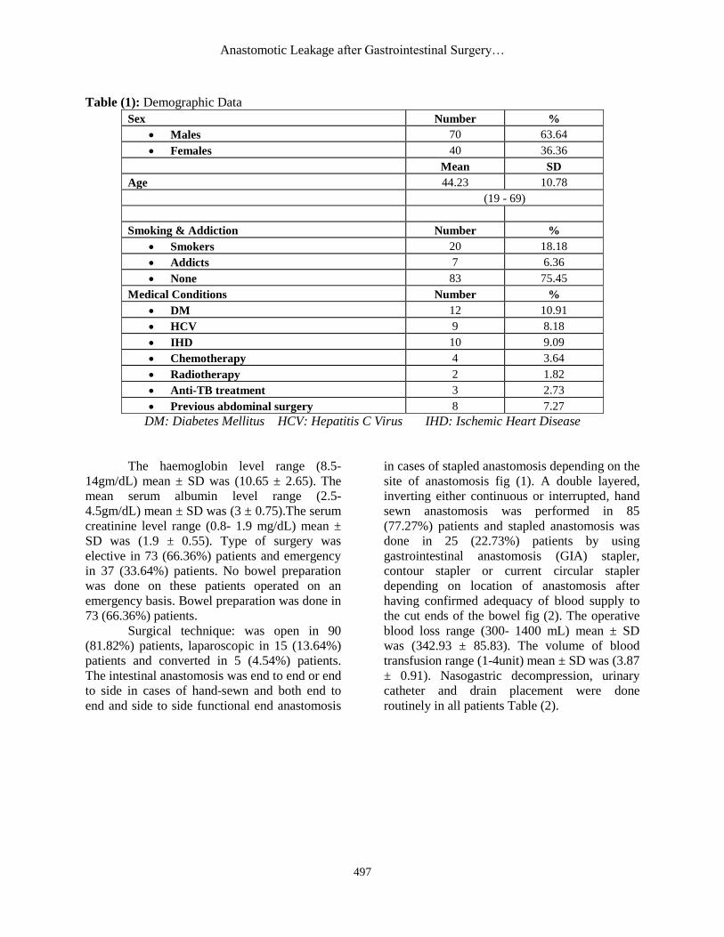
Table (1): Demographic Data
Sex Number %
Males 70 63.64
Females 40 36.36
Mean SD
Age 44.23 10.78
(19 - 69)
Smoking & Addiction Number %
Smokers 20 18.18
Addicts 7 6.36
None 83 75.45
Medical Conditions Number %
DM 12 10.91
HCV 9 8.18
IHD 10 9.09
Chemotherapy 4 3.64
Radiotherapy 2 1.82
Anti-TB treatment 3 2.73
Previous abdominal surgery 8 7.27
DM: Diabetes Mellitus HCV: Hepatitis C Virus IHD: Ischemic Heart Disease
The haemoglobin level range (8.5-
14gm/dL) mean ± SD was (10.65 ± 2.65). The
mean serum albumin level range (2.5-
4.5gm/dL) mean ± SD was (3 ± 0.75).The serum
creatinine level range (0.8- 1.9 mg/dL) mean ±
SD was (1.9 ± 0.55). Type of surgery was
elective in 73 (66.36%) patients and emergency
in 37 (33.64%) patients. No bowel preparation
was done on these patients operated on an
emergency basis. Bowel preparation was done in
73 (66.36%) patients.
Surgical technique: was open in 90
(81.82%) patients, laparoscopic in 15 (13.64%)
patients and converted in 5 (4.54%) patients.
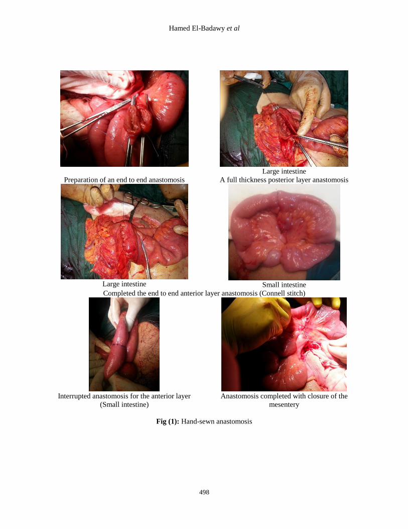
The intestinal anastomosis was end to end or end
to side in cases of hand-sewn and both end to
end and side to side functional end anastomosis
in cases of stapled anastomosis depending on the
site of anastomosis fig (1). A double layered,
inverting either continuous or interrupted, hand
sewn anastomosis was performed in 85
(77.27%) patients and stapled anastomosis was
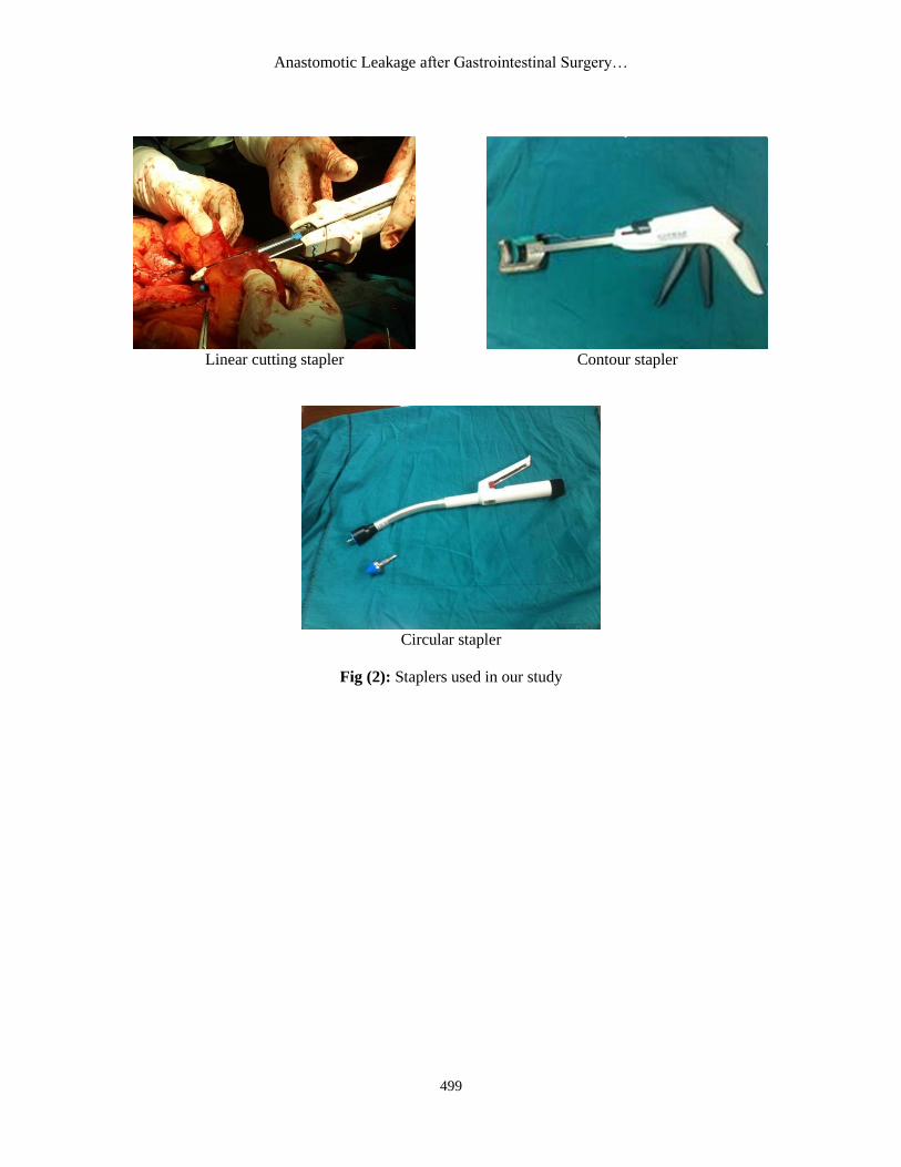
done in 25 (22.73%) patients by using
gastrointestinal anastomosis (GIA) stapler,
contour stapler or current circular stapler
depending on location of anastomosis after
having confirmed adequacy of blood supply to
the cut ends of the bowel fig (2). The operative
blood loss range (300- 1400 mL) mean ± SD
was (342.93 ± 85.83). The volume of blood
transfusion range (1-4unit) mean ± SD was (3.87
± 0.91). Nasogastric decompression, urinary
catheter and drain placement were done
routinely in all patients Table (2).
Hamed El-Badawy et al
498
Large intestine
Preparation of an end to end anastomosis A full thickness posterior layer anastomosis
Large intestine
Small intestine
Completed the end to end anterior layer anastomosis (Connell stitch)
Interrupted anastomosis for the anterior layer
(Small intestine)
Anastomosis completed with closure of the
mesentery
Fig (1): Hand-sewn anastomosis
Anastomotic Leakage after Gastrointestinal Surgery…
499
Linear cutting stapler Contour stapler
Circular stapler
Fig (2): Staplers used in our study
Hamed El-Badawy et al
500
Table (2): Preoperative, operative and postoperative data
Preoperative Data Mean SD
Preoperative Hb level 10.65 2.65
Preoperative albumin level 3.1 0.75
Preoperative creatinine level 1.09 0.55
Preoperative Bowel Preparation Number %
Yes 73 66.36
No 37 33.64
Type Of Surgery Number %
Elective 73 66.36
Emergency 37 33.64
Surgical Technique Number %
Open 90 81.82
Laparoscopic 15 13.64
Converted 5 4.55
Level Of Anastomosis Number %
Small Bowel 40 36.36
Gastrojejunostomy 11 10.00
Choledochojejunostomy 4 3.64
Large Bowel 55 50.00
Colicocolic 25 22.73
Rt Ileocolic 8 7.27
Lt Ileocolic 6 5.45
Colicorectal 8 7.27
Colicoanal 4 3.64
Ileorectal 2 1.82
Ileoanl 2 1.82
Type Of Anastomosis Number %
Hand Sewn 85 77.27
Stapled 25 22.73
Mean SD
Operative blood loss 342.93 85.83
Volume of blood transfusion 3.87 0.91
Number %
Drain placement 110 100.00
Nasogastric decompression 110 100.00
Hb = Hemoglobin Anastomotic leakage was occurred in 17 of 110 patients (15.4%) group I, no anastomotic leakage was
found in 93 patients (84.6%) group II. On postoperative day 3 significantly more patients in group I had fever
above 38oC than in group II 8/17 (47.06%) versus 13/93 (13.98%) (P 0.01). More in group I patients than in
group II patients also had transient disturbances, they included the absence of bowel movement on
postoperative day 4, 7/ 17 (41.18%), versus 10/93 (10.75%) (P 0.01). and diarrhea before postoperative day
6 5/17 (29.41%) versus 8/93 (8.60%).From postoperative day 2 to 4, amount of drainage fluid exceeding
500ml were collected significantly more from group I patients than group II patients 9/17 (52.74%) versus
12/93 (12.90%) (P 0.01). No significant difference was noted between the two groups for nasogastric fluid
aspiration 1.200 mL on 3rd
postoperative day. On postoperative day 4 significantly more from group I than
group II patients had oliguria urine output less than 400CC/ day 6/17 (35.29%) versus 15/93 (16.13%) (P
0.01). Renal failure on postoperative day 3-5 affected significantly more patients in group I than group II 3/17
(17.65%) versus 5/93 (5.38%) (P 0.01). After postoperative day 6, significantly more group I patients than
group II had leukocytosis (WBC) over 12, 000/mm3/ 14/17 (82.35%) versus 23/ 93 (24.73%) (P 0.01). Table
(3).
Anastomotic Leakage after Gastrointestinal Surgery…
501
Table (3): Predictive factors for anastomotic leak.
Leakage n = 17 No Leakage n = 93
P value Number % Number %
Postoperative
Fever 8 47.06 13 13.98
Absence of bowel
movement 7 41.18 10 10.75 < 0.01
Diarrhea 5 29.41 8 8.60
Amount of drainage
> 500 cc 9 52.94 12 12.90 < 0.01
< 500 cc 8 47.06 7 7.53
Postoperative renal failure 3 17.65 5 5.38 < 0.01
Postoperative oliguria 6 35.29 15 16.13 < 0.01
Postoperative leukocytosis 14 82.35 23 24.73 < 0.001
The mean postoperative period for diagnosis of anastomotic leakage was 9 days range (5- 16
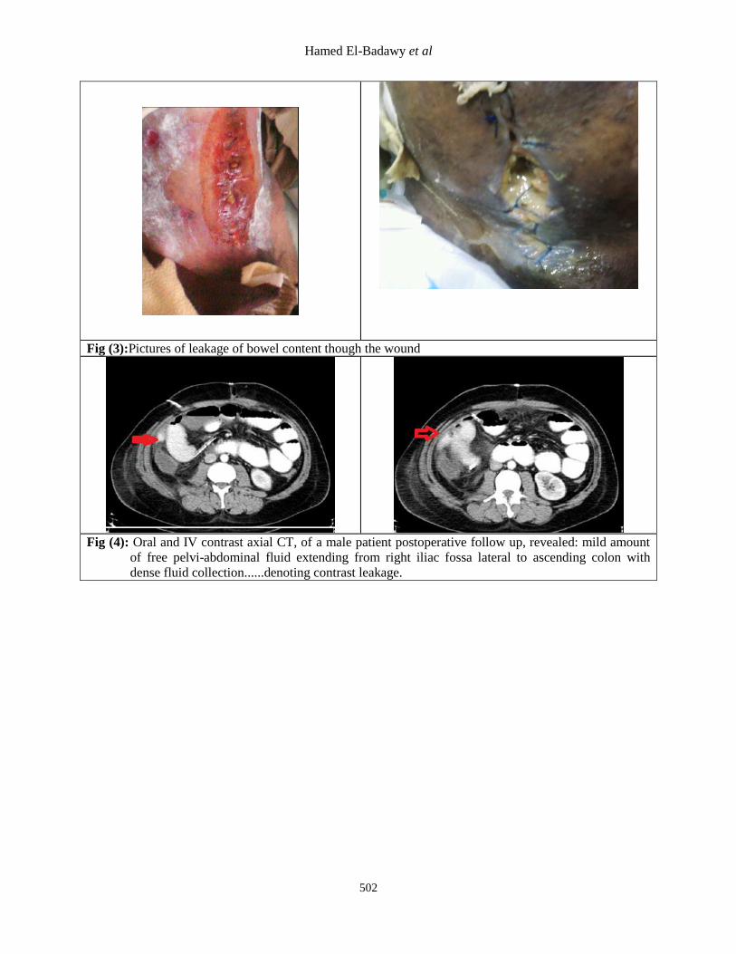
days). In 7 patients, it was identified by drain output and 4 patients were clinically diagnosed at a mean of
8 days (5-12 days) postoperatively fig (3). The remaining 6 patients were diagnosed radiologicaly at a
mean 16 day’s postoperatively. Contrast enema was obtained in 4 cases, the leak was observed in one
case, but in 3 cases the test was falsely negative. CT scan were obtained in 5 cases, the leak was correctly
diagnosed in 4 cases, but one scan was falsely negative fig (4).
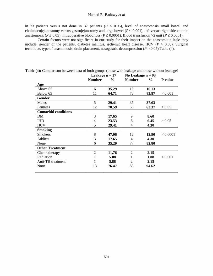
Out of the 17 patients from group I with anastomotic leakage, 9 patients required fecal diversion
after another exploratory laparotomy and washout of peritoneal cavity and repair of the leak fig (5).
5 patients were able to be managed non-operatively (typically with radiologic drainage and antibiotics) fig
(6). 2 patients had conservative management of the leak done and one patient died before reoperation and
anastomotic leakage was confirmed by (autopsy). No permanent diverting stoma was observed in this
study.
Hamed El-Badawy et al
502
Fig (3):Pictures of leakage of bowel content though the wound
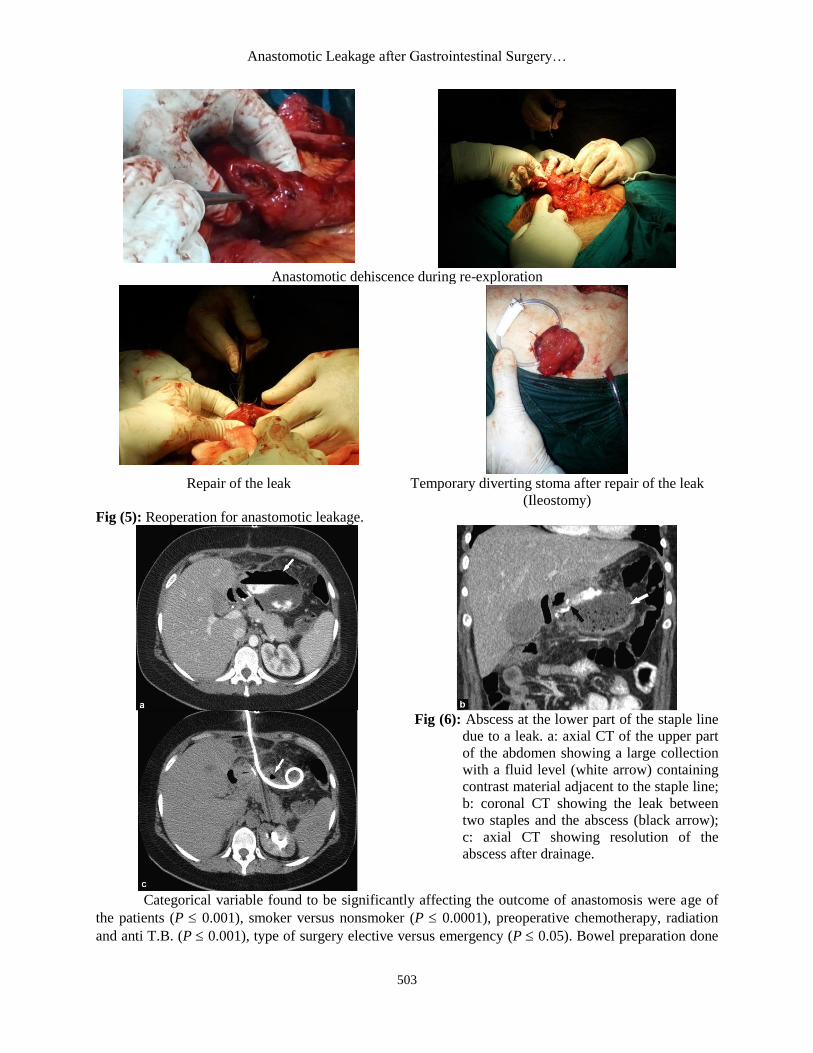
Fig (4): Oral and IV contrast axial CT, of a male patient postoperative follow up, revealed: mild amount
of free pelvi-abdominal fluid extending from right iliac fossa lateral to ascending colon with
dense fluid collection......denoting contrast leakage.
Anastomotic Leakage after Gastrointestinal Surgery…
503
Anastomotic dehiscence during re-exploration
Repair of the leak Temporary diverting stoma after repair of the leak
(Ileostomy)
Fig (5): Reoperation for anastomotic leakage.
Fig (6): Abscess at the lower part of the staple line
due to a leak. a: axial CT of the upper part
of the abdomen showing a large collection
with a fluid level (white arrow) containing
contrast material adjacent to the staple line;
b: coronal CT showing the leak between
two staples and the abscess (black arrow);
c: axial CT showing resolution of the
abscess after drainage.
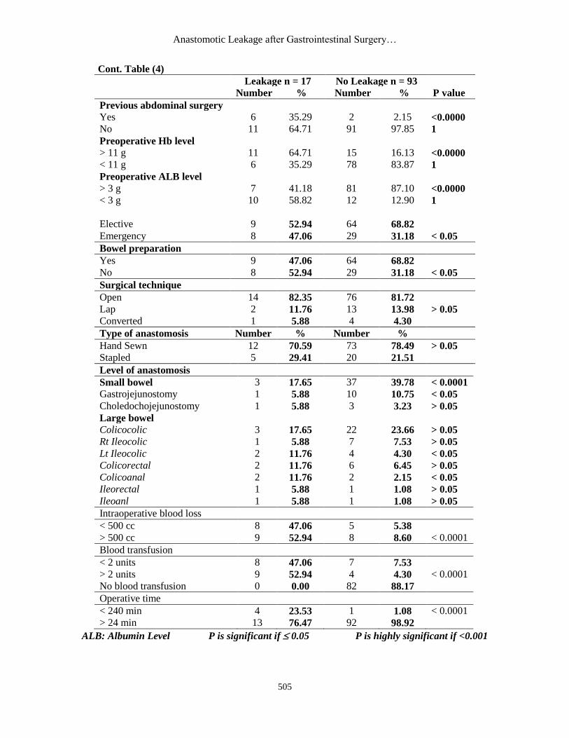
Categorical variable found to be significantly affecting the outcome of anastomosis were age of
the patients (P 0.001), smoker versus nonsmoker (P 0.0001), preoperative chemotherapy, radiation
and anti T.B. (P 0.001), type of surgery elective versus emergency (P 0.05). Bowel preparation done
Hamed El-Badawy et al
504
in 73 patients versus not done in 37 patients (P 0.05), level of anastomosis small bowel and
choledocojejunostomy versus gastrojejunstomy and large bowel (P 0.001), left versus right side colonic
anastomosis (P 0.05). Intraoperative blood loss (P 0.0001). Blood transfusion >2 unit (P 0.0001).
Certain factors were not significant in our study for their impact on the anastomotic leak: they
include: gender of the patients, diabetes mellitus, ischemic heart disease, HCV (P > 0.05). Surgical
technique, type of anastomosis, drain placement, nasogastric decompression (P > 0.05) Table (4).
Table (4): Comparison between data of both groups (those with leakage and those without leakage)
Leakage n = 17 No Leakage n = 93
P value Number % Number %
Age
Above 65 6 35.29 15 16.13
Below 65 11 64.71 78 83.87 < 0.001
Gender
Males 5 29.41 35 37.63
Females 12 70.59 58 62.37 > 0.05
Comorbid conditions
DM 3 17.65 9 8.60
IHD 4 23.53 6 6.45 > 0.05
HCV 5 29.41 4 4.30
Smoking
Smokers 8 47.06 12 12.90 < 0.0001
Addicts 3 17.65 4 4.30
None 6 35.29 77 82.80
Other Treatment
Chemotherapy 2 11.76 2 2.15
Radiation 1 5.88 1 1.08 < 0.001
Anti-TB treatment 1 5.88 2 2.15
None 13 76.47 88 94.62
Anastomotic Leakage after Gastrointestinal Surgery…
505
Cont. Table (4)
Leakage n = 17 No Leakage n = 93
P value Number % Number %
Previous abdominal surgery
Yes 6 35.29 2 2.15 <0.0000
1 No 11 64.71 91 97.85
Preoperative Hb level
> 11 g 11 64.71 15 16.13 <0.0000
1 < 11 g 6 35.29 78 83.87
Preoperative ALB level
> 3 g 7 41.18 81 87.10 <0.0000
1 < 3 g 10 58.82 12 12.90
Elective 9 52.94 64 68.82
Emergency 8 47.06 29 31.18 < 0.05
Bowel preparation
Yes 9 47.06 64 68.82
No 8 52.94 29 31.18 < 0.05
Surgical technique
Open 14 82.35 76 81.72
Lap 2 11.76 13 13.98 > 0.05
Converted 1 5.88 4 4.30
Type of anastomosis Number % Number %
Hand Sewn 12 70.59 73 78.49 > 0.05
Stapled 5 29.41 20 21.51
Level of anastomosis
Small bowel 3 17.65 37 39.78 < 0.0001
Gastrojejunostomy 1 5.88 10 10.75 < 0.05
Choledochojejunostomy 1 5.88 3 3.23 > 0.05
Large bowel
Colicocolic 3 17.65 22 23.66 > 0.05
Rt Ileocolic 1 5.88 7 7.53 > 0.05
Lt Ileocolic 2 11.76 4 4.30 < 0.05
Colicorectal 2 11.76 6 6.45 > 0.05
Colicoanal 2 11.76 2 2.15 < 0.05
Ileorectal 1 5.88 1 1.08 > 0.05
Ileoanl 1 5.88 1 1.08 > 0.05
Intraoperative blood loss
< 500 cc 8 47.06 5 5.38
> 500 cc 9 52.94 8 8.60 < 0.0001
Blood transfusion
< 2 units 8 47.06 7 7.53
> 2 units 9 52.94 4 4.30 < 0.0001
No blood transfusion 0 0.00 82 88.17
Operative time
< 240 min 4 23.53 1 1.08 < 0.0001
> 24 min 13 76.47 92 98.92
ALB: Albumin Level P is significant if 0.05 P is highly significant if <0.001
Hamed El-Badawy et al
506
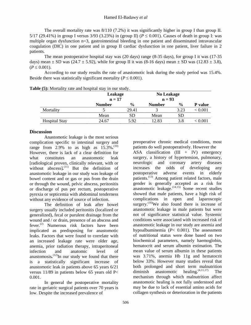
The overall mortality rate was 8/110 (7.2%) it was significantly higher in group I than group II.
5/17 (29.41%) in group I versus 3/93 (3.23%) in (group II) (P 0.001). Causes of death in group I: was
multiple organ dysfunction n=3, gastrointestinal bleeding in one patient and disseminated intravascular
coagulation (DIC) in one patient and in group II cardiac dysfunction in one patient, liver failure in 2
patients.
The mean postoperative hospital stay was (20 days) range (8-35 days), for group I it was (17-35
days) mean ± SD was (24.7 ± 5.92), while for group II it was (8-16 days) mean ± SD was (12.83 ± 3.8),
(P 0.001).
According to our study results the rate of anastomotic leak during the study period was 15.4%.
Beside there was statistically significant mortality (P 0.001).
Table (5): Mortality rate and hospital stay in our study.
Leakage
n = 17
No Leakage
n = 93
P value Number % Number %
Mortality 5 29.41 3 3.23 < 0.001
Mean SD Mean SD
Hospital Stay 24.67 5.92 12.83 3.8 < 0.001
Discussion Anastomotic leakage is the most serious
complication specific to intestinal surgery and
range from 2.9% to as high as 15.3%.(10)
However, there is lack of a clear definition for
what constitutes an anastomotic leak
[radiological proven, clinically relevant, with or
without abscess].(11)
But the definition of
anastomotic leakage in our study was leakage of
bowel content and or gas or pus from the drain
or through the wound, pelvic abscess, peritonitis
or discharge of pus per rectum, postoperative
pyrexia or septicemia with abdominal tenderness
without any evidence of source of infection.
The definition of leak after bowel
surgery usually included peritonitis (localized or
generalized), fecal or purulent drainage from the
wound and / or drain, presence of an abscess and
fever.(2)
Numerous risk factors have been
implicated as predisposing for anastomotic
leaks. Factors that were found to correlate with
an increased leakage rate were older age,
anemia, prior radiation therapy, intraperitoneal
infection and anatomic level of
anastomosis.(12)
In our study we found that there
is a statistically significant increase of
anastomotic leak in patients above 65 years 6/21
versus 11/89 in patients below 65 years old P<
0.001.
In general the postoperative mortality
rate in geriatric surgical patients over 70 years is
low. Despite the increased prevalence of
preoperative chronic medical conditions, most
patients do well postoperatively. However the
ASA classification (III + IV) emergency
surgery, a history of hypertension, pulmonary,
neurologic and coronary artery diseases
increases the odds of developing any
postoperative adverse events in elderly
patients.(13)
Among patient related factors, male
gender is generally accepted as a risk for
anastomotic leakage.(14,15)
Some recent studies
showed that male patients, have a high risk of
complications in open and laparoscopic
surgery.(16)
We also found there is increase of
anastomotic leakage in male patients but were
not of significance statistical value. Systemic
conditions were associated with increased risk of
anastomotic leakage in our study are anemia and
hypoalbuminemia (P< 0.001). The assessment
of nutritional status were done based on two
biochemical parameters, namely haemoglobin,
hematocrit and serum albumin estimation. The
mean value of serum albumin in these patients
was 3.71%, anemia Hb 11g and hematocrit
below 33%. However many studies reveal that
both prolonged and short term malnutrition
diminish anastomotic healing.(4,11,17)
The
mechanism through which malnutrition affect
anastomotic healing is not fully understood and
may be due to lack of essential amino acids for
collagen synthesis or deterioration in the patients
Anastomotic Leakage after Gastrointestinal Surgery…
507
immuno-competence.(18)
Diabetes, ischemic
heart disease and Hepatitis C virus is another
causes of anastomotic leakage in this study, but
it did not have statistical significant effect. In
this study out of 17 patients who developed
anastomotic leak 3 of them were diabetic, 5 were
HCV and 4 were ischemic heart diseases. Little
evidence indicates that diabetes affects GI
healing. A direct effect of the diabetic state of
the healing process is difficult to separate from
an impairment caused by increased abscess
formation.(19)
Smoking and addiction were the
independent risk factors associated with
anastomotic leak in our study (P< 0.0001). We
agree with Sultan et al.(3)
, Daams et al.(20)
,
Trencheva et al.(21)
. These investigators found
that there is highly significant anastomotic leak
in smoking patients.
Smoking and alcohol abuse are
important predictive factors for anastomotic
leakage after colonic and rectal resection.(20,22)
Preoperative chemotherapy, radiotherapy and
anti-tuberculous drugs are associated with highly
significant risk factors for anastomotic leak in
this study. Chemoradiation may predispose to
anastomotic problem in patients having colon
surgery, particularly in patients with
anastomosis in the pelvis. Anastomotic leak and
radiation therapy may contribute to the
formation of pelvic abscess, rendering the
neorectum stiff and noncompliant. After
reconstruction, patients may suffer from
tenesmus and fecal incontinence.(12)
Previous abdominal surgery was
independent risk factor in anastomotic leak in
our study. After open lower abdominal surgery
adhesion related problems and readmission rates
were mostly influenced by the initial site of
surgery: colon and rectal resection having the
highest relative risk of problems directly related
to adhesion.(23)
Type of surgery and bowel preparation
were independent risk factors of anastomotic
leakage in this study. We found that there is
increase of anastomotic leak in patients who
operated in emergency 8/37 versus 9/73 in
elective cases and in patients who are not
attending bowel preparation versus the prepared
cases. Several well designed prospective
randomized trials have shown that preoperative
bowel cleaning does not prevent anastomotic
leakage or wound infection in patients
undergoing open or laparoscopic colorectal
surgery.(14,24)
However, some randomized trial
have reported significant differences in
outcomes with use of oral antibacterial agents
and mechanical preparation.Irvin and
Goligher,(25)
:reported significant decrease in
anastomotic dehiscence with use of mechanical
preparation than that without mechanical bowel
preparation. Burke et al.(26)
have provided further
evidence that question the use of bowel
preparation showing no difference in outcome
after colon surgery between prepared and
unprepared patients.(3)
Surgical technique, either open,
laparoscopic, or converted were not associated
with significant difference of anastomotic
leakage in our study. Laparoscopic colon
surgery was first described by Redwine and
Sharpe(27)
and multiple level I studies show the
advantages include less intraoperative trauma,
reduction in postoperative adhesions, decreased
postoperative pain, decreased length of ileus,
better cosmesis, early discharge from hospital
and early return to work.(28,29)
Operating room
costs are significantly higher, but the difference
in overall hospital charges has not been found to
be statistically significant.(30)
The available data comparing the
anastomotic leakage rate in laparoscopic or open
operated patients showed no difference
regardless of the level of the anastomosis.(31)
We reported that patients whose surgery
converted from laparoscopic to open tended to
have longer operating time, higher morbidity
and more prolonged hospital stay.We agree with
Tjandra and Chan,(28)
about this point. Also,
there is no significant difference in anastomotic
leakage was noted between hand sewn and
stapling procedures. In relation to efficacy,
applicability and safty, it has been demonstrated
that the use of surgical stapling instruments is
comparable to that of conventional suturing
methods. In certain situation, staplers offer the
facility to achieve reconstructions that would be
difficult to be accomplished manually and their
popularity in that setting seen justifiable.(32)
A meta- analysis, concluded that there is
no difference between hand-sewn and stapled
anastomosis for the majority of outcome
measures including mortality, leak rates, local
Hamed El-Badawy et al
508
cancer recurrences and wound
infection.(20,33,34,35,36)
However, in a recent
coherence review ileocolic stapler anastomosis
were associated with fewer leaks than hand sewn
anastomosis.(37)
On contrary: Cheng et al.(38)
concluded
that: stapled anastomosis showed a trend to a
higher leakage rate, but the difference did not
reach statistical significance.
We noted that prolonged operative time
was associated with highly significant rate of
anastomotic leaks (P< 0.0001). In a series of 541
colorectal anastomosis between 1999 and 2004
at a single colorectal unit, univariate analysis
show that a prolonged operating time had a odd
ratio of 2.8 for developing anastomotic
leakage.(39)
Many studies showed that prolonged
operating time correlated with higher
anastomotic leak.(11,20,29)
A highly statistically significant relation
was found between intra operative blood loss,
intra operative blood transfusion and
anastomotic leak. (P< 0.0001) we agree with
Kirchhof et al.,(11)
; Kiran et al., (40)
about this
point.
Blood transfusion are known to have an
immuno- suppressive effect. As a result tumor
growth may be enhanced, the incidence of tumor
recurrence may be high, there can be prolonged
allograft survival in transplantation procedures
and increased susceptibility to infection.(41,42)
In
the peritoneal cavity there could be delayed
healing of anastomosis and increased incidence
of intraperitoneal sepsis.(43)
Routine nasogastric decompression and
abdominal drains in patients undergoing a
procedure involving an intestinal anastomosis
remain controversial. Abdominal drains and
nasogastric tubes were routinely inserted in all
patients in this study. In retrospective and
prospective randomized, controlled trial, routine
use of a nasogastric tube conferred no significant
advantage.(44,45)
In fact, there was a trend toward
an increased incidence of respiratory tract
infections after routine gastric decompression.(17)
Nonetheless, one study found that nearly 20% of
patients required insertion of gastric tube in the
early postoperative period.(44)
The value of prophylactic drainage in
intestinal anastomosis has been studies
extensively currently available data from
randomized controlled trials point out that a
routine prophylactic drainage provides no
benefit after uncomplicated major colon and
rectal surgery.(46)
On contrary, a no drain policy
was associated with less and a fewer
anastomotic leaks, these studies underscore the
low sensitivity of drains in detecting leakage and
bleeding, which questions the putative warning
function of a prophylactic drain. In summary,
there is sufficient evidence showing that routine
drainage colorectal anastomosis does not prevent
leaks or other complications.(47,48)
This finding to
the contrary, many surgeons elect to place an
intra-abdominal drain to the pelvis after an
anterior resection or a coloanal anastomosis
because of the higher than unusual risk that a
fluid collection will develop. Drainage is rarely
helpful, indeed easy, after a gastric or small
bowel anastomosis. Drains are indicated,
however, after emergency operations for
peritonitis or trauma in which it was necessary
to close or anastomose damaged or inflamed
bowel.(17)
Left side anastomosis, colorectal and
ileoanal anastomosis were the independent risk
factors associated with anastomotic leak in our
study. Most studies comparing high and low
anterior resections have shown that the level of
anastomosis is the most predictive factor for
leakage. Higher leak rate are typically reported
for low pelvic anastomosis or anastomosis to the
anal canal.(3,7,21,49)
The present study found a clinical
leakage rate of 15.4%. This rate is at the higher
level of incidence reported by several
investigators which range from 2.8%-
15%.(7,38,50,51,52,53)
Sultan et al(3)
reported 15%
anastomotic leakage in their study in agree with
our results. The reason behind the higher rate of
leakage in our study were not proximally
diverted while in rest of the studies patients
population was mixed i.e. proximally diverted as
well as not diverted.
The mean postoperative period for
diagnosis of anastomotic leakage was a 9 days
range [5-16 days] in our study. Anastomotic
leakage typically becomes clinically apparent
between the 5th and 8
th postoperative day, but
many exceptions exist, with one study even
reporting a mean of the 12th postoperative day
for the diagnosis of anastomotic leakage.(7)
Interestingly in recent study anastomotic leaks
Anastomotic Leakage after Gastrointestinal Surgery…
509
were more often after hospital discharge.(54)
Clinical signs of systemic inflammatory
response syndrome, fever, ileus and pain are
frequent but have low positive predictive value
for anastomotic leak, when observed separately.
In a study by Dendulk et al.(55)
these clinical
features were combined into a clinical scoring
system (Dutch leakage score), with patients were
scored daily in a systematical and uniform way.
Points are attributed to certain clinical criteria
i.e. fever, heart rate, nutritional status, signs of
ileus, gastric retention, type of intake and
laboratory findings i.e. C- reactive protein
(CRP) level, leucocytes, kidney function. In our
study we also recorded the fluid collected by
nasogastric aspiration and abdominal drainage
and urine output. After applying the score
system retrospectively on a historical cohort, the
score was used prospectively: it was shown that
patients with a high score were prone to
anastomotic leakage requiring intensive clinical
observation or radiological evaluation. This
scoring system reduced delay in diagnosis of
anastomotic leak from 4 to 1.5 days, decreasing
false negative diagnostic imaging representing a
major factor of delay in diagnosis.(56)
Water soluble enemas or CT scans are
widely used for diagnosis of anastomotic leak,
CT scanning appear to be far more helpful than
contrast enema in the radiologic diagnosis of the
leak. CT scan were obtained in 5 cases, the leak
was correctly diagnosed in 4 cases but one scan
was falsely negative. On contrary contrast
enema was obtained in 4 cases, the leak was
observed in one case, but in 3 cases the test was
falsely negative. CT scan does appear to be the
radiologic procedure of choice to diagnose an
anastomotic leak after intestinal surgery when
clinical finding alone insufficient.(7)
When facing and treating patients with
anastomotic leak, surgeons have to take into
account many different aspects i.e, age, health
status and current clinical condition of the
patient, extent of dehiscence, time between
operation and reoperation, indication of primary
resection and localization of the anastomosis.
These variable lead to individualization of
treatment strategies and in comparable outcome.
However: few studies, showing that surgeons
believe that the anastomosis can be repaired
rather than dismantled, have paved the way for a
trial in which next to mortality and morbidity,
preservation of the anastomosis could be one of
the endpoints.(20,57)
The overall mortality rate in our study
was 8/110 (7.2%). It was significantly higher in
patients with anastomotic leak 5/17 (28.41%)
versus 3/93 (3.23%) in patients without
anastomotic leak (P< 0.001). In comparison with
other studies we approximate with the study of
Sultan et al.(3)
, they reported 15.79% mortality
rate of their patients with anastomotic leak and
high in comparison with the study of Hyman et
al.(7)
, they found mortality rate 5.7% of their
patients, Cheng et al.(38)
the mortality rate 1.4%,
Trenchval et al.(21)
the mortality rate 0.9% Alves
et al.(50)
the mortality rate 12% and Buch et
al.(58)
the mortality rate was 12.9% of their
patients with anastomotic leakage. In four of
five deaths, leaks occurred in very ill patients
undergoing emergency procedure and appeared
to be premorbid events.
There was no case with permanent
diverting stoma in this study. On contrary
Brisinda et al.(8)
reported that anastomotic
leakage has been associated with a 6-39%
mortality rate and a 10-100% risk of permanent
stoma.
Morbidity of dramatically increased
opposed patients without colonic anastomotic
leakage and leads to reoperations, radiological
intervention and permanent stoma in 56%.(59)
Conclusion Postoperative gastrointestinal anastomotic
leak is a very serious complication that has great
clinical impact on patients, putting surgeons in
dilemmas of detection and management.
There is multiple risk and predictive
factors associated with occurrence of leak were
suspected in this study such as: older patients,
preoperative anemia, hypoalbuminemia,
immunesuppressive therapy, smoking, surgery
performed in an emergency setting, without
adequate bowel preparation, long operative time,
intraoperative blood loss and blood transfusion
and low pelvic anastomosis, but many factors
remain unclear.
The presentation of anastomotic leakage
varying from severe peritonitis and leakage of
bowel content through the wound or from the
drain to asymptomatic (small pelvic abscess).
Hamed El-Badawy et al
510
Early detection and expediently
treatment is very helpful to improve the patients
outcome but death after leak is most often a
substitute for a critically ill patients and was
infrequently the actual cause of death and so
every effort needs to be made to bring down the
mortality rates and hospital stay associated with
anastomotic leak.
References 1. Kaidar-Person O, Rosenthal RJ, Wexner
SD et al (2008). Compression anastomosis:
history and clinical considerations. Am J Surg.
195(6):818-26.
2. Bruce J, Krukowski ZH, Al-Khairy G et al
(2001). Systematic review of the definition and
measurement of anastomotic leak after
gastrointestinal surgery. Br J Surg. 88(9):1157-
68.
3. Sultan R, Chawla T and Zaidi M (2014).
Factors affecting anastomotic leak after
colorectal anastomosis in patients without
protective stoma in tertiary care hospital.J Pak
Med Assoc. 64(2):166-70.
4. Bieleki K and Gajda A (1999). The causes and
prevention of anastomotic leak after colorectal
surgery: Klinickaonkology; 25-30.
5. Gorissen KJ, Benning D, Berghmans T et al
(2012). Risk of anastomotic leakage with non-
steroidal anti-inflammatory drugs in colorectal
surgery. Br J Surg. 99(5):721-7.
6. Boccola MA, Lin J, Rozen WM et al (2010). Reducing anastomotic leakage in oncologic
colorectal surgery: an evidence-based review.
Anticancer Res. 30(2):601-7.
7. Hyman N, Manchester TL, Osler T et al
(2007). Anastomotic leaks after intestinal
anastomosis: it's later than you think. Ann Surg.
245(2):254-8.
8. Brisinda G, Vanella S, Cadeddu F et al (2009).
End-to-end versus end-to-side stapled
anastomoses after anterior resection for rectal
cancer. J Surg Oncol. 99(1):75-9.
9. Pickleman J, Watson W, Cunningham Jet al
(1999). The failed gastrointestinal anastomosis:
an inevitable catastrophe? J Am Coll Surg. 188
(5): 473-82.
10. Chambers WM and Mortensen NJ (2004). Postoperative leakage and abscess formation
after colorectal surgery. Best Pract Res Clin
Gastroenterol; 18 (5): 865-80.
11. Kirchhoff P, Clavien PA and Hahnloser D
(2010).Complications in colorectal surgery: risk
factors and preventive strategies. Patient Saf
Surg.4(1):5.
12. Nasirkhan MU, Abir F, Longo W et al (2006).
Anastomotic disruption after large bowel
resection. World J Gastroenterol;12(16):2497-
504.
13. Leung JM and Dzankic S (2001). Relative
importance of preoperative health status versus
intraoperative factors in predicting postoperative
adverse outcomes in geriatric surgical patients. J
Am Geriatr Soc. 49(8):1080-5.
14. Jung SH, Yuc S, Choip W et al (2008). Risk
factors and oncolgic impact of anastomtic leak-
age after rectal cancer surgery. Dis colon rectum;
51: 902- 908.
15. Matthiessen P, Hallböök O, Andersson M et
al (2004). Risk factors for anastomotic leakage
after anterior resection of the rectum. Colorectal
Dis. 6 (6): 462-9.
16. Kirchhoff P, Dincler S and Buchmann P
(2008).A multivariate analysis of potential risk
factors for intra- and postoperative complications
in 1316 elective laparoscopic colorectal
procedures. Ann Surg. 248(2):259-65.
17. Britton J (2003). Intestinal anastomosis ACS
surgery: principle and practice; 29- 1012.
18. Vagholkar RK (2001). Healing of anastomosis
in the gastrointestinal tract: retrospective study
of 35 cases: Bombary hospital J. 43 (2): 269- 79.
19. van der Ham AC, Kort WJ, Weijma IM et al
(1992). Healing of ischemic colonic
anastomosis: fibrin sealant does not improve
wound healing. Dis Colon Rectum; 35(9):884-
91.
20. Daams F, Luyer M and Lange JF (2013). Colorectal anastomotic leakage: aspects of
prevention, detection and treatment. World J
Gastroenterol; 19(15):2293-7.
21. Trencheva K, Morrissey KP, Wells M et al
(2013).Identifying important predictors for
anastomotic leak after colon and rectal resection:
prospective study on 616 patients. Ann Surg.
257(1):108-13.
22. Sørensen LT, Jørgensen T, Kirkeby LT et al
(1999). Smoking and alcohol abuse are major
risk factors for anastomotic leakage in colorectal
surgery. Br J Surg. 86(7):927-31.
23. Parker MC (2004).Epidemiology of adhesions:
the burden.Hosp Med. 65(6):330-6.
24. Wille-Jørgensen P, Guenaga KF, Matos D et
al (2005). Pre-operative mechanical bowel
cleansing or not? an updated meta-analysis.
Colorectal Dis. 7(4):304-10.
25. Irvin TT and Goligher JC (1973). Aetiology of
disruption of intestinal anastomoses. Br J Surg.
60(6):461-4.
Anastomotic Leakage after Gastrointestinal Surgery…
511
26. Burke P, Mealy K, Gillen P et al (1994).
Requirement for bowel preparation in colorectal
surgery. Br J Surg. 81(6):907-10.
27. Redwine DB andSharpe DR (1991).
Laparoscopic segmental resection of the sigmoid
colon for endometriosis. J Laparoendosc Surg.
1(4):217-20.
28. Tjandra JJ and Chan MK (2006). Systematic
review on the short-term outcome of
laparoscopic resection for colon and
rectosigmoid cancer. Colorectal Dis. 8(5):375-
88.
29. Chen HH, Wexner SD, Iroatulam AJ et al
(2000). Laparoscopic colectomy compares
favorably with colectomy by laparotomy for
reduction of postoperative ileus. Dis Colon
Rectum; 43(1):61-5.
30. Dwivedi A, Chahin F, Agrawal S et al (2002). Laparoscopic colectomy vs. open colectomy for
sigmoid diverticular disease. Dis Colon Rectum;
45(10):1309-14.
31. Breukink S, Pierie J and Wiggers T
(2006).Laparoscopic versus open total
mesorectal excision for rectal cancer. Cochrane
Database Syst Rev. (4): CD005200.
32. Calin MD, Bălălău C, Popa F et al (2013). Colic anastomotic leakage risk factors. J Med
Life;6(4):420-3.
33. Slieker JC, Daams F, Mulder IM et al (2013). Systematic review of the technique of colorectal
anastomosis. JAMA Surg. 148(2):190-201.
34. Alberts JC, Parvaiz A and Moran BJ
(2003).Predicting risk and diminishing the
consequences of anastomotic dehiscence
following rectal resection. Colorectal Dis.
5(5):478-82.
35. Lustosa SA, Matos D, Atallah AN et al (2002).
Stapled versus handsewn methods for colorectal
anastomosis surgery: a systematic review of
randomized controlled trials. Sao Paulo Med
J. 120(5):132-6.
36. Demetriades D, Murray JA, Chan LS et al
(2002). Hand sewn versus stapled anastomosis in
penetrating colon injuries requiring resection: a
multicenter study. J Trauma; 52(1):117-21.
37. Choy PY, Bissett IP, Docherty JG et al (2007).
Stapled versus hand sewn methods for ileocolic
anastomoses. Cochrane Database Syst Rev. (3):
CD004320.
38. Cheng YT, Ching CT, Chone CW et al
(2010).The risk factor of anastomotic leakage
and influence of fecal diversion after resection of
rectal cancer. J Soc Colon rectal Surgery; 21: 9-
16.
39. Lipska MA, Bissett IP, Parry BR et al (2006).
Anastomotic leakage after lower gastrointestinal
anastomosis: men are at a higher risk. ANZ J
Surg; 76(7):579-85.
40. Kiran RP, Delaney CP, Senagore AJ et al
(2004). Operative blood loss and use of blood
products after laparoscopic and conventional
open colorectal operations. Arch Surg.
139(1):39-42.
41. Tadros T, Wobbes T and Hendriks T (1993).
Opposite effects of interleukin-2 on normal and
transfusion-suppressed healing of experimental
intestinal anastomoses. Ann Surg. 218(6):800-8.
42. Waymack JP, Balakrishnan K, McNeal N et
al (1986). Effect of blood transfusions on
macrophage-lymphocyte interaction in an animal
model. Ann Surg. 204(6):681-5.
43. Wobbes T, Bemelmans BL, Kuypers JH et al
(1990). Risk of postoperative septic
complications after abdominal surgical treatment
in relation to perioperative blood transfusion.
Surg Gynecol Obstet. 171(1):59-62.
44. Reasbeck PG, Rice ML and Herbison GP
(1984). Nasogastric intubation after intestinal
resection. Surg Gynecol Obstet. 158(4):354-8.
45. Burg R, Geigle CF, Faso JM et al (1978).
Omission of routine gastric decompression. Dis
Colon Rectum; 21(2):98-100.
46. Sagar PM, Hartley MN, Macfie J et al (1995).
Randomized trial of pelvic drainage after rectal
resection. Dis Colon Rectum; 38(3):254-8.
47. Karliczek A, Jesus EC, Matos D et al (2006).
Drainage or nondrainage in elective colorectal
anastomosis: a systematic review and meta-
analysis. Colorectal Dis. 8(4):259-65.
48. Petrowsky H, Demartines N, Rousson V et al
(2004). Evidence-based value of prophylactic
drainage in gastrointestinal surgery: a systematic
review and meta-analyses. Ann Surg.
240(6):1074-84
49. Alves A, Panis Y, Trancart D et al (2002).
Factors associated with clinically significant
anastomotic leakage after large bowel resection:
multivariate analysis of 707 patients. World J
Surg. 26(4):499-502.
50. Eberl T, Jagoditsch M, Klingler A et al
(2008). Risk factors for anastomotic leakage
after resection for rectal cancer. Am J Surg. 196
(4): 592-8.
51. Konishi T, Watanabe T, Kishimoto J et al
(2006). Risk factors for anastomotic leakage
after surgery for colorectal cancer: results of
prospective surveillance. J Am Coll Surg.
202(3):439-44.
Hamed El-Badawy et al
512
52. Walker KG, Bell SW, Rickard MJ et al
(2004). Anastomotic leakage is predictive of
diminished survival after potentially curative
resection for colorectal cancer. Ann
Surg. 240(2):255-9.
53. Rullier E, Laurent C, Garrelon JL et al
(1998). Risk factors for anastomotic
leakage after resection of rectal cancer. Br J
Surg. 85(3):355-8.
54. Nicksa GA, Dring RV, Johnson KH et al
(2007). Anastomotic leaks: what is the best
diagnostic imaging study? Dis Colon Rectum;
50(2):197-203.
55. Dendulk M, Noter SL, Hendriks ER et al
(2009). Improved diagnosis and treatment of
anastomotic leakage after colorectal surgery. Eur
J Surg Oncol. 35(4):420-6.
56. Doeksen A, Tanis PJ, Vrouenraets BC et al
(2007). Factors determining delay in
relaparotomy for anastomotic leakage after
colorectal resection. World J Gastroenterol;
13(27):3721-5.
57. Phitayakorn R, Delaney CP, Reynolds HL et
al (2008). Standardized algorithms for
management of anastomotic leaks and related
abdominal and pelvic abscesses after colorectal
surgery. World J Surg. 32(6):1147-56.
58. Buchs NC, Gervaz P, Secic M et al
(2008).Incidence, consequences, and risk factors
for anastomotic dehiscence after colorectal
surgery: a prospective monocentric study. Int
J Colorectal Dis. 23(3):265-70.
59. Lindgren R, Hallböök O, Rutegård J et al
(2011). What is the risk for a permanent stoma
after low anterior resection of the rectum for
cancer? A six-year follow-up of a multicenter
trial. Dis Colon Rectum. 54(1):41-7.