anatomical and physiological foundations of cerebellar information processing
TRANSCRIPT

R E V I E W S
NATURE REVIEWS | NEUROSCIENCE VOLUME 6 | APRIL 2005 | 297
One of the major challenges of systems neuroscienceis to understand the neural basis of perception andbehaviour. Considerable progress has been made, forinstance, with regard to the analysis of the neural mech-anisms that underlie visual perception. In large part,this has been achieved by using a combination ofanatomical tract tracing and physiological characteriza-tion of neuronal receptive fields1. By comparison, ourunderstanding of movement control remains rather lesscomplete. Nevertheless, over the past decade or two, asimilar combination of systems level anatomical andphysiological approaches has been used to unravelsome of the intricacies of an important sensorimotorcontrol system, namely the cerebellum.
Fundamental to the operation of any CNS structureis the information processing that it accomplishes. Thecerebellum receives a wide variety of sensory inputsand generates motor-related outputs according to inter-nal rules of computation. These rules are determinedby the internal connectivity of cerebellar neuronalnetworks and the intrinsic properties of cerebellarneurons. Consequently, the information content of itsinputs together with the structural organization of theinternal circuitry of the cerebellum imposes constraintson theoretical models of cerebellar function. Effective
communication between anatomists, physiologists andmodellers is therefore essential for understanding howthe cerebellum contributes to the control of movementand other processes.
With this outlook in mind, the aim of the presentreview is to summarize recent advances in our under-standing of the anatomy and physiology of an importantregion of the cerebellum — the PARAVERMIS — and itsinfluence on voluntary limb movements. The paravermishas abundant connections with the spinal cord, and wepresent evidence to indicate that a key organizingprinciple is its subdivision into an array of multizonalmicrocomplexes (MZMCs). The functional organizationof these microcomplexes is thought to reflect spinalwithdrawal reflex organization. As a result, individualcerebellar microcomplexes could be kept informedabout inappropriate movements — that is, ‘MOTOR
ERRORS’ — that are related to elementary movements of alimb, such as those that result from contractions of singlemuscles. By incorporating recent findings on intrinsiccerebellar cortical connectivity, we also speculate onhow this modular arrangement might have a role incerebellar contributions to movement control. It isimportant to emphasize, however, that the modelpresented here is derived mainly from studies of the
ANATOMICAL AND PHYSIOLOGICALFOUNDATIONS OF CEREBELLARINFORMATION PROCESSINGRichard Apps* and Martin Garwicz ‡
Abstract | A coordinated movement is easy to recognize, but we know little about how it isachieved. In search of the neural basis of coordination, we present a model of spinocerebellarinteractions in which the structure–functional organizing principle is a division of the cerebelluminto discrete microcomplexes. Each microcomplex is the recipient of a specific motor errorsignal — that is, a signal that conveys information about an inappropriate movement. Thesesignals are encoded by spinal reflex circuits and conveyed to the cerebellar cortex throughclimbing fibre afferents. This organization reveals salient features of cerebellar informationprocessing, but also highlights the importance of systems level analysis for a fullerunderstanding of the neural mechanisms that underlie behaviour.
PARAVERMIS
A region on either side of themidline of the cerebellum thatlies lateral to the vermis andmedial to the hemisphere. Itcontains the cerebellar corticalzones C1, C2 and C3 and receivesclimbing fibre input from theinferior olive and projects to thenucleus interpositus. Here, theterm is used to denote thefunctionally related C1, C3 and Y(but not the C2) zones.
*Sensorimotor ControlGroup, Department ofPhysiology, University ofBristol, University Walk,Bristol BS8 1TD, UK.‡Division of Neuroscience,Department of ExperimentalMedical Science, BMC F10,Lund University,Tornav. 10, 221 84, Sweden.Correspondence to M.G.e-mail:[email protected]:10.1038/nrn1646Published online 15 March 2005

MOTOR ERROR
In the case of motor commands,the difference between the actualmotor command and the correctcommand, or between theintended and achievedmovement. A simple example isthe retinal slip signal, in whichthis difference is detecteddirectly at the sensory surface byspecialized retinal ganglion cells.
GAIN
The amplification factor thatregulates the relationshipbetween input and output, forinstance, in a reflex circuit.
TIMING THEORY
Here, the term refers toBraitenberg’s idea that parallelfibres provide delay lines forconverting spatial patterns intotemporal patterns.
LEARNED PATTERN
RECOGNITION THEORIES
Here, the term refers to thetheories of Marr and Albus, inwhich the cerebellum is viewedas a spatial pattern recognitiondevice with learning capacity.
CONTROL THEORY
Here, the term refers to aconceptual framework whereinengineering control principlesare applied to the modelling ofCNS functions.
298 | APRIL 2005 | VOLUME 6 www.nature.com/reviews/neuro
R E V I E W S
neurons (historically starting with the TIMING THEORY21 andthe LEARNED PATTERN RECOGNITION THEORIES22,23) to those moti-vated more by CONTROL THEORY24–26. The model presentedhere builds on a specific tradition that emphasizes thedivision of the cerebellum into a collection of ‘modules’defined by structure–function relationships. Thesemodules are thought to form the basis for informationprocessing performed by the cerebellum2,27,28.
Basic structure of the cerebellar cortexThroughout its highly convoluted extent, the cerebellumcan be divided into three cortical layers with the samebasic neuronal circuitry everywhere, which involves fivemain cell types (FIG. 1). The most conspicuous of theseare the Purkinje cells, which form an orderly monolayerinterposed between the granular and molecular layers,extending their planar dendritic trees into the molecularlayer above. As these cells are the sole output neurons ofthe cerebellar cortex they are central to cerebellar corticalinformation processing.
The granular layer below the Purkinje cells derives itsname from the small, densely packed granule cells thatsend their axons into the molecular layer, where theybifurcate to become parallel fibres (FIG. 1). These courseparallel to the long axis of each folium and as a resultthey intersect the fan-like dendritic trees of manyPurkinje cells. Mossy fibre afferents target granule cellsand, therefore, excite the Purkinje cells indirectly throughthe granule cell–parallel fibre pathway, which causes the
paravermal cerebellum and its connectivity with thespinal cord. The manner in which cerebellar control sys-tems are connected to other structures in the CNS mightvary, and this, in turn, might influence the role of anygiven cerebellar region2. Nevertheless, it is generallyaccepted (for reasons given below) that findings based onthe investigation of one particular region of the cerebel-lum should be applicable to the cerebellum as a whole.
Concepts and models of cerebellar functionThe importance of the cerebellum in the coordinationof movement is undisputed3–8, and a growing body ofevidence indicates that it might also be involved incertain cognitive processes9,10. Cerebellar networks showlong-term synaptic plasticity11–14, which indicates thatexperience-dependent adaptive and learning processesare also a salient feature of cerebellar function15–17. Suchadaptive capacity is a key feature of many current theoriesof cerebellar function.
Indeed, modelling has a long tradition in cerebellarstudies and models differ in many respects (see REF. 18 fora review). Some models address the involvement of thecerebellum in specific reflex behaviours, such as the adap-tive regulation of GAIN in the vestibulo–ocular reflex (seeREF. 19 for a recent review), or the role of the cerebellum inclassical conditioning of eye-blink reflexes (see REF. 20 for arecent review). More general cerebellar models rangefrom those inspired chiefly by cerebellar cytoarchitectureand the physiological properties of its constituent
Purkinje cell
Purkinje cell
Purkinjecell
Parallel fibre
Stellatecell
Basket cell
Golgi cell
Mossy fibre
Mossyfibre
Climbing fibre
Climbingfibre
Purkinje cell axon
Granule cell
Granularlayer
Purkinjecell layer
Molecularlayer
Granularlayer
Purkinjecell layer
Molecularlayer
Granulecell
Cerebellarnuclear
cell
To thalamusand descending
motor tractsFrominferiorolive
From brain stemnuclei and spinal cord
Parallelfibre
–
+
+
+
++
++
+
Figure 1 | Basic structure of the cerebellar cortex. There are two main afferents to the cerebellar cortex: climbing fibres, which makedirect excitatory contact with the Purkinje cells, and mossy fibres, which terminate in the granular layer and make excitatory synapticcontacts mainly with granule cells, but also with Golgi cells. In some cases, the stem axons of climbing and mossy fibres also providecollaterals to the cerebellar nuclei en route to the cerebellar cortex. The ascending axons of the granule cells branch in a T-shapedmanner to form the parallel fibres, which, in turn, make excitatory synaptic contacts with Purkinje cells and molecular layer interneurons— that is, stellate cells and basket cells. Typically, parallel fibres extend for several millimetres along the length of individual cerebellarfolia134,135. With the exception of granule cells, all cerebellar cortical neurons, including the Purkinje cells, make inhibitory synapticconnections with their target neurons. Modified, with permission, from REF. 1 © (2004) Sinauer Associates.

NATURE REVIEWS | NEUROSCIENCE VOLUME 6 | APRIL 2005 | 299
R E V I E W S
but the contact is so extensive that climbing fibres gen-erate the largest depolarizing event seen in any neuron:a highly characteristic burst of impulses known as aclimbing fibre response32 or complex spike33.
Functional microanatomy of cerebellar circuitryGiven the uniform structure of the cerebellar cortex,the basic neural computation performed is assumed tobe similar throughout, whether used for the control ofautonomic functions, limb movements or higherfunctions such as language. Notwithstanding regionaldifferences in chemoarchitecture (for example, in thecerebellar cortical distribution of molecular markerssuch as zebrin34–36), it follows that functional differ-ences between various parts of the cerebellar cortexmust arise primarily, if not exclusively, from local differ-ences in input and output connectivity. It is, therefore,understandable that considerable emphasis has beenplaced on the study of cerebellar cortical input and out-put pathways (for ‘historical’ references, see REFS 37,38).Levels of resolution have been gradually refined, andthe modern key organizing principle, on the basis ofdetailed studies mainly in cats and rats, is a division of the cerebellar cortex into a series of longitudinallyoriented strips or ‘sagittal zones’. Individual zones are typically 1–2 mm in width, running across thecerebellar lobules for many millimetres in the rostro-caudal plane28,37,39,40 (FIG. 2). The Purkinje cells in eachzone receive climbing fibre input from a circum-scribed region of the inferior olive and, in turn, sendoutput to a circumscribed region in the cerebellarnuclei, thereby forming discrete olivo–cortico–nuclearcomplexes2 (FIG. 2).
For the olivocerebellar climbing fibre input to eachcerebellar cortical zone, there is a correspondingdetailed topography. In brief, rostral and caudal sub-divisions of the contralateral inferior olive map ontozones located in the lateral (paravermal and hemi-spheral) and medial (vermal) parts of the ipsilateralcerebellar cortex. More detailed anatomical tract tracingstudies in rats and cats indicate that a given cerebellarcortical zone receives climbing fibre input from a discretecluster of olive cells that often form a rostrocaudallyelongated column in the olive41–46. Also, olivocerebellaraxons branch preferentially in the rostrocaudal axisso that individual olive cells typically provide a singleclimbing fibre to each of several Purkinje cells at differ-ent points along the length of a single cerebellar corticalzone47. Climbing fibres therefore impose a very preciseorder on cerebellar cortical organization, which pre-sumably has important implications for function.Therefore, it is perhaps not surprising that the integrityof the climbing fibre projection is vital to normalcerebellar contributions to movement control. If the cerebellum is deprived of its climbing fibre input,severe disorders to movement result that are similar inmany respects to those that arise after damage to thecerebellum itself 48–50. So, understanding the functionalorganization of climbing fibres is likely to be essentialto establishing how the cerebellum accomplishes itsvarious roles.
Purkinje cells to discharge ‘simple spikes’ (conventionalaction potentials). They also contact various types ofinterneuron in the cerebellar cortex, both directly andindirectly through the parallel fibres (not shown in FIG. 1).
The other main class of cerebellar afferent is theclimbing fibres, which arise exclusively from the inferiorolive, a well-defined complex of sub-nuclei in the ventralpart of the caudal brain stem (for further details, see REFS
29–31). In marked contrast to the indirect influence ofmossy fibres, the climbing fibres make direct synapticcontact with Purkinje cells (FIG. 1). Moreover, eachPurkinje cell receives input from just one climbing fibre,
V
rPML
Cerebellar cortical sagittal zones
Inferior olive Cerebellar nuclei
D2 Y D1 C3 C2 C1 B X A
Hemisphere Paravermis Vermis
dIPO vIPO D D/NIA NIArDAO cDAO rMAO mMAO cMAO NIP LVN NIP/FN FN
Olivocerebellar Corticonuclear
Spino–olivary pathways Descending motor tracts
Rostral
Figure 2 | Connectivity of the cerebellum. The top panel shows a dorsal view of the catcerebellum, indicating the approximate location of different sagittal zones on the cerebellar surface.In the simplified block diagrams below, matching colours show, for individual cerebellar corticalzones, the sites of origin of climbing fibres in the contralateral inferior olive, and the correspondingcorticonuclear output targets in the ipsilateral cerebellar nuclei. Different regions of the inferior olivereceive signals from the spinal cord through an array of spino–olivary pathways, and individualcerebellar nuclei influence descending motor pathways with different responsibilities in motorcontrol. As a result, zones located in different regions of the cerebellar cortex are thought to beassociated with different aspects of motor control. For example, the C1, C3 and Y zones receiveinput from the postsynaptic dorsal column pathway58 through the rostral part of the dorsalaccessory olive and output through the nucleus interpositus anterior (NIA) to the rubrospinal andcorticospinal tracts. These tracts are especially concerned with the control of voluntary limbmovements, such as walking and goal-directed reaching136,137. Lesions in experimental animalsindicate that the paravermis is important for precision limb movements such as skilled walking7,138
and goal-directed reaching139–143. An important principle seems to be that coordination is achievedby anticipatory control and compensation of inter-joint interactions characteristic of multi-joint limbmovements141. A diminished capacity to generate such compensatory activity in a predictivemanner seems to be a cardinal cerebellar symptom in all species studied, including man144,145.Conspicuously, cerebellar lesions result in deficits both of isolated reach and isolated grasp, and ofthe coupling between these two components of a goal-directed movement146. cDAO, caudal partof dorsal accessory olive; cMAO, medial accessory olive; D, dentate nucleus; dlPO, dorsal lamellaof the principal olive; FN, fastigial nucleus; LVN, lateral vestibular nucleus; mMAO, middle part ofmedial accessory olive; NIP, nucleus interpositus posterior; rDAO, rostral part of dorsal accessoryolive; rMAO, rostral medial accessory olive; rPML, rostral folia of the paramedian lobule of theposterior lobe; V, lobule Va–c of the anterior lobe; vlPO, ventral lamella of the principal olive.

300 | APRIL 2005 | VOLUME 6 www.nature.com/reviews/neuro
R E V I E W S
MZMCs are fundamental cerebellar processing units. Interms of functional organization, different parts of theolive convey information from one or several specificspino–olivo–cerebellar pathways and, consequently,each zone can be readily identified with electrophysio-logical mapping techniques39,40,51. Within each zone,smaller units known as ‘microzones’ can also be readilyidentified electrophysiologically52–55. These are definedby the specific functional characteristics of their climb-ing fibre input. Therefore, a microzone is a narrowlongitudinal strip of cerebellar cortex within which allPurkinje cells receive climbing fibre-mediated inputwith similar receptive field identity. In the case of theparavermal cerebellum, each climbing fibre has a par-ticular receptive field on the skin, usually located onone of the ipsilateral limbs (FIG. 3). This results in an intricate but highly reproducible map within thecerebellar cortex that is particularly well studied in the forelimb area of the paravermal C3 zone of thecat54,56–58. Microzones are also the defining componentsof olivo–cortico–nuclear microcomplexes2,27, whichmight be thought of as the cerebellar counterparts ofcerebral cortical columns.
Both electrophysiological and anatomical studies incats have shown that the stem axons of olive cells canbranch to innervate microzones in different cerebellarcortical zones and/or in different parts of the samezone57,59. For example, some axons branch widely toinnervate microzones located in the anterior and poste-rior lobes45,60. This olivocerebellar divergence has twoimportant implications. First, it provides a structuralbasis for a functional linkage of two or more microzoneswith the same climbing fibre receptive field, but with anindependent cerebellar cortical location. Second, it indi-cates that the map related to the ipsilateral body surfacegenerated by climbing fibre input that terminates in theC3 zone is at least partly repeated in the cerebellar cor-tical C1 and Y zones (FIG. 2). The functional organizationof climbing fibre input to the C1 zone supports thisassumption61.
An important additional finding is that the multipleparavermal cerebellar cortical representations definedby climbing fibre input to the C1, C3 and Y zonesappear to converge onto a single representation in oneof the paravermal output nuclei: the anterior divisionof nucleus interpositus (NIA)62. This convergenceseems to occur between parts of zones with commonclimbing fibre input, regardless of whether they areseparated mediolaterally or rostrocaudally in theexpanse of the cerebellar cortical sheet. Taken together,these observations led to the hypothesis that many, ifnot all, of the basic structural–functional units of theparavermal cerebellum are multizonal microcomplexes(MZMCs)62,63. Such an arrangement, whereby a strictcorrespondence is maintained between input and out-put in spatially separate microzones, is an extension ofthe ideas originally put forward by Oscarsson27 andelaborated by Ito2. The modification here is that thereis a specific convergence of information to the sameregion of a cerebellar nucleus that arises from multiplesimilar microzones in the cerebellar cortex.
1 mm
Va
Vb
VcC2
C2
C3
C1 pf
pv
a
b
c
1
1
7
6
1 6
71
Lateral Medial
Figure 3 | Mapping of cerebellar cortical microzones. a | A dorsal view of the catparavermal cerebellum in the region of lobules Va–c. The boundaries of individual zones aredelimited by dashed lines. A mediolateral sequence of Purkinje cell recordings was made attwo different rostrocaudal levels in the C3 zone, and the climbing fibre receptive fields on theipsilateral forelimb were mapped using quantifiable nociceptive input. b,c | Each set of figurinesof the dorsal and ventral aspects of the ipsilateral forelimb shows the receptive fields on theskin obtained from the two corresponding colour-coded sequences of recording tracks. Darkblue areas denote regions of skin that generated maximal response; light blue areas denotetotal extent of receptive field. Note the transition of receptive fields within each mediolateralsequence, defining the boundary between adjacent microzones (boxed), and that individualmicrozones that are present at both rostrocaudal levels are arranged in the same mediolateralorder (linked by dashed lines). See main text for further details. pv; paravermal vein; pf, primaryfissure. Data from REF. 54.

NATURE REVIEWS | NEUROSCIENCE VOLUME 6 | APRIL 2005 | 301
R E V I E W S
More specifically, each MZMC can be defined as twoor more microzones located in different parts of theparavermal cerebellar cortex that receive climbingfibre input with similar receptive field characteristics(olivocerebellar divergence), and that provide cerebellarcorticonuclear output to a common group of cerebellarnuclear neurons (cerebellar corticonuclear convergence;FIG. 4a). The most direct evidence to support thisarrangement so far has been obtained from cats byusing electrophysiological techniques in combinationwith a bidirectional double-tracer method64. Injectionsof tracer were made into two cerebellar cortical regionsin the paravermal C1 and C3 zones with similarclimbing fibre receptive field characteristics. Thedegree of overlap in NIA between the two territoriesoccupied by anterogradely labelled cerebellar cortico-nuclear terminals was in strict proportion to the numberof olive cells that were double-labelled with retrogradetracer. This shows a close correspondence betweenolivocerebellar divergence and cerebellar corticonuclearconvergence from functionally similar but spatiallyseparate cerebellar cortical areas, which is entirely inagreement with the proposed MZMC organization ofthe paravermis.
MZMC structure allows parallel information processing.What is the functional significance of the distributednature of the MZMCs? Besides climbing fibres, theother main input to the cerebellum is the mossy fibreprojection (FIG. 1), which arises from a wide variety ofsources, including the spinal cord65, numerous brainstem nuclei (especially the pons; see REF. 66 for a review)and the cerebellum itself 67–70. The organization ofmossy fibres in relation to cerebellar cortical micro-zones is of particular interest with regard to the spatialdistribution of individual paravermal MZMCs. At leasthypothetically, microzones in different parts of thecerebellar cortex that are associated with an individualMZMC could process in parallel and integrate infor-mation derived from mossy fibre inputs that arise fromdifferent origins. But is there any evidence for such anarrangement?
So far, the most detailed anatomical tracer studiesthat address this possibility have been confined toinvestigating differences in mossy fibre inputs at thezonal level of resolution. For example, the C1 and C3zones in cats have been found to have significant quan-titative differences in the density of their mossy fibreprojections from the basilar pontine nuclei, the lateralreticular nucleus and the nucleus reticularis tegmentipontis71,72. This indicates that different mossy fibre-mediated information might be processed in these twozones, potentially by microzones that belong to thesame MZMC. However, perhaps the most striking dis-tinction found so far is that all of the paravermal zonesdiffer in the extent to which they are targets for corti-cally directed axons of cerebellar nuclear cells, whichprobably terminate as mossy fibres in the granular layer.The nucleus interpositus posterior (NIP) seems to bethe most important source of these nucleocortical pro-jections, and there is a degree of non-reciprocity in the
a
b
Cerebellarcortex
Olivocerebellardivergence
rDAO
NIA
Posterior lobe Anterior lobe
C1 zonerPML
C1 zonelobule V
Cerebellarcortex
Cerebellarnuclei NIANIPD
Corticonuclearconvergence
Figure 4 | Parallel processing in cerebellarmicrocomplexes. a | The basic structural design of aparavermal multizonal microcomplex (MZMC) as deducedfrom electrophysiological mapping studies62 and supportedby neuroanatomical tract tracing experiments64,147. Eachparavermal MZMC is defined by divergence of information inthe olivocerebellar projection and convergence ofinformation in the corresponding corticonuclear projection(see text for further details). b | Schematic diagramsummarizing the cerebellar corticonuclear and nucleocorticalconnections of the C1 zone in the forelimb-receiving parts ofthe anterior and posterior lobes of the cat cerebellum.Downward arrows indicate the corticonuclear projectionsthat target the cerebellar nuclei. Upward arrows indicate thecorresponding nucleocortical projections. For the latter, thedepth of shading of each arrow indicates the density of theprojection from each of the cerebellar nuclei; darker arrowsindicates greater density. Note that the C1 zone in theforelimb-receiving part of the anterior lobe (lobule V) receivesno nucleocortical projection, whereas the same zone in thehomologous part of the posterior lobe receives an extensivenucleocortical projection. This is consistent with thepossibility that individual microzones that belong to the sameMZMC, but are located in different parts of the cerebellarcortex, receive distinct patterns of mossy fibre inputs (seetext for further details). D, Dentate nucleus; NIA, nucleusinterpositus anterior; NIP, nucleus interpositus posterior;rDAO, rostral part of the dorsal accessory olive; rPML, rostralfolia of paramedian lobule; lobule V, lobule Va–c. Panel amodified, with permission, from REF. 148 © (1998) AmericanPhysiological Society. Panel b data from REFS 68–70.
302 | APRIL 2005 | VOLUME 6 www.nature.com/reviews/neuro
R E V I E W S
This implies that output from different cerebellarcortical zones (the C2 zone and also the C1 and Dzones to some extent; see FIG. 2) is fed back to the gran-ular layer of one portion of the C1 zone, but not toanother. Such findings are consistent with the notionthat the structural organization of the paravermalcerebellar cortex allows similar climbing fibre inputsto integrate with mossy fibre signals that arise fromdifferent sources.
Functional significance of climbing fibresMany different functions have been suggested for climb-ing fibres (see REF. 73 for a review), but two conceptsremain central in the context of movement control.First, that climbing fibres mediate motor error signals2
and, second, that climbing fibre activity is instrumentalin the induction of synaptic plasticity underlying motoradaptation and motor learning74. These concepts areoften linked to one another, and both are supported,and were, in fact, inspired, by theoretical considerationsof cerebellar cortical infrastructure22,23.
Types of information mediated by climbing fibres.Experimentally, more or less straightforward relation-ships between the occurrence of climbing fibre signalsand various types of unexpected perturbation ofmovement have been described74–77 (however, see REF. 78
for an alternative view). Although these and similarfindings have been taken as support for the ERROR-
DETECTION HYPOTHESIS, theorists have raised concerns asto the plausibility and physiological origin of a truemotor error signal. In particular, the nature of such asignal does not seem compatible with the ‘sensory’characteristics of climbing fibre responses24,79. A keyissue, therefore, concerns whether climbing fibre sig-nals should instead be thought to signal sensory errors(defined as the sensory consequences of a motorerror), and, if so, how these might be transformed intomotor error signals. According to some theories ofcerebellar function, such a transformation is a pre-requisite for the use of sensory information to formand update internal cerebellar models of movementsor movement components25,26 (but see REFS 79,80 for analternative view).
As a possible solution to this motor error problem,it has been proposed that climbing fibre signalsencode sensory information in a motor frame ofreference24,81,82. If this were the case, then it might beexpected that climbing fibre responses would haveboth sensory and motor characteristics. Experimentalevidence consistent with this possibility has beenobtained in the cerebellar ventral PARAFLOCCULUS83. Inthis system, patterns of climbing fibre activity duringthe ocular following response indicate not only thatclimbing fibre signals represent sensory input (visualstimuli associated with RETINAL SLIP), but also that theiractivation seems to relate to the direction of eye move-ments, and in particular, to the axis of contraction ofindividual eye muscles. This indicates that the climb-ing fibre input to the cerebellar ventral paraflocculusis conveyed by a sensorimotor brain stem system that
topography of the projection68–70. For example, in theanterior lobe of the cat cerebellum, the C1 zonereceives little or no such projection, whereas in theposterior lobe, the same zone has an appreciablenucleocortical projection, mainly from NIP, but also, toa lesser extent, from NIA and dentate nucleus (FIG. 4b).
ERROR-DETECTION HYPOTHESIS
The general idea that climbingfibres (detect and) convey signalsthat reflect errors in motorperformance (posture as well asovert movement).
a
c
b
367
74664
23
1008546
95
74
0 0
33
5b 5e 5e 5g 5h 2d 7d
TRIFCR FDP PL FCU ECU ECRL
0.87 0.82 0.80 0.78 0.93 0.92 0.82
100
50
00 50
SNWR receptive field (%)100
MZM
C re
cept
ive
field
(%)
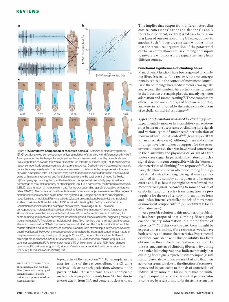
Figure 5 | Quantitative comparison of receptive fields. a | Samples of electromyographic(EMG) activity evoked by noxious mechanical stimulation of skin sites with different sensitivity (left).A sample receptive field map of a single palmar flexor muscle constructed by quantification ofEMG responses shown on the ventral side of the left forelimb of the cat (right). Numbers indicateresponse magnitude as a percentage of maximal response. Dashed lines indicate mathematicallyderived iso-response levels. This procedure was used to determine the receptive fields that areshown in a simplified form in c (bottom row) such that dark blue areas denote the receptive fieldareas with maximal response and light blue areas denote the total extent of receptive fields. b | Example graph plotting the quantitative data on receptive field sensitivity (expressed as apercentage of maximal response) of climbing fibre input to a paravermal multizonal microcomplex(MZMC) as a function of the equivalent data for the corresponding spinal nociceptive withdrawalreflex (SNWR). The correlation coefficient obtained provides an objective measure of the degree ofsimilarity between receptive fields in the two systems. c | Sample nociceptive climbing fibrereceptive fields of individual Purkinje cells (top, based on complex spike activity) and individualforelimb muscles (bottom, based on EMG activity) both using the method described in a.Correlation coefficients for the examples shown were, on average, 0.85. The closecorrespondence indicates that individual climbing fibre afferents convey information about theskin surface representing an imprint of withdrawal efficacy of a single muscle. In addition, thesame climbing fibre receives convergent input from group II muscle afferents, originating mainly inthe same muscle84. Therefore, an individual climbing fibre monitors both the input and the outputelement of an individual SNWR module (compare with FIG. 10). The functional significance of themuscle afferent input is not known, as cutaneous and muscle afferent input interactions have notbeen investigated. However, the convergence emphasizes the integrated sensorimotor nature ofthe paravermal climbing fibre input. 5b, e, g, h, 2d and 7d, denote classification of differentclimbing fibre microzones (see REF. 54 for details). ECRL, extensor carpi radialis longus; ECU,extensor carpi ulnaris; FCR, flexor carpi radialis; FCU, flexor carpi ulnaris; FDP, flexor digitorumprofundus; PL, palmaris longus; TRI, triceps. Panels a and c modified, with permission, from REF. 84 © (2002) Blackwell Publishing Ltd.

NATURE REVIEWS | NEUROSCIENCE VOLUME 6 | APRIL 2005 | 303
R E V I E W S
fibre signals that are relevant to the more complexissue of limb movement control. Quantitative tech-niques have been used to compare the spatial encod-ing of sensory information mediated by climbingfibres that terminate in the paravermal cerebellumwith sensory encoding in a previously well-character-ized sensorimotor system that mediates spinal noci-ceptive withdrawal reflexes (SNWR; FIG. 5). In brief,correlation analysis of quantified receptive fieldmaps84 showed that the skin receptive fields of climb-ing fibres that terminate in individual microzones inthe paravermal cerebellar cortex54 and the receptivefields of individual modules of the SNWR system85
‘pre-processes’ the sensory input, thereby generatingclimbing fibre signals with properties that are transitionalbetween sensory and motor information.
Spinal reflex and climbing fibre relations. Despitetheir explanatory value for the regulation of eyemovements, the findings on the ventral paraflocculusare not easily extrapolated to other parts of the motorsystem. This is because eye movements are relativelysimple: they are limited to motion in two dimensionsand involve movement of a constant mass. Recently,however, progress has been made with regard tounderstanding the nature and origin of climbing
PARAFLOCCULUS
In experimental animals, thedorsal and ventral paraflocculusare the two most caudal lobulesof the cerebellar hemisphere.
RETINAL SLIP
An unwanted movement of thevisual image on the retina thatoccurs, for instance, when themovement of the eyes isinadequate to follow a movingobject.
X
X
Y
Y
ZZ
aA B
C
c d e
b
Vt
V Vnα
N
Vn = V × cos α
Plantar
Medial
Gastrocnemius Peroneus longus Tibialis anterior
Motor output:withdrawalfield
Sensory input:receptivefield
Stim.
Figure 6 | Sensorimotor transformations in spinal nociceptive withdrawal reflex modules. A | A method to document andanalyse how movements caused by the contraction of single muscles move a skin surface in three dimensions. a | Movements ofthe right hind paw of the rat were elicited by intra-muscular stimulation (stim.) of individual muscles and documented with twocameras placed at right angles to each other. Dots indicate the points on the skin that were traced by motion analysis. b | The shapeof each paw segment was determined by photographing sections of a hardened wax model of the entire paw. c | Cross-section of adigit segment: the movement of the skin surface was decomposed to determine the actual withdrawal of the skin surface, definedas the inward vector indicated by Vn. N, normal; Vt, tangential vector. d | Sample of withdrawal vectors Vn along the skin surface ofthe proximal phalanx of digit V on contraction of a given muscle. e | Distribution of Vn on the whole hind paw on contraction of thesame muscle. Shading indicates areas of low (light blue), medium (mid blue) and high (dark blue) degrees of withdrawal. B |Simplified diagram of a spinal nociceptive withdrawal reflex module in the cat. The skin receptive field on the left forelimb — asdetermined by recording and quantification of the electromyographic response in a single muscle on nociceptive stimulation of theskin (see FIG. 5) — is shown on the right (dark blue area denotes the receptive field area with maximal response and light blue areadenotes the total extent of the receptive field). Arrows indicate schematically the magnitude of withdrawal of the different parts of thereceptive field on contraction of the muscle depicted to the left. C | Comparison of quantified withdrawal fields (as in A) andquantified skin receptive fields (as in B) for three muscles in the rat hindlimb shows high levels of similarity (correlation coefficientsranging 0.81–0.88). Panels A and C modified, with permission, from REF. 86 © (1994) Springer. Panel B data from REF. 132.

304 | APRIL 2005 | VOLUME 6 www.nature.com/reviews/neuro
R E V I E W S
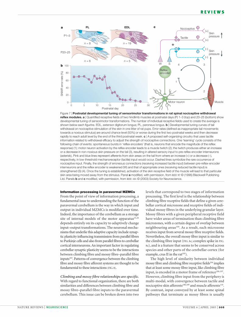
Both SNWRs and climbing fibres signal motor errors. Tobetter understand the action-based nature of sensoryencoding in SNWR modules, it is useful to considertheir postnatal development97–99. The sensorimotortransformations performed in each module are notinnate but instead are functionally adapted by a post-natal learning process that occurs during active sleep.Specifically, as individual muscles are brought to twitchby spontaneous activity in spinal sensorimotor circuits,the ensuing tactile feedback is used to tune the connec-tion strengths in individual SNWR modules100. Thechange of tactile input from the withdrawn skin areathat occurs in conjunction with the spontaneous singlemuscle movements has an adaptive effect on the reflexmodule101. By this process, each skin receptive fieldbecomes a sensory IMPRINT of the withdrawal efficacy ofan individual muscle (FIG. 7).
In individual SNWR modules, the precise sensori-motor relationship between the distribution of sensitivityon the skin of noxious (or tactile102) inputs and thewithdrawal efficacy of the muscle86 provides a theoreticalfoundation for the detection of motor errors. First, theprobability that the skin receptive field of a particularreflex module will be stimulated (for example, by hittingan external object), and therefore be activated at a givenpoint in time, is inversely proportional to the level ofcontraction of the muscle of that module (as musclecontraction will tend to withdraw the relevant skin areafrom the stimulus). Second, if a point in the receptivefield does encounter a noxious stimulus, the degree ofactivation of the module for a given stimulus intensity isin direct proportion to the extent to which the musclehas the capacity to withdraw the stimulated point fromthe stimulus. Therefore, the more inadequate the con-traction of a muscle is — that is, the larger the motorerror of that specific muscle — the stronger the recep-tive field activation will be. In other words, the degree towhich the sensory receptive field is activated duringmovement provides precise information about theextent to which an elemental part of the motor systemhas made an error.
By analogy to input to individual SNWR modules,signals conveyed to the paravermal MZMCs throughascending climbing fibre pathways84 would seem to beencoded in a way that is directly related to motoroutput86. The information contained in climbing fibresignals arising from the spinal cord does not represent anerror signal in sensory coordinates that must be convertedfrom a sensory into a motor error. That conversion hasalready taken place in the spinal cord.As a result, climbingfibres that terminate in individual paravermal MZMCsseem to provide information about motor errors thatrelate to the action of single muscles. Overall, the action-based features of climbing fibre sensory signals seem to bebroadly consistent with those stated by the CEREBELLAR
FEEDBACK-ERROR-LEARNING MODEL24,81,82. Despite differenceswith respect to phylogenetic age and the complexity ofmotor control carried out, both limb and eye movement83
cerebellar control systems seem to involve similar prin-ciples of sensory encoding, which indicates that theseprinciples might apply more generally.
were highly similar. Therefore, climbing fibre signalsthat arise from the spinal cord might be pre-processedby SNWRs. The latter system consists of 35–40 separatemodules, each comprising a specific skin receptivefield, a segmental multi-synaptic reflex arc and a specific output muscle85. Each module performs adetailed sensorimotor transformation that results in a graded withdrawal of the limb (or part of the limb),such that its particular receptive field on the skin ismoved away from the stimulus. For any given SNWRmodule, the input strength has a characteristic patternon the skin that mimics the pattern of ‘WITHDRAWAL
EFFICACY’ when the output muscle of the module contracts86 (FIG. 6).
The similarity between the receptive fields of climb-ing fibres and SNWRs might explain why the cerebellarparavermis is divided into microzones in the first place— that is, why it is modular in functional organization.An action-based representation of sensory informationshould yield a highly processed (computational) mapin the cerebellar cortex rather than a faithful represen-tation of the body surface with an orderly sequence ofreceptive fields, as in a conventional sensory map that is‘somatotopical’ in the strict sense of the word87. In fact,skin areas displaced by the action of single muscles donot necessarily form a continuum on the body surface.On the contrary, a system organized on the basis of amovement-related representation of the skin is inher-ently modular if such a system operates through anarray of individual muscles. The implications of thisorganization for cerebellar information processing areconsidered below.
However, before returning to this issue, it is ofinterest to note that the close correspondencebetween SNWR modules and paravermal climbingfibres might also be relevant to the observation thatclimbing fibre pathways that arise from the spinalcord are subject to substantial central control, espe-cially during active movements such as walking88–91 orreaching for an object92,93. The ‘gating’ of transmissionoccurs only at certain times, specific to the particularmovement. During locomotion, for instance, skinafferents from the ipsilateral forepaw are most likelyto transmit information through ascending climbingfibre pathways to the C1 and C3 zones in lobule V ofthe cerebellar cortex in the swing phase of the stepcycle88–90. Although gating and its temporal patternseem to be fundamental characteristics of climbingfibre pathways in general, its mechanisms and func-tional significance are not well understood94,95.Notably, however, a similar phenomenon has beenreported for spinal reflex pathways: the swing phaseof the step cycle is also the time when activation ofthe same skin afferents is most likely to elicit reflexwithdrawal of the limb96. This similarity in the pat-tern of modulation provides additional evidence forthe possibility of a close functional link betweenascending climbing fibre pathways and spinal reflexcircuits, and indicates that the movement-relatedgating might have a common origin, presumably atthe level of the spinal cord.
WITHDRAWAL EFFICACY
The motor effect of a muscle interms of its movement awayfrom an external reference point,which can be represented as aquantitative topographical mapbased on an analysis of thedisplacement of many points onthe skin during the contractionof a single limb muscle.
IMPRINT
In the SNWR system there is,for each individual muscle,a close relationship between thedistribution of sensitivity in thereceptive field on the skin andthe pattern of skin withdrawalcaused by muscle contraction.Therefore, the output of thesingle muscle reflex componentappears to be ‘imprinted’ on thespinal reflex circuitry that carriesout the input–outputtransformation.
CEREBELLAR FEEDBACK-ERROR-
LEARNING MODEL
A cerebellar model proposed byKawato and colleagues thatspecifically addresses the issue ofhow sensory errors might betransformed into motor errors.
NATURE REVIEWS | NEUROSCIENCE VOLUME 6 | APRIL 2005 | 305
R E V I E W S
levels that correspond to two stages of informationprocessing. The first level is the relationship betweenclimbing fibre receptive fields that define a given cere-bellar cortical microzone and receptive fields of indi-vidual mossy fibres in the underlying granular layer.Mossy fibres with a given peripheral receptive fieldhave wider areas of termination than climbing fibremicrozones, with a certain degree of overlap betweenneighbouring areas104. As a result, each microzonereceives input from several mossy fibre receptive fields.Nevertheless, the overall mossy fibre input is similar tothe climbing fibre input (FIG. 8c; complex spike in FIG.
8a), and is a feature that seems to be conserved acrossspecies and other parts of the cerebellar cortex (forexample, crus II in the rat105).
The high level of similarity between individualmossy fibre and climbing fibre receptive fields104 impliesthat at least some mossy fibre input, like climbing fibreinput, is encoded in a motor frame of reference106,107.However, climbing fibre input from the periphery ismulti-modal, with convergence between tactile andnociceptive skin afferents108,109 and muscle afferents110.By contrast, input conveyed by at least some spinalpathways that terminate as mossy fibres is usually
Information processing in paravermal MZMCsFrom the point of view of information processing, afundamental issue to understanding the function of theparavermal cerebellum is the way in which input andoutput in individual MZMCs is modified over time.Indeed, the importance of the cerebellum as a storagesite of internal models of the motor apparatus25,26
depends entirely on its capacity to adaptively changeinput–output transformations. The neuronal mecha-nisms that underlie this adaptive capacity include synap-tic plasticity influencing transmission from parallel fibresto Purkinje cells and also from parallel fibres to cerebellarcortical interneurons. An important factor in regulatingcerebellar synaptic plasticity seems to be the interactionsbetween climbing fibre and mossy fibre–parallel fibreinputs103. Patterns of convergence between the climbingfibre and mossy fibre afferent systems are thought to befundamental to these interactions (FIG. 8).
Climbing and mossy fibre relationships are specific.With regard to functional organization, there are bothsimilarities and differences between climbing fibre andmossy fibre–parallel fibre inputs to the paravermalcerebellum. This issue can be broken down into two
100
80
60
40
20
00 5 10 15 20 25 30
SN
WR
err
or r
ate
(%)
Postnatal day
a c
bW S
Nociceptive(+)
(+)
(–)
(–)
Tactile
Skinpressure
Skinpressure
Reflexencoder
1
2
34
Motor neuron Muscle
P1–3
P20–25
n = 8 n = 19
PL EDL
n = 6 n = 5
Figure 7 | Postnatal developmental tuning of sensorimotor transformations in rat spinal nociceptive withdrawalreflex modules. a | Quantified receptive fields of two hindlimb muscles at postnatal days (P) 1–3 (top) and 20–25 (bottom) showdevelopmental tuning of sensorimotor transformations. The number of individual receptive fields used to create the average isshown below each figurine. EDL, extensor digitorum longus; PL, peroneus longus. b | Developmental tuning curves of tailwithdrawal on nociceptive stimulation of the skin in one litter of rat pups. Error rates (defined as inappropriate tail movementstowards a noxious stimulus) are around chance level (50%) or worse during the first two postnatal weeks and then decreaserapidly to reach adult level by the end of the third postnatal week. c | A proposed self-organizing circuitry that uses tactileinformation related to withdrawal efficacy to adjust the strength of nociceptive connections. One ‘learning’ cycle consists of thefollowing chain of events: spontaneous bursts in ‘reflex encoders’ (that is, neurons that encode the magnitude of the reflexresponse) (1); motor neuron activation by the reflex encoder leads to a muscle twitch (2); the twitch produces either an increaseor a decrease in non-noxious skin pressure on the tail (3), resulting in altered sensory input to pre-reflex encoder interneurons(asterisk). Pink and blue lines represent afferents from skin areas on the tail from where an increase (+) or a decrease (–),respectively, in low-threshold mechanoreceptor (tactile) input would occur. Dashed lines symbolize the rare occurrence ofnociceptive input. Finally, the strength of erroneous connections (receiving increased tactile input) between pre-reflex-encoderinterneurons and the reflex encoder is weakened (W) and that of appropriate ones (receiving reduced tactile input) isstrengthened (S) (4). Once the tuning is established, activation of the skin receptive field of the muscle will lead to that particularskin area being moved away from the stimulus. Panel a modified, with permission, from REF. 97 © (1996) Blackwell PublishingLtd. Panels b and c modified, with permission, from REF. 101 © (2003) Society for Neuroscience.
306 | APRIL 2005 | VOLUME 6 www.nature.com/reviews/neuro
R E V I E W S
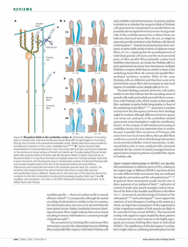
and cerebellar cortical interneurons. At present, opinionis divided as to whether the receptive fields of Purkinjecells generated by transmission in parallel fibres areprimarily due to inputs from local or non-local granulecells. In the cerebellar paravermis, evidence from catsindicates that local mossy fibres do not, as might beexpected, provide excitation to the Purkinje cells directlyoverlying them112. Instead, local interneurons have exci-tatory receptive fields similar to those of subjacent mossyfibres (FIG. 8b,c), implying that the ascending branch ofindividual granule cell axons and the most proximalparts of their parallel fibres primarily contact localinhibitory interneurons.As a result, the Purkinje cells in agiven paravermal microzone have interneuron-mediatedinhibitory receptive fields that are similar to those of theunderlying mossy fibres. By contrast, the parallel fibre-mediated excitatory receptive fields of the samePurkinje cells are different and therefore seem to bederived from mossy fibres that terminate in non-localregions of cerebellar cortex (simple spike in FIG. 8a).
The latter finding contrasts, however, with earlierstudies in rats that indicate that the ascending axons ofgranule cells make particularly powerful direct connec-tions with Purkinje cells, which results in their parallelfibre-mediated receptive fields being similar to those ofthe underlying mossy fibres113–115. At present, it remainsunclear how the discrepancy between these findingsmight be resolved, although differences between species(cat versus rat) and parts of the cerebellum studied(paravermis versus hemisphere) might be contributoryfactors. In the context of information processing incerebellar circuits, this is an important issue to resolve,because if parallel fibre excitation of Purkinje cellsarises from non-local mossy fibres, then parallel fibrescould serve a central role in distributing informationacross several cerebellar cortical microzones. As dis-cussed below, this, in turn, could provide a neuronalsubstrate for the control of muscle synergies based onspatial and/or temporal patterns of parallel fibre inputsto Purkinje cells.
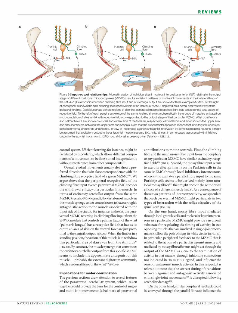
Input–output relationships in MZMCs are specific.Microstimulation in different parts of NIA, relating tothe output stage of different MZMCs, has been shownto evoke different limb movements that are mediatedthrough the red nucleus and the rubrospinal tract116–118.In general, these movements usually involve two orthree segments of an ipsilateral limb116, indicative ofcontrol of multi-joint muscle synergies, such as retrac-tion of the limb at the shoulder and flexion at the elbow(FIG. 9). Anatomical and physiological features of therubrospinal119,120 and corticospinal121 tracts support theexistence of such divergence. Looking at the system as awhole, an important consequence of this organizationis that a significant number of limb muscles are likely tobe controlled from several MZMCs. The functionaloverlap with regard to output implied by these patternsof connectivity is in stark contrast to the highly segre-gated microzonal climbing fibre input to individualMZMCs. The significance of this discrepancy is unclear,but it might relate to conflicting demands placed on the
modality specific — that is, it is either tactile or muscleafferent-related104. Consequently, although the spatialencoding of information is similar in the two systems,the information does not seem to be derived from thesame spinal circuits. Rather, similarities between climb-ing and mossy fibres might indicate that action-basedencoding of sensory information is a common principleof spinal networks111.
The second level of climbing fibre and mossy fibreinteractions concerns the relationship between climbingfibre and parallel fibre input to individual Purkinje cells
CS
SS
IN
MF
a
b
c
n = 17 n = 8
n = 18 n = 9
n = 15 n = 5
n = 36 n = 10
Purkinjecell
Mossyfibre
Climbingfibre
Parallelfibre
Interneuron
ab
c
A B
Figure 8 | Receptive fields in the cerebellar cortex. A | Schematic diagram of recordingsites in Purkinje cells, interneurons (IN) and mossy fibres (MF) at right angles to the surfacethrough the C3 zone in the paravermal cerebellar cortex. Mossy fibre input was provided bythe external component of the cuneo–cerebellar tract104,149. B | Typical receptive fieldcharacteristics of neuronal elements in two microzones (left and right columns) are depictedon the ventral and dorsal surface of the left cat forelimb. a | Complex spike (CS) and simplespike (SS) receptive fields of Purkinje cells. b | Receptive fields of nearby interneurons. c |Receptive fields of mossy fibre terminals immediately below the Purkinje cell layer. Note thatin each microzone, the CS response (due to climbing fibre activity) of individual Purkinje cellshas a similar receptive field on the skin of the ipsilateral forelimb as local inhibitoryinterneurons and mossy fibre terminals in the subjacent granular layer. By contrast, theexcitatory SS receptive field of the same Purkinje cells (due to mossy fibre–granulecell–parallel fibre input) is different. Values shown with each pair of limb figurines denote thenumbers of units that contributed to producing the average receptive field map. Panel Bmodified, with permission, from REF. 112 © (2001) Blackwell Publishing Ltd and REF. 150 ©(2003) Taylor and Francis.
NATURE REVIEWS | NEUROSCIENCE VOLUME 6 | APRIL 2005 | 307
R E V I E W S
contributions to motor control). First, the climbingfibre and the main mossy fibre input from the peripheryto any particular MZMC have similar excitatory recep-tive fields104 (FIG. 8). Second, the mossy fibre input seemsto exert its effect primarily on the Purkinje cells in thesame MZMC through local inhibitory interneurons,whereas the excitatory parallel fibre input to the samePurkinje cells seems to be derived mainly from non-local mossy fibres112 that might encode the withdrawalefficacy of a different muscle (FIG. 8). As a consequence ofthese two patterns of interconnectivity, we speculatethat each paravermal MZMC might participate in twotypes of interaction with the reflex circuitry of thespinal cord (FIG. 10).
On the one hand, mossy fibre input mediatedthrough local granule cells and molecular layer interneu-rons in a particular MZMC might provide a neuronalsubstrate for regulating the timing of activity in twoopposing muscles that are involved in single-joint move-ments (follow the path of signs in white circles in FIG. 10).In particular, peripheral feedback to the MZMC that isrelated to the action of a particular agonist muscle andmediated by mossy fibre afferents might act through theoutput of the MZMC as a cue to the termination ofactivity in that muscle (through inhibitory connectionsnot indicated in FIG. 10; FIG. 9 legend) and influence theonset of antagonist muscle activity. In this respect, it isrelevant to note that the correct timing of transitionsbetween agonist and antagonist activity associatedwith single-joint movements123 is disrupted followingcerebellar damage124.
On the other hand, similar peripheral feedback couldbe conveyed through the parallel fibres to influence the
control system. Efficient learning, for instance, might befacilitated by modularity, which allows different compo-nents of a movement to be fine-tuned independentlywithout interference from other components122.
Overall, evoked movements usually also show a pre-ferred direction that is in close correspondence with theclimbing fibre receptive field of a given MZMC116. Weargue above that the peripheral receptive field of theclimbing fibre input to each paravermal MZMC encodesthe withdrawal efficacy of a particular limb muscle. Interms of excitatory cerebellar output from the sameMZMC (see also FIG. 9 legend), the distal-most muscle inthe muscle synergy under control seems to have a roughlyantagonistic action to the muscle associated with theinput side of the circuit. For instance, in the cat, the para-vermal MZMC receiving its climbing fibre input from theSNWR module that controls a palmar flexor of the wrist(palmaris longus) has a receptive field that has as itscentre an area of skin on the ventral forepaw just prox-imal to the central footpad (FIG. 9a).When the limb is in astanding position, the action of this muscle is to withdrawthis particular area of skin away from the stimulus84
(FIG. 6B). By contrast, the muscle synergy that constitutesthe excitatory cerebellar output from this specific MZMCseems to include the approximate antagonist of thismuscle — probably the extensor digitorum communis,which is a dorsal flexor of the wrist116 (FIG. 9a).
Implications for motor coordinationThe previous sections draw attention to several featuresof the paravermal cerebellar system, which, takentogether, could provide the basis for the control of single-and multi-joint movements (a hallmark of paravermal
a
Purkinjecell
Climbingfibre
rDAO
NIA
cb
Purkinjecell
Climbingfibre
rDAO
Purkinjecell
Climbingfibre
rDAO
NIA NIA
Figure 9 | Input–output relationships. Microstimulation of individual sites in nucleus interpositus anterior (NIA) relating to the outputstage of different multizonal microcomplexes (MZMCs) results in distinct patterns of multi-joint movements in the ipsilateral limb ofthe cat. a–c | Relationships between climbing fibre input and nucleofugal output are shown for three example MZMCs. To the rightof each panel is shown the skin climbing fibre receptive field of an individual MZMC, depicted on a dorsal and ventral view of theipsilateral forelimb. Dark blue areas denote regions of skin that generated maximal response; light blue areas denote total extent ofreceptive field. To the left of each panel is a skeleton of the same forelimb showing schematically the groups of muscles activated onmicrostimulation of sites in NIA with receptive fields corresponding to the output stage of that particular MZMC. Wrist dorsiflexorsand palmar flexors are shown on dorsal and ventral side of the forearm, respectively; elbow flexors and extensors on the upper arm;and shoulder flexors between the upper arm and scapula. Note that the experimental approach means that inhibitory influences onspinal segmental circuitry go undetected. In view of ‘reciprocal’ agonist/antagonist innervation by some rubrospinal neurons, it mightbe assumed that excitatory output to the antagonist muscle (see also FIG. 10) is, at least in some cases, associated with inhibitoryoutput to the agonist (not shown). rDAO, rostral dorsal accessory olive. Data from REF. 116.

308 | APRIL 2005 | VOLUME 6 www.nature.com/reviews/neuro
R E V I E W S
be caused by the mossy fibre input related to the action ofa particular muscle (FIG. 10, lower right panel) As theoutput stage of the non-local MZMC would affect analtogether different muscle synergy, this kind of inter-action could relate to the coordination of activity acrossmultiple joints of the limb.
In the model in FIG. 10, climbing fibre-dependent andclimbing fibre-independent plasticity could dynamicallyand adaptively regulate both the local MZMC and non-local MZMC interactions, in the former case by influenc-ing synaptic transmission between the ascending axonsof granule cells and local molecular layer interneurons,and in the latter by influencing synaptic transmissionbetween parallel fibres and Purkinje cells103. In thisrespect, the gating of transmission in ascending climbingfibre pathways might allow only behaviourally relevanttraining signals to be forwarded to the cerebellum duringmovement94. Also at the behavioural level, informationprocessing within and between MZMCs resembles,respectively, the non-associative and the associativelearning used in cerebellar adaptation of gain in thevestibulo–ocular reflex and in classical conditioning ofthe eye-blink reflex. Conceptual similarities between ourmodel and that of Kawato and co-workers (see above forreferences) have already been pointed out.
Note also that, from a historical perspective, thescheme in FIG. 10, by virtue of the two pathways — the oneacting within the same MZMCs and the other acrossMZMCs — would seem to reconcile an important con-ceptual dichotomy in the early literature125. Holmes5,6
(and before him Luciani126) emphasized the control bythe cerebellum of individual movement elements andphysical variables such that movement coordination as awhole equalled the sum of the regulation of many parts.On the other hand, Babinski3,4 (and before himFlourens127) advocated a special organizing principle ofcoordination per se, by which the individual movementelements were coupled, the whole therefore being greaterthan the sum of the parts. These seemingly contradic-tory views are compatible with the pathways indicatedin the present model (see signs in white and grey circlesin FIG. 10). The interaction between MZMCs is also inkeeping with a long-held view that the parallel fibresare an important substrate for the coordination of dif-ferent body parts125,128–131 (although see REFS 113–115 fora different point of view).
Concluding remarksAlthough interpretations of how the networks outlinedin FIG. 10 might operate during active movement arespeculative, the model as such highlights the need forfurther anatomical and physiological data that are essen-tial for a fuller understanding of this system. Unchartedterritories include the organization of mossy fibre inputsfrom, for example, deep tissue receptors or cerebral corti-cal systems, and how during active movement, patternsof activity in these systems relate to the functional orga-nization of MZMCs. The functional significance of themovement-related gating of transmission in climbingfibre pathways, and how this might influence cerebellaroperation, also remains uncertain. There are also some
Purkinje cells in a different MZMC (follow the path ofsigns in grey circles in FIG. 10). Therefore, with some delayrelative to the effects on the local MZMC described above(because of the slow conduction time in the parallelfibres), the opposite effects on a non-local MZMC would
SNWR
Cerebellarcortex
n-IGrCIGrC
Parallel fibre Parallelfibre
NIA
a b c
Purkinjecell
Climbingfibre
+ +–
++
– –
Interneuron
MossyfibreRubrospinal tract
Spinalgreymatter
Antagonist
Agonist
Figure 10 | Spinocerebellar interactions and motor coordination. Functional interrelationshipbetween cerebellar microcomplexes and spinal reflex modules. Spinal nociceptive withdrawalreflex (SNWR) module (lower left panel): input–output characteristics of one example module. Skininput from a receptive field on the ventral side of the left forearm and paw (light green) and muscleinput from a palmar flexor (orange; see FIG. 5 legend) converge in the spinal grey matter and sendoutput to the muscle of the module (palmar flexor, agonist; green). Ascending pathways: skininformation is relayed through mossy fibres (light green dashed line), and integrated skin andmuscle afferent information is relayed through climbing fibres (dark green dashed line; inferior oliverelay not shown). Cerebellar cortex (upper panel): input–output characteristics of one examplemicrocomplex (b; not shown as multizonal for simplicity). Output from nucleus interpositusanterior (NIA) is mediated through the rubrospinal tract (red) to influence a group of muscles thatact on several joints of the limb (lower left panel, shown in red, including antagonist). The Purkinjecells to the left (a) and right (c) illustrate non-local microcomplexes. Two hypothetical routes ofsignal flow within microcomplexes (b; signs in white circles) and between microcomplexes (b,c; signs in grey circles) are labelled as (+) and (–) signs to indicate excitation and inhibitionrespectively. In the microcomplex in part b, the local granule cell input (lGrC; see receptive field inlower panel) provides inhibitory input to the overlying Purkinje cells by way of a local interneuroninput and also excitatory parallel fibre input to a non-local microcomplex (c). In a similar way, theexcitatory parallel fibre input to the Purkinje cells in the microcomplex in part b originates in non-local granule cells (n-lGrC) located in a different microcomplex (a). Arrows show the direction ofinformation flow (see text for further details). Interestingly, in classical conditioning of the eye-blinkreflex20, there is strong evidence that climbing fibres convey information to the cerebellar cortexabout the unconditioned stimulus, which elicits a protective reflex (the eye-blink). Such a role is inkeeping with the function of climbing fibres proposed in the present model.

NATURE REVIEWS | NEUROSCIENCE VOLUME 6 | APRIL 2005 | 309
R E V I E W S
presented here, the encoding of afferent information isof direct relevance to the modular organization ofcerebellar networks, which might be thought of as aconsequence of the integrated sensorimotor role of thecerebellum. This role does not seem to conform to aclear distinction between ‘sensory’ and ‘motor’ pro-cessing (for an alternative view, see REF. 133). On thecontrary, we believe that, to inform the debate aboutthe complex nature of cerebellar topography38, cere-bellar afferent representations should be viewed ascomputational action-based maps rather than purelyas sensory maps of the body surface.
Finally, it is also relevant to note that the precise butcomplex modular organization of cerebellar systems,exemplified by the paravermal MZMCs, highlights theimportance of using an independent scheme to classify,as far as possible, the neurons recorded in behaviouralstudies. Differences with respect to input and outputimply that each functional module contributes in a spe-cific way to movement coordination. These differentcontributions might be expected to be associated withdifferences in neuronal firing patterns in relation to theperformance of particular movements. Such relation-ships might well be obscured, or even lost, when pool-ing firing patterns from groups of neurons that do notbelong to the same functional unit. One of the chal-lenges for future cerebellar studies must be to relateneuronal spike trains in awake, behaving animals tofunctional localization.
important conceptual shortcomings. For instance, byanalogy to the SNWR system, encoding of sensoryinformation in the paravermal MZMC system seems tohave the standing position of the limb as a frame of ref-erence. Displacement of the most distal skin surfaces bymuscle contraction depends on whether the limb is mak-ing contact with the ground86,132, and it remains unclearhow the paravermal MZMC system operates when thelimb is off the ground, during, for example, a reach-to-grasp movement. In this situation, the biomechanicaleffects of contraction of several limb muscles differ sig-nificantly from those during the standing position.
Despite these shortcomings, the findings indicatethat climbing fibres that relay ascending signals conveyinformation about motor errors. Such informationcould, without further processing, be used as trainingsignals for the adaptive programming of cerebellarnetworks. This is supported by a comparison of ourown data on climbing fibre input to the paravermalcerebellum with data on climbing fibre input to theventral paraflocculus83. In addition, on the basis ofdetailed analyses of input conveyed by the dorsal spin-ocerebellar tract, Bosco and Poppele106,107 have pre-sented evidence that mossy fibre input might also beencoded in a motor frame of reference. Taken together,these findings imply that the encoding of sensoryinformation in an action-based frame of referencemight be a central organizing principle of ascendingcerebellar afferent systems in general. In the model
1. Purves, D. et al. Neuroscience 3rd edn (Sinauer Associates,Sunderland, Massachusetts, 2004).
2. Ito, M. The Cerebellum and Neural Control (Raven, NewYork, 1984).
3. Babinski, J. De l‘asynergie cerebelleuse. Rev. Neurol.7, 806–816 (1899).
4. Babinski, J. Asynergie et inertie cerebelleuses. Rev. Neurol.14, 685–686 (1906).
5. Holmes, G. The symptoms of acute cerebellar injuries due togunshot injuries. Brain 40, 461–535 (1917).
6. Holmes, G. The cerebellum of man. Brain 62, 1–30 (1939).7. Chambers, W. W. & Sprague, J. M. Functional localization in
the cerebellum II: somatotopic organization in cortex andnuclei. Arch. Neurol. Psychiatry 74, 653–680 (1955).
8. Dow, R. S. & Moruzzi, G. The Physiology and Pathology ofthe Cerebellum (Minnesota Univ. Press, Minneapolis,1958).
9. Thach, W. T. On the specific role of the cerebellum in motorlearning and cognition: clues from PET activation and lesionstudies in man. Behav. Brain Sci. 19, 411–431 (1996).
10. Schmahmann, J. D. The Cerebellum and Cognition(Academic, San Diego, 1997).
11. Ekerot, C.-F. & Kano, M. Long-term depression of parallelfibre synapses following stimulation of climbing fibres. BrainRes. 342, 357–360 (1985).
12. Ito, M. Long-term depression. Annu. Rev. Neurosci.12, 85–102 (1989).
13. Ito, M. Cerebellar long-term depression: characterization,signal transduction, and functional roles. Physiol. Rev.81, 1143–1195 (2001).
14. Hansel, C., Linden, D. J. & D’Angelo, E. Beyond parallel fiberLTD: the diversity of synaptic and non-synaptic plasticity inthe cerebellum. Nature Neurosci. 4, 467–475 (2001).
15. Ito, M., Shiida, T., Yagi, N. & Yamamoto, M. Visual influence onrabbit horizontal vestibulo–ocular reflex presumably effectedvia the cerebellar flocculus. Brain Res. 65, 170–174 (1974).
16. Robinson, D. A. Adaptive gain control of vestibuloocular reflexby the cerebellum. J. Neurophysiol. 39, 954–969 (1976).
17. Thach, W. T. A role for the cerebellum in learning movementcoordination. Neurobiol. Learn. Mem. 70, 177–188 (1998).
18. Houk, J. C., Buckingham, J. T. & Barto, A. G. Models of thecerebellum and motor learning. Behav. Brain Sci. 19,368–383 (1996).
19. Boyden, E. S., Katoh, A. & Raymond, J. L. Cerebellum-dependent learning: the role of multiple plasticitymechanisms. Annu. Rev. Neurosci. 27, 581–609 (2004).
20. Hesslow, G. & Yeo, C. H. in A Neuroscientist‘s Guide toClassical Conditioning (ed. Moore, J. W.) 86–146 (Springer,New York, Berlin, Heidelberg, 2002).
21. Braitenberg, V. & Atwood, R. P. Morphological observationson the cerebellar cortex. J. Comp. Neurol. 109, 1–33 (1958).
22. Marr, D. A theory of cerebellar cortex. J. Physiol. (Lond.)202, 437–470 (1969).
23. Albus, J. S. A theory of cerebellar function. Math. Biosci. 10,25–61 (1971).
24. Kawato, M. & Gomi, H. A computational model of fourregions of the cerebellum based on feedback-error learning.Biol. Cybern. 68, 95–103 (1992).
25. Wolpert, D. M. & Kawato, M. Multiple paired forward andinverse models for motor control. Neural Net. 11,1317–1329 (1998).
26. Wolpert, D. M., Miall, R. C. & Kawato, M. Internal models inthe cerebellum. Trends Cogn. Sci. 2, 338–347 (1998).
27. Oscarsson, O. Functional units of the cerebellum — sagittalzones and microzones. Trends Neurosci. 2, 143–145 (1979).
28. Voogd, J. & Bigaré, F. in The Inferior Olivary Nucleus:Anatomy and Physiology (eds Courville, J., de Montigny, C.& Lamarre, Y.) 207–234 (Raven, New York, 1980).
29. Armstrong, D. M. Functional significance of connections ofthe inferior olive. Physiol. Rev. 54, 358–417 (1974).
30. Brodal, A. & Kawamura, K. Olivocerebellar projection: areview. Adv. Anat. Embryol. Cell Biol. 64, 1–140 (1980).
31. Armstrong, D. M. Topographical localisation in theprojections from the inferior olive to the paravermal cortexof the anterior lobe and paramedian lobule in thecerebellum of the cat. A brief review. Arch. Ital. Biol. 128,183–207 (1990).
32. Eccles, J. C., Llinas, R. & Sasaki, K. The excitatory synapticaction of climbing fibres on the Purkinje cells of thecerebellum. J. Physiol. (Lond.) 182, 268–296 (1966).
33. Thach, W. T. Somatosensory receptive fields of single units incat cerebellar cortex. J. Neurophysiol. 30, 675–696 (1967).
34. Hawkes, R. An anatomical model of cerebellar modules.Prog. Brain Res. 114, 39–52 (1997).
35. Herrup, K. & Kuemerle, B. The compartmentalization of thecerebellum. Annu. Rev. Neurosci. 20, 61–90 (1997).
36. Sugihara, I. & Shinoda, Y. Molecular, topographic, andfunctional organization of the cerebellar cortex: a study withcombined aldolase C and olivocerebellar labeling. J. Neurosci. 24, 8771–8785 (2004).
37. Voogd, J. & Glickstein, M. The anatomy of the cerebellum.Trends Neurosci. 21, 370–375 (1998).
38. Manni, E. & Petrosini, L. A century of cerebellar somatotopy:a debated representation. Nature Rev. Neurosci. 5,241–249 (2004).
39. Oscarsson, O. in The Inferior Olivary Nucleus: Anatomy andPhysiology (eds Courville, J., de Montigny, C. & Lamarre, Y.)279–289 (Raven, New York, 1980).
40. Andersson, G., Ekerot, C.-F., Oscarsson, O. &Schouenborg, J. in Cerebellum and Neuronal Plasticity (edsGlickstein, M., Yeo, C. & Stein, J.) 165–173 (Plenum, NewYork, London, 1987).
41. Apps, R. Columnar organisation of the inferior oliveprojection to the posterior lobe of the rat cerebellum. J. Comp. Neurol. 302, 236–254 (1990).
42. Trott, J. R. & Apps, R. Lateral and medial sub-divisionswithin the olivocerebellar zones of the paravermal cortex inlobule Vb/c of the cat anterior lobe. Exp. Brain Res. 87,126–140 (1991).
43. Trott, J. R. & Apps, R. Zonal organisation within the projectionfrom the inferior olive to the rostral paramedian lobule of thecat cerebellum. Eur. J. Neurosci. 5, 162–173 (1993).
44. Atkins, M. J. & Apps, R. Somatotopical organization withinthe climbing fibre projection to the paramedian lobule andcopula pyramidis of the rat cerebellum. J. Comp. Neurol.389, 249–263 (1997).
45. Apps, R. Rostrocaudal branching within the climbing fibreprojection to forelimb-receiving areas of the cerebellarcortical C1 zone. J. Comp. Neurol. 419, 193–204 (2000).
46. Pardoe, J. & Apps, R Structure–function relations of twosomatotopically corresponding regions of the rat cerebellarcortex: olivo–cortico–nuclear connections. Cerebellum 1,165–184 (2002).
47. Sugihara, I., Wu, H. S. & Shinoda, Y. The entire trajectoriesof single olivocerebellar axons in the cerebellar cortex andtheir contribution to cerebellar compartmentalization. J. Neurosci. 21, 7715–7723 (2001).
48. Wilson, W. C. & Magoun, H. W. The functional significanceof the inferior olive. J. Comp. Neurol. 83, 69–77 (1945).

310 | APRIL 2005 | VOLUME 6 www.nature.com/reviews/neuro
R E V I E W S
49. Murphy, M. G. & O‘Leary, J. L. Neurological deficit in catswith lesions of the olivocerebellar system. Arch. Neurol. 24,145–157 (1971).
50. Llinas, R., Walton, K., Hillman, D. E. & Sotelo, C. Inferior olive:its role in motor learning. Science 190, 1230–1231 (1975).
51. Garwicz, M., Ekerot, C.-F. & Schouenborg, J. Distribution ofcutaneous nociceptive and tactile climbing fibre input tosagittal zones in cat cerebellar anterior lobe. Eur. J. Neurosci. 4, 289–295 (1992).
52. Andersson, G. & Oscarsson, O. Climbing fiber microzonesin cerebellar vermis and their projection to different groups ofcells in the lateral vestibular nucleus. Exp. Brain Res. 32,565–579 (1978).
53. Andersson, G. & Oscarsson, O. Projections to lateralvestibular nucleus from cerebellar climbing fiber zones. Exp.Brain Res. 32, 549–564 (1978).
54. Ekerot, C.-F., Garwicz, M. & Schouenborg, J. Topographyand nociceptive receptive fields of climbing fibres projectingto the cerebellar anterior lobe in the cat. J. Physiol. (Lond.)441, 257–274 (1991).An electrophysiological demonstration of a largenumber of microzones in the paravermal C3 zone,based on a detailed analysis of distributions ofsensitivity in the nociceptive receptive fields ofindividual climbing fibres. Owing to the particularcharacteristics of the receptive fields, they aresuggested to reflect movement rather than to relaysensory information from the periphery.
55. Hesslow, G. Correspondence between climbing fibre inputand motor output in eyeblink related areas in cat cerebellarcortex. J. Physiol. (Lond.) 476, 229–244 (1994).
56. Ekerot, C.-F. & Larson, B. The dorsal spino–olivocerebellarsystem in the cat. II. Somatotopical organization. Exp. BrainRes. 36, 219–232 (1979).
57. Ekerot, C.-F. & Larson, B. Branching of olivary axons toinnervate pairs of sagittal zones in the cerebellar anteriorlobe of the cat. Exp. Brain Res. 48, 185–198 (1982).
58. Ekerot, C.-F., Garwicz, M. & Schouenborg, J. Thepostsynaptic dorsal column pathway mediates cutaneousnociceptive information to cerebellar climbing fibres in thecat. J. Physiol. (Lond.) 441, 275–284 (1991).
59. Armstrong, D. M., Harvey, R. J. & Schild, R. F.Cerebello–cerebellar responses mediated via climbingfibres. Exp. Brain Res. 18, 19–39 (1973).
60. Rosina, A. & Provini, L. Somatotopy of climbing fiberbranching to the cerebellar cortex in cat. Brain Res.289, 45–63 (1983).
61. Ekerot, C.-F. & Larson, B. The dorsal spino–olivocerebellarsystem in the cat. I. Functional organization andtermination in the anterior lobe. Exp. Brain Res. 36,201–217 (1979).
62. Garwicz, M. & Ekerot, C.-F. Topographical organization ofcerebellar cortical projection to nucleus interpositus anteriorin the cat. J. Physiol. (Lond.) 474, 245–260 (1994).An electrophysiological study in anaesthetized cats inwhich MZMCs were first proposed. The topography ofevoked field potentials in the NIA indicated thatmicrozones in the C1, C3 and Y zones with similarclimbing fibre input project to a common set ofnuclear neurons.
63. Garwicz, M. Cerebellar control of forelimb movements:modular organization revealed by nociceptive and tactileclimbing fibre input. Thesis, Lund Univ. (1992).
64. Apps, R. & Garwicz, M. Precise matching of olivo–corticaldivergence and cortico–nuclear convergence betweensomatotopically corresponding areas in the medial C1 andmedial C3 zones of the paravermal cerebellum. Eur. J. Neurosci. 12, 205–214 (2000).A combined electrophysiological mapping andbidirectional tracer study in cats showing that,consistent with the MZMC hypothesis, there was aclose correlation between the incidence of double-labelled olive cells and the extent of overlap betweencorticonuclear terminal fields.
65. Oscarsson, O. in Handbook of Sensory Physiology. Vol. II:Somatosensory System (ed. Iggo, A.) 339–380 (Springer,New York, 1973).
66. Brodal, P. & Bjaalie, J. G. Salient anatomic features of thecortico–ponto–cerebellar pathway. Prog. Brain Res. 114, 227–249 (1997).
67. Tolbert, D. L., Bantli, H. & Bloedel, J. R. Anatomical andphysiological evidence for a cerebellar nucleo–corticalprojection in the cat. Neuroscience 1, 205–217 (1976).
68. Trott, J. R., Apps, R. & Armstrong, D. M. Topographicalorganisation within the cerebellar nucleocortical projection tothe paravermal cortex of lobule Vb/c in the cat. Exp. BrainRes. 80, 415–428 (1990).
69. Trott, J. R., Apps, R. & Armstrong, D. M. Zonal organizationof cortico–nuclear and nucleo-cortical projections of theparamedian lobule of the cat cerebellum. I. The C1 zone.Exp. Brain Res. 118, 298–315 (1998).
A combined electrophysiological andneuroanatomical tracing study in cats showing that, inmarked contrast to the forelimb part of the C1 zone inthe anterior lobe, the homologous part of the C1 zonein the posterior lobe has substantial nucleocorticalprojections. This is consistent with the idea thatdifferent parts of the same zone (possibly containingsimilar microzones) have different mossy fibre inputs.
70. Trott, J. R., Apps, R. & Armstrong, D. M. Zonal organizationof cortico–nuclear and nucleo-cortical projections of theparamedian lobule of the cat cerebellum. II. The C2 zone.Exp. Brain Res. 118, 316–330 (1998).
71. King, V., Armstrong, D. M., Apps, R. & Trott, J. R. Numericalaspects of pontine, lateral reticular and inferior olivaryprojections to two paravermal cortical zones of the catcerebellum. J. Comp. Neurol. 390, 537–551 (1998).
72. Herrero, L., Pardoe, J. & Apps, R. Pontine and lateralreticular projections to the c1 zone in lobulus simplex andparamedian lobule of the rat cerebellar cortex. Cerebellum1, 185–199 (2000).
73. Simpson, J. I., Wylie, D. R. & De Zeeuw, C. I. On climbingfibre signals and their consequence(s). Behav. Brain Sci. 19,384–398 (1996).
74. Gilbert, P. F. C. & Thach, W. T. Purkinje cell activity duringmotor learning. Brain Res. 128, 309–328 (1977).
75. Andersson, G. & Armstrong, D. M. Complex spikes in Purkinjecells in the lateral vermis (b zone) of the cat cerebellum duringlocomotion. J. Physiol. (Lond.) 385, 107–134 (1987).
76. Ojakangas, C. L. & Ebner, T. J. Purkinje cell complex andsimple spike changes during a voluntary arm movementlearning task in the monkey. J. Neurophysiol. 68,2222–2236 (1992).
77. Kitazawa, S., Kimura, T. & Yin, P. B. Cerebellar complexspikes encode both destinations and errors in armmovements. Nature 392, 494–497 (1998).
78. Horn, K. M., Pong, M. & Gibson, A. R. Discharge of inferiorolive cells during reaching errors and perturbations. BrainRes. 1008, 137–138 (2004).
79. Porrill, J., Dean, P. & Stone, J. V. Recurrent cerebellararchitecture solves the motor-error problem. Proc. R. Soc.Lond. B 271, 789–796 (2004).
80. Dean, P., Porrill, J. & Stone, J. V. Visual awareness and thecerebellum: possible role of decorrelation control. Prog.Brain Res. 144, 61–75 (2004).
81. Kawato, M. & Wolpert, D. Internal models for motor control.Novartis Found. Symp. 218, 291–304 (1998).
82. Kawato, M. Internal models for motor control and trajectoryplanning. Curr. Opin. Neurobiol. 9, 718–727 (1999).
83. Kobayashi, Y. et al. Temporal firing patterns of Purkinje cellsin the cerebellar ventral paraflocculus during ocular followingresponses in monkeys II. Complex spikes. J. Neurophysiol.80, 832–848 (1998).A single-unit study in awake monkeys of climbingfibre and simple spike activity in the ventralparaflocculus during ocular following responses.Results indicate that climbing fibre signals haveproperties that are transitional between sensory andmotor information.
84. Garwicz, M., Levinsson, A. & Schouenborg, J. Commonprinciples of sensory encoding in spinal reflex modules andcerebellar climbing fibers. J. Physiol. (Lond.) 540,1061–1069 (2002).Quantitative methods were used in anaesthetizedcats to compare characteristics of individual SNWRsand signals carried by individual climbing fibres tocerebellar cortical microzones. High positivecorrelations between reflex input and climbing fibreskin receptive fields and between reflex output and climbing fibre muscle afferent input indicated that climbing fibre signals that arise from the spinalcord might be functionally linked to the SNWRs.
85. Schouenborg, J. & Kalliomäki, J. Functional organization ofthe nociceptive withdrawal reflexes. I. Activation of hindlimbmuscles in the rat. Exp. Brain Res. 83, 67–78 (1990).
86. Schouenborg, J. & Weng, H.-R. Sensorimotortransformation in a spinal motor system. Exp. Brain Res.100, 170–174 (1994).A highly detailed three-dimensional analysis of thedisplacement of a large number of points on the skinduring contractions caused by single limb muscles inthe rat. By comparing the maps generated in this waywith the nociceptive input from the skin mediated byspinal reflex pathways to single limb muscles, theprecise relationships between the motor effect of anindividual muscle and its sensory receptive field wererevealed.
87. Kaas, J. H. Topographic maps are fundamental to sensoryprocessing. Brain Res. Bull. 44, 107–112 (1997).
88. Lidierth, M. & Apps, R. Gating in the spino–olivocerebellarpathways to the c1 zone of the cerebellar cortex duringlocomotion in the cat. J. Physiol. (Lond.) 430, 453–469 (1990).
89. Apps, R., Hartell, N. A & Armstrong D. M. Step phase-related excitability changes in spino–olivocerebellar paths tothe c1 and c3 zones in cat cerebellum. J. Physiol. (Lond.)483, 687–702 (1995).
90. Apps, R. & Lee, S. Gating of transmission in climbing fibrepaths to cerebellar cortical C1 and C3 zones in the rostralparamedian lobule during locomotion in the cat. J. Physiol.(Lond.) 516, 875–883 (1999).
91. Pardoe, J., Edgley, S. A., Drew, T. & Apps, R. Changes inexcitability of ascending and descending inputs to cerebellarclimbing fibres during locomotion. J. Neurosci. 24,2656–2666 (2004).
92. Horn, K. M., Van Kan, P. L. & Gibson, A. R. Reduction ofrostral dorsal accessory olive responses during reaching. J. Neurophysiol. 76, 4140–4151 (1996).
93. Apps, R., Atkins, M. J. & Garwicz, M. Gating of cutaneousinput to cerebellar climbing fibres during a reaching task incat. J. Physiol. (Lond.) 502, 203–214 (1997).
94. Apps, R. Movement-related gating of climbing fibre input tocerebellar cortical zones. Prog. Neurobiol. 57, 537–562(1999).
95. Apps, R. Gating of climbing fibre input to cerebellar corticalzones. Prog. Brain Res. 124, 201–211 (2000).
96. Forssberg, H., Grillner, S. & Rossignol, S. Phase dependentreflex reversal during walking in chronic spinal cats. BrainRes. 85, 103–107 (1975).
97. Holmberg, H. & Schouenborg, J. Postnatal development ofthe nociceptive withdrawal reflexes in the rat: a behaviouraland electromyographic study. J. Physiol. (Lond.) 493,239–252 (1996).
98. Holmberg, H. & Schouenborg, J. Developmental adaptationof withdrawal reflexes to early alteration of peripheralinnervation in the rat. J. Physiol. (Lond.) 495, 399–409(1996).
99. Holmberg, H., Schouenborg, J., Yu, Y. B. & Weng, H. R.Developmental adaptation of rat nociceptive withdrawalreflexes after neonatal tendon transfer. J. Neurosci. 17,2071–2078 (1997).
100. Petersson, P., Waldenstrom, A., Fahraeus, C. &Schouenborg, J. Spontaneous muscle twitches duringsleep guide spinal self-organization. Nature 424, 72–75(2003).
101. Waldenström, A., Thelin, J., Thimansson, E., Levinsson, A. &Schouenborg, J. Developmental learning in a pain-relatedsystem: evidence for a cross-modality mechanism. J. Neurosci. 23, 7719–7725 (2003).
102. Weng, H. R. & Schouenborg, J. On the cutaneousreceptors contributing to withdrawal reflex pathways in thedecerebrate spinal rat. Exp. Brain Res. 118, 71–77 (1998).
103. Jörntell, H. & Ekerot C.-F. Reciprocal bi-directionalplasticity of parallel fiber receptive fields in cerebellarPurkinje cells and their afferent interneurons. Neuron 34,797–806 (2002).
104. Garwicz, M., Jörntell, H. & Ekerot, C.-F. Cutaneousreceptive fields and topography of mossy fibres andclimbing fibres projecting to cat cerebellar C3 zone. J. Physiol. (Lond.) 512, 277–293 (1998).
105. Brown, I. E. & Bower, J. M. Congruence of mossy fiber andclimbing fiber tactile projections in the lateral hemispheres ofthe rat cerebellum. J. Comp. Neurol. 429, 59–70 (2001).
106. Bosco, G. & Poppele, R. E. Reference frames for spinalproprioception: kinematics based or kinetics based? J. Neurophysiol. 83, 2946–2955 (2000).
107. Bosco, G. & Poppele, R. E. Proprioception from aspinocerebellar perspective. Physiol. Rev. 81, 539–568(2001).A review article that summarizes this group’simportant studies on the dorsal spinocerebellar tract(DSCT) system in the cat. A key conclusion is thatmossy fibre inputs to the cerebellum conveyed by theDSCT system seem to encode whole-hindlimbkinematics.
108. Ekerot, C.-F., Gustavsson, P., Oscarsson, O. &Schouenborg, J. Climbing fibres projecting to cat cerebellaranterior lobe activated by cutaneous A and C fibres. J. Physiol. (Lond.) 386, 529–538 (1987).
109. Ekerot, C.-F., Oscarsson, O. & Schouenborg, J. Stimulationof cat cutaneous nociceptive C fibres causing tonic andsynchronous activity in climbing fibres. J. Physiol. (Lond.)386, 539–546 (1987).
110. Jörntell, H., Garwicz, M. & Ekerot, C.-F. Relation betweencutaneous climbing fibre receptive fields and muscle afferentinput to climbing fibres projecting to the cerebellar C3 zonein the cat. Eur. J. Neurosci. 8, 1769–1779 (1996).
111. Levinsson, A., Holmberg, H., Broman, J., Zhang, M. &Schouenborg, J. Spinal sensorimotor transformation:relation between cutaneous somatotopy and a reflexnetwork. J. Neurosci. 22, 8170–8182 (2002).
112. Ekerot, C.-F. & Jörntell, H. Parallel fibre receptive fields ofPurkinje cells and interneurons are climbing fibre-specific.Eur. J. Neurosci. 13, 1303–1310 (2001).

NATURE REVIEWS | NEUROSCIENCE VOLUME 6 | APRIL 2005 | 311
R E V I E W S
A single-unit mapping study in decerebrate catsshowing that skin receptive fields of climbing fibresand local interneurons in individual microzones aresimilar, whereas the parallel fibre receptive fields ofthe same Purkinje cells are different. An importantconclusion is that excitatory input to Purkinje cellsdepends on input from non-local mossyfibres/granule cells and is mediated over somedistance by parallel fibres.
113. Llinás, R. in The Cerebellum, New Vistas (Experimental BrainResearch Supplements 6) (eds Palay, S. L. & Chan-Palay, V.)189–194 (Springer, Berlin, Heidelberg, New York, 1982).
114. Bower, J. M. & Woolston, D. C. Congruence of spatialorganization of tactile projections to granule cell and Purkinjecell layers of cerebellar hemispheres of the albino rat: verticalorganization of cerebellar cortex. J. Neurophysiol. 49,745–766 (1983).
115. Cohen, D. & Yarom, Y. Patches of synchronized activity inthe cerebellar cortex evoked by mossy-fiber stimulation:questioning the role of parallel fibers. Proc. Natl Acad. Sci.USA 95, 15032–15036 (1998).
116. Ekerot, C.-F., Jörntell, H. & Garwicz, M. Functional relationbetween corticonuclear input and movements evoked onmicrostimulation in cerebellar nucleus interpositus anterior inthe cat. Exp. Brain Res. 106, 365–376 (1995).
117. Giuffrida, R. et al. Single muscle organization of interposito-rubral projections. Exp. Brain Res. 39, 261–267 (1980).
118. Rispal-Padel, L., Cicirata, F. & Pons, C. Cerebellar nucleartopography of simple and synergistic movements in the alertbaboon (Papio papio). Exp. Brain Res. 47, 365–380 (1982).
119. Mewes, K. & Cheney, P. D. Facilitation and suppression ofwrist and digit muscles from single rubromotoneuronal cellsin the awake monkey. J. Neurophysiol. 66, 1965–1977(1991).
120. Belhaj-Saïf, A., Karrer, J. H. & Cheney, P. D. Distribution andcharacteristics of poststimulus effects in proximal and distalforelimb muscles from red nucleus in the monkey. J. Neurophysiol. 79, 1777–1789 (1998).
121. Fetz, E. E., Cheney, P. D., Mewes, K. & Palmer, S. Control offorelimb muscle activity by populations ofcorticomotoneuronal and rubromotoneuronal cells. Prog.Brain Res. 80, 437–449 (1989).
122. Mussa-Ivaldi, F. A. Modular features of motor control andlearning. Curr. Opin. Neurobiol. 9, 713–717 (1999).
123. Hallett, M., Shahani, B. T. & Young, R. R. EMG analysis ofstereotyped voluntary movements in man. J. Neurol.Neurosurg. Psychiatry 38, 1154–1162 (1975).
124. Hore, J., Wild, B. & Diener, H. C. Cerebellar dysmetria at theelbow, wrist and fingers. J. Neurophysiol. 65, 563–571(1991).
125. Thach, W. T., Goodkin, H. P. & Keating, J. G. The cerebellumand adaptive coordination of movement. Annu. Rev.Neurosci. 15, 403–442 (1992).
126. Luciani, L. in Human Physiology Ch. 8, 467 (Macmillan,London, 1915). (Translated by F. A. Welby.)
127. Flourens, P. Recherches experimentales sur les propriétés etles fonctions du systéme nerveux, dans les animauxvertébres. (Crevot, Paris, 1824).
128. Eccles, J. C., Ito, M. & Szentágothai, J. The Cerebellum as aNeuronal Machine (Springer, Berlin, 1967).
129. Braitenberg, V., Heck, D. & Sultan, F. The detection andgeneration of sequences as a key to cerebellar function:experiments and theory. Behav. Brain Sci. 20, 229–245(1997).
130. Garwicz, M. & Andersson, G. Spread of synaptic activityalong parallel fibres in cat cerebellar anterior lobe. Exp. BrainRes. 88, 615–622 (1992).
131. Isope, P. & Barbour, B. Properties of unitary granulecell–Purkinje cell synapses in adult rat cerebellar slices. J. Neurosci. 22, 9668–9678 (2002).
132. Levinsson, A., Garwicz, M. & Schouenborg, J. Sensorimotortransformation in cat nociceptive withdrawal reflex system.Eur. J. Neurosci. 4327–4332 (1999).
133. Bower, J. M. Is the cerebellum sensory for motor’s sake, ormotor for sensory’s sake: the view from the whiskers of arat? Prog. Brain Res. 114, 463–496 (1997).
134. Brand, S., Dahl, A.-L. & Mugnaini, E. The length of theparallel fibres in the cat cerebellar cortex. An experimentallight and electron microscopic study. Brain Res. 26, 39–58(1976).
135. Pichitpornchai, C., Rawson, J. A. & Rees, S. Morphology ofparallel fibres in the cerebellar cortex of the rat: anexperimental light and electron microscopic study withbiocytin. J. Comp. Neurol. 342, 206–220 (1994).
136. Lawrence, D. G. & Kuypers, H. G. J. M. The functionalorganization of the motor system in the monkey. I. The effects of bilateral pyramidal lesions. Brain 91, 1–14(1968).
137. Lawrence, D. G. & Kuypers, H. G. J. M. The functionalorganization of the motor system in the monkey. II. Theeffects of lesions of the brain-stem pathways. Brain 91,15–36 (1968).
138. Yu, J. & Eidelberg, E. Recovery of locomotor function oncats after localized cerebellar lesions. Brain Res. 273,121–131 (1983).
139. van Kan, P. L. E., Houk, J. C. & Gibson, A. R. Outputorganization of intermediate cerebellum of the monkey. J. Neurophysiol. 69, 57–73 (1993).
140. Gibson, A. R., Horn, K. M., Stein, J. F. & van Kan, P. L.Activity of interpositus neurons during a visually guidedreach. Can. J. Physiol. Pharmacol. 74, 499–512 (1996).Recordings of individual neurons in the forelimbregion of the NIA in awake, behaving monkeyssupported the general idea that this cerebellarnucleus is important for the control of handmovements used in a reach-to-grasp movement.
141. Cooper, S. E., Martin, J. H. & Ghez, C. Effects of inactivationof the anterior interpositus nucleus on the kinematic anddynamic control of multijoint movement. J. Neurophysiol.84, 1988–2000 (2000).
Reversible lesions with muscimol of the NIA in awakecats showed that the cerebellar paravermal system isimportant in the anticipatory control of interactionsbetween different limb segments involved in areaching task.
142. Monzée, J. & Smith, A. M. Responses of cerebellarinterpositus neurons to predictable perturbations applied toan object held in a precision grip. J. Neurophysiol. 91,1230–1239 (2004).
143. Monzée, J., Drew, T. & Smith, A. M. Effects of muscimolinactivation of the cerebellar nuclei on precision grip. J. Neurophysiol. 91, 1240–1249 (2004).
144. Bastian, A. J., Martin, T. A., Keating, J. G. & Thach, W. T.Cerebellar ataxia: abnormal control of interaction torquesacross multiple joints. J. Neurophysiol. 76, 492–509 (1996).
145. Bastian, A. J., Zackowski, K. M. & Thach, W. T. Cerebellarataxia: torque deficiency or torque mismatch betweenjoints? J. Neurophysiol. 83, 3019–3030 (2000).
146. Zackowski, K. M., Thach, W. T. & Bastian, A. J. Cerebellarsubjects show impaired coupling of reach and graspmovements. Exp. Brain Res. 146, 511–522 (2002).
147. Garwicz, M., Apps, R. & Trott, J. R. Micro-organization ofolivocerebellar and corticonuclear connections of theparavermal cerebellum in the cat. Eur. J. Neurosci. 8,2726–2738 (1996).
148. Garwicz, M., Ekerot, C.-F. & Jörntell, H. Organizationalprinciples of cerebellar neuronal circuitry. News Physiol. Sci.13, 26–32 (1998).
149. Cooke, J. D., Larson, B., Oscarsson, O. & Sjölund, B. Originand termination of cuneocerebellar tract. Exp. Brain Res. 13,339–358 (1971).
150. Ekerot, C.-F. & Jörntell, H. Parallel fiber receptive fields: a keyto understanding cerebellar operation and learning.Cerebellum 2, 101–109 (2003).
AcknowledgementsThe authors are grateful to J. Schouenborg for his comments on anearlier version of the manuscript. Many thanks are also due to R. Bissett for her help in the drafting of figures. We thank the UKMedical Research Council (R.A.) and the Swedish Research Counciland the Segerfalk Foundation (M.G.) for their financial support.
Competing interests statementThe authors declare no competing financial interests.
Online links
FURTHER INFORMATIONApps’ Homepage: http://www.bris.ac.uk/Depts/Physiology/new/pi/richard_apps/apps_intro.htmGarwicz’s Homepage:http://www.research.med.lu.se/en_projektdetaljer.php?Proj=62Access to this interactive links box is free online.