anatomical description of the lungs of burmeister’s ... aava... · parasitology, 108, pp 343-349...
TRANSCRIPT

Anatomical description of the lungs of Burmeister’s porpoise (Phocoena spinipinnis) Dr. Ismael Concha, DVM, MS Santo Tomás University (Campus Santiago) Avenida Ejército 146, Santiago. Chile. Código postal: 8370003. [email protected] Introduction: Porpoise are the smallest cetaceans, reaching body lengths up to 2.5 meters (8.2 ft). Burmeister’s porpoises (Burmeister, 1865) are endemic to the coast of South America and are in the data deficient Red List category (1) This specie is related to dolphins, and the most obvious visible difference between the two groups is that porpoises have flattened, spade-shaped teeth distinct from the conical teeth of dolphins, and shorter beaks. They have small, rounded heads and obtuse jaws instead of beaks. Phocoena spinipinnis have small bumps, known as tubercles, on the leading edge of the dorsal fin (2). One of the most remarkable structures in the lungs of cetacea are the sphincters in the respiratory bronchioles. The sphincters have been described in the common porpoise, bottlenose dolphin, and in white whale (3). The porpoises appear to be readily colonized by lungworms (4) Material and method: A juvenile male porpoise was found by the staff of Marine Life Experience Center Chile during January of 2011. The animal was stranded at the beach and died because its injuries. The same day of the recollection was donated to us. The cetacean was frozen within 24 hrs of its death, and one week after the necropsy was performed in our laboratory. Numerous organs were preserved for further studies. The lungs were cleaned with water, insufflated using an air pump and pigmented for an external anatomical description. Results and discussion: The lungs showed an elliptic shape, with an acute cranial apex and a caudal sharpened end. The right lung was slightly bigger and its apex extends farther than the left lung. The costal surface of the lungs displayed a smooth face without interlobar fissures; it was not possible to indentify lobes as in domestic animals. This surface presents a cranial-caudal obtuse longitudinal margin that separated a dorsal flattened horizontal face and a convex lateral face. The medial surface presents the vertebral and mediastinal parts, there was a notorious cardiac impression on each lung but no cardiac notch was found. The right lung showed a groove for the esophagus, and the cranial and caudal vena cava. The right bronchus is divided into cranial and caudal bronchi before it enters the lung, similar to the tracheal and principal right bronchi of ruminants. Cranial to the tracheal bronchus a deep groove for the azygos vein was observed. Four papillary shape processes were found, two related with the cranial (tracheal) hilus and two with the principal hilus. The left lung presents in its medial surface a notorious aortic groove and a unique hilus with two papillary processes caudally located. The diaphragmatic surface was very oblique in both lungs. The description presented in this abstract represent a good starting point to make future anatomical studies in this specie. 1-http://www.iucnredlist.org/apps/redlist/details/17029/0 date of consult July 1, 2011 2-Read, Andrew (1999). Porpoises. Stillwater, MN, USA:Voyageur Press. ISBN 0-89658-420-8. 3-J. R. Goudappel & E.J. Slijper Microscopic Structure of the Lungs of the Bottlenose Whale. Nature 182, 479 (16 August 1958) | doi:10.1038/182479a0 4-J. A. Balbuena, P. E. Aspholm, K. I. Andersen and A. Bjørge (1994). Lung-worms (Nematoda: Pseudaliidae) of harbour porpoises (Phocoena phocoena) in Norwegian waters: patterns of colonization. Parasitology, 108, pp 343-349 doi:10.1017/S0031182000076186
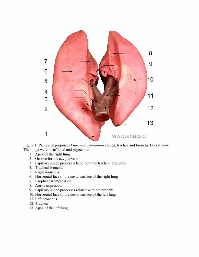
Figure 1: Picture of porpoise (Phocoena spinipinnis) lungs, trachea and bronchi. Dorsal view. The lungs were insufflated and pigmented.
1. Apex of the right lung 2. Groove for the azygos vein 3. Papillary shape process related with the tracheal bronchus 4. Tracheal bronchus 5. Right bronchus 6. Horizontal face of the costal surface of the right lung 7. Esophageal impression 8. Aortic impression 9. Papillary shape processes related with the bronchi 10. Horizontal face of the costal surface of the left lung 11. Left bronchus 12. Trachea 13. Apex of the left lung
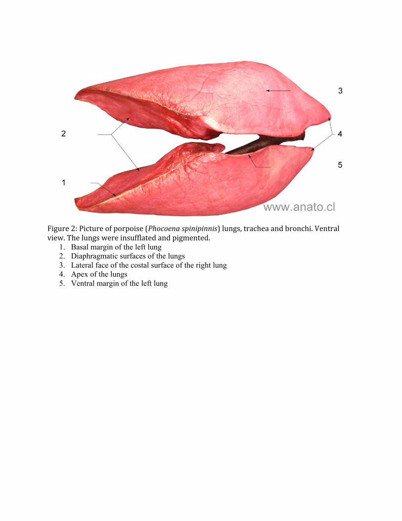
Figure 2: Picture of porpoise (Phocoena spinipinnis) lungs, trachea and bronchi. Ventral view. The lungs were insufflated and pigmented.
1. Basal margin of the left lung 2. Diaphragmatic surfaces of the lungs 3. Lateral face of the costal surface of the right lung 4. Apex of the lungs 5. Ventral margin of the left lung
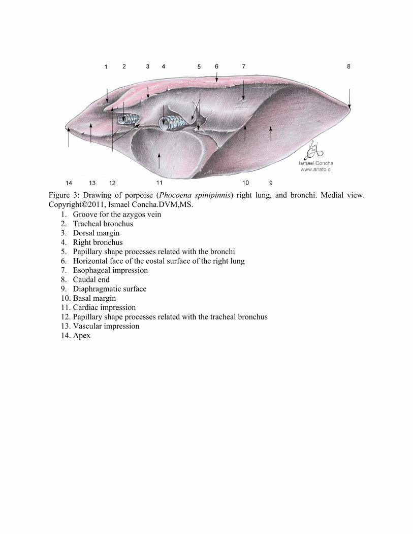
Figure 3: Drawing of porpoise (Phocoena spinipinnis) right lung, and bronchi. Medial view. Copyright2011, Ismael Concha.DVM,MS.
1. Groove for the azygos vein 2. Tracheal bronchus 3. Dorsal margin 4. Right bronchus 5. Papillary shape processes related with the bronchi 6. Horizontal face of the costal surface of the right lung 7. Esophageal impression 8. Caudal end 9. Diaphragmatic surface 10. Basal margin 11. Cardiac impression 12. Papillary shape processes related with the tracheal bronchus 13. Vascular impression 14. Apex

Figure 4: Drawing of porpoise (Phocoena spinipinnis) left lung, and bronchus. Medial view. Copyright2011, Ismael Concha.DVM,MS.
1. Aortic impression 2. Papillary shape processes related with the left bronchus 3. Dorsal margin 4. Left bronchus 5. Horizontal face of the costal surface of the left lung 6. Apex 7. Cardiac impression 8. Basal margin 9. Diaphragmatic surface 10. Caudal end