anatomy and pathology of the rotator interval and pathology of the...–extension of rotator cuff...
TRANSCRIPT
Anatomy and Pathology of the
Rotator Interval
R. Grace Bhardwaj
1 May 2008

Historical perspective
• Term “rotator interval” used by shoulder
surgeons to describe coracoid perforation of the
anterior rotator cuff; a triangular interval results
– Attributed to Neer (1970)
• Role in
– Glenohumeral instability
– Stabilization of the long head biceps tendon
– Inflammatory capsular conditions (adhesive
capsulitis)
Overview
• Normal anatomy– Borders
– Contents
• Biomechanics– Anatomic (cadaveric)
– Clinical
• Pathology– Rotator cuff tears
– Biceps sling
– CHL, SGHL, long head biceps tendon
– Capsular inflammation (adhesive capsulitis)
Overview
• Normal anatomy– Borders
– Contents
• Biomechanics– Anatomic (cadaveric)
– Clinical
• Pathology
– Rotator cuff tears
– Biceps sling
– CHL, SGHL, long head biceps tendon
– Capsular inflammation (adhesive capsulitis)

Hunt SA, Kwon YW, Zuckerman JD: The Rotator Interval: Anatomy, Pathology, and Strategies for Treatment. J Am Acad Orthop Surg 2007:15;218-227.
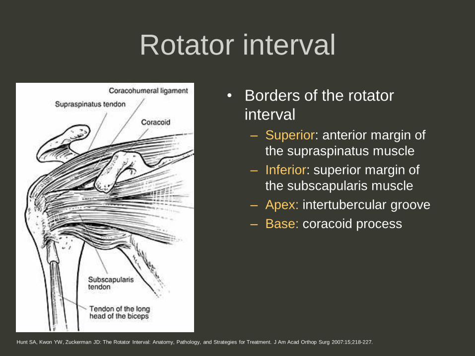
Rotator interval
• Triangular space created by
interposition of the coracoid process
between the supraspinatus and
subscapularis muscles
Hunt SA, Kwon YW, Zuckerman JD: The Rotator Interval: Anatomy, Pathology, and Strategies for Treatment. J Am Acad Orthop Surg 2007:15;218-227.
Rotator interval
• Borders of the rotator
interval
– Superior: anterior margin of
the supraspinatus muscle
– Inferior: superior margin of
the subscapularis muscle
– Apex: intertubercular groove
– Base: coracoid process
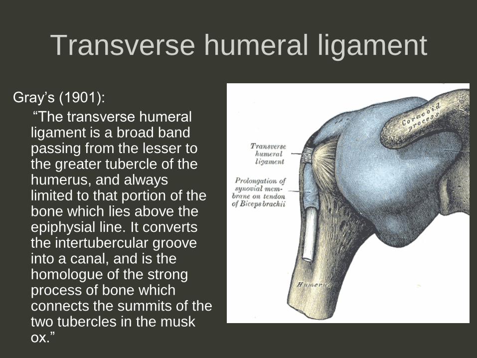
Transverse humeral ligament
Gray’s (1901):
“The transverse humeral ligament is a broad band passing from the lesser to the greater tubercle of the humerus, and always limited to that portion of the bone which lies above the epiphysial line. It converts the intertubercular groove into a canal, and is the homologue of the strong process of bone which connects the summits of the two tubercles in the musk ox.”

Transverse humeral ligament?
• Meyer (1920s): 2 observations– In shoulders with biceps tendon dislocation, the tissue described
as THL was intact
– Biceps dislocation was consistently medial (underneath or into
the subscapularis tendon substance)
• Others (Slatis and Aalto, 1979; Krief 2004) have
suggested that coracohumeral ligament
disruption is necessary for biceps tendon
dislocation
– No clear anatomic or histologic description of
the THL
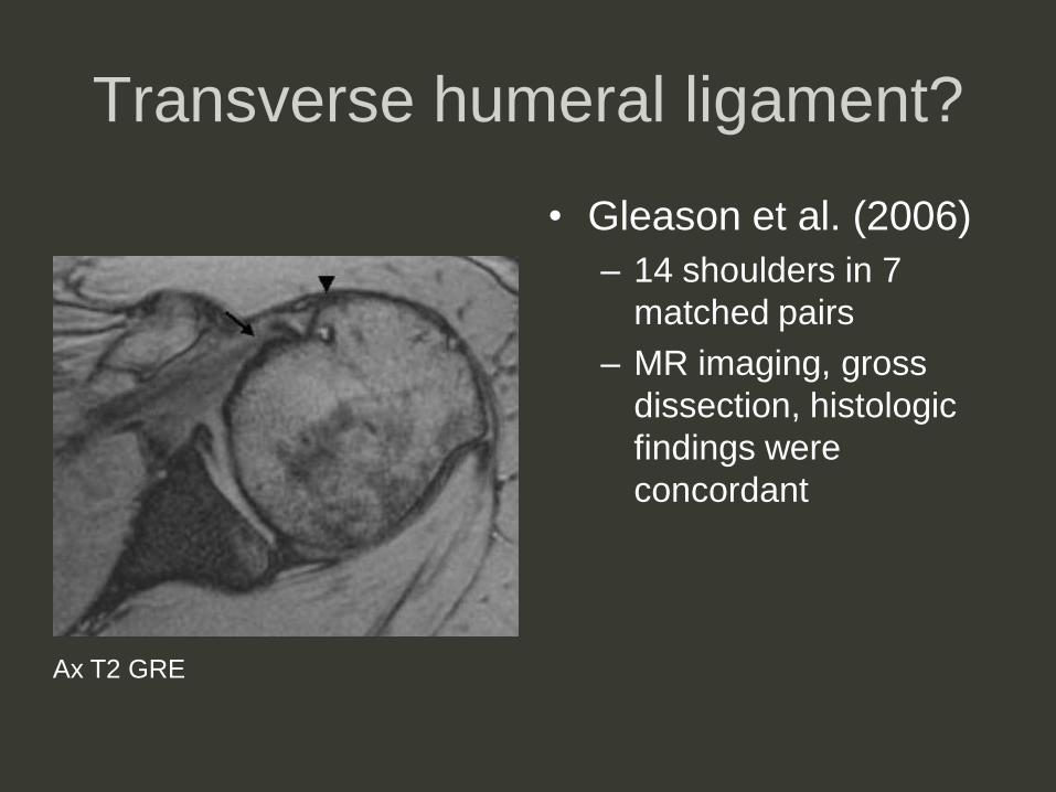
Transverse humeral ligament?
• Gleason et al. (2006)
– 14 shoulders in 7
matched pairs
– MR imaging, gross
dissection, histologic
findings were
concordant
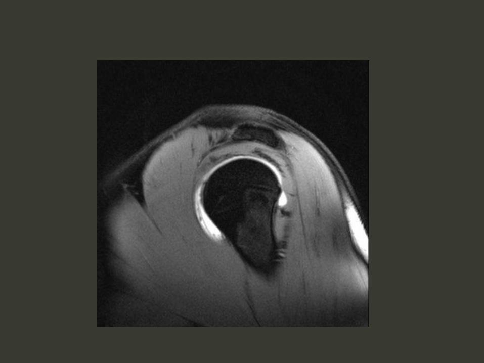
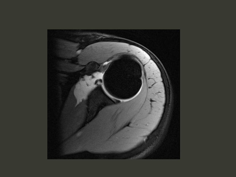
Ax T2 GRE

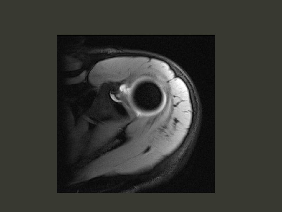
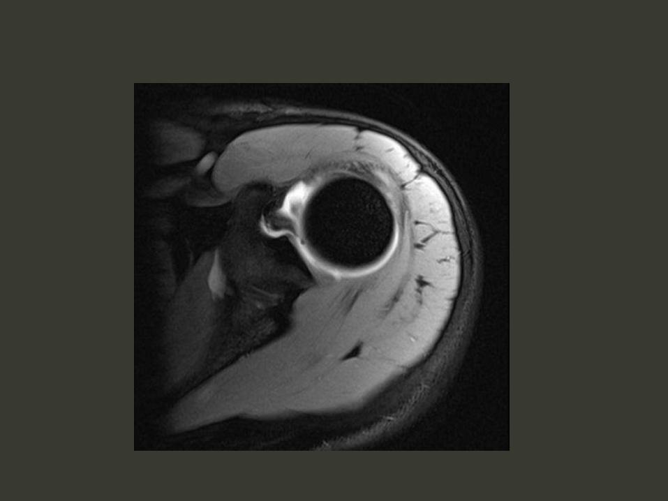
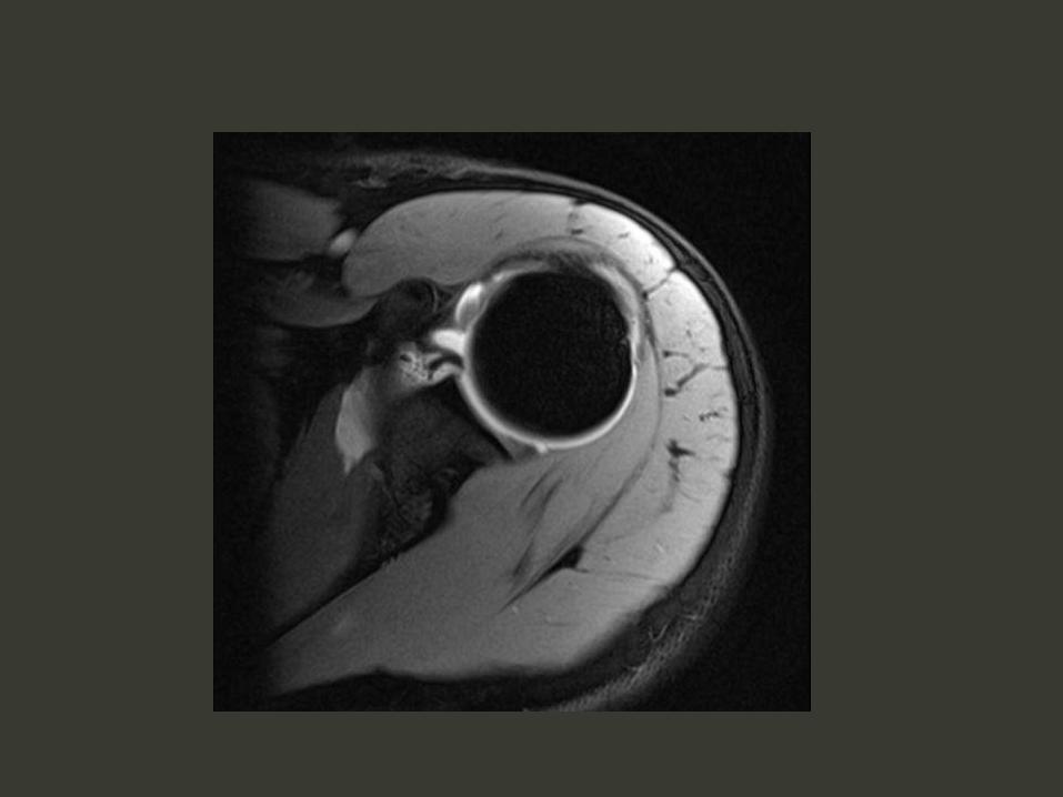
Transverse humeral ligament?
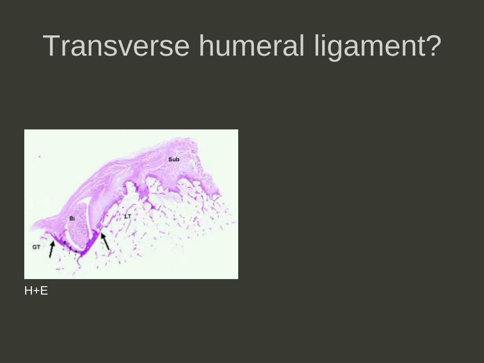
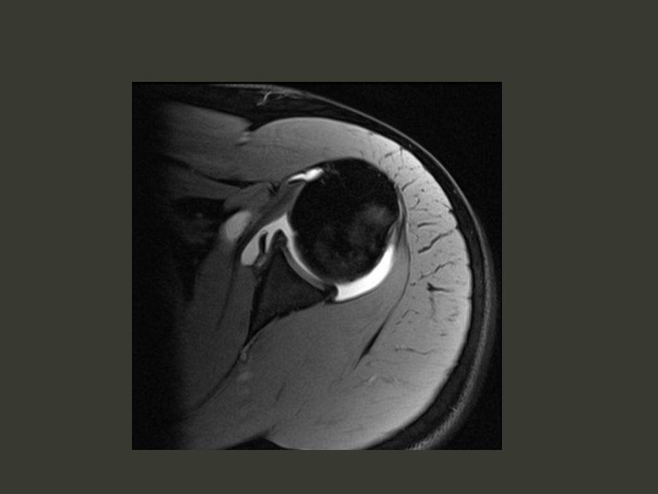
Transverse humeral ligament?
H+E
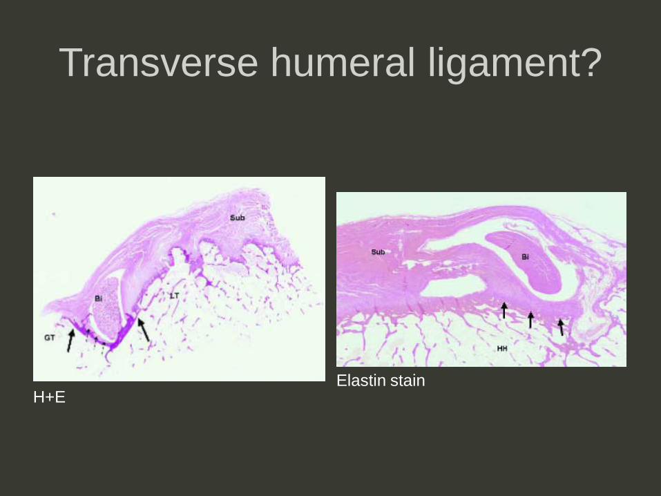
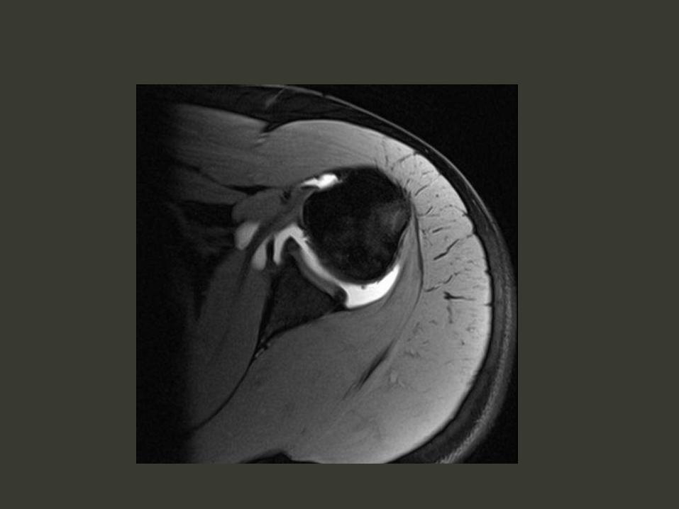
Transverse humeral ligament?
H+EElastin stain

Separate “THL” not confirmed
H+EElastin stain
Rotator interval contents
• Coracohumeral ligament
• Superior glenohumeral ligament
• Biceps tendon, long head
• Reinforced by, confluent with overlying
capsule

Rotator interval contents
• Coracohumeral ligament
• Superior glenohumeral ligament
• Long head biceps tendon
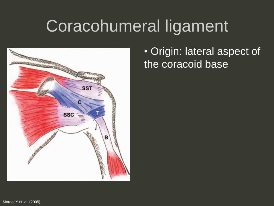
Coracohumeral ligament
• Origin: lateral aspect of
the coracoid base
Morag, Y et. al. (2005)
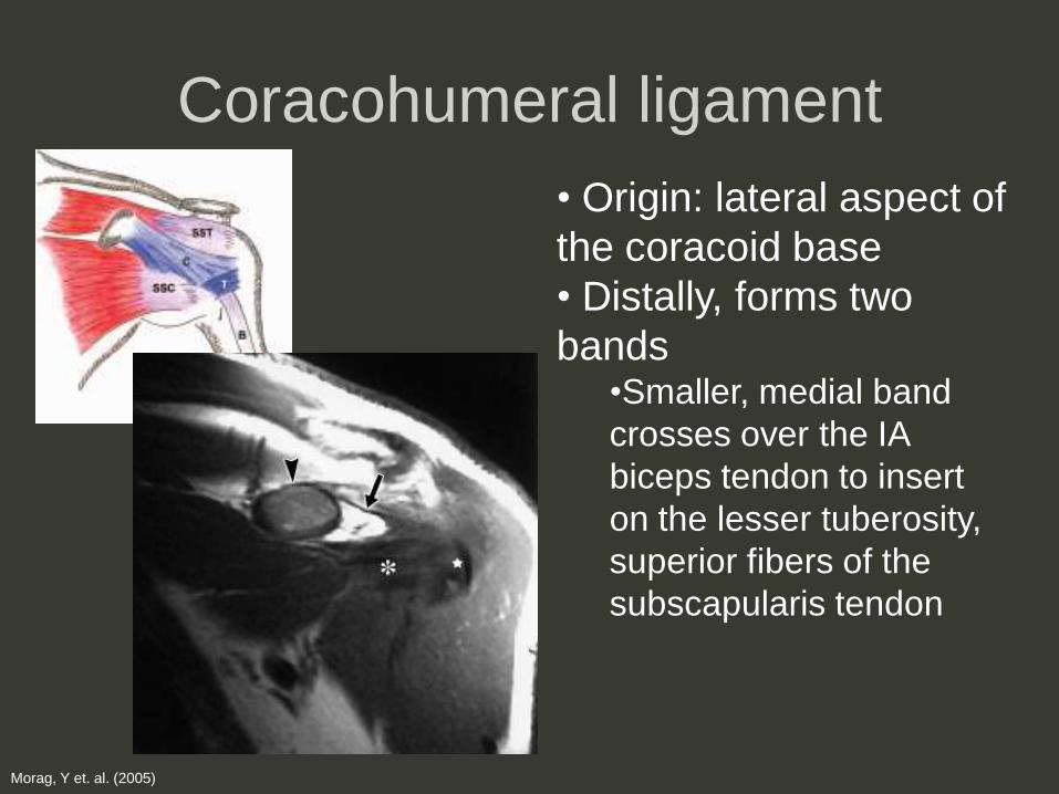
Coracohumeral ligament
• Origin: lateral aspect of
the coracoid base
• Distally, forms two
bands•Smaller, medial band
crosses over the IA
biceps tendon to insert
on the lesser tuberosity,
superior fibers of the
subscapularis tendon
Morag, Y et. al. (2005)

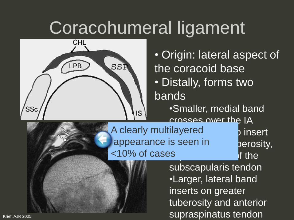
Coracohumeral ligament
• Origin: lateral aspect of
the coracoid base
• Distally, forms two
bands•Smaller, medial band
crosses over the IA
biceps tendon to insert
on the lesser tuberosity,
superior fibers of the
subscapularis tendon
•Larger, lateral band
inserts on greater
tuberosity and anterior
supraspinatus tendonKrief, AJR 2005
A clearly multilayered
appearance is seen in
<10% of cases
Coracohumeral ligament
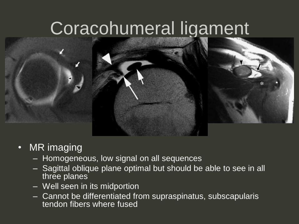
• MR imaging– Homogeneous, low signal on all sequences
– Sagittal oblique plane optimal but should be able to see in all three planes
– Well seen in its midportion
– Cannot be differentiated from supraspinatus, subscapularis tendon fibers where fused
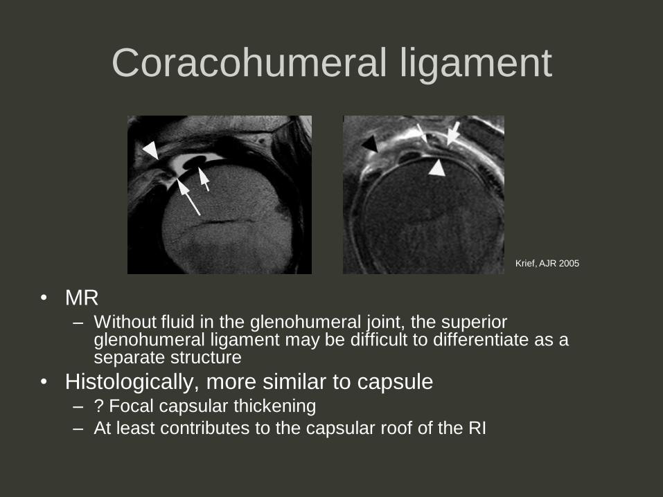
Coracohumeral ligament
• MR– Without fluid in the glenohumeral joint, the superior
glenohumeral ligament may be difficult to differentiate as a separate structure
• Histologically, more similar to capsule– ? Focal capsular thickening
– At least contributes to the capsular roof of the RI
Krief, AJR 2005
Rotator interval contents
• Coracohumeral ligament
• Superior glenohumeral ligament
• Biceps tendon, long head

Superior glenohumeral ligament
• Origin: superior
tubercle of the glenoid
(anterior to the biceps
tendon)
• Insertion:
superolateral lesser
tuberosity (deep to
superior border of
subscapularis tendon)
www.yess.uk.com/images/anatomy/ghj_ligs.jpg
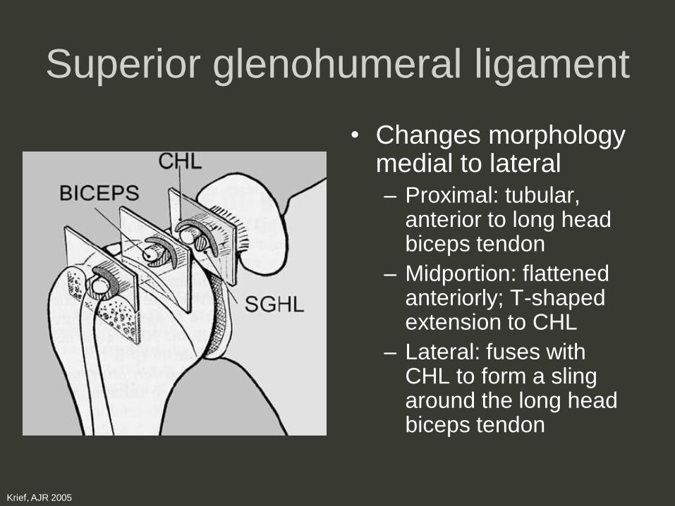
Superior glenohumeral ligament
• Changes morphology medial to lateral– Proximal: tubular,
anterior to long head biceps tendon
– Midportion: flattened anteriorly; T-shaped extension to CHL
– Lateral: fuses with CHL to form a sling around the long head biceps tendon
Krief, AJR 2005

Superior glenohumeral ligament
• MR– Uniform low signal intensity
– Anterior to long head biceps tendon on axial images
– Cannot differentiate from CHL where fused distally
– Best seen in the presence of intraarticular fluid
Morag 2005Bigoni 2004
Rotator interval contents
• Coracohumeral ligament
• Superior glenohumeral ligament
• Biceps tendon, long head
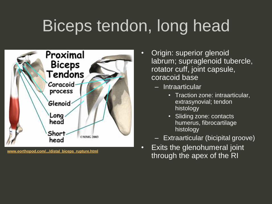
Biceps tendon, long head
• Origin: superior glenoid labrum; supraglenoid tubercle, rotator cuff, joint capsule, coracoid base
– Intraarticular
• Traction zone: intraarticular, extrasynovial; tendon histology
• Sliding zone: contacts humerus, fibrocartilage histology
– Extraarticular (bicipital groove)
• Exits the glenohumeral joint through the apex of the RI
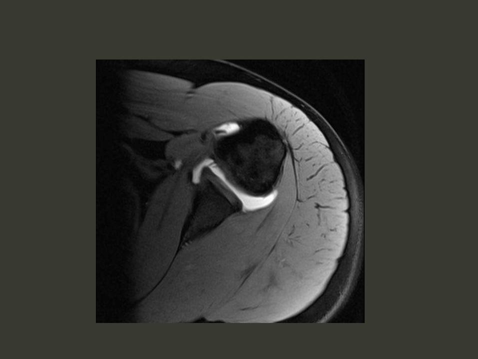
www.eorthopod.com/.../distal_biceps_rupture.html
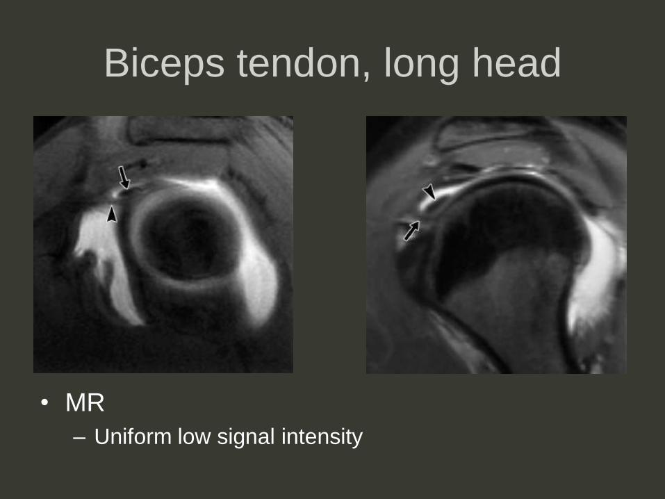
Biceps tendon, long head
• MR
– Uniform low signal intensity
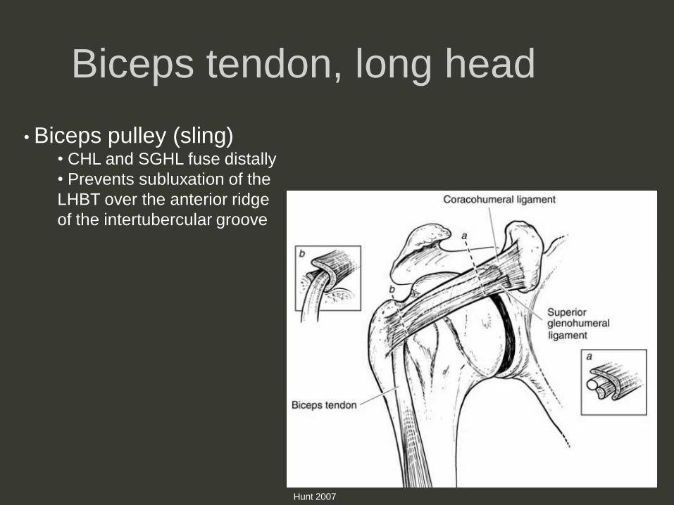
Biceps tendon, long head
• Biceps pulley (sling)• CHL and SGHL fuse distally
• Prevents subluxation of the
LHBT over the anterior ridge
of the intertubercular groove
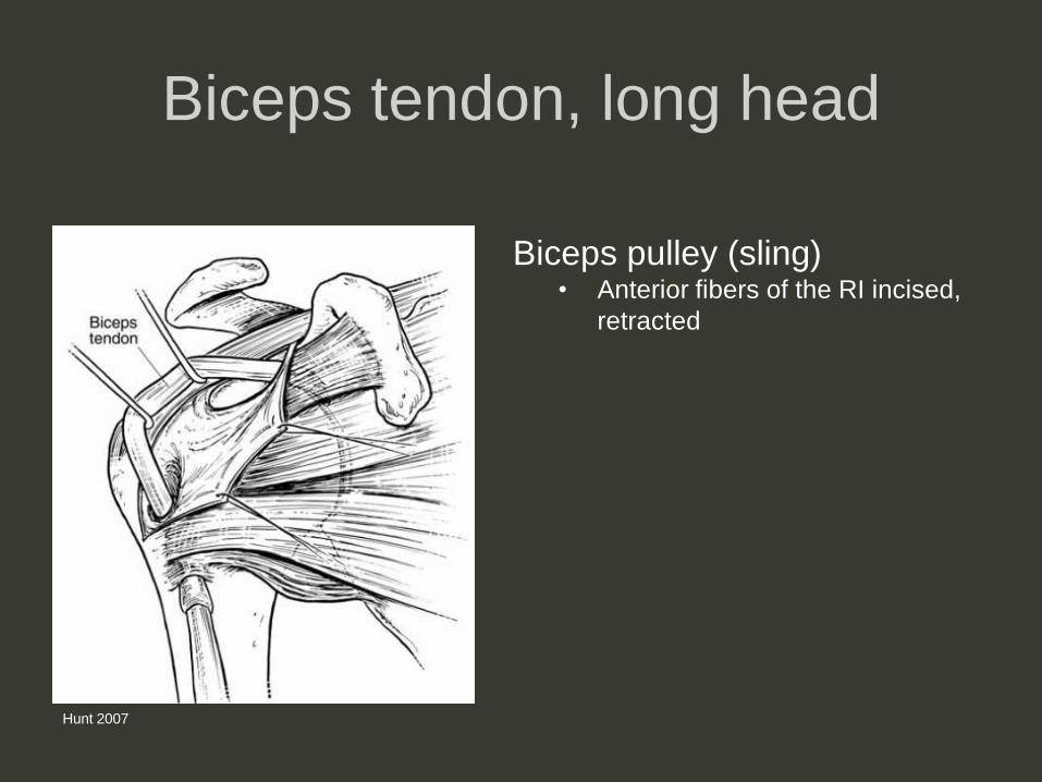
Hunt 2007
Hunt 2007
Biceps tendon, long head
Biceps pulley (sling)• Anterior fibers of the RI incised,
retracted
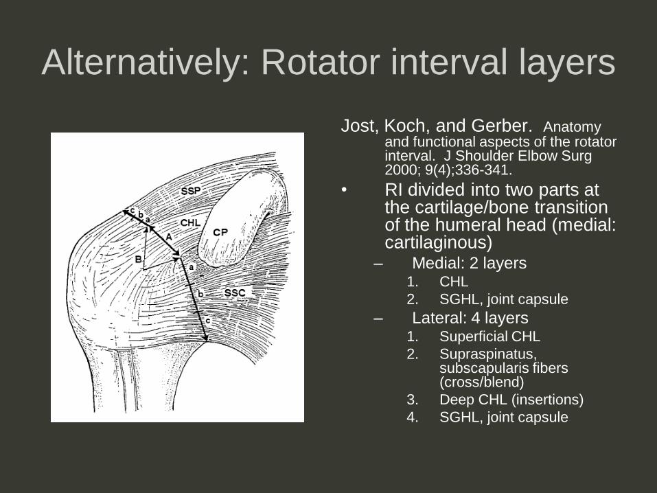
Alternatively: Rotator interval layers
Jost, Koch, and Gerber. Anatomy and functional aspects of the rotator interval. J Shoulder Elbow Surg 2000; 9(4);336-341.
• RI divided into two parts at the cartilage/bone transition of the humeral head (medial: cartilaginous)
– Medial: 2 layers1. CHL
2. SGHL, joint capsule
– Lateral: 4 layers1. Superficial CHL
2. Supraspinatus, subscapularis fibers (cross/blend)
3. Deep CHL (insertions)
4. SGHL, joint capsule
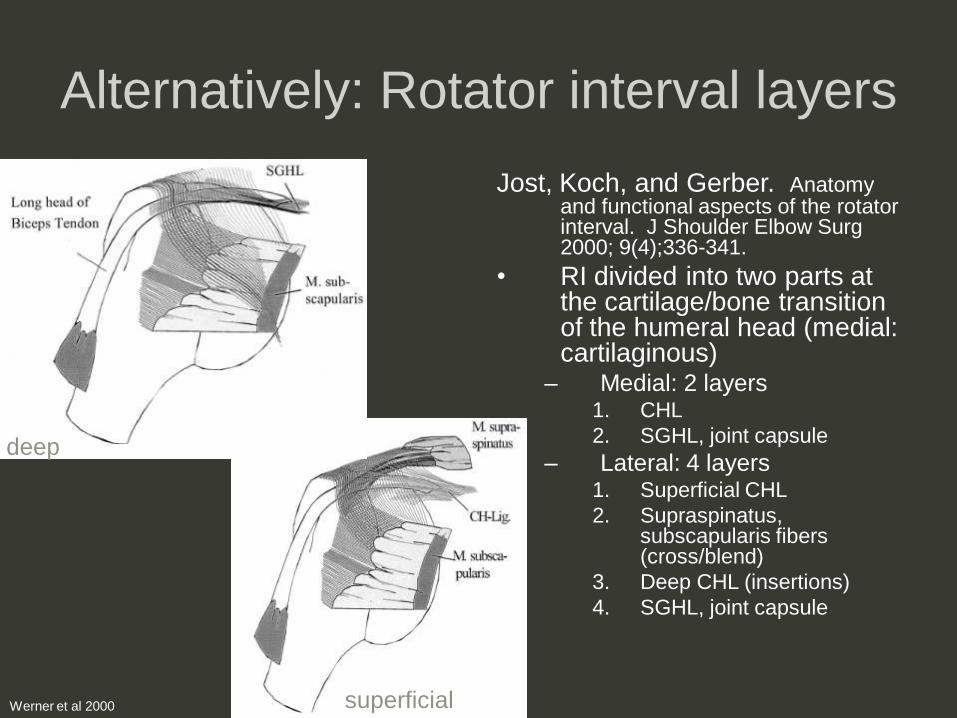
deep
superficialWerner et al 2000
Overview
• Normal anatomy– Borders
– Contents
• Biomechanics– Anatomic (cadaveric)
– Clinical
• Pathology
– Rotator cuff tears
– Biceps sling
– CHL, SGHL, long head biceps tendon
– Capsular inflammation (adhesive capsulitis)
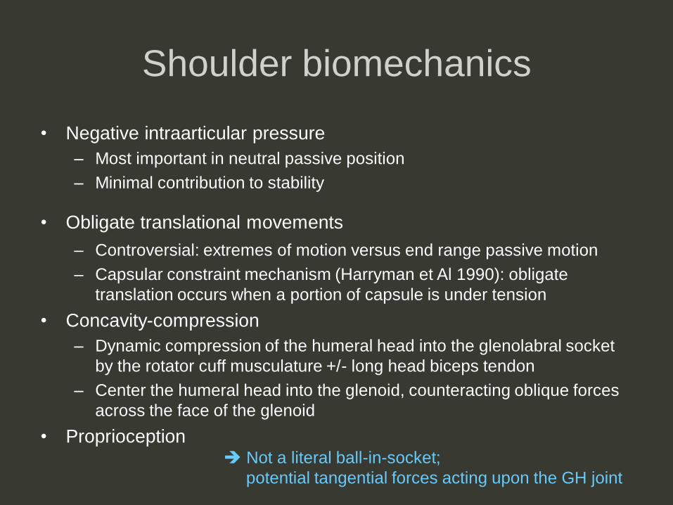
Shoulder biomechanics
• Negative intraarticular pressure
– Most important in neutral passive position
– Minimal contribution to stability
• Obligate translational movements
– Controversial: extremes of motion versus end range passive motion
– Capsular constraint mechanism (Harryman et Al 1990): obligate
translation occurs when a portion of capsule is under tension
• Concavity-compression
– Dynamic compression of the humeral head into the glenolabral socket
by the rotator cuff musculature +/- long head biceps tendon
– Center the humeral head into the glenoid, counteracting oblique forces
across the face of the glenoid
• Proprioception Not a literal ball-in-socket;
potential tangential forces acting upon the GH joint
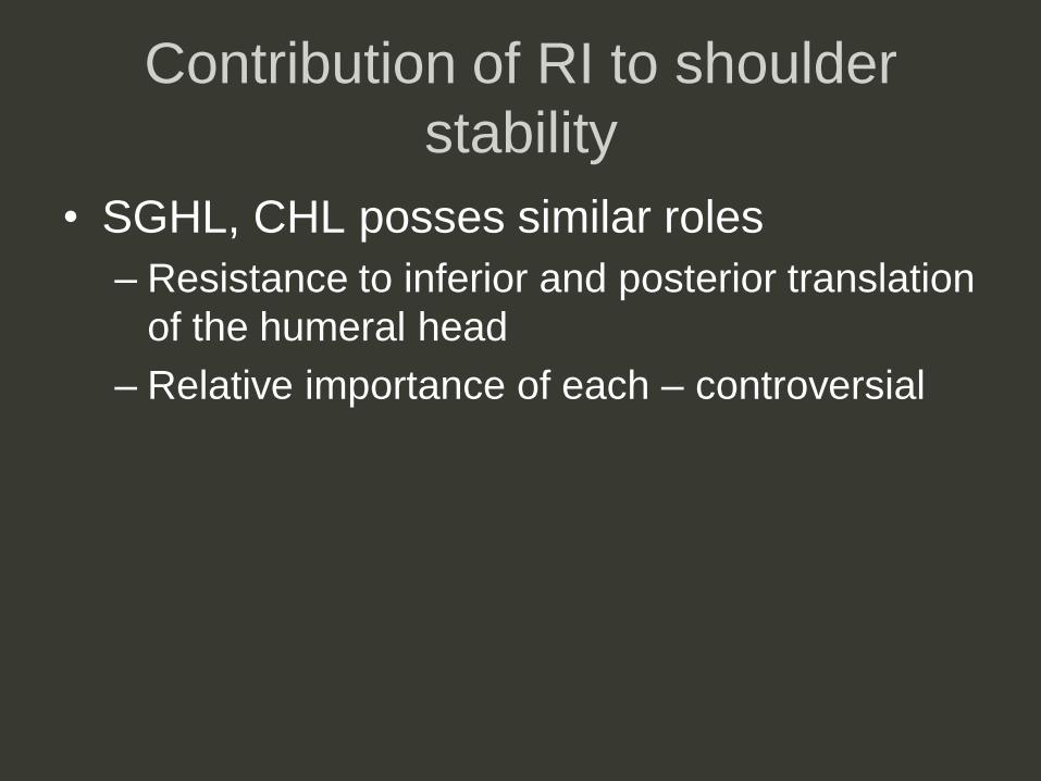
Contribution of RI to shoulder
stability
• SGHL, CHL posses similar roles
– Resistance to inferior and posterior translation
of the humeral head
– Relative importance of each – controversial
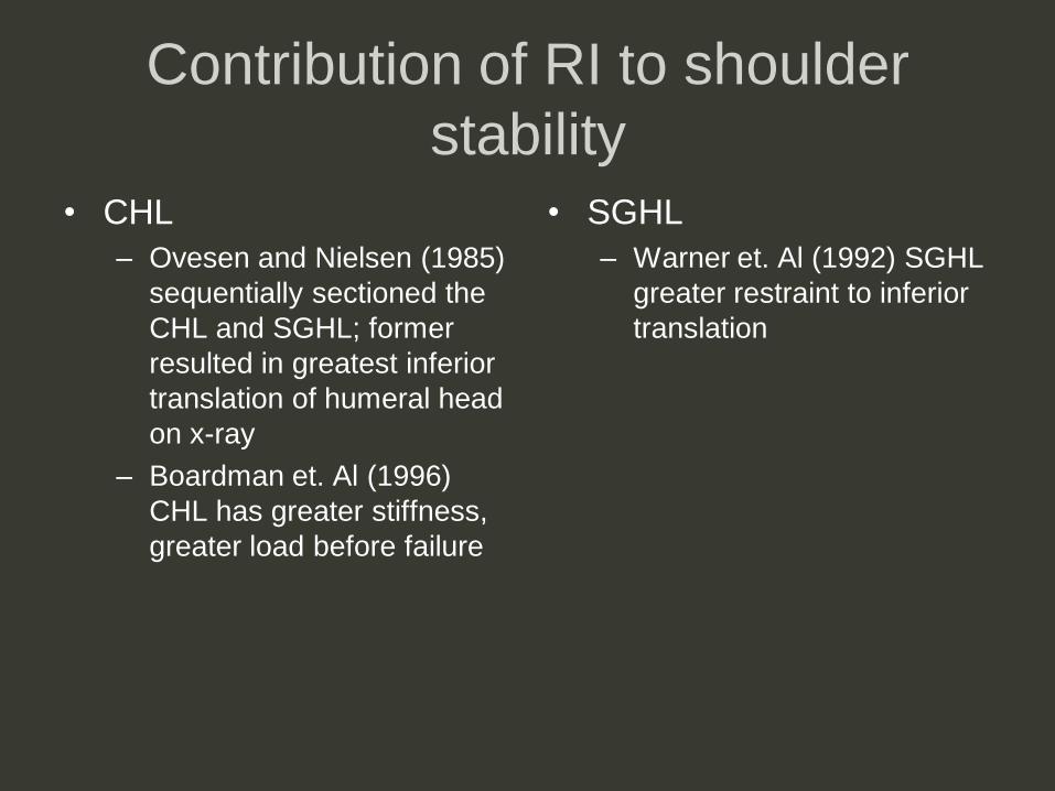
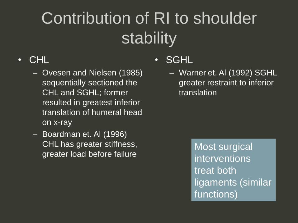
Contribution of RI to shoulder
stability
• CHL
– Ovesen and Nielsen (1985)
sequentially sectioned the
CHL and SGHL; former
resulted in greatest inferior
translation of humeral head
on x-ray
– Boardman et. Al (1996)
CHL has greater stiffness,
greater load before failure
• SGHL
– Warner et. Al (1992) SGHL
greater restraint to inferior
translation
Most surgical
interventions
treat both
ligaments (similar
functions)
Contribution of RI to shoulder
stability
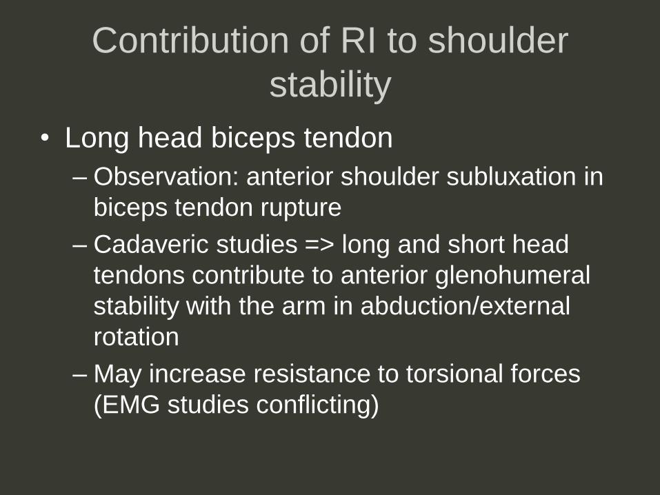
• Long head biceps tendon
– Observation: anterior shoulder subluxation in
biceps tendon rupture
– Cadaveric studies => long and short head
tendons contribute to anterior glenohumeral
stability with the arm in abduction/external
rotation
– May increase resistance to torsional forces
(EMG studies conflicting)

Harryman et al. (1992)
• Perhaps the first comprehensive cadaveric study to evaluate RI function
• Sectioning the RI capsule (CHL/SGHL) increased the ranges of flexion, extension, adduction, external rotation– Humeral head tended to translate posteroinferior wrt
glenoid after sectioning
• Imbrication decreased these ranges of motion
• (Abduction, internal rotation relaxed the RI capsule; sectioning/imbrication did not alter)
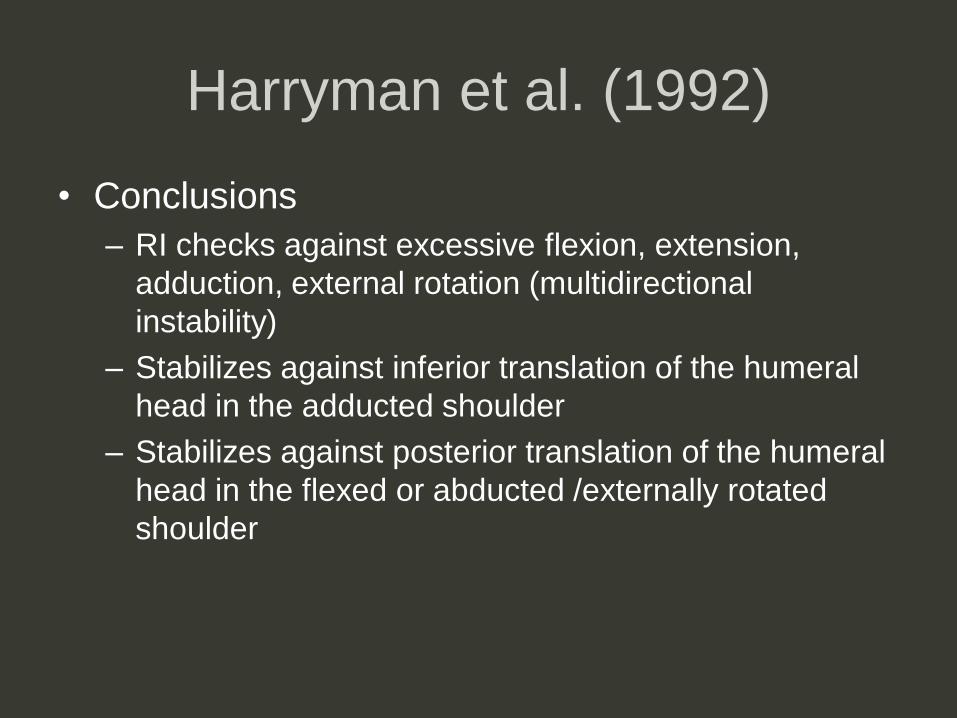
Harryman et al. (1992)
• Conclusions
– RI checks against excessive flexion, extension,
adduction, external rotation (multidirectional
instability)
– Stabilizes against inferior translation of the humeral
head in the adducted shoulder
– Stabilizes against posterior translation of the humeral
head in the flexed or abducted /externally rotated
shoulder
Harryman et al. (1992)
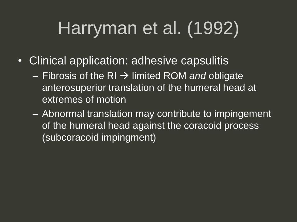
• Clinical application: adhesive capsulitis
– Fibrosis of the RI limited ROM and obligate
anterosuperior translation of the humeral head at
extremes of motion
– Abnormal translation may contribute to impingement
of the humeral head against the coracoid process
(subcoracoid impingment)
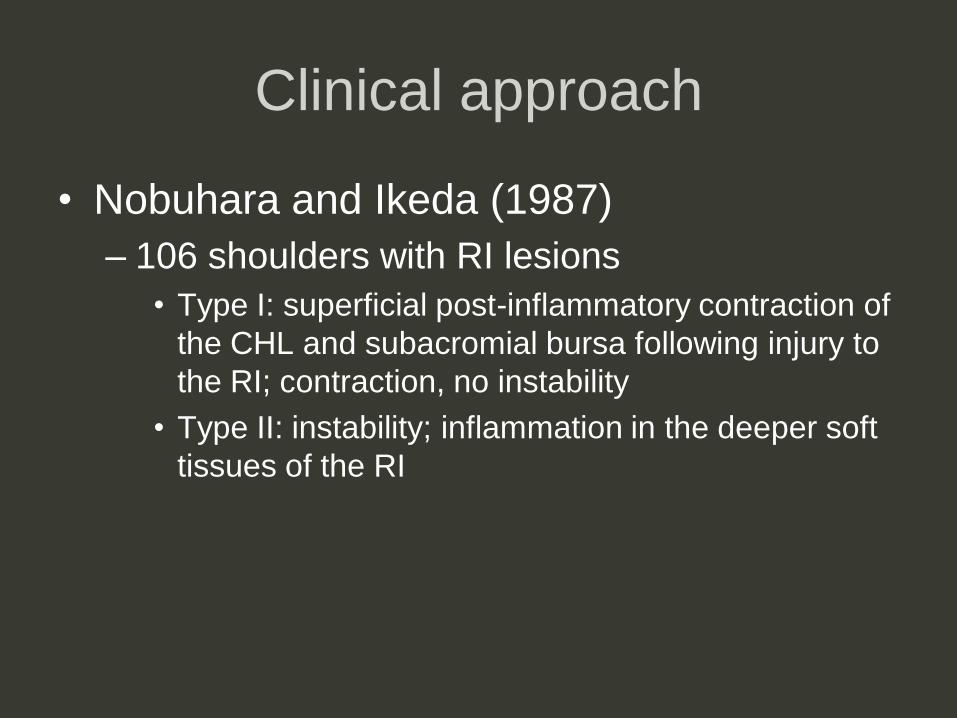
Clinical approach
• Nobuhara and Ikeda (1987)
– 106 shoulders with RI lesions
• Type I: superficial post-inflammatory contraction of
the CHL and subacromial bursa following injury to
the RI; contraction, no instability
• Type II: instability; inflammation in the deeper soft
tissues of the RI
Clinical approach
• Nobuhara and Ikeda (1987)
– 106 shoulders with RI lesions
• Type I
– Restriction of passive external rotation or forward flexion
of the shoulder
– Adhesive capsulitis; postoperative tightness
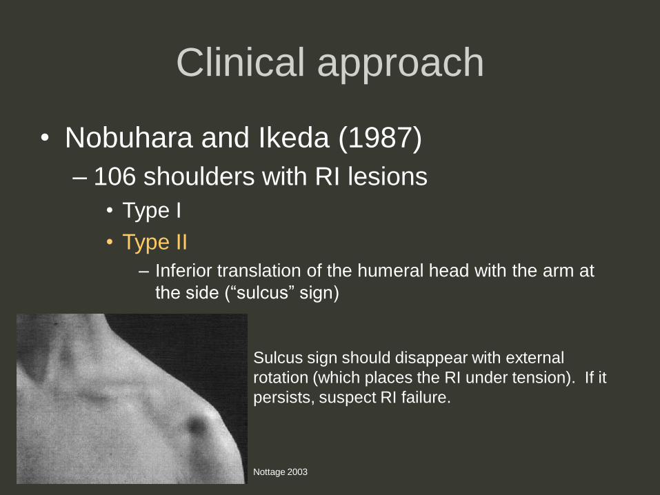
Clinical approach
• Nobuhara and Ikeda (1987)
– 106 shoulders with RI lesions
• Type I
• Type II
– Inferior translation of the humeral head with the arm at
the side (“sulcus” sign)
Sulcus sign should disappear with external
rotation (which places the RI under tension). If it
persists, suspect RI failure.
Nottage 2003
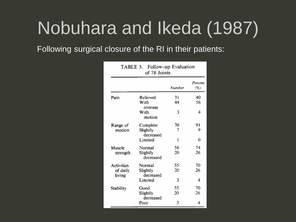
Nobuhara and Ikeda (1987)Following surgical closure of the RI in their patients:
Summary of clinical findings
• Rotator interval too tight (fibrosis)
– Alterations in glenohumeral obligate translation
– Superior cuff complaints, pain (internal impingement)
• Rotator interval too loose (defect)
– Posteroinferior glenohumeral instability, pain
Overview
• Normal anatomy– Borders
– Contents
• Biomechanics– Anatomic (cadaveric)
– Clinical
• Pathology– Rotator cuff tears
– Biceps sling
– CHL, SGHL, long head biceps tendon
– Capsular inflammation (adhesive capsulitis)

RI pathology
• Includes:
– Extension of rotator cuff tear
• Anterior supraspinatus tendon
• Superior subscapularis tendon
– Long head of the biceps tendon, intraarticular
– Coracohumeral ligament
– Superior glenohumeral ligament
– RI capsule
RI and rotator cuff tear
• Anterior extension of a supraspinatus
tendon tear can involve the rotator interval
– If involves the coracohumeral ligament, can
also result in biceps tendon subluxation
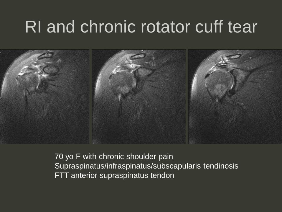
RI and chronic rotator cuff tear
70 yo F with chronic shoulder pain
Supraspinatus/infraspinatus/subscapularis tendinosis
FTT anterior supraspinatus tendon
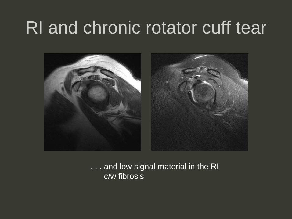
RI and chronic rotator cuff tear
. . . and low signal material in the RI
c/w fibrosis
RI and rotator cuff tear
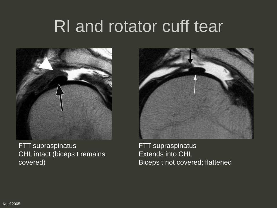
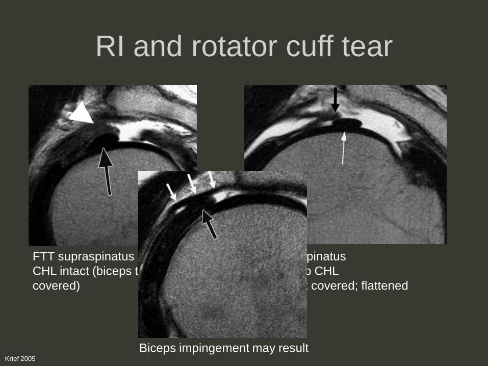
FTT supraspinatus
CHL intact (biceps t remains
covered)
FTT supraspinatus
Extends into CHL
Biceps t not covered; flattened
Krief 2005
RI and rotator cuff tear
FTT supraspinatus
CHL intact (biceps t remains
covered)
FTT supraspinatus
Extends into CHL
Biceps t not covered; flattened
Krief 2005
Biceps impingement may result
Biceps pulley lesions
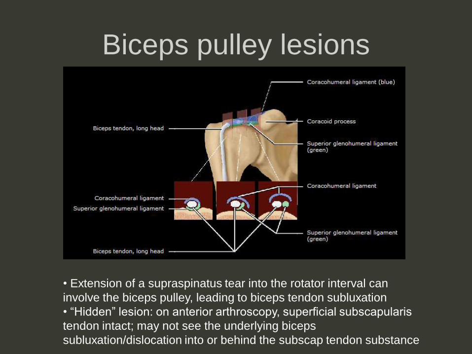
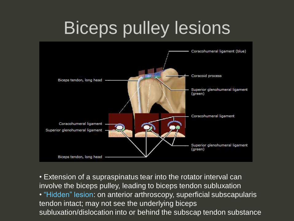
• Extension of a supraspinatus tear into the rotator interval can
involve the biceps pulley, leading to biceps tendon subluxation
• “Hidden” lesion: on anterior arthroscopy, superficial subscapularis
tendon intact; may not see the underlying biceps
subluxation/dislocation into or behind the subscap tendon substance
Biceps pulley lesions
• Extension of a supraspinatus tear into the rotator interval can
involve the biceps pulley, leading to biceps tendon subluxation
• “Hidden” lesion: on anterior arthroscopy, superficial subscapularis
tendon intact; may not see the underlying biceps
subluxation/dislocation into or behind the subscap tendon substance
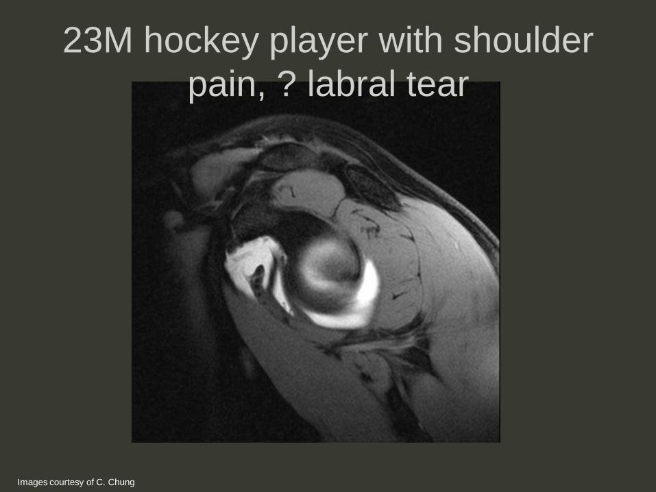
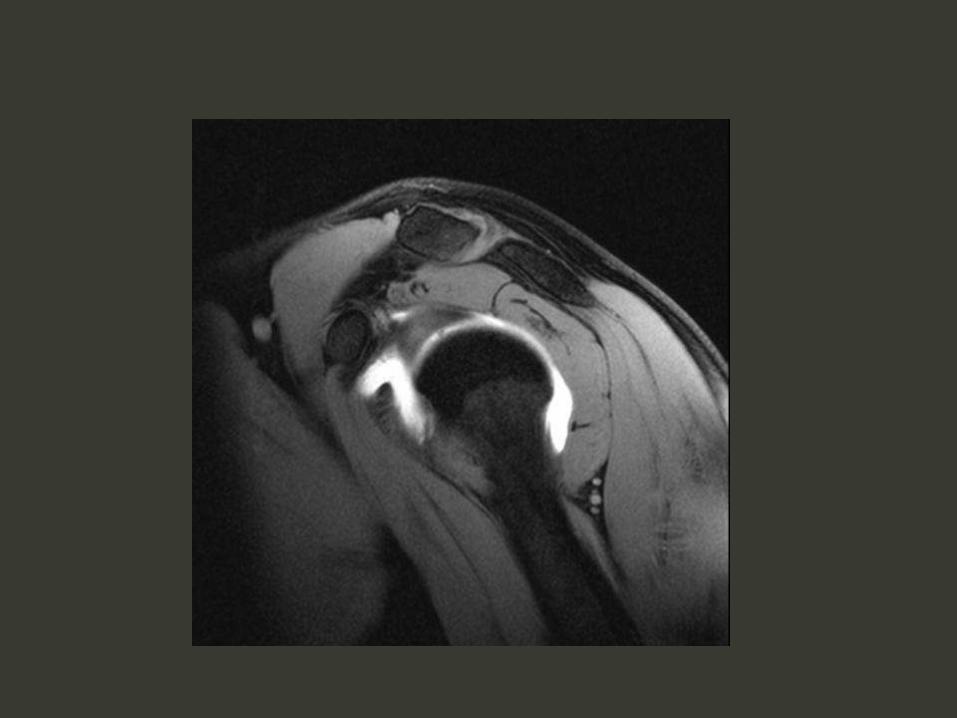
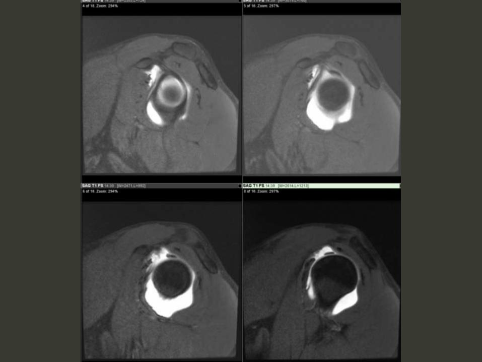
23M hockey player with shoulder
pain, ? labral tear
Images courtesy of C. Chung
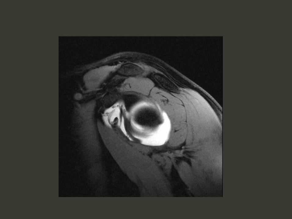
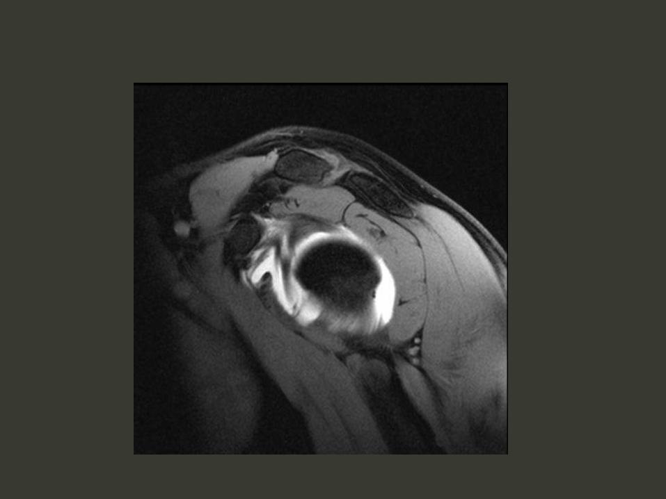


Biceps pulley lesion
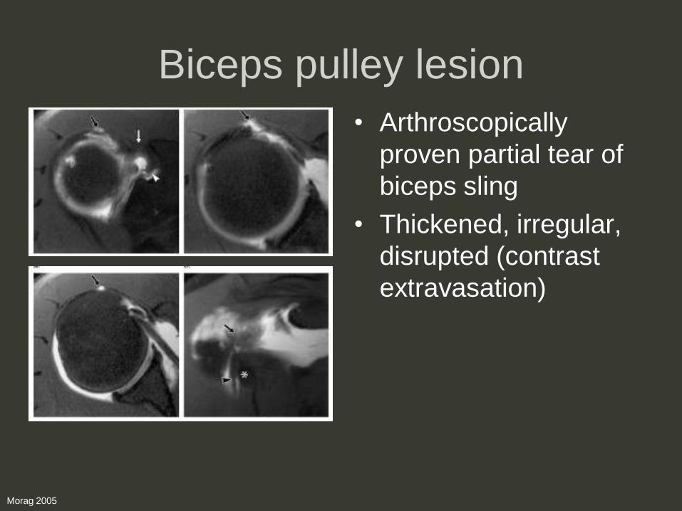
Biceps pulley lesion
• Arthroscopically
proven partial tear of
biceps sling
• Thickened, irregular,
disrupted (contrast
extravasation)
Morag 2005
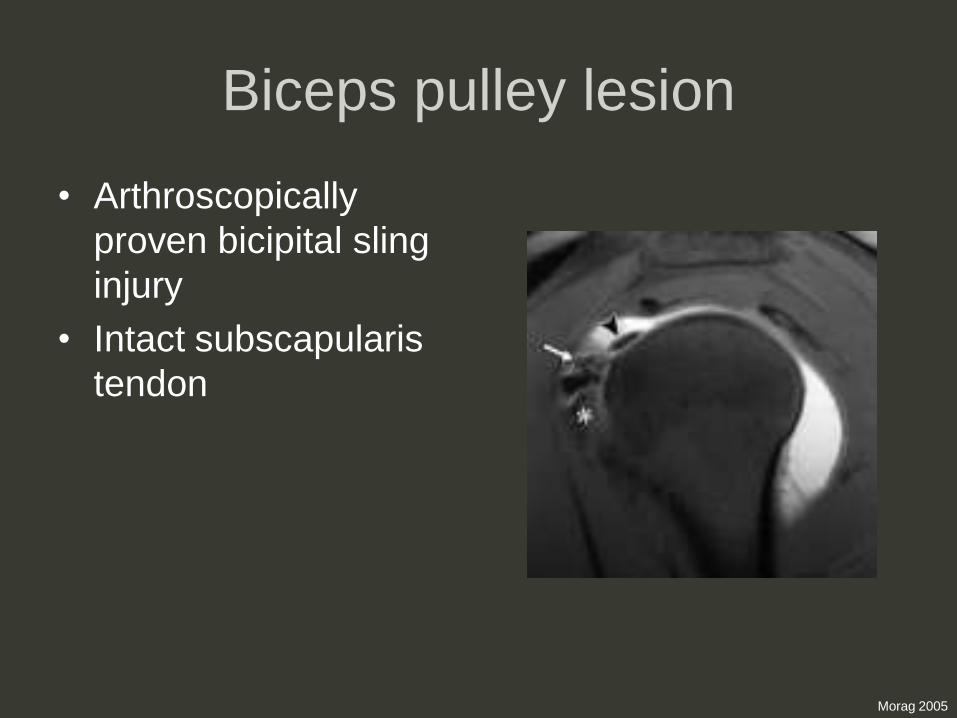
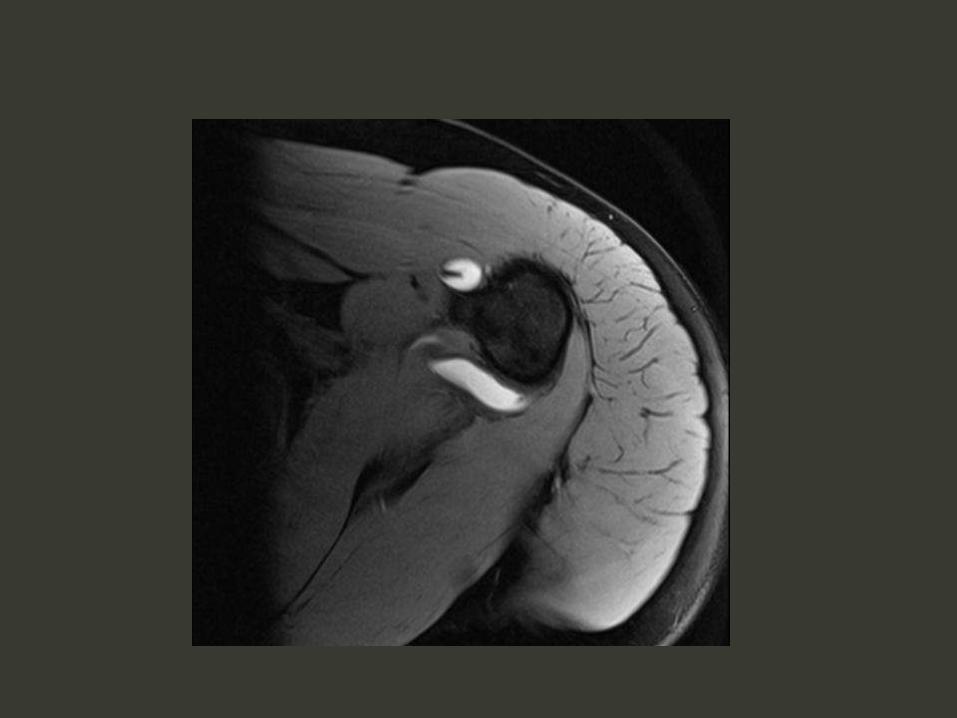
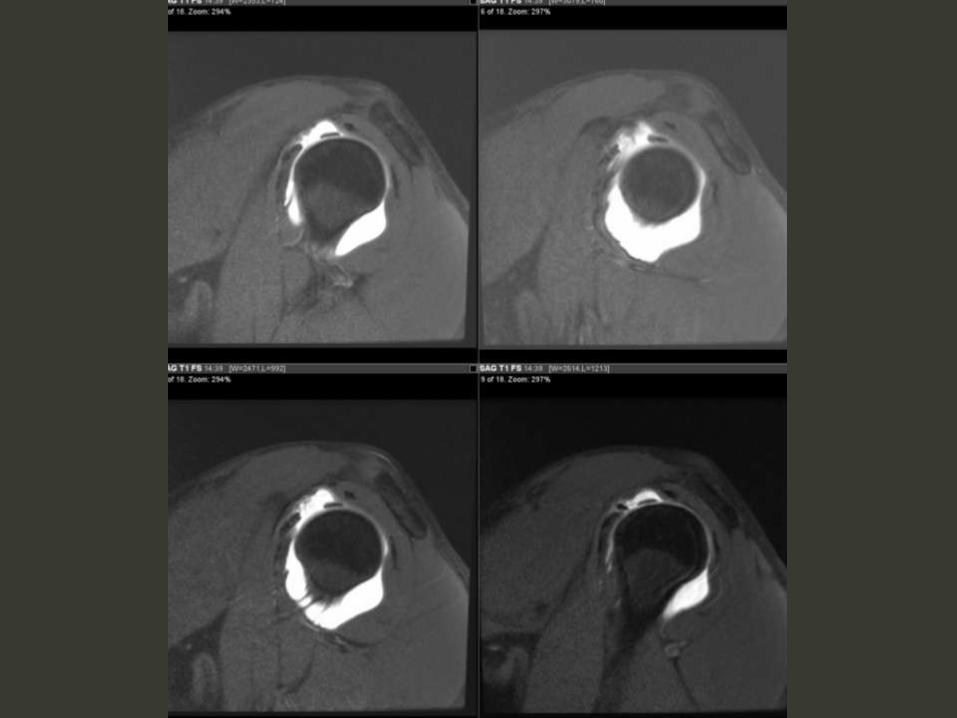
Biceps pulley lesion
• Arthroscopically
proven bicipital sling
injury
• Intact subscapularis
tendon
Morag 2005

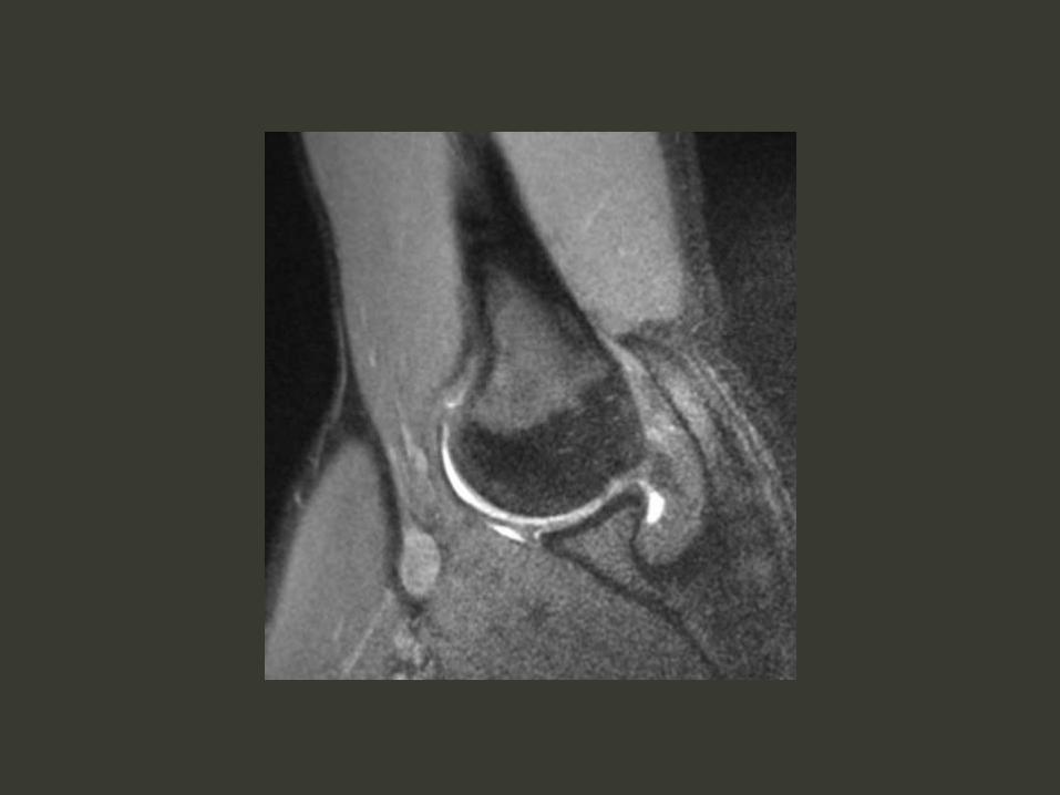
RI lesion and SLAP tear
Beltran and Kim 2003
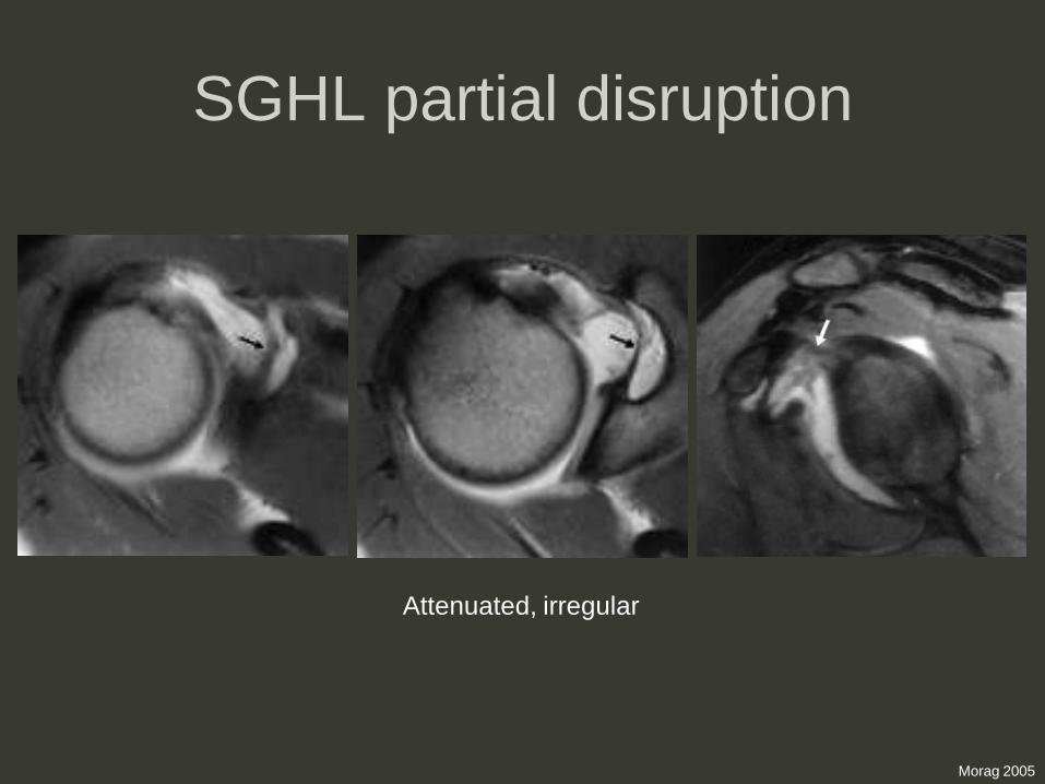
SGHL partial disruption
Morag 2005
Attenuated, irregular
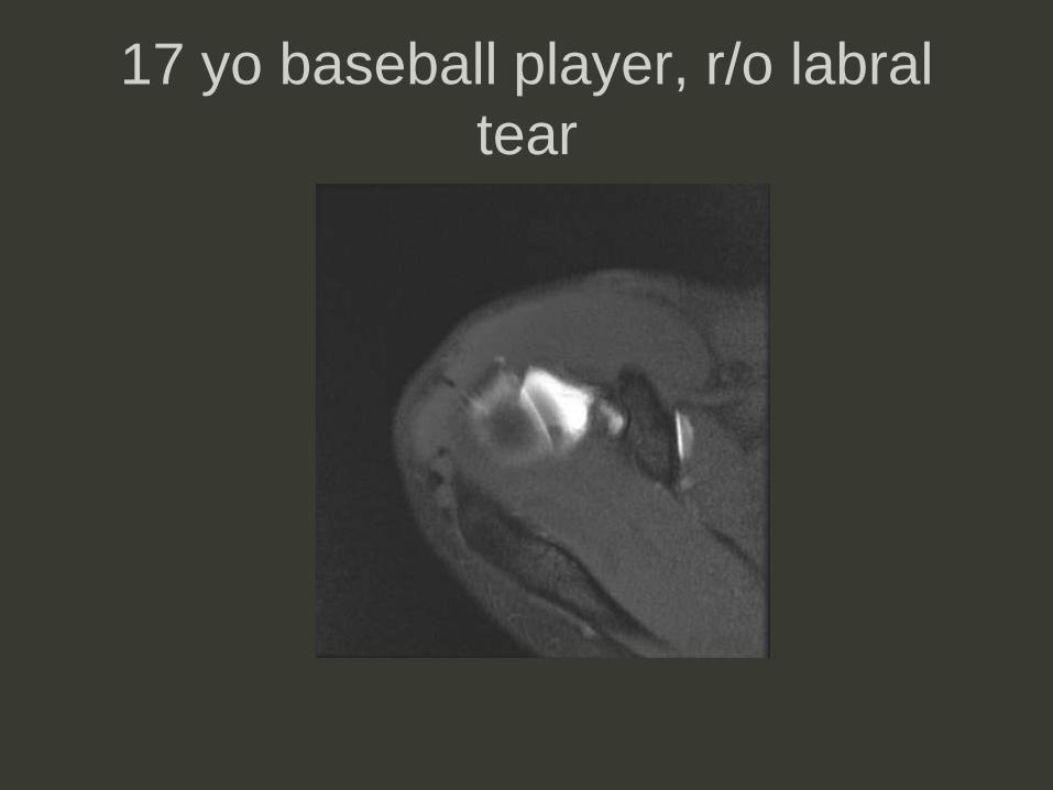
17 yo baseball player, r/o labral
tear
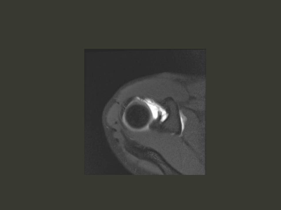
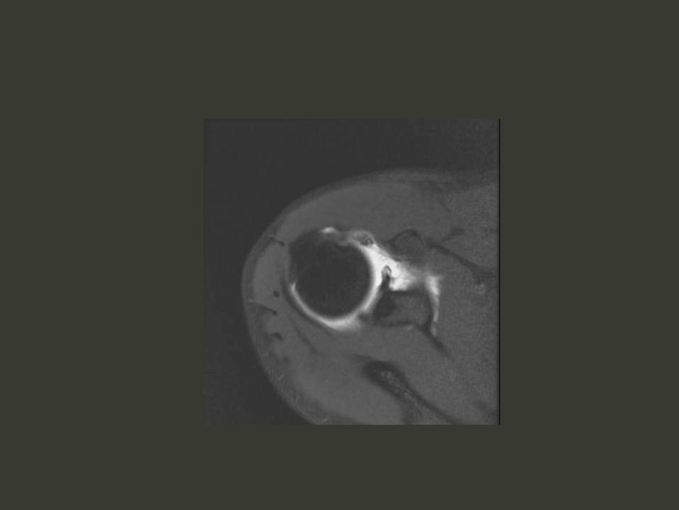
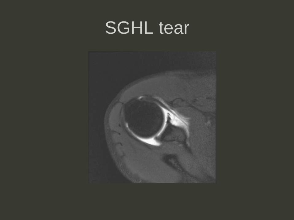
SGHL tear
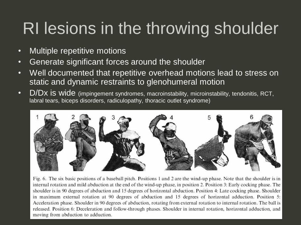
RI lesions in the throwing shoulder
• Multiple repetitive motions
• Generate significant forces around the shoulder
• Well documented that repetitive overhead motions lead to stress on static and dynamic restraints to glenohumeral motion
• D/Dx is wide (impingement syndromes, macroinstability, microinstability, tendonitis, RCT,
labral tears, biceps disorders, radiculopathy, thoracic outlet syndrome)
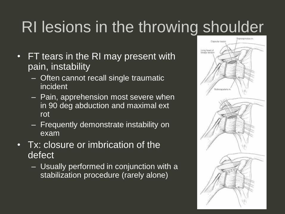
RI lesions in the throwing shoulder
• FT tears in the RI may present with pain, instability– Often cannot recall single traumatic
incident
– Pain, apprehension most severe when in 90 deg abduction and maximal ext rot
– Frequently demonstrate instability on exam
• Tx: closure or imbrication of the defect– Usually performed in conjunction with a
stabilization procedure (rarely alone)
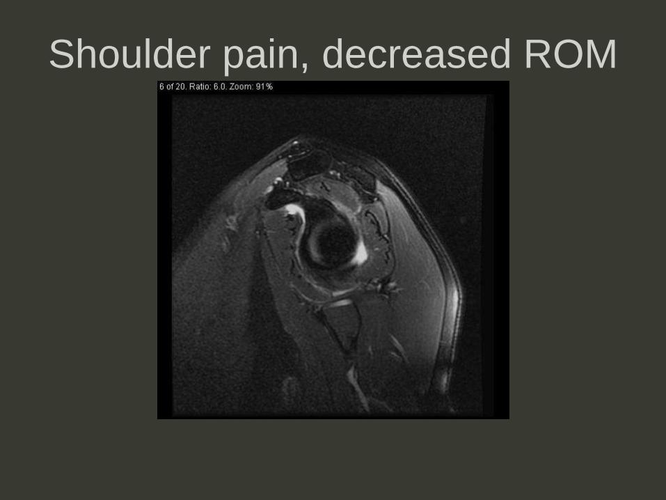
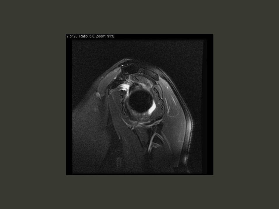
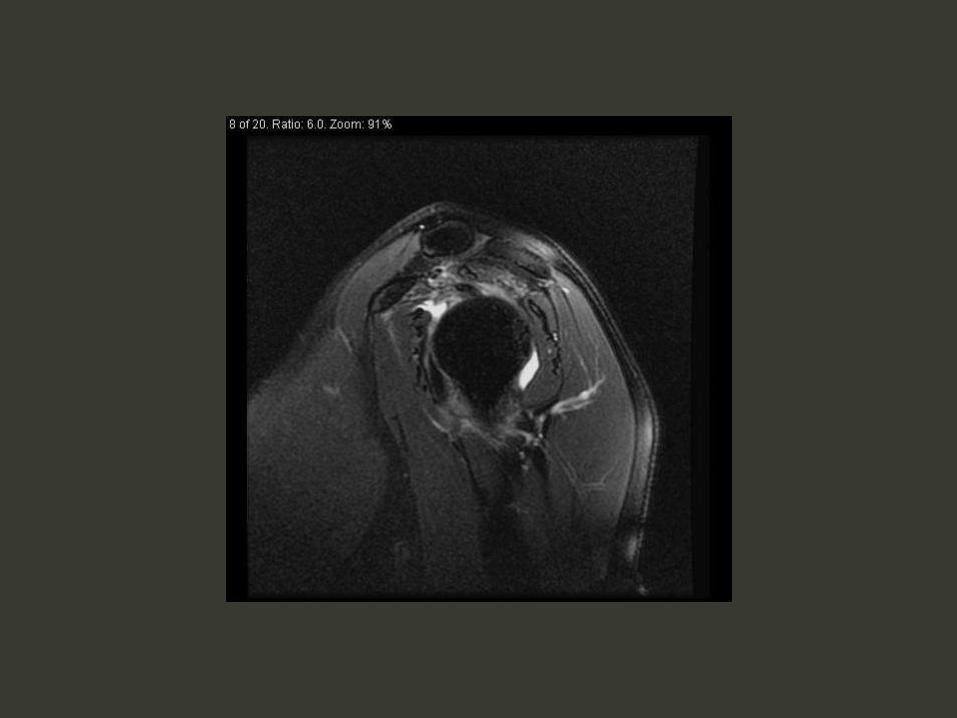
Shoulder pain, decreased ROM
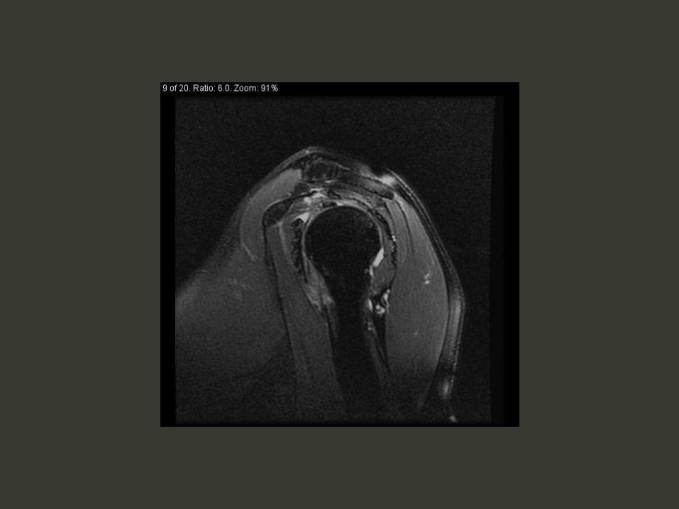

Adhesive capsulitis
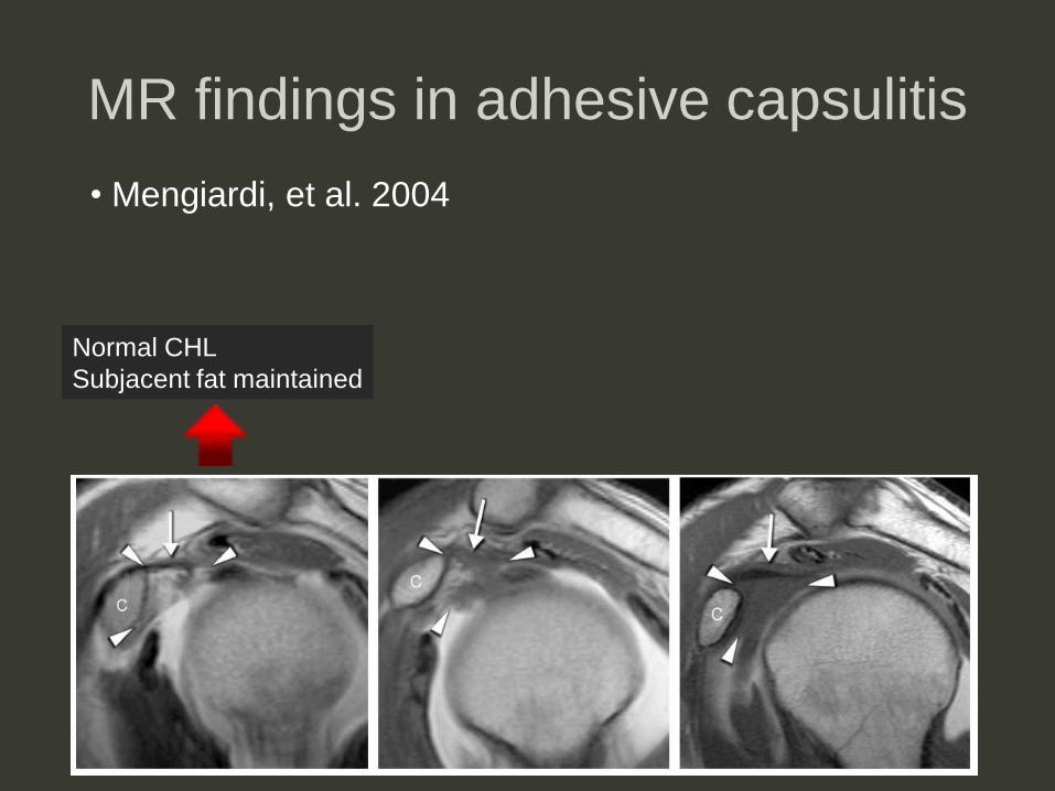
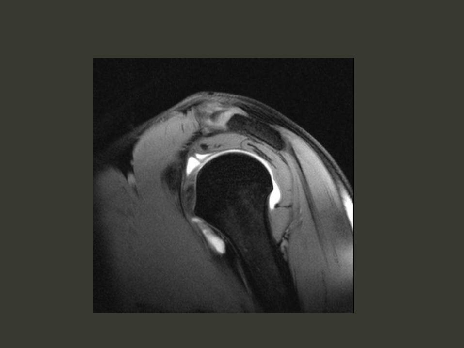
MR findings in adhesive capsulitis
Normal CHL
Subjacent fat maintained
• Mengiardi, et al. 2004
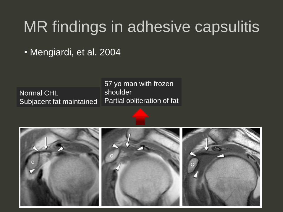
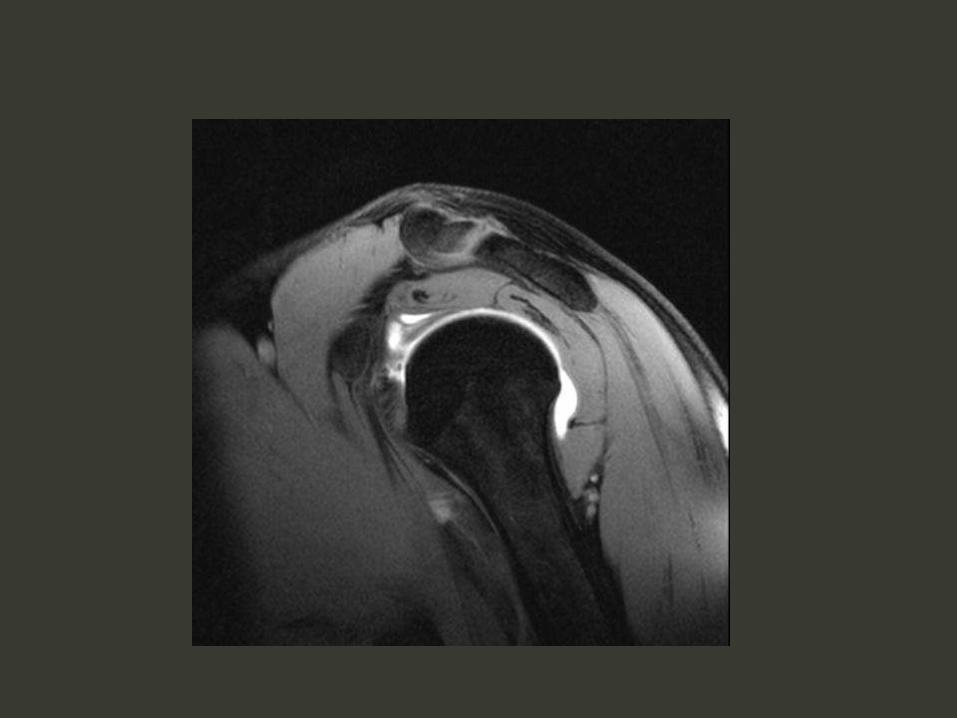
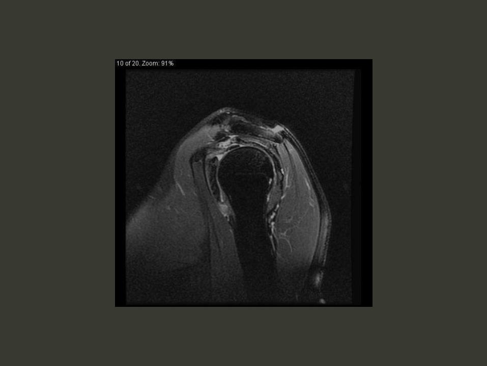
MR findings in adhesive capsulitis
Normal CHL
Subjacent fat maintained
57 yo man with frozen
shoulder
Partial obliteration of fat
• Mengiardi, et al. 2004

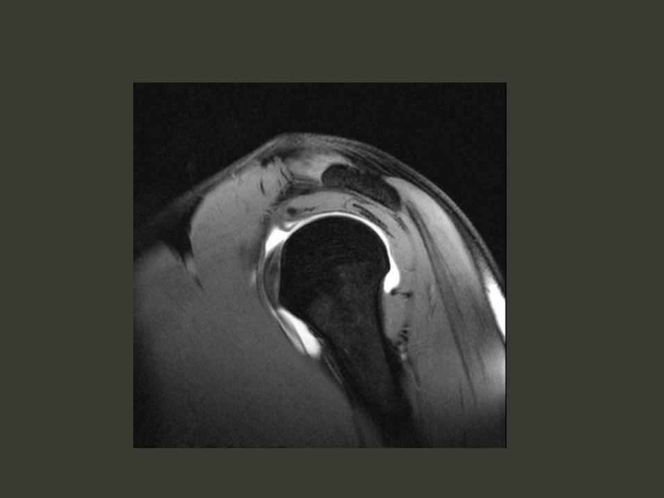
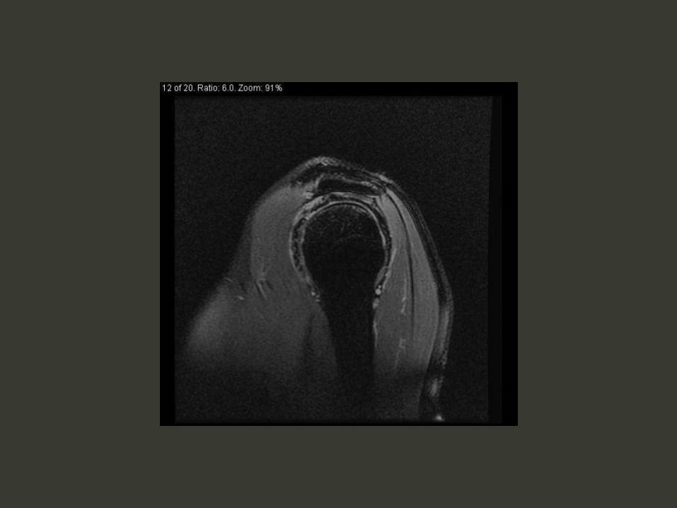
MR findings in adhesive capsulitis
Normal CHL
Subjacent fat maintained
57 yo man with frozen
shoulder
Partial obliteration of fat
55 yo pt with frozen
shoulder
Complete obliteration
of fat (subcoracoid
triangle sign)
• Mengiardi, et al. 2004
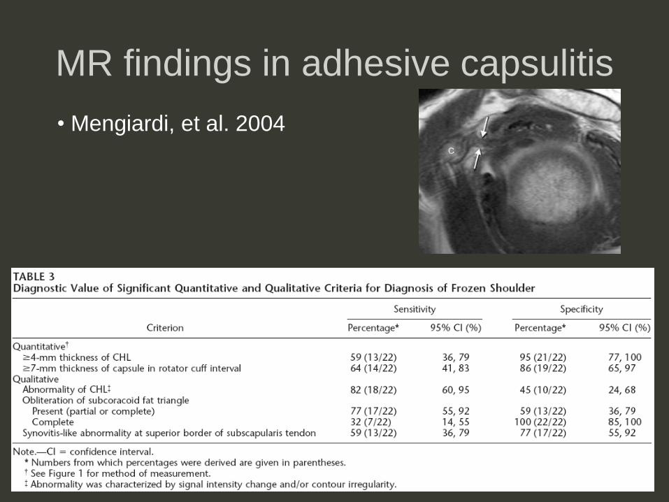
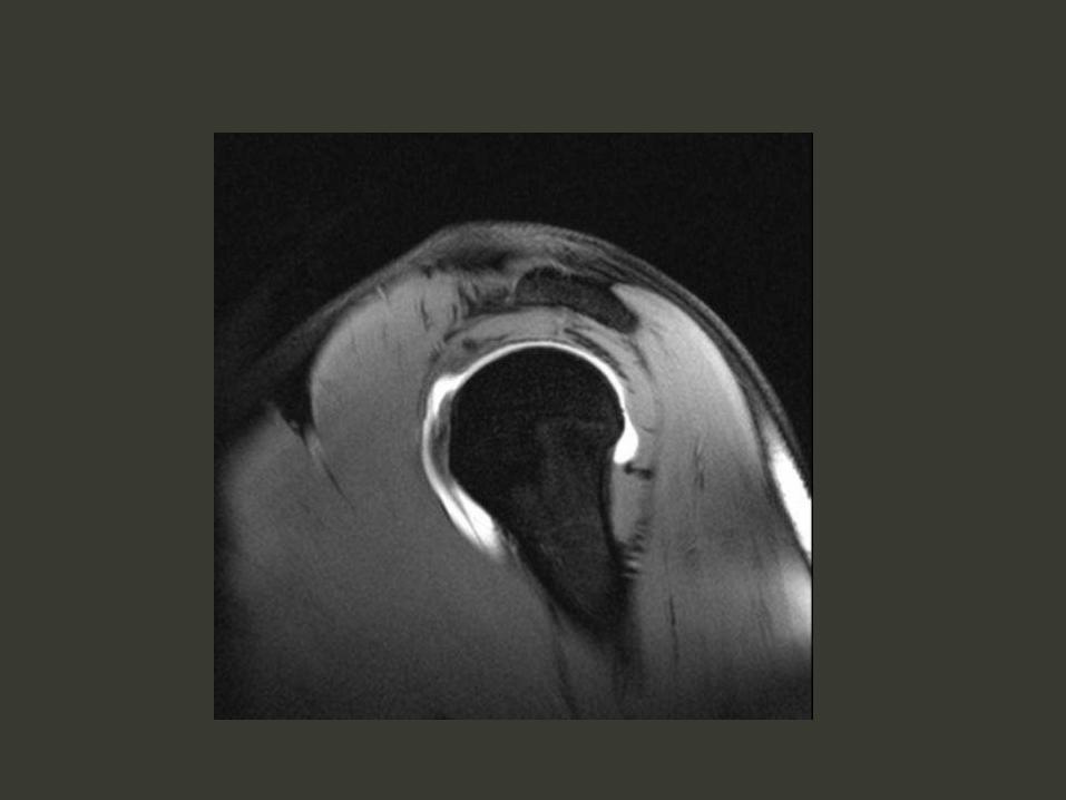
MR findings in adhesive capsulitis
• Mengiardi, et al. 2004
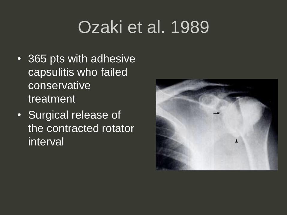
Ozaki et al. 1989
• 365 pts with adhesive
capsulitis who failed
conservative
treatment
• Surgical release of
the contracted rotator
interval

Ozaki et al. 1989
• 365 pts with adhesive
capsulitis who failed
conservative
treatment
• Surgical release of
the contracted rotator
interval
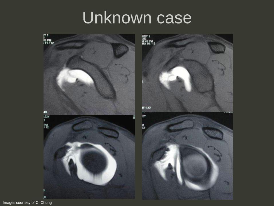
Unknown case
Images courtesy of C. Chung
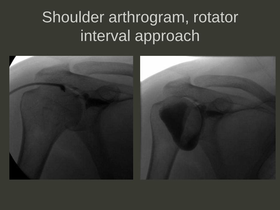
Shoulder arthrogram, rotator
interval approach
Iatrogenic RI “lesion”
• Concept is also of significance with
arthroscopy
– RI is regularly used as the anterior portal in
shoulder arthroscopy
– But capsulorraphy without RI closure in a pt
with RI defect can result in recurrent
postoperative symptoms
Summary
• Normal anatomy controversial
• Biomechanic significance controversial
• Pain, instability can result from RI pathology
• RI lesions often in association with other shoulder pathologies (eg RCT, SLAP)
• “Hidden” lesions can potentially be seen with MR
• Missed RI lesion can have clinical repercussions (inadequate surgical repair recurrent pain/instability)

References• Special thanks to Christine Chung for contributing images!
• Beltran, J, and DHM Kim. MR imaging of shoulder instability injuries in the athlete. Magn Res Imaging Clin N Am 2003; 11:221-238.
• Bigoni, BJ, and CB Chung. MR imaging of the rotator cuff interval. Magnetic Resonance Imaging Clinics of North America 2004; 12:61-73.
• Chung, CB, JR Dwek, GJ Cho, N Lektrakul, D Trudell, and D Resnick. Rotator cuff interval: Evaluation with MR imaging and MR arthrography of the shoulder in 32 cadavers. Journal of Computer Assisted Tomography 2000; 24(5):738-743.
• Doukas, WC and KP Speer. Anatomy, pathophysiology, and biomechanics of shoulder instability. Operative Techniques in SportsMedicine 2000; 8(3):179-187.
• Dumontier, C, A Sautet, O Gagey, and A Apoil. Rotator interval lesions and their relation to coracoid impingement syndrome. J Shoulder Elbow Surg 1999; 8(2):130-135.
• Fitzpatrick, MJ, SE Powell, JE Tibone, and FR Warren. The anatomy, pathology, and definitive treatment of rotator interval lesions: Current concepts. Arthroscopy 2003; 19(10):70-79.
• Gleason, PD, et. Al. The transverse humeral ligament: A separate anatomical structure or a continuation of the osseous attachment of the rotator cuff? Am J Sports Med 2006; 34:72-77.
• Jost, B, PP Koch, and C Gerber. Anatomy and functional aspects of the rotator interval. Journal of Shoulder and Elbow Surgery 2000; 9:336-341.
• Jost, B, and C Gerber. What the shoulder surgeon would like to know from MR imaging. Magn Res Imaging Clin N Am 2004; 12:161-168.
• Harryman, DT, JA Sidles, SL Harris, and FA Matsen. The role of the rotator interval capsule in passive motion and stability of the shoulder.
• Hunt, SA, YW Kwon, and JD Zukerman. The rotator interval: Anatomy, pathology, and strategies for treatment. J Acad Orthop Surg2007; 15:218-227.
• Krief, OP. MRI of the rotator interval capsule. AJR 2005; 184:1490-1494.
• Mengiardi, B, CWA Pfirrmann, G. Gerber, J Hodler, and M Zanetti. Frozen shoulder: MR arthrographic findings. Radiology 2004;233:486-492.
• Morag, Y, et. Al. MR arthrography of roatator interval, long head of the biceps brachii, and biceps pulley of the shoulder. Radiology 2005; 235:21-30.
• Nottage, WM. Rotator interval lesions: Physical exam, imaging, arthroscopic findings, and repair. Techniques in Shoulder & Elbow Surgery 2003; 4(4):175-184.
• Nobuhara, K, and H. Ikeda. Rotator interval lesion. Clinical Orthopaedics and Related Research. 1987; 223: 44-50.
• Ozaki, J, et. Al. Recalcitrant chronic adhesive capsulitis of the shoulder. JBJS 1989; 71-A(10):1511-1515.
• Paulson, MM, NF Watnik, and DM Dines. Coracoid impingement syndrome, rotator interval reconstruction, and biceps tenodesis in the overhead athlete. Orthop Clin N Amer 2001; 32(3).
• Werner, A, R Mueller, D Boehm, and F Gohlke. The stabilizing sling for the long head of the biceps tendon in the rotator cuff interval: A histoanatomic study. AJS 2000; 28(1): 28-31.