anatomy and physiology of farm animals 7 th ed by frandson
TRANSCRIPT

Anatomy and Physiology of Farm Animals
Rowen D. FrandsonW. Lee Wilke
Anna Dee Fails
SEVENTH EDITION
A N A T O M Y A N D P H Y S I O L O G Y O F
Farm AnimalsS E V E N T H E D I T I O N

A N A T O M Y A N D P H Y S I O L O G Y O F
Farm AnimalsS E V E N T H E D I T I O N

Rowen D. Frandson, BS, DVM, MSProfessor Emeritus, Department of Anatomy and Neurobiology
College of Veterinary Medicine and Biomedical Sciences
Colorado State University
Fort Collins, Colorado
W. Lee Wilke, DVM, PhDAssociate Professor Emeritus, Department of Biomedical Sciences
College of Veterinary Medicine and Biomedical Sciences
Colorado State University
Fort Collins, Colorado
Anna Dee Fails, DVM, PhDAssistant Professor, Department of Biomedical Services
College of Veterinary Medicine and Biomedical Sciences
Colorado State University
Fort Collins, Colorado
A John Wiley & Sons, Inc., Publication

Seventh Edition fi rst published 2009© 2009 Wiley-Blackwell
Blackwell Publishing was acquired by John Wiley & Sons in February 2007. Blackwell’s publishing program has been merged with Wiley’s global Scientifi c, Technical, and Medical business to form Wiley-Blackwell.
Editorial Offi ce2121 State Avenue, Ames, Iowa 50014-8300, USA
For details of our global editorial offi ces, for customer services, and for information about how to apply for permission to reuse the copyright material in this book, please see our website at www.wiley.com/wiley-blackwell.
Authorization to photocopy items for internal or personal use, or the internal or personal use of specifi c clients, is granted by Blackwell Publishing, provided that the base fee is paid directly to the Copyright Clearance Center, 222 Rosewood Drive, Danvers, MA 01923. For those organizations that have been granted a photocopy license by CCC, a separate system of payments has been arranged. The fee codes for users of the Transactional Reporting Service are ISBN-13: 978-0-8138-1394-3/2009.
Designations used by companies to distinguish their products are often claimed as trademarks. All brand names and product names used in this book are trade names, service marks, trademarks or registered trademarks of their respective owners. The publisher is not associated with any product or vendor mentioned in this book. This publication is designed to provide accurate and authoritative information in regard to the subject matter covered. It is sold on the understanding that the publisher is not engaged in rendering professional services. If professional advice or other expert assistance is required, the services of a competent professional should be sought.
First Edition, 1965, Lippincott Williams & WilkinsSecond Edition, 1974, Lippincott Williams & WilkinsThird Edition, 1981, Lippincott Williams & WilkinsFourth Edition, 1986, Lippincott Williams & WilkinsFifth Edition, 1992, Lippincott Williams & WilkinsSixth Edition, 2003, Lippincott Williams & Wilkins
Library of Congress Cataloging-in-Publication Data
Wilke, W. Lee. Anatomy and physiology of farm animals / W. Lee Wilke, Anna Dee Fails, R.D. Frandson. – 7th ed. p. ; cm. R.D. Frandson’s name appears fi rst on the 6th ed. Includes bibliographical references and index. ISBN-13: 978-0-8138-1394-3 (alk. paper) ISBN-10: 0-8138-1394-8 (alk. paper) 1. Veterinary anatomy. 2. Veterinary physiology. I. Fails, Anna Dee. II. Frandson, R. D. III. Title. [DNLM: 1. Anatomy, Veterinary. 2. Animals, Domestic–physiology. SF 761 W681a 2009] SF761.F8 2009 636.089′2–dc22 2008051665
A catalog record for this book is available from the U.S. Library of Congress.
Set in 10.5/12pt Berkeley by SNP Best-set Typesetter Ltd., Hong KongPrinted and bound in Singapore
DisclaimerThe publisher and the author make no representations or warranties with respect to the accuracy or completeness of the contents of this work and specifi cally disclaim all warranties, including without limitation warranties of fi tness for a particular purpose. No warranty may be created or extended by sales or promotional materials. The advice and strategies contained herein may not be suitable for every situation. This work is sold with the understanding that the publisher is not engaged in rendering legal, accounting, or other professional services. If professional assistance is required, the services of a competent professional person should be sought. Neither the publisher nor the author shall be liable for damages arising herefrom. The fact that an organization or Website is referred to in this work as a citation and/or a potential source of further information does not mean that the author or the publisher endorses the information the organization or Website may provide or recommendations it may make. Further, readers should be aware that Internet Websites listed in this work may have changed or disappeared between when this work was written and when it is read.
1 2009
To Zelda, Eric, Stephanie and Mark —R.D.F.
To M.W. Poore —A.D.F.
To Aline and Willard —W. L.W.

TABLE OF CONTENTS
Chapter 3
Embryology 51Development of Germ Layers 52Neurulation 53Differentiation of Other Tissues 54
Chapter 4
The Skeletal System 59Functions of Bones 59Terminology 62Classifi cation of Bones According to Gross
Appearance 64Axial Skeleton 64Appendicular Skeleton 71
Chapter 5
Microscopic Anatomy and Growth and Development of Bone 77Microscopic Anatomy and Formation of
Bone 77Ossifi cation 80Physiology of Bone 82Fractures and Fracture Healing 83Other Pathologic Conditions 85
Chapter 6
Joints 87Classifi cation of Joints 87
Preface xiAcknowledgments xiii
Chapter 1
Introduction to Anatomy and Physiology 3Descriptive Terms Useful in the Study of
Anatomy 5Microscopic Anatomy: Animal Cells and
Tissues 7The General Plan of the Animal Body 14
Chapter 2
Anatomy and Physiology of the Cell 17Properties of Life 18Chemical Composition of the Cell 19Microscopic Study of the Cell 25The Cell Membrane 28Transport Across Cell Membranes 31Membrane Potentials and Excitable
Cells 36Membrane Receptors and Intracellular
Signaling 38Cytoplasm and Cytoplasmic Organelles 39Nucleus 42Cell Division 48Regulation of Cell Growth and
Replication 50
vii

viii / ANATOMY AND PHYSIOLOGY OF FARM ANIMALS
Movements of Joints 90Types of Synovial Joints 91Joints of the Axial Skeleton 92Joints of the Appendicular Skeleton 92Pathology of Joints and Related
Structures 101
Chapter 7
Anatomy of the Muscular System 105Types of Muscle Tissue 105Functions of the Muscular System 106Skeletal Muscle Organization 106Muscles of the Thoracic Limb 113Muscles of the Pelvic Limb 120Muscles of the Head 126Muscles of the Trunk and Neck 127
Chapter 8
Microscopic Anatomy and Physiology of Muscle 131Skeletal Muscle 131Smooth Muscle 142Cardiac Muscle 146
Chapter 9
Anatomy of the Nervous System 149Microscopic Neuroanatomy 151Embryology 153Central Nervous System 156Peripheral Nervous System 164Autonomic Nervous System 169Enteric Nervous System 171
Chapter 10
Physiology of the Nervous System 173Physiology of the Nerve Impulse 173Synaptic Transmission 175Neurotransmitters 177Neural Control of Skeletal Muscle 177Physiology of the Autonomic Nervous
System 181Regeneration and Repair in the Nervous
System 184
Chapter 11
Sense Organs 185Sensory Receptors 186Somatosensation 187Visceral Sensations 189Chemical Senses 190Hearing and Balance 192Vision 199
Chapter 12
Endocrinology 207Hormones and Their Receptors 208Cellular Effects of Peptide Hormones 211Cellular Effects of Steroid and Thyroid
Hormones 212Negative and Positive Feedback
Regulation 212Hypothalamopituitary Axis 213Hormones of the Neurohypophysis 215Hormones of the Adenohypophysis 215Other Endocrine Glands 220
Chapter 13
The Integument 223Integument 223Skin 223Adnexa of the Skin 226Modifi ed Epidermis 228Coat Color in Horses 233Wool 234
Chapter 14
The Equine Foot and Passive Stay Apparatus 237Structure of the Foot 238Function 249Stay Apparatus 250
Chapter 15
Blood and Other Body Fluids 257Blood 257Plasma and Serum 265Blood pH 266

TABLE OF CONTENTS / ix
Hemostasis and Coagulation 266Lymph 269Serous Fluids 269
Chapter 16
Body Defenses and the Immune System 271Nonspecifi c Defenses 272Specifi c Immune Response 273B Lymphocytes 274Immunoglobulins 275T Cells and Cell-Mediated Immunity 275Lymphocyte Origin, Development, and
Residence 277Active and Passive Immunities 277Immunological Surveillance 277Lymphatic System 278
Chapter 17
Anatomy of the Cardiovascular System 283Heart 283Vessels 289Pulmonary Circulation 289Systemic Circulation 291Veins 295Fetal Circulation 297
Chapter 18
Physiology of the Heart and Circulation 301Basic Design and Function of the
Cardiovascular System 301Cardiac Cycle 303Electrical Activity of the Heart 306Cardiac Output and Its Regulation 308Structure and Function of Blood
Vessels 310Regulation of Arterial Blood Pressure and
Blood Volume 312Cardiovascular Function During Exercise and
Hypovolemia 315
Chapter 19
The Respiratory System 317Upper Respiratory Tract 318Thorax 325Physiology of Respiration 328
Chapter 20
Anatomy of the Digestive System 335Organization of the Digestive System 335Mouth 337Pharynx 343Esophagus 344Nonruminant Stomach 345Ruminant Stomach 346Small Intestine 350Large Intestine 354Peritoneal Features 355Accessory Digestive Organs 355
Chapter 21
Physiology of Digestion 361Pregastric Physiology 363Ruminant Forestomach 365Gastric Physiology 367Physiology of the Small Intestine, Exocrine
Pancreas, and Liver 368Physiology of the Cecum and Colon 373Rectum and Defecation 374
Chapter 22
Nutrition and Metabolism 375Nutrition 375Metabolism 376
Chapter 23
The Urinary System 383Anatomy of the Kidney 383Ureters, Urinary Bladder, and Urethra 387Micturition 388Overview of Function and Histology of the
Kidneys 389Glomerular Filtration 391Proximal Tubule Transport 392

x / ANATOMY AND PHYSIOLOGY OF FARM ANIMALS
Concentration and Dilution of Urine: Role of the Loop of Henle and Collecting Duct Transport 393
Sodium, Potassium, and Aldosterone 397Urine Acidifi cation 398Regulation of Acid-Base Balance 398
Chapter 24
Anatomy of the Male Reproductive System 401Testis 402Epididymis 405Ductus Deferens 405Scrotum 405Inguinal Canal 405Descent of the Testis 406Castration 408Accessory Sex Glands 408Penis 409Prepuce 411Muscles of the Male Genitalia 412Blood and Nerve Supply of the Male
Genitalia 412
Chapter 25
Physiology of Male Reproduction 413Seminiferous Tubules and
Spermatogenesis 413Epididymis 417Semen and Semen Technology 417Hormones of Male Reproduction 418Erection and Ejaculation 419
Chapter 26
Anatomy of the Female Reproductive System 421Ovaries 421Uterine Tubes 423Uterus 424Vagina 427Vestibule and Vulva 427Blood and Nerve Supply of the Female
Reproductive Tract 427
Chapter 27
The Ovary and Estrous Cycles 429Oogenesis 429
Ovulation 433Corpus Luteum 434Phases of the Estrous Cycle 435Specifi cs of Selected Estrous Cycles 436
Chapter 28
Pregnancy and Parturition 439Fertilization 439Implantation and Placentation 442Hormones of Pregnancy 444Pregnancy Diagnosis 445Parturition 446Fetal Presentations and Delivery 447Dystocia 448
Chapter 29
Anatomy and Physiology of the Mammary Glands 449Mammary Glands of the Cow 450Microscopic Anatomy of the Mammary
Gland 454Mammary Glands of Swine 455Mammary Glands of Sheep and Goats 456Mammary Glands of the Horse 456Physiology of Lactation 456
Chapter 30
Poultry 463Integument 464Body Design 465Skeleton and Bone 466Musculature 468Gastrointestinal System 468Respiratory System 471Cardiovascular System 474Lymphatic System 475Urinary System 475Female Reproductive System 478Male Reproductive System 481Sex Chromosomes 482Reproduction and Photoperiods 482
Appendix: Abbreviations 483Bibliography 487Index 489
PREFACE
The fi rst edition of Anatomy and Physiology of Farm Animals combined accuracy with simplicity and clarity of expression and gained considerable acceptance among veterinary and veterinary techni-cian students as well as those majoring in the animal sciences and vocational agriculture. Later editions were revised to increase the value of the book to fi rst-year veterinary students. Now in its seventh edition, this text maintains a strong reputation by achieving a balance in both depth and scope of its subject.
Summary of Key Features
This edition includes a number of new or updated features that further enhance the appeal of the text.
Species Orientation. As in the fi rst six editions, general principles of anatomy and physiology are discussed as they apply to farm animals. Important species differences are described, with the most attention given to the horse and the cow. The sheep, goat, and hog are described where important and relevant species differences exist. When the goat is not mentioned specifi cally, it may be assumed that the goat is similar to the sheep.
Poultry Chapter. This edition includes a new chapter on anatomy and physiology of poultry. The chapter discusses all body systems, with an emphasis on anatomical features and physiological processes unique to birds. This addition further expands the scope of the text and increases its usefulness to students in a variety of diverse programs of study.
New Art. The art program in the text continues to be revised and enlarged. Approximately 60 new line drawings have been added, and many of these are original to this edition. The use of radiographic images to illustrate anatomical features is also a new addition to this edition.
Cellular and Molecular Mechanisms. In keeping with current trends in physiology and medi-cine, cellular and molecular mechanisms in physiological processes are emphasized, but attempts are made to illustrate the relationships between these mechanisms and phenomena that can be observed in intact animals. Where controversial subjects are discussed, the generally accepted view is given in greatest detail.
xi
xii / ANATOMY AND PHYSIOLOGY OF FARM ANIMALS
Clinical Extracts. Clinical extracts, material especially useful in a clinical setting, are high-lighted in blue throughout the text. These extracts help students understand the practical value of anatomy and physiology and serve to illustrate mechanistic links between these basic sciences and clinical conditions.
Nomina Anatomica Veterinaria. Every effort has been made to bring the anatomical nomencla-ture used in this text into accordance with the fi fth edition of the Nomina Anatomica Veterinaria. Exceptions are made only when a different term is in such common usage as to argue for an alter-native name.
Glossary and Terminology. Because abbreviations may be confusing and diffi cult to remember, a glossary of commonly used abbreviations is included in the appendix. Technical terms are used throughout the book, but most terms not found in an ord nary college dictionary are defi ned within the text.
R.D. Frandson Fort Collins, Colorado

ACKNOWLEDGMENTS
Acknowledgment of all sources of information and assistance in the evolution of this book from its fi rst edition in 1964 to this, the seventh edition, is impossible. However, I would like to thank specifi cally the following colleagues and friends for their many and varied contributions.
Dr. Y.Z. Abdelbaki, Dr. T.H. Belling, Jr., Miss Elsie Bergland, Dr. R.W. Davis, Dr. G.P. Epling, Dr. R.A. Kainer, and Dr. H. Meyer.
The many publishers who loaned illustrations and tables.Dr. Charles W. Miller for the section in the fourth edition on the use of ultrasound.Dr. Sandra Pitcaithley for assistance with microscopic images for this and the previous (sixth)
edition.Dr. Anna Fails and Dr. Lee Wilke for rewriting the fi fth edition as coauthors of the sixth and
seventh editions, and Dr. Fails for her original artwork for the sixth and seventh editions.
xiii


A N A T O M Y A N D P H Y S I O L O G Y O F
Farm AnimalsS E V E N T H E D I T I O N

INTRODUCTION TO ANATOMY AND PHYSIOLOGY
The term anatomy has come to refer to the science that deals with the form and struc-
ture of all organisms. Literally, the word means to cut apart; it was used by early anatomists when speaking of complete dissection of a cadaver.
In contrast to anatomy, which deals primar-ily with structure, physiology is the study of the integrated functions of the body and the func-tions of all its parts (systems, organs, tissues, cells, and cell components), including biophys-ical and biochemical processes.
When anatomy and physiology courses are taught separately, the approach to the laboratory portion of each course is considerably different. Study in a typical gross anatomy laboratory is based primarily on dissection of animal cadav-ers. These usually have been preserved by embalming, and one or more parts of the vas-cular system have been injected with a colored material to facilitate identifi cation of the vessels. Careful dissection coupled with close observa-tion gives the student a concept of the shape, texture, location, and relations of structures visible to the unaided eye that can be gained in no other way. Similarly, the use of the micro-scope with properly prepared tissue sections on slides is essential for understanding structures that are so small they cannot be seen without optical or electron microscopic assistance.
In the physiology laboratory, the student studies the response of whole animals, isolated
Descriptive Terms Useful in the Study of Anatomy
Microscopic Anatomy: Animal Cells and Tissues
Epithelial Tissues
Connective Tissues
Muscle Tissue
Nervous Tissue
The General Plan of the Animal Body
Chapter 1
3

4 / ANATOMY AND PHYSIOLOGY OF FARM ANIMALS
organs, or individual cells to changes in their environment (both internal and external).
Changes may be induced by almost any agent or manipulation, for example, drugs, changes in temperature or altitude, surgical modifi cations (such as neutering), and changes in diet. Monitoring of the responses may be as simple as monitoring changes in body weight or as complex as measuring the electrical poten-tial across the cell membrane of a single cell.
Anatomists and physiologists working in research use some of the same techniques that are used in teaching laboratories but with con-siderable refi nement. Both types of scientists use equipment and methods developed in the physical sciences, particularly chemistry and physics. The anatomist applies the principles of physics to the use of microscopes and applies knowledge of chemistry in the staining of various parts of cells and tissues. The combina-tion of chemistry and microscopic anatomy is known as histochemistry.
Although anatomy and physiology are com-monly pursued as more or less independent disciplines, they are both facets of the study of the animal body. A thorough knowledge of structure imparts much information about its function. However, a mere description of struc-ture without describing function would be of little practical value. Conversely, it is impossible to gain a thorough understanding of function without a basic knowledge of structure.
The science of anatomy has become so extensive that it is now divided into many spe-cialized branches. In fact, Dorland’s Medical Dic-tionary defi nes 30 subdivisions of anatomy. This text chiefl y describes gross (macroscopic) anatomy. This is the study of the form and relations (relative positions) of the structures of the body that can be seen with the unaided eye. Comparative anatomy is a study of the structures of various species of animals, with particular emphasis on those characteristics that aid in classifi cation. Embryology is the study of developmental anatomy, covering the period from conception (fertilization of the egg) to birth. Another large branch of anatomy consists of the study of tissues and cells that can be seen only with the aid of a microscope. This is known as microscopic anatomy, or histology.
The most recent development in the study of anatomy is ultrastructural cytology, which deals with portions of cells and tissues as they are visualized with the aid of the electron microscope. The term fi ne structure is used fre-quently in reference to structures seen in elec-tron micrographs (photographs made with the electron microscope).
Our approach to the study of anatomy will be chiefl y by systems—systematic anatomy. To name the study, the suffi x -ology, which means branch of knowledge or science, is added to the root word referring to the system. Table 1-1
Table 1-1. Nomenclature for Systematic Anatomy
System Name of Study Chief Structures
Skeletal system Osteology Bones
Articular system Arthrology Joints
Muscular system Myology Muscles
Digestive system Splanchnology Stomach and intestines
Respiratory system Splanchnology Lungs and airways
Urinary system Splanchnology Kidneys and urinary bladder
Reproductive system Splanchnology Ovaries and testes
Endocrine system Endocrinology Ductless glands
Nervous system Neurology Brain, spinal cord, and nerves
Circulatory system Cardiology Heart and vessels
Sensory system Esthesiology Eye and ear

INTRODUCTION TO ANATOMY AND PHYSIOLOGY / 5
indicates the commonly accepted systems, the name of the study of those systems, and the chief structures involved in each system.
Physiology has also become so extensive in scope that many areas of specialization are recognized. Like anatomy, these may be based on body systems (e.g., neurophysiology, gas-trointestinal physiology, cardiovascular phy-siology, respiratory physiology, endocrine physiology, and reproductive physiology) or the level of biological organization (cell physi-ology and organismal physiology). All of these subdivisions become the parts of such overall areas of study as applied physiology, compara-tive physiology, pathophysiology, medical physiology, and mammalian physiology. We will be concerned with these systems and studies as they relate specifi cally to farm animals.
Descriptive Terms Useful in the Study of Anatomy
When giving geographic locations, we make use of certain arbitrary frames of reference known as meridians of latitude and longitude. However, since an animal is rarely oriented exactly with a line on the earth’s surface, our frames of reference must be in relation to the animal itself and must apply regardless of the position or direction of the animal (Fig. 1-1). Many terms of direction differ signifi -cantly between human and domestic animal anatomy because of the orientation of bipedal versus quadrupedal stance. Although use of human anatomical nomenclature in quadru-peds usually leads to confusion, the terms anterior, posterior, superior, and inferior are frequently used to describe the eye and aspects of dental anatomy of both human
Rostral
Cranial
Dorsal
Caudal
Distal
Proximal
Pla
nta
rC
audal
Cra
nial
Do
rsal
Ventral
Palm
ar
Caudal
Cra
nia
lD
ors
al
Medial
Lateral
Transverse Planes
MedianPlane
SagittalPlane
FrontalPlane
Figure 1-1. Directional terms and planes of the animal body.

6 / ANATOMY AND PHYSIOLOGY OF FARM ANIMALS
beings and domestic animals (see Chapters 11 and 12).
Cranial is a directional term meaning toward the head. The shoulder is cranial to the hip; it is closer to the head than is the hip.
Caudal means toward the tail. The rump is caudal to the loin.
Rostral and caudal are directional terms used in reference to features of the head to mean toward the nose (rostral) or toward the tail (caudal).
The median plane is an imaginary plane passing through the body so as to divide the body into equal right and left halves. A beef carcass is split into two halves on the median plane.
A sagittal plane is any plane parallel to the median plane. The median plane is sometimes called the midsagittal plane.
A transverse plane is at right angles to the median plane and divides the body into cranial and caudal segments. A cross-section of the body would be made on a transverse plane. The cinch of a saddle defi nes a transverse plane through the thorax of a horse.
A horizontal plane is at right angles to both the median plane and transverse planes. The horizontal plane divides the body into dorsal (upper) and ventral (lower) segments. If a cow walks into a lake until the water comes above the chest, the surface of the water is in a hori-zontal plane in relation to the cow.
In addition to the planes of reference, other descriptive terms are valuable in defi ning an area we wish to discuss.
Medial is an adjective meaning close to or toward the median plane. The heart is medial to the lungs; it is closer to the median plane than are the lungs. The chestnut is on the medial aspect (inside) of a horse’s limb; it is on the side closest to the median plane.
Lateral is the antonym of medial; it means away from the median plane. The ribs are lateral to the lungs, that is, farther from the median plane.
Dorsal means toward or beyond the back-bone or vertebral column. The kidneys are dorsal to the intestines; they are closer to the vertebral column. Dorsum is the noun referring
to the dorsal portion or back. A saddle is placed on the dorsum of a horse.
Ventral means away from the vertebral column or toward the midabdominal wall. The udder is the most ventral part of the body of a cow, the part of the body farthest from the vertebral column.
Deep and internal indicate proximity to the center of an anatomical structure. The humerus (arm bone) is deep in relation to all other struc-tures in the arm.
Superfi cial and external refer to proximity to the surface of the body. Hair is superfi cial to all other structures of the body.
Proximal means relatively close to a given part, usually the vertebral column, body, or center of gravity. Proximal is generally used in reference to an extremity or limb. The carpus or knee is proximal to the foot.
Distal means farther from the vertebral column, and like proximal, it is generally used in reference to portions of an extremity. The hoof is distal to the carpus or knee.
The suffi x -ad is used to form an adverb from any of the above-named directional terms, indicating movement in the direction of or toward, as in dorsad, ventrad, caudad, and craniad, that is, respectively, toward the dorsum, toward the belly, toward the tail, and toward the head. For example, the superfi cial digital fl exor tendon inserts on the distal limb (the adjective distal describes noun limb), but it passes distad as it runs along the palmar aspect of the manus (the adverb distad describes the verb passes).
In describing the thoracic limb (forelimb) distal to (below) the carpus, palmar refers to the fl exor or caudal surface. Dorsal is used in this region to refer to the opposite (cranial) side. In describing the pelvic limb (hindlimb) distal to the hock, plantar refers to the caudal surface, and dorsal here, too, refers to the side directly opposite (the cranial side).
Prone refers to a position in which the dorsal aspect of the body or any extremity is upper-most. Pronation refers to the act of turning toward a prone position.
Supine refers to the position in which the ventral aspect of the body or palmar or plantar

INTRODUCTION TO ANATOMY AND PHYSIOLOGY / 7
aspect of an extremity is uppermost. Supina-tion refers to the act of turning toward a supine position.
The term median is often confused with medial. Both words are used as adjectives when describing anatomical structures. Median means on the midline (as in the median plane, or the median artery). Medial is subtly different, as it means toward the midline and is a term of rela-tivity (as it implies that there is a lateral).
Microscopic Anatomy: Animal Cells and Tissues
All living things, both plants and animals, are constructed of small units called cells. The sim-plest animals, such as the ameba, consist of a single cell that is capable of performing all functions commonly associated with life. These functions include growth (increase in size), metabolism (use of food), response to stimuli (such as moving toward light), contraction (shortening in one direction), and reproduction (development of new individuals of the same species).
A typical cell consists of three main parts, the cytoplasm, the nucleus, and the cell mem-brane (Fig. 1-2). Detailed structure of the indi-vidual cell is described in Chapter 2. Tissues are discussed in this chapter.
In complex animals, certain cells specialize in one or more the functions of the animal
body. A group of specialized cells is a tissue. For example, cells that specialize in conducting impulses make up nerve tissue. Cells that spe-cialize in holding structures together make up connective tissue. Various tissues are associated in functional groups called organs. The stomach is an organ that functions in digestion of food. A group of organs that participate in a common enterprise make up a system. The stomach, liver, pancreas, and intestines are all part of the digestive system.
The primary types of tissues include (1) epi-thelial tissues, which cover the surface of the body, line body cavities, and form glands; (2) connective tissues, which support and bind other tissues together and from which, in the case of bone marrow, the formed elements of the blood are derived; (3) muscle tissues, which specialize in contracting; and (4) nervous tissues, which conduct impulses from one part of the body to another.
Epithelial Tissues
In general the epithelial tissues are classifi ed as simple (composed of a single layer) or strati-fi ed (many-layered). Each of these types is further subdivided according to the shape of the individual cells within it (Fig. 1-3). Simple epithelium includes squamous (platelike) cells, cuboidal (cubic) cells, columnar (cylin-drical) cells, and pseudostratifi ed columnar cells.
Simple squamous epithelium consists of thin, platelike cells. They are much expanded in two directions but have little thickness. The edges are joined somewhat like mosaic tile cov-ering a fl oor. A layer of simple squamous epi-thelium has little tensile strength and is found only as a covering layer for stronger tissues. Simple squamous epithelium is found where a smooth surface is required to reduce friction. The coverings of viscera and the linings of body cavities and blood vessels are all composed of simple squamous epithelium.
Cuboidal epithelial cells are approximately equal in all dimensions. They are found in some ducts and in passageways in the kidneys. Figure 1-2. A cell as seen with a light microscope.

A. B.
C. D.
E. F.
G. H.
Figure 1-3. Primary types of epithelial tissues. A) Simple squamous. B) Simple squamous in tubular arrangement. C) Simple cuboidal. D) Simple cuboidal arranged as a duct. E) Simple columnar. F) Pseudostratifi ed columnar with cilia. G) Transitional. H) Stratifi ed squamous.
8
INTRODUCTION TO ANATOMY AND PHYSIOLOGY / 9
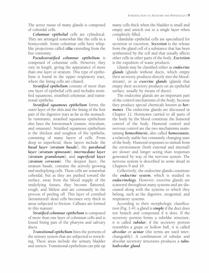
The active tissue of many glands is composed of cuboidal cells.
Columnar epithelial cells are cylindrical. They are arranged somewhat like the cells in a honeycomb. Some columnar cells have whip-like projections called cilia extending from the free extremity.
Pseudostratifi ed columnar epithelium is composed of columnar cells. However, they vary in length, giving the appearance of more than one layer or stratum. This type of epithe-lium is found in the upper respiratory tract, where the lining cells are ciliated.
Stratifi ed epithelium consists of more than one layer of epithelial cells and includes strati-fi ed squamous, stratifi ed columnar, and transi-tional epithelia.
Stratifi ed squamous epithelium forms the outer layer of the skin and the lining of the fi rst part of the digestive tract as far as the stomach. In ruminants, stratifi ed squamous epithelium also lines the forestomach (rumen, reticulum, and omasum). Stratifi ed squamous epithelium is the thickest and toughest of the epithelia, consisting of many layers of cells. From deep to superfi cial, these layers include the basal layer (stratum basale), the parabasal layer (stratum spinosum), intermediate layer (stratum gra nulosum), and superfi cial layer (stratum corneum). The deepest layer, the stratum basale, contains the actively growing and multiplying cells. These cells are somewhat cuboidal, but as they are pushed toward the surface, away from the blood supply of the underlying tissues, they become fl attened, tough, and lifeless and are constantly in the process of peeling off. This layer of cornifi ed (keratinized) dead cells becomes very thick in areas subjected to friction. Calluses are formed in this manner.
Stratifi ed columnar epithelium is composed of more than one layer of columnar cells and is found lining part of the pharynx and salivary ducts.
Transitional epithelium lines the portions of the urinary system that are subjected to stretch-ing. These areas include the urinary bladder and ureters. Transitional epithelium can pile up
many cells thick when the bladder is small and empty and stretch out to a single layer when completely fi lled.
Glandular epithelial cells are specialized for secretion or excretion. Secretion is the release from the gland cell of a substance that has been synthesized by the cell and that usually affects other cells in other parts of the body. Excretion is the expulsion of waste products.
Glands may be classifi ed either as endocrine glands (glands without ducts, which empty their secretory products directly into the blood-stream), or as exocrine glands (glands that empty their secretory products on an epithelial surface, usually by means of ducts).
The endocrine glands are an important part of the control mechanisms of the body, because they produce special chemicals known as hor-mones. The endocrine glands are discussed in Chapter 12. Hormones carried to all parts of the body by the blood constitute the humoral control of the body. Humoral control and nervous control are the two mechanisms main-taining homeokinesis, also called homeostasis, a relatively stable but constantly changing state of the body. Humoral responses to stimuli from the environment (both external and internal) are slower and longer acting than responses generated by way of the nervous system. The nervous system is described in some detail in Chapters 9 and 10.
Collectively, the endocrine glands constitute the endocrine system, which is studied in endocrinology. However, exocrine glands are scattered throughout many systems and are dis-cussed along with the systems to which they belong, such as the digestive, urogenital, and respiratory systems.
According to their morphologic classifi ca-tion (Fig. 1-4), a gland is simple if the duct does not branch and compound if it does. If the secretory portion forms a tubelike structure, it is called tubular; if the secretory portion resembles a grape or hollow ball, it is called alveolar or acinar (the terms are used inter-changeably). A combination of tubular and alveolar secretory structures produces a tubu-loalveolar gland.

10 / ANATOMY AND PHYSIOLOGY OF FARM ANIMALS
Figure 1-4. Types of exocrine glands and comparison of simple and compound glands. A) Simple tubular gland. B) Simple coiled tubular gland. C) Simple branched tubular gland. D & E) Simple acinar/alveolar glands and simple branched acinar/alveolar glands. F) Compound tubular gland. G & H) Compound acinar/alveolar glands. Com-pound tubuloacinar/tubuloalveolar glands consist of either a mixture of tubular and acinar/alveolar secretory units or tubular secretory units “capped” by acini or alveoli. (Reprinted with permission of Wiley-Blackwell from Eurell, J.A. and Frappier, B.L. Dellmann’s Textbook of Veterinary Histology. 6th ed. Ames, IA: Blackwell Publishing Profes-sional, 2006.)
Compound glands often are subdivided grossly into lobes, which in turn may be further subdivided into lobules. Hence, the connective tissue partitions (called septa) are classifi ed as interlobar septa if they separate lobes and as interlobular septa if they separate lobules. Similar terminology may be applied to ducts draining lobes or lobules of glands, that is,
interlobar ducts and interlobular ducts, respectively.
Another classifi cation of glands is based on the manner in which their cells elaborate their secretion. By this classifi cation, the most common type is the merocrine gland. Mero-crine glands pass their secretory products through the cell wall without any appreciable

INTRODUCTION TO ANATOMY AND PHYSIOLOGY / 11
loss of cytoplasm or noticeable damage to the cell membrane. The holocrine gland is the least common type. After the cell fi lls with secretory material, the entire holocrine gland cell dis-charges to the lumen of the gland to constitute the secretion. Sebaceous glands associated with hair follicles of the skin are the most common holocrine glands. An intermediate form of secretion in which a small amount of cytoplasm and cell membrane is lost with the secretion is sometimes described for the prostate and some sweat glands. Such glands are called apocrine glands.
Connective Tissues
Connective tissues, as the name implies, serve to connect other tissues. They give form and strength to many organs and often provide pro-tection and leverage. Connective tissues include
elastic tissue, collagenous (white fi brous) tissue, reticular (netlike) tissue, adipose (fat) tissue, cartilage, and bone.
Elastic tissue contains kinked fi bers that tend to regain their original shape after being stretched. This tissue is found in the ligamen-tum nuchae, a strong band that helps to support the head, particularly in horses and cattle. Elastic tissue also is found in the abdominal tunic, in the ligamenta fl ava of the spinal canal, in elastic arteries, and mixed with other tissues wherever elasticity is needed.
Collagenous (white fi brous) tissue is found throughout the body in various forms. Indi-vidual cells (fi broblasts) produce long protein-aceous fi bers of collagen, which have remarkable tensile strength. These fi bers may be arranged in regular repeating units, or laid down in a more random, irregular arrangement.
In dense regular connective tissue (Fig. 1-5), the fi bers are arranged in parallel bundles,
Figure 1-5. Longitudinal section through a tendon showing the histological appearance of dense regular connec-tive tissue. (Left) notice the line of nuclei (arrow), indicating the loose connective tissue surrounding blood vessels and nerves. Hematoxylin and eosin stain, ×226. At higher power (right), spindle-shaped fi broblasts can be seen among collagen fi bers. Hematoxylin and eosin stain, ×660. (Reprinted with permission of Wiley-Blackwell from Dellmann, H.D. and Brown, E.M. Textbook of Veterinary Histology. 2nd ed. Philadelphia: Lea & Febiger, 1981.)
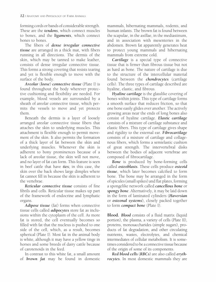
12 / ANATOMY AND PHYSIOLOGY OF FARM ANIMALS
forming cords or bands of considerable strength. These are the tendons, which connect muscles to bones, and the ligaments, which connect bones to bones.
The fi bers of dense irregular connective tissue are arranged in a thick mat, with fi bers running in all directions. The dermis of the skin, which may be tanned to make leather, consists of dense irregular connective tissue. This forms a strong covering that resists tearing and yet is fl exible enough to move with the surface of the body.
Areolar (loose) connective tissue (Plate I) is found throughout the body wherever protec-tive cushioning and fl exibility are needed. For example, blood vessels are surrounded by a sheath of areolar connective tissue, which per-mits the vessels to move and yet protects them.
Beneath the dermis is a layer of loosely arranged areolar connective tissue fi bers that attaches the skin to underlying muscles. This attachment is fl exible enough to permit move-ment of the skin. It also permits the formation of a thick layer of fat between the skin and underlying muscles. Whenever the skin is adherent to bony prominences because of a lack of areolar tissue, the skin will not move, and no layer of fat can form. This feature is seen in beef cattle that have ties; in this case, the skin over the back shows large dimples where fat cannot fi ll in because the skin is adherent to the vertebrae.
Reticular connective tissue consists of fi ne fi brils and cells. Reticular tissue makes up part of the framework of endocrine and lymphatic organs.
Adipose tissue (fat) forms when connective tissue cells called adipocytes store fat as inclu-sions within the cytoplasm of the cell. As more fat is stored, the cell eventually becomes so fi lled with fat that the nucleus is pushed to one side of the cell, which, as a result, becomes spherical (Plate I). Most fat in the animal body is white, although it may have a yellow tinge in horses and some breeds of dairy cattle because of carotenoids in the feed.
In contrast to this white fat, a small amount of brown fat may be found in domestic
mammals, hibernating mammals, rodents, and human infants. The brown fat is found between the scapulae, in the axillae, in the mediastinum, and in association with mesenteries in the abdomen. Brown fat apparently generates heat to protect young mammals and hibernating mammals from extreme cold.
Cartilage is a special type of connective tissue that is fi rmer than fi brous tissue but not as hard as bone. The nature of cartilage is due to the structure of the intercellular material found between the chondrocytes (cartilage cells). The three types of cartilage described are hyaline, elastic, and fi brous.
Hyaline cartilage is the glasslike covering of bones within joints. This type of cartilage forms a smooth surface that reduces friction, so that one bone easily glides over another. The actively growing areas near the ends of long bones also consist of hyaline cartilage. Elastic cartilage consists of a mixture of cartilage substance and elastic fi bers. This type of cartilage gives shape and rigidity to the external ear. Fibrocartilage consists of a mixture of cartilage and collage-nous fi bers, which forms a semielastic cushion of great strength. The intervertebral disks between the bodies of adjacent vertebrae are composed of fi brocartilage.
Bone is produced by bone-forming cells called osteoblasts. These cells produce osteoid tissue, which later becomes calcifi ed to form bone. The bone may be arranged in the form of spicules (small spikes) and fl at plates, forming a spongelike network called cancellous bone or spongy bone. Alternatively, it may be laid down in the form of laminated cylinders (Haversian or osteonal systems), closely packed together to form compact bone (Plate I).
Blood. Blood consists of a fl uid matrix (liquid portion), the plasma, a variety of cells (Plate II), proteins, monosaccharides (simple sugars), pro-ducts of fat degradation, and other circulating nutrients, wastes, electrolytes, and chemical intermediates of cellular metabolism. It is some-times considered to be a connective tissue because of the origin of some of its components.
Red blood cells (RBCs) are also called eryth-rocytes. In most domestic mammals they are

INTRODUCTION TO ANATOMY AND PHYSIOLOGY / 13
nonnucleated biconcave disks that contain the protein hemoglobin. The main function of the RBCs is to carry hemoglobin. Hemoglobin in turn has the primary function of carrying oxygen from the lungs to all tissues of the animal. At the tissue level, oxygen is released to the cells, while carbon dioxide, which is produced by the cells, diffuses into the blood to be carried back to the lungs, where it can be eliminated during breathing. Anemia is a reduc-tion in the concentration of functional RBCs in the blood. It can result from a loss of red cells (as in hemorrhage), insuffi cient RBC produc-tion, or inappropriate or premature degrada-tion of the red cells.
White cells (also called leukocytes) are one of the body’s fi rst lines of defense against infec-tion. They include agranulocytes and granulo-cytes. Agranulocytes are of two kinds: monocytes, large cells that engulf and destroy foreign particles, and lymphocytes, which usually are smaller and are associated with immune responses. An excess of agranulocytes tends to be associated with chronic types of diseases.
Granulocytes (polymorphonuclear leuko-cytes) are of three types and are described according to their affi nity for different stains. Granules in neutrophils stain indifferently; basophils have dark-staining granules when stained with common blood stains; and eosino-phils have red-staining granules. Blood plate-lets (thrombocytes) are small, irregularly shaped cellular fragments that are associated with the clotting of the blood. Mammalian platelets lack a nucleus.
Plasma is the fl uid part of unclotted blood. Plasma is particularly useful as a substitute for blood in transfusions because the proteins in it give it the same osmotic pressure as blood. Plasma therefore will not escape from blood vessels as readily as a salt solution.
Serum is the supernatant fl uid that remains after a clot forms and incorporates the cellular components of blood. It is similar to plasma but lacks most of the clotting factors. Serum is sometimes administered for prevention and treatment of diseases because it contains the antibody fractions of the blood.
Muscle Tissue
The three types of muscle tissue are skeletal, smooth, and cardiac (Plate I; Fig. 1-6). Both skeletal and cardiac muscle cells consist of
Figure 1-6. Types of muscle tissue. A) Smooth muscle. B) Skeletal muscle. C) Cardiac muscle. (Courtesy of Sandra Pitcaithley, DVM.)
14 / ANATOMY AND PHYSIOLOGY OF FARM ANIMALS
fi bers that under the microscope show charac-teristic cross-striations, so both are classifi ed as striated muscle. Smooth muscle cells lack dis-tinct cross-striations.
Each skeletal muscle cell must have its own nerve supply, and when stimulated, the whole fi ber contracts. This is the all-or-none law of muscle contraction. However, the force of con-traction depends on the state of the fi ber at any one moment. For example, is it already fatigued? Is it warmed up? Is it stretched? Striated skeletal muscle tissue plus some connective tissue makes up the fl esh of meat-producing animals.
Smooth muscle cells are spindle-shaped cells that contain one centrally located nucleus per cell. Smooth muscle is found in the walls of the digestive tract, in the walls of blood vessels, and in the walls of urinary and repro-ductive organs. These cells contract more slowly than skeletal muscle and in response to a variety of stimuli, although they are not under volun-tary control.
Cardiac muscle is also known as involuntary striated muscle because it is not usually under conscious control, yet it does have cross- striations. The heart muscle is composed of a complex branched arrangement of cardiac muscle cells. Modifi ed muscle cells called Pur-kinje fi bers conduct impulses within the heart, much as nerve fi bers do in other parts of the body.
Nervous Tissue
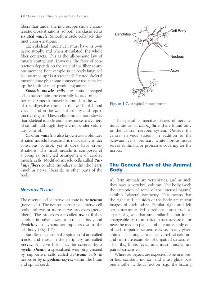
The essential cell of nervous tissue is the neuron (nerve cell). The neuron consists of a nerve cell body and two or more nerve processes (nerve fi bers). The processes are called axons if they conduct impulses away from the cell body and dendrites if they conduct impulses toward the cell body (Fig. 1-7).
Bundles of axons in the spinal cord are called tracts, and those in the periphery are called nerves. A nerve fi ber may be covered by a myelin sheath, a specialized wrapping created by supportive cells called Schwann cells in nerves or by oligodendrocytes within the brain and spinal cord.
The special connective tissues of nervous tissue are called neuroglia and are found only in the central nervous system. Outside the central nervous system, in addition to the Schwann cells, ordinary white fi brous tissue serves as the major protective covering for the nerves.
The General Plan of the Animal Body
All farm animals are vertebrates, and as such they have a vertebral column. The body (with the exception of some of the internal organs) exhibits bilateral symmetry. This means that the right and left sides of the body are mirror images of each other. Similar right and left structures are called paired structures, such as a pair of gloves that are similar but not inter-changeable. Most unpaired structures are on or near the median plane, and of course, only one of each unpaired structure exists in any given animal. The tongue, trachea, vertebral column, and heart are examples of unpaired structures. The ribs, limbs, eyes, and most muscles are paired structures.
Wherever organs are expected to be in more-or-less constant motion and must glide past one another without friction (e.g., the beating
Axon
Nucleus
Cell BodyDendrites
Figure 1-7. A typical motor neuron.

INTRODUCTION TO ANATOMY AND PHYSIOLOGY / 15
heart and moving gut), a serosal cavity is present. The simple squamous epithelium lining various body cavities is also called meso-thelium, and the cavities have within them only a scant amount of fl uid to facilitate free move-ment of the tissues. The diaphragm divides the embryonic body cavity into a thoracic cavity and the abdominopelvic cavity. Each of these are further subdivided.
The thoracic cavity contains the pericardial sac, which surrounds the heart, and two pleural sacs, which surround the two lungs. These sacs are formed by a serous membrane, the pleura, a layer of simple squamous epithelium with under-lying connective tissue, moistened with the small amount of fl uid within the cavity of the sac.
The abdominopelvic cavity is somewhat arbitrarily divided into the abdominal and pelvic cavities. The abdominal cavity contains the kidneys, most of the digestive organs, and a variable amount of the internal reproductive organs in both sexes. The pelvic cavity contains
the terminal part of the digestive system (the rectum) and all of the internal portions of the urogenital system not found in the abdominal cavity. The abdominal and pelvic cavities are continuous with one another, and the brim of the pelvis marks the transition. The serous membrane that surrounds the abdominal viscera and part of the pelvic viscera is called peritoneum.
A transverse section through the abdominal cavity illustrates the general plan of the body as a tube (the digestive tract and its derivatives) within a tube (the body wall) (Fig. 1-8). Nor-mally there are few air-fi lled spaces in the animal body except in the respiratory system and the ear. However, for the sake of clarity, many illustrations show a considerable separa-tion between structures that in the animal body are actually in contact.
The layers of the body wall and the layers of the digestive tract show a striking similarity, although in reverse order. Layers of the body
Skin
FasciaStriated Muscle
Retroperitoneal
Fascia
Parietal Peritoneum Visceral
Peritoneum
Submucosal
Connective
Tissue
Smooth Muscle
Mucous Membrane
Subserous
Connective Tissue
Transverse
Fascia
Figure 1-8. Cross-section of the body wall and digestive tract.
16 / ANATOMY AND PHYSIOLOGY OF FARM ANIMALS
wall from outside inward are (1) epithelium (epidermis of the skin), (2) connective tissue (dermis and fascia), (3) muscle (striated), (4) connective tissue (transverse fascia), and (5) mesothelium (parietal peritoneum). The layers of the gut wall from outside inward are (1) mesothelium (visceral peritoneum), (2) con-nective tissue (subserous connective tissue), (3) muscle (smooth), (4) connective tissue (sub-mucosa), and (5) epithelium (mucous mem-brane) (Fig. 1-8).
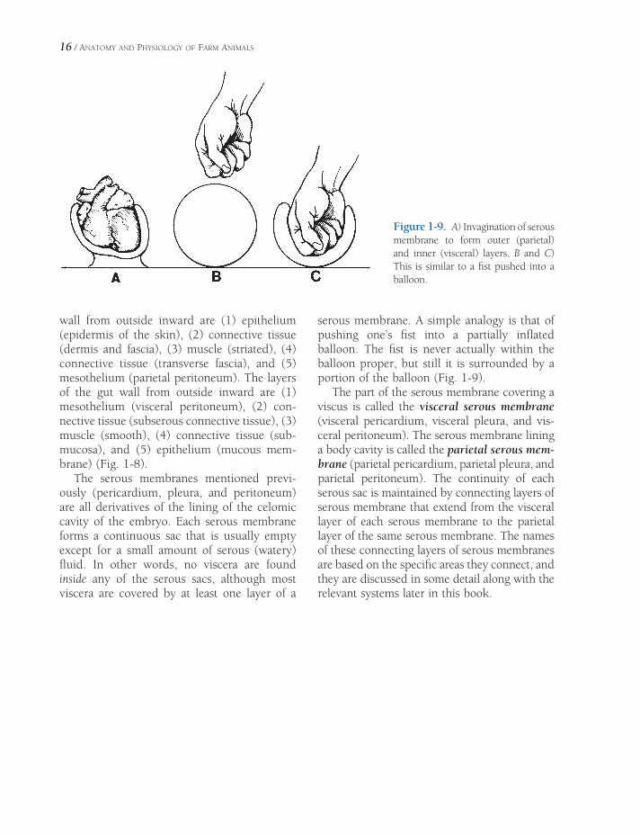
The serous membranes mentioned previ-ously (pericardium, pleura, and peritoneum) are all derivatives of the lining of the celomic cavity of the embryo. Each serous membrane forms a continuous sac that is usually empty except for a small amount of serous (watery) fl uid. In other words, no viscera are found inside any of the serous sacs, although most viscera are covered by at least one layer of a
serous membrane. A simple analogy is that of pushing one’s fi st into a partially infl ated balloon. The fi st is never actually within the balloon proper, but still it is surrounded by a portion of the balloon (Fig. 1-9).
The part of the serous membrane covering a viscus is called the visceral serous membrane (visceral pericardium, visceral pleura, and vis-ceral peritoneum). The serous membrane lining a body cavity is called the parietal serous mem-brane (parietal pericardium, parietal pleura, and parietal peritoneum). The continuity of each serous sac is maintained by connecting layers of serous membrane that extend from the visceral layer of each serous membrane to the parietal layer of the same serous membrane. The names of these connecting layers of serous membranes are based on the specifi c areas they connect, and they are discussed in some detail along with the relevant systems later in this book.
Figure 1-9. A) Invagination of serous membrane to form outer (parietal) and inner (visceral) layers. B and C) This is similar to a fi st pushed into a balloon.

ANATOMY AND PHYSIOLOGY OF THE CELL
Membrane Receptors and Intracellular Signaling
Cytoplasm and Cytoplasmic OrganellesCytoplasm
The Golgi Apparatus
The Endoplasmic Reticulum and
Ribosomes
Mitochondria
Lysosomes
Other Structures
NucleusStructure of the Nucleus
DNA and DNA Replication
RNA: Transcription and Translation
Biotechnology
Cell DivisionMitosis
Meiosis
Regulation of Cell Growth and Replication
Properties of LifeChemical Composition of the Cell
Water
Proteins
Lipids
Carbohydrates
Inorganic Substances
Acids, Bases and pH
Microscopic Study of the CellLight Microscopy
Electron Microscopy
The Cell MembraneStructure of the Membrane
Intercellular Contact and Adhesion
Transport Across Cell MembranesSimple and Facilitated Diffusion
Osmosis
Active Transport
Membrane Potentials and Excitable CellsResting Membrane Potential
Excitable Cells and Action Potentials
Chapter 2
17

18 / ANATOMY AND PHYSIOLOGY OF FARM ANIMALS
Discovery of living cells would have been diffi cult, if not impossible, before Zacharias
Jansen of the Netherlands invented the com-pound microscope in 1590. Robert Hooke of England used the term cell to describe the cavi-ties he saw in sections of cork. In 1665, Hooke published a description of cork cells based on a study done with his improved compound microscope.
In 1839 Matthias Schleiden, a German bota-nist, and Theodor Schwann, an animal anato-mist, formulated the cell theory, which set forth the concept that “the elementary parts of all tissues are formed of cells in an analogous, though very diversifi ed, manner, so that it may be asserted that there is one universal principle of development for the elementary parts of organisms, however different, and that this principle is the formation of cells.”
The word cell comes from the Latin cella meaning small chamber. In biology, particu-larly animal biology, the term cell refers more specifi cally to the individual units of living structure rather than the compartments that may contain them. There actually are no compartments as such in most tissues (with the exception of bone and cartilage), but the living units, cells, are found in groups in which mainly adjacent cells restrain individual cells. As early as 1772, Corti observed the jellylike material in the cell that later was called protoplasm.
Properties of Life
It is diffi cult to give a satisfactory defi nition of life. However, the cell is the functional unit of all animal life. It is the unit that makes up all tissues, organs, and systems, which in turn make up the total animal. Therefore, the prop-erties of the cell are equated with those of life. These properties include homeostasis, growth, reproduction, absorption, metabolism, secre-tion, irritability, conductivity, and contractility. The last two characteristics, however, are not properties of all cells. Conductivity is an impor-tant functional characteristic of both nerve and
muscle cells, whereas contractility is a property of muscle cells.
Homeostasis is the tendency for living things to attempt to maintain a state of relative stabil-ity. At the whole-animal level or at the cellular level, all living things respond to stresses placed upon them by changes in their environment. Their responses are attempts to maintain a state of homeostasis.
Growth is increase in size. Increase in size of a cell or organ beyond normal is called hypertrophy. An increase in the size of a structure due to an increase in the number of cells is called hyperplasia. A decrease in size from normal is called atrophy. Failure of a tissue or organ to develop is called aplasia, while incomplete development or defective development of a tissue or organ is called hypoplasia.
Reproduction of a cell or of an organism implies the ability to produce more cells or more organisms that are essentially the same as the original. Some fully differentiated cells, for instance nerve cells, do not normally retain the ability to reproduce in the adult.
Cells may be found in solutions whose com-position is quite different from that of the fl uid within the cells. To maintain intracellular homeostasis in these conditions, the passage of particles and water in and out of the cell must be regulated. Absorption is the process of taking dissolved materials or water through the cell membrane into the substance of the cell. This can be a passive process dependent on the forces of diffusion and osmosis, an active process requiring the expenditure of energy from adenosine triphosphate, or the result of electrochemical ionic forces and affi nities that require no direct expenditure of energy. All three can occur at the same time across the same cell membrane.
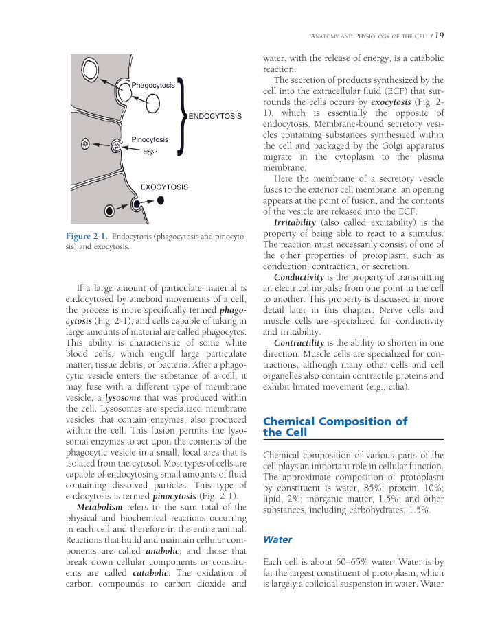
Endocytosis is another means by which extracellular materials may enter a cell. In endocytosis the exterior cell membrane moves to surround extracellular materials in a mem-brane pocket (Fig. 2-1). This membrane vesicle detaches from the inner surface of the cell membrane and moves into the interior of the cell.
ANATOMY AND PHYSIOLOGY OF THE CELL / 19
If a large amount of particulate material is endocytosed by ameboid movements of a cell, the process is more specifi cally termed phago-cytosis (Fig. 2-1), and cells capable of taking in large amounts of material are called phagocytes. This ability is characteristic of some white blood cells, which engulf large particulate matter, tissue debris, or bacteria. After a phago-cytic vesicle enters the substance of a cell, it may fuse with a different type of membrane vesicle, a lysosome that was produced within the cell. Lysosomes are specialized membrane vesicles that contain enzymes, also produced within the cell. This fusion permits the lyso-somal enzymes to act upon the contents of the phagocytic vesicle in a small, local area that is isolated from the cytosol. Most types of cells are capable of endocytosing small amounts of fl uid containing dissolved particles. This type of endocytosis is termed pinocytosis (Fig. 2-1).
Metabolism refers to the sum total of the physical and biochemical reactions occurring in each cell and therefore in the entire animal. Reactions that build and maintain cellular com-ponents are called anabolic, and those that break down cellular components or constitu-ents are called catabolic. The oxidation of carbon compounds to carbon dioxide and
water, with the release of energy, is a catabolic reaction.
The secretion of products synthesized by the cell into the extracellular fl uid (ECF) that sur-rounds the cells occurs by exocytosis (Fig. 2-1), which is essentially the opposite of endocytosis. Membrane-bound secretory vesi-cles containing substances synthesized within the cell and packaged by the Golgi apparatus migrate in the cytoplasm to the plasma membrane.
Here the membrane of a secretory vesicle fuses to the exterior cell membrane, an opening appears at the point of fusion, and the contents of the vesicle are released into the ECF.
Irritability (also called excitability) is the property of being able to react to a stimulus. The reaction must necessarily consist of one of the other properties of protoplasm, such as conduction, contraction, or secretion.
Conductivity is the property of transmitting an electrical impulse from one point in the cell to another. This property is discussed in more detail later in this chapter. Nerve cells and muscle cells are specialized for conductivity and irritability.
Contractility is the ability to shorten in one direction. Muscle cells are specialized for con-tractions, although many other cells and cell organelles also contain contractile proteins and exhibit limited movement (e.g., cilia).
Chemical Composition of the Cell
Chemical composition of various parts of the cell plays an important role in cellular function. The approximate composition of protoplasm by constituent is water, 85%; protein, 10%; lipid, 2%; inorganic matter, 1.5%; and other substances, including carbohydrates, 1.5%.
Water
Each cell is about 60–65% water. Water is by far the largest constituent of protoplasm, which is largely a colloidal suspension in water. Water
Phagocytosis
Pinocytosis
EXOCYTOSIS
ENDOCYTOSIS}Figure 2-1. Endocytosis (phagocytosis and pinocyto-sis) and exocytosis.

20 / ANATOMY AND PHYSIOLOGY OF FARM ANIMALS
acts as a solvent for inorganic substances and enters into many biochemical reactions.
Most body water is within cells, and this fl uid volume is called intracellular fl uid. This fl uid volume is about 40% of body weight. The remaining fl uid (about 20% of body weight), found outside of cells, is termed extracellular fl uid (ECF). Most ECF (about 15% of body weight) surrounds cells throughout the body and is termed interstitial fl uid. Unique inter-stitial fl uids include the cerebrospinal fl uid, the fl uid in the joints, the fl uid in the eyes (aqueous and vitreous humors), and the serous fl uid in the visceral spaces (i.e., pericardial, pleural, and peritoneal spaces). Blood plasma, a specifi c type of ECF, is about 5% of total body weight. The percentages for the different types of body fl uids vary from one animal to another. Factors affecting these percentages include condition (amount of fat), age, state of hydration, and species.
Water is constantly lost from the body, and it must be replenished if the animal is to remain in water balance and not become dehydrated. Most is lost via the urine, but it is also lost in the feces and by evaporation from body sur-faces, such as the skin and respiratory passages. Water replacement is almost entirely by drink-ing, because minimal amounts of water are pro-duced in the bodies of domestic animals as a result of cellular metabolism (metabolic water).
Proteins
After water, proteins are the next largest con-stituent of protoplasm. Proteins are complex high-molecular-weight colloidal molecules consisting primarily of amino acids that are polymerized (joined) into polypeptide chains (Fig. 2-2A). The union of amino acids within a protein molecule is by way of a peptide linkage, a bond between the amino (NH2) group of one amino acid and the carboxyl (COOH) group of another amino acid, with the elimination of water. A small chain of amino acids is called a peptide. A polypeptide is a chain of more than 50 amino acids connected by peptide linkages,
and a chain that contains more than 100 amino acids is called a protein.
The peptide linkages between amino acids in a protein are somewhat fl exible, and this permits the chain to bend into various three-dimensional shapes (Fig. 2-2B). These confi gu-rations may become relatively stable, because chemical attractions, or bonds, form between amino acids at various points in the chain. The three-dimensional shape of a protein is an important determinant of its biologic function, because the shape can determine what segments
COOH
H2N
Leucine
Arginine
PhenylalineHistidine
Lysine
A
B
ChemicalBonds
Figure 2-2. A) A chain of amino acids joined by pep-tide bonds to form a protein. B) A large protein. Each fi lled circle represents a single amino acid. Chemical bonds between amino acids at distant points in the chain produce the three-dimensional shape of the protein molecule.
ANATOMY AND PHYSIOLOGY OF THE CELL / 21
of the protein chain are exposed and available to interact with other molecules.
Amino acids, and thus proteins, contain carbon, hydrogen, oxygen, and nitrogen. Pro-teins may also contain other elements such as sulfur, phosphorus, or iron. Simple proteins yield only amino acids or their derivatives upon hydrolysis. The simple proteins, and examples of each, are as follows:
1. Albumins (plasma albumin, milk lactalbumin)2. Globulins (plasma globulins, globulins in
plant seeds)3. Protamines (in sperm cells)4. Histones (with nucleoproteins in cell
nuclei)5. Albuminoids (collagen and elastin of
connective tissue)
Conjugated proteins consist of simple pro-teins combined with a component that is not a protein or amino acid. Rather, it is called a prosthetic group. The conjugated proteins and examples of each are as follows:
1. Glycoproteins: Includes mucopolysaccha-rides and oligosaccharides as the carbohy-drate prosthetic group (in connective tissue and salivary mucus)
2. Lipoproteins: Prosthetic group is lipid (in blood plasma and egg yolk)
3. Nucleoproteins: Nucleic acid prosthetic group (in cell nuclei, chromosomes, and viruses)
4. Chromoproteins: Fe-porphyrin prosthetic group (hemoglobin, cytochromes)
5. Metalloproteins: Contain iron, zinc, or copper (blood transferrin, ferritin, carbonic anhydrase)
6. Phosphoproteins: Phosphate prosthetic group (casein in milk, vitellin in eggs)
Most proteins can be classifi ed as structural proteins or as reactive proteins. Structural pro-teins include these fi brous proteins: collagens, which are the major proteins of connective tissue and which represent about 30% of the total protein content of the animal body; elas-tins, which are present in elastic tissues such as the ligamentum nuchae, the abdominal tunic,
and some arteries; and keratins, which are the proteins of wool, hair, horns, and hoofs. Reac-tive proteins include enzymes, protein hor-mones, histones associated with nucleic acids in the nucleus of cells, and contractile proteins in muscle (actin and myosin). Many varieties of proteins are found in blood plasma. Functions of plasma proteins include the transport of sub-stances such as hormones and lipids in the blood, contributing to the process of blood coagulation, and creating an effective osmotic pressure difference between the plasma and interstitial fl uid. Plasma proteins also include antibodies, which are produced by certain blood cells and are part of an overall immune response.
All cell membranes contain proteins, and like plasma proteins, the proteins in cell mem-branes have a variety of functions. These include serving as membrane receptors for hormones and drugs, contributing to the transport of water and particles into and out of cells, acting as membrane-bound enzymes, and serving as markers to permit the immune system to recognize cells as normal or abnormal body components.
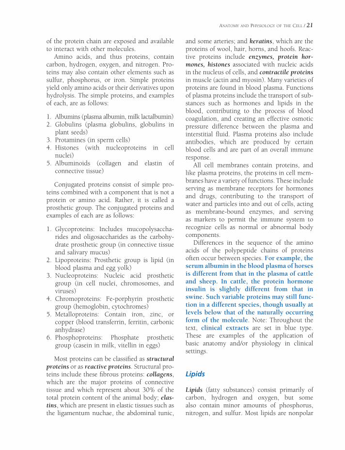
Differences in the sequence of the amino acids of the polypeptide chains of proteins often occur between species. For example, the serum albumin in the blood plasma of horses is different from that in the plasma of cattle and sheep. In cattle, the protein hormone insulin is slightly different from that in swine. Such variable proteins may still func-tion in a different species, though usually at levels below that of the naturally occurring form of the molecule. Note: Throughout the text, clinical extracts are set in blue type. These are examples of the application of basic anatomy and/or physiology in clinical settings.
Lipids
Lipids (fatty substances) consist primarily of carbon, hydrogen and oxygen, but some also contain minor amounts of phosphorus, nitrogen, and sulfur. Most lipids are nonpolar

22 / ANATOMY AND PHYSIOLOGY OF FARM ANIMALS
molecules and thus are insoluble in water. The four primary chemical types of lipids in animals are fatty acids, triglycerides or triacylglycerols, phospholipids, and steroids.
Fatty acids are chains of covalently bound carbon atoms with hydrogens attached (Fig. 2-3). If each carbon atom has four single covalent bonds, the fatty acid is saturated. If any carbon atom has fewer than four single bonds, the fatty acid is unsaturated. A polyunsaturated fatty acid has multiple carbon atoms with fewer than four single bonds. Animal tissues tend to have higher amounts of saturated fatty acids than do vegetable oils.
Prostaglandins and leukotrienes, derived from fatty acids, are produced by a variety of cells throughout the body. In many cases, these serve as local messengers that permit one cell to affect the function of another nearby. Both prostaglandins and leukotrienes are local mes-sengers in the process of infl ammation, and prostaglandins regulate ovarian function in some species.
Triglycerides consist of a glycerol molecule with three fatty acids attached (Fig. 2-4). Also known as neutral fats, triglycerides are the primary form of lipid storage in adipose tissue in animals. Fatty acids must be detached from glycerol before they can undergo further metab-olism. This detachment is the function of enzymes known as lipases. Because triglycer-ides are not soluble in water, most are not transported as individual molecules in blood plasma. For transport, they are combined with other lipids and proteins into relatively large particles known as lipoproteins. In this form they can be transported from site to site within the body.
The glycerol and fatty acids derived from the breakdown of triglycerides are all sources of energy. Glycerol can serve as a substrate for the glycolytic pathway in the cytosol. Fatty acids enter the mitochondria, where they are broken down into two carbon units, which become acetyl coenzyme A (acetyl CoA). The metabolism of acetyl CoA within the mito-chondria ultimately results in the production of the high-energy compound adenosine tri-phosphate (ATP). Details about the role of the mitochondria in the production of ATP are described in the section on organelles later in this chapter.
Phospholipids are similar to triglycerides except that a molecule containing a phosphate group has replaced one of the three fatty acids. The replacement of the nonpolar (hydropho-bic) fatty acid with a nonlipid polar (hydro-philic) molecule creates a unique compound
H
C
C
C
H
H
H
H
_
_
_
_
_
_
O CO
_=
CH2_
CH2
_ CH2_
CH2
_ CH2_
CH2
_ CH2_
CH2
_ CH2_
CH2
_ CH3_
O CO
_=
CH2_
CH2
_ CH2_
CH2
_ CH2_
CH2
_ CH2_
CH2
_ CH2_
CH2
_ CH3_
O CO
_=
CH2_
CH2
_ CH2_
CH2
_ CH2_
CH2
_ CH2_
CH2
_ CH2_
CH2
_ CH3_
+H2O
+H2O
+H2O
H
C
C
C
H
H
H
H
_
_
_
OH
OH
OH
_
_
_
TriglycerideGlycerol
Saturated Fatty Acid:
Polyunsaturated Fatty Acid:
H-O-C-C-C-C-HO H H H-= - -
H H H
- - -
H-O-C-(CH2)3-C=C-C-C=C-C-C=C-C-C=C-(CH2)4-C-HO H H H H H H H H H H H H
H H HH
-= - - - - - - - - - - -
- - - -
Figure 2-3. Saturated and polyunsaturated fatty acids. The 4-carbon saturated fatty acid is butyric acid, and the 20-carbon fatty acid is arachidonic acid.
Figure 2-4. Three fatty acids combined with glycerol to form a triglyceride.
ANATOMY AND PHYSIOLOGY OF THE CELL / 23
with two regions that vary in water solubility. The phosphate-containing region becomes water soluble and the remainder of the phos-pholipid molecule is water insoluble. This unique characteristic is important in the role of phospholipids in the structure of cell mem-branes. Cell membranes throughout the body primarily consist of phospholipids.
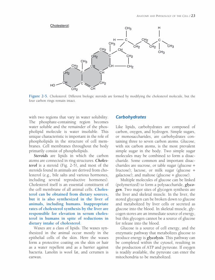
Steroids are lipids in which the carbon atoms are connected in ring structures. Choles-terol is a steroid (Fig. 2-5), and most of the steroids found in animals are derived from cho-lesterol (e.g., bile salts and various hormones, including several reproductive hormones). Cholesterol itself is an essential constituent of the cell membrane of all animal cells. Choles-terol can be obtained from dietary sources, but it is also synthesized in the liver of animals, including humans. Inappropriate rates of cholesterol synthesis by the liver are responsible for elevation in serum choles-terol in humans in spite of reductions in dietary intake of cholesterol.
Waxes are a class of lipids. The waxes syn-thesized in the animal occur mostly in the epithelial cells of the skin. Here the waxes form a protective coating on the skin or hair as a water repellent and as a barrier against bacteria. Lanolin is wool fat, and cerumen is earwax.
Carbohydrates
Like lipids, carbohydrates are composed of carbon, oxygen, and hydrogen. Simple sugars, or monosaccharides, are carbohydrates con-taining three to seven carbon atoms. Glucose, with six carbon atoms, is the most prevalent simple sugar in the body. Two simple sugar molecules may be combined to form a disac-charide. Some common and important disac-charides are sucrose, or table sugar (glucose + fructose); lactose, or milk sugar (glucose + galactose); and maltose (glucose + glucose).
Multiple molecules of glucose can be linked (polymerized) to form a polysaccharide, glyco-gen. Two major sites of glycogen synthesis are the liver and skeletal muscle. In the liver, the stored glycogen can be broken down to glucose and metabolized by liver cells or secreted as glucose into the blood. In skeletal muscle, gly-cogen stores are an immediate source of energy, but this glycogen cannot be a source of glucose for release into the blood.
Glucose is a source of cell energy, and the enzymatic pathway that metabolizes glucose to produce energy is glycolysis. This pathway can be completed within the cytosol, resulting in the production of ATP and pyruvate. If oxygen is readily available, the pyruvate can enter the mitochondria to be metabolized.
HOCH2
C
CH3
H
CH
CH2
CH2
C
CH
CH2
CH
CH
CH2
CH2
CH CH2
CH2C
CH3
CH
C
CH2
CH3
CH2
CH2
C
H
CH3
CH3
Cholesterol:
Figure 2-5. Cholesterol. Different biologic steroids are formed by modifying the cholesterol molecule, but the four carbon rings remain intact.
24 / ANATOMY AND PHYSIOLOGY OF FARM ANIMALS
The sugar deoxyribose is found in combina-tion with a base (purine or pyrimidine) and a phosphate, forming DNA (deoxyribonucleic acid). DNA is the carrier of all genetic informa-tion from generation to generation and from cell to cell and ultimately controls all functions of the cell. DNA is found almost exclusively in the nucleus of the cell. A related substance, RNA (ribonucleic acid), includes the sugar ribose combined with a base and a phosphate. RNA is intimately associated with synthesis of all cell proteins.
Even though carbohydrates are a major part of the diet of most animals, the amount of car-bohydrates in animal’s bodies is relatively small. Carbohydrates make up less than 1% of most cells.
Inorganic Substances
Of the atoms or elements found in protoplasm, more than 99% are hydrogen, carbon, oxygen, and nitrogen contained in the organic com-pounds described earlier. Protoplasm also has inorganic compounds containing iodine, iron, phosphorus, calcium, chlorine, potassium, sulfur, sodium, magnesium, copper, manga-nese, zinc, cobalt, chromium, selenium, molyb-denum, fl uorine, silicon, tin, and vanadium. Of the 24 elements found in the body cells, 20 represent less than 1% of the total amount of elements in living tissue.
An electrolyte is any molecular substance that in solution dissociates into its electrically charged components, called ions. For example, this occurs when sodium chloride in solution dissociates into Na+ and Cl−. The solution can then carry an electrical charge and current.
The major ions found within cells in order of abundance, expressed in milliequivalents per liter of fl uid, are potassium (K+) 140 mEq/L; phosphate (HPO4
2−), 75 mEq/L; magnesium (Mg2+), 60 mEq/L; sodium (Na+) 10 mEq/L; bicarbonate (HCO3
−) 10 mEq/L; and chloride (Cl−) 4 mEq/L.
A milliequivalent is one-thousandth of an equivalent. An equivalent weight is the weight
in grams that will displace or react with 1 gram atomic weight of hydrogen ion (H+ = 1.008 g).
The practical importance of this concept is that laboratory reports and records of mea-surements of body fl uid electrolyte and ion concentrations are often expressed as milli-equivalents per liter (mEq/L). Another way of expressing measurements is in mg%, or milli-grams per 100 milliliters (mg/100 mL). Mea-surements may also be expressed as milligrams per deciliter (mg/dL); a deciliter is 100 mL, or a tenth of a liter. A liter is 1000 mL, or 1.06 quarts.
Bone contains about 65% inorganic material by volume. Most of this mineral material is in the form of hydroxyapatite crystals with a molecular formula Ca10(PO4)6(OH)2. In addi-tion, sodium, magnesium, and iron may be incorporated in the mineral structure.
Acids, Bases, and pH
An acid is a compound that is capable of ion-izing and releasing a hydrogen ion. The pH of a solution is a measure of the concentration of H+. However, pH is reported as the negative of the logarithm to the base 10 of the H+ concen-tration in moles, so the greater the H+ concen-tration, the more negative, or lower, the pH. Concentrations of H+ in normal body fl uids are much lower than other electrolytes. A typical H+ concentration in plasma is 4 × 10⁻⁹ moles per liter, or 4 nanomoles per liter, equivalent to a pH of 7.45. A nanomole is one-millionth of a millimole.
A base is a compound that is capable of reducing the concentration of hydrogen ions in a solution by combining with them. When bases are added to solutions, the H+ concentra-tion is reduced, so the pH rises. Chemical buffer solutions contain both acids and bases and therefore are capable of either releasing or combining with H+. This dual ability tends to provide a relatively stable pH.
All body fl uids, intracellular and extra-cellular, contain mixtures of several chemical

ANATOMY AND PHYSIOLOGY OF THE CELL / 25
buffers. These buffers act simultaneously to maintain a relatively stable pH within their respective fl uids. This stability is critical for normal metabolic processes and enzymatic reactions.
Microscopic Study of the Cell
Cells range in diameter from about 10 to 100 μm (micrometers). Cells that are actively multiplying range from about 20 to 30 μm in diameter. Table 2-1 lists the relationships among metric units of measurement used for microscopy. (For example, 1 μm is one-thou-sandth of a millimeter, and there are about 25 mm in 1 inch; thus, approximately 1000 cells, each 25 μm in diameter, could be lined up between the 1- and 2-inch marks of a ruler.) Sizes of cells vary considerably from one type of cell to another, but with the exception of the yolks of birds’ eggs (which are considered single cells), the distance from the center of the cell to some portion of the cell membrane (surface of the cell) is seldom more than a few micrometers.
The outer cell membrane is thin, 7–10 nm. Regardless of its composition, a membrane of this dimension can have little tensile strength; this is another reason cells must be small.
The uniformly small size of cells and the much smaller sizes of structures within the cell have made effective study of cells diffi cult. As noted earlier, the existence of cells was not confi rmed before the microscope was invented.
Details of the actual structure of the various parts of cells were not known with any degree of certainty until after the development of the electron microscope. The study of gross anatomy goes back several centuries, but under-standing of the fi ner structure of the animal body awaited more recent technological developments.
Light Microscopy
Some cells are in tissues that are thin enough to be illuminated from one side and observed with a microscope from the opposite side. This is true of the web of the foot of the frog, the mesentery of the intestine, and a few other tissues. In these instances, living cells can be observed directly, and this technique is useful for the study of blood circulation. Specifi c cells may also be taken from a living animal and grown on artifi cial medium by tissue culture. These cells may then be studied in the living state, even at high magnifi cations.
Except for the forgoing situations, cells usually are studied after undergoing some degree of manipulation, so that what is actually seen with the microscope bears little resem-blance to the living cell. A typical treatment of tissue before it can be examined with a light microscope in cludes the following:
1. Fixation with an agent, such as an aldehyde, that will cross-link the tissue proteins and prevent further changes in the tissue, such as autolysis and bacterial degradation. Alternatively, tissue may be frozen in liquid
Table 2-1. Metric Linear Measurements
Unit Abbreviation Relationships
Meter m 1 m = 102 cm, 103 mm, 106 μm, 109 nm, 1010 Å
Centimeter cm 1 cm = 10−2 m, 101 mm, 104 μm, 107 nm, 108 Å
Micrometer (micron) μm 1 μm = 10−6 m, 10−4 cm, 10−3 mm, 103 nm, 104 Å
Nanometer nm 1 nm = 10−9 m, 10−7 cm, 10−6 mm, 10−3 μm, 10 Å
Angstrom Å 1 Å = 10−10 m, 10−8 cm, 10−7 mm, 10−4 μm, 10−1 nm

26 / ANATOMY AND PHYSIOLOGY OF FARM ANIMALS
nitrogen to prevent such degenerative changes.
2. Embedding the tissue in a material that will permit cutting very thin sections. Paraffi n is used for producing sections of 5–10 μm thickness; sections as thin as 1–2 μm can be obtained by embedding in a plastic, such as glycol methacrylate. Since most embedding media are not water soluble, the fi xed tissue must be dehydrated and then infi ltrated with some material such as xylene, which is miscible with the embedding medium. Frozen tissues need not be embedded.
3. Sectioning the tissue into very thin slices so that the sections may be placed on a glass slide. A microtome is used for this purpose. It consists of a sharp blade and a mechanism for moving the tissue past the blade and then advancing it a defi ned distance after each cutting. Frozen tissues are sectioned in a cryostat, which is a microtome housed in a freezer cabinet.
4. Staining the section so that different cells or different parts of cells can be differentiated according to color. Hematoxylin and eosin are stains commonly used together, and this treatment is described as an H & E stain. The hematoxylin tends to stain acidic portions of a cell dark blue or purple (these basophilic areas include the cell nucleus, which contains nucleic acids), and the eosin tends to stain the basic portions of a cell pink to red (these acidophilic areas include much of the more basic protein within the cell). Wright’s stain, used to stain blood cells (Plates I and II), stains basophilic areas blue with methylene blue and acidophilic areas red with eosin. Sections can also be treated with a variety of chemical solutions to demonstrate the presence of certain types of chemicals or the activity of enzymes in the tissue or cell, a technique called histochemistry (Fig. 2-6). The presence of specifi c types of molecules can be determined by exposing the section to a solution containing antibodies to those molecules. This technique is called immunocytochemistry.
5. The last step, of course, is the actual examination of the stained section of tissue on the slide by means of a microscope and light transmitted through the section.
This approach to the study of the animal body has been standard for many years and will continue to be useful regardless of newer devel-opments. However, some factors should be kept in mind when studying sections or pho-tographs of sections.
The relationship of the tissue sections to the actual tissue is about the same as that of a sack of potato chips to a growing potato. Both the sections and the potato chips have been pro-cessed so that actual resemblance to the origi-nal structure is limited. Both are seen in two dimensions, length and width, with thickness relatively unimportant for visualization. Recent technological advances such as confocal micro-scopy and computer reconstruction provide three-dimensional views of cellular and tissue structure.
The light microscope can magnify objects to a maximum of about 1500 times the original size. This is known as the magnifi cation, or power, of the microscope. Resolving power refers to the property of showing two objects as separate structures. The light microscope can resolve (separate) two structures that are as
Figure 2-6. Serial sections of horse triceps brachii muscle histochemically stained for enzymatic activity. The same muscle cells (I and II) are visible in both sections. A) Calcium-dependent ATPase activity. B) Activity of a mitochondrial oxidative enzyme.

ANATOMY AND PHYSIOLOGY OF THE CELL / 27
close as approximately 0.2 μm (about 200 nm). This resolving power depends greatly on the wavelength of the light used to observe the tissue and the optical quality of the objective lens of the microscope.
Other developments in light microscopy include phase contrast and fl uorescence microscopy. Phase contrast microscopy can be used with unstained and/or living cells, because it depends on differences in refraction of various parts of a cell for image formation. Fluorescence microscopy is often teamed with immunocytochemistry, in which antibody-labeled cells are identifi ed by causing them to fl uoresce upon exposure to light of a specifi c wavelength.
X-ray diffraction is used to study the struc-ture of inorganic and organic crystals and the molecular structure of biologic substances such as DNA, collagen, and hemoglobin. It consists of passing a beam of x-rays through the sub-stance and recording the diffraction pattern (scattering of the beam) on a photographic emulsion.
Electron Microscopy
Electron microscopes do not use visible light for the delineation of structures as in the light microscope; they use a beam of electrons focused by electromagnetic lenses. The electron beam may pass through a thin specimen in the transmission electron microscope or be refl ected from the surface of an object and studied with the scanning electron microscope. The images with the electron microscope, however, are only black and white. (For an example of a scanning electron micrograph, see Fig. 15-2.)
The scanning electron microscope is a ver-satile instrument with a magnifi cation range from ×15 to ×10,000 and a resolution in the vicinity of 10 nm. Depth of fi eld with the scan-ning electron microscope is much greater than with any light microscope. Preparation of specimens for observation with the scanning electron microscope is relatively simple. Non-metallic biologic material generally is dehy-
drated and coated with a thin layer of metallic gold before it is placed in the scanning electron microscope.
The transmission electron microscope is capable of much higher magnifi cation (as much as ×1 million), with an effective resolution of 0.1 nm. By the use of photographic enlarge-ment and projection techniques, the magnifi ca-tions can exceed 1 million and still show good detail. (A typical transmission electron micro-graph is shown in Fig. 2-9.) Because so much more detail can be seen in a small area, tissue preparation for transmission electron micros-copy is much more exacting and time-consum-ing than for light microscopy.
The best means of fi xation is to apply a fi xa-tive (commonly glutaraldehyde followed by osmium tetroxide) to the living tissue or biopsy specimen. The time from the living state to immersion in the fi xative should not exceed 2 minutes, and the size of tissue should not exceed 1 mm on a side. Osmium tetroxide acts both as a fi xative and as a stain. Other heavy metals, including lead, may be used as so-called stains. The term stain may be used somewhat loosely, because the areas where the metals concentrate inhibit the passage of electrons, giving an electron-dense appearance that shows up as a dark area in the fi nal photographic print. After fi xation, the tissue is dehydrated and infi ltrated with plastic and embedded in plastic for sectioning. The sections are cut extremely thin (less than 30 nm), placed on a grid, and examined with the electron microscope.
The picture of the typical cell viewed with the electron microscope still shows most of the structures described by light microscopy but in much greater detail. The typical cell seen in light microscopy consists of a nucleus and cyto-plasm surrounded by the cell membrane (Fig. 2-7). The nucleus contains a nucleolus and chromatin material, which forms into chromo-somes during cell division. A membrane called the nuclear envelope surrounds the nucleus. The cytoplasm contains a number of structures, or organelles, including the endoplasmic retic-ulum, Golgi apparatus, mitochondria, and inclusions.

28 / ANATOMY AND PHYSIOLOGY OF FARM ANIMALS
The Cell Membrane
Structure of the Membrane
The outer cell membrane, also known as plasma membrane, and other membranes within the cell primarily consist of phospholipids, pro-teins, and cholesterol. According to the fl uid mosaic model, the phospholipids are arranged with their polar (hydrophilic) ends facing the protein layers, while their nonpolar (hydropho-bic) ends face each other in the center of the membrane (Fig. 2-8). Because of the hydropho-bic properties of the phospholipids, the mem-brane forms a water-impermeable barrier that separates the interior of the cell from the ECF.
This barrier protects the cell by preventing the simple diffusion of water and water-soluble particles. Plasma membranes contain varying amounts of cholesterol, which is found between the phospholipid molecules and adjusts the fl u-idity and fl exibility of the membrane.
The protein composition of the outer cell membrane is extremely variable among differ-ent types of cells, and this variation has a great deal of infl uence on functional differences between cells. Some membrane proteins are fi rmly inserted into the membrane among the phospholipids (Fig. 2-8). These are integral proteins; they may be arranged so that they extend completely across the membrane. Pro-teins that are exposed to both the cytosol inside
Secretory vesicle
Golgi complex
centrosome
nuclearenvelope
nucleolus
smoothendoplasmicreticulum
lipid inclusion
nuclear pore
mitochondrion
roughendoplasmicreticulum
Figure 2-7. The general organization of a cell. The nucleus contains a distinct nucleolus. (Reprinted with permis-sion of Wiley-Blackwell from Dellmann, H.D., Eurell, J. Textbook of Veterinary Histology. 5th edition. Baltimore: Lippincott, Williams & Wilkins, 1998.)

ANATOMY AND PHYSIOLOGY OF THE CELL / 29
the cell and the extracellular fl uid surrounding the cell are transmembrane proteins (Fig. 2-8). Many transmembrane proteins are involved in transport processes to move substances across the cell membrane. In Figure 2-8 a transmem-brane protein forms a channel to permit passage across the cell membrane. Proteins may also be found bound to the surface of the membrane, and such proteins are classifi ed as peripheral proteins (Fig. 2-8). Most of these are on the cytosolic surface of the membrane, and they are often found bound to an integral protein.
The functions of cell membrane proteins include (1) transport of substances across the membrane; (2) provision of a site of binding for substances found in extracellular fl uid, such as hormones; (3) contribution to the formation of cell-to-cell junctions; (4) provision of enzymes with active sites facing either the cytosol or the extracellular fl uid; and (5) identifi cation or rec-ognition of cell type or cell origin by other cells. These functions are discussed in more detail later in this chapter.
Structural modifi cations of the cell mem-brane occur largely on the free surface of cells (a surface not adjacent to any other cells). These modifi cations usually increase the cell surface and presumably function in absorption or secretion. The striated or brush border seen in light microscopy appears in electron micro-graphs to consist of uniform fi ngerlike projec-tions called microvilli. Less regular projections,
called stereocilia, are irregular branched exten-sions of the cell cytoplasm that are not motile.
Motile cilia (kinocilia) are complex elon-gated, fi ngerlike projections from cell surfaces found in areas where material is moved past the surface, as in the linings of the trachea and the uterine tubes. Each cilium is associated with a basal body that resembles a centriole normally seen in the cytoplasm of all cells.
Intercellular Contact and Adhesion
All vertebrates develop from division of a single cell, the fertilized egg. Unicellular animals also develop by division of a single parent cell. When the parent cell of a unicellular animal divides, the resulting daughter cells each go their own separate way, but the daughter cells of the fertilized ovum of a multicellular animal stay together and eventually differentiate into cells making up different tissues.
The ability of multiple individual cells to remain together and function as a tissue or organ depends on local modifi cations of the outer cell membrane. In some cases, these modifi cations simply physically connect one cell to another. In other cases, these modifi ca-tions connect cells and form a passageway for exchanges between them. The cell membrane modifi cations entail focal accumulations of spe-cifi c membrane proteins termed cell adhesion
IntegralProteins
CholesterolExtracellular
Intracellular
ChannelPeripheral Protein
PhospholipidsFigure 2-8. Fluid mosaic model of the cell membrane and proteins associated with the membrane.

30 / ANATOMY AND PHYSIOLOGY OF FARM ANIMALS
molecules. The areas of cell membranes involved in intercellular contact and adhesion were named when they were initially examined by light and electron microscopy.
In electron micrographs, desmosomes appear as local thickenings of adjacent plasma mem-branes, with tiny fi brils radiating from the thickening into the cytoplasm of the cell (Fig. 2-9). Desmosomes tightly bind adjacent cells by the interactions between membrane proteins extending out from the surface of the cell mem-branes. They are seen as a single site or as a more extensive beltlike circle surrounding cells.
Tight junctions appear as an area or zone where the plasma membranes of two adjacent
cells immediately adhere to each other. These are often found just below the free surface of epithelial cells. Each tight junction passes com-pletely around the periphery of the cell at the same level. Tight junctions restrict the move-ment of water or dissolved materials into the space between adjacent cells.
Gap junctions are formed by membrane proteins that extend between adjacent cells to form a passage for exchange of small molecules and ions (Fig. 2-10). The exchange of ions permits one cell to affect the electrical activity of the adjacent cell. These types of exchanges have special functional importance in cardiac muscle and certain types of smooth muscle in the gastrointestinal tract.
Figure 2-9. Two desmosomes connecting the membranes of two cells. (Reprinted with permission of Wiley-Blackwell from Dell-mann, H. D. and Eurell J. Textbook of Veteri-nary Histology. 5th ed. Baltimore: Lippincott Williams & Wilkins, 1998.)
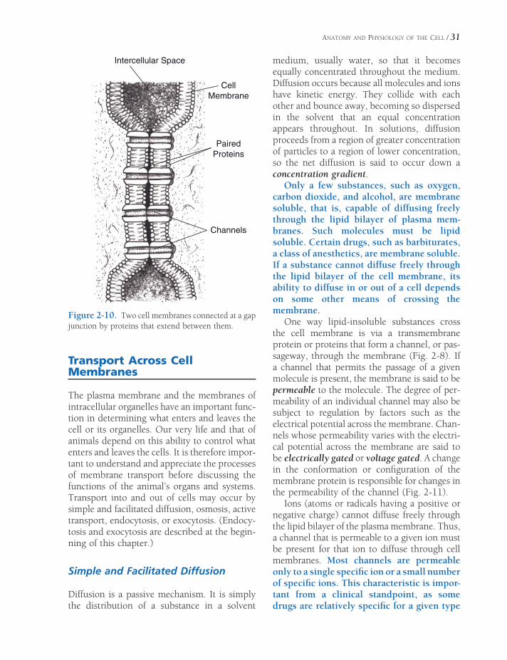
ANATOMY AND PHYSIOLOGY OF THE CELL / 31
Transport Across Cell Membranes
The plasma membrane and the membranes of intracellular organelles have an important func-tion in determining what enters and leaves the cell or its organelles. Our very life and that of animals depend on this ability to control what enters and leaves the cells. It is therefore impor-tant to understand and appreciate the processes of membrane transport before discussing the functions of the animal’s organs and systems. Transport into and out of cells may occur by simple and facilitated diffusion, osmosis, active transport, endocytosis, or exocytosis. (Endocy-tosis and exocytosis are described at the begin-ning of this chapter.)
Simple and Facilitated Diffusion
Diffusion is a passive mechanism. It is simply the distribution of a substance in a solvent
medium, usually water, so that it becomes equally concentrated throughout the medium. Diffusion occurs because all molecules and ions have kinetic energy. They collide with each other and bounce away, becoming so dispersed in the solvent that an equal concentration appears throughout. In solutions, diffusion proceeds from a region of greater concentration of particles to a region of lower concentration, so the net diffusion is said to occur down a concentration gradient.
Only a few substances, such as oxygen, carbon dioxide, and alcohol, are membrane soluble, that is, capable of diffusing freely through the lipid bilayer of plasma mem-branes. Such molecules must be lipid soluble. Certain drugs, such as barbiturates, a class of anesthetics, are membrane soluble. If a substance cannot diffuse freely through the lipid bilayer of the cell membrane, its ability to diffuse in or out of a cell depends on some other means of crossing the membrane.
One way lipid-insoluble substances cross the cell membrane is via a transmembrane protein or proteins that form a channel, or pas-sageway, through the membrane (Fig. 2-8). If a channel that permits the passage of a given molecule is present, the membrane is said to be permeable to the molecule. The degree of per-meability of an individual channel may also be subject to regulation by factors such as the electrical potential across the membrane. Chan-nels whose permeability varies with the electri-cal potential across the membrane are said to be electrically gated or voltage gated. A change in the conformation or confi guration of the membrane protein is responsible for changes in the permeability of the channel (Fig. 2-11).
Ions (atoms or radicals having a positive or negative charge) cannot diffuse freely through the lipid bilayer of the plasma membrane. Thus, a channel that is permeable to a given ion must be present for that ion to diffuse through cell membranes. Most channels are permeable only to a single specifi c ion or a small number of specifi c ions. This characteristic is impor-tant from a clinical standpoint, as some drugs are relatively specifi c for a given type
Intercellular Space
CellMembrane
PairedProteins
Channels
Figure 2-10. Two cell membranes connected at a gap junction by proteins that extend between them.
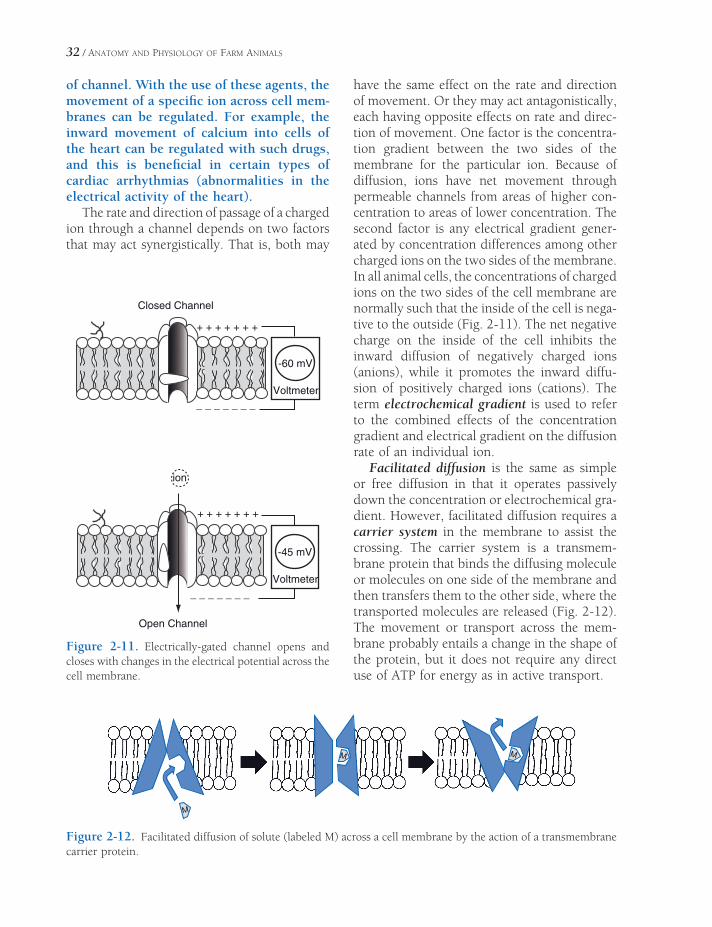
32 / ANATOMY AND PHYSIOLOGY OF FARM ANIMALS
of channel. With the use of these agents, the movement of a specifi c ion across cell mem-branes can be regulated. For example, the inward movement of calcium into cells of the heart can be regulated with such drugs, and this is benefi cial in certain types of cardiac arrhythmias (abnormalities in the electrical activity of the heart).
The rate and direction of passage of a charged ion through a channel depends on two factors that may act synergistically. That is, both may
have the same effect on the rate and direction of movement. Or they may act antagonistically, each having opposite effects on rate and direc-tion of movement. One factor is the concentra-tion gradient between the two sides of the membrane for the particular ion. Because of diffusion, ions have net movement through permeable channels from areas of higher con-centration to areas of lower concentration. The second factor is any electrical gradient gener-ated by concentration differences among other charged ions on the two sides of the membrane. In all animal cells, the concentrations of charged ions on the two sides of the cell membrane are normally such that the inside of the cell is nega-tive to the outside (Fig. 2-11). The net negative charge on the inside of the cell inhibits the inward diffusion of negatively charged ions (anions), while it promotes the inward diffu-sion of positively charged ions (cations). The term electrochemical gradient is used to refer to the combined effects of the concentration gradient and electrical gradient on the diffusion rate of an individual ion.
Facilitated diffusion is the same as simple or free diffusion in that it operates passively down the concentration or electrochemical gra-dient. However, facilitated diffusion requires a carrier system in the membrane to assist the crossing. The carrier system is a transmem-brane protein that binds the diffusing molecule or molecules on one side of the membrane and then transfers them to the other side, where the transported molecules are released (Fig. 2-12). The movement or transport across the mem-brane probably entails a change in the shape of the protein, but it does not require any direct use of ATP for energy as in active transport.
+ + + + + + +
Voltmeter
-60 mV
_ _ _ _ _ _ _
Voltmeter
-45 mV
+ + + + + + +
_ _ _ _ _ _ _
ion
Closed Channel
Open Channel
Figure 2-11. Electrically-gated channel opens and closes with changes in the electrical potential across the cell membrane.
M
M
M
Figure 2-12. Facilitated diffusion of solute (labeled M) across a cell membrane by the action of a transmembrane carrier protein.
ANATOMY AND PHYSIOLOGY OF THE CELL / 33
Sugars, especially glucose, depend on facili-tated diffusion to enter cells by joining with carrier proteins upon reaching the lipid bilayer of the membrane. The glucose carrier complex transports glucose down the glucose concentra-tion gradient to the inside of the cell mem-brane. Here the carrier releases the glucose to enter the cell. The carrier remains in the mem-brane and reconfi gures itself so that it is avail-able for more transport. Other substances besides glucose, such as amino acids, also depend on facilitated diffusion to cross cell membranes.
The rate at which facilitated diffusion occurs also depends on the number of carrier proteins available in the membrane. In the case of glucose, the speed of entry into many cells, such as skeletal muscle, is greatly increased by the hormone insulin, which the pancreas secretes. Insulin facilities the entry of glucose into skeletal muscle, in part by increasing the number of carrier proteins in the cell mem-brane of these cells.
Osmosis
Osmosis is movement of water across mem-branes. Like many solutes, water does not diffuse freely through the lipid bilayer of cell membranes but rather must diffuse through water channels formed by transmembrane pro-teins. These proteins are aquaporins. If the intracellular fl uid within a cell has a higher concentration of undiffusible solutes than the interstitial fl uid bathing the cell, water will move into the cell from the interstitial fl uid until the concentrations are the same on both sides of the membrane. As the water moves in, the volume in the cell increases. The driving force moving water from the solution on the side of the lower solute concentration to the side with the higher solute concentration is osmotic pressure.
The osmotic pressure of an aqueous solution can be measured by using a U-tube in which the two sides are separated by a semipermeable membrane, which is permeable only to water (Fig. 2-13). The solution is placed in one side
of the U-tube, and distilled water is placed in the other. The force of osmosis moves water through the membrane from the side contain-ing distilled water to the side containing the solution. This movement continues until the hydrostatic pressure generated by the increased height of the fl uid column on the solution side is equal to the osmotic force (Fig. 2-13). The units of osmotic pressure may be given in cen-timeters of water (height of water column), or converted to millimeters of mercury (mm Hg; height of a column of mercury creating an equivalent amount of hydrostatic pressure).
Osmotic pressure is an important mecha-nism in maintaining cellular volume by deter-mining whether water will enter or leave cells. If the concentration of solutions on each side of a membrane is the same, as seen with cells in blood, the bathing fl uid is said to be isotonic (isosmotic) in relation to the cells. This means that the osmotic pressure is the same on both sides of the membrane. A 0.9% solution of sodium chloride is considered to be isotonic with mammalian red blood cells and for this reason is called a normal or physiologic saline solution. Normal saline can be used to moisten exposed tissues, such as open wounds, without damaging the cells.
If the bathing fl uid has a lower osmotic pres-sure than the cells, it is said to be hypotonic, and water tends to cross the membrane and enter the cells. In the case of red blood cells in hypotonic plasma, the water entering the cells can swell and fi nally burst them, a condition called hemolysis.
Red blood cells in hypertonic plasma (more concentrated than the cell cytoplasm) lose water to the plasma and become wrinkled. The wrinkling of red cells is called crenation. In relation to mammalian cells, a solution less concentrated than 0.9% sodium chloride is said to be hypotonic; one more concentrated than 0.9% sodium chloride is said to be hypertonic; and of course a 0.9% sodium chloride solution is isotonic.
The osmotic pressure of a solution is deter-mined by the number of solute particles: the more solute particles in a volume of fl uid, the greater the osmotic pressure. The number of

34 / ANATOMY AND PHYSIOLOGY OF FARM ANIMALS
DistilledWater
GLUCOSE MOLECULES
selectivelypermeablemembrane
VolumeDecreased
VolumeIncreased
OsmoticForce
Chamber B Chamber A
Chamber B Chamber AFigure 2-13. Osmosis and osmotic pres-sure. A membrane impermeable to solute particles prevents their diffusion from chamber A into chamber B. Distilled water from chamber B migrates into chamber A until the difference in the height of the water columns equals the osmotic pressure of the solution in chamber A.
particles is determined by the molar concentra-tion of the solution and by the number of ions formed if the solute is an electrolyte. For example, glucose is not an electrolyte, having one particle per molecule, but sodium chloride is an electrolyte, giving two particles (Na+ and Cl−) per molecule when placed in solution. A 1-molar solution of sodium chloride has twice the osmotic pressure of a 1-molar solution of glucose, because the sodium chloride solution has twice the number of particles in solution.
These concepts of osmosis and osmotic pres-sure become important when intravenous fl uids are administered to animals for problems such as dehydration, anorexia, milk fever, and diar-rhea. Furthermore, they are important princi-ples in normal functions of animals, such as the fl ow of blood and lymph, the excretion of
wastes in the urine by the kidneys, and the digestion and absorption of food, as is dis-cussed in subsequent chapters.
Active Transport
Some molecules and ions can move across cell membranes (either in or out of cells) against concentration or electrical gradients. The term against, as used here, means that the particles are moving in the direction opposite that of diffusion. This movement across the cell mem-brane consumes energy produced by the cell and is called active transport.
The best-recognized example of a primary active transport system is the sodium– potassium (Na–K) pump. This pump is actually
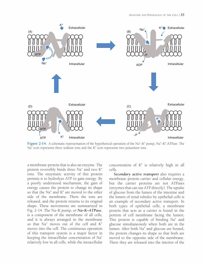
ANATOMY AND PHYSIOLOGY OF THE CELL / 35
a membrane protein that is also an enzyme. The protein reversibly binds three Na+ and two K+ ions. The enzymatic activity of this protein permits it to hydrolyze ATP to gain energy. By a poorly understood mechanism, the gain of energy causes the protein to change its shape so that the Na+ and K+ are moved to the other side of the membrane. There the ions are released, and the protein returns to its original shape. These movements are summarized in Fig. 2-14. The Na–K pump, or Na–K–ATPase, is a component of the membrane of all cells, and it is always arranged in the membrane so that Na+ moves out of the cell and K+ moves into the cell. The continuous operation of this transport system is a major factor in keeping the intracellular concentration of Na+ relatively low in all cells, while the intracellular
concentration of K+ is relatively high in all cells.
Secondary active transport also requires a membrane protein carrier and cellular energy, but the carrier proteins are not ATPases (enzymes that can use ATP directly). The uptake of glucose from the lumen of the intestine and the lumen of renal tubules by epithelial cells is an example of secondary active transport. In both types of epithelial cells, a membrane protein that acts as a carrier is found in the portion of cell membrane facing the lumen. This protein is capable of binding Na+ and glucose simultaneously when both are in the lumen. After both Na+ and glucose are bound, the protein changes its shape so that both are moved to the opposite side of the membrane. There they are released into the interior of the
Extracellular
Extracellular
(A)
(D) (C)
(B)Extracellular
Extracellular
K+
K+
K+
K+
Na+
Na+
ATP
ATP
ATP
ADP
~P
~P
Intracellular
Intracellular
Intracellular
Intracellular
Figure 2-14. A schematic representation of the hypothetical operation of the Na+–K+ pump, Na+–K+-ATPase. The Na+ icon represents three sodium ions and the K+ icon represents two potassium ions.
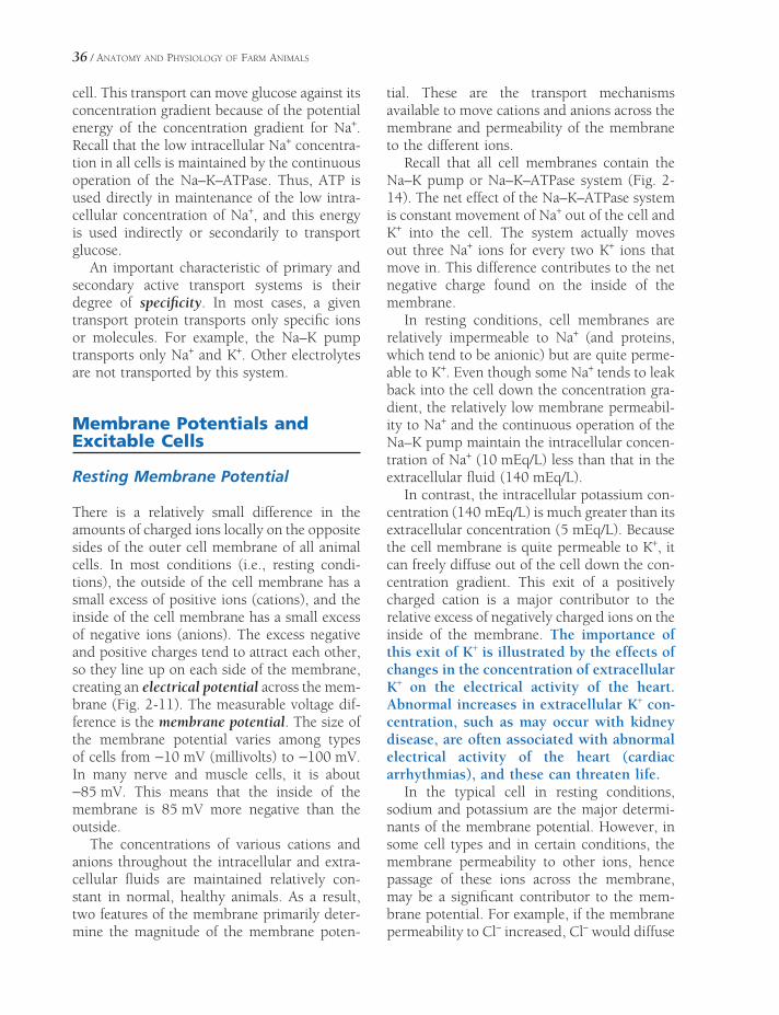
36 / ANATOMY AND PHYSIOLOGY OF FARM ANIMALS
cell. This transport can move glucose against its concentration gradient because of the potential energy of the concentration gradient for Na+. Recall that the low intracellular Na+ concentra-tion in all cells is maintained by the continuous operation of the Na–K–ATPase. Thus, ATP is used directly in maintenance of the low intra-cellular concentration of Na+, and this energy is used indirectly or secondarily to transport glucose.
An important characteristic of primary and secondary active transport systems is their degree of specifi city. In most cases, a given transport protein transports only specifi c ions or molecules. For example, the Na–K pump transports only Na+ and K+. Other electrolytes are not transported by this system.
Membrane Potentials and Excitable Cells
Resting Membrane Potential
There is a relatively small difference in the amounts of charged ions locally on the opposite sides of the outer cell membrane of all animal cells. In most conditions (i.e., resting condi-tions), the outside of the cell membrane has a small excess of positive ions (cations), and the inside of the cell membrane has a small excess of negative ions (anions). The excess negative and positive charges tend to attract each other, so they line up on each side of the membrane, creating an electrical potential across the mem-brane (Fig. 2-11). The measurable voltage dif-ference is the membrane potential. The size of the membrane potential varies among types of cells from −10 mV (millivolts) to −100 mV. In many nerve and muscle cells, it is about −85 mV. This means that the inside of the membrane is 85 mV more negative than the outside.
The concentrations of various cations and anions throughout the intracellular and extra-cellular fl uids are maintained relatively con-stant in normal, healthy animals. As a result, two features of the membrane primarily deter-mine the magnitude of the membrane poten-
tial. These are the transport mechanisms available to move cations and anions across the membrane and permeability of the membrane to the different ions.
Recall that all cell membranes contain the Na–K pump or Na–K–ATPase system (Fig. 2-14). The net effect of the Na–K–ATPase system is constant movement of Na+ out of the cell and K+ into the cell. The system actually moves out three Na+ ions for every two K+ ions that move in. This difference contributes to the net negative charge found on the inside of the membrane.
In resting conditions, cell membranes are relatively impermeable to Na+ (and proteins, which tend to be anionic) but are quite perme-able to K+. Even though some Na+ tends to leak back into the cell down the concentration gra-dient, the relatively low membrane permeabil-ity to Na+ and the continuous operation of the Na–K pump maintain the intracellular concen-tration of Na+ (10 mEq/L) less than that in the extracellular fl uid (140 mEq/L).
In contrast, the intracellular potassium con-centration (140 mEq/L) is much greater than its extracellular concentration (5 mEq/L). Because the cell membrane is quite permeable to K+, it can freely diffuse out of the cell down the con-centration gradient. This exit of a positively charged cation is a major contributor to the relative excess of negatively charged ions on the inside of the membrane. The importance of this exit of K+ is illustrated by the effects of changes in the concentration of extracellular K+ on the electrical activity of the heart. Abnormal increases in extracellular K+ con-centration, such as may occur with kidney disease, are often associated with abnormal electrical activity of the heart (cardiac arrhythmias), and these can threaten life.
In the typical cell in resting conditions, sodium and potassium are the major determi-nants of the membrane potential. However, in some cell types and in certain conditions, the membrane permeability to other ions, hence passage of these ions across the membrane, may be a signifi cant contributor to the mem-brane potential. For example, if the membrane permeability to Cl− increased, Cl− would diffuse
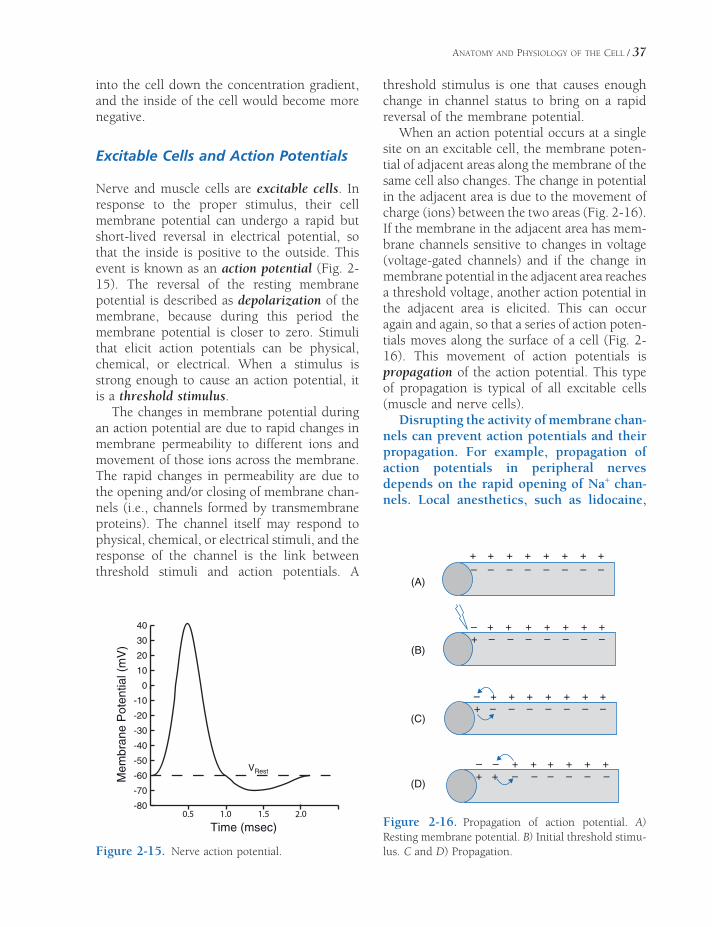
ANATOMY AND PHYSIOLOGY OF THE CELL / 37
into the cell down the concentration gradient, and the inside of the cell would become more negative.
Excitable Cells and Action Potentials
Nerve and muscle cells are excitable cells. In response to the proper stimulus, their cell membrane potential can undergo a rapid but short-lived reversal in electrical potential, so that the inside is positive to the outside. This event is known as an action potential (Fig. 2-15). The reversal of the resting membrane potential is described as depolarization of the membrane, because during this period the membrane potential is closer to zero. Stimuli that elicit action potentials can be physical, chemical, or electrical. When a stimulus is strong enough to cause an action potential, it is a threshold stimulus.
The changes in membrane potential during an action potential are due to rapid changes in membrane permeability to different ions and movement of those ions across the membrane. The rapid changes in permeability are due to the opening and/or closing of membrane chan-nels (i.e., channels formed by transmembrane proteins). The channel itself may respond to physical, chemical, or electrical stimuli, and the response of the channel is the link between threshold stimuli and action potentials. A
threshold stimulus is one that causes enough change in channel status to bring on a rapid reversal of the membrane potential.
When an action potential occurs at a single site on an excitable cell, the membrane poten-tial of adjacent areas along the membrane of the same cell also changes. The change in potential in the adjacent area is due to the movement of charge (ions) between the two areas (Fig. 2-16). If the membrane in the adjacent area has mem-brane channels sensitive to changes in voltage (voltage-gated channels) and if the change in membrane potential in the adjacent area reaches a threshold voltage, another action potential in the adjacent area is elicited. This can occur again and again, so that a series of action poten-tials moves along the surface of a cell (Fig. 2-16). This movement of action potentials is propagation of the action potential. This type of propagation is typical of all excitable cells (muscle and nerve cells).
Disrupting the activity of membrane chan-nels can prevent action potentials and their propagation. For example, propagation of action potentials in peripheral nerves depends on the rapid opening of Na+ chan-nels. Local anesthetics, such as lidocaine,
-80
-70
-60
-50
-40
-30
-20
-10
0
10
20
30
40
0.5 1.0 1.5 2.0
Time (msec)
Mem
bran
e P
oten
tial (
mV
)
VRest
+ + +
–– – – – – – –
+ + + + +
– +
(A)
(B)
(C)
(D)
+–+ – – – – – –
+ + + + +
– + +–+ – – – – – –
+ + + + +
– – +–+ + – – – – –
+ + + + +
Figure 2-15. Nerve action potential.
Figure 2-16. Propagation of action potential. A) Resting membrane potential. B) Initial threshold stimu-lus. C and D) Propagation.
38 / ANATOMY AND PHYSIOLOGY OF FARM ANIMALS
block these sodium channels and prevent action potential propagation when applied along a peripheral nerve.
Membrane Receptors and Intracellular Signaling
The cell membrane protects the cell from the extracellular environment by restricting exchange between the intracellular and extra-cellular fl uid spaces. However, cells must also be able to detect the presence of certain chemi-cal substances in the extracellular fl uid and alter appropriate intracellular functions when these chemicals are present. These abilities are essential for communication between individ-ual nerve cells, between nerve and muscle cells, and between cells of endocrine glands and the cells affected by the hormones secreted by endocrine glands. The recognition of the spe-cifi c chemical in the extracellular fl uid and the response to its presence are based on the inter-action between the chemical and a membrane protein. Ligand is the general term applied to any chemical that is capable of interacting with, or binding to, a membrane protein or mem-brane receptor. Important characteristics of the ligand–receptor relationship are reversibility, specifi city, affi nity, and saturation.
The binding of a ligand to a membrane receptor is usually reversible, for it is due to relatively weak chemical attractions. The weak-ness of this binding is also a reason for some of the other characteristics of the ligand–receptor relationship. Membrane receptors can bind to only a limited number of particular ligands or chemicals; that is, receptors demonstrate speci-fi city for certain ligands. However, even though a receptor may be capable of binding any of a number of ligands, the affi nity, or strength of binding, between the receptor and ligand may not be the same for both chemicals. Ligands that form a strong bond with a receptor are described as having a high affi nity for the receptor.
The number of membrane receptors on any given cell or cell population is fi nite. Thus, it is possible to provide enough ligand that all receptor molecules have ligand bound to them.
A receptor is described as occupied when it has a ligand bound to it. Saturation of receptors occurs when all receptors are occupied with ligand.
These concepts and characteristics of ligands and receptors are the basis for a variety of pharmaceutical agents. For example, b-blocker drugs are ligands that bind to b-adrenergic receptors but do not produce any biologic response. However, because the b-blocker occupies the receptor, the binding of any other ligand is prevented. Thus, b-blockers are used when it is desir-able to reduce the biologic activity of b-adrenergic receptor stimulation. The binding of the b-blocker to the b-receptor prevents normal endogenous agents (epinephrine and norepinephrine) from binding to the recep-tor and bringing about a biologic response.
The binding of a ligand to a membrane receptor is but the fi rst step in the process by which a chemical in the extracellular fl uid can alter the function of a cell without entering it. The second step depends on the particular type of membrane receptor. In some cases, the membrane receptor is also a membrane channel. The binding of a ligand to these types of recep-tors is associated with a change in the permea-bility of the channel; hence, these channels are described as being ligand-gated. Other mem-brane receptors are also enzymes that are acti-vated by the binding of a ligand. These enzymes have active sites facing the interior of the cell, so intracellular functions are changed as a result of the ligand–receptor interaction.
Yet another way that the function of a cell can be changed after a ligand binds membrane receptors involves another specifi c group of membrane proteins known as G proteins. G proteins are within the cell membrane and closely associated with certain receptor pro-teins. The G protein acts as an intermediate in the chain of events between ligand binding and cellular response. As a result of the ligand–receptor binding, the associated G protein acts to activate or inhibit an enzyme or membrane channel. The potential actions of G proteins are summarized in Fig. 2-17.
Adenyl cyclase is a cell membrane enzyme whose activity is regulated by ligand–receptor

ANATOMY AND PHYSIOLOGY OF THE CELL / 39
interactions. In some cases, the enzyme also acts as the cell membrane receptor, but in other cases, the enzyme is linked to a receptor via a G protein. Adenyl cyclase catalyzes the intracel-lular formation of cyclic adenosine monophos-phate (cAMP), which in turn can activate intracellular kinases. These activated kinases can activate other intracellular proteins to change cellular function. In this general scheme, cAMP is said to be a second messenger in that it transfers a chemical signal from the mem-brane to the interior of the cell. cAMP is the second messenger for many of the peptide hor-mones that bind to membrane receptors. Examples include parathyroid hormone, gluca-gon, and luteinizing hormone.
Membrane proteins may also act as recep-tor sites for attachment of disease-causing agents, such as viruses. Some viruses bind to appropriate membrane receptors on sus-ceptible cells, which then endocytose the virus receptor complex. This provides a means by which the virus can infect the sus-ceptible cell.
Cytoplasm and Cytoplasmic Organelles
Cytoplasm
Cytoplasm is the material fi lling the inside of the cell and containing the intracellular organ-elles and nucleus (Fig. 2-18). Organelles are
intracellular structures organized for a parti-cular function within the cell. These include the Golgi apparatus, endoplasmic reticulum (smooth and rough), mitochondria, centrioles, free ribosomes, lysosomes, peroxisomes, and a variety of crystals, granules, and droplets (formerly called inclusions, collectively). The relatively liquid component of cytoplasm is cytosol; the organelles are arranged within the cytosol by a complex system of intra -cellular fi laments and microtubules called the cytoskeleton.
Dissolved or suspended within the fl uid cytosol are a variety of proteins, sugars, and salts. Many of the proteins are enzymes that function in the metabolic activities of the cell. Within some cells are proteins that function as cytosolic receptors binding ligands that have gained access to the cytosol after passing through the cell membrane.
The Golgi Apparatus
The Golgi apparatus varies in size and location in cells of different tissues but generally appears as a stack of fl attened membranous sacs (lamel-lae) near the nucleus (Fig. 2-18). It functions as the site of the fi nal stages of synthesis and packaging of secretory products of the cell. Within the Golgi, secretory products are enclosed within a membrane vesicle for tempo-rary storage in the cell or for transport to the plasma membrane, where exocytosis releases
Extracellular Fluid
Cytoplasm
B
GIGSA
Figure 2-17. The G proteins associated with membrane receptors can be stimulatory (GS) or inhibitory (GI). The G proteins will act on associated membrane enzymes, A, or channels, B, after the appropriate ligand binds to the membrane receptor that is exposed to the extracellular fl uid.

40 / ANATOMY AND PHYSIOLOGY OF FARM ANIMALS
the products into the extracellular fl uid as a form of secretion. Mucopolysaccharides may form in the Golgi apparatus, and glycoproteins are terminally synthesized there as combina-tions of carbohydrates and proteins that have been transported to the Golgi apparatus by the smooth and rough endoplasmic reticulum.
The Endoplasmic Reticulum and Ribosomes
The endoplasmic reticulum is a membranous network found throughout the cytoplasm of the cell (Fig. 2-18). It was fi rst described in the endoplasm (the cytoplasm deepest in the cell), giving rise to the name endoplasmic reticulum. Although still called the endoplasmic reticu-lum, it has been observed in all parts of the cytoplasm and may be continuous with the outer nuclear membrane. The endoplasmic
reticulum is in the form of tubules and sheets, with occasional enlarged sacs or vesicles called cisternae.
In some sites, the endoplasmic reticulum is associated with ribosomes, which appear like beads along the membrane. Granular or rough endoplasmic reticulum (Fig.2-18) is endoplas-mic reticulum associated with ribosomes; agranular, or smooth endoplasmic reticulum (Fig. 2-18), has no ribosomes associated with it.
Mitochondria
Mitochondria are ovoid organelles about 10 μm long. The double membrane of the mitochon-drion, with the cristae projecting into the inte-rior, provides a large surface area for attachment of enzymes (Fig. 2-18). Studies of fragmented mitochondria indicate that all of the enzymes
Figure 2-18. General organization of a typical cell. (Reprinted with permission of Wiley-Blackwell from Tortora, G.J. and Derrickson, B., Principles of Anatomy and Physiology, 11th ed. Hoboken, NJ: John Wiley & Sons, 2006.)

ANATOMY AND PHYSIOLOGY OF THE CELL / 41
associated with oxidation of nutrients to carbon dioxide, ATP, and water are found in the mito-chondria. Thus, all of the enzymes and coen-zymes involved in the tricarboxylic acid cycle (also called the Krebs cycle, or citric acid cycle) are largely local to the mitochondria.
Oxidation during the tricarboxylic acid cycle releases carbon dioxide and hydrogen atom pairs (H2). The H2 furnishes its electrons to the mitochondrial electron transport system to drive a series of reduction reactions, culminat-ing in the formation of water and storage of the energy in the form of ATP. The ATP is formed by the oxidative phosphorylation of ADP (ade-nosine diphosphate), which adds one inorganic phosphate molecule to ADP, creating a higher-energy compound. The energy incorporated into ATP becomes available for any cellular activity that requires energy, such as protein synthesis, muscle contraction, and active trans-port. Energy is released during the reconver-sion of ATP to ADP and an inorganic phosphate. Most of the cellular processes requiring energy take place outside of the mitochondria.
Since the mitochondria produce the energy for the cell, it follows that the more mitochon-dria in a cell, the more active the cell can be. Mitochondrial ATP production depends on oxygen, so highly active cells also need a ready supply of oxygen. The enzymes of the glyco-lytic pathway, using glucose as a substrate, can produce ATP without oxygen in the cytoplasm. However, this pathway is less effi cient than mitochondrial production; it produces less ATP per molecule of substrate.
Mitochondria contain their own DNA and RNA for reproducing themselves. They also carry on partial synthesis of proteins and lipids and have their own ribosomes. Mitochondrial reproduction can be stimulated by increased demands for cell energy and is not dependent on cellular division.
Lysosomes
Lysosomes are membrane-bound vesicles of digestive (hydrolytic) enzymes. They are larger than ribosomes but smaller than mitochondria,
ranging in diameter from 0.25 to 0.75 μm. Lysosomes apparently originate from the endo-plasmic reticulum and the Golgi apparatus.
Lysosomes contain a variety of enzymes that degrade all types of biologic molecules. Nor-mally, the membrane of the lysosome prevents lysosomal enzymes from acting on molecules within the cytoplasm. However, in certain con-ditions, the enzymes are released into the cytosol, which may then lyse (destroy) the cell itself.
Cytoplasmic vesicles formed by the phago-cytosis of extracellular material may fuse with lysosomes, thereby permitting the enzymatic digestion of the contents of the vesicle while protecting the cell itself from lysis. White blood cells, which act as scavenger cells by phagocy-tizing bacteria, dead tissue, and damaged cell debris, contain many lysosomes. Lysosomes also engulf and degrade intracellular organelles. This is a means by which individual cells can remove and recover components of damaged parts of themselves. The only mammalian cells that are known not to contain lysosomes are the red blood cells.
When cells contain inactive lysosomes, disease can follow. An example is Pompe’s disease, in which the lysosomes cannot digest glycogen. Also, changes caused by sunburn occur when the ultraviolet light of the sun rup-tures lysosomes in the skin cells.
Other Structures
Peroxisomes are smaller than lysosomes and are most numerous in cells of the liver and kidney. Peroxisomes contain enzymes responsible for degrading lipids, alcohols, and a variety of potentially toxic substances. Hydrogen peroxide is produced within the peroxisome as a result of this enzymatic action. Other enzymes within the peroxisome rapidly degrade the potentially toxic hydrogen peroxide to protect the cell.
Microtubules, intermediate fi laments, and microfi laments are rodlike organelles that make up the cytoskeleton, which primarily functions to determine the shape of the cell and assist with cell movement. Microtubules are scattered throughout the cytoplasm in most cells and are

42 / ANATOMY AND PHYSIOLOGY OF FARM ANIMALS
the largest and most rigid of the three cytoskel-etal components (Fig. 2-19). Microtubules are spindle fi bers in cell division, motile elements in cilia, and assisters of transport of molecules within some cells, such as in the processes of neurons (nerve cells). Intermediate fi laments are primarily found in association with special-ized cell-to-cell junctions, such as desmosomes (Fig. 2-9).
Microfi laments are thinner than microtu-bules, but they make up most of the cytoskel-eton. Microfi laments are composed of actin, a protein involved in cell movement and muscle contraction.
The centriole is a short cylinder about 0.2 μm wide and 0.4 μm long. Centrioles, composed of nine triplets of microtubules, usually occur at the bases of cilia, where they are called basal bodies. A pair of centrioles, the centrosome, also occurs in all cells near the nucleus and organizes the microtubules, which form the mitotic spindle during cell division.
Nucleus
Structure of the Nucleus
The nucleus contains the genetic material of the cell encoded in molecules of DNA. With light
microscopy, DNA and its associated proteins are seen as a more diffusely staining chromatin in the nondividing cell and as chromosomes in the dividing cell. The nuclei of somatic cells contain the information necessary for deter-mining the form and structure of new cells, and the nuclei of sex cells contain the information necessary to determine the characteristics of a new individual. The nucleoli consist largely of clustered DNA, which codes for ribosomal RNA; the nucleoli are seen as densely staining spherical bodies in the nucleus (Fig. 2-18).
The nuclear envelope (Fig. 2-18), which sur-rounds the cell nucleus, is composed of two distinct membranes separated by about 20 nm. The outer membrane is continuous with the endoplasmic reticulum. Pores (small gaps or interruptions) in the nuclear envelope permit exchange between the protoplasm of the nucleus (nucleoplasm) and the cytoplasm outside the nucleus, including the movement of RNA synthesized in the nucleus out into the cytoplasm.
The functional activity and the continued life of the cell depend on the presence and functional integrity of a nucleus. A cell from which the nucleus has been removed (enucle-ated) gradually ceases activity, atrophies, and fi nally dies. However, if the nucleus is replaced with a nucleus from a cell from the same species
Figure 2-19. Microtubules in axons of neu-rosecretory neurons. (Reprinted with permis-sion of Wiley-Blackwell from Dellmann, HD and Eurell, J. Textbook of Veterinary Histology. 5th ed. Baltimore: Lippincott Williams & Wilkins, 1998.)

ANATOMY AND PHYSIOLOGY OF THE CELL / 43
prior to irreversible atrophy, function of the cell can be restored. The only cells in higher animals that do not have nuclei are mature red blood cells. This lack of nucleus is associated with their short life span, only 120 days.
The primary functions of the nucleus are (1) to regulate protein synthesis in the cell, thereby regulating the biochemical activities of the cell, and (2) to ensure the passage of genetic mate-rial (the chromosomes and their component genes) to subsequent generations of cells and/or organisms.
DNA and DNA Replication
The genetic material necessary to direct cellular functions is primarily composed of chains of DNA. The chains of DNA are formed by joining small units (nucleotides), each containing a phosphate, a sugar (deoxyribose), and either a purine or pyrimidine base. The purine bases in DNA are adenine and guanine, and the pyrimidine bases are thymine and cytosine (Fig. 2-20).
Watson and Crick determined the structure of DNA to be a double helix, something like a spiral staircase or twisted metal ladder. The outside rails consist of two long chains of sugar–phosphate molecules, and the rungs are
made up of paired bases that hold the two parts of the double helix together. Adenine is always paired with thymine, and guanine is always paired with cytosine. The two strands are joined by hydrogen bonds between the bases (Fig. 2-21). The two strands of the DNA double helix are not identical but are complementary. In other words, whenever adenine appears on one strand, thymine is in the same position on the opposite strand, and whenever guanine is on one strand, cytosine is in the same position on the other strand.
To pass on the genetic information to the next generation of cells or animals, the DNA double helix must be replicated. Replication of DNA begins with unwinding of the helix and splitting of the chain at the point of junction of
Figure 2-20. General structure of a nucleotide (sub-units of DNA and RNA).
Figure 2-21. The basic structure of DNA. There are four nucleotides. The symbols in the upper right show how the nucleotides pair in DNA. (Adapted with per-mission from Cohen, B.J. and Wood, D.L. Memmler’s The Human Body in Health and Disease. 9th ed. Phila-delphia: Lippincott Williams & Wilkins, 2000.)
O
-O
H
CC
N
NH2
OCH2
N
O-
P O
O
H
CH
OH
C
H
C
H
Typical deoxyribonucleotide:
Base (cytosine)
Sugar (deoxyribose)
Phosphate
Cytosine (C)
Thymine (T)
Adenine (A)
Guanine (G)
Sugar unitPhosphate unit
Nitrogen base (G)

44 / ANATOMY AND PHYSIOLOGY OF FARM ANIMALS
complementary bases. Each separated strand serves as a template, or model, for the forma-tion of its complementary strand, which pro-duces two double DNA helices, replicas of the original. Each new double helix consists of one strand of the original double helix and one newly synthesized strand (Fig. 2-22). Errors in the duplication of DNA strands during replica-tion give rise to genetic mutations. Errors may occur spontaneously, or their frequency may be increased through the effects of numerous external factors, or mutagens (e.g., ionizing radiation, exposure to certain chemicals).
The genetic information in DNA is coded by the specifi c sequence of purine and pyrimidine bases in a DNA molecule. This sequential arrangement of bases and its control of hered-ity, both on the cellular and the species level, have been called the genetic code, or the lan-guage of life. The interpretation of this code results in the synthesis of specifi c proteins. The only cellular constituent whose synthesis is specifi cally directed by the genetic code is protein. Such proteins include those that are
secreted as cellular products, those that are found in the cell membrane, and those that function within the cytosol or within cellular organelles.
The DNA code is said to be a triplet code, for each group of three nucleotides in the DNA chain ultimately calls for a specifi c amino acid in the process of protein synthesis. There are approximately 20 amino acids in the cell. With four bases that may be included in a triplet, there are more than enough potential triplet codes to represent the 20 amino acids. Other triplet codes in the DNA serve as signals to demarcate the segment of the DNA chain that represents a particular protein and to regulate the initial and terminal steps in protein synthesis.
A gene is a segment of DNA that contains the triplet codes for all amino acids in one or more proteins and the signal sequences neces-sary to regulate the processing of the DNA segment. However, interspersed within a gene are also sequences of nucleotides that are not regulators of the process and do not contain
C
CTG CA T
T
GG
A
G
T
C
A
T
G
C
A
C
G
G
A
C
C
GC
T
G
T
C
T
C
G
A
T
T
C G A C
G
G
C
CT
C
C G A G C T AA
C
GG
A T G
A
G C T G A
G A C
C T G
C
DNApolymerase
Parent
ReplicaA
T
G
C
Adenine
Thymine
Guanine
Cytosine
Figure 2-22. Replication of DNA.

ANATOMY AND PHYSIOLOGY OF THE CELL / 45
necessary triplet codes. These noncoding, non-regulatory segments are introns. The DNA coding segments in a gene are exons. A single gene may have multiple exons and introns throughout its length.
RNA: Transcription and Translation
The processes by which the genetic code is interpreted and proteins are synthesized require the participation of three forms of RNA (ribo-nucleic acid). The three forms of RNA are mes-senger RNA (mRNA), transfer RNA (tRNA), and ribosomal RNA (rRNA). Like DNA, all three forms of RNA consist of nucleotide units that contain a sugar (ribose), a phosphate, and a purine or pyrimidine base. The two purines that are found in DNA, adenine and guanine, are also found in RNA, as is the pyrimidine cytosine. However, RNA does not contain the pyrimidine thymine that is found in DNA. Instead, RNA contains the pyrimidine uracil. Whereas the structure of DNA was two strands or chains of nucleotides joined together in a double helix, RNA exists only as a single strand.
The fi rst step in the interpretation of the genetic code, transcription, results in the for-mation of a mRNA. This process is similar to DNA replication except that DNA now serves as a template for the synthesis of a mRNA instead of a new complementary strand of DNA. A specifi c nuclear enzyme, RNA poly-merase II, and other nuclear proteins collec-tively known as general transcription factors bind together at a specifi c site on the DNA to initiate the transcription of a specifi c gene. The site on the DNA at which binding occurs and transcription begins is known as a promoter. A special segment of DNA adjacent to the pro-moter region contains a start sequence of nucle-otides to signal RNA polymerase II to begin the synthesis of mRNA. Other regulatory proteins that act as enhancer or repressor transcription factors can also infl uence the rate of transcrip-tion of a specifi c gene. These regulatory tran-scription factors may bind to sites on DNA that are distant from the promoter region, but
because of the folding and curling of the DNA strand, they can interact with the proteins bound at the promoter region.
Using the DNA as a template, RNA poly-merase II synthesizes a single complementary strand of nucleotides. As with DNA, each group of three nucleotides in the newly synthe-sized mRNA is the code for a specifi c amino acid. Each group of three nucleotides in the mRNA is a codon. When the end of the segment of DNA that represents a particular protein is reached, a stop sequence in the DNA terminates the mRNA synthesis, and the mRNA strand detaches from the RNA poly-merase. Figure 2-23 summarizes the steps of transcription.
Recall that the segment of DNA that repre-sents a gene and serves as the template for mRNA contains both exons and introns (i.e., both coding and noncoding regions). Thus, the newly synthesized mRNA must be processed to remove the segments that correspond to the introns in the DNA. This processing is done before the mRNA exits the nucleus and enters the cytoplasm, where protein synthesis will take place. The segments of mRNA that corre-spond to the noncoding introns are excised, and the segments that correspond to the coding exons are spliced together by a complex con-sisting of RNA and a protein called a spliceo-some. During the splicing, the spliceosome sometimes omits segments of the initial mRNA that correspond to some of the coding exons. This omission results in alternately spliced mRNAs and ultimately the synthesis of differ-ent proteins from the transcription of a single gene.
Within the cytoplasm, the processed mRNA binds to a ribosome. This binding occurs at a specifi c end of the mRNA under the direction of a start codon at that site. The start codon also signals the binding of an initial tRNA with a specifi c amino acid attached. There is at least one specifi c tRNA for each of the approximately 20 amino acids in the cell. After the fi rst tRNA is bound, a second tRNA with its attached amino acid arrives and binds to the next codon in line. Ribosomal enzymes then detach the amino acid from the fi rst tRNA and link the two
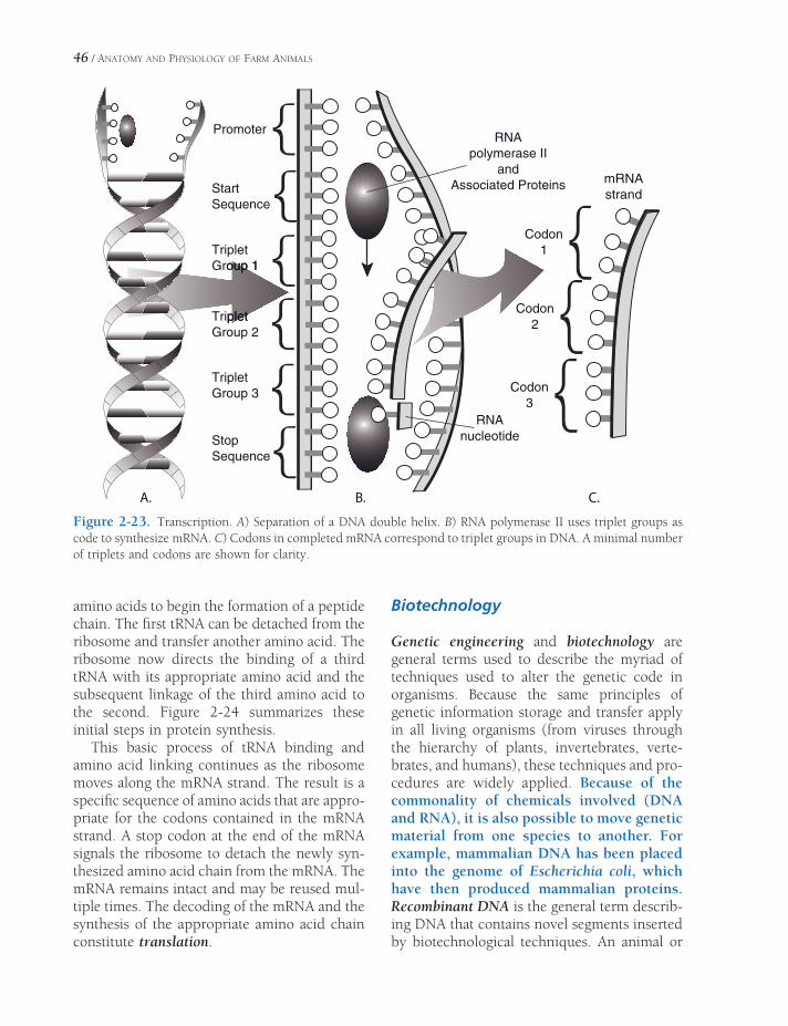
46 / ANATOMY AND PHYSIOLOGY OF FARM ANIMALS
StartSequence
StopSequence
TripletGroup 1oup 1
TripletpletGroup 2
TripletGroup 3
RNApolymerase II
andAssociated Proteins
Codon1
Codon2
Codon3
mRNAstrand
RNAnucleotide
Promoter
B.A. C.
Figure 2-23. Transcription. A) Separation of a DNA double helix. B) RNA polymerase II uses triplet groups as code to synthesize mRNA. C) Codons in completed mRNA correspond to triplet groups in DNA. A minimal number of triplets and codons are shown for clarity.
amino acids to begin the formation of a peptide chain. The fi rst tRNA can be detached from the ribosome and transfer another amino acid. The ribosome now directs the binding of a third tRNA with its appropriate amino acid and the subsequent linkage of the third amino acid to the second. Figure 2-24 summarizes these initial steps in protein synthesis.
This basic process of tRNA binding and amino acid linking continues as the ribosome moves along the mRNA strand. The result is a specifi c sequence of amino acids that are appro-priate for the codons contained in the mRNA strand. A stop codon at the end of the mRNA signals the ribosome to detach the newly syn-thesized amino acid chain from the mRNA. The mRNA remains intact and may be reused mul-tiple times. The decoding of the mRNA and the synthesis of the appropriate amino acid chain constitute translation.
Biotechnology
Genetic engineering and biotechnology are general terms used to describe the myriad of techniques used to alter the genetic code in organisms. Because the same principles of genetic information storage and transfer apply in all living organisms (from viruses through the hierarchy of plants, invertebrates, verte-brates, and humans), these techniques and pro-cedures are widely applied. Because of the commonality of chemicals involved (DNA and RNA), it is also possible to move genetic material from one species to another. For example, mammalian DNA has been placed into the genome of Escherichia coli, which have then produced mammalian proteins. Recombinant DNA is the general term describ-ing DNA that contains novel segments inserted by biotechnological techniques. An animal or

Figure 2-24. Protein synthesis. A) Transcription results in processed mRNA that exits nucleus. B) Translation of the mRNA produces a peptide chain. C) The completed peptide is released.
47
48 / ANATOMY AND PHYSIOLOGY OF FARM ANIMALS
plant that contains DNA from another organ-ism is said to be transgenic.
The discovery of a group of enzymes known as restriction nucleases was a key factor in the development of recombinant DNA tech-niques. These enzymes cut DNA into shorter segments by splitting the linkages between nucleotides. The enzymes do not act at random sites through the DNA strand; instead, each individual nuclease acts at a specifi c site termed its restriction site. If DNA from two different organisms are treated with the same nuclease, the nuclease will fragment the DNA at similar restriction sites in both. This yields segments of DNA with similar characteristics at their ends, but the sequences within the segments may be quite different. Because the ends of the seg-ments from the two organisms are similar, DNA ligase (an enzyme that reestablishes the nucleotide linkages) can be used to join the different DNA segments. The fi nal result is a DNA strand that contains DNA from two dif-ferent organisms.
To produce a transgenic organism, recombi-nant DNA must be inserted into an organism’s genome. In domestic animals, this has been accomplished by microinjection of recombi-nant DNA into a pronucleus of single-cell embryos. The pronucleus is a nucleuslike struc-ture in the embryo that contains genetic mate-rial from one parent. One-cell embryos have two pronuclei that ultimately fuse so that the genetic material from the two parents can be joined in the new individual. Recombinant DNA has also been transferred into embryos by infecting early-stage embryos with retroviruses containing recombinant DNA in their genome. Retroviruses are a specifi c group of viruses that insert their own genetic material into the genome of organisms they infect. As they insert their own genetic material, they also insert the recombinant DNA.
Clones are genetically identical individuals produced by asexual means. Cloning has been accomplished by splitting an early-stage multi-cell embryo into single cells, which continue their development into identical individuals. This technique has been successfully used in domestic animals. Clones have also been pro-duced by nuclear transfer. In this technique,
nuclei obtained from cells of adult animals are transferred into oocytes with their original nucleus removed. The oocytes with the trans-ferred nucleus can be placed in the uterus of an appropriate female for gestation.
Cell Division
Mitosis
Each day millions of cells in the body of any normal animal die and are degraded or sloughed from epithelial surfaces. These cells must be replaced if normal life is to continue, and the replacement cells must be replicas of the original cells. Mitosis, the division of somatic cells to produce two daughter cells, includes the dupli-cation of genetic material for each daughter cell. Even though the process of cell division is nor-mally a continuous process, here it has been divided into periods, or phases, for ease of com-munication about the process. The active phases are primarily based on nuclear changes visible by light microscopy. They are prophase, meta-phase, anaphase, and telophase (Fig. 2-25).
Interphase. The period between active cell divisions is the interphase. It may vary from a matter of hours in actively proliferating tissue to an almost permanent condition in cells that no longer divide, such as mature cardiac muscle cells. The replication of DNA during interphase prepares the cell to begin mitosis.
Prophase. Prophase, the fi rst of the active phases, is characterized by condensation of chromatin into twisted fi lamentous threads (chromatids). (The term mitosis comes from the Greek word mitos, meaning thread.) Also during prophase, the nuclear envelope and the nucleolus begin to break down and disappear, and the two centrioles move to opposite poles of the cell. Microtubules become organized and arranged in a fan shape, radiating outward from the centrioles to the equator of the cell. This arrangement is the mitotic spindle.
Metaphase. Metaphase is the period when the nuclear envelope and nucleolus totally disap-

ANATOMY AND PHYSIOLOGY OF THE CELL / 49
pear. The chromatids move and line up across the cell’s equator in the middle of the spindle, and the spindle microtubules attach to the cen-tromere region of the chromatids.
Anaphase. Anaphase is the stage in which each centromere divides, separating the two chromatids, now properly called chromosomes again. The cell now contains twice as many chromosomes as it had originally. Half of the chromosomes begin to migrate toward one cen-triole at a pole of the spindle, and the other half migrates to the other centriole.
Telophase. Telophase begins when half of the chromosomes have been drawn by the micro-tubules to each pole of the cell. A nuclear enve-lope forms around each set of daughter chromosomes, and a nucleolus appears in each new nucleus. The spindle tubules disappear, and the chromosomes begin to unwind into fi laments. Ultimately the chromosomes lose their visible identity and become the chromatin of the interphase period.
The cell itself next divides into two daughter cells. The division of the cytoplasm is called cytokinesis. It starts with invagination of the
Figure 2-25. States of mitosis in cells of the whitefi sh blastula: 1) Interphase, with cell center adjacent to nucleus. 2) Early prophase, showing development of astral centers. 3) Late prophase, with astral centers at opposite ends of the cell. 4) Metaphase. 5) Early anaphase. 6) Late anaphase. 7) Early telophase. 8) Mid telophase, showing cleav-age furrow. 9) Telophase interphase following separation of daughter cells. (Courtesy of Phillip G. Coleman, Michigan State University.)

50 / ANATOMY AND PHYSIOLOGY OF FARM ANIMALS
plasma membrane around the equator of the cell and ends by pinching off the two halves, with a nucleus in each half, creating the daugh-ter cells. Each centriole is also replicated, and each daughter cell is now a replica of the parent cell. Mitosis is complete.
Meiosis
Meiosis (reduction division) differs from mitosis in a number of ways. It occurs during gametogenesis, the formation of ova in the female (oogenesis) and spermatozoa in the male (spermatogenesis). These processes are discussed in detail in Chapters 25 and 27. Since fertilization doubles the number of chromo-somes in the fertilized ovum, equal numbers being contributed by the male and female, there must be a mechanism to reduce the somatic, or diploid, number of chromosomes in each gamete prior to fertilization.
Meiosis not only reduces the diploid number of chromosomes by half to the haploid number, it also increases the genetic variability of the offspring by crossing over. Homologous chro-mosomes in the primary sex (germ) cells pair up during prophase of meiosis. Homologous chromosomes are similar chromosomes that were contributed by the two parents of the indi-vidual. These paired homologous chromosomes may then cross over and exchange similar areas, resulting in two chromosomes that are different from either parent chromosome.
Regulation of Cell Growth and Replication
Most cell types in the body can grow beyond normal size (hypertrophy). However, not all cell types in the mature animal have the same
ability to replicate and produce two new daugh-ter cells. Some cell types, for example the epi-thelium lining the small intestine and certain blood cells, are continuously replicating to replace cells that are lost from the body or die. Other cell types, for example cardiac and skel-etal muscle cells, do not normally replicate and produce new daughter cells. What determines the ability of cells to replicate and divide is not fully understood. However, a variety of chemi-cal signals stimulate cell growth and, in some cases, cell division. Chemical signals with these capabilities are said to be growth factors. Typi-cally, growth factors stimulate only certain populations of cells. For example, colony-stim-ulating growth factors stimulate bone marrow cells to produce blood cells, while insulin-like growth factors stimulate cartilage proliferation at growth plates in bone to promote an increase in body size.
Unregulated cell growth and replication are a factor in the development of cancer. A mass of cells undergoing uncontrolled growth is a tumor. A tumor is said to be benign if it is local and does not invade other tissues. Malignant tumors are capable of invading surrounding tissues and spread-ing to other sites throughout the body (metastasis).
Sometimes normal cells must undergo spon-taneous, or programmed, death and be removed without an infl ammatory response so that normal function of a tissue or organ can proceed. For example a corpus luteum formed in an ovary during an estrus cycle must be removed before another cycle can begin. The term apoptosis is applied to spontaneous or programmed death of normal cells. In some cases the development of tumors may also include a decrease in the rate of normal apoptosis.

EMBRYOLOGY
Embryology is the study of the early prenatal development of an animal. As a descriptive
discipline, it has been largely supplanted in college curricula by developmental biology, a discipline that tends to focus on the cellular, genetic, and molecular events that underlie the development of the embryo. Nonetheless, knowledge of the morphological changes that are the subject of embryology can make anatomy easier to understand and certainly make understanding of birth defects more logical.
Development begins with the fertilization of the egg (ovum) by a spermatozoon to form a zygote (see Chapter 28). The ovum and sper-matozoon each contribute half of the nuclear chromosomes to the newly formed zygote. The cells of the zygote undergo division, migration, and differentiation to become successively a morula, a blastula, a gastrula, and then an embryo. Strictly speaking, the period of the embryo ends when the various organs and organ systems are formed. The embryo then becomes a fetus that more or less resembles an adult of the same species. The period of the fetus primarily entails increase in size and func-tional differentiation of organs. In cattle, the embryo becomes a fetus approximately at the end of the second month of gestation. The fetus becomes a neonate (newborn animal) at partu-rition (birth).
Development of Germ LayersNeurulationDifferentiation of Other Tissues
Chapter 3
51

52 / ANATOMY AND PHYSIOLOGY OF FARM ANIMALS
Development of Germ Layers
The one-celled zygote undergoes the fi rst mitotic divisions, known as cleavage, shortly after fertilization. Cleavage increases the number of cells (called blastomeres) without increasing the volume of the developing embryo, so that after each cell division, the daughter cells have smaller cytoplasmic mass. However, the nuclei of the daughter cells are normal in size and contain a full complement of chromosomes. The cluster of small cells resulting from cleavage has a lobulated appear-ance resembling a berry; hence, the name morula (Latin, small mulberry) is given to this stage (Fig. 3-1).
When the morula reaches the uterus, a cavity, the blastocele, forms within it, trans-forming the morula into a hollow ball called a blastula. The blastula comprises a layer of cells, the trophoblast, surrounding the blastocele, into which a collection of cells, the inner cell
mass, protrudes. The inner cell mass eventually forms the body of the embryo. The trophoblast will develop into the extraembryonic tissues, including the placenta.
Because they retain the potential to become any cell of the embryo (excluding the extraem-bryonic tissues), the cells of the inner cell mass are often described as being pluripotent. It is these cells of the early embryo, the so-called embryonic stem cells, that are the subject of such intense interest and debate in the scientifi c community (see p. 57).
It is the blastula stage that is collected from donor animals for embryo transfer.
The portion of the inner cell mass closest to the trophoblast is the epiblast, and the portion adjacent to the blastocele is the hypoblast. As the inner cell mass develops, a cavity forms dorsal to the epiblast; this is the amniotic cavity.
Sometime before the end of the second week of development, the epiblast begins to thicken
Zygote
2-Cell Stage
4-Cell Stage
Morula
Blastula
Figure 3-1. Development from zygote to blastula.

EMBRYOLOGY / 53
with proliferating cells on the longitudinal axis of the embryo. This thickening is the primitive streak, and here the epiblast cells migrate into the interior of the embryo, taking up residence deep to the outer layer of cells (now called ectoderm) and displacing the hypoblast to create a deep layer, the endoderm. Between the ectoderm and endoderm the third and fi nal germ cell layer, the mesoderm, is established. This migration of cells is gastrulation (Fig. 3-2); with it the embryo establishes the three primary cell lines that will give rise to all of the tissues in the adult body.
The ectoderm on the dorsal surface of the embryo will become epidermis and nervous tissue. The endoderm is the germ cell line des-tined to become the lining of the gastrointesti-nal and respiratory systems, the epithelial parts of glands associated with the digestive system,
and parts of the reproductive system. The mesoderm gives rise to muscle, the skeleton, urinary and cardiovascular systems, and parts of the reproductive system.
Neurulation
Beginning around the second week of develop-ment, the mesoderm on the midline of the developing embryo condenses into a longitudi-nal rod, the notochord. The notochord in ver-tebrates is essential to inducing the formation of the overlying nervous system and the differ-entiation of adjacent mesoderm into defi nitive vertebrae.
Ectoderm overlying the notochord is induced by it to thicken, forming the neural plate. From this point, these cells, destined to become the
Figure 3-2. Gastrulation. A) and B) Around the time of implantation, when the embryo embeds in the wall of the uterus, the inner cell mass becomes a disk of two distinct layers, epiblast and hypoblast. C) Embryo viewed from above, as indicated by the arrow in B. Cells of the epiblast begin to proliferate and migrate toward the longi-tudinal primitive streak on the dorsal midline. D) Cross-section through the region of the primitive streak. Migrating cells move to the interior of the embryo, where they become mesoderm.
Uterine Lining
Inner Cell Mass
Trophoblast
A.
B.
.D.C
Amniotic Cavity
Epiblast
Hypoblast
View depicted by arrow in B (dorsal view)
Primitive Streak Ecotoderm
Mesoderm
Dorsal view of embryo C in cross-section
Endoderm Migrating Cells
Primitive Streak
Arrows indicatedirection of
migrating cells
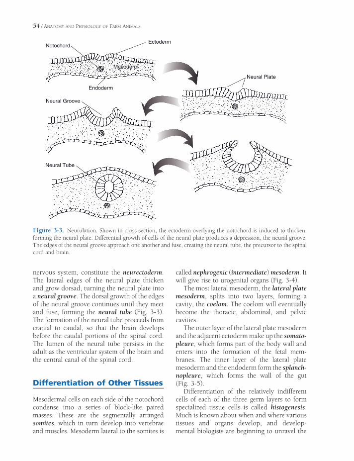
54 / ANATOMY AND PHYSIOLOGY OF FARM ANIMALS
nervous system, constitute the neurectoderm. The lateral edges of the neural plate thicken and grow dorsad, turning the neural plate into a neural groove. The dorsal growth of the edges of the neural groove continues until they meet and fuse, forming the neural tube (Fig. 3-3). The formation of the neural tube proceeds from cranial to caudal, so that the brain develops before the caudal portions of the spinal cord. The lumen of the neural tube persists in the adult as the ventricular system of the brain and the central canal of the spinal cord.
Differentiation of Other Tissues
Mesodermal cells on each side of the notochord condense into a series of block-like paired masses. These are the segmentally arranged somites, which in turn develop into vertebrae and muscles. Mesoderm lateral to the somites is
called nephrogenic (intermediate) mesoderm. It will give rise to urogenital organs (Fig. 3-4).
The most lateral mesoderm, the lateral plate mesoderm, splits into two layers, forming a cavity, the coelom. The coelom will eventually become the thoracic, abdominal, and pelvic cavities.
The outer layer of the lateral plate mesoderm and the adjacent ectoderm make up the somato-pleure, which forms part of the body wall and enters into the formation of the fetal mem-branes. The inner layer of the lateral plate mesoderm and the endoderm form the splanch-nopleure, which forms the wall of the gut (Fig. 3-5).
Differentiation of the relatively indifferent cells of each of the three germ layers to form specialized tissue cells is called histogenesis. Much is known about when and where various tissues and organs develop, and develop -mental biologists are beginning to unravel the
Figure 3-3. Neurulation. Shown in cross-section, the ectoderm overlying the notochord is induced to thicken, forming the neural plate. Differential growth of cells of the neural plate produces a depression, the neural groove. The edges of the neural groove approach one another and fuse, creating the neural tube, the precursor to the spinal cord and brain.
NotochordEctoderm
Mesoderm
Endoderm
Neural Plate
Neural Groove
Neural Tube

Figure 3-4. Differentiation of mesoderm. Somites form most medially. These will give rise to the muscles and bones associated with the vertebral column. Lateral to the somites is the intermediate mesoderm, which will dif-ferentiate into tissues of the urogenital system. Most laterally, the lateral plate mesoderm will become part of the gastrointestinal wall and body wall.
55
Ectoderm Notochord
Endoderm Mesoderm
Somite
Closing Neural Groove
Somites
Lateral Plate Mesoderm
PrimitiveGut
Intermediate Mesoderm
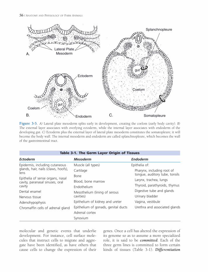
56 / ANATOMY AND PHYSIOLOGY OF FARM ANIMALS
Lateral PlateMesoderm
gutCoelom
Splanchnopleure
gut
Somatopleure
Coelom
Endoderm
Ectoderm
A.
.C.B
Figure 3-5. A) Lateral plate mesoderm splits early in development, creating the coelom (early body cavity). B) The external layer associates with overlying ectoderm, while the internal layer associates with endoderm of the developing gut. C) Ectoderm plus the external layer of lateral plate mesoderm constitutes the somatopleure; it will become the body wall. The internal mesoderm and endoderm are called splanchnopleure, which becomes the wall of the gastrointestinal tract.
Table 3-1. The Germ Layer Origin of Tissues
Ectoderm Mesoderm Endoderm
Epidermis, including cutaneous glands, hair, nails (claws, hoofs), lens
Epithelia of sense organs, nasal cavity, paranasal sinuses, oral cavity
Dental enamel
Nervous tissue
Adenohypophysis
Chromaffi n cells of adrenal gland
Muscle (all types)
Cartilage
Bone
Blood, bone marrow
Endothelium
Mesothelium (lining of serous cavities)
Epithelium of kidney and ureter
Epithelium of gonads, genital ducts
Adrenal cortex
Synovium
Epithelia of:
Pharynx, including root of tongue, auditory tube, tonsils
Larynx, trachea, lungs
Thyroid, parathyroids, thymus
Digestive tube and glands
Urinary bladder
Vagina, vestibule
Urethra and associated glands
molecular and genetic events that underlie development. For instance, cell surface mole-cules that instruct cells to migrate and aggre-gate have been identifi ed, as have others that cause cells to change the expression of their
genes. Once a cell has altered the expression of its genome so as to assume a more specialized role, it is said to be committed. Each of the three germ lines is committed to form certain kinds of tissues (Table 3-1). Differentiation

EMBRYOLOGY / 57
occurs as these cells assume the appearance and functions characteristic of the cell type to which they are committed.
Embryonic stem cells are capable of dif-ferentiating into multiple tissue types, a characteristic called pluripotency. Unlike most differentiated tissues, they can survive in long-term culture (outside the body). Embryonic stem cells are profoundly inter-esting to biomedical researchers, as it appears that cultured stem cells can be induced to differentiate into a wide variety of tissue types. This technique offers promise of a novel way of treating diseases involving loss of normal tissue. A few of the many possibilities include using stem cells to
create (1) hematopoietic cells to regenerate normal blood precursors for treatment of blood and immune disorders, (2) cartilage and bone to replace damaged skeletal com-ponents, and (3) neuronal precursors to replace the neurons lost in Parkinson’s disease or Alzheimer’s disease. Research with human embryonic stem cells is fraught with contentious ethical considerations, since these cells are generally acquired from aborted embryos and fetuses. Research using animal embryos tends to be less ethically problematic, but it will naturally lead to intense debate as techniques developed in animal models are extrapolated for use in human medicine.


THE SKELETAL SYSTEM
The study of the bones that make up the skeleton, or framework of the body, is oste-
ology. The skeleton gives a basis for the external structure and appearance of most vertebrate animals as we know them (Figs. 4-1 and 4-2). All mammals share a basic body plan with striking similarities in skeletal structure. Differ-ences refl ect adaptations to specifi c lifestyles.
The skeleton of a living animal is made up of bones that are themselves living structures. They have blood vessels, lymphatic vessels, and nerves; they are subject to disease; they can undergo repair; and they adjust to changes in stress. The functions of bones include provid-ing protection, giving rigidity and form to the body, acting as levers, storing minerals, and forming the cellular elements of blood.
Functions of Bones
Protection of vital organs is one of the impor-tant functions of bones. The central nervous system is protected by the skull and vertebral column; the heart and lungs, by the rib cage; and internal parts of the urogenital system, by the pelvis.
In the vertebrates, locomotion, defense, offense, grasping, and other activities of this type depend largely upon the action of muscles that attach to levers. Almost without exception, these levers are made of bone and are integral parts of the skeleton.
Functions of BonesTerminologyClassifi cation of Bones According to Gross
AppearanceAxial Skeleton
Skull
Vertebral Column
Sternum and Ribs
Appendicular SkeletonThoracic Limbs
Pelvic Limbs
Chapter 4
59

Proximal sesamoid b.
Facial bones
Cranial bones
Cervical vertebrae
Axis (C2)
L1
Atlas (C1)
ScapulaSacrum
Caudal vertebrae
Ischium
Femur
Patella
Fibula
Tibia
Tarsus (hock)
Metatarsus
Ulna
Distal sesamoid b.
Proximal phalanx(long pastern b.)
Middle phalanx(short pastern b.)
Distal phalanx(coffin bone)
Carpus
Radius
Elbow
Sternum
Humerus
Mandible
Ilium
Pubis
Stifle
Fetlock joint
Coffin joint
Shoulder
Thoracic vertebrae
Costalcartilages
Lumbar vertebrae Sacral vertebrae
Pastern joint
Metacarpus
Figure 4-1. Skeleton of the horse. (Adapted with permission of Wiley-Blackwell from Spurgeon’s Color Atlas of Large Animal Medicine. Baltimore: Lippincott Williams & Wilkins, 1999, p. 5.)
60

Frontal b.
Zygomatic arch
Zygomatic b.
Nasal b.
Maxilla
Infraorbitalforamen
Incisive b.
Mental foramenMandible
Acromion1st rib
Greater tubercle
Humerus
Manubrium of sternum
Body of sternum
Intermediate carpal b.
Ulnar carpal b. Fused 2nd & 3rd carpal b.
4th carpal b.
5th metacarpal b.
Atlas (C1)Axis (C2)
C7
T1
Scapular cartilageSpine of scapula
ScapulaT13 L1
L6Sacrum
1st caudal vertebra
Greater trochanter
Femur
Lateral condyle of tibiaand head of fibula
Tibia
Calcaneus
Lateral malleolus
Fused central and 4th tarsal b.
Fused 3rd & 4thmetatarsal b.
Ulna
Trochlea of radius
Accessory carpal b.
Radial carpal b.
4th carpal b.
Fused 2nd & 3rd carpal b.
Xiphoid process
Costal cartilage
13th rib
Coxal tuber
Body of ilium
Patella
Talus
Fused central and4th tarsal b.
Fused 2nd & 3rdtarsal b.
Fused 2nd & 3rdtarsal b.
1st tarsal b.
Proximal sesamoid
Proximal phalanx
Middle phalanx
Distal sesamoid b.
Distal phalanx
6thRib
Radius
3rd & 4th metacarpal b.
Facial tuber
Olecranon tuber
2nd metatarsal b.
Ischium
Ischiatic tuber
Pubis
Wing of ilium
Transverse process
Spinous process
Occipital b.
Nuchal crest
Lacrimal b.
Parietal b.
Temporal b.
Palatine b.
Fused
b.
Lateral epicondyle
Calcaneal tuber
(distal end of fibula)
Supraglenoid tubercle
Figure 4-2. Skeleton of the ox. (Adapted with permission of Wiley-Blackwell from Spurgeon’s Color Atlas of Large Animal Medicine. Baltimore: Lippincott Williams & Wilkins, 1999, p. 35.)

62 / ANATOMY AND PHYSIOLOGY OF FARM ANIMALS
The entire skeleton serves as a dynamic storage area for minerals, particularly calcium and phosphorus. These minerals are deposited and withdrawn as needed in the ongoing homeokinetic process. Blood formation is not strictly a function of bone itself but of the marrow within the cavity of long bones and the spongy substance of all young bones.
Terminology
Certain terms (Fig. 4-3) routinely used in refer-ence to bones, particularly long bones, include the following.
Compact (dense or cortical) bone is the hard layer that constitutes the exterior of most bones and forms almost the entire shaft of long bones.
Cancellous (spongy) bone is composed of spicules arranged to form a porous network. The spaces are usually fi lled with marrow.
The medullary cavity (marrow cavity) is the space surrounded by the cortex of a long bone. In young animals it is fi lled with red marrow (hematopoietic tissue), which gradually is replaced by yellow marrow (fat) as the animal ages.
Epiphysis refers to either end of a long bone. The end closest to the body is the proximal
egalitraC ralucitrAegalitraC ralucitrA
Articular Cartilage
Metaphysis
Metaphysis
DistalEpiphysis
ProximalEpiphysisEpiphyseal
Cartilage
Cancellous(spongy) Bone
Compact Bone
Periosteum
Endosteum
Diaphysis
MedullaryCavity
Figure 4-3. Longitudinal section of the equine femur. Left) Immature (growth plates open). Right) Mature (growth plates fused).

THE SKELETAL SYSTEM / 63
epiphysis, and the end farthest from the body is the distal epiphysis.
The diaphysis is the cylindrical shaft of a long bone between the two epiphyses.
The metaphysis of a mature bone is the fl ared area adjacent to the epiphysis.
Epiphyseal cartilage or disk (physis) is a layer of hyaline cartilage within the metaphysis of an immature bone that separates the diaphy-sis from the epiphysis. This is the only area in which a bone can lengthen.
Articular cartilage is a thin layer of hyaline cartilage that covers the articular (joint) surface of a bone.
Periosteum is a fi brous membrane that covers the surface of a bone except where artic-ular cartilage is located. Osteoblasts (bone-pro-ducing cells) of the periosteum are responsible for increases in the diameter of bones, and activity of periosteal cells is important in the healing of fractures. The periosteum is vascular and well-innervated.
Table 4-1. Bony Features
Example
Articular Projections
Head Spherical articular projection Head of femur
Condyle Approximately cylindrical articular mass
Medial & lateral femoral condyles
Trochlea Pulleylike articular mass Trochlea of distal humerus
Facet Relatively fl at articular surface Articular facets between carpal bones
Nonarticular Projections
Process General term for bony projection Spinous process or transverse process of vertebra
Tuberosity (tuber) Relatively large nonarticular projection
Deltoid tuberosity of humerus and tuber sacrale of pelvis
Tubercle (tuberculum) Smaller projection Greater and lesser tubercles of humerus
Spine Pointed projection or ridge Nasal spine of palatine bone and spine of scapula
Crest Sharp ridge Median sacral crest
Neck Cylindrical part of bone to which a head is attached
Femoral neck
Line (linea) Small ridge or mark on bone Gluteal lines of ilium
Articular Depressions
Fovea Small depression (may be articular or not)
Fovea capitis on head of femur
Glenoid Cavity Shallow articular concavity Glenoid cavity of scapula
Notch Indentation (may be articular or not) Semilunar notch of ulna and alar notch of atlas
Nonarticular Depressions
Fossa Large nonarticular depression Supraspinous fossa of scapula
Foramen Circumscribed hole in bone Foramen magnum at base of skull
Canal Tunnel through one or more bones Vertebral canal through length of vertebral column

64 / ANATOMY AND PHYSIOLOGY OF FARM ANIMALS
Endosteum is a fi brous membrane that lines the marrow cavity and osteonal canals (osteons) of a bone. Erosion of existing bone by osteo-clasts (bone-destroying cells) in the endosteum determines the size of the marrow cavity and the thickness of the diaphyseal cortex. Both periosteum and endosteum contain osteoblasts and osteoclasts (see Chapter 5).
Many of the projections from and depres-sions in bones have general names that depend to some extent on their size and function. Both projections and depressions may be articular or nonarticular. If they are articular, they form an integral part of a joint and are covered with articular cartilage. Nonarticular projections and depressions exist outside of joints. Many of them provide areas for attachment of muscle tendons or of ligaments. Table 4-1 lists some common bony features.
Classifi cation of Bones Accord-ing to Gross Appearance
Any bone may be classifi ed in one of the fol-lowing groups: long, short, fl at, sesamoid, pneumatic, or irregular.
Long bones are greater in one dimension than any other. Each consists of a relatively cylindrical shaft (the diaphysis) and two extrem-ities, the epiphyses, with a metaphysis between each epiphysis and the diaphysis. Long bones function chiefl y as levers and aid in support, locomotion, and prehension. The best examples of long bones are in the extremities. In the tho-racic limb the long bones include the humerus, radius, ulna, metacarpals, and phalanges. In the pelvic limb, the long bones are the femur, tibia, fi bula, metatarsals, and phalanges.
Short bones are cuboid, or approximately equal in all dimensions. There is no single marrow cavity, but the interior is composed of spongy bone fi lled with marrow spaces. The exterior is formed by a thin layer of compact bone. Short bones absorb concussion, and they are found in complex joints such as the carpus (the “knee” of the thoracic limb) and tarsus (hock), where a variety of movements as well as absorption of shock occur.
Flat bones are relatively thin and expanded in two dimensions. They consist of two plates of compact bone, the lamina externa and lamina interna, separated by spongy material called diploë.
Flat bones function chiefl y for protection of vital organs such as the brain (skull), the heart and lungs (scapulae and ribs), and the pelvic viscera (pelvis), but many, notably the scapulae and pelvis, provide large areas for attachment of muscles.
Sesamoid bones are so called because of their fancied resemblance to a sesame seed, although many sesamoid bones of domestic animals have decidedly un-seedlike shapes. Sesamoid bones occur along the course of tendons to reduce friction, increase leverage, or change the direction of pull. The patella (kneecap) is the largest sesamoid bone in the body.
Pneumatic bones contain air spaces or sinuses that communicate with the atmosphere. The frontal bones and maxillary bones of the skull are examples of this type of bone among mammals. Many avian bones are “pneuma-tized” as well (see Chapter 30).
Irregular bones are unpaired bones on the median plane; they include the vertebrae and some of the unpaired bones of the skull. These bones do not fi t well into any other descriptive classifi cation. They feature prominent processes and offer protection, support, and muscular attachment.
Axial Skeleton
The axial skeleton includes bones on or attached to the midline (axis) of the body and comprise the skull, vertebral column, sternum, and ribs. Table 4-2 lists the bones of the axial skeleton by regions.
Skull
The part of the skeleton within the head is the skull. It protects the brain, supports many of the sense organs, and forms passages for entry
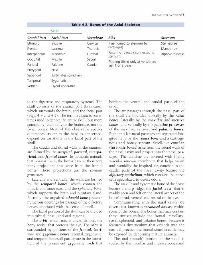
THE SKELETAL SYSTEM / 65
to the digestive and respiratory systems. The skull consists of the cranial part (braincase), which surrounds the brain, and the facial part (Figs. 4-4 and 4-5). The term cranium is some-times used to denote the entire skull, but more commonly refers only to the braincase, not the facial bones. Most of the observable species differences, as far as the head is concerned, depend on variations in the facial part of the skull.
The caudal and dorsal walls of the cranium are formed by the occipital, parietal, interpa-rietal, and frontal bones. In domestic animals that possess them, the horns have at their core bony projections that arise from the frontal bones. These projections are the cornual processes.
Laterally and ventrally, the walls are formed by the temporal bones, which contain the middle and inner ears, and the sphenoid bone, which supports the brain and pituitary gland. Rostrally, the unpaired ethmoid bone presents numerous openings for passage of the olfactory nerves associated with the sense of smell.
The facial portion of the skull can be divided into orbital, nasal, and oral regions.
The orbit, which means circle, denotes the bony socket that protects the eye. The orbit is surrounded by portions of the frontal, lacri-mal, and zygomatic bones. Frontal, zygomatic, and temporal bones all participate in the forma-tion of the prominent zygomatic arch that
borders the ventral and caudal parts of the orbit.
The air passages through the nasal part of the skull are bounded dorsally by the nasal bones, laterally by the maxillae and incisive bones, and ventrally by the palatine processes of the maxillae, incisive, and palatine bones. Right and left nasal passages are separated lon-gitudinally by the vomer bone and a cartilagi-nous and boney septum. Scroll-like conchae (turbinate bones) arise from the lateral walls of the nasal cavity and project into the nasal pas-sages. The conchae are covered with highly vascular mucous membrane that helps warm and humidify the inspired air; conchae in the caudal parts of the nasal cavity feature the olfactory epithelium, which contains the nerve cells specialized to detect odors.
The maxilla and zygomatic bone of the horse feature a sharp ridge, the facial crest, that is readily seen and felt on the lateral aspect of the horse’s head, ventral and rostral to the eye.
Communicating with the nasal cavity are diverticula, known as paranasal sinuses, within some of the bones. The bones that may contain these sinuses include the frontal, maxillary, nasal, sphenoid, and palatine bones. Because it features a diverticulum that extends into the cornual process, the frontal sinus in cattle may be exposed by dehorning mature animals.
The oral (mouth) portion of the skull is roofed by the maxillae and incisive bones and
Table 4-2. Bones of the Axial Skeleton
Skull
Cranial Part Facial Part Vertebrae Ribs Sternum
Ethmoid
Frontal
Interparietal
Occipital
Parietal
Pterygoid
Sphenoid
Temporal
Vomer
Incisive
Lacrimal
Mandible
Maxilla
Palatine
Nasal
Turbinates (conchae)
Zygomatic
Hyoid apparatus
Cervical
Thoracic
Lumbar
Sacral
Caudal
True (joined to sternum by cartilages)
False (not directly connected to sternum)
Floating (fi xed only at vertebrae; last 1 or 2 pairs)
Sternebrae
Manubrium
Xiphoid process
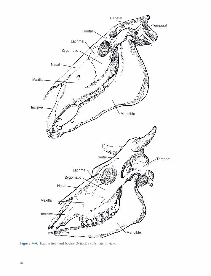
Frontal Temporal
Lacrimal
Zygomatic
Nasal
Maxilla
Incisive
Mandible
Parietal
Frontal
Temporal
Lacrimal
Zygomatic
Nasal
Maxilla
Incisive
Mandible
Figure 4-4. Equine (top) and bovine (bottom) skulls, lateral view.
66

Cornual Process
Facial Tuber
Infraorbital Foramen
Frontal
Supraorbital Foramen
Zygomatic
Lacrimal
Nasal
Maxilla
Incisive
Parietal
Temporal
Nuchal Crest
ExternalSagittal Crest
TemporalFossa
Frontal
Orbit
Nasal
Lacrimal
Zygomatic
Maxilla
Incisive
Figure 4-5. Bovine (left) and equine (right) skulls, dorsal view.
67

68 / ANATOMY AND PHYSIOLOGY OF FARM ANIMALS
by the palatine bone. The maxillae and incisive bones contain the teeth of the upper dental arcade (although the incisive bones lack teeth in ruminants).
Ventrolaterally, the mandible completes the oral portion. The mandible pivots on a fossa of the temporal bone just rostral to the opening of the ear. The mandible contains all of the lower teeth and gives attachment to some of the muscles associated with chewing and swallowing.
The hyoid apparatus is a bony framework (Fig. 4-6) that gives support to the pharynx (throat) and provides attachment to some pha-ryngeal, laryngeal, and lingual muscles. It lies between the right and left portions of the man-dible and is attached to the styloid process of each temporal bone.
Vertebral Column
The vertebral column is composed of median unpaired irregular bones called vertebrae. The
following letters are typically used to designate the respective regions:
C Cervical vertebrae: neck regionT Thoracic: chestL Lumbar: loinS Sacral: pelvis, fused vertebraeCd Caudal (coccygeal): tail
A vertebral formula for a given species con-sists of the letter symbol for each region fol-lowed by the number of vertebrae in that region in the given species. Vertebral formulas of common domestic animals and humans are shown in Table 4-3. The parts of a typical ver-tebra include the body, the arch, and the pro-cesses (Fig. 4-7).
The body is a cylindrical mass forming the ventral part of the vertebra and fl oor of the vertebral foramen.
Dorsally, the arch completes the vertebral foramen, which contains the spinal cord. When vertebrae are placed in series, the adja-
Stylohyoid Bone
Thyrohyoid Bone
Basihyoid Bone
Lingual Process
Ceratohyoid Bone
Figure 4-6. Equine hyoid apparatus. The stylohyoid bones articulate with the styloid processes of the skull, and the thyrohyoid bones articulate with the larynx. The prominent lingual process extends into the base of the tongue and affords an attachment site for some lingual muscles.
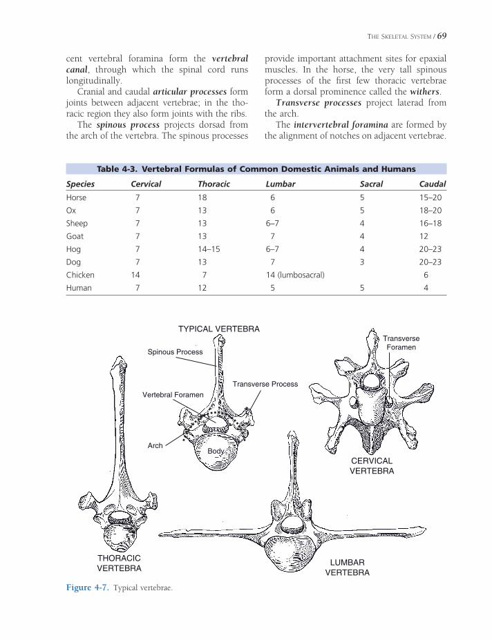
THE SKELETAL SYSTEM / 69
Table 4-3. Vertebral Formulas of Common Domestic Animals and Humans
Species Cervical Thoracic Lumbar Sacral Caudal
Horse 7 18 6 5 15–20
Ox 7 13 6 5 18–20
Sheep 7 13 6–7 4 16–18
Goat 7 13 7 4 12
Hog 7 14–15 6–7 4 20–23
Dog 7 13 7 3 20–23
Chicken 14 7 14 (lumbosacral) 6
Human 7 12 5 5 4
THORACICVERTEBRA
LUMBARVERTEBRA
CERVICALVERTEBRA
TYPICAL VERTEBRA
BodyArch
Spinous Process
Vertebral Foramen
Transverse Process
TransverseForamen
Figure 4-7. Typical vertebrae.
cent vertebral foramina form the vertebral canal, through which the spinal cord runs longitudinally.
Cranial and caudal articular processes form joints between adjacent vertebrae; in the tho-racic region they also form joints with the ribs.
The spinous process projects dorsad from the arch of the vertebra. The spinous processes
provide important attachment sites for epaxial muscles. In the horse, the very tall spinous processes of the fi rst few thoracic vertebrae form a dorsal prominence called the withers.
Transverse processes project laterad from the arch.
The intervertebral foramina are formed by the alignment of notches on adjacent vertebrae.

70 / ANATOMY AND PHYSIOLOGY OF FARM ANIMALS
Spinal nerves exit the vertebral canal via the intervertebral foramina on their way to inner-vating peripheral structures.
The cervical vertebrae have well-developed articular processes to accommodate the large range of motion of the neck. All domestic mammals have seven cervical vertebrae.
The atlas is the fi rst cervical vertebra. The spinous process is absent. The atlas articulates with the occipital condyles of the skull cranially and with the axis caudally.
The axis is the second cervical vertebra. Its spinous process forms a longitudinal sail on its dorsum. The body of the axis features a cranial projection called the dens (for its resemblance to a tooth), which articulates with the atlas in a pivot joint.
The remaining cervical vertebrae are similar to one another, with small spinous processes and rather large transverse and articular pro-cesses. With the exception of the last cervical vertebra (C7), each cervical transverse process contains a transverse foramen through which passes the vertebral artery.
Thoracic vertebrae are characterized by well-developed spinous processes and articular facets for the ribs. Costal fovea on the bodies of adjacent thoracic vertebrae form cavities for articulation with the heads of the ribs. Each transverse process also features a fovea for artic-ulation with the tubercle of the rib of the same number as the vertebra.
Lumbar vertebrae have large, fl at transverse processes that project laterad. The spinous pro-cesses are similar to those of the last few tho-racic vertebrae. The articular processes are more robust than those of the thoracic verte-brae, but not as large as the articular processes in the cervical region. The body and caudal articular processes of the last lumbar vertebra articulate with the sacrum.
The sacral vertebrae are fused to form a single wedge-shaped bone, the sacrum, which articulates with the last lumbar vertebra crani-ally, with the fi rst caudal vertebra caudally, and with the wings of the ilia craniolaterally. The intervertebral foramina of the sacrum are rep-resented by dorsal and ventral rows of sacral foramina on dorsal and ventral sides of the
sacrum. These foramina, as with other inter-vertebral foramina, give passage to spinal nerves.
Caudal vertebrae form the bony basis for the tail. Depending on the length of the tail, the number varies considerably from species to species and even within the same species. Size of the vertebrae decreases rapidly in a caudal direction, until the last few caudal vertebrae are merely small rods of bone. As animals lack the curved, fused coccyx of human beings, the term coccygeal to describe the vertebrae of the tail is discouraged.
Sternum and Ribs
The sternum forms the ventrum of the bony thorax and gives attachment to the costal car-tilages of the ribs as well as providing a bony origin for the pectoral muscles. The cranial extremity of the sternum is the manubrium; the middle portion is the body; and the caudal extremity is the xiphoid process. The sternum consists of individual bones called sternebrae that tend to fuse as age advances. The number of sternebrae (excluding the manubrium and xiphoid from the count) varies with species as follows: pig, four; ruminants and horses, fi ve; and in the dog, six.
The ribs form the lateral walls of the bony thorax. Usually, the number of pairs of ribs is the same as the number of thoracic vertebrae. Rarely, an extra rib or pair of ribs lies either cranial or caudal to the thoracic vertebrae. A typical rib consists of a shaft, a sternal extre-mity ventrally, and a vertebral extremity dorsally.
Except for the last one or two pairs of ribs, the sternal extremity is connected to the sternum by the costal hyaline cartilage; ribs so attached are called sternal (true) ribs. The ver-tebral extremity consists of a spherical head connected to the rib by a constricted neck and a tubercle that articulates with the transverse process of a thoracic vertebra. The head articu-lates with the bodies of two adjacent vertebrae at the costal fovea.
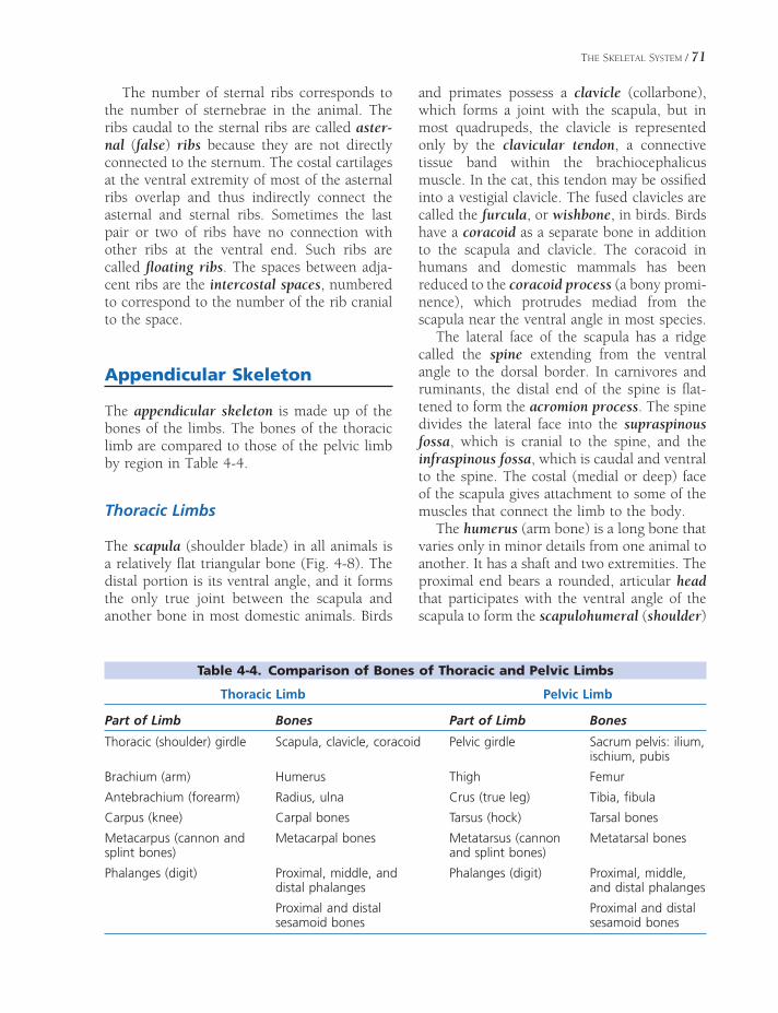
THE SKELETAL SYSTEM / 71
The number of sternal ribs corresponds to the number of sternebrae in the animal. The ribs caudal to the sternal ribs are called aster-nal (false) ribs because they are not directly connected to the sternum. The costal cartilages at the ventral extremity of most of the asternal ribs overlap and thus indirectly connect the asternal and sternal ribs. Sometimes the last pair or two of ribs have no connection with other ribs at the ventral end. Such ribs are called fl oating ribs. The spaces between adja-cent ribs are the intercostal spaces, numbered to correspond to the number of the rib cranial to the space.
Appendicular Skeleton
The appendicular skeleton is made up of the bones of the limbs. The bones of the thoracic limb are compared to those of the pelvic limb by region in Table 4-4.
Thoracic Limbs
The scapula (shoulder blade) in all animals is a relatively fl at triangular bone (Fig. 4-8). The distal portion is its ventral angle, and it forms the only true joint between the scapula and another bone in most domestic animals. Birds
and primates possess a clavicle (collarbone), which forms a joint with the scapula, but in most quadrupeds, the clavicle is represented only by the clavicular tendon, a connective tissue band within the brachiocephalicus muscle. In the cat, this tendon may be ossifi ed into a vestigial clavicle. The fused clavicles are called the furcula, or wishbone, in birds. Birds have a coracoid as a separate bone in addition to the scapula and clavicle. The coracoid in humans and domestic mammals has been reduced to the coracoid process (a bony promi-nence), which protrudes mediad from the scapula near the ventral angle in most species.
The lateral face of the scapula has a ridge called the spine extending from the ventral angle to the dorsal border. In carnivores and ruminants, the distal end of the spine is fl at-tened to form the acromion process. The spine divides the lateral face into the supraspinous fossa, which is cranial to the spine, and the infraspinous fossa, which is caudal and ventral to the spine. The costal (medial or deep) face of the scapula gives attachment to some of the muscles that connect the limb to the body.
The humerus (arm bone) is a long bone that varies only in minor details from one animal to another. It has a shaft and two extremities. The proximal end bears a rounded, articular head that participates with the ventral angle of the scapula to form the scapulohumeral (shoulder)
Table 4-4. Comparison of Bones of Thoracic and Pelvic Limbs
Thoracic Limb Pelvic Limb
Part of Limb Bones Part of Limb Bones
Thoracic (shoulder) girdle Scapula, clavicle, coracoid Pelvic girdle Sacrum pelvis: ilium, ischium, pubis
Brachium (arm) Humerus Thigh Femur
Antebrachium (forearm) Radius, ulna Crus (true leg) Tibia, fi bula
Carpus (knee) Carpal bones Tarsus (hock) Tarsal bones
Metacarpus (cannon and splint bones)
Metacarpal bones Metatarsus (cannon and splint bones)
Metatarsal bones
Phalanges (digit) Proximal, middle, and distal phalanges
Phalanges (digit) Proximal, middle, and distal phalanges
Proximal and distal sesamoid bones
Proximal and distal sesamoid bones

giPxOesroH
A
A
A
B
B
B
C
C
C
D
D
D
E
E
E
F
F
F
G
G
GHH
H
Figure 4-8. Comparative anatomy of the bones of the thoracic limb. A, Scapula; B, scapulohumeral (shoulder joint); C, humerus; D, elbow joint; E, antebra-chium (radius & ulna); F, carpus; G, metacarpus; H, digit (phalanges).
72

THE SKELETAL SYSTEM / 73
joint. The proximal end of the humerus also features a number of irregular tuberosities and tubercles, providing sites of attachment to muscles of the shoulder region. The palpable prominence produced by this end of the humerus is called the point of the shoulder. The distal end of the humerus forms a spool-like condyle that articulates with the proximal ends of the radius and ulna in the elbow.
The radius and ulna are the bones of the antebrachium (forearm). In mammals, the radius is the larger of the two, although in birds it is smaller than the ulna. The radius enters into the elbow joint proximally and the carpus distally. The radius can be felt directly beneath the skin on the medial side of the forearm.
The ulna varies in its degree of development from species to species. The prominent olecra-non process (point of the elbow) is found in all mammals proximal and caudal to the elbow joint. This process forms a lever for attachment of the muscles that extend the elbow. In the horse, the proximal portion of the shaft of the ulna is well developed but fused to the radius; the distal ulna is absent. The ox, sheep, goat, and pig each have a complete ulna, but with little or no movement between the ulna and radius. The cat and dog have considerably more movement between these complete bones, but not nearly as much as primates, who can pronate and supinate their hands through the rotation of radius and ulna relative to one another.
The carpus in all animals is a complex region that includes two rows of small bones. This region corresponds to the human wrist, and is frequently, although erroneously, called the “knee” by horsemen. Carpal bones in the proxi-mal row are called (from medial to lateral) radial, intermediate, and ulnar, whereas those in the distal row are numbered 1 to 4 from medial to lateral. In addition, an accessory carpal bone projects caudad from the lateral side of the carpus. The numbering of the carpal bones of the distal row is based on an ancestral four, but among common domestic farm animals only the pig consistently has four carpal bones in this distal row. The fi rst carpal bone of the horse, when present, is small and non–weight bearing. The fi rst carpal is not present
in ruminants, and the second and third carpal bones are fused in these species.
The metacarpus is immediately distal to the carpus. In the horse it includes a single large metacarpal (cannon) bone, the base for the third digit (corresponding to the middle fi nger), and two small metacarpal (splint) bones. The second metacarpal bone is on the medial side, and the fourth is on the lateral side. Trauma to these small bones with consequent excess bone formation results in splints. Splints in horses sometimes produce lameness, but often consti-tute only a blemish, a disfi gurement not usually associated with unsoundness (Figure 4-9).
The cannon bone of the ox and sheep is a fusion of the third and fourth metacarpal bones. A vertical groove on the dorsum of the cannon bone demarcates the embryonic line of fusion.
Figure 4-9. Medial splint. (Reprinted with permis-sion of Wiley-Blackwell from Stashak, T.S.: Adams’ Lameness in Horses. 5th ed. Baltimore, Lippincott Wil-liams & Wilkins, 2002.)
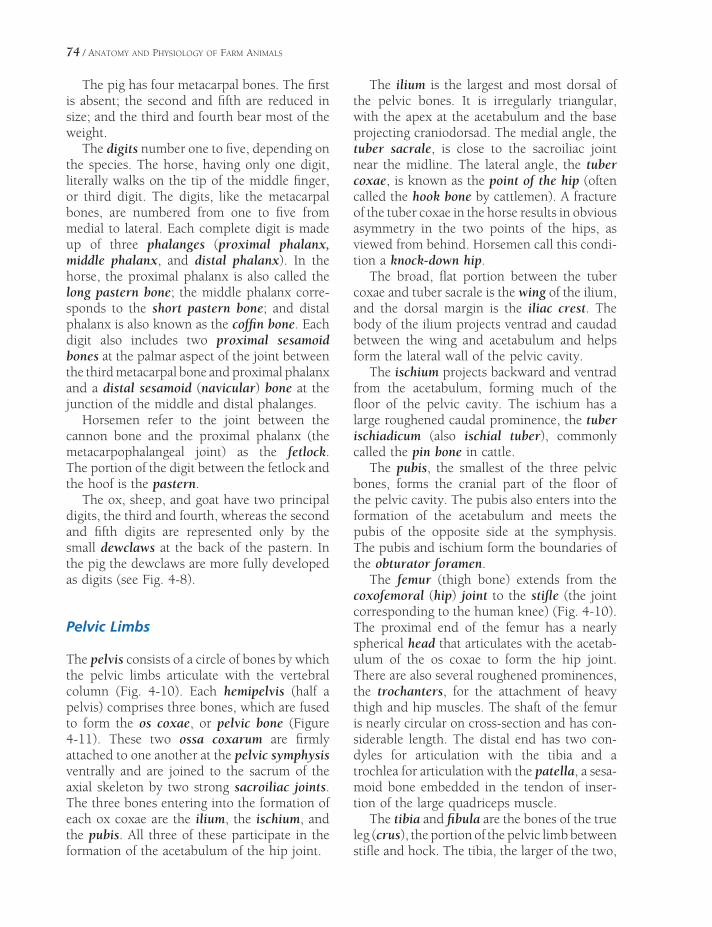
74 / ANATOMY AND PHYSIOLOGY OF FARM ANIMALS
The pig has four metacarpal bones. The fi rst is absent; the second and fi fth are reduced in size; and the third and fourth bear most of the weight.
The digits number one to fi ve, depending on the species. The horse, having only one digit, literally walks on the tip of the middle fi nger, or third digit. The digits, like the metacarpal bones, are numbered from one to fi ve from medial to lateral. Each complete digit is made up of three phalanges (proximal phalanx, middle phalanx, and distal phalanx). In the horse, the proximal phalanx is also called the long pastern bone; the middle phalanx corre-sponds to the short pastern bone; and distal phalanx is also known as the coffi n bone. Each digit also includes two proximal sesamoid bones at the palmar aspect of the joint between the third metacarpal bone and proximal phalanx and a distal sesamoid (navicular) bone at the junction of the middle and distal phalanges.
Horsemen refer to the joint between the cannon bone and the proximal phalanx (the metacarpophalangeal joint) as the fetlock. The portion of the digit between the fetlock and the hoof is the pastern.
The ox, sheep, and goat have two principal digits, the third and fourth, whereas the second and fi fth digits are represented only by the small dewclaws at the back of the pastern. In the pig the dewclaws are more fully developed as digits (see Fig. 4-8).
Pelvic Limbs
The pelvis consists of a circle of bones by which the pelvic limbs articulate with the vertebral column (Fig. 4-10). Each hemipelvis (half a pelvis) comprises three bones, which are fused to form the os coxae, or pelvic bone (Figure 4-11). These two ossa coxarum are fi rmly attached to one another at the pelvic symphysis ventrally and are joined to the sacrum of the axial skeleton by two strong sacroiliac joints. The three bones entering into the formation of each ox coxae are the ilium, the ischium, and the pubis. All three of these participate in the formation of the acetabulum of the hip joint.
The ilium is the largest and most dorsal of the pelvic bones. It is irregularly triangular, with the apex at the acetabulum and the base projecting craniodorsad. The medial angle, the tuber sacrale, is close to the sacroiliac joint near the midline. The lateral angle, the tuber coxae, is known as the point of the hip (often called the hook bone by cattlemen). A fracture of the tuber coxae in the horse results in obvious asymmetry in the two points of the hips, as viewed from behind. Horsemen call this condi-tion a knock-down hip.
The broad, fl at portion between the tuber coxae and tuber sacrale is the wing of the ilium, and the dorsal margin is the iliac crest. The body of the ilium projects ventrad and caudad between the wing and acetabulum and helps form the lateral wall of the pelvic cavity.
The ischium projects backward and ventrad from the acetabulum, forming much of the fl oor of the pelvic cavity. The ischium has a large roughened caudal prominence, the tuber ischiadicum (also ischial tuber), commonly called the pin bone in cattle.
The pubis, the smallest of the three pelvic bones, forms the cranial part of the fl oor of the pelvic cavity. The pubis also enters into the formation of the acetabulum and meets the pubis of the opposite side at the symphysis. The pubis and ischium form the boundaries of the obturator foramen.
The femur (thigh bone) extends from the coxofemoral (hip) joint to the stifl e (the joint corresponding to the human knee) (Fig. 4-10). The proximal end of the femur has a nearly spherical head that articulates with the acetab-ulum of the os coxae to form the hip joint. There are also several roughened prominences, the trochanters, for the attachment of heavy thigh and hip muscles. The shaft of the femur is nearly circular on cross-section and has con-siderable length. The distal end has two con-dyles for articulation with the tibia and a trochlea for articulation with the patella, a sesa-moid bone embedded in the tendon of inser-tion of the large quadriceps muscle.
The tibia and fi bula are the bones of the true leg (crus), the portion of the pelvic limb between stifl e and hock. The tibia, the larger of the two,

giPxOesroH
A
A
A
B
B
B
C
C
C
D
D
D
EE
E
FF
F
G G
G
H H
HII
I
Figure 4-10. Comparative anatomy of the bones of the pelvic limb. A, Pelvis; B, coxofemoral (hip) joint; C, femur; D, patella; E, stifl e (knee) joint; F, crus (tibia & fi bula); G, tarsus (hock); H, metatarsus; I, digit (phalanges).

76 / ANATOMY AND PHYSIOLOGY OF FARM ANIMALS
is palpable beneath the skin medially. The fi bula, which is much smaller, lies on the lateral side of the leg.
The tibia has an expanded proximal end that participates in the stifl e joint. Its shaft is trian-gular in cross-section. The distal end of the tibia has two concave depressions that form the hinge joint of the hock with the talus (tibiotar-sal bone).
In the dog, pig, and humans, the fi bula is a long, thin bone extending from the proximal end of the tibia to the lateral aspect of the hock. The horse has both the proximal end and a portion of the shaft, whereas only a vestige of the proximal end of the fi bula is present in domestic ruminants. All domestic species have the distal extremity of the fi bula, forming the prominent lateral malleolus of the hock. The lateral malleolus is fused to the tibia in the horse but is a separate small bone articulating with distal tibia and tarsal bones in ruminants.
Sacrum
Ilium
Acetabulum
Pubis
Ischium
TuberIschiadicum
ObturatorForamen
Ischium
Ilium
Tuber Coxae
LATERAL
DORSAL
Tuber Sacrale
Tuber Coxae
Tuber Sacrale
Pubic Symphysis
The tarsus (hock), like the carpus in the thoracic limb is composed of multiple small bones; it corresponds to the human ankle. The proximal row of tarsal bones consists of two large bones. The talus dorsally has two spool-like ridges for articulation with the tibia. The calcaneus projects proximad and caudad to form the point of the hock. The cal-caneus, which corresponds to the human heel, acts as a lever for the muscles extending the hock.
In the horse, the central row of tarsal bones is reduced to a single central tarsal bone. The bones of the distal row are numbered 1 to 4 from medial to lateral, with tarsal bones 1 and 2 fused into a single bone. In cattle, tarsal bones 2 and 3 are fused, as are the central and 4th.
The metatarsus and digits of the pelvic limb are similar to the metacarpus and digits of the thoracic limb.
Figure 4-11. The pelvis of the ox. Lateral (left) and dorsal (right) views.

MICROSCOPIC ANATOMY AND GROWTH AND
DEVELOPMENT OF BONE
Microscopic Anatomy and Formation of Bone
About a third of the weight of bone consists of an organic framework of fi brous tissue and cells. This organic matter mainly consists of collagen and polysaccharides called glycosami-noglycans (GAGs), which contain chondroitin sulfate. They give resilience and toughness to bones. The remaining two-thirds of bone weight consists of inorganic calcium and phosphorus salts in the organic framework. About 80% of these salts are calcium phosphate, and the remainder is primarily calcium carbonate and magnesium phosphate. The calcium phosphate is primarily found in hydroxyapatite crystals formed with calcium hydroxide. These salts give hardness and rigidity to bones and make them resist the passage of x-rays. If the inor-ganic salts are removed by soaking a bone in dilute acid, the resulting decalcifi ed bone will retain its original form but will be fl exible enough to be tied in a knot. On the other hand, if the organic matter is removed by char-ring in a furnace so that only the inorganic salts remain, the bone will retain its form but be brittle and break unless handled with extreme care.
Mature bone consists of osteocytes (bone cells) surrounded by an intercellular matrix composed of calcifi ed osteoid material. The
Microscopic Anatomy and Formation of Bone
Ossifi cationEndochondral (Intracartilaginous)
Ossifi cation
Intramembranous Ossifi cation
Physiology of BoneBone Mechanics and Remodeling
Calcium of Bone
Fractures and Fracture HealingOther Pathologic Conditions
Chapter 5
77
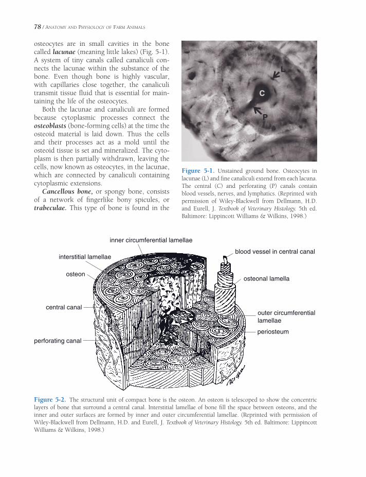
78 / ANATOMY AND PHYSIOLOGY OF FARM ANIMALS
osteocytes are in small cavities in the bone called lacunae (meaning little lakes) (Fig. 5-1). A system of tiny canals called canaliculi con-nects the lacunae within the substance of the bone. Even though bone is highly vascular, with capillaries close together, the canaliculi transmit tissue fl uid that is essential for main-taining the life of the osteocytes.
Both the lacunae and canaliculi are formed because cytoplasmic processes connect the osteoblasts (bone-forming cells) at the time the osteoid material is laid down. Thus the cells and their processes act as a mold until the osteoid tissue is set and mineralized. The cyto-plasm is then partially withdrawn, leaving the cells, now known as osteocytes, in the lacunae, which are connected by canaliculi containing cytoplasmic extensions.
Cancellous bone, or spongy bone, consists of a network of fi ngerlike bony spicules, or trabeculae. This type of bone is found in the
Figure 5-1. Unstained ground bone. Osteocytes in lacunae (L) and fi ne canaliculi extend from each lacuna. The central (C) and perforating (P) canals contain blood vessels, nerves, and lymphatics. (Reprinted with permission of Wiley-Blackwell from Dellmann, H.D. and Eurell, J. Textbook of Veterinary Histology. 5th ed. Baltimore: Lippincott Williams & Wilkins, 1998.)
perforating canal
central canal
osteon
interstitial lamellae
inner circumferential lamellae
blood vessel in central canal
osteonal lamella
outer circumferential lamellae
periosteum
Figure 5-2. The structural unit of compact bone is the osteon. An osteon is telescoped to show the concentric layers of bone that surround a central canal. Interstitial lamellae of bone fi ll the space between osteons, and the inner and outer surfaces are formed by inner and outer circumferential lamellae. (Reprinted with permission of Wiley-Blackwell from Dellmann, H.D. and Eurell, J. Textbook of Veterinary Histology. 5th ed. Baltimore: Lippincott Williams & Wilkins, 1998.)

MICROSCOPIC ANATOMY AND GROWTH AND DEVELOPMENT OF BONE / 79
extremities of long bones, where resistance to compression without excessive weight is needed. Flat bones between two layers of compact bone, as in the skull, are also cancel-lous. The spicules of bone are arranged so as to resist stresses and strains imposed on the bone by weight or pull of muscles.
Compact bone, found in the shafts of long bones, consists primarily of many laminated tubes known as osteonal systems (formally termed haversian systems). Each osteon consists of one central canal containing vessels and nerves surrounded by circular plates of bone (osteonal lamellae) forming the laminated cyl-inder (Fig. 5-2). These plates are laid down in a centripetal fashion (from the periphery toward the center). After the bone is formed, the osteo-blasts that became embedded in the bone sub-stance are called osteocytes. In general these osteons are added on the periphery of the shaft of a bone as the bone increases in diameter. Blood vessels extend from the periosteum to central canals through perforating canals (also known as Volkmann’s canals), which often travel at right angles to the central canals (Fig. 5-2).
Osteoblasts usually come from mesenchy-mal cells, the parent cells of all connective tissues. The osteoblasts divide readily, but only a portion of the new cells actually secretes osteoid substance and forms bone; the rest is held in reserve as the osteogenic layer of the periosteum and endosteum within the marrow cavity and central canals. These reserve cells divide and form more osteoblasts whenever more bone is needed, as in repair of fractures, response to stress, or growth. Because the inter-cellular matrix is unyielding, bone can be added only on the surface, and the osteocytes (mature osteoblasts) probably have lost the ability to divide.
As the shaft of a long bone enlarges in diam-eter as a result of the activity of the osteogenic layer of the periosteum, bone along the inner surface is normally resorbed to increase the size of the marrow cavity (Fig. 5-3). Resorption of bone may also occur under abnormal condi-tions, such as during a period of calcium defi -
Figure 5-3. Remodeling that occurs as a long bone increases in size. Both resorption and deposition of bone take place. A, Size and shape of young bone; B, size and shape of mature bone.
fi ciency. Whenever bone is resorbed (under normal or abnormal conditions), large multi-nucleated cells called osteoclasts (bone- destroying cells) are usually found (Fig. 5-4). These cells, derived from macrophages, take an active part in bone destruction by releasing organic acids and enzymes.

80 / ANATOMY AND PHYSIOLOGY OF FARM ANIMALS
that develops in the midshaft region of a long bone is the primary ossifi cation center (Fig. 5-5). Secondary ossifi cation centers then develop near the ends of long bones. These ossifi cation centers grow and expand, but a region of cartilage, the physis, still separates the centers during growth and development. Chon-drocytes within this region continue to prolif-erate and produce cartilage to provide this separation and thus allow for continued growth in the length of the long bone.
The midshaft region of a long bone that con-tains the primary ossifi cation center is the diaphysis; each end that contains a secondary ossifi cation center is an epiphysis (Fig. 5-5). As animals grow and mature, the region of carti-lage that separates the bony diaphysis and epiphyses continues to narrow. The epiphyseal plate, another term used to describe this region of cartilage in growing animals, emphasizes how narrow it may become.
When the cartilage in the epiphyseal plates is completely replaced by bone, increases in length, hence growth in stature of the animal, is impossible. This is epiphyseal closure. An epiphyseal line can often be seen on bones where this closure has occurred. The region of a long bone where the diaphysis and an epiphy-sis meet is a metaphysis (Fig. 5-5).
Several hormones affect the rate of growth of long bones, but growth hormone and sex hormones (androgens and estrogens) are key regulators. In general, growth hormone pro-motes elongation of long bones, and the sex hormones promote growth and epiphyseal closure. Growth hormone itself has little direct effect on chondrocytes within epiphyseal plates. It stimulates other cells within the area of the plates and in the liver to produce peptides, insulin-like growth factors (IGFs), which in turn stimulate the chondrocytes to proliferate and increase their rate of cartilage production. This provides more cartilage in which bone can form to increase the length of the long bone. The critical role of IGFs has been confi rmed in some human dwarfi sm and in African Pygmies who have normal blood levels of growth hormone but who do not grow tall due to of low levels of IGFs.
Figure 5-4. Osteoclast (solid arrow) resorbs bone. Osteoblasts (open arrows) form bone. (Reprinted with permission of Wiley-Blackwell from Dellmann, H.D. and Eurell, J. Textbook of Veterinary Histology. 5th ed. Baltimore: Lippincott Williams & Wilkins, 1998.)
Ossifi cation
Ossifi cation is the formation of true bone by deposition of calcium salts in a matrix of osteoid tissue. Calcifi cation refers to the deposition of calcium salts in any tissue. Calcifi cation of tissue other than osteoid is usually associated with some pathologic process.
Regardless of the location, the sequence of actual bone formation consists of osteoblasts laying down osteoid tissue that is subsequently calcifi ed under the infl uence of the enzyme alkaline phosphatase. A local area of bone for-mation is called a center of ossifi cation (Fig. 5-5). The environment in which bone forms determines whether the type of ossifi cation is endochondral or intramembranous.
Endochondral (Intracartilaginous) Ossifi cation
During fetal development, most of the skeleton fi rst develops as a cartilage pattern or model, and then the cartilage of this model is gradually replaced by bone. This process is called endo-chondral ossifi cation. The center of ossifi cation

MICROSCOPIC ANATOMY AND GROWTH AND DEVELOPMENT OF BONE / 81
A B
hypertrophy of chondrocytes
C D
periosteal bud
EF
primary center of ossification
secondary center of ossification
physis
epiphysis
metaphysis
diaphysis
Figure 5-5. The stages of endochondral ossifi cation of a long bone. A) A cartilage model forms initially. B) The chondrocytes in the center of the model hypertrophy. C) A bony collar begins to form around the cartilage model. D) Blood vessels from the periosteum (periosteal bud) invade the cartilage model, bringing bone-forming cells to initiate the primary center of ossifi cation. E) The physis and secondary centers of ossifi cation are established. F) The growth plate closes in the mature bone, and a confl uent marrow cavity from the epiphysis to the diaphysis is formed. (Reprinted with permission of Wiley-Blackwell from Dellmann, H.D. and Eurell, J. Textbook of Veterinary Histology. 5th ed. Baltimore: Lippincott Williams & Wilkins, 1998.)
Androgens, such as the sex hormone testos-terone, and estrogens have a variety of complex effects on the rates of bone growth. The well-recognized growth spurt associated with puberty is thought to be due to stimulatory effects of androgens and estrogens that increase in circulation at this time. Androgens appear to have a greater stimulatory effect than estrogens, and this difference is responsible, in part, for differences in body size between males and females. The stimulatory effects of androgens on growth are in part due to their ability to increase the secretion of growth hormone. While sex hormones are capable of stimulating the rate of growth, they also bring about epiph-
yseal closure, which ultimately limits body size. The mechanisms responsible for this effect are not completely understood, but differences in the magnitude of their stimulatory effect on ossifi cation versus their stimulatory effect on cartilage production may be responsible.
Intramembranous Ossifi cation
Many of the fl at bones, such as bones of the skull, are preformed in a fi brous membrane, or matrix, which is infi ltrated with osteoid tissue. The osteoid tissue calcifi es to form true bone. The layers of periosteum on either side of the

82 / ANATOMY AND PHYSIOLOGY OF FARM ANIMALS
bone then form additional bone. Like long bones of the limbs, large fl at bones in mature animals consist of compact bone surrounding a cancellous bone core.
Physiology of Bone
Bone Mechanics and Remodeling
Elasticity is the characteristic of a substance that enables it to change shape when subjected to stress but return to its original shape when the stress is removed. Mature bone is relatively inelastic. A rod of bone can be elongated only about 0.5% of its length before breaking. However, even this much deformity is not per-fectly elastic; the deformity is permanent, and the bone will not return completely to its origi-nal length if stretched near its breaking point. This characteristic of deforming under stress without returning to the original shape is exag-gerated in bone diseases such as rickets.
In addition to tension (stretching), bone may be subjected to stresses of compression, shearing, bending, and torsion (twisting). A bone will support considerably more weight in a static situation (supporting weight without moving) than under a dynamic load. A dynamic load results from impact between the bone and another object. For example, the leg bones of a horse bear a static load when the horse is stand-ing quietly, but bear a dynamic load when the horse is running, jumping, or kicking. Com-pression, bending, and shearing of the leg bones are all stresses produced by this type of activity. When a horse or other animal pivots with one or more feet bearing weight, torsion or twisting is added to the other stresses. This is seen particularly well in the action of cutting horses. Muscles and tendons that run parallel to a bone tend to act like guy wires and reduce stresses, particularly bending and shearing stresses.
Bone, even in a fresh carcass, appears hard, dense, inelastic, and almost lifeless. Actually, bone is quite a dynamic tissue, and all bone is constantly being formed and resorbed. The continuous turnover of bone in mature animals
is termed remodeling. Through remodeling, bone can shrink (atrophy), increase in size (hypertrophy), repair breaks, and rearrange its internal structure to best resist stresses and strains. In both normal and pathologic condi-tions, bone can reshape itself according to good engineering principles to sustain a maximum of stress with a minimum of bone tissue. Atrophy of bone occurs when pressure is constant and excessive or when there is little or no stress, as in weightlessness in space or when a limb is immobilized and not bearing weight. Proliferation of bone may occur in response to concussion or intermittent pres-sure. Thus, pressure can cause either atrophy or proliferation, depending on the degree and duration of stress and the maturity of the bone. Excessive pressure on growing bone slows or stops growth, while in mature bone it may stimulate either excessive growth or rearrangement of structure.
Calcium of Bone
In 100 cc of bone there is 10 g of calcium, as compared with 6 mg per 100 cc for most tissues and about 10 mg per 100 mL for blood. Thus, bone serves as a reservoir of minerals (espe-cially calcium), which are constantly being either replenished or depleted. Through the action of osteoclasts and osteocytes, calcium can be taken from this reservoir when serum calcium levels are low. The reservoir can be replenished by the action of osteoblasts and osteocytes.
The activity of the cells within bone is subject to regulation by the hormones parathyroid hormone (PTH) and calcitonin. The sources of these hormones and the regulation of their secretion are discussed in Chapter 12. The overall effect of PTH is to increase serum calcium by increasing the net release of calcium salts from bone. This effect is due in part to increased osteoclast activity and inhibition of osteoblast activity. PTH also affects osteocytes in mature bone, and these cells also play a role in the rapid release of calcium salts in response to PTH. However, the mechanism by which
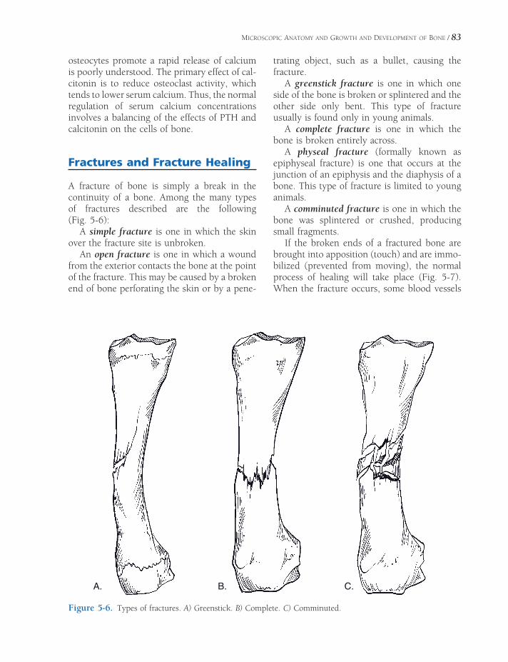
MICROSCOPIC ANATOMY AND GROWTH AND DEVELOPMENT OF BONE / 83
osteocytes promote a rapid release of calcium is poorly understood. The primary effect of cal-citonin is to reduce osteoclast activity, which tends to lower serum calcium. Thus, the normal regulation of serum calcium concentrations involves a balancing of the effects of PTH and calcitonin on the cells of bone.
Fractures and Fracture Healing
A fracture of bone is simply a break in the continuity of a bone. Among the many types of fractures described are the following (Fig. 5-6):
A simple fracture is one in which the skin over the fracture site is unbroken.
An open fracture is one in which a wound from the exterior contacts the bone at the point of the fracture. This may be caused by a broken end of bone perforating the skin or by a pene-
trating object, such as a bullet, causing the fracture.
A greenstick fracture is one in which one side of the bone is broken or splintered and the other side only bent. This type of fracture usually is found only in young animals.
A complete fracture is one in which the bone is broken entirely across.
A physeal fracture (formally known as epiphyseal fracture) is one that occurs at the junction of an epiphysis and the diaphysis of a bone. This type of fracture is limited to young animals.
A comminuted fracture is one in which the bone was splintered or crushed, producing small fragments.
If the broken ends of a fractured bone are brought into apposition (touch) and are immo-bilized (prevented from moving), the normal process of healing will take place (Fig. 5-7). When the fracture occurs, some blood vessels
.C.B.A
Figure 5-6. Types of fractures. A) Greenstick. B) Complete. C) Comminuted.

84 / ANATOMY AND PHYSIOLOGY OF FARM ANIMALS
are ruptured, releasing blood around the broken ends of the bone. This forms a clot that is invaded by connective tissue cells forming granulation tissue (term for mass of tissue con-sisting largely of fi broblasts and capillaries). The osteoblasts from the surface of the bone, from the periosteum, and from the endosteum lining the marrow cavities and haversian canals divide rapidly and produce a massive amount of osteoid tissue called a callus. The osteoid tissue fi lls the gap between the broken ends of the bone, fi lls the marrow cavity for a distance, and completely encircles the broken ends of the bone, forming an effective splint that usually
prevents movement between the segments. The callus becomes mineralized, changing into true bone. Remodeling of the callus to form a typical bone shaft with a marrow cavity completes the healing process. Misalignment of the fractured bone is corrected to some extent by the action of osteocytes and osteoclasts, which also remove excessive internal and external callus. As soon as the bone is put to use, functional orientation of the callus begins, with a tendency to straighten imperfections in the alignment of the bone. The callus will increase in size on the concave side, where stress is greatest, and tend to erode on the convex side, thus tending to correct any deformity.
The amount of spontaneous correction that is possible in fractures depends on a number of factors, including age of the animal, blood supply to the bone, degree of correction neces-sary, presence or absence of infection, and amount of damage to surrounding tissues. Excessive separation of fragments, which may be caused by too much traction or incomplete immobilization of a fracture, may result in non-union, with fi brous tissue fi lling the gap between fragments.
Quickest fracture healing occurs in young animals, particularly if the fracture site has a good blood supply and is completely immobi-lized with the ends of the fragments in apposi-tion. In humans, a fracture may heal completely within a month in an infant, but a similar frac-ture in a person past middle age may require 6 months or longer to heal.
If bone healing is delayed or a major defect has resulted from a severe fracture or surgical removal of bone, grafting or transplanting bone into the damaged area may stimulate healing. Bone for grafts may be obtained from the same animal that is receiving the graft (autogenous graft) or from another animal of the same species (allograft). Autogenous grafts are typi-cally cancellous bone obtained from a site such as the proximal end of the humerus. Allografts of cortical bone may be used relatively intact or as bone chips to fi ll defi cient areas. Autogenous grafts have an advantage in that the portion of the bone in contact with tissue fl uid may survive and the osteoblasts become active. At the same
Figure 5-7. Some stages in healing of a fracture of a long bone. A) Early soft callus replaces blood clot. B) Intermediate callus. C) Nearly healed hard callus.
MICROSCOPIC ANATOMY AND GROWTH AND DEVELOPMENT OF BONE / 85
time, osteoclasts remove the dead portions of the graft, which are replaced by healthy bone if the graft is functional and subjected to the proper amount of stress. Osteoblasts in allografts die, because the animal body tends to reject any foreign protein.
Other Pathologic Conditions
Other pathologic conditions of bones may be caused by infections, tumors, endocrine distur-bances, or nutritional imbalances. Tuberculosis of bone and osteomyelitis (infl ammation of the bone and bone marrow) are two infections sometimes seen in bone. In humans, osteomy-elitis usually is caused by staphylococci or streptococci that gain access to the bone by way of the bloodstream and develop into a general infection or by way of a wound, in which case the infection may remain local.
Benign (slow-growing, noninvasive, and not likely to cause death) bone tumors are named according to the cells of bone from which they originate. A tumor of bone tissue itself is called an osteoma. Chondromas may develop from the epiphyseal cartilage or from unabsorbed islands of cartilage that preceded the develop-ing bone. Tumors develop and grow as a result of intrinsic changes in the cells of origin, but bony growths may also occur on the surface of a bone simply in response to prolonged irrita-tion. Such growths are called exostoses.
Malignant tumors grow rapidly, metastasize (spread to other locations), and are often fatal
if not treated. Osteogenic sarcomas are malig-nant tumors of bone that most commonly develop near the ends of long bones. Sarcoma is a general term for malignant tumors originat-ing from tissues of mesodermal origin, such as connective tissue.
Osteodystrophy is a general term for any abnormality in bone development. Many osteo-dystrophies are due to some abnormality in the normal regulation of calcium and phospho-rus. The abnormality could be produced as a result of an inappropriate diet or some disease state disrupting calcium and phosphorus regulation.
Rickets in young, growing animals and osteomalacia in adults are conditions of inad-equate mineralization of osteoid. Without appropriate mineralization, bones are relatively weak and fl exible. Both rickets and osteomala-cia usually are due to a lack of vitamin D, which is required for normal absorption of calcium from the gastrointestinal tract. However, they may also be caused by an imbalance or lack of calcium and/or phosphorus in the diet. Rickets chiefl y affects the growing areas in bone, while osteomalacia, sometimes called adult rickets, affects the entire bone, since there are no rapidly growing areas in adult bone.
Achondroplasia is a hereditary condition in which the metaphyses fuse early in life, but the bones continue to increase in diameter. An animal affected with it is called an achondro-plastic dwarf. The dachshund is a breed of dogs selectively bred for this condition. Dwarfi sm in cattle closely resembles achondroplasia.

JOINTS
Syndesmology (arthrology) is the study of the articulations (unions) between bones,
which are commonly called joints. Joints may be classifi ed by a variety of schemes, usually by that of anatomy or degree of movement. The tissue that unites the bones of a joint is gener-ally fi brous tissue or cartilage. The structure and arrangement of these tissues is specialized for the joint’s specifi c task.
Classifi cation of Joints
Based on their structure and the material that unites them, joints may be classifi ed as fi brous, cartilaginous, or synovial. These classifi cations can be combined as follows.
Fibrous Joints
Fibrous joints have no joint cavity. The bones are united by fi brous tissue (Fig. 6-1).
Syndesmosis refers to a joint united by fi brous tissue that permits only slight move-ment. The normal union of the shafts of the splint bones and cannon bone of the horse is an example of syndesmosis.
A suture is the particular fi brous joint between bones of the skull. Sutures often com-pletely ossify in maturity.
A gomphosis is the specialized articula -tion of teeth in their alveoli (sockets) in the
Classifi cation of JointsFibrous Joints
Cartilaginous Joints
Synovial Joints
Other Synovial Structures
Movements of JointsTypes of Synovial JointsJoints of the Axial SkeletonJoints of the Appendicular Skeleton
Joints of the Thoracic Limb
Joints of the Pelvic Limb
Pathology of Joints and Related Structures
Chapter 6
87
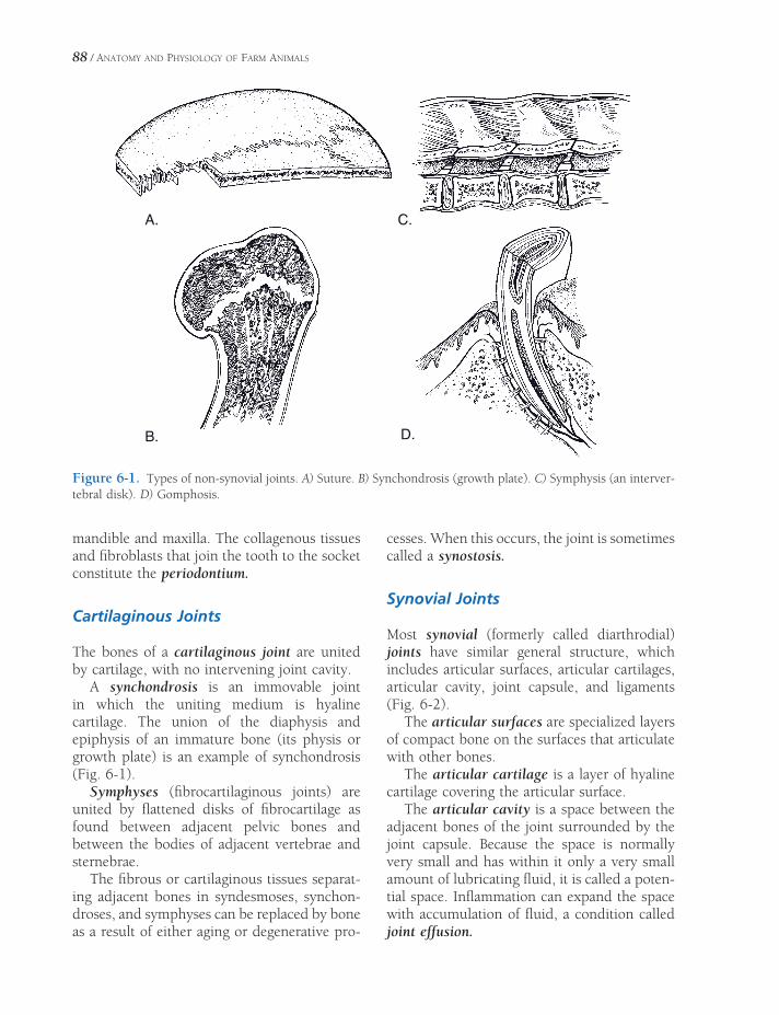
88 / ANATOMY AND PHYSIOLOGY OF FARM ANIMALS
mandible and maxilla. The collagenous tissues and fi broblasts that join the tooth to the socket constitute the periodontium.
Cartilaginous Joints
The bones of a cartilaginous joint are united by cartilage, with no intervening joint cavity.
A synchondrosis is an immovable joint in which the uniting medium is hyaline cartilage. The union of the diaphysis and epiphysis of an immature bone (its physis or growth plate) is an example of synchondrosis (Fig. 6-1).
Symphyses (fi brocartilaginous joints) are united by fl attened disks of fi brocartilage as found between adjacent pelvic bones and between the bodies of adjacent vertebrae and sternebrae.
The fi brous or cartilaginous tissues separat-ing adjacent bones in syndesmoses, synchon-droses, and symphyses can be replaced by bone as a result of either aging or degenerative pro-
cesses. When this occurs, the joint is sometimes called a synostosis.
Synovial Joints
Most synovial (formerly called diarthrodial) joints have similar general structure, which includes articular surfaces, articular cartilages, articular cavity, joint capsule, and ligaments (Fig. 6-2).
The articular surfaces are specialized layers of compact bone on the surfaces that articulate with other bones.
The articular cartilage is a layer of hyaline cartilage covering the articular surface.
The articular cavity is a space between the adjacent bones of the joint surrounded by the joint capsule. Because the space is normally very small and has within it only a very small amount of lubricating fl uid, it is called a poten-tial space. Infl ammation can expand the space with accumulation of fl uid, a condition called joint effusion.
A.
B.
C.
D.
Figure 6-1. Types of non-synovial joints. A) Suture. B) Synchondrosis (growth plate). C) Symphysis (an interver-tebral disk). D) Gomphosis.
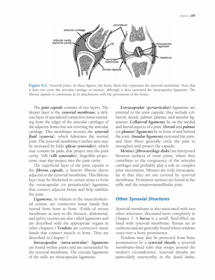
JOINTS / 89
The joint capsule consists of two layers. The deeper layer is the synovial membrane, a deli-cate layer of specialized connective tissue extend-ing from the edges of the articular cartilages of the adjacent bones but not covering the articular cartilage. This membrane secretes the synovial fl uid (synovia), which lubricates the normal joint. The synovial membrane’s surface area may be increased by folds (plicae synoviales), which may contain fat pads, that project into the joint cavity. Villi (villi synoviales), fi ngerlike projec-tions, may also project into the joint cavity.
The superfi cial layer of the joint capsule is the fi brous capsule, a heavier fi brous sleeve adjacent to the synovial membrane. This fi brous layer may be thickened in certain areas to form the extracapsular (or periarticular) ligaments that connect adjacent bones and help stabilize the joint.
Ligaments, in relation to the musculoskele-tal system, are connective tissue bands that extend from bone to bone. (Folds of serous membrane as seen in the thoracic, abdominal, and pelvic cavities are also called ligaments and are described with the appropriate organs in other chapters.) Tendons are connective tissue bands that connect muscle to bone. They are described in Chapter 7.
Intracapsular (intra-articular) ligaments are found within joints and are surrounded by the synovial membrane. The cruciate ligaments of the stifl e are intracapsular ligaments.
Extracapsular (periarticular) ligaments are external to the joint capsule; they include col-lateral, dorsal, palmar, plantar, and annular lig-aments. Collateral ligaments lie on the medial and lateral aspects of a joint. Dorsal and palmar (or plantar) ligaments lie in front of and behind the joint. Annular ligaments surround the joint, and their fi bers generally circle the joint to strengthen and protect the capsule.
Menisci ( fi brocartilage disks) are interposed between surfaces of some joints, where they contribute to the congruency of the articular cartilages and probably play a role in complex joint movements. Menisci are truly intracapsu-lar in that they are not covered by synovial membrane. Prominent menisci are found in the stifl e and the temporomandibular joint.
Other Synovial Structures
Synovial membrane is also associated with two other structures, discussed more completely in Chapter 7. A bursa is a small, fl uid-fi lled sac lined with synovial membrane. Bursae act as cushions and are generally found where tendons cross over a bony prominence.
Tendons may also be protected from bony prominences by a synovial sheath, a synovial membrane–lined tube that wraps around the tendon’s circumference. Synovial sheaths are particularly noteworthy in the distal limbs,
Figure 6-2. Synovial joints. In these fi gures, the heavy black line represents the synovial membrane. Note that it does not cover the articular cartilage or menisci, although it does surround the intracapsular ligaments. The fi brous capsule is continuous at its attachments with the periosteum of the bones.
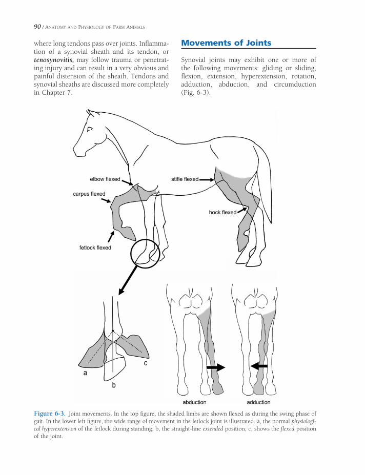
90 / ANATOMY AND PHYSIOLOGY OF FARM ANIMALS
where long tendons pass over joints. Infl amma-tion of a synovial sheath and its tendon, or tenosynovitis, may follow trauma or penetrat-ing injury and can result in a very obvious and painful distension of the sheath. Tendons and synovial sheaths are discussed more completely in Chapter 7.
Movements of Joints
Synovial joints may exhibit one or more of the following movements: gliding or sliding, fl exion, extension, hyperextension, rotation, adduction, abduction, and circumduction (Fig. 6-3).
Figure 6-3. Joint movements. In the top fi gure, the shaded limbs are shown fl exed as during the swing phase of gait. In the lower left fi gure, the wide range of movement in the fetlock joint is illustrated. a, the normal physiologi-cal hyperextension of the fetlock during standing; b, the straight-line extended position; c, shows the fl exed position of the joint.
JOINTS / 91
Gliding or sliding movement occurs between two more or less fl at surfaces in plane joints.
Flexion is movement in the sagittal plane that tends to decrease the angle between seg-ments making up a joint. The carpus must be fl exed when a horse’s front foot is picked up for trimming.
Extension is the reverse of fl exion and is movement in the sagittal plane that tends to increase the angle between segments forming the joint.
Hyperextension is movement in which the angle between segments is increased beyond 180° (a straight line). The fetlock of the horse is hyperextended while in the normal standing position (often called physiologic hyperexten-sion). Other joints do not normally hyperex-tend unless fatigued or stressed or when poor conformation provides inadequate support to the joint. Hyperextension of the equine carpus, for example, may occur late in a race, when the galloping horse is fatigued, and can result in injury to the joint. A horse whose carpi are hyperextended because of poor conformation is said to be calf-kneed, and it is prone to go lame with hard work.
Rotation consists of a twisting movement of a segment around its own axis. Shaking the head “no” is a good example of rotation, in this case between the atlas and axis of the vertebral column.
Adduction is movement toward the median plane. Abduction is movement away from the median plane.
Circumduction combines the other types of movement with the exception of rotation; it may be defi ned as a movement in which an extremity describes a cone, with the distal end of the extremity describing a circle. A horse that paddles (an undesirable outward swinging of the feet while in motion) is exhibiting circumduction.
Pronation rotates an extremity so that the dorsum is up. Supination is a movement that rotates an extremity so that the palmar or plantar aspect of the limb is up. Pronation and supination are rarely seen to any extent in domestic animals.
Types of Synovial Joints
Synovial joints are classifi ed according to the type of joint surface and movements. Simple joints involve only two articulating bones, while composite joints include more than two bones within the same joint capsule. The types of synovial joints commonly found in domestic animals include ginglymus, plane, trochoid, and spheroid. Additional types of joints, described particularly in the dog, are condy-loid, ellipsoid, and saddle.
Ginglymus (hinge) joints move only in their sagittal plane. The movements in this type of joint are fl exion, extension, and, in some joints, hyperextension. The fetlock is a good example of a ginglymus joint.
Plane joints have only a slight gliding move-ment between relatively fl at apposed surfaces. These surfaces are called facets. The joints between adjacent carpal bones are examples of plane joints.
A trochoid (pivot) joint is one in which rotary movement occurs around one axis.
Spheroid (ball and socket) joints permit movement in nearly any direction. A spherical head on one bone fi ts into a cup-shaped depres-sion in the other. Flexion, extension, adduc-tion, abduction, rotation, and circumduction are all possible in spheroid joints. The coxo-femoral (hip) joint is the best example of a spheroid joint.
In a condylar joint, convex articular condyles articulate with somewhat concave articular sur-faces. The temporomandibular and femorotibial (stifl e) joints are examples. They resemble gin-glymus joints but permit more movement.
The ellipsoid joint has an articular surface that is expanded more in one direction than another, forming an ellipse. The joint between the distal end of the radius and proximal row of carpal bones is ellipsoid. In domestic animals this has been called a ginglymus joint.
The saddle joint has surfaces that resemble an English saddle. It permits all types of move-ment except rotation. The carpometacarpal joint of the human thumb is the best example, but the interphalangeal joints of the dog are sometimes classifi ed as saddle joints.
92 / ANATOMY AND PHYSIOLOGY OF FARM ANIMALS
Joints of the Axial Skeleton
The joints of the skull are chiefl y sutures, with adjacent bones united by fi brous tissue. In old age these typically ossify, becoming synostoses. In addition to the sutures associated with the braincase, the skull also features the symphysis of the mandible, the synchondrosis at the junc-tion of the sphenoid bone and occipital bone at the base of the skull, and the temporoman-dibular joint.
The temporomandibular joint between the mandible and the temporal bone of the skull consists of an articular surface on the skull and one on the mandible, with a plate of cartilage (articular disk or meniscus) between. The tem-poromandibular joint acts as a ginglymus joint when the mouth opens and closes and as a plane joint when the jaw moves from side to side and forward and back, as in grinding food. The temporomandibular joint may be described as a condylar joint.
The atlanto-occipital joint between the occipital bone of the skull and fi rst cervical vertebra (the atlas) is strictly a ginglymus joint. Two condyles on the occipital bone fi t into corresponding depressions in the atlas. The only movements possible are fl exion and exten-sion in the sagittal plane, as in nodding the head “yes.”
Rotation of the head occurs between the atlas and axis, at the atlantoaxial joint. The dens, a toothlike projection from the cranial extremity of the axis, projects into the vertebral foramen of the atlas, where it is held by a group of strong ligaments that permit considerable rotary movement. The atlantoaxial joint is the best example of a pivot joint, in which one segment rotates around the long axis of another.
The symphyseal (fi brocartilaginous) joints between adjacent vertebrae throughout the rest of the vertebral column exhibit relatively little motion. The bodies of adjacent vertebrae are united by a heavy disk of fi brocartilage, the intervertebral disk (IVD), that is fl exible enough to permit some bending in any direc-tion, even twisting. This fi brocartilage has a soft center, the nucleus pulposus, which may abnor-
mally protrude through the surrounding annulus fi brosus into the vertebral canal. The resulting condition, a ruptured intervertebral disk, may cause signifi cant injury to the overly-ing spinal cord.
The articular processes of adjacent vertebrae have fl at surfaces that are apposed to form plane joints with limited gliding movements. These surfaces are larger and the movements more extensive near the head, decrease in the thoracic region, and are again more extensive in the lumbar region. The joints between sacral vertebrae fuse completely, and the sacrum becomes a single bone with the segments joined by synostoses.
The ribs are attached to the vertebral column by two separate joints. One is between the head of the rib and the costal fovea formed by the bodies of two adjacent thoracic vertebrae; the other is between the tubercle of the rib and a facet on the transverse process of the vertebra of the same number as the rib. The heads of the paired ribs on each side of an intervertebral space are joined by an intercapital ligament, which forms a reinforcing band across the dorsal side of the intervertebral disk. The pres-ence of the intercapital ligament helps explain why disk protrusions are rare in the thoracic region of the vertebral column.
Joints of the Appendicular Skeleton
Joints of the Thoracic Limb
The scapula has no true bony connection with the thorax. It is held in place by a number of muscles and ligaments. This type of joint is sometimes called a synsarcosis.
The shoulder (scapulohumeral) joint is spheroid. Movements in all directions, includ-ing rotation, are possible. In domestic animals, however, the arrangement of shoulder muscles practically limits movement to a hinge type of action in the sagittal plane. Thus, extension and fl exion are the chief movements. The head of the humerus is a large sphere much more extensive than the comparable cavity of the
JOINTS / 93
scapula. The joint capsule is extensive, with poorly developed collateral ligaments. Instead, the tendons of the muscles crossing the shoul-der joint on all sides act effectively as support-ive ligaments (it is these well-developed tendons that blend in human beings to form the so-called rotator cuff. This term is not used in veterinary anatomy, however.).
The elbow is a true ginglymus joint formed by the humeral condyle meeting the proximal ends of the radius and ulna. The proximal end of the radius is slightly concave and expanded to give an extensive surface for support. Com-bined with semilunar notch of the ulna, the ra dius forms a half-circle embracing the humeral condyle. In the horse and ox, move-ment in the elbow is limited to fl exion and extension. In humans and to a lesser degree in carnivores, the joint between the radius and ulna permits supination and pronation.
The carpus (Figs. 6-4 and 6-5) is a complex joint that permits fl exion and extension not only between the radius and proximal row of carpal bones (radiocarpal joint), but also to a lesser degree between the proximal and distal rows of carpal bones (midcarpal joint). The entire joint is capable of absorbing considerable shock because of the many small joints formed by adjacent carpal bones connected by short ligaments. The joint between the distal row of carpal bones and the metacarpus (carpometa-carpal joint) is almost entirely a plane joint, which allows only limited gliding movements and makes almost no contribution to the degree to which the entire carpus can fl ex.
The fi brous layer of the joint capsule of the carpus is extensive, being a long sleeve extend-ing from the radius to the metacarpus and enclosing the carpal bones. The synovial mem-brane, however, forms three separate sacs: a radiocarpal sac, a midcarpal sac, and a carpo-metacarpal sac.
In the horse, there is normally little move-ment between the large metacarpal III and the smaller metacarpals II and IV (splint bones). Excessive movement or trauma results in infl ammation at this site, creating a splint. In an acute state, the splint is a painful swelling where the shafts of the large
and small metacarpal bones meet. Later this swelling may ossify and form a bony promi-nence that may not cause any lameness at all (see Fig. 4-9).
In the ox and sheep, the third and fourth metacarpal bones are fused to form the single cannon bone, which articulates proximally with the distal carpal bones and distally with the proximal phalanges. In the dog and pig, the proximal ends of adjacent metacarpal bones abut one another in a series of plane joints (intermetacarpal joints).
The metacarpophalangeal (fetlock) joint of the horse (Fig. 6-6) is formed by the distal end of the metacarpus; the proximal end of the fi rst phalanx, or long pastern bone; and the two proximal sesamoid bones. It is a ginglymus joint that in the normal standing position is hyperextended (Fig. 6-3).
The proximal interphalangeal (pastern) joint is a ginglymus joint between the fi rst and second phalanges (the long and the short pastern bones). Although it is a ginglymus joint, it is rather limited in motion. Degenera-tive changes in this joint are called a high ringbone.
The distal interphalangeal joint (coffi n joint) is formed by the second and third pha-langes and the distal sesamoid (navicular) bone. The coffi n joint is largely encased within the hoof and is essentially a ginglymus joint. Degen-erative joint disease in this joint is commonly referred to as low ringbone. A similar pattern of articulations is followed for each digit in animals possessing more than one digit per foot (e.g., ruminants and pigs).
Joints of the Pelvic Limb
The sacroiliac joint is the only bony connec-tion between the axial and appendicular skele-tons. In the young animal, this joint exhibits features of both synchondroses and synovial joints, although its mobility is progressively diminished in the adult. The articular surface of the sacrum is held in tight apposition to the wing of the ilium by a number of short, strong ligaments. Movement in this joint is normally

Midcarpal Joint
Radiocarpal Joint
A.
B.
.D.C
I U
A
I UI U A
3
2
4
3 42&3
4
Radius
RR
Radius
MC III&IV
MC III
Carpometacarpal Joint
Figure 6-4. Carpus and distal joints of limb. A) Equine thoracic limb in fl exion. Most of the carpal fl exion derives from fl exion at radiocarpal and midcarpal joints; the carpometacarpal joint has little movement. B) Lateral view of equine thoracic limb in weight-bearing position. C) Dorsal view of the equine carpus. D) Dorsal view of the bovine carpus.
94
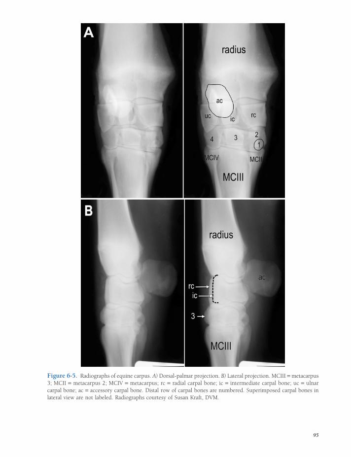
Figure 6-5. Radiographs of equine carpus. A) Dorsal-palmar projection. B) Lateral projection. MCIII = metacarpus 3; MCII = metacarpus 2; MCIV = metacarpus; rc = radial carpal bone; ic = intermediate carpal bone; uc = ulnar carpal bone; ac = accessory carpal bone. Distal row of carpal bones are numbered. Superimposed carpal bones in lateral view are not labeled. Radiographs courtesy of Susan Kraft, DVM.
95

96 / ANATOMY AND PHYSIOLOGY OF FARM ANIMALS
Figure 6-6. Radiographs of equine digit (a lateral projection) (A) and fetlock (a cranial-caudal projection) (B). MCIII = third metacarpal bone; P1 = proximal phalanx; P2 = middle phalanx; P3 = distal phalanx; PS = proximal sesamoid bones; DS = distal sesamoid (navicular) bone. Radiographs courtesy of Susan Kraft, DVM.
severely limited, but it may become more extensive just prior to parturition, when the ligaments stretch under the infl uence of the hormone relaxin (see Chapter 28). Ligaments in this area include dorsal and ventral sacroil-iac and sacrotuberous ligaments. In the ox, sheep, and horse, the latter is a strong, wide
band that extends from the sacrum to the tuber ischiadicum and helps form the lateral wall of the pelvis.
The sacroiliac joint can be partially sepa-rated (a sacroiliac subluxation) by a fall or other trauma. Such an injury produces pain and muscle spasm and often becomes a

JOINTS / 97
source of chronic soreness. Sometimes visible asymmetry in the two tubera sacrales develops, with the subluxated side displaced upward. This sign is commonly called hunt-er’s bump (Fig. 6-7).
The coxofemoral (hip) joint is the best example of a spheroid (ball and socket) joint. The head of the femur is about two-thirds of a sphere that fi ts into the less extensive acetabu-lum of the os coxae. The margin of the acetabu-lum is reinforced and deepened by a marginal cartilage.
The joint capsule of the hip joint is extensive, but not so extensive as that of the shoulder. The ligament of the femoral head (formerly round ligament) connects the head of the femur with a nonarticular area within the acetabulum. The hip joint of the horse is reinforced by an acces-sory ligament that extends from the prepubic tendon to the head of the femur. It is presumed to prevent signifi cant abduction of the pelvic limb. Movements in nearly all directions are possible in the hip joint, but as in the shoulder joint, extension and fl exion are the movements chiefl y employed in normal locomotion.
The stifl e joint corresponds to the human knee (Figs. 6-8 and 6-9). It comprises the condyles of the distal femur, the patella, and the proximal tibia. The femoral condyles are separated from the proximal tibia by two intra-articular menisci. Each meniscus is a half-moon–shaped disk that conforms to the surface of the proximal tibia and is concave on the upper surface to fi t the respective condyle of the femur. These menisci help keep the joint congruent and absorb shock. The stifl e is stabilized by medial and lateral collateral ligaments and by two intra-capsular cruciate ligaments that form an X as they cross from the tibia to the femur in the middle of the joint.
The patella (kneecap) is a sesamoid bone embedded in the tendon of insertion of the large cranial muscles of the thigh. This muscle group (see Chapter 7) is a powerful extensor of the stifl e, acting through its connection to the cranial aspect of the proximal tibia via one (pig) or three (horses and ruminants) strong patellar ligaments. In the horse, the medial patellar ligament is attached to the medial aspect of the
Figure 6-7. Hunter’s bump, an asymmetry of the tubera sacrales produced by subluxation of the sacroiliac joint. The higher tuber sacrale is the affected side. (Reprinted with permission of Wiley-Blackwell from Stashak, T. S. Adams’ Lameness in Horses. 5th ed. Baltimore: Lippincott Williams & Wilkins, 2002.)
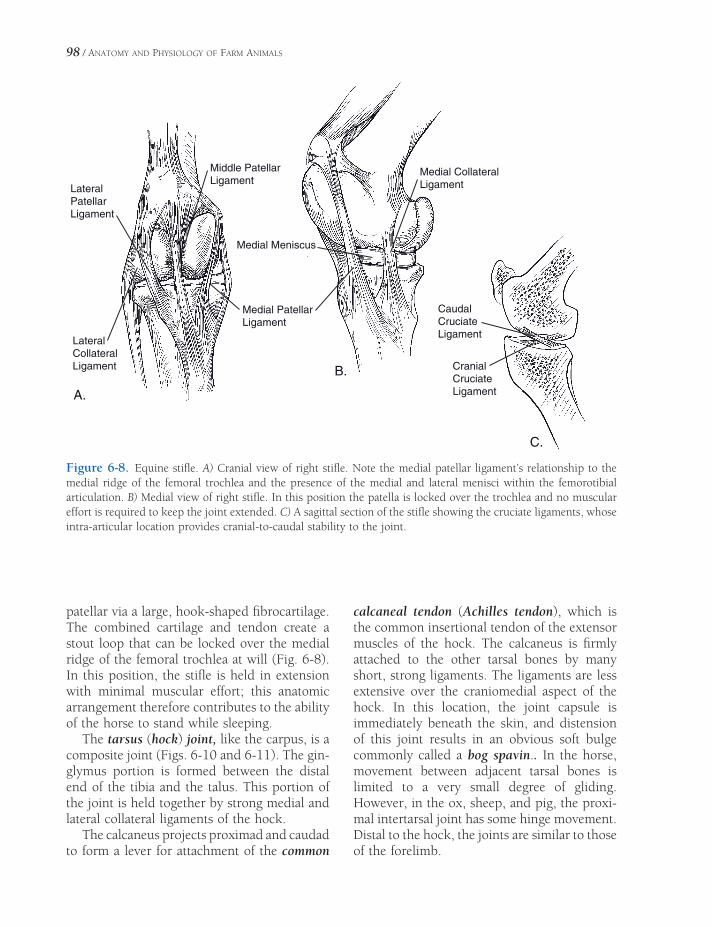
98 / ANATOMY AND PHYSIOLOGY OF FARM ANIMALS
Middle PatellarLigament
LateralPatellarLigament
Medial PatellarLigament
Medial CollateralLigament
Medial Meniscus
LateralCollateralLigament
CaudalCruciateLigament
CranialCruciateLigamentA.
B.
C.
Figure 6-8. Equine stifl e. A) Cranial view of right stifl e. Note the medial patellar ligament’s relationship to the medial ridge of the femoral trochlea and the presence of the medial and lateral menisci within the femorotibial articulation. B) Medial view of right stifl e. In this position the patella is locked over the trochlea and no muscular effort is required to keep the joint extended. C) A sagittal section of the stifl e showing the cruciate ligaments, whose intra-articular location provides cranial-to-caudal stability to the joint.
patellar via a large, hook-shaped fi brocartilage. The combined cartilage and tendon create a stout loop that can be locked over the medial ridge of the femoral trochlea at will (Fig. 6-8). In this position, the stifl e is held in extension with minimal muscular effort; this anatomic arrangement therefore contributes to the ability of the horse to stand while sleeping.
The tarsus (hock) joint, like the carpus, is a composite joint (Figs. 6-10 and 6-11). The gin-glymus portion is formed between the distal end of the tibia and the talus. This portion of the joint is held together by strong medial and lateral collateral ligaments of the hock.
The calcaneus projects proximad and caudad to form a lever for attachment of the common
calcaneal tendon (Achilles tendon), which is the common insertional tendon of the extensor muscles of the hock. The calcaneus is fi rmly attached to the other tarsal bones by many short, strong ligaments. The ligaments are less extensive over the craniomedial aspect of the hock. In this location, the joint capsule is immediately beneath the skin, and distension of this joint results in an obvious soft bulge commonly called a bog spavin.. In the horse, movement between adjacent tarsal bones is limited to a very small degree of gliding. However, in the ox, sheep, and pig, the proxi-mal intertarsal joint has some hinge movement. Distal to the hock, the joints are similar to those of the forelimb.

Figure 6-9. Lateral radiograph of equine stifl e joint. Radiographs courtesy of Susan Kraft, DVM.
Tibia
C
Central + 42 + 3
MTIII + IV
2+3
Central+ 4
TCa
Tibia
Ca
T
C 43
MT IIIMT IV
MTIII + IV
T
Tibia
.C.B.A
Figure 6-10. Hock. A) Lateral view of left bovine hock. B) Cranial view of left equine hock. C) Cranial view of left bovine hock.
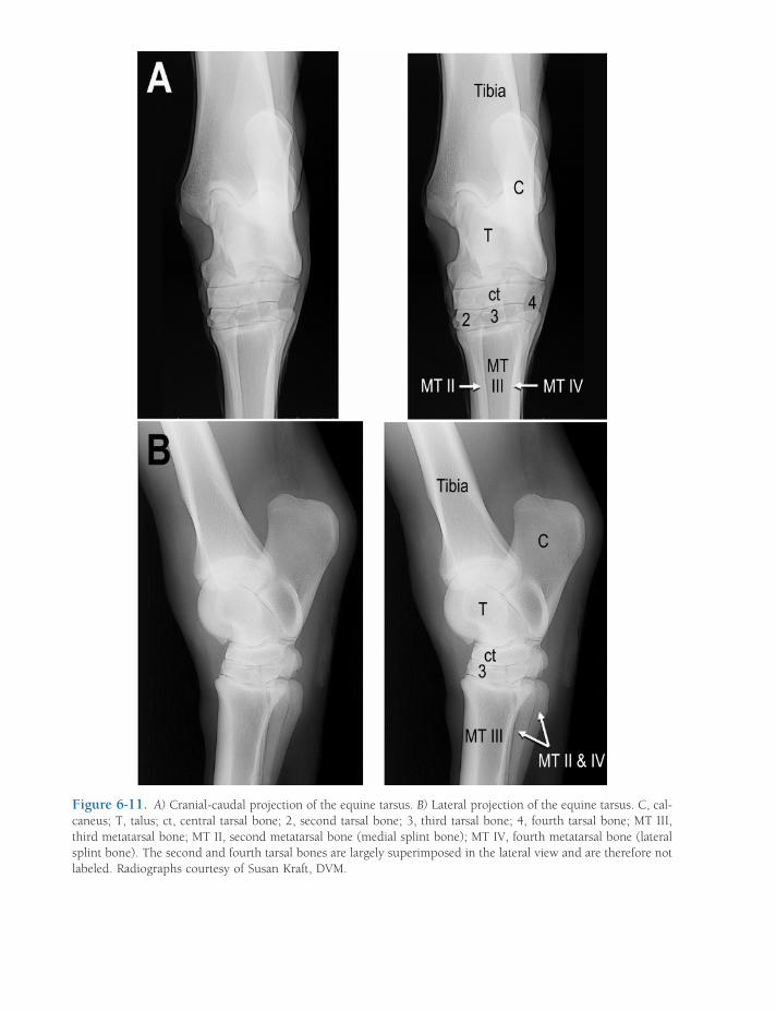
Figure 6-11. A) Cranial-caudal projection of the equine tarsus. B) Lateral projection of the equine tarsus. C, cal-caneus; T, talus; ct, central tarsal bone; 2, second tarsal bone; 3, third tarsal bone; 4, fourth tarsal bone; MT III, third metatarsal bone; MT II, second metatarsal bone (medial splint bone); MT IV, fourth metatarsal bone (lateral splint bone). The second and fourth tarsal bones are largely superimposed in the lateral view and are therefore not labeled. Radiographs courtesy of Susan Kraft, DVM.

JOINTS / 101
Pathology of Joints and Related Structures
Since synovial joints depend on free movement for effective functioning, anything that inter-feres with their mobility can produce gait abnormalities. Disorders affecting joints are commonly due to injuries, infections, or infl ammations.
A dislocation, also known as a luxation, is a condition in which articular surfaces undergo a signifi cant loss of congruency (less severe dis-locations are subluxations). Dislocation of a joint nearly always includes stretching or tearing of ligaments, and if the dislocation is severe enough, the joint capsule also may tear. The usual treatment for dislocation consists of replacing the joint in its normal position (reduc-ing the luxation), surgical repair of severely dis-rupted elements if necessary, and external support and/or fi xation. Reduction may be dif-fi cult unless the animal is fully anesthetized so that all muscles are relaxed. Early treatment is important to prevent the joint cavity from fi lling with connective tissue. Because of the excessive stretching or tearing of ligaments, recovery from a dislocation may be less satisfac-tory and take longer than recovery from a pro-perly treated fracture.
Occasionally, a reasonably functional joint, called a false joint, may develop at the site of a chronic luxation. In a false joint, the fi brous connective tissue that develops in response to the infl ammation and abnormal stresses at the luxation may provide enough stability to permit considerable movement, even though no joint capsule or cartilage develops as in true joints. False joints may also form at a fracture site if the ends of the fracture are not immobilized.
Fractures, or breaks of bones, may affect one or more of the segments making up a joint. Fractures within or close to a joint are often diffi cult to reduce adequately, since even minor discontinuities in the articular surface can lead to severe arthritic changes. Immobilization fol-lowing reduction is also often diffi cult because of the short length of at least one of the segments.
A sprain is a condition in which the liga-ments are stretched, but the joint does not remain subluxated following removal of the displacing force. The term strain is sometimes used in place of sprain, although strain is used more frequently to denote excessive stretching of a muscle or tendon. Although a considerable amount of swelling may follow a sprain, the affected joint usually recovers spontaneously if rested adequately.
Lacerations such as those from barbed wire may extend into a joint cavity, causing loss of synovial fl uid and exposing the interior of the joint to the external environment. A condition of this nature may be serious and diffi cult to treat. The danger is not from the loss of syno-vial fl uid (which is rapidly replaced), but from infection of the joint cavity. Synovial fl uid is a good medium for bacterial growth, and the many recesses of most joint cavities make drainage and treatment of an infected joint dif-fi cult. There is danger of permanent damage to the articular cartilage from infection.
Puncture wounds to joints may result from penetration by a sharp object such as a nail, wire, or wood sliver. Such wounds are espe-cially dangerous; since the wound is often not obvious, it usually drains poorly, and the envi-ronment into which the bacteria are introduced is frequently anaerobic (not exposed to air). In such conditions, a puncture wound can pre-cipitate a particularly severe infection.
Infection may also reach the joint by way of blood or lymph. Erysipelas of swine and um ib-lical infections arising from a variety of bacteria in foals frequently result in joint infections. Certain viral illnesses, for example caprine arthritis–encephalitis (CAE) virus of goats, can cause nonseptic arthritis of multiple joints.
Arthritis, or more correctly, osteoarthritis, is infl ammation of the components of a joint, causing swelling and pain; it usually accompa-nies each of the conditions previously men-tioned. Chronic infl ammation is likely to produce bony and cartilaginous changes that can permanently affect the joint’s function. Degenerative joint disease (DJD) is the medical expression often used to describe the multiple changes in a chronically infl amed joint; it

Table 6-1. Pathologic Disorders of Joints and Related Structures
Name of Condition Pathology
Bicipital bursitis Infl ammation of bursa between tendon of m. biceps brachii and humerus
Bog spavin Distension of joint capsule of hock; swelling on the craniomedial aspect of hock
Bone spavin (jack spavin) DJD of distal intertarsal and tarsometatarsal joints
Bowed tendon (tenosynovitis)
Infl ammation, usually due to stretching, of digital fl exor tendons and their synovial sheaths caudal to cannon bone; usually thoracic limbs affected
Bursitis Infl ammation of any bursa
Capped elbow Infl ammation of bursa over olecranon process (point of elbow)
Capped hock Infl ammation of bursa over calcaneus (point of hock)
Carpitis (popped knee) Infl ammation of carpal joint capsule and/or ligaments
Curb Thickening of long plantar ligament on caudal aspect of hock
Dislocation (luxation) Components of joint displaced
Fistulous withers Infl ammation or infection with draining tract from bursa over spinous process
Herniated (slipped) intervertebral disk
Prolapse of nucleus pulposus of IVD, usually into the vertebral canal; can cause spinal cord damage
Hunter’s bump Subluxation of sacroiliac joint
Laminitis (founder) Infl ammation of laminae between hoof wall and distal phalanx
Navicular disease Infl ammation of navicular bursa and bone, often with involvement of associated tendons
Osselets Periostitis of proximal phalanx and/or distal metacarpus III, with capsulitis of fetlock joint
Osteoarthritis Infl ammation of any joint. Because term includes the bony elements of joint, it is more descriptive than arthritis.
Poll evil Infl ammation or infection of bursa over atlas (C1)
Quittor Infection of collateral cartilages of distal phalanx
Ring bone Enthesiophyte formation in interphalangeal joints
Side bone Ossifi cation of collateral cartilages of distal phalanx
Splints Infl ammation and exostosis involving joint between cannon bone and a splint bone (usually medial)
Sprain Stretching of ligaments of any joint
Stifl ing (upward fi xation of the patella)
Inadvertent locking of patella over medial ridge of femoral trochlea; holds patella and hock in extension
Strain Excessive stretching of muscle and/or tendon
Subluxation Partial dislocation of any joint
Synovitis Infl ammation of any synovial membrane
Thorough-pin Synovitis of deep digital fl exor tendon sheath proximal to hock
Trochanteric bursitis Infl ammation of bursa between greater trochanter of femur and tendon of middle gluteal muscle
Wind puffs (wind gall) Synovitis of fetlock joint or tenosynovitis of digital fl exor tendons in region of fetlock

JOINTS / 103
includes loss of articular cartilage, erosion of underlying bone, and the development of bone spurs (enthesiophytes or osteophytes) around the margins of the joint. The obvious enlarge-ments associated with high and low ringbone in horses are due to the osteophytes associated with the osteoarthritic pastern and coffi n joints, respectively. These and other disorders affecting joints or related structures (mostly in horses) are listed in Table 6-1.
Osteoarthritis is commonly treated with nonsteroidal anti-infl ammatory drugs (e.g.,
aspirin, phenylbutazone), which reduce the pain associated with infl ammation but do not reverse DJD. Recently, much anecdotal and some clinical evidence has supported the use of chondroprotective agents (e.g., chondroitin sulfate, glucosamine). These substances, many of which are used as food additives, may inhibit formation of destruc-tive enzymes and prostaglandins in the dis-eased joints and increase production of the molecular constituents of synovial fl uid and cartilage.

ANATOMY OF THE MUSCULAR SYSTEM
A note on nomenclature is appropriate before undertaking this survey of the muscles of
domestic animals. Traditionally, muscles are given Latin names, and as these are foreign words, they are italicized. The Latin names are usually highly descriptive of the function and/or appearance of the muscle, and the student is encouraged to use a medical dictionary to explore their meaning. In Latin, as in many Romance languages, the noun is written fi rst, followed by adjectives that describe it. Hence, the musculus triceps brachii (literally, the three-headed muscle of the arm) begins with the noun, musculus, usually abbreviated as m., plural mm. In English, nouns are generally pre-ceded by their descriptors, so the abbreviation for muscle is placed at the end of the string of adjectives, for example, deep digital fl exor m. In Latin, the same muscle is m. fl exor digitalis pro-fundus. This text uses English or Latin names, opting for whichever is in wider use.
Types of Muscle Tissue
The three types of muscle are skeletal, smooth, and cardiac. The bulk of the muscle in the body is skeletal muscle, and it is responsible for pro-ducing the voluntary movements of the limbs, trunk, and head. It is also the muscle tissue with which we are most familiar as the meat of our domestic animals. The muscle cells (fi bers)
Types of Muscle TissueFunctions of the Muscular SystemSkeletal Muscle Organization
Muscle Attachments
Functional Grouping of Muscles
Synovial Structures
Muscles of the Thoracic LimbMuscles Acting on the Shoulder Girdle
Muscles Acting on the Shoulder Joint
Muscles Acting on the Elbow
Muscles Acting on the Carpus
Muscles Acting on the Digits
Muscles of the Pelvic LimbMuscles Acting on the Hip Joint
Muscles Acting on the Stifl e
Muscles Acting on the Hock
Muscles Acting on the Digit
Muscles of the HeadMuscles of Mastication
Muscles of Facial Expression
Other Muscles of the Head
Muscles of the Trunk and NeckExtensors of the Vertebral Column
Flexors of the Vertebral Column
Abdominal Muscles
Muscles of Respiration
Chapter 7
105
106 / ANATOMY AND PHYSIOLOGY OF FARM ANIMALS
of skeletal muscle tissue are grouped into dis-tinct organs of variable size identifi ed as indi-vidual muscles. These are usually attached to the bones of the skeleton (hence the term skel-etal muscle) and are under voluntary control of the animal. Under the microscope, skeletal muscle fi bers exhibit a characteristic striped pattern arising from the orderly arrangement of the contractile proteins within the cells (see Chapter 1, Plate I, No. 11). As a consequence, skeletal muscle is also called striated muscle.
Smooth (involuntary or unstriated) muscle is composed of muscle cells that have no stria-tions visible with a microscope. Smooth muscle is found in systems of the body with autonomic function. Thus, smooth muscle is a major com-ponent of the wall of organs of the digestive and urogenital systems and most blood vessels. Contraction of smooth muscle is an intrinsic property of the fi bers themselves, which means that contraction does not generally require stimulation by a nerve; however, the contractil-ity of smooth muscle is regulated and coordi-nated by the autonomic nervous system.
Cardiac muscle is characterized by fi bers with visible striations, so it is considered a type of striated muscle. However, cardiac muscle, like smooth muscle, contracts intrinsically and is not under voluntary control. Cardiac muscle is restricted to the heart, where it constitutes most of the thickness of the wall. Its rhythmic contraction is responsible for the circulation of blood.
Functions of the Muscular System
Vertebrate muscle has only one function: con-traction. “Relaxation” is a passive process, a lack of contraction. Thus, the varied functions of the muscular system are all based on con-traction (or shortening) of muscle fi bers. Layers of smooth muscle in the walls of the stomach and intestines contract to mix and propel food along the gastrointestinal tract; smooth muscle layers in the walls of blood vessels control the distribution of blood, which is propelled by the contraction of the cardiac muscle of the heart.
In the eye, smooth muscle fi bers adjust the diameter of the pupil and thickness of the lens for optimal vision, while in the skin, contrac-tion of smooth muscles causes the hair to stand up. Skeletal muscles permit locomotion by contracting to change the relative positions of bones during movement and by maintaining joint angles against the pull of gravity during support. The skeletal muscles of respiration move air into and out of the lungs by contract-ing to change the volume of the thoracic cavity. In addition, heat production through shivering is the result of brief repetitive contractions of skeletal muscle throughout the body.
The remainder of this chapter describes the locations, attachments, and actions of the major skeletal muscles. Figures 7-1 through 7-3 illus-trate many of these muscles of the horse and ox.
Skeletal Muscle Organization
Muscle fi bers are arranged in bundles sur-rounded by fi brous connective tissue. The con-nective tissue between individual muscle fi bers is called endomysium. The sheath surrounding bundles of muscle fi bers is called perimysium, and the connective tissue around an entire muscle is known as epimysium. The epimy-sium is the deep fascia of the muscle.
Muscle fi bers may be arranged in parallel sheets, as in the abdominal muscles, or nar-rower bands, as in the sartorius muscle on the medial side of the thigh; the muscle fi bers in these strap muscles are parallel to one another. Other arrangements of muscle fi bers include fusiform muscles and various pennate, or pen-niform (featherlike), arrangements (Fig. 7-4). In the penniform arrangements, a tendon rep-resents the quill and the muscle fi bers attaching to the tendon at an angle represent the vane of the feather. If the fi bers come from only one side, the arrangement is called unipennate; from two sides, bipennate; and from three or more sides, multipennate.
The parallel arrangement of muscle fi bers in strap muscles provides the greatest potential for overall muscle shortening but is a relatively

Temporalis m.
Parotidoauricularis m.
Splenius m.
Trapezius m. (thoracic part)Serratus ventralis m.(thoracic part)
Semitendinosus m.
Tendon of longdigital extensor m.
External jugular v.Zygomaticus m.
Parotid salivary glandRhomboideus cervicis m.
Serratus ventralis m. (cervical part)Subclavius m.
Trapezius m. (cervical part)
Latissimus dorsi m.
Thoracolumbar fascia
Serratus dorsalis caudalis m.
Coxal tuberMiddle gluteal m.
Subiliac lymph nodes
Superficial gluteal m.
Semimembranosus m.
Biceps femoris m.
Gastrocnemius m.
Soleus m.
Lateral saphenous v.
Deep head digital flexor m.
Lateral digital extensor m.
Common calcaneal tendon
Long digital extensor m. Tendon of cranial tibial m.
(cunean tendon)
Distal end of 4th metatarsalbone
Plantaranularligament
Tendon of peroneustertius m.
Cranial tibial m.Long digital extensor m.
Deep digital flexor m.: .medial head .deep head .superficial head
Medial saphenous v.
Fascia lata
Tensor fasciaelatae m.Lateral thoracic v.
Flexor carpiulnaris m.Flexor carpiradialis m.Cephalic v.Tendon ofextensor carpiobliquus m.
Superficial digitalflexor tendon
Deep digital flexortendon
Palmar anularligamentExtensor branch of
suspensory ligament
Distal end of 4thmetacarpal bone
Suspensory ligaments
Extensor carpi obliquus m.
Lateral digital extensor m.
Ulnaris lateralis m.
Common digital extensor m.
Extensor carpi radialis m.
Brachialis m.
Cephalic v.
Triceps brachii m. lateral head
Triceps brachii m. long head
Descending pectoral m.
Deltoideus m.
Omotransversarius m.
Sternocephalicus m.
Omohyoideus m.Facial v.
Buccinator m.
Depressor labiiinferioris m.
Caninus m.
Levator nasolabialis m.
Levator labii superioris m.
Masseter m.
Orbicularis oculi m.(under skin)
Facial n.
External abdominal oblique m.
( interosseus medius mm. )
Descending pectorial m.
Retinaculum
and peroneus tertius m. (deep)
Brachiocephalicus m.
Tendon of common digital extensor m.
Figure 7-1. Superfi cial muscles of the horse. (Reprinted with permission of Wiley-Blackwell from McCracken, T.O., Kainer, R.A., and Spurgeon, T.S. Spurgeon’s Color Atlas of Large Animal Anatomy. Baltimore: Lippincott Williams & Wilkins, 1999.)

Deep digital flexor m.
Orbit
Extensor carpi obliquus m.(abductor pollicis longus)
Base of cecum
Iliocostalis thoracis m.
Semispinaliscapitis m.
Digastricus m. (caudal belly)
Wing of atlas
Occipitohyoideus m.
Longus capitis m.
Maxilla
Nasal b.
Canine teethEsophagus
Intertransversarii m.Scalenus ventralis m.
Longissimus cervicis m.
Subclavius m.
Supraspinatus m.
Infraspinatus m.
Teres minor m.
Biceps brachii m.
Brachialis m.
Heart
Lateral digital extensor m.Right lung: cranial lobe
Right lung: caudal lobe
Right dorsal colon
Right ventral colonLiver
Body of cecum
Long digital extensor m. (cut)
Cranial tibial m.
Peroneus tertius m.
Lateral digital extensor m.
Short digital extensor m.
Deep digital flexor m.(deep or lateral head)
Soleus m.
Gastrocnemius m.(lateral head)
Vastus lateralis m.
Semimembranosus m.
Rectus femoris m.
Semitendinosus m.
Iliacus m.
Broad sacrotuberal ligament
Middle gluteal m.
Caudal flexure of duodenum
Longissimus thoracisand lumborum m. Spinalis thoracis m.
Rhomboideus m.
Longissimus atlantis m.
Longissimus capitis m.
Caudal capital oblique m.
Lateral cartilage ofdistal phalanx
(of quadriceps m.)
(of quadriceps m.)
(ungual cartilage)
Distal digital anular ligament
Sternothyrohyoideus m.
Figure 7-2. Deeper muscles of the horse. (Reprinted with permission of Wiley-Blackwell from McCracken, T.O., Kainer, R.A., and Spurgeon, T.S. Spurgeon’s Color Atlas of Large Animal Anatomy. Baltimore: Lippincott Williams & Wilkins, 1999.)
108

Subcutaneous abdominal v.
Deep digitalflexor m.(superficial head)
Malaris
Levator nasolabialis m.
Zygomaticus m.
Levator labiisuperioris m.
Caninus m.
Scutularis m.
Long digital extensor m.
Sacrocaudalis m.
Coccygeus m.
Frontalis m.
Orbicularisoculi m.
Facial n.
Buccinator m.
Mylohyoideus m.
Masseter m.
External jugular v.
Sternocephalicus m.
Brachiocephalicus m.
Omotransversarius m.
Deltoideus m.Brachialis m.
Cephalic v.
Triceps brachii m.
Extensor carpi radialis m.
Common digital extensor m.
Extensor carpi obliquus m.
Lateral digital extensor m.
Ulnaris lateralis m.
Ascending pectoral m.
External abdominaloblique m.
Tendon of extensor carpiobliquus m.
Superficial and deepdigital flexor tendons
Suspensory ligaments
Caudal superficialepigastric v.
Tendon of longdigital extensor m.
Parotid gland
Spine of scapulaTrapezius m.
Latissimus dorsi m.
Serratus dorsalis caudalis m. Internal abdominaloblique m.
Coxal tuber
Tensor fasciae latae m.
Middle gluteal m.
Gluteobiceps m.
Semitendinosus m.
Gastrocnemius m.
Peroneus longus m.
Peroneus tertius m.
Lateral saphenous v.
Lateral digitalextensor m.
Deep digital flexor m.
Abdominal tunicFacial a.& v.
Median v.
Serratus ventralis m.
Deep digitalflexor m.(medial head)
Dorsal ligamentof tarsus
Cranial superficialepigastric v.
Tendon of cranial tibial m.
Descending pectoral m. (lateral head)
Cranial tibial m.
Thoracolumbar fascia
Figure 7-3. Superfi cial muscles of the cow. (Reprinted with permission of Wiley-Blackwell from McCracken, T.O., Kainer, R.A., and Spurgeon, T.S. Spurgeon’s Color Atlas of Large Animal Anatomy. Baltimore: Lippincott Williams & Wilkins, 1999.)
109

110 / ANATOMY AND PHYSIOLOGY OF FARM ANIMALS
Parallel Fibers"Strap Muscle"
Fusiform Unipennate Bipennate Multipennate
Figure 7-4. Arrangements of muscle fi bers: Parallel (a strap muscle), fusiform, unipennate, bipennate, multipennate.
weak arrangement, while the pennate arrange-ment increases the power of a muscle, but at the expense of distance over which it can contract.
Muscle Attachments
If a muscle appears to arise directly from the bone, it is said to have a fl eshy attachment. In reality, the muscle fi bers attach to very short tendons, which in turn attach to the periosteum of the bone or penetrate the surface of the bone for a short distance.
Tendons, fi brous bands of collagen connect-ing muscles to bone, are composed of dense, regular connective tissue in parallel bundles. Most tendons are cords or bands that attach spindle-shaped or pennate muscles to bones. Other tendons are fl at sheets known as aponeu-roses (singular, aponeurosis), usually associ-ated with fl at muscles. The heavy fi brous sheets that cover the muscles of the loin are good examples of aponeuroses.
Most muscles have attachments to two bones (some muscles are attached to soft tissue, e.g., skin). Traditionally, the less mobile attachment
is called the origin and the more movable attachment is called the insertion. For example, the m. biceps brachii extends from the scapula to the radius. The scapula usually moves less than the radius during contraction of the biceps, so the origin of the biceps is its attachment to the scapula, and the insertion is its attachment to the radius. In the extremities, the origin usually is proximal and the insertion distal. Since the only thing a muscle can actively do is contract, it nearly always tends to bring its origin and insertion closer together, causing one or both of the bones to move.
It is important to note that distinguishing between origin and insertion for some muscles is diffi cult, as relative motion may change. For example, contraction of the m. brachiocephalicus may advance the thoracic limb when the foot is off the ground, or it may fl ex the neck to the side if the foot is bearing weight. In such cases, the distinction between origin and insertion is probably a semantic one, without much real anatomical importance.
Some muscles have distinctive divisions, called heads, which have separate origins. The triceps is an example of a muscle with multiple heads, in this case three.
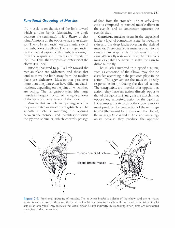
ANATOMY OF THE MUSCULAR SYSTEM / 111
Functional Grouping of Muscles
If a muscle is on the side of the limb toward which a joint bends (decreasing the angle between the segments), it is a fl exor of that joint. A muscle on the opposite side is an exten-sor. The m. biceps brachii, on the cranial side of the limb, fl exes the elbow. The m. triceps brachii, on the caudal aspect of the limb, takes origin from the scapula and humerus and inserts on the ulna. Thus, the triceps is an extensor of the elbow (Fig. 7-5).
Muscles that tend to pull a limb toward the median plane are adductors, and those that tend to move the limb away from the median plane are abductors. Muscles that pass over more than one joint often have different classi-fi cations, depending on the joint on which they are acting. The m. gastrocnemius (the large muscle in the gaskin or calf of the leg) is a fl exor of the stifl e and an extensor of the hock.
Muscles that encircle an opening, whether they are striated or smooth, are sphincters. The smooth muscle surrounding the opening between the stomach and the intestine forms the pyloric sphincter, which controls passage
of food from the stomach. The m. orbicularis oculi is composed of striated muscle fi bers in the eyelids, and its contraction squeezes the eyelids shut.
Cutaneous muscles occur in the superfi cial fascia (a layer of connective tissue) between the skin and the deep fascia covering the skeletal muscles. These cutaneous muscles attach to the skin and are responsible for movement of the skin. When a fl y rests on a horse, the cutaneous muscles enable the horse to shake the skin to dislodge the fl y.
The muscles involved in a specifi c action, such as extension of the elbow, may also be classifi ed according to the part each plays in the action. The agonists are the muscles directly responsible for producing the desired action. The antagonists are muscles that oppose that action; they have an action directly opposite that of the agonists. Synergists are muscles that oppose any undesired action of the agonists. For example, in extension of the elbow, a move-ment produced by contraction of the m. triceps brachii (the agonist for extension of the elbow), the m. biceps brachii and m. brachialis are antag-onists because they produce the opposite
Triceps Brachii Muscle
Biceps Brachii Muscle
Figure 7-5. Functional grouping of muscles. The m. biceps brachii is a fl exor of the elbow, and the m. triceps brachii is an extensor. In this case, the m. biceps brachii is an agonist for elbow fl exion, and the m. triceps brachii acts as an antagonist. Any muscles that assist elbow fl exion indirectly by stabilizing other joints are considered synergists of that movement.

112 / ANATOMY AND PHYSIOLOGY OF FARM ANIMALS
action, fl exion of the elbow. Since the long head of the triceps can fl ex the shoulder joint as well as extend the elbow, any muscle that opposes fl exion of the shoulder joint is a syner-gist of elbow fl exion. The m. supraspinatus and m. brachiocephalicus are synergists for this par-ticular action.
Whether a given muscle is classifi ed as an agonist, an antagonist, or a synergist depends entirely on the specifi c action. If fl exion (instead of extension) of the elbow is the desired action, the m. biceps brachii and m. brachialis become agonists, and the m. triceps brachii and m. anco-neus become antagonists.
Synovial Structures
Synovial structures of the body include joint capsules, bursae, and synovial (tendon) sheaths. The inner layer of each consists of a connective tissue membrane that produces synovial fl uid to reduce friction. Synovial joints were described in detail in Chapter 6.
A bursa is a synovial sac between two struc-tures that tend to rub against each other (Fig. 7-6). Clinically important bursae include (1) the bicipital bursa, between the biceps brachii tendon and the proximal end of the humerus; (2) the atlantal bursa, between the ligamentum nuchae and atlas; (3) the supraspinous bursa, between the ligamentum nuchae and the spinous process of the second thoracic verte-
bra; (4) superfi cial bursae, between the skin and olecranon process of the ulna at the point of the elbow and between the skin and super-fi cial digital fl exor tendon at the point of the hock; and (5) the navicular bursa, between the deep digital fl exor tendon and the navicular (distal sesamoid) bone. Normally, a bursa con-tains only enough fl uid to reduce friction between adjacent parts.
Infl ammation of a bursa, often associated with excessive fl uid, is called bursitis. Enlargement of bursae may be due to trauma, as is usually the case with capped hock, capped elbow (shoe boil), and carpal hygroma. Infl ammation followed by formation of a draining tract, which is fairly common, underlies the equine conditions poll evil (at the atlantal bursa) and fi stulous withers (at the supraspinous bursa).
A bursa gives adequate protection to struc-tures that move only a short distance in relation to each other. However, tendons that must travel a long distance (sometimes as much as several inches) over a bone or other structure require protection and friction-free movement for their entire length. This is afforded by a synovial sheath (Fig. 7-6).
A synovial sheath resembles an elongated bursa placed between the tendon and underly-ing tissue, with the edges of the bursa (sheath) refl ected around the tendon until they meet. This results in an inner layer of synovial mem-brane on the surface of the tendon and a super-
Figure 7-6. Synovial structures. Left) A synovial sheath surrounds a tendon over a greater distance and facilitates its frictionless movement. Refl ection of the synovial membrane creates the supporting mesotendon, through which vessels and nerves gain access to the tendon. Right) A bursa lies interposed between a tendon or ligament and a bony prominence.
ANATOMY OF THE MUSCULAR SYSTEM / 113
fi cial layer of the synovial membrane outside the tendon, forming a closed sac that contains synovial fl uid to reduce friction between the tendon and adjacent structures. The double fold of membrane formed where the edges of the synovial sheath meet is the mesotendon. Vessels and nerves to the tendon reach it by passing through the mesotendon.
Infl ammation of a synovial sheath and its tendon is called tenosynovitis. Low-grade trauma (for instance, as associated with rig-orous schooling of young horses) may result in a mild, painless tenosynovitis of the digital fl exor tendon sheath (windpuffs) or the deep digital fl exor tendon sheath proxi-mal to the hock (thoroughpin).
Muscles of the Thoracic Limb
See Table 7-1 for list of thoracic limb muscles.
Muscles Acting on the Shoulder Girdle
The scapula and shoulder can make complex movements in humans, but in domestic animals the chief movement of the proximal part of the thoracic limb is a pendulous swing forward and backward. The muscles that hold the scapula in place contribute to this swinging movement. These muscles are also critical in allowing the weight of the thorax to be sup-ported between the two thoracic limbs (Fig. 7-7). Superfi cial muscles of the scapula that link it to the bony thorax include the m. trapezius and the m. omotransversarius. The m. rhom-boideus and the m. serratus ventralis are deep.
The m. trapezius is a triangular fl at muscle that takes origin along the dorsal midline from the head to the lumbar vertebrae. The m. tra-pezius inserts chiefl y on the spine of the scapula. The portion originating cranial to the scapula helps swing the scapula forward; the one attach-ing behind draws it back.
The m. rhomboideus is a heavier muscle just deep to the trapezius. The m. rhomboideus also
originates from the dorsal midline both cranial and caudal to the scapula. The m. rhomboideus inserts on the deep (medial) face of the scapular cartilage.
The m. serratus ventralis is the largest and most important muscle attaching the thoracic limb to the trunk. It is a large, fan-shaped muscle. The origin of the m. serratus ventralis is the widest part and extends from the transverse processes of the cervical and thoracic vertebrae and from the ribs along a curved line just above the sternum as far back as the tenth costal car-tilage. The insertion is on the medial side of the dorsal portion of the scapula. The m. serratus ventralis on each side together form a sling that supports the trunk between the thoracic limbs. The cervical portion, on contraction, tends to rotate the distal part of the scapula backward, while the thoracic portion rotates it forward. The muscle’s cervical attachments allow it to lift the neck as well.
The m. omotransversarius is a separate muscle of the shoulder region in most domestic species. It takes origin from the transverse pro-cesses of the more cranial cervical vertebrae and inserts on the distal part of the spine of the scapula (clavicular tendon in the horse). With these attachments, the m. omotransversarius usually pulls the distal end of the scapula forward, although with the limb in weight-bearing position, it instead assists lateral fl exion of the neck.
Muscles Acting on the Shoulder Joint
The shoulder, being a ball-and-socket joint, can make all types of movement. In the quadruped its chief actions are extension and fl exion.
Extensors of the Shoulder. The m. brachio-cephalicus, as the name implies, extends from the head to the arm. The origin is from the occipital bone of the skull and transverse pro-cesses of the cervical vertebrae. It inserts on the lateral side of the proximal part of the humerus proximal to the deltoid tuberosity. The m. bra-chiocephalicus is the heavy muscle covering the cranial aspect of the point of the shoulder. It

Table 7-1. Muscles of the Thoracic Limb
Muscle Origin Insertion Primary Actions Innervation
m. trapezius ligaments of the dorsal midline
scapular spine lifts, advances, & retracts scapula
accessory n.
m. rhomboideus ligaments of the dorsal midline
medial side scapular cartilage
lifts & advances scapula, lifts neck
cervical & thoracic spinal nn.
m. serratus ventralis transverse processes of caudal cervical & cranial thoracic vertebrae, fi rst 8 or 9 ribs
medial side of scapula & scapular cartilage
supports trunk, lifts neck, advances & retracts scapula
cervical spinal nn, long thoracic n.
m. omotransversarius fascia of shoulder & arm
transverse processes of cranial cervical vertebrae
advances limb, lateral neck fl exion
cervical spinal nn.
m. brachiocephalicus m. cleidobrachialis m. cleidocephalicus (m. cleidomastoideus)
occipital bone, transverse processes of cervical vertebrae
medial fascia of arm & forearm, lateral side of humerus, deltoid tuberosity
lateral neck fl exion, shoulder extension, advances limb
cervical spinal nn., accessory n.
m. latissimus dorsi spinous processes of thoracic & lumbar vertebrae
medial humerus limb retraction, shoulder fl exion
thoracodorsal n.
m. pectoralis superfi cialis (superfi cial pectoral m.)
sternum & cranial costal cartilages
medial humerus & fascia of arm & forearm
limb adduction cranial & caudal pectoral nn.
m. pectoralis profundus (deep pectoral m.)
sternum, ribs, abdominal fascia
craniomedial humerus
adduction & retraction of limb, advances trunk when limb is weight-bearing
cranial & caudal pectoral nn.
m. subclavius cranial sternum & costal cartilages
epimysium of m. supraspinatus
support of trunk, shoulder stabilization
cranial pectoral nn.
m. supraspinatus supraspinous fossa of scapula
greater tubercle of humerus
stabilization & extension of shoulder
suprascapular n.
m. infraspinatus infraspinous fossa of scapula
greater tubercle of humerus
stabilization & fl exion of shoulder
suprascapular n.
m. subscapularis subscapular fossa medial humerus shoulder stabilization
subscapular n.
m. teres major caudal border of scapula
medial humerus (teres major tuberosity)
shoulder fl exion axillary n.
m. teres minor caudal border scapula
proximal to deltoid tuberosity
shoulder fl exion axillary n.
m. deltoideus scapular spine deltoid tuberosity shoulder fl exion axillary n.
m. coracobrachialis coracoid process of scapula
medial humerus shoulder extension musculocutaneous n.
114

Muscle Origin Insertion Primary Actions Innervation
m. biceps brachii supraglenoid tubercle
cranial aspect of proximal radius & ulna
elbow fl exion, shoulder extension
musculocutaneous n.
m. brachialis caudal aspect of proximal humerus
medial aspect of proximal radius
elbow fl exion musculocutaneous n.
m. triceps brachii caudal border of scapula, proximal humerus
olecranon process elbow extension, shoulder fl exion (long head only)
radial n.
m. anconeus caudolateral humerus
olecranon process elbow extension radial n.
m. tensor fasciae antibrachii
caudal border of scapula (via epimysium of triceps m.)
olecranon process, deep fascia of antebrachium
elbow extension, tensing of forearm fascia
radial n.
m. extensor carpi radialis
lateral humeral epicondyle
cranial aspect of proximal metacarpus
carpal extension radial n.
m. extensor carpi obliquus
craniolateral radius medial metacarpus
carpal extension radial n.
m. extensor digitorum communis (common digital extensor m.)
lateral humeral epicondyle
distal phalanx digital extension, carpal extension
radial n.
m. extensor digitorum lateralis (lateral digital extensor m.)
lateral collateral ligament of elbow, proximal radius & ulna
dorsal aspect of digit
digital extension, carpal extension
radial n.
m. extensor carpi ulnaris (m. ulnaris lateralis)
lateral humeral epicondyle
lateral metacarpus, accessory carpal bone
carpal fl exion (?) radial n.
m. pronator teres medial humeral epicondyle
medial aspect proximal radius
elbow fl exion median n.
m. fl exor carpi radialis
medial humeral epicondyle
palmeromedial aspect of proximal metacarpus
carpal fl exion median n.
m. fl exor carpi ulnaris medial humeral epicondyle, olecranon
accessory carpal bone
carpal fl exion ulnar n.
m. fl exor digitorum superfi cialis (superfi cial digital fl exor m.)
medial humeral epicondyle
middle phalanx digital fl exion, carpal fl exion
ulnar n.
m. fl exor digitorum profundus (deep digital fl exor m.)
medial humeral epicondyle, caudal radius, medial olecranon
palmar surface of distal phalanx
digital fl exion, carpal fl exion
median & ulnar nn.
n(n), nerve(s).
Table 7-1. Continued
115

116 / ANATOMY AND PHYSIOLOGY OF FARM ANIMALS
raises and advances the shoulder. The m. bra-chiocephalicus is also the principal extensor of the shoulder and acts as a lateral fl exor of the neck when the limb is weight bearing.
The m. brachiocephalicus is subdivided into the m. cleidobrachialis, extending from the cla-vicular tendon (representing the vestigial clav-icle) to the humerus, and the m. cleidocephalicus, extending from the clavicular tendon to the head and neck. In species other than the horse, the m. cleidocephalicus may be further subdi-vided into a mastoid part attaching to the mastoid process of the temporal bone and either an occipital part (in ruminants and pigs) or a cervical part (in carnivores). In horses, the sole attachment of the m. cleidocephalicus is to the mastoid process, and in this species the muscle is often called the m. cleidomastoideus. The m. cleidobrachialis is comparable to the anterior deltoid muscle of humans.
The m. supraspinatus originates from the supraspinous fossa of the scapula cranial to the
spine. It inserts on the greater tubercle (both greater and lesser in the horse) of the humerus. The m. supraspinatus may assist in extending the shoulder but acts chiefl y as a stabilizing ligament of the shoulder joint. This is one of the muscles that atrophies (shrinks) in sweeny in horses, a condition that results from damage to its motor innervation, the suprascapular nerve.
Flexors of the Shoulder. The m. teres major originates from the dorsal part of the caudal border of the scapula and inserts on the teres major tuberosity on the medial side of the shaft of the humerus. It is a strong fl exor of the shoulder joint.
The m. latissimus dorsi is a wide, triangular muscle that originates from the spinous pro-cesses of the thoracic and lumbar vertebrae by means of a wide aponeurosis, the thoracolum-bar fascia. It inserts with the m. teres major on the medial side of the humerus and is a strong fl exor of the shoulder. Also, it pulls the thoracic limb caudad or, if the limb is fi xed, advances the trunk.
The m. infraspinatus originates from the infraspinous fossa just caudal and ventral to the spine of the scapula. It inserts into the caudal part of the greater tubercle of the humerus. The m. infraspinatus also acts as a strong collateral ligament of the shoulder joint and may abduct, fl ex, and outwardly rotate the shoulder. This muscle also atrophies in cases of sweeny.
The m. teres minor lies just distal to the infraspinatus muscle and has the same action as the m. infraspinatus. The m. teres minor origi-nates from the distal caudal border of the scapula and inserts on the teres minor tuberos-ity of the humerus just distal to the greater tubercle of the humerus.
The m. deltoideus extends from the spine of the scapula to the deltoid tuberosity of the humerus. It is an abductor and fl exor of the shoulder joint.
Adductors of the Shoulder. The pectoral muscles form the substance of the brisket. They originate from the sternum and insert mainly on the proximal part of the humerus. Com-
Figure 7-7. Muscular suspension of the thorax between the thoracic limbs. 1, Scapula; 2, humerus; 3, radius and ulna; 4, sternum; 5, m. pectoralis profundus; 6, m. serratus ventralis; 7, m. trapezius; 8, m. rhomboi-deus. The attachment of limbs to the trunk is achieved through a synsarcosis, rather than by a bony joint.
ANATOMY OF THE MUSCULAR SYSTEM / 117
monly, they are divided into the superfi cial pectoral muscle (m. pectoralis superfi cialis) and the deep pectoral muscle (m. pectoralis profundus). These pectoral muscles are strong adductors of the forelimb, and the deep pecto-ral muscle also advances the trunk when the limb is fi xed on the ground (weight bearing).
The m. subclavius is absent in carnivores, small in ruminants, and well developed in horses and pigs. It has sometimes been consid-ered part of the deep pectoral muscle. The m. subclavius arises from the cranial sternum and costal cartilages and arcs craniodorsad to insert on the epimysium of the m. supraspinatus. This muscle is part of the support of the trunk and contributes to stabilization of the shoulder joint.
The m. coracobrachialis is a small muscle extending from the coracoid process on the medial side of the scapula to the medial side of the shaft of the humerus. The location of the muscle belly suggests a shoulder fl exor, but its attachments make this muscle an extensor of that joint.
The m. subscapularis stabilizes the shoulder on the medial side. It originates from the sub-scapular fossa on the medial side of the scapula below the attachments of the m. rhomboideus and m. serratus ventralis. It inserts on the lesser tuberosity of the humerus and provides some adduction to the shoulder joint.
Muscles Acting on the Elbow
Since the elbow is a hinge joint, the muscles acting on it are either fl exors or extensors. In quadrupeds, the extensors are stronger than the fl exors because they support the weight of the body by maintaining the limbs in extension (Fig. 7-8).
Extensors of the Elbow. The m. triceps brachii has three heads. The long head origi-nates from the caudal border of the scapula, and the medial and lateral heads originate from the respective sides of the humeral diaphysis. Car-nivores have an accessory head that also origi-nates from the humerus between the medial
and lateral heads (although this arrangement gives the carnivore four heads on this muscle, it is still called a triceps). All heads insert on the olecranon process of the ulna. The triceps is the strongest extensor of the elbow. The long head may also act to fl ex the shoulder.
The m. anconeus, deep to the m. triceps brachii, is a rather small muscle that covers the caudal aspect of the joint capsule of the elbow. It also originates on the humerus, inserts on the olecranon process, and extends the elbow.
The m. tensor fasciae antibrachii originates via a thin aponeurosis that is blended with the long head of the triceps muscle and the m. latis-simus dorsi. The fl attened muscle belly lies on the caudomedial aspect of the arm and inserts via a second aponeurosis on the olecranon and antebrachial fascia. The muscle’s name refl ects its action on the antebrachial fascia (it tenses it), but through these fascial connections the m. tensor fasciae antibrachii also assists the triceps in extension of the elbow.
Flexors of the Elbow. The m. biceps brachii originates on the supraglenoid tubercle just dorsal and cranial to the articular surface of the scapula. It inserts on (1) the radial tuberosity on the cranial aspect of the proximal radius, (2) the medial collateral ligament of the elbow, and (3) the antebrachial fascia. The tendinous blending with the antebrachial fascia forms a palpable cordlike structure on the fl exor surface of the elbow called the lacertus fi brosus. The biceps assists in holding the shoulder joint in apposition and may extend it to some extent. However, the chief action of the m. biceps brachii is fl exion of the elbow.
The m. brachialis is strictly a fl exor of the elbow since it originates on the humerus and inserts on the cranial aspect of the radius (and in some species the ulna).
As ungulates have only limited or absent ability to pronate and supinate their limbs, the m. pronator teres is reduced to a ligament in horses and only a small, weak muscle in rumi-nants and pigs. In these species, it acts primar-ily as a fl exor of the elbow. It originates on the medial epicondyle of the humerus and inserts on the medial side of the radius.

118 / ANATOMY AND PHYSIOLOGY OF FARM ANIMALS
Figure 7-8. Muscles of the thoracic limb. A) Equine. B) Bovine.
Extensor muscles of the carpus and digit (discussed in the next section), which originate on the lateral epicondyle of the humerus, may assist in fl exion of the elbow as a secondary function.
Muscles Acting on the Carpus
The carpus, like the elbow, acts essentially as a hinge joint. The extensors of the carpus lie on the craniolateral aspect of the limb, and
SuperficialPectoral M.
M. Extensor CarpiRadialis
Radius
BrachialisM.Flexor CarpiRadialis
M.Flexor CarpiUlnaris
M.Extensor CarpiObliquus M. Extensor Carpi
Radialis
Common DigitalExtensor M.
Lateral DigitalExtensor M.
M. UlnarisLateralis
M. ExtensorCarpi Obliquis
Joining of DigitalExtensor Tendons
Superficial &Deep DigitalFlexorTendonsSuspensory Ligament
Palmar Annular Ligamentof the Fetlock
M. ExtensorCarpi Radialis
Common DigitalExtensor M.
Lateral DigitalExtensor M. M. Ulnaris
Lateralis
SuperficialDigital Flexor M.
SuspensoryLigament
.B.A
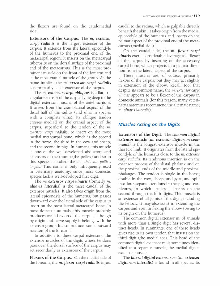
ANATOMY OF THE MUSCULAR SYSTEM / 119
the fl exors are found on the caudomedial side.
Extensors of the Carpus. The m. extensor carpi radialis is the largest extensor of the carpus. It extends from the lateral epicondyle of the humerus to the proximal end of the metacarpal region. It inserts on the metacarpal tuberosity on the dorsal surface of the proximal end of the metacarpus. This is the most pro-minent muscle on the front of the forearm and is the most cranial muscle of the group. As the name implies, the m. extensor carpi radialis acts primarily as an extensor of the carpus.
The m. extensor carpi obliquus is a fl at, tri-angular extensor of the carpus lying deep to the digital extensor muscles of the antebrachium. It arises from the craniolateral aspect of the distal half of the radius (and ulna in species with a complete ulna). Its oblique tendon crosses mediad on the cranial aspect of the carpus, superfi cial to the tendon of the m. extensor carpi radialis, to insert on the most medial metacarpal bone, which is the second in the horse, the third in the cow and sheep, and the second in pigs. In humans, this muscle is one of the well-developed abductors and extensors of the thumb (the pollex) and so in this species is called the m. abductor pollicis longus. This name is only infrequently used in veterinary anatomy, since most domestic species lack a well-developed fi rst digit.
The m. extensor carpi ulnaris (formerly m. ulnaris lateralis) is the most caudal of the extensor muscles. It also takes origin from the lateral epicondyle of the humerus, but passes downward over the lateral side of the carpus to insert on the most lateral metacarpal bone. In most domestic animals, this muscle probably produces weak fl exion of the carpus, although by origin and nerve supply it belongs with the extensor group. It also produces some outward rotation of the forearm.
In addition to these carpal extensors, the extensor muscles of the digits whose tendons pass over the dorsal surface of the carpus may act secondarily as extensors of the carpus.
Flexors of the Carpus. On the medial side of the forearm, the m. fl exor carpi radialis is just
caudal to the radius, which is palpable directly beneath the skin. It takes origin from the medial epicondyle of the humerus and inserts on the palmar aspect of the proximal end of the meta-carpus (medial side).
On the caudal side, the m. fl exor carpi ulnaris exerts considerable leverage as a fl exor of the carpus by inserting on the accessory carpal bone, which projects in a palmar direc-tion from the lateral aspect of the carpus.
These muscles are, of course, primarily fl exors of the carpus, but they may act slightly in extension of the elbow. Recall, too, that despite its common name, the m. extensor carpi ulnaris appears to be a fl exor of the carpus in domestic animals (for this reason, many veteri-nary anatomists recommend the alternate name, m. ulnaris lateralis).
Muscles Acting on the Digits
Extensors of the Digit. The common digital extensor muscle (m. extensor digitorum com-munis) is the longest extensor muscle in the thoracic limb. It originates from the lateral epi-condyle of the humerus close to the m. extensor carpi radialis. Its tendinous insertion is on the extensor process of the distal phalanx and on the proximal ends of the middle and proximal phalanges. The tendon is single in the horse; double in the cow, sheep, and goat; and split into four separate tendons in the pig and car-nivores, in which species it inserts on the second through the fi fth digits. This muscle is an extensor of all joints of the digit, including the fetlock. It may also assist in extending the carpus and even in fl exing the elbow (owing to its origin on the humerus).
The common digital extensor m. of animals with more than a single digit has several dis-tinct heads. In ruminants, one of these heads gives rise to its own tendon that inserts on the third digit (the medial toe). This head of the common digital extensor m. is sometimes iden-tifi ed as a separate muscle, the medial digital extensor muscle.
The lateral digital extensor m. (m. extensor digitorum lateralis) is found in all species. Its

120 / ANATOMY AND PHYSIOLOGY OF FARM ANIMALS
origin is just caudal to the common digital extensor m. on the lateral collateral ligament of the elbow and lateral radius and ulna. The insertion varies according to the number of digits present, and there are interspecifi c differ-ences regarding to which phalanx the tendon attaches. In pigs it inserts on the fourth and fi fth digits; in ruminants, on the middle phalanx of the fourth digit; and in the horse, on the proximal phalanx of the third (and only) digit.
Flexors of the Digit. In all animals the prin-cipal digital fl exors are the superfi cial and the deep digital fl exor muscles. The deep digital fl exor m. (m. fl exor digitorum profundus) lies the closest to the metacarpal bones. It origi-nates from the humerus, radius, and ulna. Its long, stout tendon extends distad through the carpal canal, then along the palmar side of the metacarpus to insert on the palmar surface of the distal phalanges. As with the common digital extensor tendon, the number of inser-tions depends on the number of digits, with the main tendon dividing into individual slips, one per digit, just proximal to the fetlock. The deep digital fl exor is the only muscle that fl exes the distal interphalangeal joint. Second-arily, it also fl exes the more proximal joints of the digit and the carpus. The deep digital fl exor m. also is important in supporting the fetlock.
The superfi cial digital fl exor m. (m. fl exor digitorum superfi cialis) is similar to the deep digital fl exor m., but it inserts primarily on the proximal part of the middle phalanx of each digit. In the horse, the superfi cial digital fl exor tendon inserts on the palmar aspects of the proximal end of the middle phalanx and the distal end of the proximal phalanx. Tendons of both the superfi cial and deep digital fl exor mm. can be palpated palmar to the cannon bone. The expression bowed tendons describes a traumatic condition of horses involving ten-donitis of one or both of these tendons in the cannon region (Fig. 7-9).
Interosseous muscles lie between the meta-carpal bones of carnivores and humans. In large ungulates most of the muscle tissue has been
replaced with connective tissue, and these structures are known as the suspensory liga-ments. They take origin from the palmar aspect of the proximal metacarpus and insert on the proximal sesamoid bones. The suspensory liga-ments are part of the passive support of the metacarpophalangeal joint.
Muscles of the Pelvic Limb
The hip is a ball-and-socket joint and as such can move in nearly any direction (Fig. 7-10). However, the chief movements are extension and fl exion of the femur. Adduction and abduc-tion are also fairly common movements, and some rotation is possible.
See Table 7-2 for a list of muscles of the pelvic limb.
Figure 7-9. Tenosynovitis (bowed tendon). Note swelling on the palmar aspect above the fetlock in the area of the fl exor tendons.

M. QuadricepsFemoris
M. Pectineus
M. Gracilis
Tibia
Long DigitalExtensor T.
M. Adductor M. Semimembranosus
M. Semitendinosus
M. Gastrocnemius
Popliteal M.
Deep DigitalFlexor M.
CommonCalcanealTendon
SuperficialDigital Flexor T.
Deep Digital Flexor T.
Suspensory Ligament
M. TensorFasciae Latae
Fascia Lata
Patella
3 Divisions ofFemoral Biceps M.
M. Gastrocnemius
M. SoleusLong DigitalExtensor M.
M. PeroneusLongus
M. Deep DigitalFlexor
Long DigitalExtensor Tendon
M. Deep DigitalFlexor
Calcaneus
SuperficialDigital Flexor
Tendon
Deep DigitalFlexor Tendon
M. Gluteobiceps(cut)
QuadricepsFemoris M.
M. CranialTibialis
M. PeroniusTertius
Long DigitalExtensor M.
M. PeroneusLongus
M. Lateral DigitalExtensor
Tendons of DigitalExtensor M.
.B.AFigure 7-10. Muscles of the pelvic limb. A) Equine. Left) medial view. Right) lateral view. B) Bovine: lateral view.
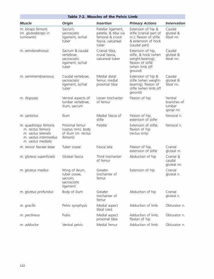
Table 7-2. Muscles of the Pelvic Limb
Muscle Origin Insertion Primary Actions Innervation
m. biceps femoris(m. gluteobiceps in ruminants)
Sacrum, sacrosciatic ligament, ischial tuber
Patellar ligament, patella, & tibia via femoral & crural fascia, calcaneal tuber
Extension of hip & stifl e (cranial part of m.); fl exion of stifl e & extension of hock (caudal part)
Caudal gluteal & tibial nn.
m. semitendinosus Sacrum & caudal vertebrae, sacrosciatic ligament, ischial tuber
Cranial tibia, crural fascia, calcaneal tuber
Extension of hip, stifl e, & hock (when weight-bearing); fl exion of stifl e (when limb off ground)
Caudal gluteal & tibial nn.
m. semimembranosus Caudal vertebrae, sacrosciatic ligament, ischial tuber
Medial distal femur, medial proximal tibia
Extension of hip & stifl e (when weight-bearing); fl exion of stifl e (when limb off ground)
Caudal gluteal & tibial nn.
m. iliopsoas Ventral aspects of lumbar vertebrae, ilium, sacrum
Lesser trochanter of femur
Flexion of hip Ventral branches of lumbar spinal nn.
m. sartorius Ilium Medial fascia of stifl e
Flexion of hip, extension of stifl e
Femoral n.
m. quadriceps femoris m. rectus femoris m. vastus lateralis m. vastus intermedius m. vastus medialis
Proximal femur (vastus mm), body of ilium (m. rectus femoris)
Patella Extension of stifl e, fl exion of hip (rectus only)
Femoral n.
m. tensor fasciae latae Tuber coxae Fascia lata Flexion of hip, extension of stifl e
Cranial gluteal m.
m. gluteus superfi cialis Gluteal fascia Third trochanter of femur
Abduction of hip Cranial & caudal gluteal nn.
m. gluteus medius Wing of ileum, tuber coxae, sacrum, sacrosciatic ligament
Greater trochanter of femur
Extension of hip Cranial gluteal n.
m. gluteus profundus Body of ilium Greater trochanter of femur
Abduction of hip Cranial gluteal n.
m. gracilis Pelvic symphysis Medial aspect tibial crest
Adduction of limb Obturator n.
m. pectineus Pubis Medial aspect proximal tibia
Adduction of limb, fl exion of hip
Obturator n.
m. adductor Ventral pelvis Medial femur Adduction of limb Obturator n.
122

Muscle Origin Insertion Primary Actions Innervation
m. quadratus femoris Ventral sacrum Caudal proximal femur
Outward rotation of hip, hip extension
Sciatic n.
External obturator m. (m. obturatorius externus)
Ventral pelvis, covering obturator foramen
Trochanteric fossa of proximal femur
Outward rotation of hip, limb adduction
Obturator n.
Internal obturator m. (m. obturatorius internus)
(Absent in ruminants) covering obturator foramen inside pelvic canal
Trochanteric fossa of proximal femur
Outward rotation of hip, hip extension
Sciatic n.
mm. gemelli Ischium Trochanteric fossa of proximal femur
Outward rotation of hip, hip extension
Sciatic n.
m. popliteus Lateral aspect of lateral femoral condyle
Caudoproximal tibia
Stifl e fl exion Tibial n.
m. gastrocnemius Caudal aspect of femoral condyles
Tuber calcanei Hock extension Tibial n.
m. fl exor digitorum superfi cialis (superfi cial digital fl exor m.)
Caudodistal femur, between heads of m. Gastrocnemius
Tuber calcanei, plantar aspect of middle phalanx
Hock extension, digital fl exion
Tibial n.
m. tibialis cranialis Craniolateral aspect proximal tibia
Medial tarsus & metatarsus
Flexion of hock Peroneal n.
m. peroneus tertius Extensor fossa of distolateral femur
Cranial aspect of distal tarsus & proximal metatarsus
Flexion of hock Peroneal n.
m. peroneus longus Head of fi bula & lateral femoral condyle
Plantar aspect of distal tarsus & metatarsus
Flexion of hock Peroneal n.
m. extensor digitorum longus (long digital extensor m.)
Extensor fossa of distolateral femur
Dorsal aspect of distal phalanx
Flexion of hock, extension of digits
Peroneal n.
m. extensor digitorum lateralis (lateral digital extensor m.)
Proximal fi bula Middle phalanx (artiodactyls), tendon of long digital extensor (Equidae)
Flexion of hock, extension of digits
Peroneal n.
m. fl exor digitorum profundus (deep digital fl exor m.)
Proximocaudal tibia
Plantar aspect distal phalanx
Extension of hock, fl exion of digits
Tibial n.
Table 7-2. Continued
123
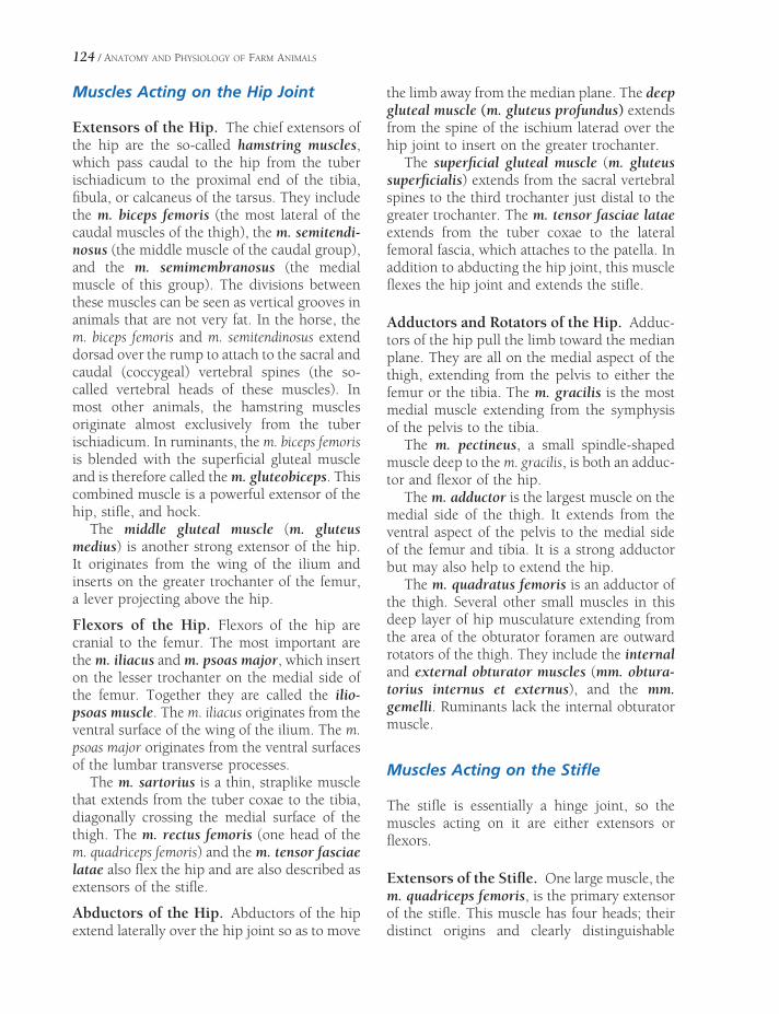
124 / ANATOMY AND PHYSIOLOGY OF FARM ANIMALS
Muscles Acting on the Hip Joint
Extensors of the Hip. The chief extensors of the hip are the so-called hamstring muscles, which pass caudal to the hip from the tuber ischiadicum to the proximal end of the tibia, fi bula, or calcaneus of the tarsus. They include the m. biceps femoris (the most lateral of the caudal muscles of the thigh), the m. semitendi-nosus (the middle muscle of the caudal group), and the m. semimembranosus (the medial muscle of this group). The divisions between these muscles can be seen as vertical grooves in animals that are not very fat. In the horse, the m. biceps femoris and m. semitendinosus extend dorsad over the rump to attach to the sacral and caudal (coccygeal) vertebral spines (the so-called vertebral heads of these muscles). In most other animals, the hamstring muscles originate almost exclusively from the tuber ischiadicum. In ruminants, the m. biceps femoris is blended with the superfi cial gluteal muscle and is therefore called the m. gluteobiceps. This combined muscle is a powerful extensor of the hip, stifl e, and hock.
The middle gluteal muscle (m. gluteus medius) is another strong extensor of the hip. It originates from the wing of the ilium and inserts on the greater trochanter of the femur, a lever projecting above the hip.
Flexors of the Hip. Flexors of the hip are cranial to the femur. The most important are the m. iliacus and m. psoas major, which insert on the lesser trochanter on the medial side of the femur. Together they are called the ilio-psoas muscle. The m. iliacus originates from the ventral surface of the wing of the ilium. The m. psoas major originates from the ventral surfaces of the lumbar transverse processes.
The m. sartorius is a thin, straplike muscle that extends from the tuber coxae to the tibia, diagonally crossing the medial surface of the thigh. The m. rectus femoris (one head of the m. quadriceps femoris) and the m. tensor fasciae latae also fl ex the hip and are also described as extensors of the stifl e.
Abductors of the Hip. Abductors of the hip extend laterally over the hip joint so as to move
the limb away from the median plane. The deep gluteal muscle (m. gluteus profundus) extends from the spine of the ischium laterad over the hip joint to insert on the greater trochanter.
The superfi cial gluteal muscle (m. gluteus superfi cialis) extends from the sacral vertebral spines to the third trochanter just distal to the greater trochanter. The m. tensor fasciae latae extends from the tuber coxae to the lateral femoral fascia, which attaches to the patella. In addition to abducting the hip joint, this muscle fl exes the hip joint and extends the stifl e.
Adductors and Rotators of the Hip. Adduc-tors of the hip pull the limb toward the median plane. They are all on the medial aspect of the thigh, extending from the pelvis to either the femur or the tibia. The m. gracilis is the most medial muscle extending from the symphysis of the pelvis to the tibia.
The m. pectineus, a small spindle-shaped muscle deep to the m. gracilis, is both an adduc-tor and fl exor of the hip.
The m. adductor is the largest muscle on the medial side of the thigh. It extends from the ventral aspect of the pelvis to the medial side of the femur and tibia. It is a strong adductor but may also help to extend the hip.
The m. quadratus femoris is an adductor of the thigh. Several other small muscles in this deep layer of hip musculature extending from the area of the obturator foramen are outward rotators of the thigh. They include the internal and external obturator muscles (mm. obtura-torius internus et externus), and the mm. gemelli. Ruminants lack the internal obturator muscle.
Muscles Acting on the Stifl e
The stifl e is essentially a hinge joint, so the muscles acting on it are either extensors or fl exors.
Extensors of the Stifl e. One large muscle, the m. quadriceps femoris, is the primary extensor of the stifl e. This muscle has four heads; their distinct origins and clearly distinguishable

ANATOMY OF THE MUSCULAR SYSTEM / 125
muscle bellies make it common practice to name them as separate muscles. The longest head, the m. rectus femoris, originates from the ilium just above the acetabulum. The other three heads, m. vastus medialis, m. vastus intermedius, and m. vastus lateralis, originate from the respective areas of the shaft of the femur. All four heads insert on the patella. The patella, being fastened to the front of the tibia by the patellar ligaments, extends the stifl e when it is pulled proximad by the m. quadriceps femoris. Because of its origin on the ilium, the m. rectus femoris also fl exes the hip.
Flexors of the Stifl e. The chief fl exors of the stifl e are the hamstring muscles, which also extend the hip (m. biceps femoris, m. semitendi-nosus, m. semimembranosus; discussed earlier). In addition, the extensor muscles of the hock that originate on the caudal surface of the distal end of the femur may also fl ex the stifl e. These muscles include the m. gastrocnemius and the superfi cial digital fl exor m. (discussed later). The m. popliteus is a relatively small muscle caudal to the stifl e. Its chief action is fl exion of the stifl e, although it may slightly rotate the leg (tibia and fi bula) mediad.
Muscles Acting on the Hock
The principal actions of the hock are extension and fl exion.
Extensors of the Hock. Extensors of the hock primarily attach to the calcaneus (point of the hock) by way of the common calcanean tendon. The m. gastrocnemius and superfi cial digital fl exor m. originate from the caudal aspect of the distal femur, and their tendons make up the bulk of the common calcanean tendon. They are joined in part by portions of the m. biceps femoris, m. gracilis, and m. semitendinosus, which also assist in extending the hock and hip and fl exing the stifl e. The deep digital fl exor m. also extends the hock.
Flexors of the Hock. Flexors of the hock include the m. tibialis cranialis and the various
peroneal muscles, whose tendons pass over the dorsal surface of the hock to insert on the tarsus and metatarsus. The m. peroneus tertius is the only named peroneal muscle in the horse. Additionally, the m. peroneus longus is found on the ox, sheep, goat, pig, and dog. The m. pero-neus brevis is found only in carnivores and humans. The digital extensors also fl ex the hock, because their tendons pass over its fl exor surface.
Muscles Acting on the Digit
Digital extensors of the thoracic limb also tend to extend the carpus, while digital extensors of the pelvic limb tend to fl ex the hock. Likewise, digital fl exors of the thoracic limb produce fl exion of the carpus, while digital fl exors of the pelvic limb produce extension the hock. Muscles acting on the digits of the pelvic limb are similar in arrangement to those of the tho-racic limb.
Extensors of the Digit. The long digital exten-sor muscle (m. extensor digitorum longus) originates from the distal end of the femur and passes distad to insert on the distal phalanx of each digit. As with the common extensor in the thoracic limb, the tendon has one part in the horse; two parts in the ox, goat, and sheep; and four parts in the pig and carnivores.
The lateral digital extensor muscle (m. extensor digitorum lateralis) lies between the extensor and fl exor groups of muscles of the crus. In the horse, its tendon joins that of the long digital extensor m. at about the middle of the cannon. In ruminants, it inserts via a separate tendon on the proximal middle pha-lanx of the fourth digit. In pigs, it resembles the ruminant condition, with the addition of a small deep part of the muscle that features a separate tendon inserting on the fi fth digit.
Flexors of the Digit. The superfi cial and deep digital fl exor mm. are arranged in the pelvic limb similarly to the thoracic limb. However, the tendon of the superfi cial digital fl exor muscle also attaches to the calcaneus.

126 / ANATOMY AND PHYSIOLOGY OF FARM ANIMALS
Muscles of the Head
Muscles of Mastication
Muscles of mastication are those that have attachments to the mandible and whose con-tractions produce the jaw movements associ-ated with chewing (Fig. 7-11). As a general rule, considerably more muscle mass (and therefore strength) is devoted to the elevation (closure) of the mandible than to depression (opening). Most of the muscles of mastication are inner-vated by the trigeminal nerve (see Chapter 9).
The m. temporalis is a strong muscle arising from the sagittal crest and the expansive tem-poral fossa of the lateral cranium and inserting on the coronoid process of the mandible. Its action is to elevate the mandible, bringing the upper and lower teeth together.
The m. masseter is especially well developed in herbivorous species. This powerful mastica-
tory muscle arises from the maxillary region of the face and the zygomatic arch. It inserts on the caudal mandible, and its primary actions are to elevate the mandible and to draw it laterad. The broad expanse of the horse’s cheek is formed by the masseter muscle.
Medial to the mandible are two pterygoid muscles (mm. pterygoideus). These arise from the ventral parts of the skull (the pterygoid and palatine bones) and insert on the mandible. These muscles assist in closing the mandible and play an important role in the side-to-side grinding movements typical of herbivore mastication.
Opening of the jaw is largely assisted by gravity, but forceful depression of the man-dible is primarily the function of the m. digastricus.
This muscle arises from the region caudal to the temporomandibular joint and inserts on the angle of the mandible. As the name implies, the
M. Brachiocephalicus
M. Splenius
Facial Nerve
M. Parotidoauricularis
M. Caudal Auricular Rostral Auricular Muscles
M. Levator Anguli Oculi Medialis
M. Orbicularis Oculi
M. Levator Nasolabialis
M. Caninus
Par
otid
Gla
nd
M. Sternomandibularis
M. Omohyoideus/Sternohyoideus
M. Masseter
M. Zygomaticus
M. Mentalis
M. Buccinator
M. Orbicularis Oris
Figure 7-11. Muscles of the equine head.

ANATOMY OF THE MUSCULAR SYSTEM / 127
muscle has two bellies, indistinctly divided near the muscle’s center.
Muscles of Facial Expression
The muscles that move the skin and append-ages of the face and head are the muscles of facial expression or mimetic muscles. These are generally thin cutaneous muscles inner-vated by the facial nerve (see Chapter 9).
The auricular muscles are roughly divided into a rostral and a caudal group. The contrac-tion of auricular muscles produces the range of ear movements characteristic of domestic animals.
The m. orbicularis oculi is a sphincter-like muscle that surrounds the palpebral fi ssure (the opening of the eyelids). Contraction of the m. orbicularis oculi produces a forceful closure of the fi ssure.
A large number of muscles produce move-ments of the lips and cheeks; these are impor-tant in prehension of food and assist with mastication by keeping food in the mouth and positioned between the teeth. A sphincter-like muscle, the m. orbicularis oris, surrounds the lips; its contraction purses the lips. The m. buc-cinator forms the wall of the cheek. Other mimetic muscles lift and depress the lips, change the shape of the nostrils, and produce other facial movements.
Other Muscles of the Head
A number of striated muscles lie within the orbit associated with the globe of the eye. These extraocular muscles (so called because they are attached to the outside of the eyeball) move the globe of the eye in the many directions of which it is capable. The extraocular muscles are described in Chapter 11 with the anatomy of the visual apparatus.
A large number of muscles associated with the pharynx and soft palate are important in deglutition (swallowing) and phonation (vocal-ization). Their various functions are to lift or depress the palate and to constrict or dilate the pharynx. This same region contains many
muscles that originate or insert on the hyoid apparatus. These act to move the hyoid appa-ratus relative to the tongue and larynx or, when the hyoid apparatus is fi xed in place, to move these latter structures relative to it.
The tongue of domestic animals is an organ capable of extraordinary movements; it is used to prehend and masticate food, drink water and suckle milk, and is used as a grooming tool. The intrinsic muscles of the tongue are arranged in fascicles that run longitudinally, transversely, and vertically, allowing the tongue to change shape in multiple planes. Extrinsic muscles of the tongue, those that arise from outside the tongue, include the m. genioglossus, which arises from the rostral part of the mandible (the genu) and inserts in the base of the tongue so that its contraction draws the entire tongue rostrad. The m. hyoglossus arises from the hyoid apparatus and inserts in the base of the tongue; it draws the tongue caudad. The m. mylohyoideus lies transversely between the rami of the mandible. It is not, therefore, strictly a muscle of the tongue, but its contraction lifts the fl oor of the mouth, hence the tongue.
Muscles of the Trunk and Neck
Extensors of the Vertebral Column
The group of muscles dorsal to the transverse processes of the vertebrae on either side of the spinous processes are the epaxial muscles. These make up the loin muscles and continue forward to the head. Collectively, they are referred to as the m. erector spinae. In domestic animals, the largest of these muscles is known as the m. longissimus. It is composed of many small bundles of muscle fi bers that extend from ver-tebral transverse processes to spinous processes, from transverse processes to transverse pro-cesses, or between spinous processes. As these attachments may extend from one vertebra to the next or overlap one or more vertebrae, there are many possibilities for naming individual muscles and a variety of individual muscle actions. As a general description, however, we can say that these muscles are responsible for

128 / ANATOMY AND PHYSIOLOGY OF FARM ANIMALS
extension, and when acting unilaterally, lateral fl exion of the vertebral column. They may also cause slight rotation (twisting) of the vertebral column, as seen when a bucking horse throws the front feet to one side and hind feet to the opposite side.
Other epaxial muscle groups include the medial transversospinalis system, whose fi bers span one or more vertebrae from transverse to spinous processes, and the iliocostalis system. The latter is the most lateral group of the epaxial muscles (Fig. 7-12).
The same general arrangement of muscles is continued into the neck, where much greater fl exibility is evidenced. The dorsal neck muscles, which extend (raise) the head and neck, are well developed to support the head. The large extensor muscles of the head origi-nate from the vertebrae in the region of the withers and insert on the occipital bone of the skull. The most superfi cial of these muscles (other than the m. trapezius, which does not originate from the vertebrae) is the m. splenius, and deep to it is the m. complexus (Figs. 7-1 to 7-3). Other muscles that actively extend the head and neck include the m. rhomboideus, continuations of the m. longissimus, and the rectus capitis and obliquus capitis groups. Deeper muscles that extend from one vertebra to the next also aid in movements of the neck. In addition to these muscles, a heavy elastic band, the ligamentum nuchae (nuchal liga-ment), reaches from the withers to the skull. The ligamentum nuchae gives considerable aid to the muscles that extend and support the head and neck.
Flexors of the Vertebral Column
Muscles ventral to the transverse processes of the vertebrae are the hypaxial muscles. They tend to fl ex the trunk, neck, and head. These include the m. sternocephalicus, which extends from the sternum to the mandible in the horse and to the mandible and mastoid process of the skull in ruminants. In addition, the m. sterno-thyroideus, m. sternohyoideus, m. longus colli, and m. longus capitis are fl exors.
Abdominal Muscles
The muscles that form the bulk of the abdomi-nal wall support the organs of digestion and many of the reproductive organs, particularly the gravid (pregnant) uterus. The abdominal muscles may act to fl ex the vertebral column. If contracting on one side only, they fl ex it lat-erally or even twist the vertebral column. These muscles are important in emptying the contents of the digestive tract (defecation), urinary tract (urination, also called micturition), and female reproductive tract at birthing (parturition). The abdominal muscles are used in regurgitation and vomiting and serve as strong muscles for forced expiration of air from the lungs, as seen during coughing or sneezing.
The abdominal muscles are arranged in layers much like plywood, with the muscle fi bers running in different directions. Most of these muscles have broad aponeurotic inser-tions that meet at the midventral line known as the linea alba (white line).
The external abdominal oblique m. (m. obliquus externus abdominis) is the most superfi cial. The fi bers of this muscle lie obliquely ventrad and caudad. Its origin is from the last few ribs and thoracolumbar (lumbodor-sal) fascia over the back and loins. The inser-tion is by means of a broad fl at tendon (aponeurosis) that meets the insertion of the muscle from the opposite side at the linea alba. Caudally, the muscle is continued by an apo-neurosis, sometimes called the inguinal liga-ment, at the junction of abdominal wall to pelvic limb. This ligament forms the superfi cial wall of the inguinal canal for the passage of the spermatic cord of the male. It contains a slit, the superfi cial inguinal ring, through which the spermatic cord passes from the inguinal canal into the scrotum.
The internal abdominal oblique m. (m. obliquus internus abdominis) is immediately deep to the external abdominal oblique muscle. Its fi bers pass obliquely ventrad and craniad, and the muscle also inserts on the linea alba by means of an aponeurosis. In some animals, this muscle forms the deep wall of the inguinal canal and also the deep inguinal ring. The most

ANATOMY OF THE MUSCULAR SYSTEM / 129
Figure 7-12. Epaxial muscles. A) Superfi cial muscles of the horse. B) Deeper muscles of the ox.
caudal group of fi bers from the internal abdom-inal oblique muscle passes through the inguinal canal with the spermatic cord and attaches to the outer covering of the testicle. This slip of muscle constitutes the cremaster m., which pulls the testicle toward the inguinal canal. In some animals, such as rodents, the testicle is
retracted into the abdominal cavity except during breeding seasons.
The m. transversus abdominis is the deepest of the abdominal muscles. It originates from the deepest layer of thoracolumbar fascia, and the fi bers are directed perpendicular to the long axis of the body to insert on the linea alba.
M. Longissimus Thoracis
M. Spinalis Thoracis
Nuchal Ligament
M. Semispinalis Capitis
M. Longissimus Capitis
M. Longissimus Cervicis
M. Iliocostalis Thoracis
Nuchal Ligament
M. Spinalis MM. Multifidi
MM. Intertranversarius
M. Scalenus
A
B

130 / ANATOMY AND PHYSIOLOGY OF FARM ANIMALS
The m. rectus abdominis forms the muscu-lar fl oor of the abdomen. It originates from the cartilages of the ribs and the sternum. The fi bers run directly caudad in a horizontal plane to attach to the pubis by means of a strong prepubic tendon. The m. rectus abdominis char-acteristically is divided by a series of tendinous intersections.
Muscles of Respiration
The muscles of respiration are either expira-tory, forcing air out of the lungs by decreasing the size of the thorax, or inspiratory, causing air to enter the lungs by increasing the size of the thorax.
The diaphragm is the chief muscle of inspi-ration. It is a dome-shaped sheet of muscle separating the thoracic and abdominal cavities. It projects into the thorax. Contraction of the fi bers of the diaphragm tends to fl atten the diaphragm and force the abdominal viscera caudad, further into the abdomen. This, in effect, increases the volume of the thorax and
lowers intrathoracic pressure, drawing air into the lungs.
The external intercostal mm. (mm. inter-costales externi) extend from each rib to the next rib behind. The fi bers are directed ventrad and caudad similarly to the external abdominal oblique muscle. When these muscles contract, they tend to rotate the ribs upward and forward, increasing the size of the thorax. The internal intercostal mm. (mm. intercostales interni), which lie deep to the external intercostal muscles, run from each rib to the next one in front, and the fi bers are directed ventrad and craniad. Although not all studies are in agree-ment, most anatomists describe their action as reducing the volume of the thorax and there-fore aiding in forced expiration. Some authori-ties believe that both the external and internal intercostal muscles may function in both inspi-ration and expiration.
As mentioned previously, the abdominal muscles may act as muscles of expiration by forcing the abdominal viscera against the diaphragm, decreasing the size of the thorax.

MICROSCOPIC ANATOMY AND
PHYSIOLOGY OF MUSCLE
The basis for movement in living cells is contractile proteins, which can convert
chemical energy into the mechanical energy of tension and motion. Muscle cells are highly specialized for contraction, and their primary constituents are contractile proteins. However, proteins with contractile properties have also been extracted from many other types of cells. For example, such proteins are responsible for the migration of some white blood cells from capillaries into peripheral tissues, for the move-ments of mitochondria, and for the movement of the cilia on some epithelial cells.
Skeletal Muscle
Structure
The skeletal muscle fi ber (also called voluntary striated muscle fi ber) is actually a long, multinu-cleated cell with visible striations. Immediately beneath the outer cell membrane (sarcolemma) are numerous nuclei, refl ecting the end-to-end fusion of shorter primitive muscle cells during development. The interior of the fi ber is packed with elongated protein strands (myofi brils), and fi lling the clefts and spaces between these strands is an extensive network of smooth endoplasmic reticulum (sarcoplasmic reticulum) and associ-ated tubular invaginations of sarcolemma (trans-verse tubules, or T tubules) (Fig. 8-1).
Skeletal MuscleStructure
Excitation, Contraction, and Relaxation
Strength of Contraction
Drugs That Affect Skeletal Muscle
Function
Types of Muscle Contraction
Smooth MuscleStructure
Stress–Relaxation
Contraction and Relaxation
Role and Sources of Calcium
Action Potentials and Slow Waves
Autonomic Innervation
Cardiac MuscleExcitation and Contraction
Cardiac Hypertrophy
Chapter 8
131
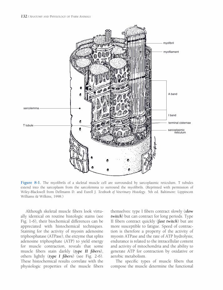
132 / ANATOMY AND PHYSIOLOGY OF FARM ANIMALS
themselves: type I fi bers contract slowly (slow twitch) but can contract for long periods. Type II fi bers contract quickly (fast twitch) but are more susceptible to fatigue. Speed of contrac-tion is therefore a property of the activity of myosin ATPase and the rate of ATP hydrolysis; endurance is related to the intracellular content and activity of mitochondria and the ability to generate ATP for contraction by oxidative or aerobic metabolism.
The specifi c types of muscle fi bers that compose the muscle determine the functional
Although skeletal muscle fi bers look virtu-ally identical on routine histologic stains (see Fig. 1-6), their biochemical differences can be appreciated with histochemical techniques. Staining for the activity of myosin adenosine triphosphatase (ATPase), the enzyme that splits adenosine triphosphate (ATP) to yield energy for muscle contraction, reveals that some muscle fi bers stain darkly (type II fi bers), others lightly (type I fi bers) (see Fig. 2-6). These histochemical results correlate with the physiologic properties of the muscle fi bers
sarcolemma
T tubule
myofibril
myofilament
A band
I band
terminal cisternae
sarcoplasmicreticulum
Figure 8-1. The myofi brils of a skeletal muscle cell are surrounded by sarcoplasmic reticulum. T tubules extend into the sarcoplasm from the sarcolemma to surround the myofi brils. (Reprinted with permission of Wiley-Blackwell from Dellmann D. and Eurell J. Textbook of Veterinary Histology. 5th ed. Baltimore: Lippincott Williams & Wilkins, 1998.)

MICROSCOPIC ANATOMY AND PHYSIOLOGY OF MUSCLE / 133
characteristics of a whole muscle. Muscles that require sustained contraction, such as the anti-gravity muscles, typically contain more slow-twitch endurance fi bers than do the muscles that contract briefl y but quickly and with great force. It is even possible to show a difference among breeds of horses with regard to the muscle fi ber composition of the same muscle; the middle gluteal muscle of the sprinting quarter horse is usually characterized by more of the large, fast-twitch, but low-endurance type II fi bers than is the same muscle of the slower but long-winded Arabian.
On casual examination with a light micro-scope, the cross-striations of skeletal muscle appear to be disks throughout the entire fi ber. However, the electron microscope shows the striations only in the myofi brils and not in the sarcoplasm (cytoplasm of muscle cell). The alternate light and dark bands of all myofi brils appear at the corresponding places in the fi ber (Fig. 8-2). The fact that corresponding bands of adjacent myofi brils are in register makes it appear that these bands extend across the whole fi ber (Figs. 8-1 and 8-2). The apparent bands of myofi brils are due to relative density
Figure 8-2. Light micrograph (A) and electron micrograph (B) of longitudinal skeletal muscle and schematic representation (C) of a sarcomere. (Reprinted with permission of Wiley- Blackwell from Dellmann D. and Eurell J. Textbook of Veteri-nary Histology. 5th ed. Balti-more: Lippincott Williams & Wilkins, 1998.)
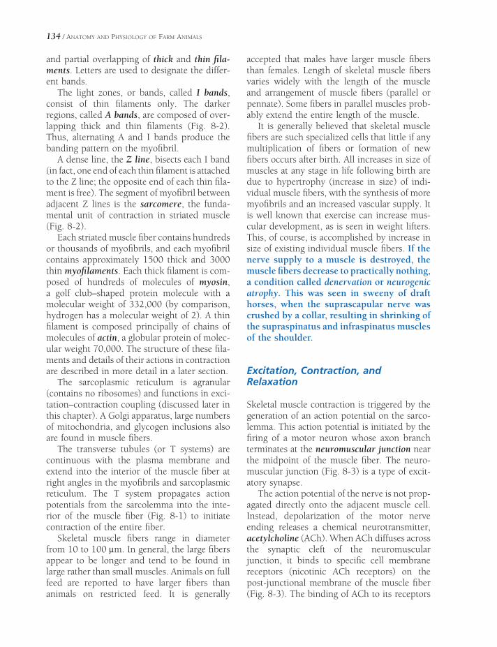
134 / ANATOMY AND PHYSIOLOGY OF FARM ANIMALS
and partial overlapping of thick and thin fi la-ments. Letters are used to designate the differ-ent bands.
The light zones, or bands, called I bands, consist of thin fi laments only. The darker regions, called A bands, are composed of over-lapping thick and thin fi laments (Fig. 8-2). Thus, alternating A and I bands produce the banding pattern on the myofi bril.
A dense line, the Z line, bisects each I band (in fact, one end of each thin fi lament is attached to the Z line; the opposite end of each thin fi la-ment is free). The segment of myofi bril between adjacent Z lines is the sarcomere, the funda-mental unit of contraction in striated muscle (Fig. 8-2).
Each striated muscle fi ber contains hundreds or thousands of myofi brils, and each myofi bril contains approximately 1500 thick and 3000 thin myofi laments. Each thick fi lament is com-posed of hundreds of molecules of myosin, a golf club–shaped protein molecule with a molecular weight of 332,000 (by comparison, hydrogen has a molecular weight of 2). A thin fi lament is composed principally of chains of molecules of actin, a globular protein of molec-ular weight 70,000. The structure of these fi la-ments and details of their actions in contraction are described in more detail in a later section.
The sarcoplasmic reticulum is agranular (contains no ribosomes) and functions in exci-tation–contraction coupling (discussed later in this chapter). A Golgi apparatus, large numbers of mitochondria, and glycogen inclusions also are found in muscle fi bers.
The transverse tubules (or T systems) are continuous with the plasma membrane and extend into the interior of the muscle fi ber at right angles in the myofi brils and sarcoplasmic reticulum. The T system propagates action potentials from the sarcolemma into the inte-rior of the muscle fi ber (Fig. 8-1) to initiate contraction of the entire fi ber.
Skeletal muscle fi bers range in diameter from 10 to 100 μm. In general, the large fi bers appear to be longer and tend to be found in large rather than small muscles. Animals on full feed are reported to have larger fi bers than animals on restricted feed. It is generally
accepted that males have larger muscle fi bers than females. Length of skeletal muscle fi bers varies widely with the length of the muscle and arrangement of muscle fi bers (parallel or pennate). Some fi bers in parallel muscles prob-ably extend the entire length of the muscle.
It is generally believed that skeletal muscle fi bers are such specialized cells that little if any multiplication of fi bers or formation of new fi bers occurs after birth. All increases in size of muscles at any stage in life following birth are due to hypertrophy (increase in size) of indi-vidual muscle fi bers, with the synthesis of more myofi brils and an increased vascular supply. It is well known that exercise can increase mus-cular development, as is seen in weight lifters. This, of course, is accomplished by increase in size of existing individual muscle fi bers. If the nerve supply to a muscle is destroyed, the muscle fi bers decrease to practically nothing, a condition called denervation or neurogenic atrophy. This was seen in sweeny of draft horses, when the suprascapular nerve was crushed by a collar, resulting in shrinking of the supraspinatus and infraspinatus muscles of the shoulder.
Excitation, Contraction, and Relaxation
Skeletal muscle contraction is triggered by the generation of an action potential on the sarco-lemma. This action potential is initiated by the fi ring of a motor neuron whose axon branch terminates at the neuromuscular junction near the midpoint of the muscle fi ber. The neuro-muscular junction (Fig. 8-3) is a type of excit-atory synapse.
The action potential of the nerve is not prop-agated directly onto the adjacent muscle cell. Instead, depolarization of the motor nerve ending releases a chemical neurotransmitter, acetylcholine (ACh). When ACh diffuses across the synaptic cleft of the neuromuscular junction, it binds to specifi c cell membrane receptors (nicotinic ACh receptors) on the post-junctional membrane of the muscle fi ber (Fig. 8-3). The binding of ACh to its receptors
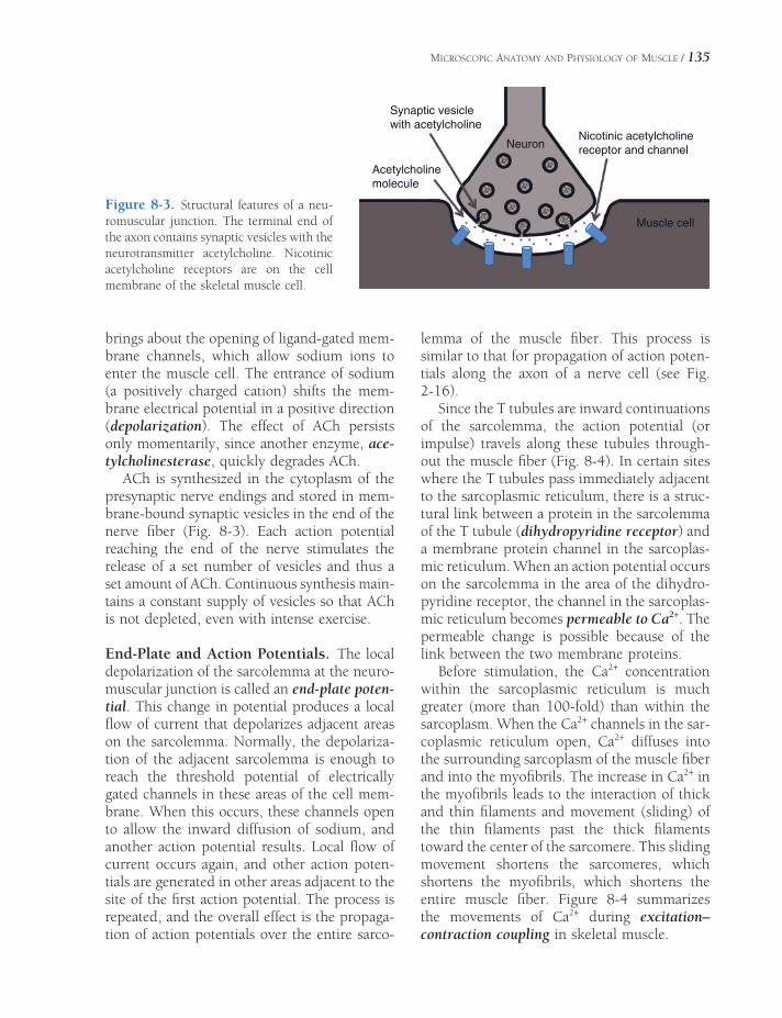
MICROSCOPIC ANATOMY AND PHYSIOLOGY OF MUSCLE / 135
brings about the opening of ligand-gated mem-brane channels, which allow sodium ions to enter the muscle cell. The entrance of sodium (a positively charged cation) shifts the mem-brane electrical potential in a positive direction (depolarization). The effect of ACh persists only momentarily, since another enzyme, ace-tylcholinesterase, quickly degrades ACh.
ACh is synthesized in the cytoplasm of the presynaptic nerve endings and stored in mem-brane-bound synaptic vesicles in the end of the nerve fi ber (Fig. 8-3). Each action potential reaching the end of the nerve stimulates the release of a set number of vesicles and thus a set amount of ACh. Continuous synthesis main-tains a constant supply of vesicles so that ACh is not depleted, even with intense exercise.
End-Plate and Action Potentials. The local depolarization of the sarcolemma at the neuro-muscular junction is called an end-plate poten-tial. This change in potential produces a local fl ow of current that depolarizes adjacent areas on the sarcolemma. Normally, the depolariza-tion of the adjacent sarcolemma is enough to reach the threshold potential of electrically gated channels in these areas of the cell mem-brane. When this occurs, these channels open to allow the inward diffusion of sodium, and another action potential results. Local fl ow of current occurs again, and other action poten-tials are generated in other areas adjacent to the site of the fi rst action potential. The process is repeated, and the overall effect is the propaga-tion of action potentials over the entire sarco-
lemma of the muscle fi ber. This process is similar to that for propagation of action poten-tials along the axon of a nerve cell (see Fig. 2-16).
Since the T tubules are inward continuations of the sarcolemma, the action potential (or impulse) travels along these tubules through-out the muscle fi ber (Fig. 8-4). In certain sites where the T tubules pass immediately adjacent to the sarcoplasmic reticulum, there is a struc-tural link between a protein in the sarcolemma of the T tubule (dihydropyridine receptor) and a membrane protein channel in the sarcoplas-mic reticulum. When an action potential occurs on the sarcolemma in the area of the dihydro-pyridine receptor, the channel in the sarcoplas-mic reticulum becomes permeable to Ca2+. The permeable change is possible because of the link between the two membrane proteins.
Before stimulation, the Ca2+ concentration within the sarcoplasmic reticulum is much greater (more than 100-fold) than within the sarcoplasm. When the Ca2+ channels in the sar-coplasmic reticulum open, Ca2+ diffuses into the surrounding sarcoplasm of the muscle fi ber and into the myofi brils. The increase in Ca2+ in the myofi brils leads to the interaction of thick and thin fi laments and movement (sliding) of the thin fi laments past the thick fi laments toward the center of the sarcomere. This sliding movement shortens the sarcomeres, which shortens the myofi brils, which shortens the entire muscle fi ber. Figure 8-4 summarizes the movements of Ca2+ during excitation– contraction coupling in skeletal muscle.
Figure 8-3. Structural features of a neu-romuscular junction. The terminal end of the axon contains synaptic vesicles with the neurotransmitter acetylcholine. Nicotinic acetylcholine receptors are on the cell membrane of the skeletal muscle cell.
Synaptic vesicle
with acetylcholine
Acetylcholine
molecule
Neuron
Muscle cell
Nicotinic acetylcholine
receptor and channel
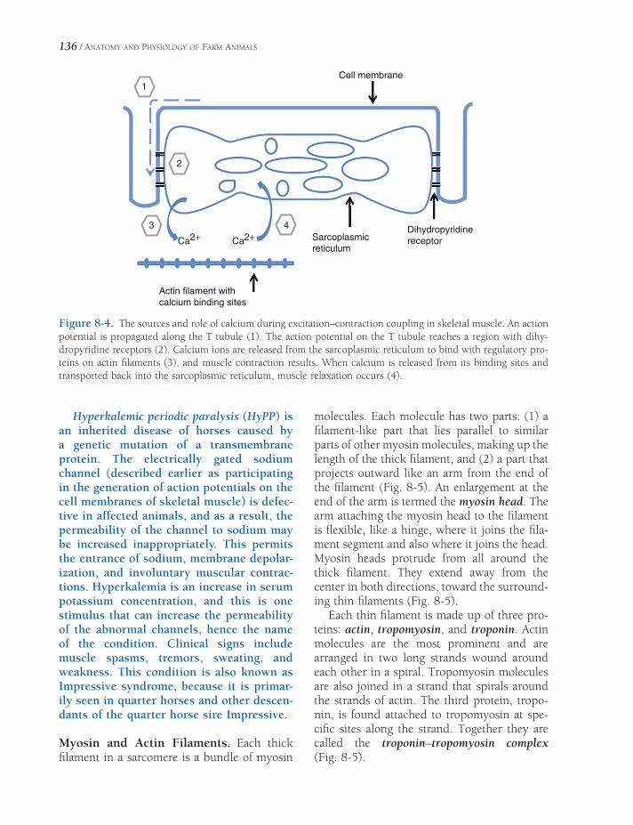
136 / ANATOMY AND PHYSIOLOGY OF FARM ANIMALS
Hyperkalemic periodic paralysis (HyPP) is an inherited disease of horses caused by a genetic mutation of a transmembrane protein. The electrically gated sodium channel (described earlier as participating in the generation of action potentials on the cell membranes of skeletal muscle) is defec-tive in affected animals, and as a result, the permeability of the channel to sodium may be increased inappropriately. This permits the entrance of sodium, membrane depolar-ization, and involuntary muscular contrac-tions. Hyperkalemia is an increase in serum potassium concentration, and this is one stimulus that can increase the permeability of the abnormal channels, hence the name of the condition. Clinical signs include muscle spasms, tremors, sweating, and weakness. This condition is also known as Impressive syndrome, because it is primar-ily seen in quarter horses and other descen-dants of the quarter horse sire Impressive.
Myosin and Actin Filaments. Each thick fi lament in a sarcomere is a bundle of myosin
molecules. Each molecule has two parts: (1) a fi lament-like part that lies parallel to similar parts of other myosin molecules, making up the length of the thick fi lament, and (2) a part that projects outward like an arm from the end of the fi lament (Fig. 8-5). An enlargement at the end of the arm is termed the myosin head. The arm attaching the myosin head to the fi lament is fl exible, like a hinge, where it joins the fi la-ment segment and also where it joins the head. Myosin heads protrude from all around the thick fi lament. They extend away from the center in both directions, toward the surround-ing thin fi laments (Fig. 8-5).
Each thin fi lament is made up of three pro-teins: actin, tropomyosin, and troponin. Actin molecules are the most prominent and are arranged in two long strands wound around each other in a spiral. Tropomyosin molecules are also joined in a strand that spirals around the strands of actin. The third protein, tropo-nin, is found attached to tropomyosin at spe-cifi c sites along the strand. Together they are called the troponin–tropomyosin complex (Fig. 8-5).
Actin filament withcalcium binding sites
3
Ca2+ Ca2+4
2
1
Sarcoplasmicreticulum
Dihydropyridinereceptor
Cell membrane
Figure 8-4. The sources and role of calcium during excitation–contraction coupling in skeletal muscle. An action potential is propagated along the T tubule (1). The action potential on the T tubule reaches a region with dihy-dropyridine receptors (2). Calcium ions are released from the sarcoplasmic reticulum to bind with regulatory pro-teins on actin fi laments (3), and muscle contraction results. When calcium is released from its binding sites and transported back into the sarcoplasmic reticulum, muscle relaxation occurs (4).
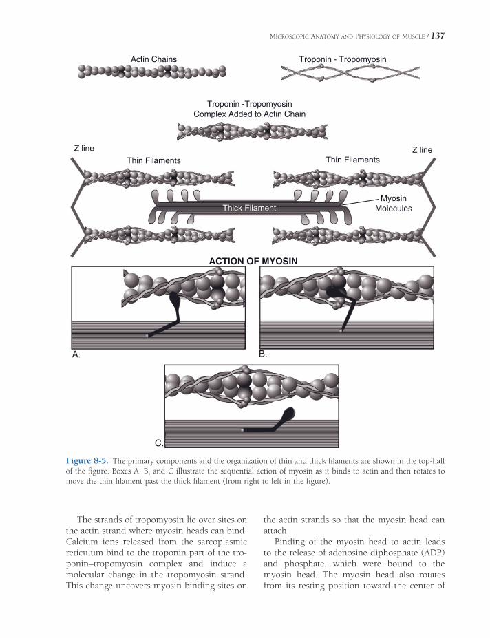
MICROSCOPIC ANATOMY AND PHYSIOLOGY OF MUSCLE / 137
Actin Chains
Thin Filaments
Thick FilamentMyosin
Molecules
ACTION OF MYOSIN
Thin FilamentsZ lineZ line
Troponin -TropomyosinComplex Added to Actin Chain
Troponin - Tropomyosin
A. B.
C.
Figure 8-5. The primary components and the organization of thin and thick fi laments are shown in the top-half of the fi gure. Boxes A, B, and C illustrate the sequential action of myosin as it binds to actin and then rotates to move the thin fi lament past the thick fi lament (from right to left in the fi gure).
The strands of tropomyosin lie over sites on the actin strand where myosin heads can bind. Calcium ions released from the sarcoplasmic reticulum bind to the troponin part of the tro-ponin–tropomyosin complex and induce a molecular change in the tropomyosin strand. This change uncovers myosin binding sites on
the actin strands so that the myosin head can attach.
Binding of the myosin head to actin leads to the release of adenosine diphosphate (ADP) and phosphate, which were bound to the myosin head. The myosin head also rotates from its resting position toward the center of

138 / ANATOMY AND PHYSIOLOGY OF FARM ANIMALS
the sarcomere; this movement pulls the actin chain to which it is bound past the thick fi la-ment (Fig. 8-5).
The myosin head remains at its fi nal angle and bound to the actin of the thin fi lament until an intact ATP molecule binds to another site on the myosin head. (This site on the myosin head is ATPase, which also promotes the hydrolysis of ATP prior to movement of the head.) With the binding of a new ATP, the myosin head detaches from the actin chain and resumes its resting angle. It is ready to repeat the process of attaching to actin, moving from resting to fi nal angle and pulling the attached thin fi la-ment farther toward the center of the sarco-mere, detaching, then binding still another ATP molecule.
The cycle of events that produces the short-ening of each sarcomere, the sliding fi lament model of muscle contraction, is summarized in Figure 8-5. The resultant contraction shortens the sarcomere and shortens the I bands. The A band always remains the same length. During shortening, the thin fi laments slide over the thick fi laments as they are drawn from both ends, pulling the Z lines closer together (Fig. 8-2). The fi laments themselves do not shorten.
Relaxation. Muscle contraction will continue as long as there is an excess of Ca2+ in the sar-coplasm, but when the effect of the action potentials on the sarcolemma ends, the Ca2+ is sequestered back into the sarcoplasmic reticu-lum (Fig. 8-4). Ion pumps in the membrane of the sarcoplasmic reticulum use the energy of ATP to pump the Ca2+ from the sarcoplasmic fl uid back into storage so that it can be ready for the next depolarization. (Without ATP the muscle cannot relax.) Only a small amount of Ca2+ is left out in the sarcoplasm of the relaxed resting muscle, not enough to act on the tropo-nin–tropomyosin complex. Therefore, during relaxation the thin and thick fi laments are dis-sociated, allowing the elasticity of the muscle to return it to its resting length, which pulls the Z lines and thin fi laments back to their original positions.
Replenishment of ATP. A great amount of ATP is needed because the energy for contrac-
tion is derived from hydrolyzed (dephosphory-lated) ATP. Also, the muscle fi ber uses ATP to sequester Ca2+ back into the sarcoplasmic retic-ulum. It is also needed for complete recovery of the membrane after depolarization—the Na–K–ATPase system.
The concentration of ATP in resting skeletal muscle is relatively small, supplying only enough energy to maintain contraction for a brief period. Since muscles continue to contract after the initial supply of ATP has been used, the resulting ADP is phosphorylated again from another source, creatine phosphate (CP).
There is normally about fi ve times as much CP as ATP stored in the sarcoplasm of skeletal muscle. When the ATP is used for contraction and relaxation, transphosphorylation occurs from CP to the resulting ADP, forming ATP again. This replenishment reaction occurs almost as fast as ATP is used. Therefore, the ATP level changes little until the concentration of CP gets low. CP acts as the immediate energy source for the resynthesis of ATP. This is a convenient means of transferring energy for muscle contraction.
The concentration of CP is also limited. Thus, if muscle contraction continues for longer than a few seconds, the CP and new ATP even-tually have to be reconstituted by the citric acid cycle (Krebs cycle) in the mitochondria of the muscle fi bers. If muscle activity outstrips the ability of the mitochondria to produce ATP aerobically, anaerobic metabolism of carbohy-drate fuel begins and lactic acid accumulates in the muscle cell. Glucose (the major carbo-hydrate fuel) is obtained from the blood supply to the muscle and from the glycogen stored in the muscle fi bers. Glycogen is broken down by glycogenolysis. Glycogenolysis and glycolysis are complex processes involving a number of reactions, enzymes, and intermedi-ate compounds.
Oxygen from the blood must also be sup-plied to the mitochondria in muscles for the citric acid cycle to operate and result in oxida-tive phosphorylation of ADP to ATP. While lactic acid was mostly being produced anaero-bically during muscle contraction, an oxygen debt was building up. This oxygen debt must

MICROSCOPIC ANATOMY AND PHYSIOLOGY OF MUSCLE / 139
be repaid during relaxation before optimal muscle activity can resume.
The chain of reactions involved in supplying energy for muscle contraction and recovery is shown in Table 8-1.
Strength of Contraction
Whenever a single muscle fi ber receives a nervous impulse and action potentials are gen-erated on the muscle fi ber, the action potentials will be propagated over the entire fi ber and cause the whole fi ber to contract. This is the all-or-none law of muscle contraction.
The all-or-none law applies to a single muscle fi ber or a single motor unit (a motor neuron and all of the muscle fi bers it supplies); it does not apply to an entire muscle, such as the m. biceps brachii. The all-or-none law also does not state that a muscle fi ber will always contract with the same speed or the same force; rather, it states that for the conditions at the time of stimulation, the muscle fi ber will con-tract to its maximum. The force of contraction does depend on the state of the fi ber at the time, that is, whether it is fatigued, stretched to its optimal length, and so on. The sliding fi la-ment mechanism is possible because of overlap of the thin and thick fi laments. This overlap permits the binding between actin and myosin. Experimental studies have demonstrated that the amount of overlap of the fi laments before contraction begins affects the contraction strength of individual muscle fi bers. When muscle fi bers are stretched before being stimu-lated, contraction strength increases up to an optimal amount of stretch. Any further stretch-
ing produces a decrease in contraction strength. This same relationship is true for the other striated muscle, cardiac muscle, and is an important factor in the regulation of cardiac contraction strength.
Summation. Each gross muscle, which is com-posed of multiple motor units and many indi-vidual muscle fi bers, is capable of contracting with varying degrees of strength. This is the result of summing the contractions in two ways. Motor unit summation (recruitment) occurs when more motor units are stimulated to contract simultaneously in the gross muscle. Therefore, more muscle fi bers and bundles are contracting and producing greater strength in the whole muscle. Temporal summation occurs when the frequency of stimulation to one or more motor units is increased. That is, the fre-quency of stimulation is such that the fi rst con-traction is not over by the time the second contraction begins. The two become additive, which increases the contraction strength (Fig. 8-6). Ordinarily, in normal muscle function both types of summation occur at the same time.
Tetany (Tetanus). When the frequency of stimulation becomes so rapid that further increases in frequency will not increase the strength of contraction, the greatest force that the muscle can develop has been reached. This is called tetany, tetanus, or tetanization. All of these terms are defi ned as a continuous tonic spasm of muscle or a steady state of contraction (Fig. 8-7). The disease caused by the toxin from Clostridium tetani, which produces spasm of the masseter muscles (lockjaw) followed by a spasm of other muscles, is also
Table 8-1. Chain of Reactions That Supply Energy for Muscle Contraction and Recovery
ATP→ ADP + phosphoric acid + energy (for immediate use in contraction)
CP→ Creatine + phosphoric acid + energy (for resynthesis of ATP from ADP)
Glucose (glycogen or blood)→ Lactic acid + energy (for resynthesis of CP from creatine and phosphoric acid)
Lactic acid + oxygen→ Water + carbon dioxide + energy (for resynthesis of ATP and CP)
ATP, adenosine triphosphate; ADP, adenosine diphosphate; CP, creatine phosphate.

140 / ANATOMY AND PHYSIOLOGY OF FARM ANIMALS
specifi cally referred to as tetanus. The Clos-tridium toxin produces skeletal muscle tetany by inhibiting the release of inhibitory neuromediators within the central nervous system. These inhibitors normally act within the spinal cord to regulate the activity of motor neurons to skeletal muscle. Without these inhibitors, any motor activity can result in spastic or tetanic contractions of the skeletal muscle. Loud noises or sudden movements can cause affected animals to tense their muscles and may induce general-ized spasms.
Fatigue. Fatigue is a decrease in work capacity caused by work itself. Fatigue may occur at the level of the individual muscle fi ber, or it may be a generalized state affecting the animal as a whole. On a whole-animal basis, resistance to fatigue entails some poorly defi ned factors such as motivation. The factors contributing to fatigue of an individual muscle fi ber have been
best studied in muscle cells in isolated in vitro preparations outside of the body. These studies indicate that fatigue is a function of the muscle cell itself and is not due to failure or fatigue of the neurons that innervate skeletal muscles.
At the individual cell level, one contributor to fatigue is a decrease in the availability of ATP, as its use increases with increased muscle contractions. However, even with strenuous prolonged exercise ATP is never totally depleted, because the intracellular generation of ATP is also increased during exercise. Increases in intracellular concentrations of various metabo-lites generated as a result of contraction also contribute to fatigue. Among these are the phosphate from the splitting of ATP to ADP and organic acids such as lactic acid. Lactic acid can diffuse out of the cell and reduce the pH of the interstitial fl uids within a muscle. Accumulations of lactic acid within muscles contribute to pain and soreness of muscles fol-lowing strenuous exercise.
Figure 8-6. Temporal summation of muscle twitches. The fi rst two contractions illustrate the force generated by a single stimulus (action potential). When a second stimulus is applied before the muscle can completely relax, summation occurs, and the force of contraction is greater (third recording).
Figure 8-7. Tetany. The interval between stimuli (action potentials) steadily decreases until no relaxation can occur between action potentials.
Forc
e
Tetany
Time= Stimulus
Forc
e
S=Stimulus Time
S S S S

MICROSCOPIC ANATOMY AND PHYSIOLOGY OF MUSCLE / 141
The rate at which ATP is generated within an exercising muscle depends on the availabil-ity of substrates for the different metabolic pathways that produce ATP. If a muscle pri-marily uses the glycolytic pathway, glucose must be readily available. Glucose may be stored within the muscle cell as glycogen or delivered via the circulation. If a muscle uses the oxidative pathway to produce ATP, oxygen must be available in addition to fatty acids or other similar substrates. The oxygen, of course, must arrive via the circulation. Adequate blood fl ow during exercise to deliver oxygen, fatty acids, and/or glucose to skeletal muscle is a key factor in resisting fatigue.
Drugs That Affect Skeletal Muscle Function
Almost as soon as ACh initiates the impulse for muscle contraction, the enzyme acetylcholines-terase inactivates the ACh. This enzyme, which degrades ACh, is found within the area of the neuromuscular junction. Agents that are capable of inhibiting the action of the acetyl-cholinesterase enzyme are referred to as anti-cholinesterases. The effect of anticholinesterases at the neuromuscular junction is to prolong the availability and effects of the ACh.
These actions may be desirable in some disease states, and neostigmine and physostig-mine are commonly used anticholinesterase drugs. However, too much anticholinesterase activity can produce a toxicity characterized by muscular spasms and asphyxiation due to spasms of skeletal muscles necessary for respi-ration. The signs of anticholinesterase toxicity also include constriction of the pupil of the eye, intestinal cramps, vomiting, and diarrhea. These signs are due to the increased availability of ACh at neural synapses within the parasym-pathetic division of the autonomic nervous system. Parasympathetic nerves are discussed in more detail in Chapter 10.
Anticholinesterase activity is the basis for some effective insecticides and the so-called nerve gases that have been studied exten-sively by the armed forces since World War
II. Most of these compounds are alkyl phos-phates, and the insecticides are also known as organic phosphates or organophosphates. The insecticides include such products as malathion, parathion, and diazinon. Prod-ucts for both external application and oral administration are included in the organo-phosphate insecticides. If improperly used, any of the organophosphates is extremely dangerous, not only to domestic animals but also to the person using it. Therefore, it is imperative that this class of insecticides be used under proper supervision and that instructions for use are followed exactly.
Another group of drugs affecting the neuro-muscular junction are the curariform drugs. These drugs act like curare, the deadly poison that some South American Indians use on their arrowheads. These agents bind to ACh recep-tors on the postjunctional membrane so that ACh cannot bind to the receptors and produce an end-plate potential. Curare is not destroyed by acetylcholinesterase. Death can result from asphyxiation because of paralysis of muscles needed for respiration. The paralysis induced by curariform drugs depends on the concentra-tion of the drug, and such agents are used therapeutically to produce varying amounts of paralysis, such as for muscle relaxation during certain types of surgery.
Botulinum toxin, which is produced by the bacterium Clostridium botulinum, also acts at the neuromuscular junction. It prevents the release of ACh from the nerve ending by preventing synaptic vesicles containing ACh from binding to the cell membrane for exo-cytosis. Flaccid paralysis results because action potentials cannot be produced on the sarcolemma to bring about muscle contrac-tion. The term limberneck has been applied to the characteristic fl accid paralysis of the neck in poultry intoxicated with botulinum toxin.
Types of Muscle Contraction
The primary function of muscle is to contract, that is, develop tension and shorten. However,

142 / ANATOMY AND PHYSIOLOGY OF FARM ANIMALS
contraction is often described as having four types: concentric (shortening), eccentric (length-ening), isometric (same length but increased tension), and isotonic (same tension but length changes).
Concentric contraction is the usual form, in which the muscle moves a bone or segment by shortening. An example is fl exion of the elbow by contraction of the m. biceps brachii.
Isometric contraction occurs naturally whenever a limb or portion of the body is held stationary against equal resistance, such as gravity. To hold the head up in a fi xed posi-tion, the dorsal neck muscles must contract isometrically.
Eccentric contraction occurs in the extensor muscles of the neck when an animal lowers its head gradually. Antagonistic muscles may also undergo eccentric contraction when unsuccess-fully opposing the actions of a prime mover.
Isotonic contraction occurs when the length of the muscle changes but the tension remains the same, primarily when a muscle lifts a given weight. The weight is constant; therefore, the tension does not change.
Rigor and Rigor Mortis. If most of the ATP in a muscle is depleted, the myosin heads cannot separate from the actin in the thin fi laments and the calcium can no longer be sequestered back into the sarcoplasmic reticulum by the calcium pump. Therefore, relaxation cannot occur because the actin and myosin fi laments are bound in a continuous contracted state. This is termed rigor, and it is sustained until more ATP is made available. Rigor mortis is rigor that occurs a few hours after death, when ATP is no longer available. The muscles of the whole animal progressively become stiff and rigid; without ATP to sequester the calcium and separate the cross-bridges, the fi laments remain locked together. The rigidity continues until cell autolysis and protein degradation break down the muscle.
Tone. The term muscle tone refers to the slight tension exhibited by all muscles at rest. It is due to the continuous transmission of impulses at
very low frequency from the spinal cord to the muscles. Tone keeps muscles in a partially con-tracted state and prevents them from becoming fl accid (fl abby), as occurs in paralysis.
When an animal becomes anxious, fearful, or excited, the muscle tone intensifi es. There-fore, the muscles become taut (tension increases) and the animal can respond faster to any stimu-lus. This is often seen in the skittish, nervous, or jumpy animal. During sleep, muscle tone is low to allow for optimal relaxation.
Smooth Muscle
Smooth muscle is sometimes called involun-tary muscle because it is found in structures that are not voluntarily or consciously regu-lated (e.g., gastrointestinal tract, blood vessels, reproductive tract). About 99% of the smooth muscle of the animal body is of the visceral type, which is also called single-unit or unitary smooth muscle. Cells of this type of smooth muscle are joined with gap junctions to provide mechanical and electrical connections between cells. The electrical connections permit the propagation of action potentials directly from cell to cell. The spreading of action potentials and the contraction that is elicited allow a group of muscle cells to act together as a single unit. This type of activity is appropriate for generalized activities of an entire organ, such as the contraction of the stomach.
About 1% of the smooth muscle of the animal body is multiunit smooth muscle (e.g., iris and ciliary body of the eye and pilomotor fi bers that erect the hair in the skin). In this type of smooth muscle, the contraction of each individual smooth muscle cell is more depen-dent on its autonomic innervation. Gap junc-tions are not prevalent between these cells.
Structure
The smooth muscle cell is a fusiform (spindle-shaped) contractile unit with a central nucleus. Size of smooth muscle fi bers varies consider-ably. Most cells are 50–250 μm long and 5–10 μm in greatest diameter. The major portion

MICROSCOPIC ANATOMY AND PHYSIOLOGY OF MUSCLE / 143
of the cell consists of sarcoplasm. No cross-striations, myofi brils, or sarcolemma are easily visible with the light microscope (see Fig. 1-6). Filaments are present as actin and myosin mol-ecules, but there are no orderly arrangements to form striations. Interactions between actin and myosin fi laments (i.e., sliding fi laments) are believed to be the basis for smooth muscle contractions, but the characteristic shortening of sarcomeres is not seen because there is no distinct organization of the fi laments. A sarco-plasmic reticulum, which accumulates calcium, is present, but it is not as extensive or as highly organized as in skeletal muscle.
Like skeletal muscle, smooth muscle cells can hypertrophy to increase the size of organs. However, unlike skeletal muscle, smooth muscle cells can also divide mitotically to increase the number of cells. For example, the increase in size of the uterine wall during preg-nancy to several times its nonpregnant volume is due in considerable measure to an increase in the amount of smooth muscle in the wall. Some of the increase is due to an increase in size of individual muscle fi bers, but there is also an increase in the number of cells. This hyper-trophy and hyperplasia in the uterus are under the infl uence of reproductive hormones. Late in pregnancy there is also a change in the structure of the cell membranes of uterine smooth muscle cells. An increase in gap junctions between cells in late pregnancy sets the stage for the mechani-cal and electrical coupling between smooth muscle cells that will be necessary for normal parturition. In general, the structure and func-tion of smooth muscle are more subject to modifi cation by external factors, such as hor-mones, than are those of skeletal muscle.
Stress–Relaxation
Smooth muscle exhibits a special property called stress–relaxation, or plasticity. This is the ability to adjust to stretching without increasing the fi nal tension or the pressure exerted on the contents within a hollow viscus surrounded by smooth muscle. As the muscle stretches, the tension increases at fi rst, but then
in a few seconds or a few minutes the smooth muscle relaxes again to its original tension, even though it is still elongated.
This stress–relaxation occurs in the stomach when it is fi lling with food, in the intestines as the processed food moves along, in the blood vessels when the blood volume increases, in the urinary bladder as it increases its volume of urine, and in the uterus as pregnancy develops.
Plasticity allows expansion of stretch within physiologic limits without an increase in pres-sure and without pain; the smooth muscle does not lose its contractile ability. The reverse occurs upon emptying of a visceral organ when the stretched muscle shortens back to its origi-nal length: all tension is lost at fi rst but returns shortly. Plasticity is believed to be due to changes in the arrangement or binding of the myosin and actin fi laments upon stretching or shortening.
Contraction and Relaxation
All skeletal muscle contraction depends on ACh release at a neuromuscular junction and the generation and propagation of action poten-tials on the cell membrane. This is not the case for smooth muscle. The stimuli that bring about smooth muscle contraction and relaxation are quite variable. Thus, smooth muscle is a much more functionally diverse tissue than skeletal muscle.
The contraction and relaxation of most smooth muscle are much slower events than the rapid muscle twitch that is characteristic of skeletal muscle. This permits the maintenance of a relatively constant pressure with the use of little cell energy. This is characteristic of smooth muscle in organs that require a constant state of tone or some degree of contraction for normal function (e.g., smooth muscle in the walls of blood vessels).
Role and Sources of Calcium
As in skeletal muscle, contraction and relax-ation of smooth muscle are linked to the Ca2+

144 / ANATOMY AND PHYSIOLOGY OF FARM ANIMALS
concentration in the cytosol of smooth muscle cells. However, how this concentration is regu-lated and the role of Ca2+ in the contraction process is quite different in the two types of muscle. Some calcium is stored in the sarco-plasmic reticulum of smooth muscle cells, but many types of smooth muscle cells also contain a signifi cant number of calcium channels in the outer cell membrane (Fig. 8-8). These calcium channels may be either voltage-gated or ligand-gated, and the smooth muscle within a given organ may have both types of channels or pri-marily only one type of channel. When these channels open in response to the appropriate stimulus (changes in membrane potential or presence of specifi c ligands), calcium can diffuse into the cell to initiate contraction (Fig. 8-8). Receptors that are specifi c for many different ligands (e.g., hormones and neurotransmitters) are found on the smooth muscle of different organs. This is important functionally, because a given hormone produces contraction only of organs with receptors for that specifi c hormone. Calcium entering from the outside via the chan-nels may stimulate the release of further calcium from the sarcoplasmic reticulum, which further strengthens the contractions.
An understanding of the role of calcium channels in the outer cell membrane of smooth muscle cells led to the develop -ment of a group of drugs termed calcium channel blockers. These agents are capable of binding to and inactivating these calcium
channels, and this tends to reduce the strength of contractions. Smooth muscle cells in the walls of many arterial blood vessels have these types of channels, and this type of drug has been proved effective in lowering blood pressure in both humans and animals.
When cytosolic Ca2+ increases within smooth muscles, calcium ions bind to a regulatory protein (calmodulin) (Fig.8-8). Unlike skeletal muscle, whose regulatory proteins are associ-ated with the actin fi laments, the calmodulin in smooth muscle is associated with the heads of myosin molecules in myosin fi laments. The binding of calcium to calmodulin activates a kinase, also associated with myosin, and this kinase phosphorylates other sites on myosin. This phosphorylation ultimately results in con-traction, so myosin phosphorylation is a key regulatory step (Fig. 8-8). This is in contrast to skeletal muscle, whose myosin does not require phosphorylation. For relaxation, the phosphor-ylation of myosin is reversed by other intracel-lular enzymes that are always present and active (Fig. 8-8). This reversal and relaxation can occur when the cytosolic calcium concentra-tion is reduced. Calcium is removed from the cytosol by the transport back into the sarco-plasmic reticulum or out of the cell. Most of this transport is done by an active transport system (Ca2+–ATPase) on the membrane of the endoplasmic reticulum and the outer cell membrane.
Figure 8-8. Sources and role of calcium ions in the initiation and regulation of smooth muscle con-traction. Dotted lines associated with calcium ions indicate move-ment by diffusion. Solid lines associated with calcium ions indicate removal of calcium from the cytosol to promote relaxation. Relaxation can also be promoted by the action of other intracellular enzymes that dephosphorylate myosin.
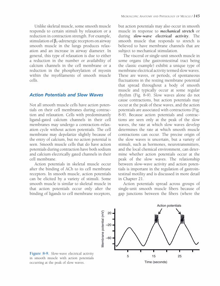
MICROSCOPIC ANATOMY AND PHYSIOLOGY OF MUSCLE / 145
Unlike skeletal muscle, some smooth muscle responds to certain stimuli by relaxation or a reduction in contraction strength. For example, stimulation of β2-adrenergic receptors on airway smooth muscle in the lungs produces relax-ation and an increase in airway diameter. In general, this type of relaxation is due to either a reduction in the number or availability of calcium channels in the cell membrane or a reduction in the phosphorylation of myosin within the myofi laments of smooth muscle cells.
Action Potentials and Slow Waves
Not all smooth muscle cells have action poten-tials on their cell membranes during contrac-tion and relaxation. Cells with predominantly ligand-gated calcium channels in their cell membranes may undergo a contraction–relax-ation cycle without action potentials. The cell membrane may depolarize slightly because of the entry of calcium, but no action potential is seen. Smooth muscle cells that do have action potentials during contraction have both sodium and calcium electrically gated channels in their cell membrane.
Action potentials in skeletal muscle occur after the binding of ACh to its cell membrane receptors. In smooth muscle, action potentials can be elicited by a variety of stimuli. Some smooth muscle is similar to skeletal muscle in that action potentials occur only after the binding of ligands to cell membrane receptors,
but action potentials may also occur in smooth muscle in response to mechanical stretch or during slow-wave electrical activity. The smooth muscle that responds to stretch is believed to have membrane channels that are subject to mechanical stimulation.
The visceral or single-unit smooth muscle in some organs (the gastrointestinal tract being the classic example) exhibit a unique type of membrane electrical activity termed slow waves. These are waves, or periods, of spontaneous fl uctuations in the resting membrane potential that spread throughout a body of smooth muscle and typically occur at some regular rhythm (Fig. 8-9). Slow waves alone do not cause contractions, but action potentials may occur at the peak of these waves, and the action potentials are associated with contractions (Fig. 8-9). Because action potentials and contrac-tions are seen only at the peak of the slow waves, the rate at which slow waves develop determines the rate at which smooth muscle contractions can occur. The precise origin of the slow waves is uncertain, but a variety of stimuli, such as hormones, neurotransmitters, and the local chemical environment, can deter-mine whether action potentials occur at the peak of the slow waves. The relationship between slow-wave activity and action poten-tials is important in the regulation of gastroin-testinal motility and is discussed in more detail in Chapter 21.
Action potentials spread across groups of single-unit smooth muscle fi bers because of gap junctions between the fi bers (where the
Action potentials
Time (seconds)
5 15
20
–20
–60
Mem
bra
ne p
ote
ntial (m
V)
25Figure 8-9. Slow-wave electrical activity in smooth muscle with action potentials occurring at the peak of slow waves.

146 / ANATOMY AND PHYSIOLOGY OF FARM ANIMALS
plasma membranes of adjacent cells touch). Thus, single-unit smooth muscle cells can be linked electrically while remaining indepen-dent chemically (no secretion of transmitter substance from cell to cell is required).
Autonomic Innervation
In visceral or single-unit smooth muscle, the fi bers of the autonomic nervous system travel between the smooth muscle cells in a branch-ing network of terminal fi brils. These fi brils have varicosities (beadlike enlargements) at intervals along their axons. When action poten-tials depolarize them, the transmitter substance is released and diffuses to the smooth muscle cell membranes of several cells, where stimula-tion occurs. In multiunit smooth muscle, a branch of an autonomic nerve innervates each muscle fi ber. This provides more direct neural control of each muscle cell, but the junction between neuron and muscle is less highly structured than the neuromuscular junction of skeletal muscle. For both types of smooth muscle, the innervation is usually dual; that is, both divisions of the autonomic nervous system innervate smooth muscle. Important excep-tions are blood vessels (arteries, arterioles, and veins), which have predominantly sympathetic innervation, and in the skin, where the pilomo-tor fi bers and sweat glands receive only sympa-thetic innervation.
Acetylcholine is released from the parasym-pathetic postganglionic nerve fi bers and norepi-nephrine from the sympathetic postganglionic fi bers. The response of smooth muscle (contrac-tion or relaxation) to these neuromediators depends on the type of autonomic receptor (see Tables 10-1 and 10-2) on the smooth muscle and the intracellular events initiated by the binding of the neuromediators to their recep-tors. For example, stimulation of β2-adrenergic receptors causes smooth muscle relaxation, while stimulation of α1-adrenergic receptors causes smooth muscle contraction. Some smooth muscle can also be stimulated to con-tract by stimulation of muscarinic receptors by ACh. Individual smooth muscle cells may have
multiple types of receptors in their cell mem-branes and respond to both autonomic neu-rotransmitters. In this case, the overall response of the smooth muscle also depends on the rela-tive levels of the different neurotransmitters.
Cardiac Muscle
Cardiac muscle (sometimes known as involun-tary striated muscle) has many anatomic char-acteristics that are similar to those of striated skeletal muscle fi bers, although the striations are fainter than in skeletal muscle. Both types of muscle consist largely of sarcoplasm, myo-fi brils, a sarcoplasmic reticulum, transverse tubules, nuclei, and a sarcolemma. The most striking difference is the tendency for cardiac muscle fi bers to branch and join, forming a network. The heart is made up of cells that are separate entities; however, unique structures, found where cardiac muscle cells meet end to end, are the intercalated disks. These disks can be seen with the light microscope (Fig. 1-6) and are interposed between muscle cells. The disks represent apposed cell membranes and gap junctions. The gap junctions provide a mechanical attachment between cells and permit electrical transmission from one cardiac muscle cell to the next. Action potentials can readily spread from cell to cell, causing cardiac muscle to act electrically and mechanically as a functional syncytium, as if it were a single cell mass.
Blood vessels and lymphatic vessels are both plentiful in cardiac muscle. A generous blood supply is essential, because most ATP produc-tion depends on aerobic metabolism. In humans disruption of the blood supply to cardiac muscle quickly results in myocardial ischemia and the symptoms of a heart attack. Cardiac muscle may also undergo necrosis (cell death) if the loss of blood supply is prolonged or extremely severe.
Excitation and Contraction
Individual cardiac muscle cells do not require nerve stimulation to contract, but action poten-

MICROSCOPIC ANATOMY AND PHYSIOLOGY OF MUSCLE / 147
tials must occur on the cell membrane. Action potentials fi rst occur spontaneously within spe-cialized myocardial pacemaker cells within the heart, and these are propagated throughout the heart by a specialized conduction system and from cell to cell via the gap junctions (at inter-calated disks). The impulse generation and conduction system is described in detail in Chapter 18. Autonomic nerves innervate the pacemaker cells, and these serve to modify the rate of spontaneous action potentials, which in turn determines contraction rate of the entire heart.
The cardiac action potential is much slower than that of skeletal muscle. It lasts for hun-dreds of milliseconds (1 msec = 1/1000 second), as opposed to 5–10 msec in skeletal muscle. Also, the contraction time in cardiac muscle lasts as long as the action potential does. Instead of a sharp spike potential, the cardiac action potential has a long plateau, which extends the time of both the action potential and the muscle contraction.
As is true for the other two types of muscle, an increase in intracellular Ca2+ must occur to bring about cardiac cell contraction. In cardiac cells the Ca2+ enters the cell via electrically
gated cell membrane channels and is also released from an extensive sarcoplasmic reticu-lum network. Thus, cardiac muscle has some similarities to both smooth and skeletal muscle. The Ca2+ binds to regulatory proteins on the actin fi laments, and contraction occurs in a manner similar to that in skeletal muscle.
Cardiac Hypertrophy
Hypertrophy (increase in cell size) occurs in cardiac muscle when the heart has excessive work to do, but like skeletal muscle, mature cardiac muscle cells do not readily regenerate or undergo hyperplasia. Living at high alti-tude may cause hypertrophy of the heart in both humans and animals, partly because of increased vascular resistance and blood pressure in the lungs. Brisket disease (high mountain disease) of cattle occurs when hypertrophy of the heart cannot adequately compensate for the increased vascular resis-tance. A common clinical sign is edema of the brisket. Cardiac hypertrophy also occurs in people and animals who undergo strenu-ous athletic training.


ANATOMY OF THE NERVOUS SYSTEM
The nervous system consists of the brain, the spinal cord, and the peripheral nerves,
which connect the various parts of the body to either the brain or spinal cord. A variety of cell types are found within the nervous system, but the primary functional cell is the neuron. A major function of the other cell types (the glia), which outnumber neurons ten to one, appears to be to maintain the cellular environment to support the activity of the neurons.
The basic functions of the nervous system can be summarized thus:
1. Initiate and/or regulate movement of body parts by initiating and/or regulating the contraction of skeletal, cardiac, and smooth muscles
2. Regulate secretions from glands3. Gather information about the external
environment and about the status of the internal environment of the body, using senses (sight, hearing, touch, balance, taste) and mechanisms to detect pain, temperature, pressure, and certain chemicals, such as carbon dioxide, hydrogen, and oxygen
4. Maintain an appropriate state of consciousness
5. Stimulate thirst, hunger, fear, rage, and sexual behaviors appropriate for survival
All functions of the nervous system require the rapid transmission of information from one site within the body to another. This trans-mission is possible because neurons have the
Microscopic NeuroanatomyEmbryologyCentral Nervous System
Brain
Meninges
Spinal Cord
Peripheral Nervous SystemSpinal Nerves
Cranial Nerves
Autonomic Nervous SystemSympathetic Nervous System
Parasympathetic Nervous System
Enteric Nervous System
Chapter 9
149

Pudendal
Cranial gluteal
Common peroneal
Tibial
Saphenous
Lateral plantar
Medial plantar
Dorsal digitals
Obturator Femoral
Genitofemoral
IlioinguinalSpinal cord
Dorsal branches of spinal
Intercostals Thoracodorsal
Subscapular Suprascapular
Vagus Accessory
Cerebellum
Cerebrum
Optic
Maxillary
Mandibular
Facial
Brachial plexus
Cranial pectoral
Musculocutaneous
Combined musculocutaneousand median
Axillary
Radial
Ulnar
Medial cutaneous antebrachial
Superficial branch of radial
Median
Dorsal digitals
Deep peroneal
Superficial peroneal
Lateral thoracic
Caudal cutaneous antebrachial
Palmar branch of ulnar
Long thoracicLumbosacral plexus
Sciatic
Plantar digitals Palmar digitals
Olfactory
Ophthalmic
Cervical
Figure 9-1. Nervous system of the bull. (Reprinted with permission of Wiley-Blackwell from McCracken T.O., Kainer R.A. and Spurgeon T.L. Spurgeon’s Color Atlas of Large Animal Anatomy. Baltimore: Lippincott Williams & Wilkins, 1999.)
ANATOMY OF THE NERVOUS SYSTEM / 151
property of excitability. This property (see Chapter 2) permits neurons to develop action potentials and rapidly propagate them along their individual cellular processes (axons). When an action potential reaches the end of an axon, the information encoded in the action potential is transmitted to another neuron or some other type of cell (notably, muscle cells). This transmission is accomplished at special-ized junctions known as synapses.
For descriptive purposes the entire nervous system (Fig. 9-1) can be divided into two parts: the central nervous system (CNS), which includes the brain and spinal cord, and the peripheral nervous system (PNS), which con-sists of cranial nerves and spinal nerves going to and from somatic (body) structures. A further distinction is the autonomic nervous system (ANS), which coordinates activity of visceral structures (smooth muscle, cardiac muscle, and glands). The ANS has elements in both the central and peripheral nervous systems, and it features both sensory and motor components.
In the PNS, sensory (afferent) nerves gather information about the external and internal envi-ronments and relay this information to the CNS. The information is obtained by specialized organs, cells, or axon terminals that react to spe-cifi c environmental energies and initiate action potentials in associated sensory axons. The spe-cialized structures that detect environmental stimuli are sensory receptors. Sensory systems are discussed more completely in Chapter 11.
The CNS receives information arriving via the PNS, integrates that information, and initi-ates appropriate movement of body parts, glan-dular secretion, or behavior in response. It may do this via voluntary or involuntary (i.e., auto-nomic or refl exive) processing. Communica-tion between the CNS and the target muscles and glands in the periphery is accomplished via motor (efferent) nerves of the PNS.
Microscopic Neuroanatomy
The individual nerve cell is called a neuron (Fig. 9-2). Neurons possess the usual features of cells, but in keeping with their function of
communication over long distances, they also exhibit a number of specializations. Each neu-ronal cell body gives rise to one or more nerve processes, cytoplasmic extensions of the cell. The nerve processes are called dendrites if they transmit electrical signals toward the cell bodies; they are called axons if they conduct electrical signals away from the cell bodies. The axon (each neuron gives rise to only one, which usually branches) arises from a conical mound of cytoplasm, the axon hillock, and its terminus branches into an arborization called the telo-dendrion. The telodendrion makes contact with other neurons or effector organs (targets). In mammals, effector organs stimulated either directly or indirectly by the telodendrion include muscle and glands.
The junction between the axon of one neuron with another neuron or target cell is the synapse. The neuron belonging to the axon is the presynaptic neuron, and the one receiving information from the axon is the postsynaptic neuron. A synapse may be between the axon of one neuron and the cell body, dendrites, and/or axon of the postsynaptic neuron. Typically, each neuron synapses with many other neurons through the extensive branching of its terminal ends and of its axon; branches of the main axon are axon collaterals.
Neurons may be classifi ed morphologically according to their number of nerve processes (Fig. 9-3). Unipolar neurons have one process; true unipolar neurons are seen only during development. Bipolar neurons have one den-drite and one axon; these are common in sensory systems. Many sensory neurons have their single dendrite and axon fused so as to give the appearance of a single process; this confi guration is pseudounipolar. Multipolar neurons have a number of dendrites in addition to their single axon. Most neurons are multipo-lar in nature.
Nervous tissue consists of not only neurons but also supportive cells. Within the CNS, these supportive cells are the neuroglia, com-prising a variety of glial cells, whereas most of the supporting tissue of the peripheral nerves is ordinary white fi brous connective tissue.

Dendrites
Nucleus
Axon Collateral
Myelin Sheath
Axon
AxonHillock
Soma of Presynaptic Neuron
Sites of Synapse
Telodendrion
Soma ofPostsynaptic
Neuron
}
Figure 9-2. Cellular anatomy of a multipolar neuron.
Figure 9-3. Morphological types of neurons. Classifi cation is based on number of processes extending from the cell body. Unipolar neurons (A) are not found in adult vertebrate nervous systems; the unipolar neuron here is depicted with a growth cone at the tip of its axon, illustrating its developmental nature. Bipolar (B) and pseudouni-polar (C) neurons are characteristic of sensory systems. Most neurons in the nervous system are multipolar (D) and can assume a variety of shapes.

ANATOMY OF THE NERVOUS SYSTEM / 153
Nerve fi bers may be myelinated or unmy-elinated. Myelinated fi bers are surrounded by a white sheath of fatty material, or myelin. The myelin sheath actually consists of many layers of cell membrane of a specialized glial cell wrapped around axons so that in cross-section the myelin sheath resembles a slice of jelly roll. In the PNS, the myelinating cell is the Schwann cell (neurolemmocyte), whereas in the CNS, the oligodendrocyte fulfi lls this function. Unmyelinated nerve fi bers are not exposed directly to the extracellular fl uid; rather, they are simply invaginated into the cell membrane of an adjacent glial cell. Axons covered in this
way are not myelinated, which is very specifi -cally the condition of being wound in multiple layers of glial cell membrane. Several unmy-elinated fi bers may be invaginated into separate areas of the same Schwann cell (Fig. 9-4).
Groups of nerve cell bodies within the CNS are generally called nuclei, while groups of nerve cell bodies in the PNS are called ganglia. Do not confuse a nucleus of the CNS with the nucleus of an individual cell. Bundles of nerve processes within the CNS are frequently called tracts, or fasciculi, and bundles of processes in the PNS are called nerves. In general terms, aggregates of neuronal cell bodies form the gray matter of the CNS, whereas regions char-acterized primarily by tracts are white matter.
Embryology
The nervous system is the fi rst organ system to begin to form in the embryo (see Chapter 3). Shortly after gastrulation, ectodermal cells on the dorsum just cranial to the primitive streak begin to proliferate and differentiate into a neural plate. The neural plate proliferates faster along its lateral margins than on the midline, creating the neural groove, the edges of which (the neural folds) ultimately meet dorsally to form the neural tube (Fig. 9-5). The entire CNS is formed from the cells of the neural tube. The lumen of the neural tube persists in the adult as the central canal of the spinal cord and as the ventricles of the brain (discussed later).
Closure of the neural tube is not simultane-ous throughout the embryo. Fusion develops fi rst at the level that will eventually become the medulla oblongata (the most caudal part of the brainstem) and proceeds craniad and caudad from there. The openings at the cranial and caudal end of the closing tube are called the rostral and caudal neuropores, respectively (see Fig. 3-4).
The rostral neuropore closes early in development; failure to do so disrupts devel-opment of the brain, leading to profound underdevelopment of the head. In its most severe form (short of embryonic death), anencephaly (a complete absence of the
Figure 9-4. A) Cross-section of the development of a myelinated axon. In the peripheral nervous system, the myelinating glial cell is a Schwann cell; in the CNS, the oligodendrocyte lays down myelin wraps. B) Some Schwann cells in the peripheral nerves envelop multi-ple axons without forming the wrappings of myelin. Axons thus embedded are considered nonmyelinated.

154 / ANATOMY AND PHYSIOLOGY OF FARM ANIMALS
cerebrum, often with concurrent absence of meninges and skull) results. The caudal neu-ropore closes later. Failure of closure in the caudal part of the neural tube results in a variety of spinal cord abnormalities called myelodysplasias. These are sometimes also associated with vertebral anomalies, such as spina bifi da.
As the edges of the deepening neural groove approach one another at the dorsal midline, a longitudinal column of cells differentiates at the union between the ectoderm and the neuroec-toderm on each side of the groove. These cells, the neural crest, end up lateral to the neural tube on each side of it and eventually form sensory and autonomic ganglion cells, Schwann cells, and other related tissues. In addition, the neural crest gives rise to a variety of other cell types, including parts of the meninges and many of the bones and muscles of the head.
Development of the spinal cord continues by an increase in the thickness of the wall of the neural tube. As cells divide and differentiate, three concentric layers of the neural tube emerge: an inner ventricular zone, a middle intermediate zone, and a superfi cial marginal zone (Fig. 9-6).
The thin ventricular zone of cells (also called ependymal or germinal zone) surrounds the lumen of the neural tube and is the site of mitosis of neuronal and glial precursors in the developing nervous system. It will ultimately form the ependyma of the central canal of the spinal cord and of the ventricles of the brain.
As cells are born in the germinal layer, they migrate outward to form the intermediate zone (also called mantle zone). The intermediate zone comprises neurons and neuroglia and becomes the gray matter near the center of the cord. The dorsal parts of the intermediate zone develop into the dorsal horns. It is here that sensory processing takes place. The ventral intermediate zone becomes the ventral horns, the location of the motor neurons whose axons will extend out into the periphery to innervate muscles and glands.
The marginal zone, which is most superfi -cial, consists of nerve processes that make up the white matter of the spinal cord. The white color comes from the fatty myelin sheaths. The spinal cord white matter is divided into dorsal, lateral, and ventral funiculi, which are delim-ited by the dorsal and ventral horns of gray matter.
Development of the brain (Fig. 9-7) begins before the neural tube is fully closed caudally. It grows rapidly throughout embryonic and
Figure 9-5. Formation of the neural tube. The thick-ened ectoderm of the neural plate develops into a groove that subsequently fuses on the dorsal side to form a closed tube. Neural crest cells adjacent to the neural folds differentiate into many tissues, including the neurons of the ganglia.

ANATOMY OF THE NERVOUS SYSTEM / 155
Marginal ZoneIntermediate Zone
Neural CanalVentricular Zone
Lateral Funiculus
Dorsal Funiculus
Dorsal Horn
Ventral Funiculus Ventral Horn
Central Canal
Figure 9-6. Cross-sectional view of the development of the spinal cord.
Figure 9-7. Dorsal view of the neural tube. The early brain divides into three vesicles that further differentiate into fi ve vesicles. These give rise to the main regions of the adult brain. Notice the optic cups (primitive retinae and optic nerves) developing from the diencephalon.
fetal life and into the neonatal period. The fi rst gross subdivisions of the brain create the three-vesicle stage. These subdivisions, which consist of three dilations of the presumptive brain, are the prosencephalon, or forebrain; mesencepha-lon, or midbrain; and rhombencephalon, or hindbrain. The prosencephalon develops lateral
extensions, the optic vesicles, the precursors of the optic nerves and retinas.
With further development, the three vesi cles differentiate into fi ve distinct regions. In this fi ve-vesicle stage of development, the pro-sencephalon further subdivides to form the telencephalon (future cerebrum) and the

156 / ANATOMY AND PHYSIOLOGY OF FARM ANIMALS
diencephalon, and the rhombencephalon divides into the metencephalon (future pons and cerebellum) and the myelencephalon (future medulla oblongata). The mesencepha-lon does not subdivide; it will become the future midbrain (often persistently called the mesencephalon).
Central Nervous System
Brain
The gross subdivisions of the adult brain include the cerebrum, cerebellum, and brain-stem (Figs. 9-8 and 9-9). The cerebrum devel-ops from the embryonic telencephalon. The components of the brainstem are defi ned in a number of ways; for our purposes, we include the diencephalon, midbrain, pons, and medulla oblongata as parts of the brainstem.
Telencephalon. The telencephalon, or cere-brum, comprises the two cerebral hemispheres, including the cerebral cortex, the basal nuclei, and other subcortical nuclei, and an aggregate of functionally related structures called the rhinencephalon. The telencephalon encloses the cavities of the lateral ventricles.
The surface area of the cerebrum in domestic mammals is increased by numerous foldings to form convex ridges, called gyri (singular gyrus), which are separated by furrows called fi ssures or sulci (singular sulcus). A particularly promi-nent fi ssure, the longitudinal fi ssure, lies on the median plane and separates the cerebrum into its right and left hemispheres. Unlike the spinal cord, in the cerebrum most of the neu-ronal cell bodies (i.e., the gray matter) are on the exterior. This layer of cerebral gray matter is called cerebral cortex. In humans and some animals, the cortical areas have been exten-sively mapped to localize specifi c sensory and
Brainstem
Cerebrum
Cerebellum
SUBDIVISIONS OF THE BRAIN
Right Hemisphere
Left Hemisphere
LongitudinalFissure
Transverse Fissure
Cerebrum
Brainstem
Figure 9-8. Subdivisions of the brain. The cerebrum consists of right and left cerebral hemispheres.

ANATOMY OF THE NERVOUS SYSTEM / 157
Figure 9-9. Midsagittal section of the brain. The lateral ventricles, one in each cerebral hemisphere, are not seen on this view, as they lie lateral to the midline.
motor functions. Anatomic regions defi ned by consistent gyri and sulci and general function are referred to as lobes. The cerebral cortex is the site at which voluntary movements are initi-ated, sensations are brought to consciousness, and higher functions, such as reasoning and planning, take place.
Deep to the cerebral cortex are aggregates of subcortical gray matter called the basal nuclei (an older term, basal ganglia, is discouraged, as the word ganglion usually refers to an accumu-lation of cell bodies outside the CNS). The basal nuclei are important in initiation and mainte-nance of normal motor activity. In humans, Huntington’s chorea and Parkinson’s disease are movement disorders caused by degenera-tion of parts of the basal nuclei.
The rhinencephalon is, from an evolutionary standpoint, one of the oldest parts of the cere-brum. It comprises a series of ventral and deep cortical structures associated primarily with the sense of smell (olfaction). The rhinencephalon has prominent connections to the parts of
the brain that control autonomic functions, emotional behaviors, and memory, a fact that accounts for the striking ability of odors to affect these functions.
Diencephalon. The diencephalon is a deriv-ative of the prosencephalon. The thalamus, epi-thalamus, hypothalamus, and the third ventricle are included in the diencephalon.
The thalamus is an important relay center for nerve fi bers connecting the cerebral hemi-spheres to the brainstem, cerebellum, and spinal cord. The epithalamus, dorsal to the thalamus, includes the pineal gland, which is an endocrine organ in mammals. Its primary secretion, melatonin, appears to be important in circadian (daily) rhythms and sleep cycles. In addition, activity of the pineal gland is likely to be important in species with markedly sea-sonal reproductive cycles.
The hypothalamus, ventral to the thalamus, surrounds the ventral part of the third ventricle and comprises many nuclei that function in autonomic activities and behavior. Attached to

158 / ANATOMY AND PHYSIOLOGY OF FARM ANIMALS
the ventral part of the hypothalamus is the hypophysis, or pituitary gland, one of the most important endocrine glands. The neuronal con-nections between the hypothalamus and the hypophysis constitute a critical point of inte-gration of the two primary communication systems of the body, the nervous and endocrine systems.
Mesencephalon. The mesencephalon, or midbrain, lies between the diencephalon ros-trally and the pons caudally. The two cerebral peduncles and four colliculi are the most prom-inent features of the midbrain.
The two cerebral peduncles, also called crura cerebri (singular crus cerebri), are large bundles of nerve fi bers connecting the spinal cord and brainstem to the cerebral hemispheres. These peduncles consist largely (but not exclu-sively) of descending motor fi ber tracts.
The colliculi (corpora quadrigemina) are four small bumps (colliculus is Latin for little hill) on the dorsal side of the midbrain. They consist of right and left rostral colliculi and right and left caudal colliculi. The rostral col-liculi coordinate certain visual refl exes, and the caudal colliculi are relay nuclei for audition (hearing).
Metencephalon. The metencephalon includes the cerebellum dorsally and the pons ventrally. The cerebellum features two lateral hemi-spheres and a median ridge called the vermis because of its resemblance to a worm. The surface of the cerebellum consists of many laminae called folia. In the cerebellum, like the cerebrum, the white matter is central, and the gray matter is peripheral in the cerebellar cortex. The cerebellum is critical to the accu-rate timing and execution of movements; it acts to smooth and coordinate muscle activity.
The pons is ventral to the cerebellum, and its surface possesses visible transverse fi bers that form a bridge from one hemisphere of the cerebellum to the other. Many other fi ber tracts and cranial nerve nuclei make up the remain-der of the pons.
Myelencephalon. The myelencephalon be -comes the medulla oblongata in the adult. It is the cranial continuation of the spinal cord, from which it is arbitrarily distinguished at the
foramen magnum. The medulla oblongata (often simply called the medulla) contains important autonomic centers and nuclei for cranial nerves.
Ventricular System. The ventricles of the brain develop from the lumen of the embryonic neural tube (Fig. 9-10). Right and left lateral ventricles lie within the respective cerebral hemispheres. They communicate with the midline third ventricle by way of two interven-tricular foramina. The third ventricle is sur-rounded by the diencephalon. It connects with the fourth ventricle by way of the narrow mesencephalic aqueduct (cerebral aqueduct) passing through the midbrain. The fourth ven-tricle, between the cerebellum above and pons and medulla below, communicates with the subarachnoid space surrounding the CNS through two lateral apertures.
Each ventricle features a choroid plexus, a tuft of capillaries that protrudes into the lumen of the ventricle. The plexus of capillaries is covered by a layer of ependymal cells that are continuous with the lining membrane of the ventricles.
The choroid plexus is responsible for forming the bulk of the cerebrospinal fl uid (CSF) that fi lls the ventricular system and surrounds the CNS. A smaller contribution to that formation is made by the ependyma lining the ventricles. CSF is continuously formed, circulated, and reabsorbed in the spaces of the nervous system; its entire volume is replaced several times a day.
The circulation of cerebrospinal fl uid begins in the two lateral ventricles (where the majority is produced). It fl ows through the interven-tricular foramina into the third ventricle, then by way of the cerebral aqueduct into the fourth ventricle, and fi nally through the lateral aper-tures into the subarachnoid space, where it surrounds both the brain and spinal cord. CSF is reabsorbed back into the venous blood via special modifi cations of the meninges called arachnoid granulations. These tiny structures protrude into the blood-fi lled dural sinuses (discussed later) and act as one-way valves for the return of CSF from the subarachnoid space to the venous blood. Reabsorption of CSF

ANATOMY OF THE NERVOUS SYSTEM / 159
Figure 9-10. Ventricular system of the brain. Arrows indicate the direction of fl ow of cerebrospinal fl uid (CSF), which is produced in the ventricles and fl ows via the lateral apertures into the subarachnoid space surrounding the brain and spinal cord. (Reprinted with permission of Wiley-Blackwell from Smith B.J. Canine Anatomy. Phila-delphia: Lippincott Williams & Wilkins, 1999.)
is passive, driven by the pressure gradient across the arachnoid granulations (i.e., the higher pressure of CSF within the subarach-noid space drives the CSF into the low pressure sinus).
Any obstruction in cerebrospinal circula-tion can injure the brain, since the choroid plexuses produce CSF independent of the pressure within the ventricular system. Buildup of CSF will expand the affected ven-tricular spaces at the expense of nervous tissue, which can be markedly compressed as a result. If this occurs during develop-ment (as often happens when the mesence-phalic aqueduct is inadequately formed), the head can become markedly enlarged and the cerebral tissue extremely compressed. This is the condition called hydrocephalus (liter-ally, water head). In an older individual whose cranial bones are fully formed and fused at the sutures, buildup of CSF does not visibly enlarge the head; the elevated pressure inside the cranial vault, however, can profoundly affect the brain within.
Meninges
The connective tissue coverings of the brain and spinal cord are the meninges (singular meninx). They include, from deep to superfi -cial, the pia mater, the arachnoid, and the dura mater (Fig. 9-11).
The pia mater, the deepest of the meninges, is a delicate membrane that invests the brain and spinal cord, following the grooves and depressions closely. The pia mater forms a sheath around the blood vessels and follows them into the substance of the CNS.
The middle meninx arises embryologically from the same layer as the pia mater but sepa-rates from it during development so that a space forms between them. Remnants of their former connection in the adult take the form of many fi laments of connective tissue that extend between them. Because of the weblike appear-ance of these fi laments, this middle layer is called the arachnoid (arachnoidea, arachnoid mater), and the connecting fi laments are the arachnoid trabeculae. Together, the pia mater
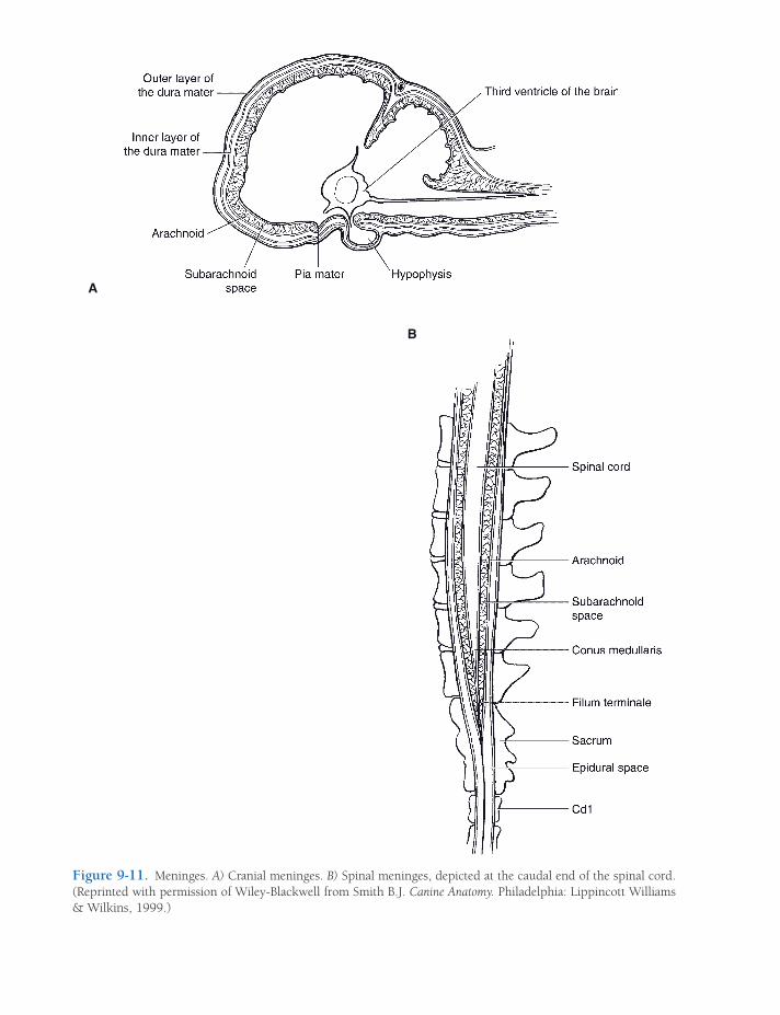
A
B
Figure 9-11. Meninges. A) Cranial meninges. B) Spinal meninges, depicted at the caudal end of the spinal cord. (Reprinted with permission of Wiley-Blackwell from Smith B.J. Canine Anatomy. Philadelphia: Lippincott Williams & Wilkins, 1999.)

ANATOMY OF THE NERVOUS SYSTEM / 161
and arachnoid constitute the leptomeninges (from the Latin word lepto, delicate), refl ecting their fi ne, delicate nature. The space between the two layers, bridged by arachnoid trabecu-lae, is the subarachnoid space. It is fi lled with CSF. It is from this space that CSF is col-lected when a spinal tap is performed.
The dura mater is the tough fi brous outer covering of the CNS. Within the cranial vault the dura mater is intimately attached to the inside of the cranial bones and so fulfi lls the role of periosteum. It also forms the falx cerebri, a median sickle-shaped fold that lies in the longitudinal fi ssure and partially separates the cerebral hemispheres. Another fold of dura mater, the tentorium cerebelli, runs trans-versely between the cerebellum and the cere-brum. In some locations within the skull, the dura mater splits into two layers divided by channels fi lled with blood. These dural sinuses receive blood from the veins of the brain and empty into the jugular veins. They are also the site of reabsorption of CSF through the arachnoid granulations back into the circulation.
The pia mater and the arachnoid of the spinal meninges are much as they are within the cranial vault. The dura mater of the spinal meninges, however, is separated from the peri-osteum of the vertebral canal by a fat-fi lled space, the epidural space.
It is into the epidural space that physi-cians and veterinarians introduce local anes-thetics to produce anesthesia in the caudal parts of the body. This procedure, epidural anesthesia, is often done for obstetric proce-dures. One common application is the injec-tion of anesthetic between the fi rst and second caudal vertebrae of the ox as an aid to repairing and reducing uterine, vaginal, or rectal prolapses.
Spinal Cord
The spinal cord (Figs. 9-12 and 9-13) is the caudal continuation of the medulla oblongata. Unlike that of the cerebrum, the spinal cord’s gray matter is found at the center of the cord, forming a butterfl y shape on cross-section. Fiber tracts, the white matter, surround this core of gray matter. A spinal cord segment is defi ned by the presence of a pair of spinal nerves. Spinal nerves are formed by the con-joining of dorsal and ventral roots, which come together as the nerve at the point where the axons exit and enter the vertebral canal.
Sensory neuronal cell bodies are present in aggregates, called dorsal root ganglia, lateral to the spinal cord. The neurons within these ganglia are pseudounipolar, and they give rise to processes that enter the dorsal horn of the
Sensory Neuron
Dorsal FuniculusCentral Canal
Dorsal Horn
Lateral HornDorsal Root
Dorsal Root Ganglion
Dorsal Branchof Spinal Nerve
Ventral Branchof Spinal Nerve
Spinal Nerve
Ventral Root
Ventral HornVentral Funiculus
Lateral Funiculus
Motor Neuron
Figure 9-12. Cross-sectional anatomy of the spinal cord and spinal nerves.

Figure 9-13. Spinal cord of the dog. View is from dorsal side, with the vertebral arches removed to expose the cord within the vertebral canal. Note the relation of the spinal nerves to the vertebral bodies. The spinal cord is shorter than the vertebral column, so that the more caudal spinal cord segments lie within vertebrae whose name and number are more cranial (e.g., spinal cord segment L6 lies within vertebra L4). The nerve roots are progres-sively longer in these more caudal parts of the vertebral column as they extend from the cord to the foramen of exit. (Reprinted with permission from Fletcher T.F. and Kitchell R.L. Anatomical studies on the spinal cord seg-ments of the dog. A.J.V.R. 1966;27:1762.)
ANATOMY OF THE NERVOUS SYSTEM / 163
spinal cord and others that unite with motor fi bers from the ventral horn neurons to become the spinal nerve extending into the periphery. The processes that extend from the spinal nerve to the spinal cord constitute the dorsal root.
The ventral root of the spinal nerve consists of motor fi bers that arise from the nerve cells primarily in the ventral horn of the spinal cord. The dorsal and ventral roots unite to form the spinal nerve close to the intervertebral foramen between adjacent vertebrae. The dorsal root ganglion is usually very close to this conjoining of dorsal and ventral roots; it frequently can be found just within the intervertebral foramen. In this location it is susceptible to compression when an intervertebral disk protrudes; to a large degree, it is this compression that causes the intense, electric pain associated with disk disease.
Tracts of the Spinal Cord. A tract is a bundle of functionally related axons in the CNS. Tracts that carry sensory information are ascending tracts, whereas those carrying motor commands are descending tracts. The white matter of the spinal cord in which the tracts are found can be roughly divided into three columns on each half of the cord: a dorsal funiculus (often called the dorsal column), a lateral funiculus, and a ventral funiculus.
Sensory Tracts. The dorsal funiculi contain afferent tracts that carry information about body position from joints, tendons, and muscles. This type of sense is called proprio-ception. Injury to this pathway produces unco-ordinated, inaccurate movements, as the cortex lacks some of the information it needs to make ongoing adjustments in the planning and exe-cution of voluntary movements. Such incoor-dination is sensory ataxia.
Proprioceptive information also ascends the spinal cord in several spinocerebellar tracts, located superfi cially on the lateral funiculus. As the name suggests, these tracts are headed for the cerebellum, where the proprioceptive information is used to help shape voluntary movements so that they are accurate and smooth.
Information about pain is carried by a large number of described pathways, but these are often grouped under the names spinothalamic tract or anterolateral system. This large group of fi bers is found in a wide band through the lateral and ventral funiculi. Some of these tracts terminate in the brainstem, where they mediate refl exes associated with painful stimuli. Others make connections that alert the entire cortex and initiate aversive behaviors. Still others are relayed directly to the parts of the cortex that create conscious awareness of the painful stimulus.
Like pain, touch and temperature sense information is carried in a variety of ascending tracts. Some of these are found in the dorsal columns and others in the anterolateral system.
Motor Tracts. Motor systems are often func-tionally grouped into two main categories: a ventromedial motor system, largely located in the ventral funiculus, and a dorsolateral motor system, found in the dorsal part of the lateral funiculus.
The ventromedial motor system primarily is responsible for activity in the axial and proxi-mal limb muscles, especially extensors and antigravity muscles. Activity in the tracts of this system assists with the support phase of gait, when limbs are in weight-bearing position with joints extended. A particularly noteworthy tract of the ventromedial motor system is the lateral vestibulospinal tract, which originates in the region of the pons and medulla.
The dorsolateral motor system is in many ways complementary to the ventromedial system. Dorsolateral tracts tend to control the muscles of the distal limb, especially the fl exors. Activity here is important in the fl exion or swing phase of gait, when limbs are lifted from the ground and advanced while fl exed. In quadrupeds, the most prominent tract in the dorsolateral motor system is the rubrospinal tract, which arises from the midbrain. In primates, and most especially humans, the corticospinal tracts arising from the motor cortex of the cerebrum are especially well developed.
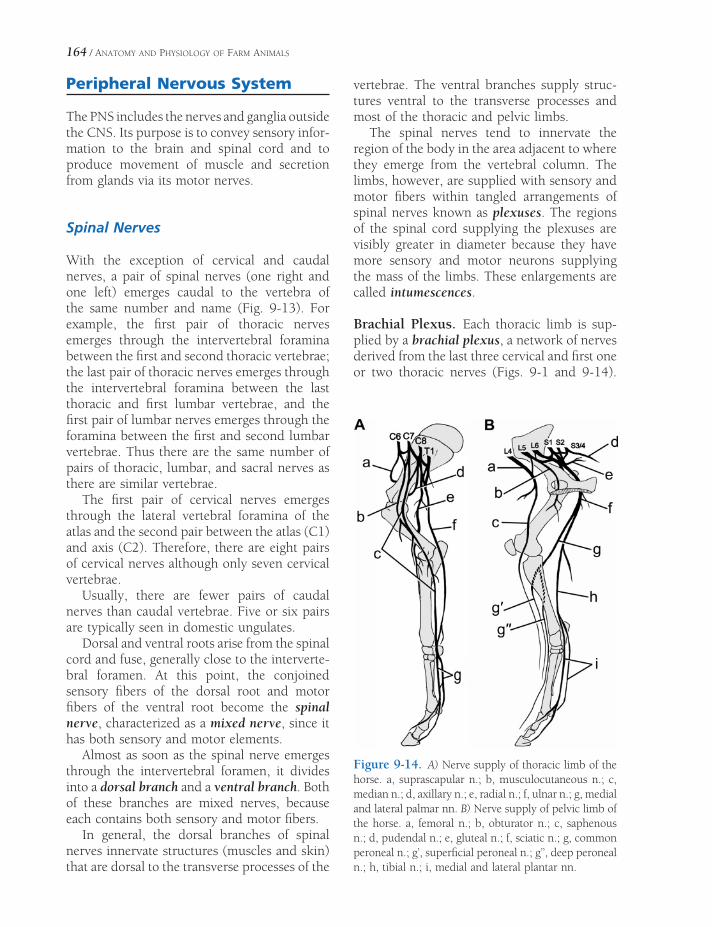
164 / ANATOMY AND PHYSIOLOGY OF FARM ANIMALS
Peripheral Nervous System
The PNS includes the nerves and ganglia outside the CNS. Its purpose is to convey sensory infor-mation to the brain and spinal cord and to produce movement of muscle and secretion from glands via its motor nerves.
Spinal Nerves
With the exception of cervical and caudal nerves, a pair of spinal nerves (one right and one left) emerges caudal to the vertebra of the same number and name (Fig. 9-13). For example, the fi rst pair of thoracic nerves emerges through the intervertebral foramina between the fi rst and second thoracic vertebrae; the last pair of thoracic nerves emerges through the intervertebral foramina between the last thoracic and fi rst lumbar vertebrae, and the fi rst pair of lumbar nerves emerges through the foramina between the fi rst and second lumbar vertebrae. Thus there are the same number of pairs of thoracic, lumbar, and sacral nerves as there are similar vertebrae.
The fi rst pair of cervical nerves emerges through the lateral vertebral foramina of the atlas and the second pair between the atlas (C1) and axis (C2). Therefore, there are eight pairs of cervical nerves although only seven cervical vertebrae.
Usually, there are fewer pairs of caudal nerves than caudal vertebrae. Five or six pairs are typically seen in domestic ungulates.
Dorsal and ventral roots arise from the spinal cord and fuse, generally close to the interverte-bral foramen. At this point, the conjoined sensory fi bers of the dorsal root and motor fi bers of the ventral root become the spinal nerve, characterized as a mixed nerve, since it has both sensory and motor elements.
Almost as soon as the spinal nerve emerges through the intervertebral foramen, it divides into a dorsal branch and a ventral branch. Both of these branches are mixed nerves, because each contains both sensory and motor fi bers.
In general, the dorsal branches of spinal nerves innervate structures (muscles and skin) that are dorsal to the transverse processes of the
vertebrae. The ventral branches supply struc-tures ventral to the transverse processes and most of the thoracic and pelvic limbs.
The spinal nerves tend to innervate the region of the body in the area adjacent to where they emerge from the vertebral column. The limbs, however, are supplied with sensory and motor fi bers within tangled arrangements of spinal nerves known as plexuses. The regions of the spinal cord supplying the plexuses are visibly greater in diameter because they have more sensory and motor neurons supplying the mass of the limbs. These enlargements are called intumescences.
Brachial Plexus. Each thoracic limb is sup-plied by a brachial plexus, a network of nerves derived from the last three cervical and fi rst one or two thoracic nerves (Figs. 9-1 and 9-14).
Figure 9-14. A) Nerve supply of thoracic limb of the horse. a, suprascapular n.; b, musculocutaneous n.; c, median n.; d, axillary n.; e, radial n.; f, ulnar n.; g, medial and lateral palmar nn. B) Nerve supply of pelvic limb of the horse. a, femoral n.; b, obturator n.; c, saphenous n.; d, pudendal n.; e, gluteal n.; f, sciatic n.; g, common peroneal n.; g’, superfi cial peroneal n.; g’’, deep peroneal n.; h, tibial n.; i, medial and lateral plantar nn.

ANATOMY OF THE NERVOUS SYSTEM / 165
The spinal cord enlargement associated with the brachial plexus lies primarily in the caudal cervical vertebrae and is consequently described as the cervical intumescence.
The brachial plexus gives rise to specifi c named nerves that innervate the muscles of the thoracic limb and supply sensation to the same general regions of the skin. Table 9-1 lists the nerves arising from the brachial plexus and the region and muscles supplied by each.
Lumbosacral Plexus. The right and left lum-bosacral plexuses supply nerves to the respec-
tive pelvic limbs (Figs. 9-1 and 9-14). The lumbosacral plexuses are made up of the ventral branches of the last few lumbar and fi rst two or three sacral nerves. The visible spinal cord enlargement here is called the lumbar intumes-cence. The nerves derived from the lumbosa-cral plexus are described in Table 9-2.
Cranial Nerves
Classically, 12 pairs of cranial nerves arising from the brain are described (Fig. 9-15). They
Table 9-1. Nerves of Brachial Plexus
Nerve Muscles Innervated Cutaneous Distribution
Suprascapular Supraspinatus & infraspinatus No sensory fi bers
Pectorals Superfi cial, deep pectoral No sensory fi bers
Subscapular Subscapularis No sensory fi bers
Musculocutaneous Biceps brachii Medial aspect of antebrachium, carpus;
craniomedial aspect of metacarpusCoracobrachialis
Brachialis
Axillary Teres minor et major Shoulder region
Deltoid
Radial Triceps brachii Craniolateral aspect of antebrachium
Anconeus
Extensor carpi radialis
Common and lateral digital extensors
Ulnaris lateralis
Extensor carpi obliquus
Supinator
Ulnar Flexor carpi ulnaris Caudal aspect of antibrachium, craniolateral
aspect of metacarpus, pastern/footDeep digital fl exor
Intrinsic mm of digit (when present)
Median Flexor carpi radialis Caudal metacarpus, pastern/foot
Superfi cial and deep digital fl exor
Pronator teres (when present)
Thoracodorsal Latissimus dorsi No sensory fi bers
Lateral thoracic Cutaneous trunci No sensory fi bers

Table 9-2. Nerves of Lumbosacral Plexus
Nerve Muscles Innervated Cutaneous Distribution
Cranial gluteal Middle and deep gluteal, tensor fasciae latae
No sensory fi bers
Caudal gluteal Superfi cial gluteal No sensory fi bers
Parts of middle gluteal, semitendinosus, biceps femoris in horse
Femoral Sartorius Medial aspect of thigh
Quadriceps femoris
Iliopsoas
Obturator Adductor No sensory fi bers
Gracilis
Pectineus
Obturator externus
Sciatic Semitendinosus et semimembranosus Sensory fi bers arise from distal branches (peroneal & tibial nn.)
Biceps femoris
Obturator internus
Gemelli
Quadratus femoris
Peroneal Tibialis cranialis Dorsal metatarsus and pastern/foot
Extensor digitorum longus et lateralis
Peroneus
Tibial Gastrocnemius Caudal crus, plantar metatarsus, pastern/foot
Flexor digitorum superfi cialis et profundus
Tibialis caudalis
Popliteus
166

Figure 9-15. Ventral view of the canine brain. (Reprinted with permission of Wiley-Blackwell from Smith B.J. Canine Anatomy. Philadelphia: Lippincott Williams & Wilkins, 1999.)
167

168 / ANATOMY AND PHYSIOLOGY OF FARM ANIMALS
Table 9-3. Synopsis of Cranial Nerves
Number Name Type Arises From Function and Distribution
I Olfactory Sensory Olfactory bulb Olfaction (smell); nasal mucosa
II Optic Sensory Diencephalon Vision; retina
III Oculomotor Motor Midbrain Motor to extraocular eye muscles; parasympathetic innervation to iris sphincter and ciliary muscles
IV Trochlear Motor Midbrain Dorsal oblique muscle of eye
V Trigemina Mixed Pons
Ophthalmic division Sensory Sensory to eye and dorsal parts of head
Maxillary division Sensory Sensory to maxillary region, nasal cavity, palates, upper teeth
Mandibular division Mixed Sensory to tongue, lower teeth and jaw; Motor to muscles of mastication
VI Abducens Motor Medulla Lateral rectus and retractor bulbi muscles of eye
VII Facial Mixed Medulla Sensory (taste) to rostral two-thirds of tongue; parasympathetic to salivary and lacrimal glands; motor to muscles of facial expression
VIII Vestibulocochlear Sensory Medulla Hearing (cochlear division) and sense of acceleration (vestibular division)
IX Glossopharyngeal Mixed Medulla Sensory (taste) to caudal third of tongue; parasympathetic to salivary glands; motor to pharyngeal muscles
X Vagus Mixed Medulla Sensory to pharyngeal, laryngeal mucosa, most viscera; parasympathetic to cervical, thoracic and most abdominal viscera; motor to pharyngeal, laryngeal muscles
XI Accessory Motor Medulla & cervical spinal cord
Motor to cervical, shoulder muscles (e.g., trapezius)
XII Hypoglossal Motor Medulla Muscles of tongue
are designated by Roman numerals, numbered from most rostral (I) to most caudal (XII). With the exception of cranial nerves I (olfactory) and II (optic), the cranial nerves arise from the mid-brain, pons, and medulla oblongata and in general resemble ordinary spinal nerves. They do not, however, have discernible dorsal and ventral roots, and some are strictly motor or
sensory (as opposed to spinal nerves, which are all mixed nerves).
Cranial nerve II, the optic nerve, only super-fi cially resembles an actual nerve of the PNS. Its fi bers are actually a tract of the CNS, invested with meninges and with myelin provided by oligodendrocytes. Features of the 12 cranial nerves are outlined in Table 9-3.

ANATOMY OF THE NERVOUS SYSTEM / 169
Autonomic Nervous System
The ANS is the part of the nervous system that regulates activity in viscera and other structures not normally under voluntary control (Fig. 9-16). The common representation of the ANS as a motor subdivision of the peripheral nervous system ignores the facts that (1) sensory fi bers from viscera make up a large proportion of the fi bers in autonomic nerves and (2) some CNS tracts and nuclei integrate and control visceral activity. Nonetheless, for purposes of this intro-duction, we consider only the peripheral motor components of the ANS. These are the nerves that infl uence activity in smooth muscle, cardiac muscle, and glands.
In motor nerves to voluntary muscle, the cell bodies of neurons directly innervating the target are found in the gray matter of the CNS, and the telodendria of these neurons make direct contact with the target. Motor nerves of the ANS, in contrast, consist of a series of two neurons. The fi rst has its cell body in the CNS, and its axon extends into the periphery, where it synapses on the cell body of a second neuron. It is the axon of the second neuron that con-tacts the visceral target. Because of this two-neuron arrangement, autonomic nerves are characterized by the presence of autonomic ganglia, peripheral collections of the cell bodies of the second neurons. Using the autonomic ganglion as a point of reference, the fi rst neuron is called preganglionic, and the second, postganglionic.
The motor output of the ANS is concerned with homeokinesis, the dynamic process of regulating the internal environment to meet the needs of the organism. As a consequence, the motor limb of the ANS is functionally and ana-tomically divided into two parts. The sympa-thetic division of the ANS prepares the organism to meet a stress by producing a combination of physiologic changes that increase available fuel molecules, blood fl ow to muscle, and cardiac output while simultaneously decreasing diges-tive processes. The parasympathetic division of the ANS is in many respects the opposite of the sympathetic division. Parasympathetic activity leads to digestion and storage of fuel molecules
and acts to bring the organism to a state of rest. The physiologic effects of the two divisions are covered more completely in Chapter 10.
Sympathetic Nervous System
Sympathetic nerve fi bers arise from thoracic and lumbar segments of the spinal cord, thus the sympathetic division is sometimes called the thoracolumbar division. Preganglionic sympathetic neurons have their cell bodies in a small lateral horn of the spinal cord gray matter, between dorsal and ventral horns. The myelinated axons of these fi bers leave via the ventral root, enter the spinal nerve, and then leave it just outside the intervertebral foramen to join a longitudinal chain of autonomic ganglia. One string of these ganglia lies on each side of the vertebral column. Each receives pre-ganglionic fi bers from the spinal nerves only in thoracic and lumbar regions, although the chains themselves extend from the cranial cer-vical region to the most caudal parts of the vertebral column. The ganglia, together with the nerve fi bers that link them longitudinally, are called the sympathetic trunk. The ganglia themselves are most correctly called the ganglia of the sympathetic trunk, although they are also called paravertebral or sympathetic chain ganglia. Cell bodies of many of the postgangli-onic sympathetic neurons are found here. From the sympathetic trunk ganglia, the unmyelin-ated axons of postganglionic neurons reach their targets either following spinal nerves or via unique autonomic nerves.
In some cases, preganglionic axons pass through the trunk without synapsing and instead synapse on other sympathetic ganglia outside the sympathetic trunk. This second group, collectively known as prevertebral or collateral ganglia, tends to be associated with the large, unpaired arterial branches of the abdominal aorta, after which they are usually named.
No sympathetic preganglionic cell bodies lie cranial to the thoracic spinal cord, so sympa-thetic innervation to head structures (e.g., the pupil, sweat glands, salivary glands) arrives at

Figure 9-16. The autonomic nervous system. Left) The parasympathetic outfl ow with cranial nerves III, VII, IX, and X and sacral spinal cord segments carrying parasympathetic fi bers. Right) The sympathetic division. Thoracic and cranial lumbar segments make contributions to the sympathetic trunk. These projections are bilateral and are shown on one side only in this drawing for purposes of clarity.

ANATOMY OF THE NERVOUS SYSTEM / 171
its targets by traveling craniad in right and left bundles of fi bers in the ventral neck. These are the cranial continuation of the sympathetic trunks in the thorax. These preganglionic sym-pathetic fi bers are bound in a connective tissue sheath with fi bers of each vagus nerve (cranial nerve X); the combined fi bers are therefore called the vagosympathetic trunk, readily iden-tifi ed dorsolateral to and parallel with the trachea (windpipe). Sympathetic fi bers in the vagosympathetic trunk synapse in the cranial cervical ganglion, found ventral to the base of the skull, and from this ganglion the postgan-glionic fi bers spread to the glands and smooth muscle of the head.
No sympathetic preganglionic cell bodies exist caudal to the midlumbar region, so sym-pathetic innervation to pelvic organs (rectum and urogenital organs) arrives via the right and left hypogastric nerves, a continuation of the caudal parts of the sympathetic trunk. The fi bers of the hypogastric nerve become admixed with parasympathetic fi bers in a diffuse network of autonomic nerves on the lateral surface of the rectum called the pelvic plexus.
The sympathetic innervation to the adrenal gland (see Chapter 11) is unique in that pre-ganglionic sympathetic fi bers synapse directly on the chromaffi n cells of the adrenal medulla without an intervening ganglion. Sympathetic stimulation causes this tissue to release cate-cholamines (epinephrine and norepinephrine) into the bloodstream, producing a widespread, pronounced, and prolonged fi ght-or-fl ight response. This is another physiologically impor-tant site at which the rapid communication system of the body (the nervous system) is integrated with the slow, more lingering com-munication system of the body (the endocrine system).
Parasympathetic Nervous System
The parasympathetic division of the autonomic nervous system arises from cranial nerves and sacral segments of the spinal cord; for this reason,
it is sometimes called the craniosacral division. Fibers of the cranial portion are distributed via four cranial nerves: the oculomotor, facial, glos-sopharyngeal, and vagus nerves. The fi rst three of these supply parasympathetic fi bers to smooth muscle and glands of the head. The vagus nerve supplies parasympathetic fi bers to the viscera of the thorax and neck and to nearly all of the abdominal viscera. The distal part of the diges-tive tract (including the transverse colon and the area caudal to it) and the pelvic viscera are innervated by parasympathetic fi bers from the sacral portion of the parasympathetic nervous system. These pelvic fi bers intermix with sym-pathetic nerves to form the pelvic plexus.
Enteric Nervous System
Neuroanatomists recognize another subdivi-sion of the nervous system, the enteric nervous sytem. This is the network of motor and sensory neurons embedded in the walls of the gastro-intestinal tract and its accessory glands (e.g., pancreas, liver). Activity in the enteric nervous system is infl uenced by the parasympathetic and sympathetic divisions of the ANS, but the system is functional without input from outside the viscera. Two dense networks of neurons are found in the walls of these organs. One, the submucosal (Meissner’s) plexus, is just deep to the inner lining of the gut. The other, the myen-teric (Auerbach’s) plexus, is found within the muscular layer.
Normally the motor output of the ANS con-trols the overall activity of the viscera, but in large part the intrinsic neurons of the enteric nervous system control local events. Sensory neurons monitor local stretch, chemical com-position of the gut’s contents, and gastro-intestinal hormones. Activity in these sensory neurons stimulates local refl ex movements, mediated by the motor neurons of the enteric nervous system. In this way, motility of the gut, dilation of local blood vessels, and secretions can be adjusted to meet the immediate local demands of digestion.


PHYSIOLOGY OF THE NERVOUS SYSTEM
Physiology of the Nerve Impulse
Nerves rapidly transmit information from one body site to another via nerve impulses (i.e., action potentials propagated along the axons of neurons within the nerves). The genesis of a resting membrane potential and the develop-ment of action potentials and their propagation are described in detail in Chapter 2 and are only briefl y reviewed here.
The resting membrane potential of neurons depends mainly on nongated potassium chan-nels in the cell membrane and the electrogenic Na–K–ATPase, or Na–K, pump. The continu-ous exit of potassium from the cell down its concentration gradient and the continuous activity of the pump maintain a difference in charge across the membrane so that the interior is negative relative to the exterior (i.e., resting membrane potential).
The axonal cell membranes of neurons contain voltage-gated sodium channels. These channels are closed at normal resting membrane potentials but rapidly open when the membrane potential is brought to threshold voltage. This results in the inward movement of sodium (down its con-centration gradient) and a large, rapid membrane depolarization (Fig. 10-1). At the peak of the action potential (maximal depolarization), the sodium channels close and some additional voltage-gated potassium channels are open. The
Physiology of the Nerve ImpulseConduction Velocity and Myelination
Synaptic TransmissionNeurotransmittersNeural Control of Skeletal Muscle
Refl exes Involving Skeletal Muscle
Contraction
Voluntary Movement and Its Regulation
Physiology of the Autonomic Nervous System
Regulation of Autonomic Nervous System
Activity
Autonomic Neurotransmitters and Their
Receptors
Regeneration and Repair in the Nervous System
Chapter 10
173

174 / ANATOMY AND PHYSIOLOGY OF FARM ANIMALS
closing of the sodium channels and the exit of additional potassium repolarizes the membrane. The exit of additional potassium produces a small afterhyperpolarization until resting condi-tions can be reestablished (Fig. 10-1).
When an action potential occurs on the axon of a neuron, the membrane potential of adja-cent areas is altered by local movement of charge. Normally, this causes the sodium chan-nels in the adjacent area to reach their thresh-old voltage, and another action potential is elicited. By this means action potentials can be propagated along axons (Fig. 2-16). Propaga-tion normally occurs in only one direction, in part because the sodium channels where the action potential just occurred are refractory to another stimulus. This refractory period is a characteristic of normal sodium channels, and the channels rapidly pass through this period so that they are no longer refractory when another normal impulse arrives.
Conduction Velocity and Myelination
As described in Chapter 9, the axons of neurons may be either unmyelinated or myelinated.
Unmyelinated axons have voltage-gated sodium channels throughout their cell membrane, and thus the propagation of action potentials along unmyelinated axons is as described earlier. The myelin sheath that surrounds myelinated axons contains the lipid sphingo-myelin, which is a good insulator against ionic fl ows. Each Schwann cell covers about a 1-mm distance along the axon, and the junction between two Schwann cells is called the node of Ranvier. No myelin is present at these nodes, and voltage-gated sodium channels are espe-cially prevalent in the axonal cell membrane at the node. Therefore, the circuits of current must fl ow from node to node, and action potentials occur only at the nodes. Since this is analogous to jumping from one node to the next, it is given the name saltatory conduction (from Latin saltare, to jump) (Fig. 10-2). This type of conduction contributes to the increased rate of impulse conduction in myelinated axons.
Conduction velocities of axons also depend on their diameter. Large-diameter axons propa-gate action potentials at higher velocities than do small-diameter axons, because large axons have less internal resistance to the fl ow of current. Large myelinated fi bers, 20 μm in diameter, approach conduction velocities of 250 mph (130 m/sec). At this rate an impulse could travel 6 feet in about 15 m/sec. The smallest unmyelinated fi bers of the body, about 0.5 μm in diameter, conduct at only about 20 inches per second (0.5 m/sec).
Action potential
gNa
gK
resting
Vo
lta
ge
or
Pe
rme
ab
ility
1 msec
Figure 10-1. Change in sodium and potassium per-meability during neuron action potential. The lines labeled gNa and gK show the respective changes in mem-brane permeabilities for sodium and potassium, and the changes are drawn to illustrate their time course relative to the action potential.
MyelinNode of Ranvier
Figure 10-2. Propagation of the action potential in a myelinated axon. Action potentials occur at each node of Ranvier.
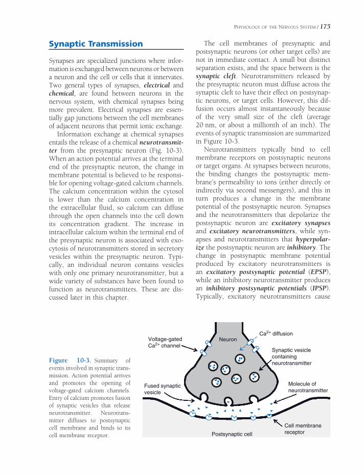
PHYSIOLOGY OF THE NERVOUS SYSTEM / 175
Synaptic Transmission
Synapses are specialized junctions where infor-mation is exchanged between neurons or between a neuron and the cell or cells that it innervates. Two general types of synapses, electrical and chemical, are found between neurons in the nervous system, with chemical synapses being more prevalent. Electrical synapses are essen-tially gap junctions between the cell membranes of adjacent neurons that permit ionic exchange.
Information exchange at chemical synapses entails the release of a chemical neurotransmit-ter from the presynaptic neuron (Fig. 10-3). When an action potential arrives at the terminal end of the presynaptic neuron, the change in membrane potential is believed to be responsi-ble for opening voltage-gated calcium channels. The calcium concentration within the cytosol is lower than the calcium concentration in the extracellular fl uid, so calcium can diffuse through the open channels into the cell down its concentration gradient. The increase in intracellular calcium within the terminal end of the presynaptic neuron is associated with exo-cytosis of neurotransmitters stored in secretory vesicles within the presynaptic neuron. Typi-cally, an individual neuron contains vesicles with only one primary neurotransmitter, but a wide variety of substances have been found to function as neurotransmitters. These are dis-cussed later in this chapter.
The cell membranes of presynaptic and postsynaptic neurons (or other target cells) are not in immediate contact. A small but distinct separation exists, and the space between is the synaptic cleft. Neurotransmitters released by the presynaptic neuron must diffuse across the synaptic cleft to have their effect on postsynap-tic neurons, or target cells. However, this dif-fusion occurs almost instantaneously because of the very small size of the cleft (average 20 nm, or about a millionth of an inch). The events of synaptic transmission are summarized in Figure 10-3.
Neurotransmitters typically bind to cell membrane receptors on postsynaptic neurons or target organs. At synapses between neurons, the binding changes the postsynaptic mem-brane’s permeability to ions (either directly or indirectly via second messengers), and this in turn produces a change in the membrane potential of the postsynaptic neuron. Synapses and the neurotransmitters that depolarize the postsynaptic neuron are excitatory synapses and excitatory neurotransmitters, while syn-apses and neurotransmitters that hyperpolar-ize the postsynaptic neuron are inhibitory. The change in postsynaptic membrane potential produced by excitatory neurotransmitters is an excitatory postsynaptic potential (EPSP), while an inhibitory neurotransmitter produces an inhibitory postsynaptic potentials (IPSP). Typically, excitatory neurotransmitters cause
Figure 10-3. Summary of events involved in synaptic trans-mission. Action potential arrives and promotes the opening of voltage-gated calcium channels. Entry of calcium promotes fusion of synaptic vesicles that release neurotransmitter. Neurotrans-mitter diffuses to postsynaptic cell membrane and binds to its cell membrane receptor.
Voltage-gatedCa2+ channel
Ca2+ diffusion
Fused synapticvesicle
Neuron
Synaptic vesiclecontainingneurotransmitter
Molecule ofneurotransmitter
Cell membranereceptorPostsynaptic cell

176 / ANATOMY AND PHYSIOLOGY OF FARM ANIMALS
membrane depolarization by increasing the membrane’s permeability to sodium, while inhibitory neurotransmitters increase the mem-brane’s permeability to either potassium or chlorine. Hyperpolarization of the cell mem-brane can be brought about by either an increased rate of potassium exit from a cell or an increased rate of chlorine entry into the cell down their respective concentration gradients.
The amount of excitatory neurotransmitter released by a single action potential in a single presynaptic neuron is constant. However, it is also typically insuffi cient to depolarize the postsynaptic neuron to a threshold voltage at which an action potential can be elicited. Therefore, to reach threshold voltage in a post-synaptic neuron, the change in voltage pro-duced by multiple EPSPs must be summated. Summation of EPSPs is classifi ed as either tem-poral or spatial. Temporal summation occurs when a single presynaptic neuron releases neu-rotransmitter repeatedly before the effect of each single release is lost. When effective sum-mation occurs, the additive effects of the mul-tiple releases are enough to produce an action potential in the postsynaptic neuron. Spatial summation occurs when simultaneous or nearly simultaneous neurotransmitter release occurs at more than one synapse on the postsynaptic
neuron. Spatial summation is possible because neuron cell bodies typically have multiple junc-tions with many presynaptic neurons.
After a neurotransmitter has been released and had its desired effect on the postsynaptic neuron, it must be removed to prevent continu-ous stimulation of the postsynaptic neuron. The specifi c mechanisms by which neurotrans-mitters are removed from neural synapses vary among neurotransmitters. However, in general these can be any one or a combination of the following: (1) enzymes in the area of the synapse degrade the neurotransmitter; (2) cell mem-brane transport systems absorb the neurotrans-mitter; or (3) the neurotransmitter diffuses away from the area of the synapse.
Typically, as described earlier, a single neuron and its dendrites contain multiple synaptic junctions and receive synaptic input from multiple presynaptic neurons, so a single neuron may receive impulses from several sources. This pattern or organization is described as convergence (Fig. 10-4). Diver-gence is the opposite: each axon branches so that synaptic connections are made with many neurons (Fig. 10-4). These organizational pat-terns permit information to be widely distrib-uted throughout a neural network (divergence) or permit multiple sources of information to be
Convergence
Divergence
Figure 10-4. Simple patterns to illustrate convergence and divergence in neural networks.

PHYSIOLOGY OF THE NERVOUS SYSTEM / 177
brought to focus on a single neuron for a focused response (convergence). These are very simple illustrations of how the organization of neuronal networks can contribute to the pro-cessing and integration of information. For example, a single neuron can be simultaneously stimulated by excitatory and inhibitory neurotransmitters from different presynaptic neurons (a converging network). The property of spatial summation permits the neuron receiv-ing these converging inputs to integrate the different stimuli and respond appropriately.
Neurotransmitters
Most neurotransmitters can be classifi ed as amino acids, monamines (modifi ed amino acids), or polypeptides. While more than 20 compounds have been proved to function as neurotransmitters (and the list will surely grow), some are especially prevalent through-out the nervous system and should receive individual consideration.
Acetylcholine (derived from the amino acid choline) is the neurotransmitter released at the neuromuscular junction on skeletal muscle (see Chapter 8) by some peripheral neurons of the autonomic nervous system (ANS) (discussed later) and found at many synapses throughout the central nervous system (CNS). Neurons releasing acetylcholine are classifi ed as cholin-ergic, and this term is also applied to synapses for which acetylcholine is the neurotransmitter. Two general classes of acetylcholine (also termed cholinergic) receptors, nicotinic and muscarinic, are found at cholinergic synapses. The enzyme acetylcholinesterase is responsible for rapidly degrading acetylcholine and thus terminating its action at cholinergic synapses.
Norepinephrine is the neurotransmitter used by most peripheral neurons in the sympathetic division of the ANS (discussed later) and at synapses at several sites in the CNS. Presynap-tic neurons and synapses using norepinephrine are termed adrenergic, and this term is also applied to cell membrane receptors that bind norepinephrine. The term adrenergic has been applied to these receptors because epinephrine,
or adrenaline, also binds to these receptors. (Adrenaline is the British term for epinephrine, which is released from the adrenal gland.) Adrenergic receptors may be classifi ed as α1, α2, β1, or β2 according to their relative binding affi nities for various adrenergic agonists. Epi-nephrine and norepinephrine are both classi-fi ed as catecholamines because of their chemical structure and because they are derived from the amino acid tyrosine. Dopamine is another cat-echolamine that functions as a neurotransmit-ter within the central and peripheral nervous systems, and specifi c dopamine receptors also exist.
γ-Aminobutyric acid (GABA) is the most prevalent inhibitory amino acid neurotransmit-ter in the CNS. Binding of GABA to its receptor produces neuronal hyperpolarization (inhibi-tion). Several agents that act as sedatives, tranquilizers, and general muscle relaxants have part of their effect via promoting GABA’s effects on the CNS. These include alcohol, barbiturates, and the benzodiaze-pines (e.g., diazepam and chlordiazepoxide). Some of these agents bind directly to GABA receptors; others seem to facilitate the action of endogenous GABA. The GABA receptors may also be the principal target for some general anesthetics.
Glutamate is the predominant excitatory neurotransmitter in the CNS, and several sub-types of glutamate receptors have been identi-fi ed. One subtype of glutamate receptor, the NMDA receptor (named for the agonist N-methyl-D-aspartate), are found in high concen-trations in areas within the brain that are involved with memory and learning. Stimula-tion of NMDA receptors is believed to bring about long-term potentiation of transmission in neural pathways in these areas.
Neural Control of Skeletal Muscle
As described in Chapter 8, each contraction of skeletal muscle requires stimulation of the muscle by a somatic motor neuron. Somatic motor neurons have their cell bodies within the

178 / ANATOMY AND PHYSIOLOGY OF FARM ANIMALS
CNS, and their axons extend from the CNS to contact skeletal muscle cells at neuromuscular junctions. Each time an action potential passes down the axon of a single motor neuron, all skeletal muscle cells that are innervated by that motor neuron are stimulated to contract. A single motor neuron can stimulate more than one skeletal muscle cell, but a single skeletal muscle cell receives input from only one motor neuron. Thus, all contractions of any single skeletal muscle cell must be initiated by action potentials arriving via the same motor neuron. A clinical classifi cation applied to the motor neuron that extends from the CNS to skele-tal muscle fi bers is lower motor neuron (LMN). (More specifi cally, this is the somatic LMN, but somatic is often omitted in common usage.) Any contraction of skeletal muscle depends on an intact and properly function-ing LMN, and the classic sign of a dysfunc-tional LMN is fl accid paralysis (a total lack of movement and muscle tone of the affected muscle).
Typically, a single neuron receives inputs from multiple other neurons because of conver-gence in neural networks. Therefore, a single LMN is subject to regulation by multiple inputs from other neurons within the CNS. Upper motor neuron (UMN) is the general term applied to a neuron within the CNS that acts to regulate the activity of a LMN. UMNs are responsible for initiating voluntary movement and maintaining relatively stable body posture and position relative to gravity so that volun-tary movements are normal. If the LMNs to a muscle are intact but some of the UMNs that normally regulate those LMNs are dysfunc-tional, both refl ex and voluntary activity of the muscle are possible, but the character of both refl exes and voluntary activity may be abnormal.
Refl exes Involving Skeletal Muscle Contraction
A refl ex action is an automatic, or unconscious, response to an appropriate stimulus. In the case of refl exes involving skeletal muscle, the
response is skeletal muscle contraction. The simplest refl ex requires two neurons. The fi rst is an afferent neuron, which detects the appro-priate stimulus and transmits that information via action potentials to the CNS. Within the CNS the afferent neuron synapses with and stimulates action potentials in an efferent neuron. The efferent neuron exits the CNS and stimulates skeletal muscle contraction. The entire neural circuit from stimulus detection to response is a refl ex arc, and in the simplest case, in which only one synapse is involved, the refl ex arc is a monosynaptic refl ex (Fig. 10-5). Very few refl exes are monosynaptic; usually one or more interneurons are interposed between the afferent and efferent neurons.
Refl exes can be classifi ed according to the segments or parts of the CNS that are required
Quadriceps
Muscle
Patellar
Ligament
Sensory
Neuron
Motor
Neuron
Figure 10-5. Patellar tendon refl ex arc is a monosyn-aptic spinal refl ex.

PHYSIOLOGY OF THE NERVOUS SYSTEM / 179
to complete the refl ex arc or circuit. Among the simplest of refl ex arcs are those that can be completed within segments of the spinal cord (spinal refl exes). A typical spinal refl ex is the patellar tendon refl ex, or knee jerk refl ex (Fig. 10-5). The knee jerk is elicited by tapping the patellar ligament (tendon), which causes the knee or stifl e to extend. The sudden stretch-ing of the muscle stimulates muscle spindles (see Chapter 11) related to the quadriceps muscle. Action potentials are transmitted along axons of primary afferent neurons and into the spinal cord by way of the dorsal root. A synapse occurs between the afferent neuron and appropriate motor neurons in the ventral gray horn of the spinal cord. Action potentials then are transmitted via the efferents to the muscle fi bers of the m. quadriceps femoris of the thigh (in this case the muscle in which the stimulus originated), causing it to contract. This type of stretch refl ex is also known as a postural refl ex because it aids in maintaining a standing position. Sudden fl exing of the stifl e stretches the quadriceps muscle, which initiates the refl ex that causes the stifl e to support the weight of the animal. The same refl ex occurs in humans when the area just below the knee is struck.
The fact that the patellar tendon refl ex arc can be completed within a limited region of the spinal cord does not mean that other segments of the CNS cannot affect the characteristics of the refl ex response. Through the actions of either inhibitory or excitatory UMNs that affect the activity of the LMNs participating in the refl ex arc, the strength or character of the refl ex response can be changed. As described earlier, the patellar tendon refl ex is an example of a postural refl ex that functions to maintain a normal posture and body position. By affecting the character of the refl ex, UMNs can also assist in the maintenance of posture and body posi-tion. More information about the regulation of UMNs is given later in this chapter.
Refl ex center is a general term used to describe the area within the CNS where afferent information is integrated to produce the effer-ent activity or refl ex response. In the case of simple spinal refl exes, the refl ex center is within
the spinal cord (i.e., the synapses are between the afferent and efferent neurons). Refl exes can be quite complex and may require integration by refl ex centers within the brain or brainstem. For example, vomiting is a refl ex that can be stimulated by a variety of diverse stimuli and that requires a series of highly coordi-nated contractions of skeletal muscles in the diaphragm, esophagus, and pharynx. These actions are coordinated by a refl ex center in the brainstem. This center receives afferent information from quite diverse areas (such as the stomach lining and the vestibular apparatus) and stimulates efferent neurons to all participating skeletal muscles. This center can be stimulated pharmacologically by the administration of any of a variety of emetic drugs.
In the dog, spinal refl exes that are used in various types of clinical examinations include the withdrawal refl ex, in which the limb is withdrawn when the footpad or toe is pinched; patellar tendon refl ex, in which extension of the stifl e results from tapping the patellar liga-ment; and extensor postural thrust refl ex, in which the limb extends when the foot is pushed toward the body, as would occur in normal body support.
Attitudinal and postural reactions include the tonic neck refl exes, in which passively extending the neck (raising the head) increases the tone of extensor muscles of the forelimbs and decreases the tone of extensor muscles of the hind limbs. This may be a partial explana-tion for the value of raising a horse’s head to prevent kicking. Conversely, when a horse gets its head down, it is much more likely to kick or buck. The extensor postural thrust enables the limbs to support the weight of the body without conscious thought on the part of the animal.
Refl exes associated with the cranial nerves include the corneal refl ex, in which the eyelids close in response to stimulation of the cornea of the eye, and the palpebral refl ex, in which gentle palpation or tapping of the eyelids causes closure. General anesthesia depresses refl ex responses, and these last two refl exes are often used to evaluate the depth of anesthesia.
180 / ANATOMY AND PHYSIOLOGY OF FARM ANIMALS
Voluntary Movement and Its Regulation
Animals cannot be asked about their intentions or thoughts while moving about. Therefore, much of what is known or assumed about vol-untary movement in animals is based on infor-mation from humans; that is, humans are the experimental animals. Much of the information from humans is based on studies of individuals with naturally occurring diseases or acquired neural defi cits due to trauma. Studies on animals with similar lesions or defi cits suggest that the underlying mechanisms are the same in humans and animals.
Using a variety of techniques and experi-ments, a large area within the cerebral cortex has been shown to be directly linked to motor activity (i.e., the motor cortex). Within this general area, more specifi c areas can be shown to control various groups of skeletal muscles. While this does not mean that neurons from this area make direct synaptic connections with skeletal muscle cells, a functional association between these areas and specifi c skeletal muscles can be demonstrated by eliciting con-tractions of specifi c muscles by stimulation of discrete areas within the motor cortex. Groups of skeletal muscles used for fi ne motor activity (e.g. fi ngers in humans) have a greater area of representation in the motor cortex than groups of muscles used for less-fi ne motor activity (e.g., bending over at the waist). The sizes of the areas in the motor cortex that represent differ-ent regions of the body are quite variable among different animals and between humans and domestic animals. For example, a horse would have a very small area representing the digits, whereas this would be large in humans. This difference is related to the different needs for fi ne motor control of the digits between horses and humans.
The precise linkage between the desire for and initiation of a specifi c voluntary movement (e.g., reaching for, grasping, and lifting a cup of coffee) and how the necessary muscles are appropriately stimulated to bring about that movement is not completely understood. What is understood about normal voluntary move-
ment, and is more relevant to animals, is that it requires a great deal of afferent information before and during the movement, and this information is used for the coordination of vol-untary movements. This afferent information is primarily derived from proprioceptors (special-ized sensory receptors designed to provide information about body position discussed in Chapter 11) in muscles, tendons, and joints throughout the body. This information is vital for coordinated movements. For example, to reach for a cup of coffee and grasp it, the posi-tion of arms, hands, and fi ngers must fi rst be known. When the cup is lifted, continuous adjustments must be made in muscle contrac-tion so that the movement is smooth regardless of the weight of the cup or the amount of coffee in it. Much of the afferent information on body position is received and used by the brain without any conscious awareness.
The cerebellum receives much of the affer-ent information about body position and ongoing movements from proprioceptors. It also receives information about movements ini-tiated in the cerebral cortex. After integrating this and other information, the cerebellum sends efferent information to multiple sites in the brain, including the motor cortex, to coordinate ongoing movements. The cerebel-lum does not initiate movements, but it is essential for normal coordination of voluntary movements.
It is clear that repetition of a pattern or sequence of movements increases the ease with which those movements can be repeated (i.e., training). This indicates that a type of learning can also occur within the neural circuits regulating voluntary movements. After such learning occurs, a series of practiced move-ments are virtually automatic and can be done without any conscious direction. Exactly how and where this learning occurs is not known, but the basal nuclei (see Chapter 9) appear to have special importance in coordinating and enabling the automatic performance of repeated movement patterns. Dopamine is an important neurotransmitter in certain basal nuclei, and Parkinson’s disease in humans is associated with the loss of dopaminergic

PHYSIOLOGY OF THE NERVOUS SYSTEM / 181
neurons in this area of the brain. Patients with Parkinson’s disease exhibit a variety of abnormal movements, and complex move-ment patterns seem to be especially affected. Treatment with L-dihydroxyphenyalanine (L-dopa), a precursor of dopamine, may relieve the symptoms, but it does not prevent further neuronal loss and progression of the condition.
All UMNs have their effects on skeletal muscle activity and movement indirectly by ultimately affecting LMN neuron activity. Because the fi nal common pathway for all movement (refl ex or voluntary) is via the LMN and because the function of UMNs is to regu-late LMN activity, abnormal movements may result from dysfunction of either LMNs or UMNs regulating the activity of a specifi c muscle. The abnormal movement may be primarily voluntary movements, refl ex-based movements, or both. By close observation of abnormal movements and with a detailed knowledge of LMNs, UMNs, and the primary functions of the sites within the CNS from where UMNs originate, clinicians can deter-mine the type and location of dysfunctional neurons causing the abnormal movements.
Physiology of the Autonomic Nervous System
Regulation of Autonomic Nervous System Activity
The ANS functions to maintain a relatively stable internal body environment, that is, to maintain a state of homeokinesis (homeosta-sis). It does so by regulating the activity of cardiac muscle, smooth muscle, and glands. The distribution of ANS nerves is widespread, including all viscera. Normally, the regulation of ANS activity occurs below the level of con-sciousness. However, emotional reactions (such as fear or excitement) and input from the cere-bral cortex also affect ANS activity.
Most organs that are innervated by the ANS have both sympathetic and parasympathetic innervations, and in most cases the effects of
the two divisions are antagonistic. For example, parasympathetic stimulation of the heart reduces heart rate, while sympathetic stimula-tion increases heart rate. The antagonistic actions are controlled to bring about appropri-ate overall regulation of the innervated organ. In the case of the heart, relatively high para-sympathetic nerve activity to the heart in an animal at rest maintains a low resting heart rate. During rest there is little sympathetic nerve activity to the heart. To increase heart rate, such as during exercise, parasympathetic nerve activity is fi rst reduced to permit an increase in heart rate, and sympathetic nerves to the heart are activated if a further increase in heart rate is necessary for more intense exercise.
Changes in ANS nerve activity can occur as a result of discrete refl exes. For example, the diameter of the pupil of the eye is controlled by an ANS refl ex initiated by changes in the amount of light detected by the retina. The circular smooth muscle fi bers of the iris con-strict the pupil and are under the control of parasympathetic nerves. Changes in the amount of light reaching the retina initiate a refl ex response to achieve a precise and proper pupil diameter.
Arterial blood pressure, salivary secretion, secretion of hydrochloric acid by the stomach, urination, and defecation are among functions regulated in part by ANS refl exes. The refl ex centers for various ANS refl ex arcs are found throughout the CNS. For example, the brain-stem has several centers regulating blood pressure, and the refl ex center for urination is in the sacral region of the spinal cord. ANS refl exes may use sympathetic nerves, parasym-pathetic nerves, or both. For example, the ANS refl ex regulation of blood pressure uses sympa-thetic nerves to blood vessels and both para-sympathetic and sympathetic nerves to the heart.
The sympathetic division of the ANS is primarily responsible for the fi ght-or-fl ight response that is associated with fear, anxiety, rage, and other strong emotions. The outcomes of this response can be predicted by imagining what changes would favor skeletal muscle activity for either fi ghting or running away.

182 / ANATOMY AND PHYSIOLOGY OF FARM ANIMALS
Outcomes include increases in heart rate, blood pressure, blood glucose, and blood fl ow to skeletal muscle; dilation of airways in the lungs (e.g., bronchi); dilation of the pupils; and decreased activity of the digestive tract.
The fi ght-or-fl ight response is a short-term event characterized by high levels of sympa-thetic nerve activity throughout the body. This widespread sympathetic activation is not the result of a discrete refl ex but is a more general sympathetic activation initiated in response to fear, anxiety, stress, and so on. The hypothala-mus and amygdala in the brain appear to be especially important sites in the initiation of the sympathetic response, but it is not clear how feelings of fear, stress, and so on affect these centers to initiate the response.
In addition to widespread increases in sym-pathetic nerve activity, the fi ght-or-fl ight response includes an increase in the release of epinephrine and norepinephrine from the adrenal medullae. Chromaffi n cells of the adrenal medullae are innervated by pregangli-onic sympathetic neurons (see Chapter 9), and these cells release their catecholamines when stimulated. In most species the primary cate-cholamine released by chromaffi n cells is epi-nephrine. Epinephrine and norepinephrine in the circulation bind to adrenergic receptors throughout the body to amplify the general effects of increased sympathetic nerve activity. The blood levels of these catecholamines are relatively low and functionally insignifi cant when animals are not undergoing a strong sym-pathetic response.
The list of events in the fi ght-or-fl ight response includes dilation of the pupil of the eye. The control of pupil size was earlier described as also being controlled by a para-sympathetic refl ex based on light reaching the retina. This is an example of how circumstances may affect which division of the ANS domi-nates the regulation of an organ or gland. If an animal is not undergoing a strong sympathetic response, the parasympathetic nerves are the primary regulator of pupil size, but during times of intense stress, sympathetic stimulation may dilate the pupil in spite of the animal being in a brightly lit environment.
Autonomic Neurotransmitters and Their Receptors
The postganglionic neurons of the parasympa-thetic division of the ANS use acetylcholine as a neurotransmitter, whereas almost all postgan-glionic neurons of the sympathetic division use norepinephrine. Acetylcholine is also the neu-rotransmitter used by preganglionic neurons in both the sympathetic and parasympathetic divisions.
The organ response to ANS stimulation depends not only on the neurotransmitter being released but also on the type of cell membrane receptor on the cells of the organ. Muscarinic and nicotinic are the two general classes of acetylcholine receptors. Nicotinic receptors are found on skeletal muscle cells at the neuromus-cular junction and in all autonomic ganglia (both sympathetic and parasympathetic), where acetylcholine is released by preganglionic neurons. Muscarinic receptors are found in most organs innervated by postganglionic para-sympathetic neurons. Stimulation of musca-rinic receptors brings about diverse cellular responses ranging from hyperpolarization of sinoatrial nodal cells to slow heart rate to con-traction of urinary bladder smooth muscle for urination. Table 10-1 lists the organs where muscarinic receptors are found and the organ response to the stimulation of those receptors by parasympathetic nerves. More details on the precise mechanisms of these effects appear in the appropriate chapters on the different body systems. Parasympathetic stimulation increases salivary gland secretion, stimulates gastrointes-tinal motility, slows heart rate, and tends to reduce cardiac output. These are often undesir-able during surgery. Muscarinic receptor antag-onists (such as atropine) are often used as preanesthetic agents to block peripheral muscarinic receptors and reduce these poten-tially harmful effects of parasympathetic stimulation.
Adrenergic receptors, which may be stimu-lated by either epinephrine or norepinephrine, also fall into two general classes, α-receptors and β-receptors. However, because of their physiologic and clinical importance, the sub-
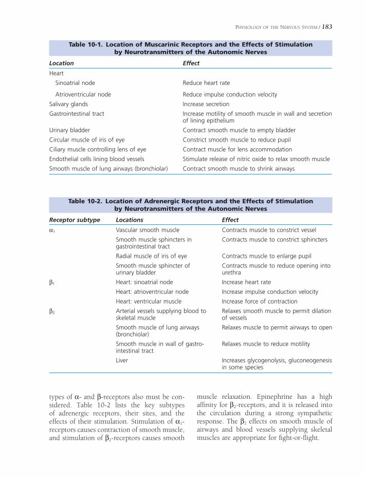
PHYSIOLOGY OF THE NERVOUS SYSTEM / 183
Table 10-1. Location of Muscarinic Receptors and the Effects of Stimulation by Neurotransmitters of the Autonomic Nerves
Location Effect
Heart
Sinoatrial node Reduce heart rate
Atrioventricular node Reduce impulse conduction velocity
Salivary glands Increase secretion
Gastrointestinal tract Increase motility of smooth muscle in wall and secretion of lining epithelium
Urinary bladder Contract smooth muscle to empty bladder
Circular muscle of iris of eye Constrict smooth muscle to reduce pupil
Ciliary muscle controlling lens of eye Contract muscle for lens accommodation
Endothelial cells lining blood vessels Stimulate release of nitric oxide to relax smooth muscle
Smooth muscle of lung airways (bronchiolar) Contract smooth muscle to shrink airways
Table 10-2. Location of Adrenergic Receptors and the Effects of Stimulation by Neurotransmitters of the Autonomic Nerves
Receptor subtype Locations Effect
α1 Vascular smooth muscle Contracts muscle to constrict vessel
Smooth muscle sphincters in gastrointestinal tract
Contracts muscle to constrict sphincters
Radial muscle of iris of eye Contracts muscle to enlarge pupil
Smooth muscle sphincter of urinary bladder
Contracts muscle to reduce opening into urethra
β1 Heart: sinoatrial node Increase heart rate
Heart: atrioventricular node Increase impulse conduction velocity
Heart: ventricular muscle Increase force of contraction
β2 Arterial vessels supplying blood to skeletal muscle
Relaxes smooth muscle to permit dilation of vessels
Smooth muscle of lung airways (bronchiolar)
Relaxes muscle to permit airways to open
Smooth muscle in wall of gastro-intestinal tract
Relaxes muscle to reduce motility
Liver Increases glycogenolysis, gluconeogenesis in some species
types of α- and β-receptors also must be con-sidered. Table 10-2 lists the key subtypes of adrenergic receptors, their sites, and the effects of their stimulation. Stimulation of α1-receptors causes contraction of smooth muscle, and stimulation of β2-receptors causes smooth
muscle relaxation. Epinephrine has a high affi nity for β2-receptors, and it is released into the circulation during a strong sympathetic response. The β2 effects on smooth muscle of airways and blood vessels supplying skeletal muscles are appropriate for fi ght-or-fl ight.

184 / ANATOMY AND PHYSIOLOGY OF FARM ANIMALS
Regeneration and Repair in the Nervous System
In mammals most neurons are fully differenti-ated at birth, although division of glial cells (including those that myelinate axons) contin-ues postnatally. With very few exceptions, neurons are incapable of mitosis, and therefore nerve cells lost to injury or disease are not replaced. Axons, however, may regenerate following injury if the neuronal cell body is healthy.
Axonal regeneration in the CNS does not usually result in recovery of function. It is believed that mature oligodendrocytes prevent regrowth and reestablishment of meaningful neuronal connections. This is why spinal cord injuries are generally irreversible. The partial recovery of function seen over time with some CNS injuries is mostly attributable to recruit-ment of remaining uninjured connections and to the individual’s ability to learn to use those remaining connections.
Axons in the peripheral nervous system (nerves) are frequently injured by crushing (as in forced extraction of a calf during dystocia) or cutting (e.g., wire cuts). Unlike the situation in the CNS, peripheral axons are capable of considerable repair. The likelihood that a given nerve injury will undergo functional recovery is correlated with (1) how close to the nerve cell body the injury is and (2) whether or not the nerve sheath is disrupted. The more proxi-mal a nerve injury, the less likely it is to recover.
In fact, axonal injury that occurs very close to the cell body may result in death of the neuron. Axonal injuries that preserve the supportive tissues of a nerve (e.g., a crushing injury with minimal disruption of the myelin and connec-tive tissue sheath) have a better chance of healing than those in which the epineurium is disturbed (e.g., a cut nerve). Minor injuries to peripheral nerves may regenerate at 2–4 mm a day, whereas more severe injuries may take many months to recover, if they recover at all.
Horses that are anesthetized or restrained in lateral recumbency should have their halter removed to avoid injury to the facial nerve. The buckles of halters are often ventral to the ear, where branches of the facial nerve are close to the skin. Facial paralysis resulting from com-pression of the nerve by a halter buckle may or may not be reversible, depending on the sever-ity and duration of the compressive injury.
Dystocia, or diffi cult birth, is common among fi rst-calf heifers bred to large bulls. The young cow may have diffi culty passing a very large calf through the pelvic canal. Because the obturator nerve passes on the medial aspect of the body of the ilium next to the reproductive tract, it is at risk for compression by the oversized calf. Over-zealous pulling on a large calf can crush the obturator nerves bilaterally, paralyzing the muscles that adduct the pelvic limbs. The cow so affected may become recumbent, with pelvic limbs splayed out laterally. Recovery depends on the severity of the injury.

SENSE ORGANS
Sensory, or afferent, systems are the means by which the nervous system receives
information about the external environment (exteroception), the internal environment (interoception), and the position and move-ment of the body (proprioception). The body uses sensory information to generate refl ex movements (e.g., a blink of the eye when it is touched, withdrawal of a limb from a hot surface, contraction of the bowel when it is stretched) without the participation of the con-scious parts of the brain. Much (but not all) sensory information is also directed to the cere-bral cortex for conscious perception.
“Sensation” is the conscious perception of sensory stimuli. It is impossible to know exactly what an animal (or another person, for that matter) sees, feels, hears, or smells. We infer what sensations an animal may experience by observing its reaction to various stimuli, by identifying homologies between human and animal sensory systems, and by imagining what we might feel in similar situations.
The experience of a given sensation as it is perceived at the cortical level has qualities that make it distinct from other types of sensations. This perceptual distinction defi nes the sensory modality. For example, the stimulus for the photoreceptors of the retina is light; the sensory modality that is experienced when photorecep-tors are stimulated is vision. Somatic sensation or somatosensation describes modalities that
Sensory ReceptorsSomatosensation
Pain
Proprioception
Touch
Visceral SensationsReferred Pain
Chemical SensesGustation
Olfaction
Hearing and BalanceExternal Ear
Middle Ear
Internal Ear
Physiology of Hearing
Mechanisms of Balance
VisionOcular Adnexa
Globe
Visual Field and Light Path
Visual Pathways of the Brain
Chapter 11
185

186 / ANATOMY AND PHYSIOLOGY OF FARM ANIMALS
arise primarily from innervation of body sur-faces and musculoskeletal elements; it includes pain, touch, temperature, and position sense (proprioception). Special senses include smell (olfaction), vision, taste (gustation), hearing (audition), and equilibrium (vestibular sensa-tions). Conscious sensory experiences arising from the viscera are limited primarily to pain, stretch, and pressure.
Certain animals have sensory systems that have no homology in human beings. For instance, migratory birds and some insects are able to sense geomagnetism of the earth, infor-mation they use for navigation. A number of species of fi sh can detect and generate electric fi elds. Other animals exploit the traditional sense modalities for highly specifi c (and expressly nonhuman) purposes. For example, cetaceans and bats both use highly modifi ed auditory circuits for sonar navigation. In spite of growing anecdotal support, however, there remains no reliable evidence that domestic animals can sense an impending earthquake through a sensory experience unavailable to humans.
Sensory Receptors
Sensory experiences begin at receptors, special-ized cells or nerve endings that detect a particu-lar aspect of the internal or external environment. They are the mechanism by which the nervous system changes some sort of environmental energy (e.g., heat, pressure, light) into the electrical activity of neurons, a process called transduction.
In somatosensory systems, the receptor is usually a specialized peripheral terminal of the primary afferent neuron (the sensory neuron extending from the central nervous system [CNS] to the periphery); for the special senses, the receptor is usually a separate specialized neural cell that synapses with the primary affer-ent. As described earlier, sensory receptors may be described by the origin of the stimulus: exteroceptors (external environment), intero-ceptors (visceral organs), and proprioceptors (position and movement sense). They may also
be described on a structural basis as encapsu-lated and nonencapsulated types. Nonencap-sulated, or free (naked) nerve endings are widely distributed and are sensitive primarily to painful stimuli. Encapsulated receptors, which vary widely in structure, are primarily concerned with touch sensations; these recep-tors are invested with specialized connective tissue capsules that impart modality specifi city to the receptor (Fig. 11-1).
The most functionally relevant classifi cation scheme for receptors is based on the type of stimulation to which a receptor best responds. In this system, there are fi ve general types of receptors: (1) mechanoreceptors, which respond to physical deformation; (2) thermo-
A
B
Figure 11-1. Encapsulated and nonencapsulated receptors. A) Naked nerve endings with Golgi stain in respiratory epithelium. These receptors transmit information about pain. B) Pacinian corpuscle, a type of touch receptor, has a connective tissue capsule surrounding the terminus of the primary afferent neuron.

SENSE ORGANS / 187
receptors, which respond to both heat and cold; (3) nociceptors, which respond to stimuli that are potentially injurious to tissue (noxious stimuli); (4) photoreceptors, which are the light receptors of the retina; and (5) chemore-ceptors, which respond to chemical changes associated with taste, smell, blood pH, and carbon dioxide concentrations in the blood. In normal circumstances, each receptor is prefer-entially sensitive to one type of stimulus; the unique stimulus to which a given receptor is most sensitive is called the adequate stimulus for that receptor.
Receptors transduce environmental energy into changes in membrane potential. The ade-quate stimulus produces a local change in mem-brane potential of the receptor, a voltage change known as the receptor potential or generator potential. The receptor potential is usually depolarizing, brought about by the opening of cation channels permeable to Na+ and K+.
The receptor potential is a graded event that spreads passively over the local membrane of the receptor; the amplitude of the potential change and the distance along the membrane that the receptor potential travels are propor-tional to the strength of the stimulus. When the
change in membrane potential reaches a critical level, the threshold, an action potential begins in the trigger zone of the sensory neuron’s peripheral process. This signal is conducted along the axon into the CNS (Fig. 11-2).
Many encapsulated receptors exhibit a phys-iologic characteristic called adaptation. With sustained stimulation, the receptors cease fi ring after their initial burst of activity. When the stimulus is withdrawn, the receptor again responds with a volley of action potentials. In doing so, the adapting receptor signals the beginning and end of the stimulus, rather than fi ring throughout its duration. Typically, touch receptors adapt rapidly; naked nerve endings—nociceptors—as a rule do not adapt but fi re continuously throughout application of a noxious stimulus.
Somatosensation
Pain
Pain is the conscious perception of noxious stimuli. A noxious stimulus is one that is capable of producing tissue damage; it can be
Figure 11-2. Somatosensory neurons are pseudounipolar in nature, with the cell body located in a sensory gan-glion. The adequate stimulus will produce a receptor potential that is proportional to the strength of the stimulus. Once threshold is reached at the trigger zone, an action potential of uniform amplitude and duration is generated and conducted in the axon toward the CNS.

188 / ANATOMY AND PHYSIOLOGY OF FARM ANIMALS
thermal, chemical, or mechanical. The receptor for noxious stimuli is the nociceptor, a naked (not encapsulated) nerve ending.
As a rule, axons transmitting noxious infor-mation are smaller and less myelinated than those carrying tactile or body position informa-tion. Activation of pain fi bers of medium diam-eter and myelination (so-called Aδ fi bers) is associated with a sharp, pricking quality of pain as reported by human beings. Activation of the smallest diameter C fi bers, which are unmy-elinated, produces a dull, burning type of pain. The preponderance of C fi bers in visceral sensory fi bers explains the burning, aching quality of visceral pain.
As indicated in Chapter 9, a number of ascending spinal cord tracts transmit informa-tion about noxious stimuli to brain structures. In addition to projecting to the cerebral cortex for conscious perception, pain pathways typi-cally have strong connections to autonomic centers in the brainstem and parts of the brain that produce increased mental alertness and behavioral and emotional responses to painful stimuli. These connections are responsible for producing signs of sympathetic stimulation (e.g., increased heart and respiratory rates, dila-tion of pupils), emotional responses, and escape behaviors (Fig. 11-3).
The ability of a given noxious stimulus to produce a perception of pain is a highly mutable property that can be modifi ed in the periphery, in the spinal cord, and in the brainstem.
The threshold of nociceptors in the periphery is not a constant. Importantly, many substances released by injured tissues and infl ammatory cells stimulate or lower the threshold of noci-ceptors. Thus, in damaged or infl amed tissue, stimuli that would normally be below thresh-old for detection may produce activity in noci-ceptive afferents. These events contribute to the development of primary hyperalgesia, a phe-nomenon wherein the perception of pain in injured tissues is increased. A dramatic example of this is made by sunburned human skin; the infl ammation of the injured skin lowers thresh-old of nociceptors so that even a light touch (for instance, contact with clothing) can acti-vate them.
Events in the dorsal horn of the spinal cord can also affect transmission of nociceptive information. Rapid, prolonged fi ring of action potentials in the primary afferent neuron can produce changes in the neuron on which it synapses, making it respond more vigorously to subsequent stimulation. This is called wind-up or spinal facilitation of pain.
Activity in spinal nociceptive pathways is also strongly infl uenced by descending antino-ciceptive systems that originate in the brain-stem. The midbrain and medulla both possess a series of midline nuclei that inhibit nocicep-tion via their connections with nociceptive pathways. These nuclei use multiple neurotrans-mitters, most notably endorphins, transmitters with powerful antinociceptive properties.
Cerebral Cortex(Conscious Perception)
Brainstem(Autonomic Centers)
Nociceptor(Naked Nerve Endings)
Muscle
Motor Neuron(Reflex Movements)
Figure 11-3. Divergence in nociceptive pathways. The primary afferent neuron brings information about painful stimuli into the CNS. From there the informa-tion produces refl ex movements and goes to the brain-stem to produce autonomic and other involuntary responses and to the cerebral cortex for conscious perception.

SENSE ORGANS / 189
Stoicism, an apparent indifference to pain, is largely determined by personality and training among both humans and animals. High-strung individuals often exhibit exaggerated reactions to stimuli that scarcely merit attention in more “laid-back” individuals. Interestingly, the effectiveness of applying a twitch to a horse’s upper lip as a method of restraint during mildly painful procedures has long been attributed to redi-recting the horse’s attention from the proce-dure to the moderately noxious stimulus of the twitch. Studies have shown, however, that the correctly applied twitch actually stimulates a release of endorphins, lowers the heart rate, and produces behavioral signs of sedation in the horse. This very old tech-nique of restraint may actually be recruiting some of the neuroanatomic pathways postu-lated to be stimulated by acupuncture.
Proprioception
Proprioception is the nonvisual perception of body position. It is a complex sensory modality that is created through the input of a variety of specialized receptors called proprioceptors. These include joint receptors (providing infor-mation on tension and pressure within joints), muscle spindles (signaling changes in muscle length), Golgi tendon organs (signaling tension in tendons), and skin mechanoreceptors (which report contact with the environment).
Ascending proprioceptive pathways project both to the cerebral cortex and the cerebellum. The cerebral cortex uses proprioception to help formulate voluntary motor plans; the cerebel-lum uses it to adjust ongoing motor movements so that they are smooth and accurate. The information carried in the separate tracts to these targets arise from the same peripheral receptors and primary afferent neurons; it is only the ultimate destination (and therefore use) that is different.
For the cerebral cortex and cerebellum to make effective use of feedback on body posi-tion to guide movements, the proprioceptive information must be delivered very rapidly to
these brain regions. Consequently, propriocep-tive tracts typically have few synapses and are composed of very large diameter, highly myelin-ated axons (called Aa fi bers). In fact, the very fastest (up to 120 m/second) axons of the entire nervous system transmit proprioceptive information.
We, and presumably animals, are generally not aware of proprioception, but it is critically important in the execution of accurate, well-coordinated movements. Injury to the proprio-ceptive pathways results in awkward, inaccurate, uncoordinated gait and movement. The inco-ordination typical of proprioceptive defi cits is referred to as ataxia.
Touch
Touch is the modality associated with non-noxious mechanical contact with the body. Touch receptors are encapsulated, and the axons that transmit touch information to the brain are typically medium in diameter and degree of myelination. Spinal cord tracts associ-ated with touch are found in all the funiculi of the cord.
Visceral Sensations
Visceral sensations involve structures within the body cavities. Most visceral afferent infor-mation is not available to consciousness but is instead important in directing the autonomic activity in viscera. Receptors of the viscera are confi ned to mechanoreceptors and chemore-ceptors. The latter as a rule do not project to cortical levels, so perceived sensations are primarily limited to pain and pressure. Since unmyelinated C fi bers are the predominant type of sensory fi ber innervating the viscera, visceral pain has a burning, aching quality.
Remarkably, the viscera tend to be rela-tively insensitive to stimuli such as crush-ing, cutting, and thermal injury. Surgical manipulation, therefore, tends to produce very little activity in sensory systems. Vis-ceral afferents do respond vigorously to

190 / ANATOMY AND PHYSIOLOGY OF FARM ANIMALS
stretch, dilation, tension, and ischemia (reduced blood fl ow), however. For this reason, cramping (increased muscular tension in the wall of a viscus) and stretch-ing due to gas accumulation are quite painful. The cramping, stretching, and/or ischemia that occur when the equine large intestine twists or is displaced can produce severe abdominal pain, called colic, in horses.
Referred Pain
In humans and presumably in animals, most visceral sensations are ill defi ned and poorly localized. Noxious stimuli originating in viscera, such as might be caused excessive dilation of a viscus, are often perceived as originating instead from a somatic region (body wall or skin), a phenomenon called referred pain. This per-ception is thought to result from the fact that information from that region of viscera con-verges on spinal cord neurons and pathways that also convey information from somatic structures. Since we are much more familiar with sensations arising from our skin and body wall, the pain is interpreted to originate in these somatic structures.
A well-known example of referred pain comes from human medicine; pain due to ischemia of heart muscle is usually perceived as radiating across the left shoulder and down the left arm. This is angina pectoris. A veterinary example may be the high degree of sensitivity in the region of the sternum that some cows exhibit with traumatic peri-tonitis caused by a wire or nail perforating the wall of the forestomach.
Chemical Senses
Chemical senses are those that detect particular molecules in the external or internal environ-ment. Chemical senses that detect molecules outside the body include gustation (taste) and olfaction (smell). Within the body, the chemi-cal senses include the detection of blood pH and carbon dioxide concentration. These latter
afferents are associated with autonomic refl exes and do not project to the cerebral cortex for perception.
Gustation
Taste, or gustation, is the modality associated with dissolved substances contacting special-ized receptor cells on the tongue and throat region. The receptors, simply called taste cells, are arranged in a group with supporting cells, a cluster that constitutes the taste bud (Fig. 11-4). Taste buds are not distributed evenly on the surface of the tongue; they are confi ned to specifi c forms of papillae (tiny projections), the vallate, foliate, and fungiform papillae, which are found on the tongue (the greatest number), soft palate, parts of the pharynx, and epiglottis of the larynx.
Sensory nerve fi bers subserving taste are dis-tributed to the rostral two-thirds of the tongue by a branch of the facial nerve, the chorda tympani. Taste in the caudal third of the tongue is conveyed by fi bers of the glossopharyngeal nerve. Somatic sensations (heat, cold, touch, pain) from the tongue are conveyed by branches
Figure 11-4. Photomicrograph of a taste bud. Indi-vidual cells include taste cells and other non-neural supportive cells. (Micrograph courtesy of Dr. Sue Kinnamon, Colorado State University, Fort Collins, CO.)
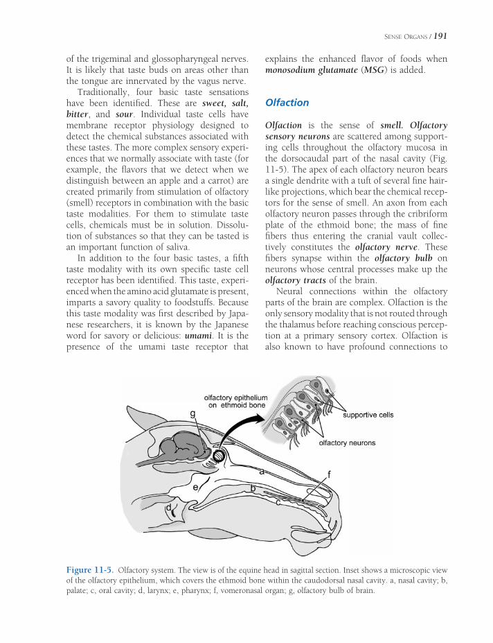
SENSE ORGANS / 191
of the trigeminal and glossopharyngeal nerves. It is likely that taste buds on areas other than the tongue are innervated by the vagus nerve.
Traditionally, four basic taste sensations have been identifi ed. These are sweet, salt, bitter, and sour. Individual taste cells have membrane receptor physiology designed to detect the chemical substances associated with these tastes. The more complex sensory experi-ences that we normally associate with taste (for example, the fl avors that we detect when we distinguish between an apple and a carrot) are created primarily from stimulation of olfactory (smell) receptors in combination with the basic taste modalities. For them to stimulate taste cells, chemicals must be in solution. Dissolu-tion of substances so that they can be tasted is an important function of saliva.
In addition to the four basic tastes, a fi fth taste modality with its own specifi c taste cell receptor has been identifi ed. This taste, experi-enced when the amino acid glutamate is present, imparts a savory quality to foodstuffs. Because this taste modality was fi rst described by Japa-nese researchers, it is known by the Japanese word for savory or delicious: umami. It is the presence of the umami taste receptor that
explains the enhanced fl avor of foods when monosodium glutamate (MSG) is added.
Olfaction
Olfaction is the sense of smell. Olfactory sensory neurons are scattered among support-ing cells throughout the olfactory mucosa in the dorsocaudal part of the nasal cavity (Fig. 11-5). The apex of each olfactory neuron bears a single dendrite with a tuft of several fi ne hair-like projections, which bear the chemical recep-tors for the sense of smell. An axon from each olfactory neuron passes through the cribriform plate of the ethmoid bone; the mass of fi ne fi bers thus entering the cranial vault collec-tively constitutes the olfactory nerve. These fi bers synapse within the olfactory bulb on neurons whose central processes make up the olfactory tracts of the brain.
Neural connections within the olfactory parts of the brain are complex. Olfaction is the only sensory modality that is not routed through the thalamus before reaching conscious percep-tion at a primary sensory cortex. Olfaction is also known to have profound connections to
Figure 11-5. Olfactory system. The view is of the equine head in sagittal section. Inset shows a microscopic view of the olfactory epithelium, which covers the ethmoid bone within the caudodorsal nasal cavity. a, nasal cavity; b, palate; c, oral cavity; d, larynx; e, pharynx; f, vomeronasal organ; g, olfactory bulb of brain.

192 / ANATOMY AND PHYSIOLOGY OF FARM ANIMALS
the limbic lobe, the part of the brain that gener-ates emotional and autonomic behaviors. Smells, therefore, are uniquely capable of elicit-ing emotions and behaviors.
There is a subset of olfactory sensory neurons outside the olfactory epithelium that innervate the mucosa of the vomeronasal organ, a diver-ticulum of the nasal cavity in the hard palate. These olfactory neurons appear to be receptors for pheromones, chemical substances that can infl uence the behavior of other individuals. Pheromones are likely especially important in reproductive behaviors. In spite of vigorous research efforts, no human pheromones have yet been unequivocally identifi ed.
Hearing and Balance
The ear can be divided into three main parts: the external, middle, and inner ears. The exter-nal ear extends from the exterior as far as the tympanic membrane (eardrum). The middle ear begins at the tympanic membrane; it is an air-fi lled space within the temporal bone. The inner ear is housed entirely in the temporal bone, forming an elaborate fl uid-fi lled system of chambers and canals (Fig. 11-6).
External Ear
The part of the ear visible on the outside of the head, the auricle, or pinna, varies considerably in shape and size between and within species. Its appearance is dictated primarily by the shape and rigidity of the auricular cartilage, an elastic cartilage that is somewhat funnel shaped. The pinna acts as a capturing device for air pressure waves, and its shape and mobil-ity are structurally important for sound local-ization. The many skeletal muscles that move the pinna are loosely categorized as rostral and caudal auricular muscles.
A tubular extension of the pinna, the carti-laginous external acoustic canal, extends from the pinna to the tympanic membrane. The external acoustic canal is lined with a modifi ed skin possessing few hairs and abundant seba-
ceous and ceruminous glands. The combined secretion of these glands is called cerumen, a brown waxy substance that protects the canal. A separate annular cartilage adjacent to the funnel-shaped base of the auricular cartilage forms most of the external acoustic canal. A third cartilage, the scutiform cartilage, lies medial to the auricular cartilage, embedded in the rostral auricular muscles. It functions as a fulcrum for the attached auricular muscles.
Middle Ear
The middle ear is an air-fi lled space, the tym-panic cavity, lined by mucous membrane and contained within the temporal bone. In most domestic animals, the middle ear features a ventrally expanded cavity, the tympanic bulla, visible on the ventral surface of the skull. The middle ear is closed to the external acoustic canal by the intact tympanic membrane and communicates with the nasopharynx via the auditory tube (formerly eustachian tube). In the horse, the auditory tube is expanded to form the large air-fi lled guttural pouch dorso-caudal to the nasopharynx. The connection between the middle ear and the pharynx is normally closed except briefl y during swallow-ing. The opening of the auditory tube brought about by swallowing or yawning permits equal-ization of air pressures between middle and external ears.
Three auditory ossicles span the middle ear from tympanic membrane to the vestibular (oval) window. From superfi cial to deep, they are the malleus (hammer), incus (anvil), and stapes (stirrup) (Fig. 11-6). These tiny bones provide a mechanical linkage from the tym-panic membrane to the vestibular window, and through them vibrations are transmitted from the former to the latter. The size and leverage of the auditory ossicles provide mechanical advantage; the energy of pressure waves strik-ing the tympanic membrane is concentrated on the smaller vestibular window, increasing the system’s sensitivity to faint stimuli.
There are also two striated muscles within the middle ear, the m. tensor tympani and the
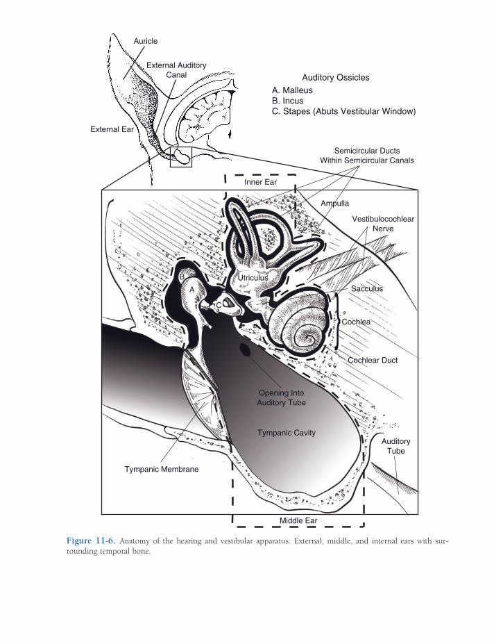
Auricle
External Ear
External AuditoryCanal Auditory Ossicles
A. MalleusB. IncusC. Stapes (Abuts Vestibular Window)
Inner Ear
Middle Ear
Semicircular DuctsWithin Semicircular Canals
Ampulla
VestibulocochlearNerve
Cochlea
Cochlear Duct
AuditoryTube
Tympanic Cavity
Opening IntoAuditory Tube
Tympanic Membrane
A
B
SacculusUtriculus
}C
Figure 11-6. Anatomy of the hearing and vestibular apparatus. External, middle, and internal ears with sur-rounding temporal bone.

194 / ANATOMY AND PHYSIOLOGY OF FARM ANIMALS
m. stapedius. These two small muscles damp vibrations of the auditory ossicles in the pres-ence of excessively loud noises. It is persistent contraction of these muscles that contributes to the temporarily reduced hearing acuity notable after, for example, attending a very loud music concert.
The chorda tympani, a branch of the facial nerve, passes through the tympanic cavity. The chorda tympani carries fi bers for taste from the rostral two-thirds of the tongue and also para-sympathetic fi bers destined for the mandibular and sublingual salivary glands. Infections of the middle ear (otitis media) can be associated with dysfunction of this nerve. Likewise, sympathetic fi bers that innervate the eye pass through the tympanic cavity; diseases affecting the middle ear can produce signs of lost sympathetic inner-vation to head structures (Horner’s syndrome), including a constricted pupil, sunken globe, and drooping of the upper eyelid.
In horses, cranial nerves VII and IX through XII and the continuation of the sym-pathetic trunk pass adjacent to the guttural pouch, separated from its interior only by mucous membrane. Diseases of the guttural pouch can therefore produce distinctive neurologic dysfunctions when these nerves are affected.
Internal Ear
The internal ear is housed entirely within the petrous temporal bone. It is a multichambered membranous sac (the membranous labyrinth) closely surrounded by a sculpted cavity of bone (the osseous labyrinth). The inner ear detects both sound and acceleration of the head (Fig. 11-6).
The membranous labyrinth in the internal ear is a system of fl uid-fi lled sacs and ducts. The primary anatomic features of the membra-nous labyrinth are (1) two enlargements, the utriculus (utricle) and sacculus (saccule); (2) three loops attached to the utriculus, the semi-circular ducts; and (3) the spiraled cochlear duct. These are fi lled with a fl uid, the endo-lymph. The membranous labyrinth is housed
in the osseous labyrinth, a similarly shaped, slightly larger excavation in the petrous tempo-ral bone. The osseous labyrinth is fi lled with perilymph.
The inner ear may be divided into two func-tional parts. In one, the osseous labyrinth forms a coiled, snail shell–shaped cochlea, inside of which is the cochlear duct, an extension of the membranous labyrinth. The cochlear duct houses the receptors for audition. These recep-tors are innervated by the cochlear division of the vestibulocochlear nerve.
The second part constitutes the vestibular apparatus. Within the utriculus, sacculus, and semicircular ducts that constitute the vestibular apparatus are the receptors that detect accelera-tions of the head, including acceleration due to gravity. Accelerations of the head contribute to the sense of equilibrium or balance. The ves-tibular apparatus is innervated by the vestibu-lar division of the vestibulocochlear nerve.
Physiology of Hearing
The environmental energy detected in audition is air pressure waves produced by vibration. These pressure waves can be described in terms of their frequency, the time between peaks of pressure waves, measured in hertz (Hz, cycles per second). Frequency determines the per-ceived pitch of sounds, with higher frequencies producing sounds of higher pitches. Air pres-sure waves are also described in terms of their amplitude, a property that refl ects the energy and consequently the loudness of these waves. Amplitude is expressed in decibels (dB), the units by which loudness is measured. The decibel scale is logarithmic, so that the loudest sounds that can be heard without discomfort (around 100 dB) are a million times as ener-getic as the faintest audible sounds.
The cochlear portion of the osseous laby-rinth resembles a snail shell (cochlea is Latin for snail). The space on the inside of the cochlea is full of perilymph, and it spirals around a central bony core, the modiolus. The corre-sponding part of the membranous labyrinth is the cochlear duct, which extends throughout
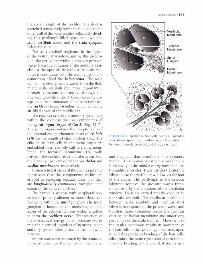
SENSE ORGANS / 195
the coiled length of the cochlea. The duct is stretched transversely from the modiolus to the outer wall of the bony cochlea, effectively divid-ing this perilymph-fi lled space into two: the scala vestibuli above and the scala tympani below the duct.
The scala vestibuli originates in the region of the vestibular window, and by this associa-tion, the perilymph within it receives pressure waves from the vibration of the auditory ossi-cles. At the apex of the cochlea the scala ves-tibuli is continuous with the scala tympani at a connection called the helicotrema. The scala tympani receives pressure waves from the fl uid in the scala vestibuli (but more importantly, through vibrations transmitted through the intervening cochlear duct); these waves are dis-sipated at the termination of the scala tympani, the cochlear (round) window, which abuts the air-fi lled space of the middle ear.
The receptor cells of the auditory system are within the cochlear duct as components of the spiral organ (organ of Corti) (Fig. 11-7). The spiral organ contains the receptor cells of the internal ear, mechanoreceptors called hair cells for the bundle of cilia on their apex. The cilia of the hair cells in the spiral organ are embedded in a relatively stiff overlying mem-brane, the tectorial membrane. The walls between the cochlear duct and the scalae ves-tibuli and tympani are called the vestibular and basilar membranes, respectively.
Cross-sectional views of the cochlea give the impression that the components within are arrayed as repeating separate units, but they are longitudinally continuous throughout the extent of the spiraled cochlea.
The hair cells synapse with peripheral pro-cesses of primary afferent neurons whose cell bodies lie within the spiral ganglion. The spiral ganglion is housed in the modiolus, and the axons of the afferent neurons within it gather to form the cochlear nerve. Transduction of the mechanical energy of air pressure waves into the electrical impulses of neurons in the auditory system takes place in the following manner.
Air pressure waves captured by the pinna are funneled down to the tympanic membrane,
and they put that membrane into vibratory motion. This motion is carried across the air-fi lled cavity of the middle ear by movements of the auditory ossicles. These ossicles transfer the vibrations to the vestibular window via the foot of the stapes. The perilymph in the osseous labyrinth receives the pressure waves trans-mitted to it by the vibrations of the vestibular window. These are carried into the cochlea by the scala vestibuli. The vestibular membrane between scala vestibuli and cochlear duct vibrates in response to the pressure waves and transfers those vibrations across the cochlear duct to the basilar membrane and underlying perilymph of the scala tympani. Movement of the basilar membrane results in movement of the hair cells in the spiral organ that rests upon it, and this produces bending of the hair cells’ cilia against the more rigid tectorial membrane. It is the bending of the cilia that results in a
VestibularMembrane
TectorialMembrane
SpiralGanglion
BasilarMembrane
A
B
C
Figure 11-7. Median section of the cochlea. Expanded view shows spiral organ within: A, cochlear duct; B, between the scala vestibuli; and C, scala tympani.

196 / ANATOMY AND PHYSIOLOGY OF FARM ANIMALS
depolarization within the hair cells. When this depolarization reaches suffi cient intensity, it initiates an action potential in the primary affer-ents of the spiral ganglion.
The ability to discriminate one pitch from another has its basis in a number of anatomic and electrical features of the spiral organ. Most simply, the basilar membrane varies in width from the base of the cochlear duct, where it is most narrow, to the apex, where it is widest. This difference in width means that each portion of the membrane has a different frequency at which it preferentially vibrates, with the widest part of the membrane vibrating at low fre-quency and the narrow part at high.
The axons of the cochlear nerve enter the brainstem with the vestibular nerve at the junc-tion of the medulla and pons and terminate in cochlear nuclei on the lateral side of the medulla. From this fi rst synapse, multiple path-ways produce auditory-mediated refl exes and conscious perception of sound. Auditory infor-mation destined for the cortex ascends bilater-ally in the medulla, pons, and midbrain. The pathways for conscious perception of sound continue on to the thalamic relay nucleus for audition, and from there fi bers project to the primary auditory cortex on the lateral aspect of the cerebrum. The result of bilaterality of the pathways is that auditory information from both cochleae reaches both left and right audi-tory cortices.
The bilaterality of auditory representa-tions in the brain means that for a brain lesion to produce complete deafness, it must affect both sides of the pathway. Such brain injuries are often incompatible with life; therefore, most deafness is peripheral (asso-ciated with the cochlear nerve or internal ear rather than the brain itself). In animals, incomplete loss of hearing is diffi cult to detect.
Disease processes that affect the ability of the tympanic membrane or auditory ossicles to transmit vibrations to the vestibular window produce conduction deafness. Those that affect the spiral organ or more proximal components of the auditory system (includ-ing the cochlear nerves, brainstem, and
auditory cortices) produce sensorineural deafness. Most inherited deafness is sensori-neural, brought about by degeneration of cochlear hair cells. Congenital deafness has been associated with white hair coats or the merle or piebald color genes in a variety of species, including dogs (most notably the Dalmatian), cats, and horses. In these indi-viduals, lack of pigment-containing cells inside the cochleae appears to be linked to degeneration of cochlear hair cells within a few weeks of birth.
Mechanisms of Balance
The vestibular system is a complex neurologic system that is concerned with maintaining a stable orientation in relation to gravity and while in motion. Its infl uence is widely distrib-uted throughout the nervous system; vestibular input is responsible for the refl ex position of eyes, neck, trunk, and limbs in reference to movement or position of the head.
The receptor organs of the vestibular system are housed in the part of the membranous laby-rinth known as the vestibular apparatus (Fig. 11-6). These receptor organs are the maculae of the utriculus and sacculus and the cristae ampullares of the semicircular ducts. Afferent information from these structures gives rise to motor refl exes that maintain stabile visual images on the retinae during movement of the head, to keep the head level with respect to gravity through neck movements, and to produce trunk and limb movements to coun-teract displacements of the head.
The utriculus and the smaller sacculus each posses a macula, a thickened oval plaque of neuroepithelium (Fig. 11-8A). The maculae consist of a population of hair cells very like those of the spiral organ. These are covered by a gelatinous sheet, the otolithic membrane, into which project the cilia of the hair cells. The surface of the otolithic membrane is studded with crystals of calcium carbonate, the otoliths, or statoconia, which increase the inertial mass of the otolithic membrane. When the head accelerates in a straight line, the individual
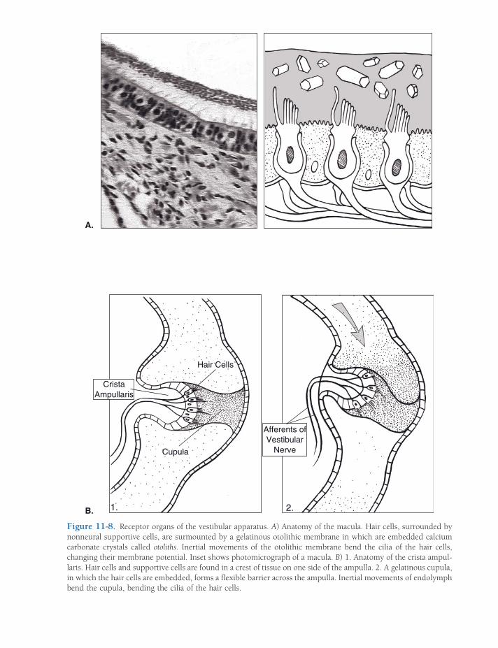
Hair Cells
Afferents ofVestibular
Nerve
CristaAmpullaris
Cupula
.B.A
1. 2.
A.
B.
Figure 11-8. Receptor organs of the vestibular apparatus. A) Anatomy of the macula. Hair cells, surrounded by nonneural supportive cells, are surmounted by a gelatinous otolithic membrane in which are embedded calcium carbonate crystals called otoliths. Inertial movements of the otolithic membrane bend the cilia of the hair cells, changing their membrane potential. Inset shows photomicrograph of a macula. B) 1. Anatomy of the crista ampul-laris. Hair cells and supportive cells are found in a crest of tissue on one side of the ampulla. 2. A gelatinous cupula, in which the hair cells are embedded, forms a fl exible barrier across the ampulla. Inertial movements of endolymph bend the cupula, bending the cilia of the hair cells.

198 / ANATOMY AND PHYSIOLOGY OF FARM ANIMALS
senses linear acceleration. The inertia of the otolithic membrane causes it to lag behind the head under conditions of linear acceleration (including the always present acceleration due to gravity); this dragging of the otolithic mem-brane bends the cilia of the underlying hair cells with shearing vectors dependent on the direction of acceleration.
Attached to the utriculus are three half- circular extensions of the membranous laby-rinth, the semicircular ducts. The ducts lie in three planes at approximately right angles to one another and are designated anterior, poste-rior, and lateral to describe their orientation. As an extension of the membranous labyrinth, each duct is fi lled with endolymph and sur-rounded by perilymph.
One end of each duct is dilated to form an ampulla, within which are housed the receptor organs of the semicircular ducts (Fig. 11-8B). One wall of the ampulla features a transverse ridge of connective tissue, the crista ampulla-ris, which supports a neuroepithelium of hair cells. Attached to the crista is a gelatinous cupula; this extends across the ampulla, forming a fl exible barrier to the fl ow of endolymph. The cilia of the hair cells are embedded in the cupula and are therefore bent by movements of it.
The semicircular ducts detect angular accel-eration (rotation), and their planes of orienta-tion roughly correspond to the X-, Y-, and Z-axes of three-dimensional space. When the head rotates, the semicircular duct lying in that rotational plane moves with it. The endolymph inside the duct, however, must overcome its inertia at the start of rotation and, as a conse-quence, briefl y lags behind the movement of the head. The fl exible cupula, acting as a dam across the ampulla, bulges in response to the endolymph’s push, and in doing so, bends the cilia of the embedded hair cells. With three pairs (right and left) of semicircular ducts detecting movement in the three planes of space, complex rotational movements of the head are encoded in the fi ring patterns of the six cristae ampullares.
Primary afferent neurons synapse with the hair cells of the vestibular apparatus. Their cell
bodies are in the vestibular ganglion, and their axons constitute the vestibular nerve, which joins the cochlear nerve to become the eighth cranial nerve. Most axons in the vestibular nerve synapse in the large vestibular nuclei of the pons and rostral medulla.
Vestibular Refl exes. If there were no mecha-nism to keep the eyes fi xed on a target when the head moved, visual images would continu-ally slip across the retina during head move-ment, and focusing on the visual fi eld would be diffi cult or impossible while the head was moving. The vestibular nuclei use information about acceleration to coordinate extraocular muscle movements with movements of the head and thereby fi x the visual image in one place on the retina as long as possible. When the excursion of the moving head carries the fi xed image out of visual range, the eyes dart ahead in the direction of movement to fi x upon a new image. This new image is held on the retina while the head continues turning until a compensatory jump ahead is again needed. This mechanism results in a cycle of slow move-ment opposite the direction of turn (eyes fi xed on target) followed by a rapid readjustment in the same direction as the turn. This oscillatory refl ex eye movement is called nystagmus.
Nystagmus is a normal refl ex, generated in response to movement of the head. Nystagmus is considered abnormal when it occurs in absence of head movement; this resting (spon-taneous) nystagmus is a sign of vestibular disease.
Axons of some neurons in the vestibular nuclei project caudad in a motor tract that infl uences activity in cervical and upper tho-racic spinal cord segments. These motor con-nections activate neck musculature and forelimb extensors, producing the vestibulocollic refl ex, which generates neck movements and forelimb extension to help keep the head level with respect to gravity and movement.
Some fi bers from the vestibular nuclei form an ipsilateral descending motor tract that extends the length of the spinal cord. This lateral vestibulospinal tract is part of the ven-tromedial motor system (see Chapter 9). Its

SENSE ORGANS / 199
activity has the primary effect of increasing tone in antigravity muscles (proximal limb extensors and axial muscles). This vestibulospinal refl ex uses vestibular information to produce limb and trunk movements that counteract the dis-placement of the head that elicits it. This mech-anism is designed to prevent tilting or falling with shifts in head position.
Animals and people with injury to one side of their vestibular system express the vestibular refl exes in the absence of appro-priate stimuli. These individuals commonly have resting nystagmus, a head tilt, and a tendency to lean, circle, or fall to one side.
Vision
The eye is an elaborate organ whose primary function is to collect and focus light upon the photosensitive retina. It lies within a cone-shaped cavity of the skull, the orbit, which houses the eyeball (globe) and a number of other soft tissue structures, the ocular adnexa (e.g., muscles, glands), that act upon the eyeball in service of its light-collecting function. Unlike the human orbit, which is a complete bony cone, the ventral portion of the orbit of domes-tic species is bounded by soft tissues, notably the pterygoid muscles.
Ocular Adnexa
Palpebrae. Two mobile folds of haired skin protect the anterior aspect of the eyeball. These are the eyelids, or palpebrae. The gap between the margins of the two palpebrae is the palpe-bral fi ssure, and its shape and size are con-trolled by the muscles of the eyelids. A layer of dense connective tissue in the palpebrae, the tarsal plate (tarsus), affords them a certain amount of rigidity. The palpebrae have thin but otherwise typical haired skin on their superfi -cial side. This skin ends at the lid margin with a sharp transition to mucous membrane. The margin may have cilia (eyelashes).
Abundant modifi ed sweat and sebaceous glands are associated with the lid margin. A
row of large modifi ed sebaceous glands, the tarsal glands (meibomian glands), is especially clinically signifi cant, as they commonly become infected, develop tumors, or become impacted. The tarsal glands are present in both palpebrae and open into a shallow furrow near the muco-cutaneous junction of the eyelid. The openings of their ducts are readily seen when the margin of the lid is everted, and the glands themselves are usually visible as yellowish-white columns under the mucous membrane on the inner surface of the lid. The tarsal glands produce an important oil layer of the tear fi lm.
The conjunctiva is the mucous membrane that lines the inside of the palpebrae and part of the anterior surface of the eyeball (excluding the clear cornea). Diseases of the conjunctiva are the most common eye disorders. The con-junctiva is rich in mucus-producing glands, lymphocytes, nerves, and blood vessels. The very small space between the eyelids and the surface of the eye is the conjunctival sac, and it is into this space that eye medications are usually instilled.
Infl ammation of the conjunctiva, or con-junctivitis, is characterized by redness and increased mucus production. Cancer eye, squamous cell carcinoma of the conjunctiva, is relatively common in Hereford, Holstein, and shorthorn breeds of cattle.
Domestic species possess a nictitating mem-brane, or third eyelid. This is a fold of mucous membrane arising from the ventromedial aspect of the conjunctival sac between the eyeball and palpebrae. It is given rigidity by a T-shaped cartilage within it, and it smooths the tear fi lm and protects the cornea.
The third eyelid has at its base a serous gland, called simply the gland of the third eyelid, which normally contributes about 50% of the tear fi lm. Ruminants, the pig, and labora-tory rodents possess another deeper gland associated with the third eyelid. This is the harderian gland. It, too, contributes to the tear fi lm.
Lacrimal Apparatus. The lacrimal apparatus comprises a series of serous, seromucoid, and mucous glands and the duct systems that drain

200 / ANATOMY AND PHYSIOLOGY OF FARM ANIMALS
their secretions from the conjunctival sac (Fig. 11-9). It provides a moist environment for the anterior surface of the eye. The lacrimal gland lies in the dorsolateral portion of the orbit; its secretion, together with that of the gland of the third eyelid, is the major contributor to the tear fi lm. In addition to the lacrimal gland and the gland of the third eyelid, the smaller, more diffuse glands of the conjunctiva and the eyelids contribute oil and mucus to the tear fi lm.
The tears drain from the conjunctival sac via two small openings on upper and lower palpe-brae near the medial corner. These are the superior and inferior lacrimal puncta, which open into short ducts (superior and inferior canaliculi) that join one another at a small sac (lacrimal sac) near the medial corner of the eye that is the origin of the nasolacrimal duct. The nasolacrimal duct travels through the bones of the face to open into the nasal cavity and/or just inside the nostril. Moisture on the nose of domestic animals is largely derived from the tears.
Extraocular Muscles. The globe of the eye moves by the action of seven striated muscles, designated extraocular muscles to distinguish
them from the intraocular muscles that lie entirely within the eyeball (Fig. 11-10).
The m. retractor bulbi arises in the caudal part of the orbit, lateral to the optic nerve, and divides into four fl at muscle bellies that insert on the equator of the eyeball in a nearly com-plete muscular cone. Contraction of the m. retractor bulbi results in retraction of the eyeball into the orbit. Also, a series of four straight muscles originate at the apex of the orbit and project to the equator of the globe, superfi cial to the four bellies of the m. retractor bulbi. They are named for their insertion on the globe: mm. rectus dorsalis, rectus ventralis, rectus media-lis, and rectus lateralis.
The m. obliquus dorsalis (dorsal oblique m.) lies between the dorsal and medial recti muscles and tapers to a thin tendon at the level of the posterior pole of the eyeball. This tendon passes around a small cartilaginous trochlea of cartilage anchored to the medial orbital wall. The trochlea redirects the pull of the tendon, which inserts on the dorsal part of the eyeball. When the dorsal oblique muscle contracts, the dorsal part of the eyeball is pulled toward the medial portion of the orbit (i.e., the left eyeball rotates counter-clockwise and the right rotates clockwise).
Figure 11-9. The lacrimal apparatus. Tears produced by the lacrimal gland and gland of the third eyelid are drained at the medial corner of the eye at superior and inferior lacrimal puncta. The two canaliculi converge at the lacrimal sac, and from there tears fl ow via the nasolacrimal duct to the vestibule of the nostril.

SENSE ORGANS / 201
dorsal rectus m.
retractor bulbi m.
lateral rectus m.optic nerve
ventral rectus m.
ventral oblique m.
OCULAR MUSCLES
levator palpebrae m.
Figure 11-10. Extraocular muscles of the right eye viewed from the medial aspect.
The m. obliquus ventralis (ventral oblique m.) originates from a fossa in the ventral portion of the orbital rim; it runs dorsolaterad, ventral to the insertion of the ventral rectus muscle; and it inserts via two short tendons on the lateral aspect of the globe. The ventral oblique muscle rotates the globe opposite to the rota-tion produced by the dorsal oblique muscle.
One other muscle found within the orbit is not strictly an extraocular muscle, since it does not act on the eyeball itself. The m. levator palpebrae superioris is the primary lifter of the upper eyelid. It arises between the origins of the dorsal oblique and dorsal rectus muscles and inserts via a wide tendon in the connective tissue within the upper eyelid.
The periorbita is a cone-shaped connective tissue sheath that surrounds the eyeball and its muscles, nerves, and vessels. Like the extraocu-lar muscles, the periorbita originates in the apex of the orbit; at the rim of the orbit, it blends with the periosteum of the facial bones.
The periorbita contains circular smooth muscle that squeezes its contents and places the eyeball forward in the orbit. Adipose tissue both within and outside the periorbita acts to cushion the orbital contents. Because of its location behind the eyeball, the adipose tissue is frequently called retrobulbar fat.
Globe
The eyeball (globe) comprises three concentric layers: the fi brous tunic, the vascular tunic, and the nervous tunic (Fig. 11-11). The three tunics of the eyeball surround several chambers fi lled with either liquid or a gelatinous material. The posterior 75% of the globe is fi lled with an acel-lular gel, the vitreous body, and as a consequence this portion is also called the vitreous chamber. The lens divides it from the anterior segment of the eyeball. The anterior segment of the eye is fi lled with a fl uid called aqueous humor.

202 / ANATOMY AND PHYSIOLOGY OF FARM ANIMALS
Figure 11-11. Sagittal section of a bovine eye. (Reprinted with permission of Wiley-Blackwell from Eurell, J.A. and Frappier, B.L. Dellmann’s Textbook of Veterinary Histology, 6th ed. Ames, IA: Blackwell Publishing Professional, 2006.)
Fibrous Tunic. The outer fi brous tunic of the eyeball is made up of a posterior opaque sclera and an anterior transparent cornea. Both are composed of very dense collagenous tissue. The sclera is white, variably tinged gray or blue; it meets the clear cornea at a transitional region called the limbus. The axons of the optic nerve pierce the sclera at the posterior pole of the eye, the area cribrosa. The tough sclera is the site of insertion for the extraocular eye muscles.
The cornea is the transparent anterior part of the fi brous tunic. It is the most powerful refracting layer of the eye (bends light more strongly than even the lens); its transparency and regular curvature are therefore critical ele-ments for focusing of light on the retina. Corneal transparency is a function of (1) lack of vascular elements and cells, (2) lack of pigment, (3) relative dehydration of the collagenous tissue, (4) a smooth optical surface (provided in con-junction with the tear fi lm), and (5) a highly regular, laminar pattern of collagen fi bers that reduces light scatter. The cornea’s anterior and posterior surfaces are covered with specialized epithelium, but most of its thickness is com-posed of collagen fi bers.
Vascular Tunic. The middle tunic of the eyeball, the vascular tunic, or uvea, is com-posed of three parts, the choroid, ciliary body, and iris. The uvea in the posterior portion of the eye is the choroid. It is highly vascular and possesses multiple layers. The deepest (closest to the center of the globe) of these is the tapetum. The tapetum of animals is a refl ective surface that bounces incoming light back onto the retina and enhances vision in low light. The shape and color of the tapetum are variable between species and individuals, but as a rule it is confi ned to the dorsal part of the posterior globe. The ventral portion of the choroid is usually not refl ective. The pig and most pri-mates lack a tapetum.
The ciliary body is the anterior continuation of the uvea. It is a circumferential thickening of the vascular tunic, and it gives rise to many fi ne suspensory ligaments that support the lens. When the muscles in the ciliary muscle con-tract, they allow the lens to assume a more spherical shape; this increases its refractive power, a change that brings close objects into focus on the retina. This process of focusing on near objects is called accommodation. The cap-

SENSE ORGANS / 203
illaries of the ciliary body produce the aqueous humor in the anterior segment of the eyeball.
The iris is the most anterior portion of the uvea and the only part of the vascular tunic normally visible in the living animal. It consists of a pigmented ring of tissue, perforated in its center by the pupil. The iris divides the aqueous-fi lled anterior segment of the eye into anterior (in front of the iris) and posterior (between the iris and lens) chambers. The iris controls the amount of light entering the posterior part of the eye by changing the size of the pupil. Con-strictor muscles, innervated by the parasympa-thetic fi bers of the oculomotor nerve, reduce the size of the pupil when they contract. Dila-tion of the pupil is accomplished by activity of the dilator muscles, innervated by sympathetic nerves.
The iris derives its color from pigmented epithelial cells primarily on the posterior surface of the iris. The typical color distribution depends on the species and is related to the coat color of the individual. In herbivores, the pigmented epithelium of the posterior surface of the iris forms nodular masses along the margin of the pupil that are visible in the ante-rior chamber. These are often very large in horses, in which species they are called corpora nigra. In cattle the same structures are usually smaller and are called iridial granules. These appear to act as something of an “internal visor” for the pupil, shading it from direct sunlight.
The site where the anterior surface of the iris meets the fi brous tunic is the iridocorneal angle (fi ltration angle); there the aqueous pro-duced by the ciliary body is reabsorbed into the venous circulation. Abnormalities here (e.g., congenital narrowing or obstruction due to infl ammation) can prevent normal egress of aqueous from the anterior chamber and result in increased intraocular pressure, a condition called glaucoma.
Nervous Tunic. The deepest layer of the eyeball is the internal (nervous) tunic or retina. The retina develops embryologically as an out-growth of the diencephalon and is therefore composed of neurons and glia. The light- sensitive part of the retina extends from just posterior to
the ciliary body to the site at which nerve fi bers exit the eyeball near the posterior pole of the globe. In clinical practice the retina is routinely viewed through the pupil with an ophthalmo-scope; the portion of the retina that can be viewed in this manner is called the fundus.
The retina has many layers. One of these is the photoreceptor layer, in which are found the specialized neural receptor cells of the visual system, the rods and cones. Each photo-receptor features an external segment that con-sists of orderly stacks of fl attened, disk-shaped plasma membrane. These are replete with phot-opigment, the molecules that are sensitive to photons of light. Photoreceptors have one of two basic shapes: either a tall cylinder or a shorter, tapering stack. The cylindrical photo-receptors are rods, equipped with a photopig-ment that renders them sensitive to very low levels of light. The tapering photoreceptors are cones, each expressing one of three known photopigments with sensitivity to a specifi c wavelength of light. Cones are, therefore, color receptors. As a rule, animals with nocturnal habits have retinas populated primarily with rods, whereas those with diurnal habits (e.g., primates and birds) have more cones, particu-larly in the region that receives light from the center of the visual fi eld.
Most domestic animals have some color vision. Their retinal cones are primarily sen-sitive to blue and yellow wavelengths of light, with few or no red-sensitive cones. Vision in these species is probably rich in blue, green, and yellow tones. The fl apping red cape wielded by the matador therefore attracts the bull by its movement and not by its color.
The photoreceptors synapse with other neurons of the retina, and visual information undergoes early neural processing within the retina. Ultimately, the axons of neurons called ganglion cells project to the optic disk, where they exit the globe and become the optic nerve.
Lens. The lens is a transparent proteinaceous biconvex disk suspended between the posterior chamber and the vitreous chamber. It is
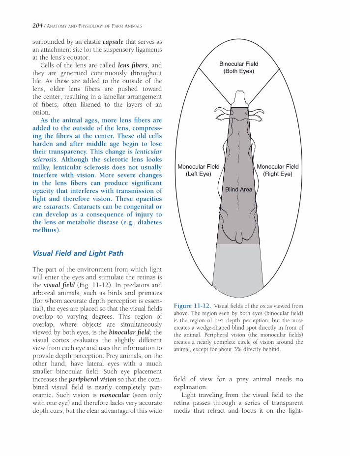
204 / ANATOMY AND PHYSIOLOGY OF FARM ANIMALS
surrounded by an elastic capsule that serves as an attachment site for the suspensory ligaments at the lens’s equator.
Cells of the lens are called lens fi bers, and they are generated continuously throughout life. As these are added to the outside of the lens, older lens fi bers are pushed toward the center, resulting in a lamellar arrangement of fi bers, often likened to the layers of an onion.
As the animal ages, more lens fi bers are added to the outside of the lens, compress-ing the fi bers at the center. These old cells harden and after middle age begin to lose their transparency. This change is lenticular sclerosis. Although the sclerotic lens looks milky, lenticular sclerosis does not usually interfere with vision. More severe changes in the lens fi bers can produce signifi cant opacity that interferes with transmission of light and therefore vision. These opacities are cataracts. Cataracts can be congenital or can develop as a consequence of injury to the lens or metabolic disease (e.g., diabetes mellitus).
Visual Field and Light Path
The part of the environment from which light will enter the eyes and stimulate the retinas is the visual fi eld (Fig. 11-12). In predators and arboreal animals, such as birds and primates (for whom accurate depth perception is essen-tial), the eyes are placed so that the visual fi elds overlap to varying degrees. This region of overlap, where objects are simultaneously viewed by both eyes, is the binocular fi eld; the visual cortex evaluates the slightly different view from each eye and uses the information to provide depth perception. Prey animals, on the other hand, have lateral eyes with a much smaller binocular fi eld. Such eye placement increases the peripheral vision so that the com-bined visual fi eld is nearly completely pan-oramic. Such vision is monocular (seen only with one eye) and therefore lacks very accurate depth cues, but the clear advantage of this wide
Binocular Field(Both Eyes)
Monocular Field(Right Eye)
Monocular Field(Left Eye)
Blind Area
Figure 11-12. Visual fi elds of the ox as viewed from above. The region seen by both eyes (binocular fi eld) is the region of best depth perception, but the nose creates a wedge-shaped blind spot directly in front of the animal. Peripheral vision (the monocular fi elds) creates a nearly complete circle of vision around the animal, except for about 3% directly behind.
fi eld of view for a prey animal needs no explanation.
Light traveling from the visual fi eld to the retina passes through a series of transparent media that refract and focus it on the light-
SENSE ORGANS / 205
sensitive retina of the posterior part of the globe. These dioptric media include the cornea, the aqueous, the lens, and the vitreous body. As indicated before, the cornea is actually the most refractive medium of the eye, but the lens is the only part of the light path with the ability to change its refractive index. This property makes it the organ of accommodation for focus on near objects. Light entering the vitreous chamber of the eye is bent by the more anterior parts of the eye in such a way that the image that is focused on the retina is inverted and reversed.
The site where the ganglion cell axons leave the eye (the optic disk) has no photoreceptors and is therefore considered the blind spot of the retina. In primates, a region dorsolateral to the optic disk is relatively free from large blood vessels and especially densely packed with photoreceptors (particularly cones). This is the region of greatest visual acuity, the macula. Domestic animals lack a macula.
Visual Pathways of the Brain
The electrical information generated by expo-sure of photoreceptors to light undergoes initial neural processing within the retina. This information ultimately leaves the eye via the optic nerve, the fi bers of which are the axons of the ganglion cells of the retina. Most of the axons of the optic nerve synapse in the thalamus, and from there visual information travels to the primary visual cortex in the occipital lobe of the brain (the most caudal part of the cerebral cortex) for conscious perception.
A smaller subset of ganglion cell axons project to other destinations in the brain. Some reach the rostral colliculi of the mesencepha-lon, where visual stimuli induce refl ex move-ments of the eyes and head. Others project to the pretectal nuclei, also in the region of the mesencephalon; these nuclei communicate with the oculomotor nuclei to coordinate the refl ex constriction of the pupils in response to light. Finally, a very small number of ganglion
cell axons project to a specifi c group of cells of the hypothalamus, the suprachiasmatic nucleus. The suprachiasmatic nucleus is the biologic clock, the part of the brain that sets circadian rhythms. Circadian rhythms are physiologic processes that vary regularly on a daily basis; prominent circadian rhythms include sleep–wake cycles, melatonin secre-tion, and body temperature fl uctuations. The suprachiasmatic nucleus has an intrinsic rhyth-micity that closely approximates 24 hours, but the projections from the retina keeps the nucle-us’s cycle entrained to the actual photoperiod of the day.
Many domestic species are seasonal breeders, meaning that their reproductive cycles are determined by the season. The most powerful determinant of the onset and cessation of breeding cycles in these species is the length of the day. The retinal projec-tions to the suprachiasmatic nucleus are the brain’s record of day length, and they there-fore determine the reproductive cycles via their infl uence on the autonomic functions of the hypothalamus. It is common agricul-tural practice to alter breeding behavior by exposing animals to artifi cial light. For instance, in the horse industry, in which an early foaling date is desirable, mares are commonly exposed to artifi cially increased day length in the winter so as to cause these spring breeders to begin fertile estrous cycles earlier than they would if exposed only to natural light.
There is a widely repeated myth among horse trainers that states that visual information from one side of the body is processed strictly on the opposite side of the brain and vice versa. Horse trainers have often cited this “fact” as a rationale for schooling horses from both sides of the body. While it is true that herbivores like horses process a majority of visual information from each half of their visual fi eld in the con-tralateral visual cortex, there are three reasons why this idea is not neurobiologically sound. One is that not all of the visual information from each eye crosses to the contralateral cortex; some is processed on the ipsilateral (same) side.
206 / ANATOMY AND PHYSIOLOGY OF FARM ANIMALS
Secondly, a small portion of the equine visual fi eld is binocular, that is, is seen simultaneously by both eyes. And fi nally, the caudal part of the corpus callosum (the large bundle of axons connecting right and left cerebral hemispheres)
connects the visual cortices of each side so that information is shared between the hemispheres. Trainers must look elsewhere for an explana-tion of why horses benefi t from training on both sides.

ENDOCRINOLOGY
As with the nervous system, the basic func-tions of the endocrine system are commu-
nication and regulation. The classic endocrine system (Figure 12-1) consists of a group of ductless glands that secrete hormones (chemi -cal messengers that function in extremely small concentrations). The hormones circulate throughout the body to bring about physiologic responses. However, this classic description does not account for other types of chemical messengers involved in other types of cell-to-cell communication and regulation. For example, normal endothelial cells of blood vessels release prostacyclin, a prostaglandin (discussed later) that acts locally to inhibit the adhesion of blood platelets. This action helps prevent platelets from forming inappropriate blood clots in normal vessels. When prostacyclin diffuses away from its site of production or is washed away by blood fl ow, it rapidly degrades, so it has no systemic effect. A local effect such as this is termed a paracrine effect. Many types of compounds, including proteins, small peptides, amines (derivatives of amino acids), derivatives of fatty acids, and even a gas (nitric oxide), have been found to function as paracrine agents.
The objective of this chapter is to introduce the basic concepts of endocrinology, including the relationships between the nervous and endocrine systems. More details on specifi c hor-mones and paracrine agents, their regulation, and their actions will be given in later chapters on the specifi c systems that the agents affect.
Hormones and Their ReceptorsChemical Classes of Hormones
Eicosanoids
Hormone Receptors
Cellular Effects of Peptide HormonesCellular Effects of Steroid and Thyroid
HormonesNegative and Positive Feedback RegulationHypothalamopituitary AxisHormones of the NeurohypophysisHormones of the Adenohypophysis
Growth Hormone
Adrenocorticotropic Hormone
Thyroid-Stimulating Hormone
Other Endocrine GlandsParathyroid Glands
Pancreatic Islets
Epiphysis (Pineal Gland)
Chapter 12
207

208 / ANATOMY AND PHYSIOLOGY OF FARM ANIMALS
Hormones and Their Receptors
Chemical Classes of Hormones
The classic hormones can be grouped accord-ing to their chemical structure as peptides, ste-roids, or amines. The amines are biochemical modifi cations of a single amino acid, tyrosine. The synthesis of steroid hormones begins with cholesterol as a substrate. Cholesterol has carbon atoms arranged in adjoining four rings,
and this ring structure is common to all steroid hormones (Fig. 12-2).
Eicosanoids
Eicosanoid is a general term for compounds that are chemical derivatives of long-chain fatty acids. Prostaglandins, thromboxanes, and leu-kotrienes are eicosanoids that function as chemical messengers. Arachidonic acid, a com-
HypothalamusPineal Gland
Pituitary
Adrenal Gland
Ovary
Testis
PancreasThyroid Gland
Parathyroid Glands
Figure 12-1. Approximate location of classic endocrine glands. Both ovary and testis are shown.
HOCH2
C
CH3
H
CH
CH2
CH2
C
CH
CH2
CH
CH
CH2
CH2
CH CH2
CH2C
CH3
CH
C
CH2
CH3
CH2
CH2
C
H
CH3
CH3
Cholesterol:
Figure 12-2. The structure of cholesterol.

ENDOCRINOLOGY / 209
ponent of cell membranes, is the precursor fatty acid in most cases. While these agents are not classic hormones, they are important chemical messengers involved in the regulation of vastly different physiologic functions. In most cases, prostaglandins, thromboxanes, and leukotri-enes act as paracrine agents, for they function near their site of origin and are rapidly metabo-lized after entering the bloodstream. This rapid metabolism in the blood is true for many para-crine agents and is a factor that contributes to their characteristic localized effect.
Prostaglandins have been isolated from nearly every tissue of the animal body, although they derive their name from the prostate gland from which they were originally isolated. There are a number of prostaglandins. Each has a slight difference in chemical structure, and each may have multiple regulatory functions in a variety of tissues. Furthermore, one prosta-glandin may have opposite physiologic effects in different organs. In general terms, prosta-glandins are implicated in regulation of blood vessel diameter, infl ammation, blood clotting, uterine contraction during parturition, and ovulation, among many others. Because pros-taglandins are important in many reproductive functions, these substances will be revisited in Chapter 27.
Leukotrienes are similar in structure to pros-taglandins, being produced from arachidonic acid via a different enzymatic pathway. There are several families of related leukotrienes, each with specifi c functions. Leukotrienes are pro-duced primarily by monocytes and mast cells and are usually associated with allergic reac-tions. Release of leukotrienes increases vascu -lar permeability and induces constriction of airways; in humans, these substances have been implicated in producing some of the most pro-longed manifestations of asthmatic attacks.
Because of the prominent role of prosta-glandins as mediators of infl ammation, drugs that inhibit prostaglandin synthesis are anti-infl ammatory. These are the nonsteroidal anti-infl ammatory drugs, or NSAIDs, of which aspirin and ibuprofen are widely used over-the-counter varieties. The side effects of these drugs (e.g., gastric ulceration and
kidney injury) occur due to the indiscrimi-nate inhibition of both undesirable effects (i.e., infl ammation) and important desirable effects (e.g., maintenance of blood fl ow to stomach and kidneys). Most NSAIDs also inhibit the formation of thromboxanes, because some of the same enzymes are involved in the synthesis pathways for pros-taglandins and thromboxanes. Aspirin reduces the synthesis of thromboxanes for blood clotting, and this is part of the ratio-nale for their use in animals and people to reduce the potential for clot formation. Newer NSAIDs have been developed to inhibit only the production of prostaglan-dins associated with infl ammation.
Hormone Receptors
Only certain specifi c populations of cells respond to an individual hormone. The term target organ is used to identify the tissue whose cells will be affected by a given hormone. Some hormones have multiple target organs, for they affect cells in several sites. For example, both skeletal muscle and liver are among the target organs for insulin. Table 12-1 lists the major endocrine glands, their hormones, and their general effects on their target organs.
Cells within target organs are capable of rec-ognizing and responding to a given hormone because they contain specifi c receptors capable of binding, or forming a chemical union, with the hormone. As described in Chapter 2, these cellular receptors may be components of the cell membrane and have a binding site exposed to the extracellular fl uid, or they may be con-tained within the cytoplasm or nucleus of cells. In either case, a receptor for a specifi c hormone must be present for a cell to respond to the hormone.
The presence and number of receptors within target cells may change in certain condi-tions. Such changes are one way that the bio-logic effect of a given hormone can be regulated. For example, levels of estrogen, a reproductive hormone, increase in circulation shortly before birth (parturition), and this stimulates an

Table 12-1. Major Endocrine Glands, the Hormones They Secrete, and Primary Action and Target Tissue of the Hormones
Endocrine Gland Hormone Action (target tissue or organ)
Hypothalamus CRH Stimulates ACTH release (adenohypophysis)
GnRH Stimulates FSH, LH release (adenohypophysis)
GHRH Stimulates GH release (adenohypophysis)
GHIH Inhibits GH release (adenohypophysis)
TRH Stimulates TSH release (adenohypophysis)
Dopamine (prolactin-inhibiting hormone)
Inhibits prolactin release (adenohypophysis)
Oxytocin and antidiuretic hormone synthesized in hypothalamus; stored, released from neurohypophysis
Adenohypophysis (anterior pituitary)
ACTH Stimulates cortical development, gluco-corticoid release (adrenal cortex)
FSH Stimulates follicular development (ovary), sperm development (testes)
LH Stimulates ovulation, development of corpus luteum, secretion by corpus luteum (ovary), secretion of androgens (testes)
GH Promotes growth in immature animals; metabolic effects on carbohydrate, lipid, protein metabolism in adults
TSH Stimulates release of thyroid hormones (follicular cells of thyroid gland)
PRL Promotes lactation (mammary gland), maternal behavior (central nervous system)
Neurohypophysis (posterior pituitary)
Oxytocin Stimulates uterine contraction, milk let-down (uterus, mammary glands)
ADH Conserves water, reduces urine volume (kidney); constricts vessels to raise blood pressure (arterioles)
Adrenal cortex Glucocorticoids Essential for normal response to stress; important roles in protein, carbohydrate metabolism (multiple organs including liver)
Mineralocorticoids (aldosterone)
Conserve Na, eliminate K (kidney)
Adrenal medulla Epinephrine, norepinephrine Augments sympathetic response to stress by actions on several organs
Thyroid follicular cells
T4, T3 Increases oxygen consumption, ATP generation (almost all cells)
Thyroid para-follicular cells
Calcitonin Promotes calcium retention (bone)
Parathyroid PTH Promotes increase in plasma calcium, reduction in plasma phosphate (bone, kidney)

ENDOCRINOLOGY / 211
Endocrine Gland Hormone Action (target tissue or organ)
Pancreatic islets: β-cells
Insulin Promotes glucose uptake; protein, lipid synthesis by various tissues, organs, including skeletal muscle, liver, adipose tissue
Pancreatic islets: α-cells
Glucagon Promotes glycogenolysis, gluconeogenesis (liver)
ACTH, corticotrophin; ADH, antidiuretic hormone, also called arginine vasopressin; CRH, corticotropin-releasing hormone; FSH, follicle-stimulating hormone; GH, growth hormone; GHIH, growth hormone–inhibiting hormone; GHRH, growth hormone–releasing hormone; GnRH, gonadotropin-releasing hormone; LH, luteinizing hormone; PRL, prolactin; PTH, parathyroid hormone; TRH, thyrotrophin-releasing hormone; TSH, thyrotrophin.
Table 12-1. Continued
increase of oxytocin receptors in the smooth muscle of the uterus. The increase in oxytocin receptors prepares the uterus so that oxytocin can promote uterine contractions when released during parturition. Without the increase in oxytocin receptors stimulated by estrogen, oxy-tocin release itself would not provide adequate uterine contractions for normal parturition. An increase in receptors on target cells is termed up-regulation, and a decrease is termed down-regulation.
Cellular Effects of Peptide Hormones
The receptors for peptide hormones are found in the cell membrane. Peptides cannot freely diffuse through the lipid bilayer of the cell membrane, so their receptors must be in the outer cell membrane to be available to the hor-mones in the extracellular fl uid. As described in Chapter 2, the binding of the hormone with the membrane receptor is the fi rst step in a series of events that brings about changes in the target cell. The subsequent events, which vary with the participating peptide hormones, include changing the permeability of mem-brane channels, stimulating or inhibiting the activity of membrane-bound enzymes, and stimulating or inhibiting the activity of intracel-lular enzymes.
When enzymatic activity is increased by a hormone–receptor interaction, the intracellular
concentration of the product of the action of that enzyme increases. For example, many peptide hormones activate the enzyme adeny-lyl cyclase, which increases the intracellular production of cyclic AMP (cAMP) by its action on adenosine triphosphate (ATP). The cAMP activates other intracellular enzymes that ulti-mately bring about the characteristic biologic response to the hormone (e.g., cellular secre-tion, cellular contraction, protein synthesis). The term second messenger is a general term for the intracellular compounds, such as cAMP, that function as an intermediate in the sequence of steps leading to the biologic response. Two other common second messengers involved in the cellular response to peptide hormones are diacylglycerol (DAG) and inositol triphos-phate (IP3). These second messengers are formed by the action of a membrane-bound enzyme, phospholipase C, on phospholipids in the cell membrane.
Ionic calcium (Ca2+) concentration in the cytosol is normally lower than that in typical extracellular fl uid, but some cells accumulate even higher concentrations of Ca2+ within their endoplasmic reticulum. Increases in cytosolic Ca2+ concentrations may result from the entry of extracellular Ca2+ through membrane chan-nels or by Ca2+ diffusing from within the endo-plasmic reticulum into the cytosol through channels within the membrane of the endoplas-mic reticulum. Increases in intracellular free Ca2+ above the typical low levels may also act as a second messenger for certain hormones.

212 / ANATOMY AND PHYSIOLOGY OF FARM ANIMALS
The increase in Ca2+ begins a series of events that ultimately results in changes in intracellu-lar enzymatic activity and a biologic response.
In many cases the biologic response to peptide hormones is rapid and relatively quickly reversed. For example, the action of antidiuretic hormone on cells in the kidney to change their permeability to water can occur within a matter of minutes. Such rapid effects are possible because pathways leading to the biologic effects may only require activation of enzymes (pro-teins) that are already in the cell. When the hormone is removed or degraded, the effects are reversed by the inactivation of these enzymes.
In some cases, the biologic response to peptide hormones is longer lasting because the intracellular pathways lead to an increase in DNA transcription and the formation of mes-senger RNA (mRNA). In these cases, the effect is prolonged by the presence of newly synthe-sized intracellular proteins. The general term for agents that directly infl uence DNA tran-scription is transcription factor. So peptide hormones may bring about changes in tran-scription factors within cells.
Cellular Effects of Steroid and Thyroid Hormones
Receptors that bind steroid and thyroid hor-mones are found either in the cytosol or in the nucleus. Steroid and thyroid hormones are lipid soluble and can reach these intracellular recep-tors by diffusing into the cell through the cell membrane. The receptor with its bound hormone acts as a transcription factor for specifi c genes within the DNA to increase or decrease the for-mation of mRNA that corresponds to the specifi c gene. Ultimately, the change in mRNA results in a change in protein production, and this brings about the biologic response.
Biologic responses to steroid and thyroid hormones typically develop more slowly but last longer than responses to peptide hormones. In part, this is because all effects are related to changes in protein synthesis. The synthesis of new protein or the degradation of protein already present requires more time than the
activation or inactivation of enzymes already present.
Negative and Positive Feedback Regulation
Assuming that adequate numbers of function-ing receptors are available, the biologic effect of any hormone is directly proportional to the concentration of the hormone in the body fl uids available to bind to the receptors. This concentration is primarily determined by two factors: the rate of hormone release from endo-crine cells and the rate of elimination from the body fl uids. In normal conditions, the concen-tration is typically determined by the rate of release. Peptide hormones and amines are stored in secretory granules by endocrine cells so that they are readily available for release. Steroid hormones are not stored and must be synthesized just prior to release.
The release, and thus the plasma concentra-tion, of most hormones is controlled by some type of negative feedback regulation. In this type of regulation, the rising levels of the hormone bring about a biologic response that inhibits further hormone release. For example, β-cells in the pancreatic islets are directly affected by the concentration of glucose in the body fl uids. An increase in glucose concentra-tion causes the β-cells to increase their release of insulin. One effect of insulin is to promote the uptake of glucose by skeletal muscle cells. As glucose is removed from the body fl uids, the stimulus for insulin release is removed, and this has a negative effect on insulin release. This negative feedback regulation of insulin release is a major factor in determining a normal plasma concentration of glucose.
The negative feedback regulation of insulin by changes in plasma glucose is a relatively simple and straightforward feedback loop. The plasma constituent, glucose, being regulated by the hormone, insulin, has a direct effect on the cells releasing the hormone. However, negative feedback loops can be quite complex and have multiple organs in the loop. Some of the more complex loops involve the hormones regulating

ENDOCRINOLOGY / 213
reproduction in domestic animals and the hypothalamus, the anterior pituitary gland, and the gonads. These are discussed further in Chapters 25 and 27.
A second type of feedback regulation, that is seen much less frequently than negative feed-back, is positive feedback regulation. In this case, the hormone brings about a biologic response that produces a further increase in the release of the hormone. This type of regulation is unusual, and it is not designed to maintain a stable or homeostatic level of some activity or blood constituent. One of the few examples of this type of regulation is the relationship between oxytocin release and dilation of the uterine cervix. An increase in oxytocin release is associated with dilation of the uterine cervix during parturition (details in chapters on repro-duction), and oxytocin acts on the smooth muscle of the uterus to increase uterine con-tractions. When the cervix dilates during par-turition and oxytocin is released, the contractions of the uterus move the fetus out of the uterus through the cervix. This further dilates the cervix, providing a greater stimulus for secre-tion of oxytocin. The overall effect is to expel the fetus when the cervix is dilated.
Hypothalamopituitary Axis
The hypothalamus is ventral to the thalamus in the diencephalon and forms the fl oor and part of the wall of the third ventricle (see Chapter 9). The pituitary gland, or hypophysis cerebri, is attached to its base by the infundibulum, a stalk of nervous tissue (primarily axons). Cell bodies of neurons whose axons form the infun-dibulum are found in the hypothalamus, and their termini abut on the capillaries in the neural part of the pituitary gland (neurohy-pophysis, posterior pituitary, or pars nervosa). Associated with the infundibulum is a unique system of arterioles and capillaries called the hypothalamohypophysial portal system. This system is a true vascular portal system in that blood from a capillary network in the hypo-thalamus fl ows through portal vessels (similar to veins) to the glandular portion of the pitu-itary (adenohypophysis, anterior pituitary, or pars distalis), where it enters a second capillary network (Fig. 12-3). During development, the pituitary gland forms by the conjoining of a ventral outpouching of the diencephalon (the future infundibulum and neurohypophysis) and a diverticulum, Rathke’s pouch, which
Supraoptic & Paraventricular Nucleiof Hypothalamus
Artery
AnteriorPituitary
Capillary Plexus
Posterior Pituitary
Portal Vessels
Capillary Plexus
AnteriorPituitary
Artery
Vein
B.A.
Figure 12-3. Relation between hypothalamus, neurohypophysis, and adenohypophysis. A) Drawing illustrates network of portal vessels. B) Drawing illustrates direct neural connection between hypothalamus and neurohypophysis.

214 / ANATOMY AND PHYSIOLOGY OF FARM ANIMALS
arises from the nearby dorsal ectoderm of the pharynx. The cells of Rathke’s pouch become the adenohypophysis.
The neurotransmitters released by hypotha-lamic neurons whose termini end in the neuro-hypophysis enter the blood and are carried to distant sites to function as systemic hormones (Fig. 12-3). These peptides are manufactured by the neuronal cell bodies within the hypo-thalamus, transported via axons into the neuro-hypophysis, and released directly into blood vessels when action potentials arrive at the telodendria.
Other hypothalamic neurons release neuro-transmitters that are carried from the hypo-thalamus to the adenohypophysis via the hypo thalamohypophysial portal system (Fig. 12-3). There, these neurotransmitters act on endocrine cells either to stimulate or to inhibit the release of other hormones (Table 12-1). While these neurotransmitters do not travel to a distant site to stimulate target cells, they are transmitted via the blood from their site of origin to their site of action. Thus, these neu-rotransmitters may be considered hormones, but they are also referred to as inhibiting or releasing factors. All of these neurotransmitter hormones except dopamine are small peptides, and the small peptides are rapidly degraded after passing through the hypothalamohy-pophysial portal system and entering the general circulation. The unique portal system permits the delivery of a relatively high concentration of the releasing or inhibiting factors to the adeno-hypophysis so that a biologic effect is possible.
The different cell types of the adenohypoph-ysis exhibit different histologic staining charac-teristics, depending on the hormone they produce. As a consequence, cells are character-ized as basophils, acidophils, or chromophobes, among other specifi c types. These endocrine cells are arranged in cords or clusters around blood-fi lled sinusoids, in keeping with their role as an endocrine organ. The neurohypoph-ysis has the typical microscopic appearance of nervous tissue, consisting of unmyelinated axons and supportive glial cells.
Historically, the pituitary gland was known as the master gland because of the large number
of hormones secreted and their wide-ranging effects. Several hormones from the adenohy-pophysis stimulate distant endocrine glands to increase production of their own hormones (Table 12-1). These stimulatory adenohypoph-yseal hormones are often called trophic or tropic hormones.
The hypothalamus functions as a crucial interface between the nervous and endocrine systems, where sensory information is inte-grated and used to regulate the endocrine output of the pituitary gland. Much of this information is related the status of the internal environment in question (e.g., extracellular fl uid osmolality, blood glucose concentration, body temperature and metabolic rate). Release of adenohypophyseal hormones can also be regulated through more direct negative feed-back loops based on the blood concentrations of the hormones involved. The hormone pro-duced by the target endocrine gland of a spe-cifi c trophic hormone can act on (1) the hypothalamus to reduce the production of its releasing factors and (2) the adenohypophysis to reduce its release of the tropic hormone (Fig. 12-4). The tropic hormones of the adeno-
TargetGland
Pituitary
Environmental Factors
Hypothalamus
TrophicHormone
(+)
( )
( )( )
(+)
Hormone
(–)
Target Gland
Figure 12-4. Potential feedback loops to regulate hypothalamic releasing hormones and tropic hormones from adenohypophysis. Stimulation of secretion indi-cated by (+) and inhibition indicated by (−).
ENDOCRINOLOGY / 215
hypophysis may also reduce hypothalamic releasing factors via a short negative feedback loop.
A synopsis of the hormones released into the general circulation by the hypophysis, the factors that regulate their release, and their general functions is presented here. Further details on these functions will be given in sub-sequent chapters when the function of their target organs is covered. All of these hormones are peptide or protein hormones based on their chemical structure.
Hormones of the Neurohypophysis
Two hormones, oxytocin and antidiuretic hor-mone, are released by the neurohypophysis.
Oxytocin is produced by the neurons of the paraventricular nucleus of the hypothalamus. When released into the bloodstream by their axon terminals, it induces contraction of target smooth muscle fi bers of the mammary gland and the uterus. As a result of its actions in the mammary gland, oxytocin aids in the phenom-enon of milk let-down, wherein suckling stim-ulates ejection of milk from the duct system of the gland.
Physical stimulation of the mammary gland (e.g., suckling) is a strong stimulus to secretion of oxytocin, but it is common for other sorts of stimuli to condition a refl ex release of the hormone. For example, dairy cows frequently learn to associate the milking parlor and the preparations for milking with milk let-down. Even before the mammary gland is handled in any way, oxy-tocin is released and milk may be seen drip-ping from the teats.
In the pregnant uterus, oxytocin acts on the myometrium (muscle of the uterus) to produce uterine contractions for expulsion of the fetus at parturition. Stretching of the cervix by the fetus stimulates further secretion of oxytocin, which then stimulates greater uterine contrac-tions. Oxytocin is sometimes administered by injection at parturition to enhance uterine contractions.
Antidiuretic hormone (ADH) is produced by neurons of the supraoptic nucleus in the hypothalamus and released from the neurohy-pophysis in response to increases in blood osmolality (concentration of dissolved sub-stances) or severe decreases in blood pressure, both of which are infl uenced by the animal’s hydration status. The water-retaining effects of ADH on the kidney are discussed in Chapter 23. ADH also produces constriction of blood vessels, an effect that gives the hormone its other name, vasopressin or arginine vasopres-sin (the form found in most mammals).
Hormones of the Adenohypophysis
Growth Hormone
The release of growth hormone (GH), also called somatotropin or somatotropic hormone, from the adenohypophysis is regulated by hypothalamic factors that either stimulate (GH-releasing hormone, or GHRH) or inhibit (GH release–inhibiting hormone, GHIH, or soma-tostatin) release. GH levels are highest in young, growing animals, but adult animals continue to secrete it. Increases in GH secretion in adults occur in response to a variety of stimuli, but probably the most important physiologic stim-ulus is a reduction in plasma glucose. In adults, GH functions as a regulator of metabolism during starvation, defi cits in plasma glucose, or hibernation. It acts to reduce protein break-down and the use of glucose for energy in skeletal muscle and to increase the mobilization of fatty acids from adipose tissue.
The role of GH in the determination of body stature in growing animals was introduced in Chapter 5. As was discussed earlier, GH itself has little direct effect on cartilage proliferation and bone growth in young animals. Its growth-promoting effects are mediated by other pep-tides, somatomedins (primarily insulin-like growth factors 1 and 2 [IGF-1 and IGF-2]), which are released by the liver and cells in the area of growth plates in bone when stimulated by GH. The somatomedins are the direct
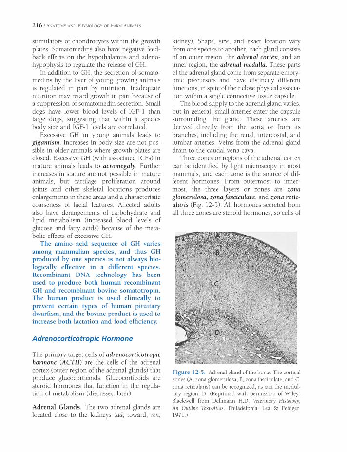
216 / ANATOMY AND PHYSIOLOGY OF FARM ANIMALS
stimulators of chondrocytes within the growth plates. Somatomedins also have negative feed-back effects on the hypothalamus and adeno-hypophysis to regulate the release of GH.
In addition to GH, the secretion of somato-medins by the liver of young growing animals is regulated in part by nutrition. Inadequate nutrition may retard growth in part because of a suppression of somatomedin secretion. Small dogs have lower blood levels of IGF-1 than large dogs, suggesting that within a species body size and IGF-1 levels are correlated.
Excessive GH in young animals leads to gigantism. Increases in body size are not pos-sible in older animals where growth plates are closed. Excessive GH (with associated IGFs) in mature animals leads to acromegaly. Further increases in stature are not possible in mature animals, but cartilage proliferation around joints and other skeletal locations produces enlargements in these areas and a characteristic coarseness of facial features. Affected adults also have derangements of carbohydrate and lipid metabolism (increased blood levels of glucose and fatty acids) because of the meta-bolic effects of excessive GH.
The amino acid sequence of GH varies among mammalian species, and thus GH produced by one species is not always bio-logically effective in a different species. Recombinant DNA technology has been used to produce both human recombinant GH and recombinant bovine somatotropin. The human product is used clinically to prevent certain types of human pituitary dwarfi sm, and the bovine product is used to increase both lactation and food effi ciency.
Adrenocorticotropic Hormone
The primary target cells of adrenocorticotropic hormone (ACTH) are the cells of the adrenal cortex (outer region of the adrenal glands) that produce glucocorticoids. Glucocorticoids are steroid hormones that function in the regula-tion of metabolism (discussed later).
Adrenal Glands. The two adrenal glands are located close to the kidneys (ad, toward; ren,
kidney). Shape, size, and exact location vary from one species to another. Each gland consists of an outer region, the adrenal cortex, and an inner region, the adrenal medulla. These parts of the adrenal gland come from separate embry-onic precursors and have distinctly different functions, in spite of their close physical associa-tion within a single connective tissue capsule.
The blood supply to the adrenal gland varies, but in general, small arteries enter the capsule surrounding the gland. These arteries are derived directly from the aorta or from its branches, including the renal, intercostal, and lumbar arteries. Veins from the adrenal gland drain to the caudal vena cava.
Three zones or regions of the adrenal cortex can be identifi ed by light microscopy in most mammals, and each zone is the source of dif-ferent hormones. From outermost to inner-most, the three layers or zones are zona glomerulosa, zona fasciculata, and zona retic-ularis (Fig. 12-5). All hormones secreted from all three zones are steroid hormones, so cells of
Figure 12-5. Adrenal gland of the horse. The cortical zones (A, zona glomerulosa; B, zona fasciculate; and C, zona reticularis) can be recognized, as can the medul-lary region, D. (Reprinted with permission of Wiley-Blackwell from Dellmann H.D. Veterinary Histology: An Outline Text-Atlas. Philadelphia: Lea & Febiger, 1971.)
ENDOCRINOLOGY / 217
all zones have ultrastructural characteristics of steroid-secreting cells.
The hormones secreted by the adrenal medulla (epinephrine and norepinephrine) are amines and are stored in secretory granules prior to release. These endocrine cells are termed chromaffi n cells because of their affi n-ity for chromium stains. As was described in Chapter 10 in the section on the autonomic nervous system, epinephrine and norepineph-rine are released from the adrenal medulla in times of stress, and this release is regulated via the autonomic nervous system. A tumor of chromaffi n cells is termed a pheochromocy-toma, and this neoplasia typically results in excessive secretion of epinephrine and nor-epinephrine. The resulting clinical signs are those of excessive stimulation of the sympa-thetic nervous system (e.g., increased heart rate and blood pressure and increased meta-bolic rate).
Hormones of the Adrenal Cortex. The zona glomerulosa secretes mineralocorticoids (pri-marily aldosterone) that function in the regula-tion of sodium and potassium balance. The regulation of balance is primarily accomplished by controlling the loss of sodium and potas-sium in urine; more details on their specifi c functions are given in Chapters 18 and 23. Mineralocorticoid secretion is not regulated by ACTH but rather by the serum potassium con-centration and the renin–angiotensin system, another group of chemical messengers dis-cussed in more detail in Chapter 18.
Glucocorticoids (primarily cortisol and cor-ticosterone) are the major secretory product of both the zona fasciculata and zona reticularis, and ACTH is the major regulator (stimulator) of their secretion. Without ACTH the fascicu-lata and reticularis both atrophy, but the glo-merulosa remains intact. The inner zones of the adrenal cortex are also a source of adrenal sex hormones (androgens and estrogens), but the rates of secretion in normal adult animals are very low and not necessary for normal repro-ductive behavior and function.
The secretion of ACTH from the adenohy-pophysis is stimulated by a hypothalamic
hormone, corticotropin-releasing hormone (CRH), and this is the most important regulator of ACTH release. However, the regulation of CRH, hence ACTH release, is extremely complex and affected by a wide variety of stimuli. ACTH increases are considered to be a classic sign of stress, and plasma levels of ACTH or cortisol are often used in experimen-tal settings to evaluate the overall stress placed on an animal by any type of physical or emotional stimulus (e.g., restraint, starvation, presence of a predator). Both ACTH and glu-cocorticoids have negative feedback effects on the pituitary and the hypothalamus to maintain normal resting blood levels of ACTH and glu-cocorticoids, but stressful stimuli can override these effects.
Glucocorticoids have many target tissues throughout the body. In general their effects on these target tissues would seem an appropriate response to counteract stressful stimuli. For example, glucocorticoids increase the rate of gluconeogenesis (glucose formation) by the liver and increase the rate of fatty acid mobili-zation from lipid tissue. In skeletal muscle protein synthesis is reduced and protein degra-dation is increased, which means that more amino acids are available for gluconeogenesis by the liver. These metabolic effects are particu-larly important during starvation.
Glucocorticoids are often used therapeu-tically to inhibit infl ammatory and immune responses. The doses used for these effects produce blood levels that are much higher than those seen in normal animals, even when they are responding to stress. Such levels and effects are described as supra-physiologic or pharmacologic. Among the many components of the infl ammatory process that are inhibited by glucocorticoids are the synthesis pathways for prostaglan-dins, leukotrienes, and thromboxanes.
Thyroid-Stimulating Hormone
The target cells for thyroid-stimulating hormone (TSH), also called thyrotropin, are endocrine cells of the thyroid that produce and release thyroxine

218 / ANATOMY AND PHYSIOLOGY OF FARM ANIMALS
(T4) and triiodothyronine (T3) when stimulated by TSH. Both are considered amine hormones, for each consists of a linkage of two iodinated tyrosine residues (Fig. 12-6). The 3 and 4 refer to the number of iodine atoms in their mole-cules. These hormones are necessary for normal growth and development in young animals, and they regulate basal metabolic rate in the adult.
Thyroid Gland. The thyroid gland is associ-ated with the proximal part of the trachea near the thyroid cartilage of the larynx. Its appear-ance varies widely among species, with the thyroid gland of most animals possessing two distinct lobes, variably connected across the midline by a strip of thyroid tissue called the isthmus. In pigs, the bulk of the gland lies pri-marily on the ventral aspect of the trachea rather than being clearly divided into lateral lobes. A connective tissue capsule covers the gland and gives rise to septa that divide the substance of the thyroid and support the vas-culature of the gland. Arterial blood supply to the thyroid and the associated parathyroid glands (discussed later) arrives as branches of the common carotid artery.
Microscopically, the thyroid gland consists of follicles, spheres lined by a simple epithe-lium of cells that ranges from cuboidal to columnar (Fig. 12-7). Thyroid follicles are fi lled with the product of the follicular lining cells, a gel-like substance called colloid, which consists of a protein–iodine complex, thyroglobulin.
The hormones T3 and T4 are stored in the colloid as iodinated tyrosine residues that are part of thyroglobulin molecules (Fig. 12-8). This type of storage is a unique among endocrine glands.
Scattered among the follicular lining cells and adjacent to them are a small subset of thyroid cells, the C cells (parafollicular cells). The C cells produce calcitonin, a peptide hormone that lowers the blood level of calcium by inhibiting the action of osteoclasts. Calcito-nin release is directly regulated by the negative feedback of serum calcium concentration on C cells, not by TSH. The physiologic importance of calcitonin in the overall regulation of serum calcium concentration is minimal compared with the role of parathyroid hormone (dis-cussed later).
Thyroxine and Triiodothyronine. Both T4
and T3 are biologically active (bind to thyroid hormone receptors), and most of the circulat-ing hormones are bound to plasma proteins. The T4 and T3 in the body fl uids that are not protein bound are the biologically active mole-cules, but they are also subject to degradation. In many species, the plasma levels of T4 are
Figure 12-6. Thyroid hormones.
Figure 12-7. Thyroid follicles of varying sizes and shapes are fi lled with colloid and lined with follicular cells. (Reprinted with permission of Wiley-Blackwell from Dellmann H.D. Textbook of Veterinary Histology. 5th ed. Baltimore: Lippincott Williams & Wilkins, 1998.)
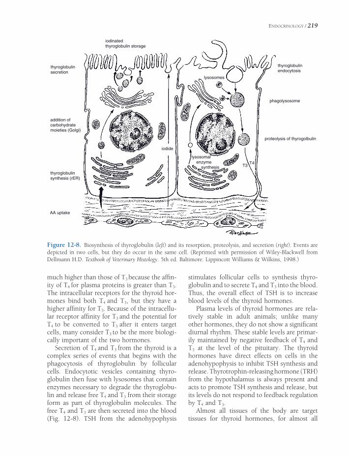
ENDOCRINOLOGY / 219
AA uptake
thyroglobulin synthesis (rER)
addition of carbohydrate moieties (Golgi)
thyroglobulin secretion
iodinatedthyroglobulin storage
thyroglobulin endocytosis
phagolysosome
lysosomes
iodide
lysosomal enzyme synthesis T3
T4
proteolysis of thyrogolbulin
Figure 12-8. Biosynthesis of thyroglobulin (left) and its resorption, proteolysis, and secretion (right). Events are depicted in two cells, but they do occur in the same cell. (Reprinted with permission of Wiley-Blackwell from Dellmann H.D. Textbook of Veterinary Histology. 5th ed. Baltimore: Lippincott Williams & Wilkins, 1998.)
much higher than those of T3 because the affi n-ity of T4 for plasma proteins is greater than T3. The intracellular receptors for the thyroid hor-mones bind both T4 and T3, but they have a higher affi nity for T3. Because of the intracellu-lar receptor affi nity for T3 and the potential for T4 to be converted to T3 after it enters target cells, many consider T3 to be the more biologi-cally important of the two hormones.
Secretion of T4 and T3 from the thyroid is a complex series of events that begins with the phagocytosis of thyroglobulin by follicular cells. Endocytotic vesicles containing thyro-globulin then fuse with lysosomes that contain enzymes necessary to degrade the thyroglobu-lin and release free T4 and T3 from their storage form as part of thyroglobulin molecules. The free T4 and T3 are then secreted into the blood (Fig. 12-8). TSH from the adenohypophysis
stimulates follicular cells to synthesis thyro-globulin and to secrete T4 and T3 into the blood. Thus, the overall effect of TSH is to increase blood levels of the thyroid hormones.
Plasma levels of thyroid hormones are rela-tively stable in adult animals; unlike many other hormones, they do not show a signifi cant diurnal rhythm. These stable levels are primar-ily maintained by negative feedback of T4 and T3 at the level of the pituitary. The thyroid hormones have direct effects on cells in the adenohypophysis to inhibit TSH synthesis and release. Thyrotrophin-releasing hormone (TRH) from the hypothalamus is always present and acts to promote TSH synthesis and release, but its levels do not respond to feedback regulation by T4 and T3.
Almost all tissues of the body are target tissues for thyroid hormones, for almost all

220 / ANATOMY AND PHYSIOLOGY OF FARM ANIMALS
have receptors for them. In mature animals the most general effect of thyroid hormones is to increase overall oxygen consumption and heat production. The basal metabolic rate is a measure of oxygen use in resting conditions, so thyroid hormones are said to increase it. The exact intracellular mechanisms responsible for these general effects are not known, but the effects are known as the calorigenic action of the thyroid hormones. The calorigenic action is associated with an overall increase in the metabolism and use of carbohydrates and lipids, which is consistent with the increased use of oxygen and heat production.
Chronic exposure of some animals to cold is associated with an increase in TSH, T4, and T3. The increased calorigenic effect of the thyroid hormones should work to maintain normal body temperature in the cold envi-ronment. The response to the cold appears to be a result of an increase in TRH release by the hypothalamus. The hypothalamus is known to be the refl ex center for other refl exes involved with the regulation of body temperature on a short-term basis (e.g., peripheral vasodilation or vasoconstriction).
Thyroid hormones are essential for normal growth and development in young animals. Two systems of special importance are the skel-etal and nervous systems. Animals with thyroid hormone defi cits (hypothyroidism) do not attain normal stature and have a variety of central nervous system abnormalities. In humans with severe defi cits at birth, mental development remains impaired throughout life, even if replacement therapy is begun when the person is 4 to 5 years of age. Cretinism is the term for the human condition caused by a congenital lack of thyroid hormone and characterized by arrested physical and mental development and lowered metabolic rate.
Other Endocrine Glands
Parathyroid Glands
The parathyroid glands are small aggregates of endocrine tissue within or near the thyroid
gland. Most domestic animals have two pairs of parathyroid glands, but the exact number and location vary with the species. Commonly, one pair is visible outside the thyroid gland and is therefore designated external parathyroids. The second pair is often buried in the substance of the thyroid, and these are the internal para-thyroids. The pig lacks grossly visible internal parathyroids.
The two types of parathyroid cells are chief cells and oxyphil cells (Fig. 12-9). Chief cells are small, usually dark cells that are associated with the production of parathyroid hormone (PTH, parathormone). The less numerous oxyphil cells are larger, with granular cyto-plasm and a small, dark nucleus. These cells have been described in horses and cattle (and humans) but are not found in other domestic species. Their function is unknown, although the fact that they appear later in life has sug-gested that they may be senescent chief cells.
Parathyroid Hormone. Parathyroid hormone (PTH), a peptide hormone, is the major con-troller of the level of blood calcium and phos-phate. It does this by stimulating release of calcium and phosphate from bone, decreasing excretion of calcium and increasing excretion
Figure 12-9. An equine parathyroid gland with two large oxyphil cells (arrows) among numerous chief cells. (Reprinted with permission of Wiley-Blackwell from Dellmann H.D. Textbook of Veterinary Histology. 5th ed. Baltimore: Lippincott Williams & Wilkins, 1998.)

ENDOCRINOLOGY / 221
of phosphate by the kidney, and promoting the formation of active vitamin D by the kidney (discussed later). In bone, PTH acts on both osteocytes and osteoclasts to promote the release of calcium phosphate. The action of PTH on osteocytes releases calcium and phos-phate from stores associated with the extracel-lular fl uids in and around bone. PTH stimulates the degradation of bone by osteoclasts while it inhibits bone formation by osteoblasts. The overall effect of PTH is to increase blood calcium concentration and lower blood phosphate con-centration by increasing its urinary excretion.
The forms of vitamin D consumed in the diet or produced in the skin by the action of ultraviolet light on precursors is not the most active form of the vitamin. These forms are further metabolized in the liver to a second precursor form that is further metabolized in the kidneys to the most biologically active form. It is this fi nal form (1,25-dihydroxycholcalcif-erol or calcitriol) that functions as the true vitamin D. PTH promotes the formation of this fi nal metabolite within the kidney. Two major functions of vitamin D are to increase the rate of calcium absorption from the gastrointestinal tract and to reduce the loss of calcium in the urine. Thus, the overall effect of vitamin D is to retain calcium in the body.
The only signifi cant regulator of PTH release is the concentration of ionized calcium in the blood plasma. Normally, about 50% of plasma calcium is bound to plasma proteins (primarily albumin), and this bound calcium is not bio-logically active. The other 50% is unbound and exists as calcium ions. Chief cells detect decreases in the concentration of ionized calcium, and they respond by increasing their secretion of PTH. The resulting rise in plasma calcium has a negative feedback effect on further PTH secretion.
Diets insuffi cient in calcium are uncom-mon in domestic animals other than carni-vores, but they can occur, especially with diets formulated primarily on grain prod-ucts, which typically have high phosphorus and low calcium. In such cases, the chroni-cally low intake of dietary calcium stimu-lates increased secretion of PTH to keep
blood calcium levels adequate for nerve and muscle function. Calcium is removed from the bone matrix, and this decalcifi cation can lead to bone deformities and osteoporosis. This creates the medical condition nut-ritional secondary hyperparathyroidism. In young animals, growth may be disrupted and limb and spine abnormalities may develop, a manifestation of hyperparathy-roidism sometimes called rickets. A particu-lar form of nutritional hyperparathyroidism commonly called bran disease or bighead is seen in horses fed excessive amounts of cereal by-products (e.g., bran) in conjunc-tion with calcium-poor hay. Bone tissue that has been decalcifi ed by high levels of circu-lating PTH is frequently replaced with soft, bulky fi brous tissue; this is especially promi-nent in the fl at bones of the face and man-dible, giving rise to the appearance of an enlarged head. Prompt correction of the dietary imbalance may improve most symp-toms (e.g., lameness) of bran disease, although the enlarged facial features are usually permanent.
Pancreatic Islets
The pancreas of domestic animals is a bilobed gland adjacent to the proximal part of the duo-denum (small intestine). The pancreas is an important exocrine gland whose enzymatic secretions are delivered to the lumen of the duodenum by one or two ducts. Scattered throughout the substance of the pancreas are small masses of endocrine tissue called pancre-atic islets (formerly the islets of Langerhans).
The pancreatic islets are clumps of pale-staining cells, arranged in irregular cords sepa-rated by capillaries (Fig. 12-10). Special stains are used to demonstrate the types of epithelial cells found in the pancreatic islets. The known number of distinct kinds of cells is still growing, but the two best characterized are the α-cells and β-cells. The β-cells are the more numerous (about 75% of all cells in the islet), and they produce the hormone insulin. α-Cells produce the hormone glucagon.
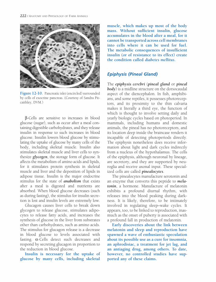
222 / ANATOMY AND PHYSIOLOGY OF FARM ANIMALS
β-Cells are sensitive to increases in blood glucose (sugar), such as occur after a meal con-taining digestible carbohydrates, and they release insulin in response to such increases in blood glucose. Insulin lowers blood glucose by stimu-lating the uptake of glucose by many cells of the body, including skeletal muscle. Insulin also stimulates skeletal muscle and liver cells to syn-thesize glycogen, the storage form of glucose. It affects the metabolism of amino acids and lipids, for it stimulates protein synthesis in skeletal muscle and liver and the deposition of lipids in adipose tissue. Insulin is the major endocrine stimulus for the state of anabolism that exists after a meal is digested and nutrients are absorbed. When blood glucose decreases (such as during fasting), the stimulus for insulin secre-tion is lost and insulin levels are extremely low.
Glucagon causes liver cells to break down glycogen to release glucose, stimulates adipo-cytes to release fatty acids, and increases the synthesis of glucose in the liver from substrates other than carbohydrates, such as amino acids. The stimulus for glucagon release is a decrease in blood glucose to levels associated with fasting. α-Cells detect such decreases and respond by secreting glucagon in proportion to the reduction in blood glucose.
Insulin is necessary for the uptake of glucose by many cells, including skeletal
muscle, which makes up most of the body mass. Without suffi cient insulin, glucose accumulates in the blood after a meal, for it cannot be transported across cell membranes into cells where it can be used for fuel. The metabolic consequences of insuffi cient insulin (or of resistance to its effect) create the condition called diabetes mellitus.
Epiphysis (Pineal Gland)
The epiphysis cerebri (pineal gland or pineal body) is a midline structure on the dorsocaudal aspect of the diencephalon. In fi sh, amphibi-ans, and some reptiles, it possesses photorecep-tors, and its proximity to the thin calvaria makes it literally a third eye, the function of which is thought to involve setting daily and yearly biologic cycles based on photoperiod. In mammals, including humans and domestic animals, the pineal has no photoreceptors, and its location deep inside the braincase renders it incapable of detecting photoperiods directly. The epiphysis nonetheless does receive infor-mation about light and dark cycles indirectly from a nucleus of the hypothalamus. The cells of the epiphysis, although neuronal by lineage, are secretory, and they are supported by neu-roglia and receive axonal input. These special-ized cells are called pinealocytes.
The pinealocytes manufacture serotonin and an enzyme that converts this peptide to mela-tonin, a hormone. Manufacture of melatonin exhibits a profound diurnal rhythm, with releases into the blood peaking during dark-ness. It is likely, therefore, to be intimately involved in regulating sleep–wake cycles. It appears, too, to be linked to reproduction, inas-much as the onset of puberty is associated with a profound fall in production of melatonin.
Early discoveries about the link between melatonin and sleep and reproduction have spawned a wave of enthusiastic speculation about its possible use as a cure for insomnia, an aphrodisiac, a treatment for jet lag, and an antiaging drug, among others. To date, however, no controlled studies have sup-ported any of these claims.
Figure 12-10. Pancreatic islet (encircled) surrounded by cells of exocrine pancreas. (Courtesy of Sandra Pit-caithley, DVM.)
THE INTEGUMENT
Integument
The integumentary system comprises the skin with its adnexal structures (e.g., hair and glands), horns, hoofs, claws, and other modifi -cations of the epithelial covering of the body. The skin is an important protective barrier, reducing water loss, invasion of microorgan-isms, and abrasive trauma. For many species, it is an important organ of thermoregulation through perspiration, control of cutaneous blood fl ow, and disposition of the hair coat. Modifi cations of the integument are used for protection (claws and horns) and provide a tough covering to the feet where they contact the ground (hoofs and footpads).
All of the components of the integument may be considered modifi cations of the surface epithelium, derived from the embryonic ecto-derm, in conjunction with an underlying vas-cularized component derived from mesoderm (see Chapter 3). In fully developed skin, these become the superfi cial epidermis and deep dermis, respectively. Specialized integumen-tary structures, such as hoofs and horns, like-wise have homologous superfi cial epithelial and deep connective tissue components.
Skin
Skin covers the outside of the animal and is continuous with mucous membranes at oral, anal, and urogenital orifi ces, the vestibule of the
IntegumentSkin
Epidermis
Dermis
Hypodermis
Adnexa of the SkinHair
Glands
Modifi ed EpidermisHooves
Horns
Dewclaws
Chestnuts and Ergots
Coat Color in HorsesWool
Chapter 13
223

224 / ANATOMY AND PHYSIOLOGY OF FARM ANIMALS
nostril, and the palpebral fi ssure; these sites are characterized by a mucocutaneous junction. Thickness of skin varies both between species and on a given individual, being generally thick-est where it is most exposed (e.g., on the back) and thinner in protected regions (e.g., the groin). The skin adheres tightly to underlying struc-tures in some locations, but in others is loosely attached to allow for considerable movement. The looseness of skin attachment is exploited by veterinarians who frequently inject medications or fl uids for rehydration into the space under-neath the skin (a subcutaneous injection).
Epidermis
The outer layer of skin, the epidermis, is an avascular stratifi ed squamous epithelium that is
nearly free of nerve endings. In most areas it can be divided into several histologic layers (Fig. 13-1). They are as follows, from deep to superfi cial: (1) A layer of mitotically active cuboidal or columnar cells, the stratum basale, follows the contour of the underlying dermis, to which it is closely applied. (2) The stratum spinosum has a spiny appearance because of its desmosomes (intercellular bridges) connecting adjacent cells. (3) The stratum granulosum consists of spindle-shaped cells containing basophilic keratohyalin granules. (4) The stratum lucidum, which is variably present, comprises cells that are poorly stainable. (5) The stratum corneum consists of layers of dead, fl attened cells. Presence and rela-tive thickness of each layer is refl ected in the overall thickness of the skin (Fig. 13-1).
Cells in the stratum basale undergo mitotic division, which pushes the more superfi cial
Figure 13-1. Feline skin. 1) Hairy skin from lumbar region. Note multiple hair follicles near bottom of micro-graph. 2) Hairless skin from nose; epidermis is thicker in this region. 3) Footpad. Epidermis is markedly thickened in this region, with most of the increase due to thickening of the stratum corneum. A, Flaking keratin; B, stratum corneum; C, stratum lucidum; D, stratum granulosum; E, stratum spinosum; F, stratum basale; G, dermis; H, hair follicle. (Reprinted with permission of Wiley-Blackwell from Dellmann H.D. and Brown E.M. Textbook of Veterinary Histology. 3rd ed. Philadelphia: Lea & Febiger, 1981.)

THE INTEGUMENT / 225
layers still farther from the blood vessels in the underlying dermis. As distance from nutrients increases, the cells fl atten and die, leaving a dense mat of their primary constituent, the fi brous protein keratin. The drying and harden-ing of the superfi cial cells, a process called both keratinization and cornifi cation, renders the surface of the skin tough and resistant to drying. As the stratum basale continuously adds cells to overlying layers, the stratum corneum fl akes off and is replaced. The rate at which this occurs can be infl uenced by trauma or disease pro-cesses. A callus is a local increase in thickness in response to continuous trauma.
Dermis
The epidermis forms an undulating sheet with fi ngerlike projections, the epidermal pegs, which project into the underlying connective tissue, the dermis. The dermis (also known as the corium, especially where it is associated
with modifi cations like hoofs and horns) bears ridges and nipplelike projections (the dermal papillae) that interdigitate with the overlying epidermis (Fig. 13-2); these are most especially prominent in weight-bearing structures, such as footpads and hoofs. The interface between epidermal pegs and dermal papillae provides increased surface area for formation of a strong junction between these two layers. A blister is a local disruption of this association between layers, usually due to repeated trauma or thermal injury.
Arteries, veins, capillaries, and lymphatics of the skin are contained in the dermis. Sensory nerve fi bers, in addition to supplying the dermis, may extend a short distance into the epidermis. Sympathetic nerves provide motor innervation to blood vessels, glands, and arrec-tor pili muscles of hair follicles in the dermis. These structures do not receive parasympa-thetic innervation.
Color of skin is due to the pigment granules generated in the cytoplasm of the resident
Sebaceous Gland
EPIDERMIS
DERMIS
HYPODERMIS
Sweat Gland
Blood Vessels
Figure 13-2. Skin anatomy.

226 / ANATOMY AND PHYSIOLOGY OF FARM ANIMALS
pigment cells, melanocytes. These cells in the stratum basale produce the pigment, melanin, which is brown, yellowish-brown, or black. Packets of melanin pigment are manufactured by the melanocytes and transferred to sur-rounding cells of the epidermis; the same process incorporates pigment into cells that cornify into hairs. The expression of different colors in skin and hair comes primarily from the relative amount of melanin produced in the melanocytes rather than from differences in numbers of melanocytes or presence of other pigments. This expression can be infl uenced by certain pituitary hormones, notably melano-cyte-stimulating hormone (MSH) and adreno-corticotropic hormone (ACTH) (see Chapter 12).
Absence of pigment in the skin (albinism), which may be partial or total, arises from a genetic inability of melanocytes to manufac-ture pigment. Lack of pigment can render the skin and surface mucous membranes more susceptible to actinic damage (i.e., cel-lular damage due to ultraviolet light), hence formation of carcinoma (cancer) of the skin or other exposed epithelia. Cancer eye, or squamous cell carcinoma of the conjunctiva of the eye, is common in white-faced cattle (e.g., Hereford) living at high elevations, where the ultraviolet component of sunlight is little attenuated.
Hypodermis
In nearly all areas of the body, a layer of loose connective tissue separates the dermis from underlying structures. This areolar connective tissue, known variously as the superfi cial fascia, subcutis, or hypodermis, permits move-ment of the skin without tearing. Where the skin is tightly attached to underlying bone or muscle, a dimple on the body surface may be seen. This is a “tie,” as is seen where the dermis is attached to the spinous processes of verte-brae. Variable amounts of fat, the panniculus adiposus, are present in the hypodermis, with species-dependent distribution and relative abundance. The panniculus adiposus is an
especially notable feature of pigs; on the dorsum of the pig, it is called backfat.
Adnexa of the Skin
Hair
Hair is a defi ning characteristic of mammals. All common domestic mammals except the pig have abundant hair. There are three main types of hair on domestic mammals: (1) guard hairs, which form the smooth outer coat; (2) wool hairs, also called the undercoat, which are fi ne and often curly; and (3) tactile hairs, long stiff hairs with specialized innervation that renders them effective as organs of touch.
An individual hair arises from a modifi cation of the epidermis, the hair follicle (Fig. 13-3). The follicle invaginates from the surface of the skin as a double-layered root sheath that sur-rounds the hair and terminates in a hair bulb of epidermal origin. The hair bulb surrounds a small knob of dermis called the dermal papilla. The internal epithelial root sheath intimately covers the root of the hair and is continuous with the epithelial cells covering the dermal papilla. The external epithelial root sheath surrounds the internal root sheath, is continu-ous with the epidermis, and gives rise to the sebaceous glands that are associated with hair follicles. The division of the epithelial cells cov-ering the dermal papilla generates the hair itself. Growth and multiplication of these cells extrude the hair from the follicle, causing it to grow.
An individual hair has a medulla at its center, surrounded by a scaly cortex, outside of which is a thin cuticle. All parts of the hair are com-posed of compressed, keratinized epithelial cells. The bulk of the hair comprises the cortex, which consists of several layers of cornifi ed cells. The amount and type of melanin in corti-cal cells determine whether the hair will be black, brown, or red. The cuticle is a single layer of thin, clear cells covering the surface of the cortex. The medulla may contain pigment, which has little effect on hair color, but air spaces between medullary cells are believed to

THE INTEGUMENT / 227
Figure 13-3. Single hair follicle. Epidermal cells adjacent to the dermal papilla give rise to the keratin of the hair. (Reprinted with permission of Wiley-Blackwell from Eurell, J.A. and Frappier, B.L. Dellmann’s Textbook of Veterinary Histology, 6th ed. Ames, IA: Blackwell Publishing Professional, 2006.)
give a white or silver color to the hair if the cortex lacks pigment. Wool hairs lack a medulla or have only a very small one, accounting for their fi ne, fl exible nature.
Tactile hairs, used as probes or feelers, are also called sinus hairs because a large blood-fi lled sinus surrounds the deep portions of the follicle. These hairs are thicker and usually
longer than guard hairs and are most com-monly found on the face, around lips and eyes. These hairs are particularly well supplied with sensory nerve endings that are sensitive to the movement of the hair.
When a hair is ready to shed, the epithelial cells over the papilla stop multiplying and become cornifi ed. The papilla atrophies, and
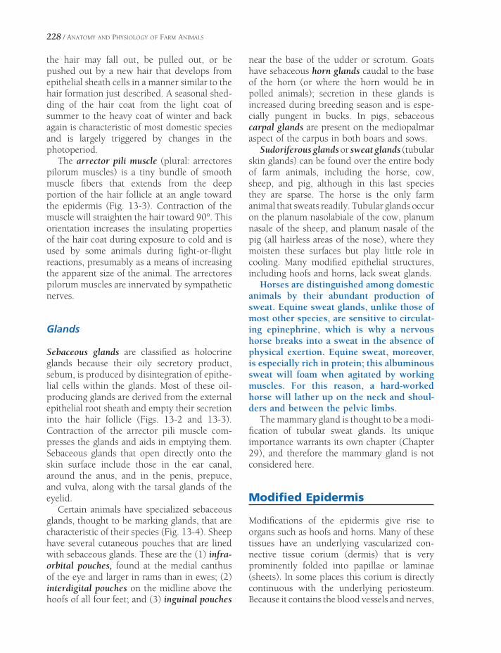
228 / ANATOMY AND PHYSIOLOGY OF FARM ANIMALS
the hair may fall out, be pulled out, or be pushed out by a new hair that develops from epithelial sheath cells in a manner similar to the hair formation just described. A seasonal shed-ding of the hair coat from the light coat of summer to the heavy coat of winter and back again is characteristic of most domestic species and is largely triggered by changes in the photoperiod.
The arrector pili muscle (plural: arrectores pilorum muscles) is a tiny bundle of smooth muscle fi bers that extends from the deep portion of the hair follicle at an angle toward the epidermis (Fig. 13-3). Contraction of the muscle will straighten the hair toward 90°. This orientation increases the insulating properties of the hair coat during exposure to cold and is used by some animals during fi ght-or-fl ight reactions, presumably as a means of increasing the apparent size of the animal. The arrectores pilorum muscles are innervated by sympathetic nerves.
Glands
Sebaceous glands are classifi ed as holocrine glands because their oily secretory product, sebum, is produced by disintegration of epithe-lial cells within the glands. Most of these oil-producing glands are derived from the external epithelial root sheath and empty their secretion into the hair follicle (Figs. 13-2 and 13-3). Contraction of the arrector pili muscle com-presses the glands and aids in emptying them. Sebaceous glands that open directly onto the skin surface include those in the ear canal, around the anus, and in the penis, prepuce, and vulva, along with the tarsal glands of the eyelid.
Certain animals have specialized sebaceous glands, thought to be marking glands, that are characteristic of their species (Fig. 13-4). Sheep have several cutaneous pouches that are lined with sebaceous glands. These are the (1) infra-orbital pouches, found at the medial canthus of the eye and larger in rams than in ewes; (2) interdigital pouches on the midline above the hoofs of all four feet; and (3) inguinal pouches
near the base of the udder or scrotum. Goats have sebaceous horn glands caudal to the base of the horn (or where the horn would be in polled animals); secretion in these glands is increased during breeding season and is espe-cially pungent in bucks. In pigs, sebaceous carpal glands are present on the mediopalmar aspect of the carpus in both boars and sows.
Sudoriferous glands or sweat glands (tubular skin glands) can be found over the entire body of farm animals, including the horse, cow, sheep, and pig, although in this last species they are sparse. The horse is the only farm animal that sweats readily. Tubular glands occur on the planum nasolabiale of the cow, planum nasale of the sheep, and planum nasale of the pig (all hairless areas of the nose), where they moisten these surfaces but play little role in cooling. Many modifi ed epithelial structures, including hoofs and horns, lack sweat glands.
Horses are distinguished among domestic animals by their abundant production of sweat. Equine sweat glands, unlike those of most other species, are sensitive to circulat-ing epinephrine, which is why a nervous horse breaks into a sweat in the absence of physical exertion. Equine sweat, moreover, is especially rich in protein; this albuminous sweat will foam when agitated by working muscles. For this reason, a hard-worked horse will lather up on the neck and shoul-ders and between the pelvic limbs.
The mammary gland is thought to be a modi-fi cation of tubular sweat glands. Its unique importance warrants its own chapter (Chapter 29), and therefore the mammary gland is not considered here.
Modifi ed Epidermis
Modifi cations of the epidermis give rise to organs such as hoofs and horns. Many of these tissues have an underlying vascularized con-nective tissue corium (dermis) that is very prominently folded into papillae or laminae (sheets). In some places this corium is directly continuous with the underlying periosteum. Because it contains the blood vessels and nerves,

BA
C
ED
Figure 13-4. Specialized sebaceous glands. A) Infraorbital pouch of the sheep. B) Interdigital pouch of the sheep. C) Inguinal pouch of the sheep. D) Horn glands of the goat. E) Carpal glands of the pig.

230 / ANATOMY AND PHYSIOLOGY OF FARM ANIMALS
the corium is often called the sensitive part of the hoof or horn. The insensitive portions of these structures are derivatives of the overlying epithelium. Nonetheless, it is helpful to keep in mind that the substance of the hoof wall, the horn, and other epidermal modifi cations is generated by the deepest layer of the epithe-lium (homologous to the stratum basale of skin) and not by the underlying corium.
Hooves
Hoofed animals are ungulates (L. unguis, nail), and most common farm mammals fall in this category. A defi ning characteristic of ungulates is the presence of a well-developed hoof associated with the distal phalanx (Fig. 13-5). Although the hoofs of pigs, ruminants, and horses differ signifi cantly in their gross appear-ance, they share certain features. Like the skin from which they are derived, hoofs have an outer avascular epidermal layer and an inner vascularized dermis; the dermis of hoofs and horns is more commonly called corium.
Different parts of the epidermis and corium of the hoof are named for their location. The outside of the hoof is covered by a thin, waxy layer called the periople. The thick hoof wall grows from a belt of epidermis at the coronary band, the region where haired skin becomes hoof. The deep side of the hoof wall is inti-mately connected to the underlying corium, which blends with the periosteum of the distal phalanx. The connection between hoof wall and corium is characterized by interdigitating sheets of hoof wall and corium. These are the laminae, of which there are insensitive laminae (part of the epidermis) and sensitive laminae (part of the corium). The laminae are especially elaborately developed in the equine hoof.
The part of the hoof in contact with the ground features a horny sole (extensive in the horse, smaller in other domestic ungulates) and a softer bulb of the hoof. Deep to the bulb of the hoof is a shock-absorbing modifi cation of the subcutis called the digital cushion. The bulb forms a large part of the palmar/plantar
aspect of the feet of ruminants and pigs, in which it bears a considerable proportion of the animal’s weight. In contrast, the equine hoof features a keratinized V-shaped frog, which is more fl exible than the adjacent sole of the hoof but harder than the bulbs of other ungulates.
The equine hoof is a highly specialized structure and is so important that it will be covered in depth in Chapter 14. The following is a brief overview of the anatomy of the hoofs of artiodactyls, the even-toed ungulates (Fig. 13-6).
A convenient terminology for the digits of even-toed animals is to refer to the digits by number (III and IV in ruminants and pigs), and then relate each digit to the midline of the respective foot. The axial side of the digit is the side closest to the midline of the foot and the abaxial side is the side farthest from the midline of the foot.
The hoof wall consists of a nearly vertical axial portion that refl ects sharply caudad at the toe (tip of the hoof) to be continuous with the abaxial portion of the wall. The abaxial hoof wall is convex and the axial wall is concave, and both the axial and abaxial surfaces are continu-ous with the bulb of the hoof. The lateral digit bears more weight than the medial one (as a clinical consequence, most foot lameness in dairy cattle is referable to the lateral hoof). In contrast to the horse, the sole and bulb of the foot carry a great deal of weight relative to the walls and toe. The bulb of the porcine hoof is especially prominent, providing a larger pro-portion of the weight-bearing surface than in ruminants.
Horns
Horns of cattle and sheep are formed over the cornual process, a bony core that projects from the frontal bone of the skull (Fig. 13-7). Both male and female cattle of horned breeds have horns, although the female animal’s horns are smaller. In most horned sheep and goat breeds, both males and females have them, although in a few breeds only rams or bucks are horned. Animals that lack horns naturally are polled.

Figure 13-5. Anatomy of the equine hoof. This median section of the hoof illustrates the transition from haired skin to hoof at the coronary band and the relationship between the horny parts of the hoof and the underlying bony structures. (Reprinted with permission of Wiley-Blackwell from Stashak T.S. Adams’ Lameness in Horses. 5th ed. Baltimore: Lippincott Williams & Wilkins, 2002.)
PIG
OX
Dewclaw
Bulb
Sole
Toe
Axial Surface
Abaxial Surface
Figure 13-6. Palmar view of the feet of the pig (left) and the ox (right). The accessory digits (dewclaws) of the pig are well developed relative to those of the ox. The bulb of the porcine hoof also makes up a larger portion of the bottom of the foot. Surfaces adjacent to the midline are described as axial, whereas those toward the outside of the hoof are abaxial.

232 / ANATOMY AND PHYSIOLOGY OF FARM ANIMALS
Haired Skin
Horn
Combined Corium & Periosteum
Cornual Diverticulum of Frontal Sinus
Figure 13-7. Longitudinal section of a horn. The horn is supported by a bony core, the corneal process of the frontal bone, which is invaded by a diverticulum of the frontal sinus. Periosteum and corium are blended on the surface of the cornual process.
The corium of the horn completely envelops the cornual process and blends with its perios-teum. The horn itself consists of dense keratin, much like the hoof wall, and elongates from the base. A soft type of horn called the epikeras covers the surface of the horn at the base and extends a variable distance toward the apex of the horn. The epikeras resembles periople of the hoof.
Variations in level of nutrition of the animal are refl ected in variations in rapidity of horn growth, resulting in a series of rings on the horn. These alternations in thickness of the horn may refl ect seasonal stresses, notably the stress of calving in cows. The age of a cow bearing calves annually may be estimated by counting the rings on the horn.
Dehorning can be accomplished by destroy-ing the corium when only buttons (the small cutaneous primordium of the future horn) are present in the young animal (usually less than 8 weeks for cattle). This is most typically done by surgical removal of the button or by its destruction with a hot iron or with caustic material. For humane reasons, veterinarians administer a local anesthetic block of the
cornual nerve before dehorning. After the horn has started to develop, the entire corium and cornual process must be removed along with the horn epidermis and a small amount of adja-cent skin to ensure complete dehorning. If any parts are left, an irregular horn stub (a scur) may develop. Dehorning after 3 or 4 months of age risks opening the frontal sinus to the outside, as the cornual diverticulum enlarges into the bony core of the horn as the animal matures.
Dewclaws
The accessory digits, commonly known as dew-claws, of ruminants correspond to digits II (medial) and V (lateral) (Fig. 13-6). Ruminant dewclaws lack well-developed phalanges; their hoofs have a wall and small bulb. Ruminant dewclaws do not bear weight and as a rule have little clinical signifi cance. Dairy farmers occa-sionally have the medial dewclaws on the pelvic limbs removed as a prophylactic measure against injury to the udder by these horny growths. The dewclaws of pigs, like the weight-

THE INTEGUMENT / 233
bearing digits, have three phalanges and a smaller but well-developed hoof. Porcine dew-claws occasionally make contact with the ground when the pig stands on soft surfaces.
Chestnuts and Ergots
Chestnuts are hornlike growths on the medial sides of horses’ limbs. The front chestnuts are proximal to the carpus, and the hind chestnuts are slightly distal to the hocks. The chestnuts are thought to be vestigial metacarpal and metatarsal footpads.
Ergots are small projections of cornifi ed epi-thelium in the center of the palmar (plantar) part of the fetlock of the horse. The tuft of hair at the fetlock hides the ergot in most instances.
Coat Color in Horses
The description of equine coat color can vary with the breed or the part of the country. The following guidelines are provided to familiarize you with some of the more common colors and the terms used to describe them, but it should be understood that legitimate disagreements will arise between knowledgeable equestrians.
Most solid colors (no spots) are derived from variations on and dilutions of black, bay, gray, and chestnut.
True black horses are rare except in a few breeds (e.g., Shire and Percheron). The body hairs and all the points are completely black without brown or white hair intermixed. Black coats frequently bleach with prolonged expo-sure to sunlight, rendering them less deeply black.
The bay horse’s body color ranges from tan through red to reddish-brown. The points (mane, tail, lower limbs, and ear rims) are black; the black limbs may not be apparent on individuals with extensive white markings. Many adjectives can be added to the basic term “bay,” but fortunately most of them are self-explanatory. Blood bay, red bay, mahogany bay, and sandy bay are just some examples of
these terms. Very dark bays (sometimes called black bay or mahogany bay) are described as brown in some registries (notably the Jockey Club, which registers Thoroughbred horses).
A gray horse’s coat has an admixture of white and darker (generally black) hairs. The skin is black. Many gray horses are dark at foaling and gradually accumulate white hairs as they age, lightening the entire coat. When the darker hairs are red, the gray may have a pink cast, and depending on the breed and the indi-vidual’s preference, these horses may be referred to as rose gray or roan (discussed later). Dap-pling is characterized by a pattern of dark and light circles in the coat with the centers lighter. Any color can be dappled, but the term is most commonly applied to gray horses.
Chestnut horses are some shade of red or brown, varying from diluted shades that are nearly yellow to a deep liver chestnut. The points on the chestnut may be the same as the body color, lighter (sometimes even fl axen), or slightly darker, but they are never black. As with bay, many descriptive adjectives can be added to chestnut to describe the color more specifi cally. One variation of chestnut is sorrel, most often encountered in reference to the Quarter Horse. In this breed, sorrel is used for light chestnut shades, including red chestnut, whereas the term chestnut is reserved for dark (liver) shades.
Palomino describes a light-colored body with lighter mane and tail. The most desirable palo-mino horses have a body color of “newly minted gold” with white mane and tail. This ideal is uncommon, though, and most palominos have body hairs in some less golden shade of yellow. All palominos’ manes and tails are white or cream. Strong dilutions of palomino can be nearly white in appearance. These diluted horses, described as cremello, often arise from palomino to palomino crosses. A buckskin’s body color is yellow or gold, and points are black. Many horses with this color also carry dun markings (discussed later), most prominently the dorsal stripe. The American Quarter Horse Association registers these horses as buckskin only if they lack the dun markings. The American Buckskin Registry Association, on the other hand, registers

234 / ANATOMY AND PHYSIOLOGY OF FARM ANIMALS
(and actually considers ideal) horses as buck-skins with the dun markings.
The term dun has been used loosely to describe a variety of diluted colors, but a more restrictive (and more common) use limits the term to mean colors that are characterized by the dun markings. These “line-backed” duns always have a dark dorsal stripe running from the mane to the base of the tail. Other, less universal markings include a shoulder bar, transverse stripes on the caudal aspect of the forearm and sometimes the hock (so-called zebra stripes), and concentric darker rings on the forehead (cobwebbing). There are many shades of dun, described by a variety of adjec-tives (e.g., yellow dun, red dun). One notable variety of dun is called grullo (grew’ yoh) or grulla (grew’ yuh), the terms masculine and feminine, respectively. Grullos have black points, and the body is a slate color, often with a bluish cast.
Roan hair coats are composed of dark hairs mixed evenly with white hairs over most of the body. Unlike grays, roans do not lighten with age, although the seasonal variation in a given roan’s appearance may be considerable. The specifi c terms used to describe roans depend on the background color into which the white hair is mixed. The roan pattern on black horses, for instance, may be described as blue roan, on bay as red roan, on the sorrel coat as strawberry roan.
The defi ning characteristic of the pinto is large, irregular patches of white against a solid color. This pattern can take one of two basic forms, overo or tobiano. Distinguishing between these two patterns can be diffi cult, and in fact, some individuals have characteristics of both. The tobiano is a white horse splashed with darker spots. White patches extend over the midline of the back, and the head is solid colored, although there may be white facial markings. Generally, all four limbs are white. Spots are usually smooth-edged and rounded. The overo is a colored horse splashed with white. White rarely extends over the midline of the back. Generally at least one and often all four limbs are the dark color. The head and face
are often broadly marked with white. The markings are irregular and splashy. A further set of descriptors used to describe pinto coat patterns distinguishes between black and white (piebald) and brown and white (skewbald) pintos. Overo and tobiano refer to pattern dis-tribution, while skewbald and piebald refer to color combination.
A Paint is fundamentally a Quarter Horse with pinto coloration. The Paint Horse Associa-tion registers only horses with Paint or Quarter Horse lineage. A Paint may be double- registered as a pinto, but not all pintos are registrable as Paints.
The Appaloosa is also distinguished by its characteristic coat pattern. The Nez Perce Indians of northeastern Oregon and northern Idaho, particularly in the region of the Palouse River, whence the breed derives its name, used selective breeding to develop the Appaloosa. In addition to the classic spotted hair coat, three distinctive pigmentation patterns are common in Appaloosas: (1) the sclera of the eye is white, (2) skin is mottled black and white (this is most obvious around lips, eyes, and genitalia), and (3) hoofs are striped vertically in black and white. The most striking thing about the Appa-loosa is, of course, its showy, spotted hair coat (although not all registered Appaloosas have spots!). This may assume many patterns, each of which is described with specialized terms by afi cionados of the breed. Many Appaloosas feature a solid body color with a white and/or spotted blanket over the hips and loins. White horses with dark spots over the entire body are leopard Appaloosas.
Wool
As the name implies, wool is composed of the wool hairs, or undercoat, of animals bred for their ability to produce usable fi ber. The hair coat of important wool-producing breeds char-acteristically lacks any appreciable guard hairs, so that the fl eece is soft and curly, made up of long, fi ne hairs. The grade of wool is related to the fi ber diameter, crimp (a reference to the

THE INTEGUMENT / 235
waviness), and length of the fi bers, with fi ne, wavy, long fi bers being most desirable. Prior to processing, the wool has an oily feel imparted by lanolin, a product of cutaneous sebaceous glands. Lanolin is sometimes called wool grease or wool wax. It is removed from the fi ber when the fl eece is cleaned and used in a variety of ointments, emollients, and skin products.
Most commercial wool is acquired from sheep, with the merino and the rambouillet being the most commercially important breeds. Angora and cashmere goats and camelids (camels, llamas, and alpacas) are also used for wool. The long, fi ne wool of the Angora goat is called mohair, whereas that of the cashmere goat is used to make cashmere wool.


THE EQUINE FOOT AND PASSIVE STAY APPARATUS
The foot of the horse is a highly modifi ed single digit, adapted for rapid running
across grasslands. The structures from the carpus (which lay people frequently call the knee) distad in the thoracic limb of the horse correspond to the wrist and hand of humans. These structures constitute the manus, and an examination of the bony components will reveal the same basic plan for both species: two rows of carpal bones, a single fully formed metacar-pal bone for each digit (of which there is only one in the horse), and three phalanges within each digit. Likewise, the hock and more distal structures in the equine pelvic limb are homol-ogous to the ankle and foot of humans; these constitute the pes. From the cannon bone distad, the equine manus and pes are nearly identical, except in a few details of blood supply and innervation. In common usage, the term foot is used to describe both manus and pes (Fig. 14-1). Among domestic horses, most lameness is referable to the foot (and most of these are associated with the forefoot, which bears more weight).
The hoof, presented in general terms in the preceding chapter, is only one part of the complex that is the equine foot. This chapter discusses the detailed anatomy of the hoof and the other components of the foot, along with the specialized ligamentous apparatus (stay apparatus and reciprocal apparatus) character-istic of the limbs of Equidae.
Structure of the FootBones and Cartilages
Cornifi ed Tissues
Tendons
Ligaments
Synovial Structures
FunctionConcussion and Storage of Energy
Stay ApparatusThoracic Limb
Pelvic Limb
Chapter 14
237

238 / ANATOMY AND PHYSIOLOGY OF FARM ANIMALS
Structure of the Foot
Bones and Cartilages
Anatomy of the carpus and tarsus are discussed in Chapters 4 and 6. In the thoracic limb, the large metacarpal III (cannon bone) and medial (II) and lateral (IV) metacarpal bones (splints) articulate proximally with the carpus, and the cannon bone articulates distally with the proxi-mal phalanx and two proximal sesamoid bones. Similarly, in the pelvic limb metatarsals II to IV articulate proximally with the tarsus, and the cannon bone (metatarsal III) articulates distally with the proximal phalanx and the two proxi-mal sesamoid bones. As a rule, metatarsal III is somewhat longer and rounder in cross-section than metacarpal III.
The three phalanges include (1) the proxi-mal phalanx (long pastern bone), (2) the middle phalanx (short pastern bone), and (3) the distal phalanx (coffi n bone) (Fig. 14-2).
The proximal phalanx articulates with the cannon bone at the fetlock (metacarpophalan-geal and metatarsophalangeal joints) and with the middle phalanx at the pastern (proximal interphalangeal joint). Middle and distal pha-langes articulate at the coffi n joint (distal inter-phalangeal joint).
The two proximal sesamoid bones lie at the palmar/plantar surface of the fetlock. The unpaired distal sesamoid (navicular) bone lies at the palmar or plantar aspect of the coffi n joint.
The distal phalanx features medial and lateral palmar/plantar processes to which are attached rhomboidal ungual cartilages (formerly collat-eral cartilages) (Fig. 14-3). The dorsal margins of these cartilages extend proximal to the hoof, where they are palpable under the skin near the heels of the foot. Flexibility of the ungual car-tilages probably aids in pumping blood away from the foot.
Trauma (by either direct injury or chronic heavy work) may ossify the ungual carti-
MANUS
PES
Figure 14-1. Comparative anatomy of human and equine limbs. In the thoracic limb, the manus comprises the carpus and elements more distal (corresponding to the human wrist and hand). In the pelvic limb, the pes includes the tarsus and elements more distal (the human ankle and foot). The horse therefore stands on a single digit, homologous to the human fi nger or toe.

THE EQUINE FOOT AND PASSIVE STAY APPARATUS / 239
Figure 14-2. Sagittal section of the equine digit. (Reprinted with permission of Wiley-Blackwell from Stashak T.S. Adams’ Lameness in Horses. 5th ed. Baltimore: Lippincott Williams & Wilkins, 2002.)
lages; this produces a condition called sidebone (Fig. 14-4). Lameness may or may not accompany sidebone. Penetrating injury in the region of the coronary band may lead to infection of the ungual cartilage and the development of chronic draining tracts. This is called quittor.
Cornifi ed Tissues
The hoof is a cornifi ed modifi cation of epider-mis, under which lies a vascular layer, the
corium (Fig. 14-5). The region where hairy skin changes to hoof is the coronary band (or coronet). The hoof wall is the portion of the hoof that is visible when the horse is standing. It is divided into a toe in front and medial and lateral quarters on the sides, and medial and lateral heels behind that turn sharply forward at the angles to be continued by the bars on the bottom of the hoof (Fig. 14-6).
Pigmentation of the germinal layer of epidermis determines the color of the hoof wall. White hoofs are found where the hair at the coronary band is also white, and dark

240 / ANATOMY AND PHYSIOLOGY OF FARM ANIMALS
hoofs are associated with dark hair in this area. The widely held conviction that black hoofs are stronger than nonpigmented (white) hoofs has no scientifi c basis but remains a strongly held belief with much anecdotal support.
A thin band of perioplic corium at the coro-nary band is associated with the layer of epider-mis that produces the thin, waxy periople (stratum tectorium) on the surface of the hoof wall. A wider band of coronary corium under-lies the portion of the epidermis that generates the bulk of the hoof wall (often called the stratum medium). The coronary corium features very prominent papillae that interdigitate with the coronary epidermis; hoof wall produced by epidermis adjacent to these papillae assumes a tubular confi guration. This tubular horn can be distinguished from surrounding intertubular horn on microscopic examination. Both contrib-ute to the stratum medium (Fig. 14-7).
The periosteum on the convex surface of the distal phalanx blends with longitudinal leaves
Figure 14-3. Lateral view of the equine digit. The ungual cartilage (lateral cartilage of distal phalanx) is stabilized by four ligaments. (Reprinted with permission of Wiley-Blackwell from Stashak T.S. Adams’ Lameness in Horses. 5th ed. Baltimore: Lippincott Williams & Wilkins, 2002.)
of corium called the laminar corium, which, because it is well innervated, is often called the sensitive laminae of the hoof. The sensitive laminae interdigitate with epidermal laminae (Figs. 14-7 and 14-8), which, because they are not innervated, are described as insensitive laminae. The large surface area afforded by the thousands of interdigitating laminae creates a strong connection between the distal phalanx and the hoof wall. Most of the weight of the horse is transferred by the laminae to the hoof wall rather than directly to the sole of the foot.
Infl ammation of the laminae is laminitis. Because the hoof is a relatively closed space, such infl ammation is extremely painful. Laminitis of suffi cient severity can result in detachment of insensitive from sensitive laminae so that the intimate association between hoof wall and distal phalanx is lost (Fig. 14-9). In such a case, the distal phalanx may rotate downward, and the hoof wall grows abnormally, producing an irregular
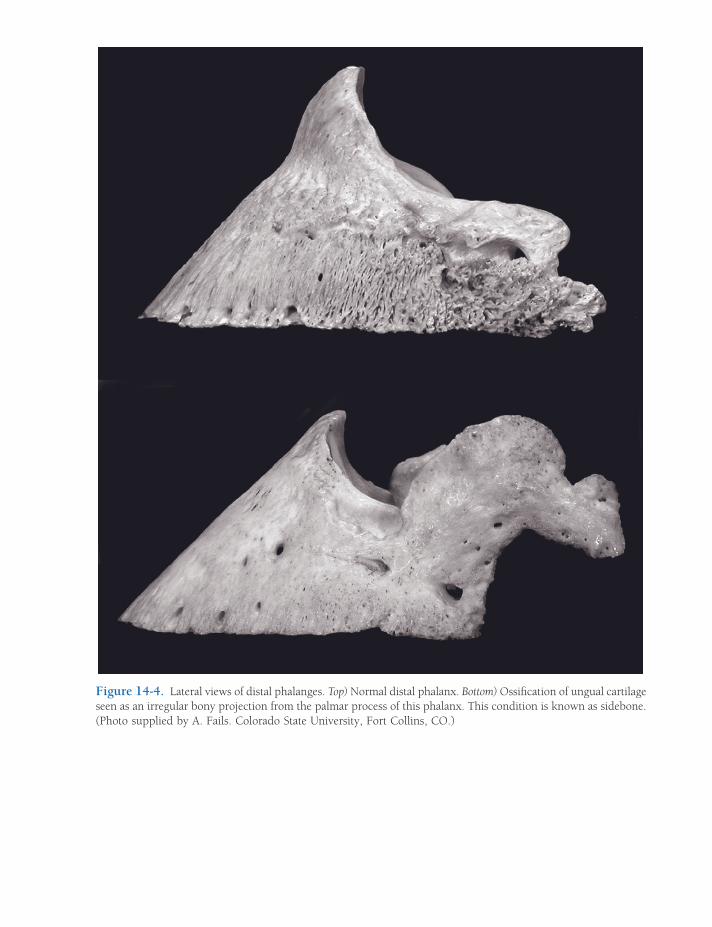
Figure 14-4. Lateral views of distal phalanges. Top) Normal distal phalanx. Bottom) Ossifi cation of ungual cartilage seen as an irregular bony projection from the palmar process of this phalanx. This condition is known as sidebone. (Photo supplied by A. Fails. Colorado State University, Fort Collins, CO.)

242 / ANATOMY AND PHYSIOLOGY OF FARM ANIMALS
Periopliccorium
Coronary corium
Laminar corium
Solarcorium
Perioplic sulcus
Coronary sulcus
Stratummedium
Epidermallaminae ofstratuminternum
Corium of the frog
Frog stay
Internal surfaceof the sole
White line
Figure 14-5. Dissected view of the relation of the hoof to the underlying corium. (Reprinted with permission of Wiley-Blackwell from Stashak T.S. Adams’ Lameness in Horses. 5th ed. Baltimore: Lippincott Williams & Wilkins, 2002.)
fl ared wall and up-curled toe. This chronic condition is founder.
The sole of the foot is a concave keratinized plate that attaches to the palmar/plantar surface of the third phalanx. It includes the entire ground surface of the foot not occupied by the wall or the frog (discussed later). Normally, the concavity of the sole allows the wall and frog to bear most of the weight and wear (Fig. 14-6).
Laypeople and farriers recognize a narrow band of the deepest part of the stratum medium, which is typically slightly lighter in coloration than the rest of the hoof wall. This is the white line. The white line is useful as a landmark for driving nails in shoeing. A properly directed nail started at or outside the white line will not touch any sensitive structures of the foot.
The frog (cuneus unguis) is a specialized, wedge-shaped pad near the heels of the foot (Fig. 14-6). The horn of the frog is relatively pliable, and the compression of the frog against the ground is important for return of blood from the foot. Deep grooves (collateral sulci or para-cuneal sulci) demarcate the sides of the frog from adjacent sole, and a single central sulcus corresponds to a spine of tissue called the frog stay on the dorsal (deep) side of the frog (Fig. 14-5). Deep to the frog is the digital cushion, a thick wedge of fi brofatty tissue (Fig. 14-2).
Tendons
No muscle bellies extend below the carpus or tarsus in the mature horse. The tendons of
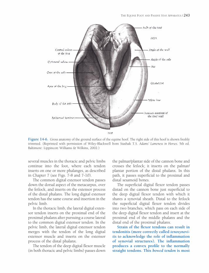
THE EQUINE FOOT AND PASSIVE STAY APPARATUS / 243
Figure 14-6. Gross anatomy of the ground surface of the equine hoof. The right side of this hoof is shown freshly trimmed. (Reprinted with permission of Wiley-Blackwell from Stashak T.S. Adams’ Lameness in Horses. 5th ed. Baltimore: Lippincott Williams & Wilkins, 2002.)
several muscles in the thoracic and pelvic limbs continue into the foot, where each tendon inserts on one or more phalanges, as described in Chapter 7 (see Figs. 7-8 and 7-10).
The common digital extensor tendon passes down the dorsal aspect of the metacarpus, over the fetlock, and inserts on the extensor process of the distal phalanx. The long digital extensor tendon has the same course and insertion in the pelvic limb.
In the thoracic limb, the lateral digital exten-sor tendon inserts on the proximal end of the proximal phalanx after pursuing a course lateral to the common digital extensor tendon. In the pelvic limb, the lateral digital extensor tendon merges with the tendon of the long digital extensor muscle and inserts on the extensor process of the distal phalanx.
The tendon of the deep digital fl exor muscle (in both thoracic and pelvic limbs) passes down
the palmar/plantar side of the cannon bone and crosses the fetlock; it inserts on the palmar/plantar portion of the distal phalanx. In this path, it passes superfi cial to the proximal and distal sesamoid bones.
The superfi cial digital fl exor tendon passes distad on the cannon bone just superfi cial to the deep digital fl exor tendon with which it shares a synovial sheath. Distal to the fetlock the superfi cial digital fl exor tendon divides into two branches, which pass on each side of the deep digital fl exor tendon and insert at the proximal end of the middle phalanx and the distal end of the proximal phalanx.
Strain of the fl exor tendons can result in tendonitis (more correctly called tenosynovi-tis to acknowledge the role of infl ammation of synovial structures). The infl ammation produces a convex profi le to the normally straight tendons. This bowed tendon is most

Periople
Coronary corium
Stratum mediumof the hoof wall
Interdigitation ofsensitive laminaeand insensitivelaminae
Stratum medium
Laminar corium
Coronaryband
Figure 14-7. Relationship between the corium of the coronary region (coronet) and the hoof wall. Insert) The gross location of this section. Note the cross-sectional appearance of the laminae. (Reprinted with permission of Wiley-Blackwell from Stashak T.S. Adams’ Lameness in Horses. 5th ed. Baltimore: Lippincott Williams & Wilkins, 2002.)
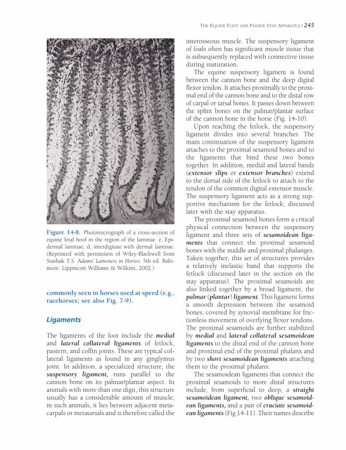
THE EQUINE FOOT AND PASSIVE STAY APPARATUS / 245
commonly seen in horses used at speed (e.g., racehorses; see also Fig. 7-9).
Ligaments
The ligaments of the foot include the medial and lateral collateral ligaments of fetlock, pastern, and coffi n joints. These are typical col-lateral ligaments as found in any ginglymus joint. In addition, a specialized structure, the suspensory ligament, runs parallel to the cannon bone on its palmar/plantar aspect. In animals with more than one digit, this structure usually has a considerable amount of muscle; in such animals, it lies between adjacent meta-carpals or metatarsals and is therefore called the
interosseous muscle. The suspensory ligament of foals often has signifi cant muscle tissue that is subsequently replaced with connective tissue during maturation.
The equine suspensory ligament is found between the cannon bone and the deep digital fl exor tendon. It attaches proximally to the proxi-mal end of the cannon bone and to the distal row of carpal or tarsal bones. It passes down between the splint bones on the palmar/plantar surface of the cannon bone in the horse (Fig. 14-10).
Upon reaching the fetlock, the suspensory ligament divides into several branches. The main continuation of the suspensory ligament attaches to the proximal sesamoid bones and to the ligaments that bind these two bones together. In addition, medial and lateral bands (extensor slips or extensor branches) extend to the dorsal side of the fetlock to attach to the tendon of the common digital extensor muscle. The suspensory ligament acts as a strong sup-portive mechanism for the fetlock, discussed later with the stay apparatus.
The proximal sesamoid bones form a critical physical connection between the suspensory ligament and three sets of sesamoidean liga-ments that connect the proximal sesamoid bones with the middle and proximal phalanges. Taken together, this set of structures provides a relatively inelastic band that supports the fetlock (discussed later in the section on the stay apparatus). The proximal sesamoids are also linked together by a broad ligament, the palmar (plantar) ligament. This ligament forms a smooth depression between the sesamoid bones, covered by synovial membrane for fric-tionless movement of overlying fl exor tendons. The proximal sesamoids are further stabilized by medial and lateral collateral sesamoidean ligaments to the distal end of the cannon bone and proximal end of the proximal phalanx and by two short sesamoidean ligaments attaching them to the proximal phalanx.
The sesamoidean ligaments that connect the proximal sesamoids to more distal structures include, from superfi cial to deep, a straight sesamoidean ligament, two oblique sesamoid-ean ligaments, and a pair of cruciate sesamoid-ean ligaments (Fig 14-11). Their names describe
Figure 14-8. Photomicrograph of a cross-section of equine fetal hoof in the region of the laminae. e, Epi-dermal laminae; d, interdigitate with dermal laminae. (Reprinted with permission of Wiley-Blackwell from Stashak T.S. Adams’ Lameness in Horses. 5th ed. Balti-more: Lippincott Williams & Wilkins, 2002.)

Figure 14-9. Chronic laminitis (founder) is characterized by loss of congruity between epidermal and dermal laminae. The distal phalanx is rotated away from the hoof wall by the distractive force of the deep digital fl exor tendon. Top) Normal hoof in sagittal section. Arrows emphasize the parallel surfaces of the hoof wall and distal phalanx. Bottom) Hoof with chronic laminitis and rotation of the distal phalanx. (Photo supplied by A. Fails. Colorado State University, Fort Collins, CO)
246

Figure 14-10. Palmar view of the equine foot, illustrating the elements of the suspensory ligament and proximal sesa-moids. The suspensory ligament and the ligaments of the proximal sesamoids form a continuous ligamentous band that passes from the carpus and proximal metacarpus to the proximal and middle phalanges. This part of the stay appa-ratus is the most important support for the fetlock joint.
247
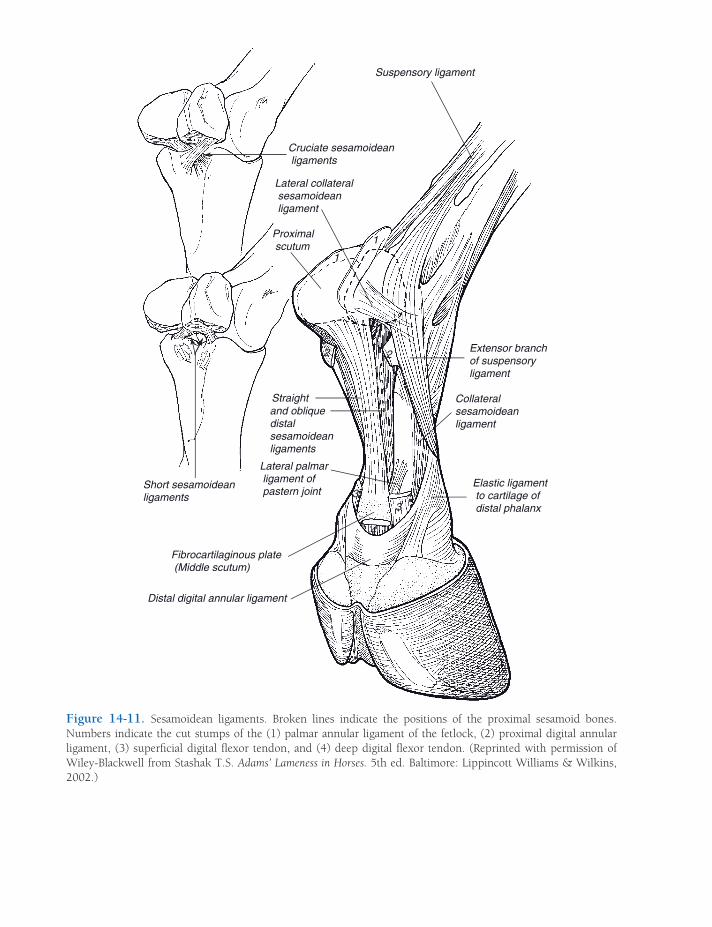
Figure 14-11. Sesamoidean ligaments. Broken lines indicate the positions of the proximal sesamoid bones. Numbers indicate the cut stumps of the (1) palmar annular ligament of the fetlock, (2) proximal digital annular ligament, (3) superfi cial digital fl exor tendon, and (4) deep digital fl exor tendon. (Reprinted with permission of Wiley-Blackwell from Stashak T.S. Adams’ Lameness in Horses. 5th ed. Baltimore: Lippincott Williams & Wilkins, 2002.)
Suspensory ligament
Cruciate sesamoidean ligaments
Lateral collateral sesamoidean ligament
Proximal scutum
Straight and oblique distal sesamoidean ligaments
Lateral palmar ligament of pastern joint
Short sesamoidean ligaments
Fibrocartilaginous plate (Middle scutum)
Distal digital annular ligament
Extensor branch of suspensory ligament
Collateral sesamoidean ligament
Elastic ligament to cartilage of distal phalanx
1
1
2 2
33
4
THE EQUINE FOOT AND PASSIVE STAY APPARATUS / 249
their gross appearance; the straight ligament attaches to the middle phalanx, and the others to the proximal phalanx. As a group, these liga-ments are often called the distal sesamoidean ligaments, but as this name sounds as though they are associated with the distal sesamoid (navicular) bone, it is probably best avoided.
The distal sesamoid bone (navicular bone) has a number of ligaments associated with it. Medial and lateral collateral ligaments attach the navicular to the distal phalanx, and an addi-tional unpaired ligament (impar ligament) extends from the distal sesamoid to the solar surface of the distal phalanx. The proximal face of the navicular bone is connected to the middle phalanx and the deep digital fl exor tendon by the T ligament (Fig. 14-2).
The many ligaments and tendons of the equine digit are bound together with a number of encircling annular ligaments. These are rea-sonably discrete thickenings of the local deep fascia. The palmar/plantar annular ligament arises from the proximal sesamoids and wraps around the palmar/plantar aspect of the fetlock, where its collagenous fi bers blend with the fl exor tendon sheath. The proximal digital annular ligament is more distal, forming a supportive sleeve on the palmar/plantar aspect of the pastern. Finally, the distal digital annular liga-ment forms a sling around the deep digital fl exor tendon near the insertion of the superfi cial digital fl exor tendon, holding the deep digital fl exor tendon close to the pastern (Fig. 14-11).
Synovial Structures
The tendons of the superfi cial and deep digital fl exor muscles share a synovial sheath that has its most proximal extent some 5 to 8 cm above the fetlock and that extends distad to the middle of the middle phalanx. The navicular bursa lies between the navicular bone and the deep digital fl exor tendon. Infl ammation of this bursa is one component of navicular disease.
Navicular disease is a common cause of forelimb lameness in Quarter Horses and Thoroughbreds. A variety of pathologies are described with navicular disease, and not
every affected horse shows all of them. These include erosion of the articular carti-lages of the navicular bone, bursitis of the navicular bursa, adhesions between the deep digital fl exor tendon and navicular bone, and erosions or necrosis of the navicular bone. There is a hereditary component to the disease, probably related to a certain conformational type, often described as a heavy horse on small feet with upright pas-terns, which exposes the navicular bone and associated structures to excessive concus-sive forces. Improper trimming of the hoof, so that the toe is left too long and/or heels overshortened, increases the stress on the deep digital fl exor tendon and may aggra-vate a predisposition to navicular disease.
The structure of the synovial joints between the phalanges and between the cannon and proximal phalanx is typical (see Chapter 6). The joint cavity of the fetlock is especially vo luminous to accommodate the wide range of motion in this ginglymus joint. Part of the joint cavity extends proximad between the cannon bone and suspensory ligament. Accumulation of excess synovial fl uid within this palmar (plantar) recess may be associated with the trauma of hard training. This produces a visible distension of the recess called wind puffs or wind galls.
Function
The primary function of the foot can be summed up in the word locomotion. A highly adapted aid to effi cient locomotion, the foot absorbs concussion, stores energy in its elastic tissues, and provides leverage for muscles that insert on the bones within it. A secondary function of the equine foot is standing support. The famous ability of horses to sleep while standing owes itself primarily to the ligamentous structures of the foot and other more proximal parts of the limb.
Concussion and Storage of Energy
A large part of the mechanism for absorbing concussion depends on angulation of the joints
250 / ANATOMY AND PHYSIOLOGY OF FARM ANIMALS
of the limbs at the time of impact and immedi-ately following contact of the foot with the ground. The muscles, tendons, and ligaments act as springs that absorb the shock of impact by permitting some fl exion of the shoulder and elbow and hyperextension of fetlock, pastern, and coffi n joints. Some of the energy of the foot striking the ground is stored as ligamentous and tendinous structures stretch; this energy is released as the foot leaves the ground. The rebound of ligaments and tendons straightens the joints and aids in lifting the foot, so that very little energy is expended on these parts of gait.
The hoof and its contents absorb concussion because of the elasticity of the hoof wall, ungual cartilages, digital cushion, and frog. As the frog strikes the ground, both the digital cushion and the frog are compressed, widening and thin-ning them. Pressure on the bars, the ungual cartilages, and the wall spreads the heels and forces blood out of the vascular bed of the foot. The direct cushioning effect of the frog and digital cushion is enhanced by the resiliency of the wall and the hydraulic shock-absorbing effect of the blood in the hoof. At the same time that the hoof is spread by frog pressure, blood is forced out of the vascular structures of the foot, which not only absorbs concussion but also pumps blood out of the foot and into the veins of the leg against gravity. This pumping action of the foot is an important means of returning venous blood from the foot to the general circulation.
Horses confi ned to a stall or otherwise immobilized for extended periods are often deprived of the pumping benefi ts of the active foot, with the result that tissue fl uid accumulates in the distal limb. This gives the appearance of swelling, which is collo-quially referred to as stocking up, in the pas-terns, fetlocks, and sometimes even more proximal. The swelling, however, is not due to infl ammation, is not painful, and very quickly resolves when the horse is encour-aged to exercise for a time.
Flexibility of the hoof is also an important consideration in correct shoeing. For most purposes, the shoe should be nailed only as far back as the quarters so that the heels are
free to expand over the top of the shoe. For the same reason, most farriers apply shoes that are slightly wider in the heel than the foot to which they are affi xed. By this means, the expanding heels of the hoof are able to spread and still maintain contact with the shoe.
Stay Apparatus
Ligamentous structures that store and release energy during locomotion are also found proxi-mal to the foot. These elements of the stay apparatus are also the elements of the limb that permit the horse to stand with minimal physi-cal effort.
To understand the function of the stay appa-ratus, it is necessary to keep several concepts central: (1) Ligaments, while elastic, do not stretch nearly as much as muscle. Therefore tendons, because they connect with muscles, offer much less resistance to stretch than do ligaments. (2) When joints are loaded (i.e., bearing weight), they tend to collapse. (3) To keep joints from collapsing with minimal mus-cular effort, ligamentous straps must cross the joint to counteract the tendency to fold during weight bearing (Fig 14-12).
Thoracic Limb
The thoracic limb is in part affi xed to the trunk by the fan-shaped m. serratus ventralis, extend-ing from the scapula to the ribs. This muscle is characterized by a thick, tendinous layer that is capable of supporting the trunk without con-traction of the muscle fi bers. The weight of the trunk is thereby supported effortlessly by a slinglike structure composed of right and left mm. serrati ventrales (see Fig. 7-7); by this con-struct, much of the weight of the horse is trans-ferred to the thoracic limbs.
When bearing weight, the shoulder joint tends to fl ex, and the fetlock, pastern, and coffi n joints tend to hyperextend, while the carpus and elbow are relatively stable when loaded in the extended position. Components of the stay apparatus cross all of these joints,

THE EQUINE FOOT AND PASSIVE STAY APPARATUS / 251
counteracting their tendency to collapse under load (Fig. 14-13).
The extensor surface of the shoulder is crossed by the tendon that is the origin of the m. biceps brachii, a very broad, partly cartilagi-nous tendon that is continuous with a very dense fi brous band running through the length of the muscle belly. This fi brous band is con-tinuous with the tendons of insertion, including the lacertus fi brosus, a substantial band that blends into the deep fascia (epimysium) of the m. extensor carpi radialis. This feature of the m. biceps brachii creates a continuous ligamentous connection from the scapula through the length of the brachium, across the elbow, and to the proximal metacarpus via its connection with the tendon of the m. extensor carpi radialis. It is this band of connective tissue that will, without muscular effort, counteract fl exion of the shoul-der when the limb is bearing weight. This same continuous band, through its connection with the m. extensor carpi radialis, contributes further to the stability of the carpus in extension.
The elbow is surprisingly stable when fully extended and loaded. The m. triceps brachii, the primary extensor of the elbow, contributes to
this stability by maintaining a slight amount of tone, even when the horse is sleeping on his feet.
The rest of the stay apparatus is primarily concerned with holding the fetlock and to a lesser extent pastern and coffi n joints in a phys-iologic position. Without a ligamentous check on the palmar aspect of these joints, the fetlock would drop to the ground and the toe would point upward, with the sole off the ground when the limb was loaded.
The primary support of the fetlock and pastern joints comprises the suspensory liga-ment, the proximal sesamoid bones, and the ligaments of the proximal sesamoid bones. Recall that these structures form a continuous ligamentous connection between the palmar aspect of the carpus and proximal metacarpus distal to the proximal and middle phalanges.
The tendons of both digital fl exor muscles offer additional support to the fetlock and pastern joints, and the tendon of the deep digital fl exor muscle resists hyperextension of the coffi n joint. Remember, though, that muscle tends to stretch easily. These digital fl exors can support the distal joints without muscular effort
Figure 14-12. When the appendicular skeleton is loaded with weight (A), the joints tend to collapse (B). The stay apparatus is a series of ligamentous bands that cross the joints and passively prevent this collapse (C).

Figure 14-13. Stay apparatus of the thoracic limb. (Reprinted with permission of Wiley-Blackwell from Stashak T.S. Adams’ Lameness in Horses. 5th ed. Baltimore: Lippincott Williams & Wilkins, 2002.)

THE EQUINE FOOT AND PASSIVE STAY APPARATUS / 253
because they both feature accessory ligaments, ligamentous connections between more proxi-mal bones and the insertions of the tendons.
The superfi cial digital fl exor muscle pos-sesses a ligamentous head that attaches to the caudal aspect of the distal radius and joins the muscle’s tendon near the carpus. This acces-sory ligament of the superfi cial digital fl exor muscle is commonly known as the radial or proximal check ligament, and its presence creates a continuous, ligamentous band from the radius to the insertion of the tendon on the proximal and middle phalanges. This provides additional support to fetlock and pastern.
The deep digital fl exor muscle also features an accessory ligament, this one attaching to the caudal part of the carpal joint capsule and blending with the muscle’s tendon just distal to the carpus. This accessory ligament of the deep digital fl exor muscle is more commonly called the carpal or distal check ligament. It creates a continuous ligamentous band that extends from the carpus to the distal phalanx, support-ing all of the joints in the digit.
Injury to the fl exor tendons produces some dropping of the fetlock as some of the resistance to hyperextension is lost. If injury includes the deep digital fl exor tendon, the toe is likely to come off the ground, as this tendon is the only one resisting hyperexten-sion of the coffi n joint. The most devastating injuries, however, involve the suspensory ligament and/or the proximal sesamoid bones. Fractures of the proximal sesamoids are the most common of all fractures in the forelimb. If the fractures are complete (trans-verse fractures of both sesamoids), or if the suspensory ligament is transected, the liga-ment loses its connections to the phalanges and the fetlock will drop to the ground. Such an injury is often irreparable and may neces-sitate destruction of the horse.
Pelvic Limb
Distal to the tarsus, the stay apparatus in the pelvic limb is more or less identical to that of the thoracic limb (Fig. 14-14). The accessory
ligament of the deep digital fl exor muscle (tarsal check ligament) arises from the plantar aspect of the tarsus and proximal metatarsus; this accessory ligament is rather long and very slender and may even be absent in some indi-viduals. The superfi cial digital fl exor muscle of the pelvic limb is nearly entirely tendinous and therefore needs no accessory ligament to create a continuous band from origin (caudal distal femur) to insertion on proximal and middle phalanges. Furthermore, the tendon of the superfi cial digital fl exor muscle inserts on the tuber calcanei, providing, in effect, a check ligament at this point.
For the pelvic limb to support weight without collapsing, the stifl e and hock must be pre-vented from fl exing. This is accomplished by one mechanism to lock the stifl e in extension and a second mechanism (the reciprocal appa-ratus) that guarantees that the hock will always fl ex and extend in unison with the stifl e. The reciprocal apparatus provides that as long as the stifl e is locked in extension, the hock will be likewise.
Recall that the equine patella features a hook-shaped medial parapatellar cartilage and three patellar tendons that insert on the tibial tuberosity (Fig. 6-8). During movement, the patella glides up and down the femoral trochlea as the quadriceps muscle contracts and relaxes, respectively. When the horse stands at rest, however, the patella is drawn proximad so that the parapatellar cartilage and medial patellar tendon are hooked over the large medial ridge of the trochlea. In this position, the stifl e is held in extension with minimal muscular effort. Typically, the horse activates this mechanism in one pelvic limb, resting the other with the toe on the ground; this posture is sometimes referred to as standing “hip-shot.”
The hock and stifl e normally fl ex and extend in unison because of the reciprocal apparatus, a combination of ligamentous structures con-necting the femur with the tarsus and metatar-sus (Fig. 14-15). On the cranial aspect of the crus, the peroneus tertius, an entirely ligamen-tous structure, extends from the region of the lateral femoral condyle to insertions on the tarsus and proximal metatarsus. In doing so, it

Figure 14-14. Stay apparatus of the pelvic limb. (Reprinted with permission of Wiley-Blackwell from Stashak T.S. Adams’ Lameness in Horses. 5th ed. Baltimore: Lippincott Williams & Wilkins, 2002.)
254

Figure 14-15. Reciprocal apparatus. (Reprinted with permission of Wiley-Blackwell from Stashak T.S. Adams’ Lameness in Horses. 5th ed. Baltimore: Williams & Wilkins, 2002.)
255
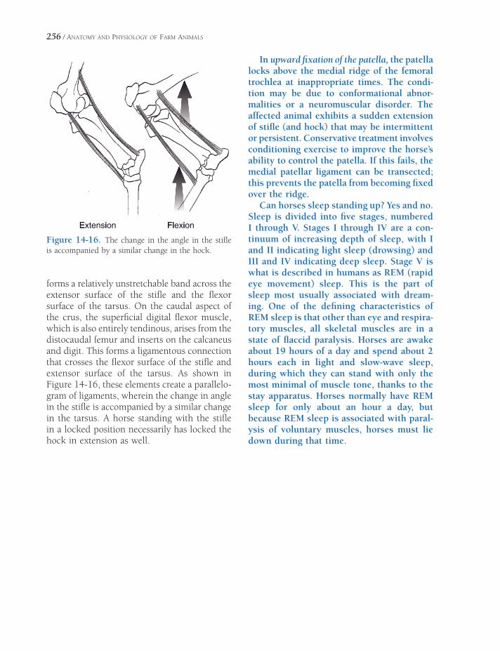
256 / ANATOMY AND PHYSIOLOGY OF FARM ANIMALS
forms a relatively unstretchable band across the extensor surface of the stifl e and the fl exor surface of the tarsus. On the caudal aspect of the crus, the superfi cial digital fl exor muscle, which is also entirely tendinous, arises from the distocaudal femur and inserts on the calcaneus and digit. This forms a ligamentous connection that crosses the fl exor surface of the stifl e and extensor surface of the tarsus. As shown in Figure 14-16, these elements create a parallelo-gram of ligaments, wherein the change in angle in the stifl e is accompanied by a similar change in the tarsus. A horse standing with the stifl e in a locked position necessarily has locked the hock in extension as well.
ExtensionE i FlexionFl i
In upward fi xation of the patella, the patella locks above the medial ridge of the femoral trochlea at inappropriate times. The condi-tion may be due to conformational abnor-malities or a neuromuscular disorder. The affected animal exhibits a sudden extension of stifl e (and hock) that may be intermittent or persistent. Conservative treatment involves conditioning exercise to improve the horse’s ability to control the patella. If this fails, the medial patellar ligament can be transected; this prevents the patella from becoming fi xed over the ridge.
Can horses sleep standing up? Yes and no. Sleep is divided into fi ve stages, numbered I through V. Stages I through IV are a con-tinuum of increasing depth of sleep, with I and II indicating light sleep (drowsing) and III and IV indicating deep sleep. Stage V is what is described in humans as REM (rapid eye movement) sleep. This is the part of sleep most usually associated with dream-ing. One of the defi ning characteristics of REM sleep is that other than eye and respira-tory muscles, all skeletal muscles are in a state of fl accid paralysis. Horses are awake about 19 hours of a day and spend about 2 hours each in light and slow-wave sleep, during which they can stand with only the most minimal of muscle tone, thanks to the stay apparatus. Horses normally have REM sleep for only about an hour a day, but because REM sleep is associated with paral-ysis of voluntary muscles, horses must lie down during that time.
Figure 14-16. The change in the angle in the stifl e is accompanied by a similar change in the hock.

BLOOD AND OTHER BODY FLUIDS
Single-celled organisms that live in seawater have an external environment that provides
all of the needs of the organisms, such as food, disposal of excreted wastes, and relatively con-stant conditions for maintenance of life. As the complexity of organisms increases, the problem of supplying each cell with a proper environ-ment becomes more acute. Higher forms of animals have developed circulating blood, and the fl uids derived from it, as a means of main-taining a relatively constant environment for all cells.
Blood
Most of the functions of blood are included in the following list:
1. Distribution of nutrients absorbed from the digestive tract
2. Transport of oxygen from the lungs to cells throughout the body
3. Transport of carbon dioxide from meta-bolizing cells to the lungs
4. Transport of waste products from meta-bolizing cells to the kidneys for excretion
5. Transport of hormones from endocrine glands to target cells
6. Assistance in body temperature control by transporting heat from deeper structures to the surface of the body
7. Assistance in maintaining a constant pH of body fl uids by providing chemical buffers
BloodFormed Elements of Blood and
Hematopoiesis
Erythrocytes
Platelets
Leukocytes
Plasma and SerumBlood pHHemostasis and Coagulation
Platelets and the Endothelium
Intrinsic and Extrinsic Coagulation
Pathways
LymphSerous Fluids
Chapter 15
257

258 / ANATOMY AND PHYSIOLOGY OF FARM ANIMALS
8. Assistance with the prevention of excessive loss of blood from injuries by providing proteins and other factors necessary for blood coagulation
9. Assistance with the defense of the body against disease by providing antibodies, cells, and other factors of body defense (see Chapter 16)
Blood consists of cells and other cell-like formed elements suspended in a fl uid called plasma. Some of its functions are specifi c to individual cells; for example, erythrocytes are primarily responsible for the transport of oxygen. Other functions, such as assisting with body defense, involve a variety of blood cells and other plasma components. Blood volume is the total amount of blood in an animal’s body, including formed elements and plasma. Typical values given as a percentage of body weight are 7–9%. Lean, muscular, athletic animals tend to have higher percentages than animals with more body fat. (Specifi c gravity is an index, or ratio, of the weight of a substance to the weight of an equal volume of water. A specifi c gravity greater than 1 means that an equal volume of the substance weighs more than water, at 1 g/mL. Blood and plasma have slightly higher specifi c gravities than water, pri-marily because of the blood cells and proteins, but the slight difference is usually disregarded when estimating blood or plasma volumes based on body weight.)
A large number of plasma proteins are sus-pended in the plasma, and electrolytes (e.g., Na+, K+, and Cl−) and other substances (e.g., glucose and urea) are dissolved in the plasma. While there are some minor species differences (e.g., adult ruminants typically have slightly lower values for blood glucose than other mammals), the values in Table 15-1 are typical normal ranges for the major chemical constitu-ents of mammalian plasma.
Plasma is a subdivision of a larger body fl uid compartment known as extracellular fl uid, or ECF (see Chapter 2). Interstitial fl uid, fl uid outside of cells but not within vessels, is the other subdivision of the ECF compartment. The primary difference between interstitial fl uid and plasma is that plasma contains a much higher concentration of proteins that cannot easily diffuse through the walls of capillaries.
Formed Elements of Blood and Hematopoiesis
The formed elements of the blood include erythrocytes (red blood cells), leukocytes (white blood cells), and platelets. Because erythrocytes and platelets both lack nuclei, they are not typical cells (see Plate II).
Hematopoiesis is the formation and devel-opment of all formed elements of blood (eryth-rocytes, leukocytes, and platelets), and all of these have a common ancestor, pluripotent
Table 15-1. Representative Values for Selected Constituents of Typical Mammalian Plasma
Constituents Units of Measurement Typical Range
Sodium mEq/L 135–155
Potassium mEq/L 3.0–5.5
Chloride mEq/L 95–110
Bicarbonate mEq/L 22–26
Total protein g/dL 6–8
Albumin g/dL 3–4
Blood urea nitrogen mg/dL 10–25
Creatinine mg/dL 1.0–1.5
Glucose mg/dL 70–100
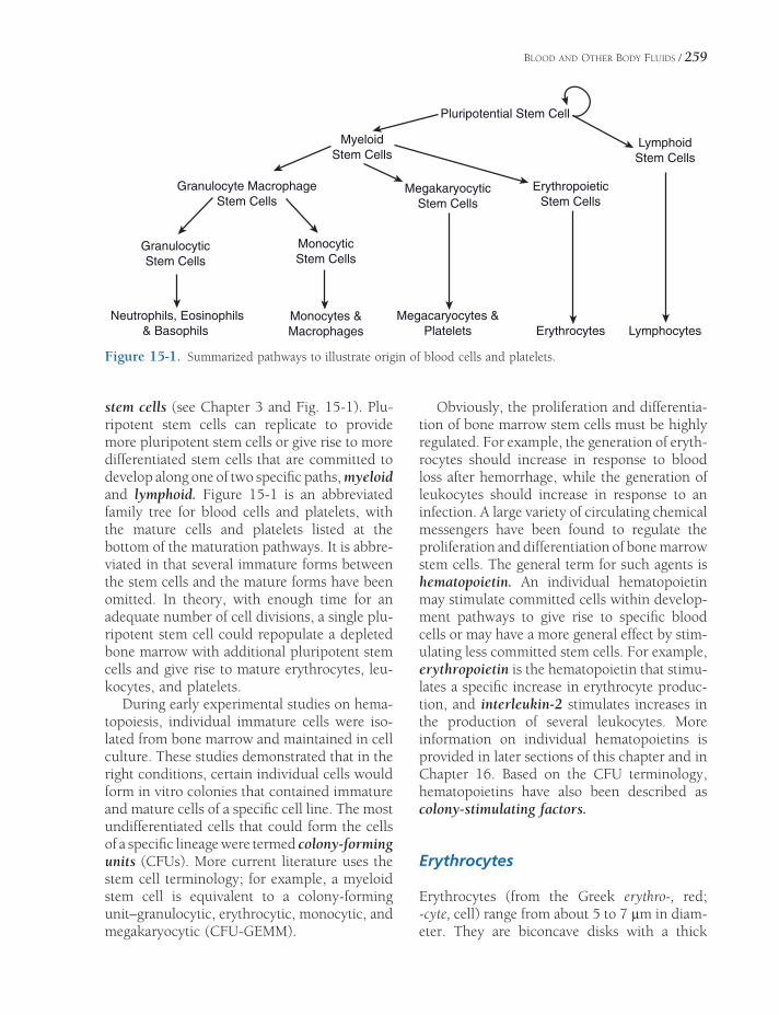
BLOOD AND OTHER BODY FLUIDS / 259
stem cells (see Chapter 3 and Fig. 15-1). Plu-ripotent stem cells can replicate to provide more pluripotent stem cells or give rise to more differentiated stem cells that are committed to develop along one of two specifi c paths, myeloid and lymphoid. Figure 15-1 is an abbreviated family tree for blood cells and platelets, with the mature cells and platelets listed at the bottom of the maturation pathways. It is abbre-viated in that several immature forms between the stem cells and the mature forms have been omitted. In theory, with enough time for an adequate number of cell divisions, a single plu-ripotent stem cell could repopulate a depleted bone marrow with additional pluripotent stem cells and give rise to mature erythrocytes, leu-kocytes, and platelets.
During early experimental studies on hema-topoiesis, individual immature cells were iso-lated from bone marrow and maintained in cell culture. These studies demonstrated that in the right conditions, certain individual cells would form in vitro colonies that contained immature and mature cells of a specifi c cell line. The most undifferentiated cells that could form the cells of a specifi c lineage were termed colony-forming units (CFUs). More current literature uses the stem cell terminology; for example, a myeloid stem cell is equivalent to a colony-forming unit–granulocytic, erythrocytic, monocytic, and megakaryocytic (CFU-GEMM).
Obviously, the proliferation and differentia-tion of bone marrow stem cells must be highly regulated. For example, the generation of eryth-rocytes should increase in response to blood loss after hemorrhage, while the generation of leukocytes should increase in response to an infection. A large variety of circulating chemical messengers have been found to regulate the proliferation and differentiation of bone marrow stem cells. The general term for such agents is hematopoietin. An individual hematopoietin may stimulate committed cells within develop-ment pathways to give rise to specifi c blood cells or may have a more general effect by stim-ulating less committed stem cells. For example, erythropoietin is the hematopoietin that stimu-lates a specifi c increase in erythrocyte produc-tion, and interleukin-2 stimulates increases in the production of several leukocytes. More information on individual hematopoietins is provided in later sections of this chapter and in Chapter 16. Based on the CFU terminology, hematopoietins have also been described as colony-stimulating factors.
Erythrocytes
Erythrocytes (from the Greek erythro-, red; -cyte, cell) range from about 5 to 7 μm in diam-eter. They are biconcave disks with a thick
Figure 15-1. Summarized pathways to illustrate origin of blood cells and platelets.
Pluripotential Stem Cell
MyeloidStem Cells
LymphoidStem Cells
Granulocyte MacrophageStem Cells
MegakaryocyticStem Cells
ErythropoieticStem Cells
GranulocyticStem Cells
MonocyticStem Cells
Neutrophils, Eosinophils& Basophils
Monocytes &Macrophages
Megacaryocytes &Platelets Erythrocytes Lymphocytes
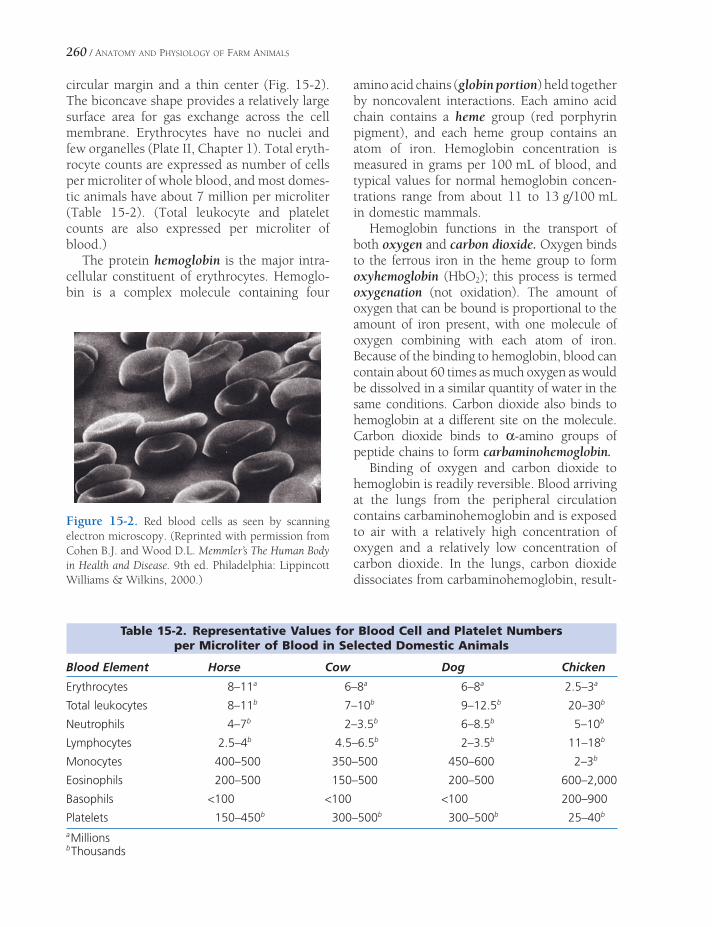
260 / ANATOMY AND PHYSIOLOGY OF FARM ANIMALS
circular margin and a thin center (Fig. 15-2). The biconcave shape provides a relatively large surface area for gas exchange across the cell membrane. Erythrocytes have no nuclei and few organelles (Plate II, Chapter 1). Total eryth-rocyte counts are expressed as number of cells per microliter of whole blood, and most domes-tic animals have about 7 million per microliter (Table 15-2). (Total leukocyte and platelet counts are also expressed per microliter of blood.)
The protein hemoglobin is the major intra-cellular constituent of erythrocytes. Hemoglo-bin is a complex molecule containing four
amino acid chains (globin portion) held together by noncovalent interactions. Each amino acid chain contains a heme group (red porphyrin pigment), and each heme group contains an atom of iron. Hemoglobin concentration is measured in grams per 100 mL of blood, and typical values for normal hemoglobin concen-trations range from about 11 to 13 g/100 mL in domestic mammals.
Hemoglobin functions in the transport of both oxygen and carbon dioxide. Oxygen binds to the ferrous iron in the heme group to form oxyhemoglobin (HbO2); this process is termed oxygenation (not oxidation). The amount of oxygen that can be bound is proportional to the amount of iron present, with one molecule of oxygen combining with each atom of iron. Because of the binding to hemoglobin, blood can contain about 60 times as much oxygen as would be dissolved in a similar quantity of water in the same conditions. Carbon dioxide also binds to hemoglobin at a different site on the molecule. Carbon dioxide binds to α-amino groups of peptide chains to form carbaminohemoglobin.
Binding of oxygen and carbon dioxide to hemoglobin is readily reversible. Blood arriving at the lungs from the peripheral circulation contains carbaminohemoglobin and is exposed to air with a relatively high concentration of oxygen and a relatively low concentration of carbon dioxide. In the lungs, carbon dioxide dissociates from carbaminohemoglobin, result-
Table 15-2. Representative Values for Blood Cell and Platelet Numbers per Microliter of Blood in Selected Domestic Animals
Blood Element Horse Cow Dog Chicken
Erythrocytes 8–11a 6–8a 6–8a 2.5–3a
Total leukocytes 8–11b 7–10b 9–12.5b 20–30b
Neutrophils 4–7b 2–3.5b 6–8.5b 5–10b
Lymphocytes 2.5–4b 4.5–6.5b 2–3.5b 11–18b
Monocytes 400–500 350–500 450–600 2–3b
Eosinophils 200–500 150–500 200–500 600–2,000
Basophils <100 <100 <100 200–900
Platelets 150–450b 300–500b 300–500b 25–40b
a Millionsb Thousands
Figure 15-2. Red blood cells as seen by scanning electron microscopy. (Reprinted with permission from Cohen B.J. and Wood D.L. Memmler’s The Human Body in Health and Disease. 9th ed. Philadelphia: Lippincott Williams & Wilkins, 2000.)

BLOOD AND OTHER BODY FLUIDS / 261
ing in the formation of hemoglobin, which can bind oxygen to become oxyhemoglobin. When the blood containing oxyhemoglobin returns to the peripheral tissues that are relatively defi -cient in oxygen, the bound oxygen is released to the tissues. Carbon dioxide can then bind to hemoglobin to continue the cycle.
Methemoglobin is a true oxidation product of hemoglobin that is unable to transport oxygen because the iron is in the ferric (Fe3+) rather than the ferrous (Fe2+) state. Certain chemicals, such as nitrites and chlorates, produce methemoglobinemia (methemoglo-bin in the blood). Nitrate poisoning has been reported in cattle grazing on highly fertil-ized plant growth. In these cases, nitrates in the plants are converted to nitrites in the rumen and cause the formation of methemo-globin when absorbed into the blood.
Carboxyhemoglobin is a more stable compound formed when carbon monoxide combines with hemoglobin. The affi nity of hemoglobin for carbon monoxide is 210 times that of its affi nity for O2. The carboxy-hemoglobin is unable to carry oxygen, and the animal essentially suffocates, although the blood is typically cherry red.
Erythropoiesis and Degradation of Eryth-rocytes. Erythrocyte formation, called ery-thropoiesis, is regulated by the hormone erythropoietin, a glycoprotein hormone that is released from specifi c cells of the kidneys in response to reduced oxygen delivery. For example, when animals move from low alti-tudes to high altitudes, where less oxygen is available, erythropoietin release is increased. Erythropoietin acts on stem cells in the bone marrow to increase the production of erythro-cytes. The increase in erythrocyte number is not immediate (it requires some days) because mature erythrocytes must be formed from stimulated stem cells before release from the bone marrow. The larger number of erythro-cytes increases oxygen delivery to the kidneys, and this has a negative feedback effect on erythropoietin release. It is via this feedback that a stable rate of erythrocyte production is maintained.
Recombinant human erythropoietin (rhEPO) has been used in appropriate clini-cal cases to stimulate erythrocyte produc-tion in both humans and animals. However, an antibody response to the human molecule occurs in more than 25% of animals, and these antibodies neutralize endogenous erythropoietin as well as the rhEPO. The inappropriate use of rhEPO has been reported in normal human athletes in an attempt to increase their exercise capacity. The potential for abusive use in animal ath-letes also exists.
Mature erythrocytes of mammals have no nuclei. Immature forms in the bone marrow do have nuclei, but these are lost during the latter stages of development. The appearance of large numbers of nucleated erythrocytes in the cir-culation is an indication that immature forms are being inappropriately released from the bone marrow. In birds and reptiles, nuclei nor-mally persist in the red cells throughout the life of the cells.
Typically, erythrocytes circulate for only 3 to 4 months after their release from the bone marrow. The removal and recycling of senes-cent erythrocytes is one function of a group of spe cialized cells, the monocyte–macrophage system (formally known as the reticuloendo-thelial system). The macrophages of this system are found fi xed or in residence in several sites throughout the body but are especially preva-lent in the spleen and liver. These macrophages are derived from circulating monocytes, hence the name of the system. The macrophages phagocytize intact erythrocytes or cellular debris and hemoglobin that are released when erythrocytes disintegrate in the blood. The macrophages degrade the globin portion of hemoglobin and release the resulting amino acids into the circulation. Iron is removed from the heme portion and released into the blood, where it is transported in combination with a protein, transferrin. Cells that require iron have cell membrane receptors that bind trans-ferrin, and this is the means by which cells can take up iron from the circulation. This includes hematopoietic cells in the bone marrow, which have the highest metabolic need for iron on a

262 / ANATOMY AND PHYSIOLOGY OF FARM ANIMALS
daily basis. Iron from the circulation also accu-mulates in the liver in a retrievable storage form, ferritin.
After the protein and iron are removed from hemoglobin, a green pigment, biliverdin, remains. This is reduced to bilirubin (a yellow-ish pigment), which is transported to the liver in combination with blood albumin. The liver conjugates it; then it passes to the gallbladder in the bile and fi nally to the intestine, where most of it is reduced to bilinogens. These are either excreted in the feces (giving the brown color to feces), or reabsorbed into the blood and then excreted in the urine as urobilinogen. The reduction in the intestine is accomplished by resident microorganisms.
Clinical Conditions and Procedures Involv-ing Erythrocytes. Icterus (jaundice) is a syn-drome characterized by a yellowish discoloration of skin, mucous membranes, and/or sclera. It results from accumulation of bilirubin in the blood and may be caused by liver damage, by occlusion of the bile ducts, or by an increased rate of erythrocyte destruction. In case of either liver damage or blockage of the bile ducts, the bile pigments are not secreted into the intestine but are resorbed into the circulatory system, causing icterus. When blood damage is excessive, as in some parasitic blood diseases, such as anaplasmosis, the bile pigments are liberated into the blood faster than the liver can conjugate and secrete them, and icterus results.
Hemolysis is the breakdown of erythrocytes and the release of hemoglobin. Bacterial toxins, snake venoms, blood parasites, and hypotonic solutions can cause so much hemolysis that the hemoglobin in plasma produces a reddish color, and the condition is called hemoglobine-mia. The hemoglobin can then be excreted in the urine, and this is termed hemoglobinuria (red water). Hemolysis can also be produced in a blood sample by physically disrupting the erythrocytes (e.g., forceful and rapid expulsion of blood from a syringe).
Hemagglutination is clumping of erythro-cytes. Erythrocytes have membrane proteins that act as antigens, and agglutination results if
erythrocytes with specifi c antigens are added to a solution containing appropriate antibodies. Blood typing is a general term describing the procedures used to identify the antigen or anti-gens in a given blood sample. Clumping may occur during transfusions of blood within the same species, such as between humans, if blood of the wrong type (i.e., containing the wrong combination of antigens) is used. At least seven blood types have been identifi ed in horses, and it is desirable to ensure that there is adequate matching of types before attempting transfu-sions between horses. Blood typing has also been done in cattle and dogs, but usually a single transfusion can be done between two cattle or between two dogs with little diffi culty. However, repeated transfusions can be prob-lematic, because the initial transfusion may provide unmatched antigens that stimulate the formation of antibodies. Blood types are inher-ited in all species, and they are often used to establish and monitor pedigrees.
Anemia (from the Greek an-, without; -emia, blood) results if either the number of functional erythrocytes or the quantity of hemoglobin per unit of blood is below normal. Anemia may be due to defi cient blood formation, as with dis-eases of the bone marrow, kidney disease with inadequate production of erythropoietin, or poor nutrition, including dietary defi ciency of iron, copper, vitamins, or amino acids. Anemia may also be caused by accelerated loss or destruction of erythrocytes, such as with hem-orrhage or parasites.
The hematocrit (packed cell volume) is the percentage by volume of whole blood that is erythrocytes. Routine hematocrit determina-tions require a glass tube treated to inhibit blood clotting (hematocrit tubes). Hematocrit tubes containing blood are centrifuged until the blood cells are packed in the lower end of the tube (Fig. 15-3). Hematocrits typically range from 35 to 45 for most mammalian species and are generally considered to be an indicator of the total erythrocyte count.
Hemoconcentration is a decrease in the fl uid component of blood with a resulting increase in the ratio of cells to fl uid. It is indicated by an excessively high erythrocyte count (polycy-
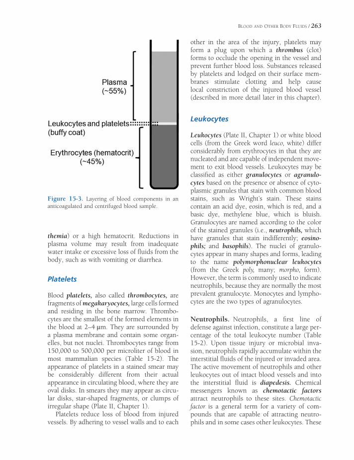
BLOOD AND OTHER BODY FLUIDS / 263
themia) or a high hematocrit. Reductions in plasma volume may result from inadequate water intake or excessive loss of fl uids from the body, such as with vomiting or diarrhea.
Platelets
Blood platelets, also called thrombocytes, are fragments of megakaryocytes, large cells formed and residing in the bone marrow. Thrombo-cytes are the smallest of the formed elements in the blood at 2–4 μm. They are surrounded by a plasma membrane and contain some organ-elles, but not nuclei. Thrombocytes range from 150,000 to 500,000 per microliter of blood in most mammalian species (Table 15-2). The appearance of platelets in a stained smear may be considerably different from their actual appearance in circulating blood, where they are oval disks. In smears they may appear as circu-lar disks, star-shaped fragments, or clumps of irregular shape (Plate II, Chapter 1).
Platelets reduce loss of blood from injured vessels. By adhering to vessel walls and to each
other in the area of the injury, platelets may form a plug upon which a thrombus (clot) forms to occlude the opening in the vessel and prevent further blood loss. Substances released by platelets and lodged on their surface mem-branes stimulate clotting and help cause local constriction of the injured blood vessel (described in more detail later in this chapter).
Leukocytes
Leukocytes (Plate II, Chapter 1) or white blood cells (from the Greek word leuco, white) differ considerably from erythrocytes in that they are nucleated and are capable of independent move-ment to exit blood vessels. Leukocytes may be classifi ed as either granulocytes or agranulo-cytes based on the presence or absence of cyto-plasmic granules that stain with common blood stains, such as Wright’s stain. These stains contain an acid dye, eosin, which is red, and a basic dye, methylene blue, which is bluish. Granulocytes are named according to the color of the stained granules (i.e., neutrophils, which have granules that stain indifferently; eosino-phils; and basophils). The nuclei of granulo-cytes appear in many shapes and forms, leading to the name polymorphonuclear leukocytes (from the Greek poly, many; morpho, form). However, the term is commonly used to indicate neutrophils, because they are normally the most prevalent granulocyte. Monocytes and lympho-cytes are the two types of agranulocytes.
Neutrophils. Neutrophils, a fi rst line of defense against infection, constitute a large per-centage of the total leukocyte number (Table 15-2). Upon tissue injury or microbial inva-sion, neutrophils rapidly accumulate within the interstitial fl uids of the injured or invaded area. The active movement of neutrophils and other leukocytes out of intact blood vessels and into the interstitial fl uid is diapedesis. Chemical messengers known as chemotactic factors attract neutrophils to these sites. Chemotactic factor is a general term for a variety of com-pounds that are capable of attracting neutro-phils and in some cases other leukocytes. These
Figure 15-3. Layering of blood components in an anticoagulated and centrifuged blood sample.

264 / ANATOMY AND PHYSIOLOGY OF FARM ANIMALS
factors may be produced by invading microor-ganisms, by leukocytes after interacting with microorganisms, or by damaged tissue.
Neutrophils are phagocytes; they engulf invading bacteria to destroy them. The destruc-tion involves the action of enzymes in the intra-cellular granules of the neutrophils and cell membrane–bound enzymes that are activated when phagocytic vesicles are completely formed. During phagocytosis, neutrophils may also release enzymes that contribute to the local infl ammation. In the process of responding to the potential infection, many of the attracted neutrophils are destroyed. Pus, the semiliquid material that results from the collective responses to a microbial invasion, may contain neutrophils and cellular debris. An abscess is an accumulation of pus that has been isolated by the formation of surrounding connective tissue.
Neutrophilia is an increase in the number of circulating neutrophils in the blood that occurs with bacterial infections. Circulating neutro-phils can increase as a result of an immediate release of existing neutrophils from the bone marrow and an increase in neutrophil produc-tion. Neutropenia refers to an abnormally low number of circulating neutrophils. The word endings -philia and -penia can be combined with the root names of the other leukocytes to indicate similar conditions (e.g., eosinophilia is an increase in eosinophils).
Eosinophils. Eosinophils contain red-staining cytoplasmic granules. Normally, eosinophils are less than 10% of total leukocytes (Table 15-2), but they may increase in number with allergic conditions and parasitism. Eosinophils are ameboid and somewhat phagocytic. Their primary function seems to be the regulation of allergic responses and tissue responses to para-sites. They act by removing antigen–antibody complexes, which stimulate allergic responses, and by inhibiting some of the mediators of allergic responses, such as histamine.
Basophils. Basophils contain blue-staining granules and are rarely seen in normal blood.
The granules of basophils contain multiple compounds, including heparin, which pre-vents blood clotting, and histamine, which relaxes smooth muscle of blood vessels and constricts smooth muscle in airways. Cells that are very similar but distinct, mast cells, are found in many sites throughout the body but are especially prevalent in connective tissue below an epithelial lining exposed to the exter-nal environment (e.g., dermis, wall of airways, and wall of gastrointestinal tract). Basophils and mast cells release the contents of their granules during allergic responses, and these chemicals contribute to the characteristic tissue responses. The release of the granules is medi-ated in part by the binding of specifi c antibod-ies and the associated allergen (antigen).
Monocytes. Monocytes are the largest of the circulating leukocytes. They are phagocytic and develop into even larger macrophages when they exit vessels and enter the tissues. Like neutrophils, monocytes are attracted by che-motactic factors to areas of tissue injury and microbial invasion. In addition to phagocytosis of tissue debris and microbes, macrophages have a major role in the overall initiation and regulation of infl ammatory and immune responses. During their response to tissue injury or microbial invasion, macrophages release numerous chemical messengers that coordinate the function of other cells respond-ing to injury or invasion. Macrophages also function in the processing of antigens, a neces-sary step in the initiation of an immune response. These functions are discussed in more detail in Chapter 16.
Lymphocytes. In most species, lymphocytes are the second most prevalent circulating leu-kocyte after neutrophils, but they are more prevalent than neutrophils in ruminants (Table 15-2). Based on their functions, three general types or groups of lymphocytes have been iden-tifi ed (B lymphocytes, T lymphocytes, and NK, or natural killer cells), but these types cannot be identifi ed by differences in appearance. In blood smears, lymphocytes vary in size, with a

BLOOD AND OTHER BODY FLUIDS / 265
relatively large nucleus surrounded by a small amount of cytoplasm (Plate II, Chapter 1). Lymphocytes function in specifi c immune responses and immune surveillance, and these functions are discussed in Chapter 16.
Differential White Blood Cell Counts. Dif-ferential counts indicate the percentage of each type of white cell in the blood sample (Table 15-2). The various types of leukocytes have different functions and respond differently to various types of infections or diseases, so dif-ferential counts can be useful for diagnosis. Differential counts taken over time can also be used to evaluate the response of an animal to infection or disease. A differential count is made by spreading a drop of whole blood thinly on a glass slide to form a blood smear. The smear is dried and stained with a blood stain, such as Wright’s stain. After staining is complete, the slide is examined with a micro-scope and the number of white cells of each kind is tabulated until a predetermined total number of white cells have been counted. The number counted is usually a multiple of 100, and the percentage of each leukocyte type observed in a given sample of blood is called the differential leukocyte count or differential white cell count. In reference laboratories, both total red and white cell counts are semiauto-matically determined by sophisticated labora-tory equipment.
Plasma and Serum
When a sample of blood is treated with an anticoagulant to prevent clotting and permit-ted to stand in a tube undisturbed, the cells gradually settle to the bottom, leaving a straw-colored fl uid above. This fl uid portion of the blood is plasma. When blood is allowed to clot, the cells are trapped in a meshwork of clotting proteins, leaving a yellow fl uid, the serum. Essentially, serum is plasma minus the plasma proteins responsible for producing the clot.
Plasma is about 92% water and 8% other substances. The kidneys are responsible for
maintaining constant proportions of water and other constituents of the plasma by the selective fi ltration and reabsorption of water and other substances from the blood plasma. The total osmolality of plasma at normal body tempera-ture is about 290 mOsm/kg. Osmolality is a measure of the number of osmotically active particles (not the mass of the particles) per unit of solute. The two predominant particles in plasma are sodium and chloride ions (Table 15-1), and these contribute the most to the total osmolality of plasma or serum.
The plasma proteins consist of two major types: albumin and globulins. Albumin is the most prevalent plasma protein and is the pre-dominant protein synthesized by the liver. Many small compounds and electrolytes (e.g., calcium ions) bind to albumin and circulate in plasma in this bound form. This prevents their rapid loss in the urine. Because albumin and other large proteins do not readily pass through capillary walls, they also provide an effective osmotic force to prevent excessive fl uid loss from capillaries into the interstitium.
The globulins in serum or plasma may be classifi ed according to their migration (separa-tion) by electrophoresis. α-Globulins and β-globulins are classes that are synthesized in the liver. Members of these classes have a variety of functions, including transport in a manner similar to albumin, body defense (see Chapter 16), and blood clotting. Many of the globulin proteins are inactive precursors of enzymes or substrates for enzymes involved in blood clot-ting (discussed later in this chapter).
The γ-globulins are synthesized by cells of the immune system. Most of the known circulating antibodies are included in the γ-globulin frac-tion. The γ-globulin content of the blood there-fore increases following vaccination and during recovery from disease. Immune serum or hyper-immune serum can be produced by repeatedly inoculating an animal with a specifi c antigen. Serum from that animal can then be injected into an animal susceptible to the same disease to provide passive protection for as long as the antibodies remain in the susceptible animal. This provides merely temporary immunity.

266 / ANATOMY AND PHYSIOLOGY OF FARM ANIMALS
Blood pH
A typical pH range for blood is 7.35 to 7.45, just slightly on the alkaline side of neutral. The pH of blood is kept within rather narrow limits by a variety of mechanisms that include contri-butions by the kidneys (see Chapter 23) and the respiratory system (see Chapter 19). Several chemical buffer systems in the plasma also con-tribute to the control of blood pH. The most important of these is the bicarbonate buffer system, and the bicarbonate ion is the base in this system. The bicarbonate ion is the second most prevalent anion in plasma (Table 15-1). In acidosis or acidemia the blood pH is abnor-mally low, and in alkalosis or alkalemia the pH is abnormally high.
Hemostasis and Coagulation
Hemostasis, the stoppage of bleeding, may involve three basic reactions: (1) constriction by the smooth muscle of the injured vessel to reduce the opening, (2) formation of a platelet plug to occlude the opening, and (3) clot for-mation to complete occlusion of the opening. Injuries to vessels do not require the formation of a clot (coagulation) if hemostasis can be achieved by the fi rst two reactions.
Platelets and the Endothelium
When a vessel is injured and the endothelial cell lining is damaged so that the underlying connective tissue is exposed, platelets adhere to collagen and other proteins in the connective tissue. This platelet adhesion results from binding between platelet cell membrane pro-teins and the connective tissue. The cell mem-brane of adhered platelets undergoes alterations, and secretory granules are also released. Plate-lets that have undergone these reactions are termed activated platelets. The presence of activated platelets stimulates other platelets to adhere to those already present. The collection of platelets forms a platelet plug that may be suffi cient (together with local vasoconstriction) to occlude extremely small openings in damaged
vessels and bring about hemostasis. Platelet aggregation is the term applied to the overall sequence of events responsible for the forma-tion of the platelet plug.
Platelet aggregation is also subject to regula-tion by two different eicosanoids, thromboxane A2
(TXA2) and prostacyclin (PGI2). TXA2 is a stimulant of platelet aggregation, and prostacy-clin inhibits platelet aggregation. The primary source of TXA2 in the area of a damaged vessel is adhered platelets that increase their synthesis of TXA2 after adhesion. The primary source of prostacyclin is intact, undamaged endothelial cells. As the platelet plug grows and extends to areas where endothelial cells remain intact, the local concentration of prostacyclin generated by undamaged endothelial cells acts to stop the growth of the platelet plug. TXA2 and serotonin (also released by adhered platelets) are both vasoconstrictors, stimulating smooth muscle contraction to assist with hemostasis.
Aspirin inhibits the formation of eico-sanoids by binding to and inhibiting cyclo-oxygenase, an enzyme necessary for their synthesis. TXA2 and prostacyclin are both eicosanoids, so aspirin initially reduces the formation of both. However, platelets do not have nuclei and cannot synthesize new enzymes, while nucleated endothelial cells can synthesize additional cyclooxygenase. Because of these differences in the two cell types, treatment with aspirin at an appropri-ate dose and an appropriate schedule pref-erentially reduces thromboxane synthesis by platelets. This is the basis for the use of aspirin when it is desirable to reduce the tendency for blood coagulation (e.g., humans with cardiac diseases and animals with heartworms.)
Some of the substances released by aggregat-ing platelets and some of the cell membrane components exposed by aggregated platelets act to promote coagulation. Thus, with larger sites of injury, more platelets aggregate and more stimulants of clotting are present in the local area. Coagulation is initiated when the local concentration of these substances reaches some critical level. Platelets are required for normal coagulation in response to vascular

BLOOD AND OTHER BODY FLUIDS / 267
damage. Clotting may also be stimulated outside of the body when a foreign surface, such as the glass surface of a test tube, induces the same reactions as exposure to collagen.
Intrinsic and Extrinsic Coagulation Pathways
The ultimate product of blood coagulation is a relatively solid gel plug (clot or thrombus). This plug may be red or somewhat clear. The color varies with the number of erythrocytes and other blood cells trapped in the clot. Erythro-cytes and leukocytes are not necessary for coag-ulation, and they may or may not be present in a clot.
A clot is relatively solid because of interlac-ing strands of fi brin (a protein polymer) that
are covalently cross-linked. Trapped within this network are other blood components (e.g., platelets, leukocytes, erythrocytes), but the key element is the fi brin. Thus, the most basic explanation of coagulation is that it is a series of biochemical reactions to produce and stabi-lize a protein polymer, fi brin.
The series or chain of biochemical reactions that links initial exposure to collagen or a surface other than normal endothelium (e.g., glass surface of a blood draw tube) to a stabi-lized fi brin network is the intrinsic clotting pathway, or intrinsic cascade (Fig. 15-4). It is intrinsic because all substances necessary for the cascade are present in the circulation. This pathway includes several proteolytic enzymes (clotting factors) normally in the plasma in an inactive form. When one of these inactive forms converts to an active form, it activates the next
Figure 15-4. Intrinsic and extrinsic pathways leading to the generation and stabilization of fi brin, which forms the framework of a clot. Two factors, VIII and V, are cofactors and are shown only in their active forms. Note that Ca2+ and activated platelets are cofactors at several points in the cascade.
INTRINSIC PATHWAY EXTRINSIC PATHWAY
Contact with collagen or surface
other than normal endothelium
Tissue damage and presence of
tissue factor (thromboplastin)
Tissue factor + VII
VIIa
IXIXa Ca2+
Ca2+
VIIIa
Ca2+
Va
Prothrombin Thrombin
Fibrinogen Fibrin
Fibrin stabilizedinto a firm clot
IX
XI XIa
XIIaXII
PlateletActivation
X
XIII
XIIIa
XaX

268 / ANATOMY AND PHYSIOLOGY OF FARM ANIMALS
enzyme in the cascade. This type of cascade allows for amplifi cation at multiple steps so that many molecules of fi brin can be generated as the result of the activation of one initial molecule. The factor numbers refer to the sequence in which they were discovered, not the sequence of their involvement in the cascade. The numbered factors have also been identifi ed by common name (Table 15-3). As shown in Figure 15-4, the fi rst step in the intrinsic cascade is the activation of factor XII. This could occur when a vessel is damaged and the underlying tissue is exposed or when blood is drawn into an untreated glass tube.
Coagulation can also be initiated when a protein from interstitial fl uid (tissue factor or tissue thromboplastin) forms an active complex with an inactive plasma protein, factor VII (Fig. 15-4). Tissue thromboplastin is a component of cell membranes of various cell types and apparently may be released from injured cells. The factor VII–tissue factor complex activates factors X and IX. Factor X is a component of the intrinsic cascade, so from this point on, the pathway to fi brin formation and linkage is the same as in the intrinsic cascade. The cascade that is initiated by tissue thromboplastin is the
extrinsic cascade, or extrinsic pathway. The fi nal product of both the extrinsic and intrinsic cascades is a fi brin clot, and some clotting factors are common to both. The only differ-ences are some of the factors found in the early steps of the cascades (Fig. 15-4).
Calcium ions are required as cofactors at various steps in both cascades (Fig. 15-4), and several anticoagulants used to prevent blood clotting outside the body do so by binding calcium ions. These include sodium citrate, potassium citrate, ammonium citrate, and eth-ylenediaminetetraacetic acid (EDTA). EDTA is usually in the form of a sodium or potassium salt.
Clot formation at the site of injury reduces blood loss and occludes the opening in the damaged vessel. This tends to remove stimuli necessary for continuation of coagulation by covering the exposed collagen and preventing the entry of tissue fl uid into the vessel. The undamaged endothelial cells adjacent to the damaged area also secrete prostacyclin, an inhibitor of platelet adhesion and aggregation. If coagulation continued unchecked beyond the site of injury, blood vessels throughout the body would be occluded inappropriately, and blood fl ow would be halted. Two additional plasma proteins, protein C and antithrombin III, prevent coagulation from continuing inap-propriately. Protein C circulates in an inactive form that is activated by thrombin. Thrombin forms as part of both the intrinsic and extrinsic pathways and is also responsible for one of the fi nal steps in both, the production of fi brin from fi brinogen (Fig. 15-4). Thus, as formation of thrombin by the coagulation pathways pro-motes clot formation, thrombin also restrains the process so that it does not become uncon-trolled. Antithrombin III is also inactive by itself, but when bound to heparin (present on normal endothelial cell membranes), the heparin–antithrombin III combination inacti-vates thrombin. Here again, the presence of intact, healthy endothelial cells acts to prevent or halt coagulation. Inhibition or inactivation of thrombin is a very effi cient means of inhibit-ing coagulation in that thrombin has a positive feedback effect on coagulation by activating
Table 15-3. International Nomenclature of Coagulation Factors with Synonyms
Factor a Synonyms
I Fibrinogen
II Prothrombin
III Tissue thromboplastin
IV Calcium
V Proaccelerin; labile factor
VII Proconvertin; stable factor
VIII Antihemophilic globulin; antihemophilic factor A
IX Christmas factor; antihemophilic factor B
X Stuart-Power factor
XI Plasma thromboplastin antecedent; antihemophilic factor C
XII Hageman factor
XIII Fibrin stabilizing factoraInternational nomenclature.

BLOOD AND OTHER BODY FLUIDS / 269
several clotting factors that precede it in the cascade.
In many cases, the damage to small vessels can be repaired so that normal blood fl ow can again occur. These repair mechanisms include removal of the clot and proliferation of endo-thelial cells to reestablish normal vascular lining. A key element in clot removal is the activation of the fi brinolytic system. This system is similar to the clotting cascade in that an inac-tive plasma protein or proenzyme, plasmino-gen, is activated to plasmin, which converts fi brin to soluble fragments. There appear to be several different pathways by which plasmino-gen can be activated to plasmin, but these are not as well understood as the activation of clot-ting factors. However, the presence of fi brin may stimulate several of these pathways. It seems that as fi brin is generated, it also begins to initiate its own ultimate destruction. Plas-minogen is also activated by tissue plasmino-gen activator, an enzyme secreted by normal, intact endothelial cells.
The majority of the plasma factors for both the coagulation and fi brinolytic cascades are synthesized in the liver, and this synthesis is vitamin K dependent. Vitamin K is a lipid-soluble essential vitamin whose normal intesti-nal absorption requires bile salts. If the liver is diseased or the bile duct is obstructed, the resultant lack of bile in the gut reduces the absorption of lipids and vitamin K. Bacteria in the mammalian intestine also synthesize absorb-able vitamin K.
Dicumarol is a toxic form of a general class of compounds known as coumarins. Dicumarol, found in spoiled sweet clover hay or silage, inhibits the clotting of blood. It is antagonistic to vitamin K and reduces the synthesis of vitamin K–dependent clot-ting factors. Among these is prothrombin. Sweet clover poisoning is the hemorrhagic condition resulting from excessive dicuma-rol ingestion. Clinical signs are related to reductions in the ability of blood to clot. Small cuts or bruises may result in bleeding that is diffi cult to stop. Warfarin is another coumarin and vitamin K antagonist that is used commercially in rodent poisons.
Lymph
Normally there is very little net egress of fl uid as blood fl ows through capillaries. Recall that the capillaries are relatively impermeable to plasma proteins, and these generate an effective osmotic force to retain fl uid within the vessels. However, there is a relatively small net loss of both protein and fl uid from capillaries. These are normally taken up by a system of small lymphatic vessels, and the resultant fl uid is lymph.
Lymphatic vessels begin as blind-ending vessels similar in structure to capillaries, and these join to form larger vessels resembling veins. The direction of lymph fl ow is from small lymphatics to large ones. The largest of the lymphatic vessels join with large veins just cranial to the heart, and here all lymph returns to the blood. Edema is an abnormal accumula-tion of fl uid within the interstitial space. Block-age of lymphatic vessels can produce edema within the area normally drained by those lymphatics.
Lymph is a clear, colorless liquid somewhat similar to blood plasma, from which it is derived. Lymph usually contains numerous lymphocytes, inorganic salts, glucose, proteins, and other nitrogenous substances. Neutrophilic leukocytes are not normally present in great numbers except during acute infections.
Lymph derived from the intestine during digestion may contain large quantities of lipids, giving it a milky appearance. This milky lymph, called chyle, results from the absorption of lipids into the lacteals, the small lymphatics of the intestine.
Serous Fluids
Serous fl uids in the body cavities include peri-toneal, pleural, and pericardial fl uid. Normally, these fl uids are present as a thin fi lm that reduces friction between apposed surfaces. Infl ammation or infection of the serous mem-branes causes increased production of serous fl uids. Examples are pleuritis (pleurisy), peri-tonitis, and pericarditis.

BODY DEFENSES AND THE IMMUNE SYSTEM
Body defense is protection against injury; immunity is protection against foreign micro-
organisms or the harmful effect of antigenic sub-stances (antigens). Antigens are molecules that can stimulate an immune response directed at that specifi c molecule, and in most cases anti-gens are either components of foreign cells or secretions by microorganisms. While the ability to mount immune responses is part of the body’s defenses, it is not the only means by which the body is defended. A number of nonspecifi c defenses, general defense mechanisms that do not need to recognize specifi c antigens to be effective, also protect the body against injury.
Foreign refers to cells or substances that are not self. Self refers to cells and substances that are normal components of an animal’s body and that normally do not elicit an immune response. The ability to differentiate between foreign and self is a critical function of the immune system. Autoimmune disorders occur when the immune system erroneously identi-fi es self tissues or antigens as foreign and mounts an inappropriate immune response. The immune system must also be able to iden-tify and eliminate self cells that have changed so that they may be harmful. The identifi cation and elimination of these altered cells is protec-tion against the development of cancers.
The immune system may be defi ned as all of the structures and cells involved in providing immune protection. This is not an anatomically
Nonspecifi c DefensesSpecifi c Immune ResponseB LymphocytesImmunoglobulinsT Cells and Cell-Mediated ImmunityLymphocyte Origin, Development, and
ResidenceActive and Passive ImmunitiesImmunological SurveillanceLymphatic System
Lymphatic Vessels
Lymph Nodes
Spleen
Thymus
Tonsils
Chapter 16
271
272 / ANATOMY AND PHYSIOLOGY OF FARM ANIMALS
defi ned system, as cells of this system can be found throughout the body in many tissues. Lymphocytes are the primary cell type involved in an immune response, and the wide distri-bution of lymphocytes throughout the body provides them ready access to invading micro-organisms and newly introduced antigens. Lymphocytes are not a uniform group of cells, and the different subtypes of lymphocytes have specifi c roles in the overall generation and reg-ulation of an immune response. However, in general, the primary responses are production of circulating antibodies (humoral response), generation of lymphocytes capable of removing the potentially harmful cell (cellular response), or both.
Infl ammation can be defi ned as the response of tissues to injury. The classic signs of acute infl ammation (swelling, pain, heat, and redness) are produced as a result of the injury and the tissue response. Chronic infl ammatory responses may not demonstrate these classic signs. The desired outcome of the response to injury is complete repair with the restoration of tissues to their original state. Obviously, this may or may not be possible, depending on the severity and type of injury and the ability of tissues to respond. While the effects of injury produce local changes that initiate an infl am-matory response, cells attracted to the area of injury (e.g., leukocytes and macrophages) also participate in infl ammatory responses.
Nonspecifi c Defenses
Epithelia that cover surfaces exposed to the external environment function as protective barriers to prevent entry of injurious agents, such as microbes and chemicals. This barrier function can be enhanced by epithelial secre-tions, such as hydrochloric acid in the stomach and nonspecifi c antimicrobial agents in saliva (e.g., lysozyme). Physical disruption of the barrier or loss of epithelial function can provide an entryway for injurious agents to reach the body fl uids and possibly spread.
When microbes or injurious agents pene-trate an epithelial barrier, their presence and
any tissue damage that they produce initiate a response, infl ammation. The initial phases of this response are immediate and similar regard-less of the type or identity of the microbe or agent. Thus, these are nonspecifi c responses, or innate responses. The terms nonspecifi c immune response and innate immune response have also been used. A specifi c or acquired immune response entails identifi cation of the specifi c microbe or agent and development of a response directed at that specifi c agent.
Local tissue phagocytes (macrophages) are among the fi rst cells to respond during a non-specifi c response; they attempt to engulf and destroy any foreign microbe or substance. The primary mode of destruction is by intracellular fusion of phagocytic vesicles with enzyme- containing lysosomes. An accumulation of neu-trophils in the area is another component of the rapid nonspecifi c response. Recall that neutro-phils are attracted to an area of injury and infl ammation by chemotactic factors and those chemotactic factors may include substances released from damaged cells and by the invad-ing microbes. Chemotactic factors are also released by tissue macrophages as part of their response to foreign microbes.
The release of chemotactic factors by respond-ing macrophages is one example of a general scheme by which nonspecifi c and specifi c immune responses are regulated. That is, many of the cells participating in the response secrete chemical messengers that affect the function of other responding cells. In this example, macro-phages already responding secrete chemical agents that act as chemotactic factors to attract neutrophils. In this way, higher levels of mac-rophage activity in response to greater numbers of microbes can promote the accumulation of greater numbers of neutrophils.
Cytokine is the general term applied to all chemical messengers (primarily proteins) that regulate cells involved in any immune response. Responding cells produce many of the cyto-kines, but other cell types also secrete them. More than 100 compounds have been classifi ed as cytokines, which provides some insight to the complexity of these responses. The various cytokines that function as chemotactic factors

BODY DEFENSES AND THE IMMUNE SYSTEM / 273
are collectively termed chemokines. Interleu-kin-1 is a specifi c cytokine that also functions as a pyrogen (an agent that produces fever). The pyrogen effect of interleukin-1 is but one example of a systemic effect of a cytokine that is unrelated to cells of the immune system. Many of the systemic changes characteristic of a diseased animal are produced by cytokines leaving a site of infl ammation and entering the general circulation.
Specifi c chemokines also attract more mac-rophages to areas of infl ammation. Since mac-rophages are derived from circulating monocytes, part of this effect requires that monocytes migrate from vessels and develop into tissue macrophages. The monocyte response is slower than that of neutrophils, so an accumulation of infl ammatory cells that is primarily neutrophils is characteristic of an acute infl ammatory response. An accumulation of macrophages is more characteristic of a chronic infl ammatory response.
Another characteristic of an acute infl amma-tory response is an increase in local blood fl ow and in movement of fl uid and plasma protein into the interstitial space. Agents that affect blood vessels in the local area of infl ammation bring about these changes. Locally produced agents include eicosanoids (see Chapter 12) that are produced by a variety of cell types.
Plasma proteins delivered to a site of infl am-mation include complement (also known as complement proteins). These are a group of plasma proteins that are normally inactive in circulation (similar to the clotting factors), but some can be activated by the presence of certain polysaccharide components of the outer cover-ings of bacteria. The activated complement pro-teins can activate other complement proteins (similar to the clotting cascades). The activated complement proteins have a number of effects that contribute to the local nonspecifi c response. These include (1) leukocyte chemotaxis, (2) direct attack on bacteria by increasing the per-meability of their cell walls, (3) stimulation of histamine release from mast cells, and (4) opso-nization. Opsonization is the facilitation of engulfment by phagocytes, and any agent that can perform this facilitation is termed an
opsonin. The local release of histamine causes a further increase in blood fl ow and accumula-tion of interstitial fl uid and plasma proteins by its actions on blood vessels.
One unique aspect of viral infections is that viruses can replicate only within cells. Viruses must enter cells of the animal’s body and use the cell’s own synthetic processes to form new viruses to infect other cells. Two types of non-specifi c defense mechanisms function to prevent viral infection and replication. Interferons are polypeptides produced and secreted by cells containing viruses, and almost all types of cells produce interferons after being infected by viruses. Interferons are a means by which an infected cell can prevent further spread of the viral infection because they act on other cells in the area to prevent viruses from using the syn-thetic pathways of newly infected cells to produce new viruses. Viruses may enter cells protected by interferons, but they cannot replicate within protected cells. Interferons are not specifi c for individual viruses, so interferons produced in response to infection by one virus will protect against infection by a different virus.
Natural killer (NK) cells are a specifi c type of lymphocyte that can recognize and destroy cells infected with viruses. This recognition does not appear to be based on viral antigens, so it is not specifi c for any given virus. The NK cells destroy the infected cell by the secretion of substances known as perforins and granzymes while in direct contact with the infected cell. These secretions act on the cell membrane and enter the infected cell to destroy it. While NK cells act in a nonspecifi c manner, their numbers are normally relatively low and are only increased during a specifi c immune response.
Specifi c Immune Response
Innate immune responses lack specifi city and memory, two important characteristics of spe-cifi c immune responses. Immunologic specifi c-ity means that the response is directed at a specifi c antigen, while immunologic memory refers to the ability to have a fast, amplifi ed response after an initial exposure to an antigen.
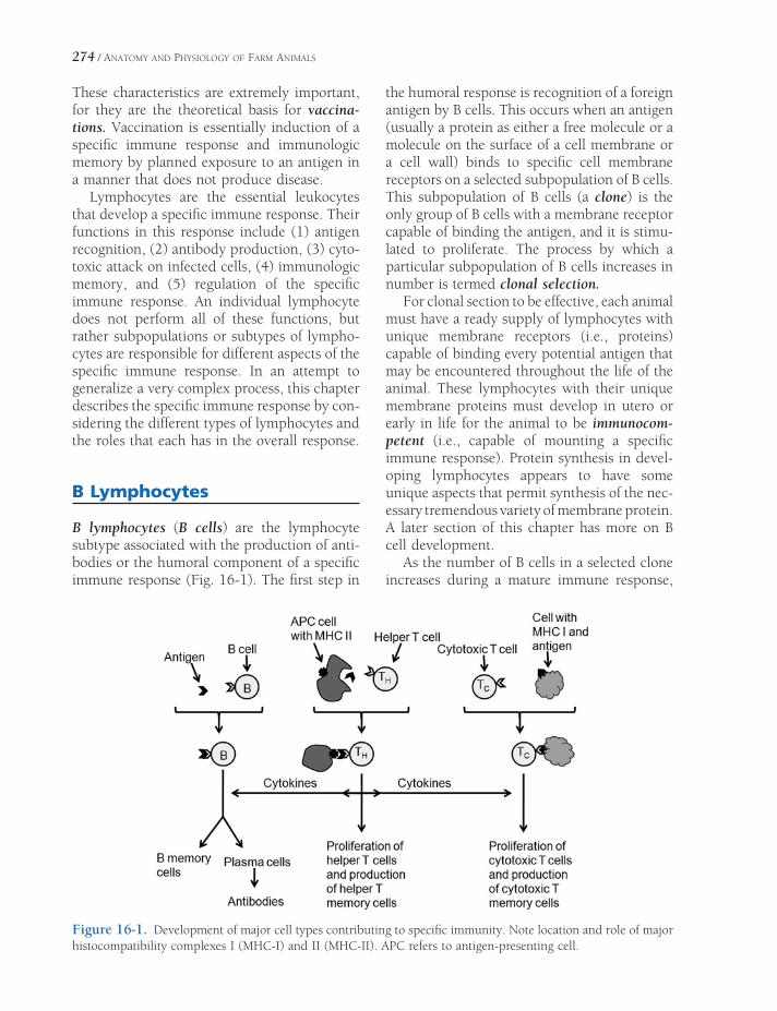
274 / ANATOMY AND PHYSIOLOGY OF FARM ANIMALS
These characteristics are extremely important, for they are the theoretical basis for vaccina-tions. Vaccination is essentially induction of a specifi c immune response and immunologic memory by planned exposure to an antigen in a manner that does not produce disease.
Lymphocytes are the essential leukocytes that develop a specifi c immune response. Their functions in this response include (1) antigen recognition, (2) antibody production, (3) cyto-toxic attack on infected cells, (4) immunologic memory, and (5) regulation of the specifi c immune response. An individual lymphocyte does not perform all of these functions, but rather subpopulations or subtypes of lympho-cytes are responsible for different aspects of the specifi c immune response. In an attempt to generalize a very complex process, this chapter describes the specifi c immune response by con-sidering the different types of lymphocytes and the roles that each has in the overall response.
B Lymphocytes
B lymphocytes (B cells) are the lymphocyte subtype associated with the production of anti-bodies or the humoral component of a specifi c immune response (Fig. 16-1). The fi rst step in
the humoral response is recognition of a foreign antigen by B cells. This occurs when an antigen (usually a protein as either a free molecule or a molecule on the surface of a cell membrane or a cell wall) binds to specifi c cell membrane receptors on a selected subpopulation of B cells. This subpopulation of B cells (a clone) is the only group of B cells with a membrane receptor capable of binding the antigen, and it is stimu-lated to proliferate. The process by which a particular subpopulation of B cells increases in number is termed clonal selection.
For clonal section to be effective, each animal must have a ready supply of lymphocytes with unique membrane receptors (i.e., proteins) capable of binding every potential antigen that may be encountered throughout the life of the animal. These lymphocytes with their unique membrane proteins must develop in utero or early in life for the animal to be immunocom-petent (i.e., capable of mounting a specifi c immune response). Protein synthesis in devel-oping lymphocytes appears to have some unique aspects that permit synthesis of the nec-essary tremendous variety of membrane protein. A later section of this chapter has more on B cell development.
As the number of B cells in a selected clone increases during a mature immune response,
Figure 16-1. Development of major cell types contributing to specifi c immunity. Note location and role of major histocompatibility complexes I (MHC-I) and II (MHC-II). APC refers to antigen-presenting cell.

BODY DEFENSES AND THE IMMUNE SYSTEM / 275
some of the B cells begin to differentiate into plasma cells. Plasma cells can synthesize and secrete antibodies specifi c for the antigen that stimulated the development of the selected clone. Thus, each plasma cell secretes only one antibody. Other B cells in the selected clone develop into B memory cells (Fig. 16-1). These cells remain in the body for extended periods (years or perhaps throughout life). Since these cells developed from a clone specifi c for a given antigen, the ready supply of them means that the next time this specifi c antigen is encoun-tered (e.g., a second infection or exposure after a vaccination), the immune response can be accelerated and amplifi ed.
The development of B memory cells and plasma cells and the secretion of antibodies are modulated by numerous cytokines. Cytokines, as the chemical messengers (proteins) secreted by a variety of cells in response to injury or microbial invasion, modulate the activity of cells of the immune system. One cell type that secretes cytokines essential for a normal humoral response is the helper T cell (Fig. 16-1). T cells are another of the general classes of lymphocytes, and helper T cells are a specifi c subtype of T cells. The characteristics and functions of helper T cells are described later in this chapter.
Immunoglobulins
Immunoglobulin is a general term for a protein that can bind to an antigen; it includes both antibodies (circulating immunoglobulins) and those found in the cell membranes of B cells. Immunoglobulins fall into fi ve major classes
based on their chemical and functional charac-teristics (Table 16-1). Immunoglobulin (Ig) G is the predominant circulating immunoglobu-lin. IgG has a variety of functions, including (1) binding free circulating antigens to reduce their potential for harm (neutralization) and promote their removal by phagocytes, (2) binding anti-gens associated with bacterial cell walls and promoting their phagocytosis, (3) binding anti-gens and activating complement to promote infl ammation and destruction of harmful microbes, and (4) acting as an agglutinin to clump particulates so that they can be more effectively phagocytized. IgG does not actually destroy any harmful substance or agent; it pro-motes destruction or removal indirectly.
IgE is the class of immunoglobulins associ-ated with most allergic responses. When an allergen (an agent capable of inducing an aller-gic response) is fi rst encountered, specifi c IgE is produced and incorporates into the cell mem-brane of mast cells. When the same allergen is again encountered, the allergen binds to the specifi c IgE on the mast cells, and this stimulates the mast cells to release their secretory granules containing histamine and produce leukotrienes. These agents bring about many of the cell and tissue responses characteristic of allergies.
T Cells and Cell-Mediated Immunity
T cells are the type of lymphocyte associated with the component of an immune response known as cell-mediated immunity. Whereas the humoral response involves antibodies that
Table 16-1. General Characteristics of Classes of Immunoglobulins
Class General Characteristics
IgG Most abundant immunoglobulin in circulation. Protective against bacteria and viruses, neutralizes toxins, and activates complement. Provides for passive immunity in newborns.
IgA Primarily found in secretions associated with epithelial surfaces and mucous membranes where it provides localized protection against bacteria and viruses.
IgE Contributes to allergic responses by binding to mast cells and basophils to promote release of mediators of allergic reactions. Also contributes to protection against parasites.
IgM First immunoglobulin produced by plasma cells in response to exposure to antigen. Contributes to natural antibodies such as those associated with blood cell typing.

276 / ANATOMY AND PHYSIOLOGY OF FARM ANIMALS
can have their effect at sites distant from their site of production, the cell-mediated immune response requires that the responding T cells be in contact with the cells bearing the foreign antigen. As with B cells, T cells have various subtypes, each with its specifi c functions. The subtypes include cytotoxic T cells, helper T cells, memory T cells (Fig. 16-1), and NK T cells. NK T cells and helper T cells are intro-duced earlier in this chapter.
As with the humoral response, antigen rec-ognition initiates the cell-mediated response. However, antigen recognition by T cells requires that the antigenic material be presented as part of a complex on the cell membrane of other immune cells. This complex consists of the antigenic material and intrinsic cell membrane proteins known as major histocompatibility complex (MHC) proteins.
The genes that code for MHC proteins are found on a single chromosome, and MHC pro-teins are continuously synthesized in all cells of an animal’s body except for erythrocytes, which do not have nuclei and are not capable of protein synthesis. After synthesis, MHC pro-teins enter the outer cell membrane so that a portion is exposed to the exterior. As MHC proteins prepare for insertion in the cell mem-brane, they form complexes with antigenic materials found within the cell. When the complex enters the membrane, the antigen is exposed to the exterior. The exposed antigen is then in a position to be recognized by T cells.
Cell membranes hold two major classes of MHC proteins, class I and class II (Fig. 16-1). Class I MHC proteins are found in all cells except for erythrocytes. Class I MHC proteins continuously present potential antigenic mate-rials on the surface of all cells. These materials include peptides and other antigenic materials normally produced in cells, as well as antigenic materials produced as a result of an abnormal-ity in cell function. If a foreign cell (i.e., not self) is introduced into an animal’s body, the antigenic material presented by its class I MHC proteins is unique for that foreign cell and differs from endogenous self cells. When viruses infect normal cells and alter their synthetic pathways, they change the antigenic materials
presented by the class I proteins of the infected cell. By recognizing these new antigens, T cells recognize foreign or infected cells, allowing ini-tiation of a cellular immune response directed at cells bearing the foreign antigen.
Only selected cell types have class II MHC proteins. These include lymphocytes, free and fi xed macrophages, microglia in the central nervous system, and a population of cells in the spleen and lymph nodes known as dendritic cells. Cells that contain class II MHC proteins are termed antigen-presenting cells (APCs), for they process and present antigens derived from foreign material or microbes (Fig. 16-1). These antigens are derived from microbes or material that the APCs take up by phagocytosis or pino-cytosis. APCs process materials in phagocytic or pinocytic vesicles to produce antigens that are complexed to class II MHC proteins and inserted into the APC cell membrane.
As with B cells, clones of T cells exist that are specifi c for a given antigen, and the process of antigen recognition permits a specifi c clone to be selected. However, the selection and acti-vation of T cells also requires interaction between the MHC proteins holding the anti-genic material and other proteins in the cell membranes of the various T cells known as cluster of differentiation markers (CD markers). These marker proteins determine the interactions that are possible for the different types of T cells. Helper T cells have CD4 markers (Fig. 16-1), which interact with class II MHC proteins, and cytotoxic T cells have CD8 markers, which interact with class I MHC pro-teins. Thus, helper T cells can recognize anti-genic material presented by APCs, and cytotoxic T cells can detect foreign antigenic material pre-sented by almost any type of cell (Fig. 16-1).
After recognizing antigens presented by APCs, the selected helper T cells are activated by interactions between other of their mem-brane proteins and membrane proteins found in the APCs. This interaction (costimulation) leads to full helper T cells activation. The activated helper T cells multiply and produce a subset to serve as memory cells (memory TH cells). A major function of the other activated helper T cells is to secrete cytokines to promote and

BODY DEFENSES AND THE IMMUNE SYSTEM / 277
amplify all aspects of the innate and specifi c immune responses. This includes attraction and stimulation of additional macrophages and NK cells, promoting the action of cytotoxic T cells, and promoting the development of selected B cells. A key cytokine produced by helper T cells to stimulate macrophages, NK cells, cytotoxic cells, and B cells is interleukin-2.
Costimulation by cells presenting the spe-cifi c antigen to selected cytotoxic T cells is also required for full activation of those cytotoxic T cells. The activated cytotoxic T cells also undergo a series of cell divisions to provide increased numbers of the selected clone and also produce a subset to serve as memory cells (memory TC
cells). Activated cytotoxic cells (killer cells) destroy cells bearing the antigen for which they were selected. Physical contact and binding between the cells bearing the antigen and the cytotoxic T cells are required for the destruction of antigen-bearing cells (Fig. 16-1), and secretions from the cytotoxic T cells, including perforin to disrupt the cell membrane of the antigen-bearing cells, accomplish it.
Lymphocyte Origin, Development, and Residence
Just as all lymphocytes are originally derived from pluripotent stem cells that subsequently became lymphoid stem cells in the bone marrow (see Fig. 15-1), the three general classes of lym-phocytes (B cells, T cells, and NK cells) are all derived from lymphoid stem cells.
The differentiation of lymphoid stem cells to the types of mature lymphocytes begins early during embryonic development. Cells destined to become T cells leave the bone marrow and travel to the thymus, where secretions called thymic hormones act on them. These are not classic hormones, for they are local to the thymus, and lymphocytes must come to the thymus to be acted on. The thymic hormones guide the further development of the lympho-cytes to become T cells. As part of the develop-ment in the thymus, only T cells capable of recognizing foreign antigens are selected to survive. The selected T cells that leave the
thymus populate lymphoid structures through-out the body, including tonsils, lymph nodes, the spleen, and collections of lymphocytes in the intestinal wall. These structures are respon-sible for producing T cells in adult animals (discussed later).
Lymphoid stem cells destined to become B cells also undergo differentiation during embry-onic development. In birds, lymphoid cells destined to become B cells leave the bone marrow and travel to the bursa of Fabricius, which is similar to the thymus but associated with the intestinal tract. Here the cells undergo maturation and selection similar to that which happens to pre–T cells in the thymus. Mature B cells leaving the bursa in birds also populate peripheral lymphoid structures, and it is these structures that produce B cells in adult birds. An organ that is the functional equivalent to the bursa has not been identifi ed in mammals. It is thought that B cells mature in the bone marrow in mammals and leave the marrow to populate the peripheral lymphoid structures. As with T cells, the peripheral lymphoid structures produce B cells in adult animals. The develop-ment of NK cells probably occurs in a manner similar to that for B cells.
Active and Passive Immunities
Active immunity is a state of immunity to a specifi c antigen achieved by the response of one’s own immune system. Active immunity may include both humoral and cell-mediated responses. Passive immunity is a state of tem-porary immunity achieved by the transfer of immunoglobulins or T cells from an animal with active immunity to another that has not encountered the antigen involved. Passive immunity is a temporary state because the immunoglobulins and/or T cells are degraded or destroyed over time.
Immunological Surveillance
NK cells and cytotoxic T lymphocytes (killer cells) both recognize and destroy cells with foreign antigens in their membranes. Cancer

278 / ANATOMY AND PHYSIOLOGY OF FARM ANIMALS
cells have surface antigens that are not present in normal cells and thus can be recognized as foreign. It is believed that cancer cells routinely develop in the body but are soon recognized and destroyed by the killer cells that recognize the abnormal antigens. This process of recogni-tion and destruction is termed immunological surveillance. Clinically evident cancers develop when this surveillance is inadequate.
Lymphatic System
The lymphatic system includes the lymphoid tissues (e.g., lymph nodes and nodules and the spleen) and the lymphatic vessels distributed throughout the body (Fig. 16-2). It drains tissue fl uid (called lymph within the lymphatic system) and is a framework for the circulation, production, and maturation of immune cells. The tissue fl uid–draining function of the lym-phatic system augments the venous circulation and therefore assists in the control of interstitial fl uid pressures. The lymphatic system is also an important component of immunologic defense of the body, as movement of lymph brings microorganisms and other foreign substances into contact with immune cells.
Lymphoid tissue consists of accumulations of lymphocytes trapped in the spaces between fi bers of reticular connective tissue. The lym-phoid tissue may be scattered diffusely in some regions (as is commonly found in mucous membranes), may appear as nodules (as in the intestinal submucosa), or may be encapsulated to form specifi c organs, including lymph nodes, the spleen, thymus, and tonsils. The lymphatic vessels and tissues are arranged so that tissue fl uid is exposed to aggregates of immune cells, which scrutinize the fl uid for foreign cells and substances, thereby assisting in the control of infection.
Lymphatic Vessels
The lymphatic vessels constitute a one-way pathway that parallels the venous system and eventually empties into the cranial vena cava or some of its tributaries. The smallest lymphatics
begin blindly between tissue cells as lymphatic capillaries, which collect the tissue fl uid not absorbed by the venous system. When the tissue fl uid enters the lymphatic vessels, it is known as lymph, which consists of fl uid origi-nally derived from the blood, a variety of blood cells, and sometimes microbes. Lymphatic vessels carry lymph back to the great veins of the heart.
Lymph Nodes
Lymph nodes are discrete knots of lymphoid tissue scattered along the course of lymph vessels. Lymph nodes fi lter the lymph and act as one of the fi rst defenses against infection by harboring lymphocytes, plasma cells, and macrophages.
Each lymph node is surrounded by a connec-tive tissue capsule that sends numerous connec-tive tissue trabeculae into the substance of the node (Fig. 16-3). The node is roughly divided into cortex, paracortex, and medulla, with large numbers of lymphocytes and macrophages in all three. Lymphocytes in the cortex are arranged in nodules (Fig. 16-3). Dark- staining groups are primary nodules, and light-staining groups are secondary nodules. Secondary nodules are areas of rapid B cell proliferation, and for that reason their cores are called germinal centers. The paracortex, deep to the cortex, is populated pri-marily with T lymphocytes and dendritic cells.
Lymphocytes in the medullary portion of the lymph node are arranged in medullary cords rather than nodules (Fig. 16-3). These tend to be primarily accumulations of plasma cells.
Immediately deep to the capsule of the node is a space, the subcapsular sinus, which com-municates with other sinuses of the cortex and medulla. Lymph delivered by afferent lymph vessels enters the subcapsular sinus and is slowly fi ltered through the cortex and medulla, to emerge fi nally at the hilus of the node, where blood vessels and nerves enter and the efferent lymphatic vessels emerge, carrying the lymph that has percolated through the node. This arrangement is ideally suited for the presentation to immune cells of antigens col-lected in the tissue fl uid.
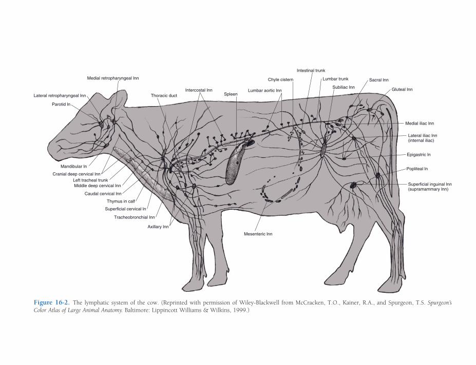
Parotid ln
Mandibular ln
Axillary lnn
Superficial cervical ln
Thymus in calf
Thoracic ductIntercostal lnn
Spleen
Subiliac lnn Gluteal lnn
Lateral iliac lnn(internal iliac)
Medial iliac lnn
Epigastric ln
Popliteal ln
Lumbar aortic lnn
Superficial inguinal lnn(supramammary lnn)
Left tracheal trunk
Chyle cistern
Intestinal trunk
Mesenteric lnn
Medial retropharyngeal lnn Lumbar trunk
Caudal cervical lnn
Sacral lnn
Tracheobronchial lnn
Lateral retropharyngeal lnn
Middle deep cervical lnn
Cranial deep cervical lnn
Figure 16-2. The lymphatic system of the cow. (Reprinted with permission of Wiley-Blackwell from McCracken, T.O., Kainer, R.A., and Spurgeon, T.S. Spurgeon’s Color Atlas of Large Animal Anatomy. Baltimore: Lippincott Williams & Wilkins, 1999.)
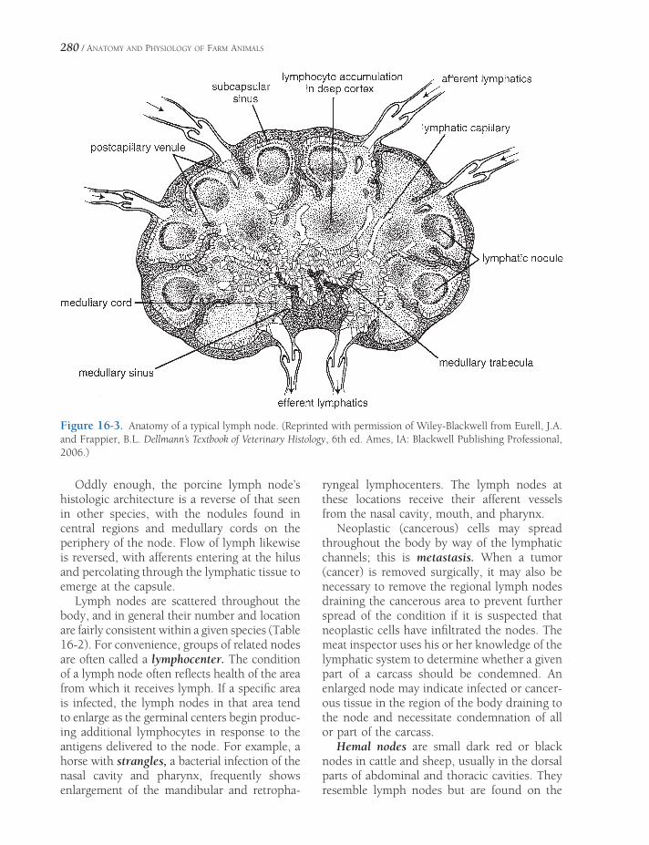
280 / ANATOMY AND PHYSIOLOGY OF FARM ANIMALS
Figure 16-3. Anatomy of a typical lymph node. (Reprinted with permission of Wiley-Blackwell from Eurell, J.A. and Frappier, B.L. Dellmann’s Textbook of Veterinary Histology, 6th ed. Ames, IA: Blackwell Publishing Professional, 2006.)
Oddly enough, the porcine lymph node’s histologic architecture is a reverse of that seen in other species, with the nodules found in central regions and medullary cords on the periphery of the node. Flow of lymph likewise is reversed, with afferents entering at the hilus and percolating through the lymphatic tissue to emerge at the capsule.
Lymph nodes are scattered throughout the body, and in general their number and location are fairly consistent within a given species (Table 16-2). For convenience, groups of related nodes are often called a lymphocenter. The condition of a lymph node often refl ects health of the area from which it receives lymph. If a specifi c area is infected, the lymph nodes in that area tend to enlarge as the germinal centers begin produc-ing additional lymphocytes in response to the antigens delivered to the node. For example, a horse with strangles, a bacterial infection of the nasal cavity and pharynx, frequently shows enlargement of the mandibular and retropha-
ryngeal lymphocenters. The lymph nodes at these locations receive their afferent vessels from the nasal cavity, mouth, and pharynx.
Neoplastic (cancerous) cells may spread throughout the body by way of the lymphatic channels; this is metastasis. When a tumor (cancer) is removed surgically, it may also be necessary to remove the regional lymph nodes draining the cancerous area to prevent further spread of the condition if it is suspected that neoplastic cells have infi ltrated the nodes. The meat inspector uses his or her knowledge of the lymphatic system to determine whether a given part of a carcass should be condemned. An enlarged node may indicate infected or cancer-ous tissue in the region of the body draining to the node and necessitate condemnation of all or part of the carcass.
Hemal nodes are small dark red or black nodes in cattle and sheep, usually in the dorsal parts of abdominal and thoracic cavities. They resemble lymph nodes but are found on the
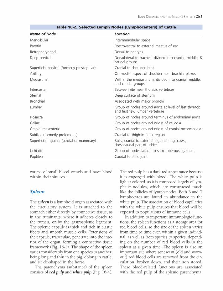
BODY DEFENSES AND THE IMMUNE SYSTEM / 281
Table 16-2. Selected Lymph Nodes (Lymphocenters) of Cattle
Name of Node Location
Mandibular Intermandibular space
Parotid Rostroventral to external meatus of ear
Retropharyngeal Dorsal to pharynx
Deep cervical Dorsolateral to trachea, divided into cranial, middle, & caudal groups
Superfi cial cervical (formerly prescapular) Cranial to shoulder joint
Axillary On medial aspect of shoulder near brachial plexus
Mediastinal Within the mediastinum, divided into cranial, middle, and caudal groups
Intercostal Between ribs near thoracic vertebrae
Sternal Deep surface of sternum
Bronchial Associated with major bronchi
Lumbar Group of nodes around aorta at level of last thoracic and fi rst few lumbar vertebrae
Iliosacral Group of nodes around terminus of abdominal aorta
Celiac Group of nodes around origin of celiac a.
Cranial mesenteric Group of nodes around origin of cranial mesenteric a.
Subiliac (formerly prefemoral) Cranial to thigh in fl ank region
Superfi cial inguinal (scrotal or mammary) Bulls, cranial to external inguinal ring; cows, dorsocaudal part of udder
Ischiatic Group of nodes lateral to sacrotuberous ligament
Popliteal Caudal to stifl e joint
course of small blood vessels and have blood within their sinuses.
Spleen
The spleen is a lymphoid organ associated with the circulatory system. It is attached to the stomach either directly by connective tissue, as in the ruminants, where it adheres closely to the rumen, or by the gastrosplenic ligament. The splenic capsule is thick and rich in elastic fi bers and smooth muscle cells. Extensions of the capsule, trabeculae, penetrate into the inte-rior of the organ, forming a connective tissue framework (Fig. 16-4). The shape of the spleen varies considerably from one species to another, being long and thin in the pig, oblong in cattle, and sickle-shaped in the horse.
The parenchyma (substance) of the spleen consists of red pulp and white pulp (Fig. 16-4).
The red pulp has a dark red appearance because it is engorged with blood. The white pulp is lighter colored, as it is composed largely of lym-phatic nodules, which are constructed much like the follicles of lymph nodes. Both B and T lymphocytes are found in abundance in the white pulp. The association of blood capillaries with the white pulp ensures that blood will be exposed to populations of immune cells.
In addition to important immunologic func-tions, the spleen functions as a storage area for red blood cells, so the size of the spleen varies from time to time even within a given individ-ual, as well as from species to species, depend-ing on the number of red blood cells in the spleen at a given time. The spleen is also an important site where senescent (old and worn-out) red blood cells are removed from the cir-culation, broken down, and their iron stored. These blood-related functions are associated with the red pulp of the splenic parenchyma.
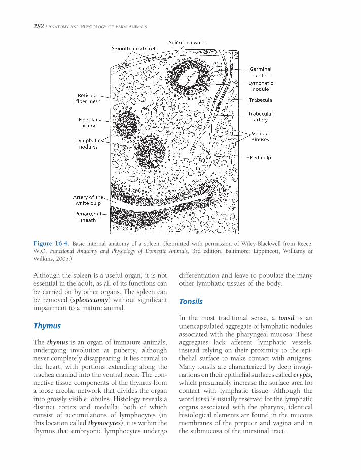
282 / ANATOMY AND PHYSIOLOGY OF FARM ANIMALS
Although the spleen is a useful organ, it is not essential in the adult, as all of its functions can be carried on by other organs. The spleen can be removed (splenectomy) without signifi cant impairment to a mature animal.
Thymus
The thymus is an organ of immature animals, undergoing involution at puberty, although never completely disappearing. It lies cranial to the heart, with portions extending along the trachea craniad into the ventral neck. The con-nective tissue components of the thymus form a loose areolar network that divides the organ into grossly visible lobules. Histology reveals a distinct cortex and medulla, both of which consist of accumulations of lymphocytes (in this location called thymocytes); it is within the thymus that embryonic lymphocytes undergo
differentiation and leave to populate the many other lymphatic tissues of the body.
Tonsils
In the most traditional sense, a tonsil is an unencapsulated aggregate of lymphatic nodules associated with the pharyngeal mucosa. These aggregates lack afferent lymphatic vessels, instead relying on their proximity to the epi-thelial surface to make contact with antigens. Many tonsils are characterized by deep invagi-nations on their epithelial surfaces called crypts, which presumably increase the surface area for contact with lymphatic tissue. Although the word tonsil is usually reserved for the lymphatic organs associated with the pharynx, identical histological elements are found in the mucous membranes of the prepuce and vagina and in the submucosa of the intestinal tract.
Figure 16-4. Basic internal anatomy of a spleen. (Reprinted with permission of Wiley-Blackwell from Reece, W.O. Functional Anatomy and Physiology of Domestic Animals, 3rd edition. Baltimore: Lippincott, Williams & Wilkins, 2005.)

ANATOMY OF THE CARDIOVASCULAR SYSTEM
The cardiovascular system consists of the heart and a system of vessels for distribu-
tion of the blood to the tissues of the body and to the lungs for exchange of gases. Whether or not the blood is oxygenated, vessels that carry blood away from the heart are called arteries, and vessels that carry blood toward the heart are called veins. Circulation to the lungs (pul-monary circulation) is functionally and ana-tomically separate from circulation to the rest of the body (systemic circulation). Conceptu-ally, it is therefore useful to regard the heart as two separate pumps housed within the same organ; one is a low-pressure pump that directs blood returning from the body to the lungs (i.e., the pulmonary circulation), and the other is a high-pressure pump that distributes blood to the systemic circulation.
Heart
The heart is a cone-shaped hollow muscular structure. The base is directed dorsad or cra-niodorsad and is attached to other thoracic structures by large arteries, veins, and the peri-cardial sac. The apex of the heart is directed ventrad and is entirely free within the pericar-dial sac. In the living animal, the right side of the heart is on the right side of the body but
HeartPericardium
Cardiac Anatomy
VesselsBlood Vessels
Lymphatic Vessels
Pulmonary CirculationSystemic Circulation
Aorta
Arterial Distribution to the Head
Arterial Distribution to Thoracic Limb
Arterial Distribution to Pelvic Limb
VeinsCranial Vena Cava
Caudal Vena Cava
Portal System
Fetal Circulation
Chapter 17
283

284 / ANATOMY AND PHYSIOLOGY OF FARM ANIMALS
oriented more cranial than the left side, which is left and somewhat caudal (Fig. 17-1).
Pericardium
The heart is partially surrounded by a serous membrane called the pericardium. The pericar-dium, like other serous tissues (the pleura and peritoneum), creates a closed cavity (the peri-cardial space) that contains only a small amount of fl uid for lubrication. The heart is invaginated into the pericardium much like a fi st thrust into an infl ated balloon (see Fig. 1–9). This arrange-ment results in two distinct layers of pericar-dium (Fig. 17-1). The inner layer, which is intimately adherent to the outer surface of the heart, is called visceral pericardium, or epicar-dium. The outer layer, called parietal pericar-dium, is continuous with the visceral layer at the base of the heart and is reinforced by a superfi cial fi brous layer (the fi brous pericar-dium), which in turn is covered by a layer of
mediastinal pleura (also called pericardial pleura). The parietal pericardium, fi brous peri-cardium, and mediastinal pleura together form the pericardial sac, which is grossly identifi -able as a thin but tough tissue surrounding the heart.
In cattle, the apex of the heart contacts the dome of the diaphragm, and the reticu-lum in the abdominal cavity lies on the caudal side of the diaphragm. Sharp metallic objects (most commonly, bits of wire) that are swallowed often accumulate in the retic-ulum. The contractions of this organ can cause these foreign bodies to penetrate the adjacent diaphragm and the pericardial sac, resulting in an infection of the sac called traumatic pericarditis, one manifestation of hardware disease. The tissues of the pericar-dium thicken, and fl uid builds up within the pericardial sac, which leads to heart failure in affected cattle. Hardware disease is usually prevented by administering a magnet by mouth; the magnet, which tends
Figure 17-1. Orientation of the heart within the thorax. The right side of the heart lies more cranial than the left. The surface of the heart is covered with visceral pericardium, which the pericardial space sepa-rates from the parietal pericardium. The parietal peri-cardium is attached by fi brous tissue to overlying pericardial pleura (not shown). These three layers con-stitute the pericardial sac.

ANATOMY OF THE CARDIOVASCULAR SYSTEM / 285
to remain in the reticulum, gathers swal-lowed metallic objects and prevents them from migrating through the wall of the forestomach.
Cardiac Anatomy
The heart wall consists of three layers: an outer serous covering called epicardium, an inner endothelial lining called endocardium, and a thick muscular layer called myocardium.
The epicardium is the same as the visceral layer of pericardium. The endocardium is a layer of simple squamous endothelial cells that lines the chambers of the heart, covers the heart valves, and is continuous with the lining of the blood vessels. The myocardium consists of cardiac muscle, described in Chapter 8.
The heart is divided into right and left sides, which correspond to the low-pressure (pulmo-nary circulation) and high-pressure (systemic circulation) systems mentioned earlier. Each side has two chambers: an atrium, which receives blood by way of large veins, and a ventricle, which pumps blood from the heart through a large artery (Fig. 17-2). The atria are thin-walled chambers, each of which features an appendage called an auricle.
The myocardium of the ventricles, which pump blood back into vascular beds, is much thicker than that of the atria. The wall of the left ventricle is also thicker than that of the right; blood ejected from the left side during its contraction is under higher pressure than that ejected from the right ventricle. The right ventricle does not quite reach the apex of the heart, as the apex is formed entirely by the more muscular left ventricle. The myocardium between the two chambers is the ventricular septum.
Between the atrium and the ventricle of each side is an atrioventricular valve, or A-V valve (Fig. 17-2). The left A-V valve is occasionally called the bicuspid valve, because in humans it has two distinct fl aps, or cusps. Another more commonly used synonym is mitral valve, because of its imagined resemblance to a bish-
op’s miter, or two-sided hat. The right A-V valve is also called the tricuspid valve because in humans it has three fl aps or cusps. The thin valve leafl ets are attached to the inner wall of the ventricle at the junction of atrium and ven-tricle. The free margins of the cusp are tethered to the interior of the ventricular wall by fi brous cords called chordae tendineae. The chordae tendineae attach to small muscular protrusions called papillary muscles that project into the lumina of the ventricles. These chordae tendin-eae prevent the valve from everting into the atrium when the ventricle contracts and closes the A-V valve by forcing blood against the ven-tricular side of the valve (Fig. 17-3A).
Each ventricle’s outfl ow tract features a semilunar valve that ensures blood fl ows only from the ventricle into the artery and not in the opposite direction. The semilunar valves have three cuplike leafl ets, with convex side facing the ventricle (Fig. 17-3B). The aortic valve lies at the junction of the left ventricle and aorta; the pulmonary valve is at the junction of the right ventricle and pulmonary trunk.
Blood returning to the heart from the sys-temic circulation is delivered to the right atrium by the cranial and caudal venae cavae (singu-lar vena cava) (Fig. 17-4). From the right atrium, this deoxygenated blood passes through the right A-V valve into the right ventricle. From the right side, the right ventricle wraps around the cranial side of the heart and termi-nates as the funnel-shaped conus arteriosus. The conus arteriosus is the origin of the pul-monary trunk, from which it is divided by the pulmonary valve.
Just distal to the pulmonary valve, the pul-monary trunk divides into right and left pulmo-nary arteries, carrying deoxygenated blood to the respective lungs.
A variable number of pulmonary veins return blood from the lungs to the left atrium. From the left atrium, blood passes through the left A-V valve into the thick-walled left ventri-cle. The left ventricle pumps the blood past the aortic valve into the aorta. The aorta and its branches carry oxygenated blood to all parts of the body (Fig. 17-4).
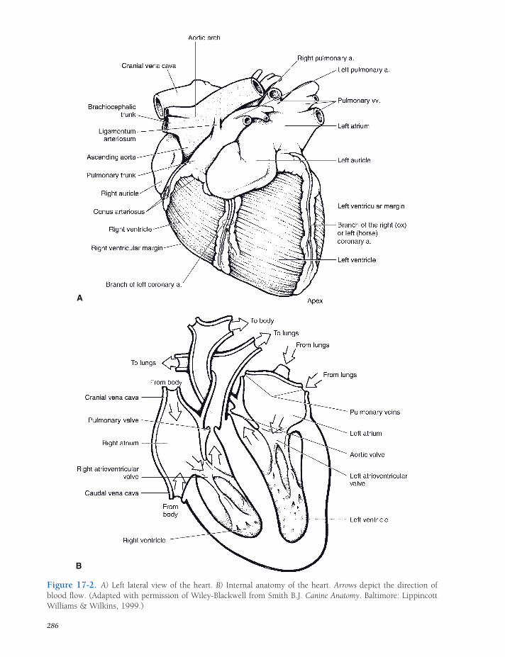
A
B
Figure 17-2. A) Left lateral view of the heart. B) Internal anatomy of the heart. Arrows depict the direction of blood fl ow. (Adapted with permission of Wiley-Blackwell from Smith B.J. Canine Anatomy. Baltimore: Lippincott Williams & Wilkins, 1999.)
286

Figure 17-3. Cardiac valves. A) Function of A-V valves. During relaxation of the ventricle (left), the valve leafl ets open to allow blood to fl ow from the atrium into the ventricle (arrow). During ventricular contraction (right), valve leafl ets are forced closed. The chordae tendineae prevent the margins of the valve from everting back into the atrium. B) A semilunar valve. Viewed from above, the valve consists of three cuplike leafl ets. During relaxation of the ventricle, the greater pressure within the vessel closes the valve. When the ventricle contracts, leafl ets part and blood fl ows through the valve (arrow).
287
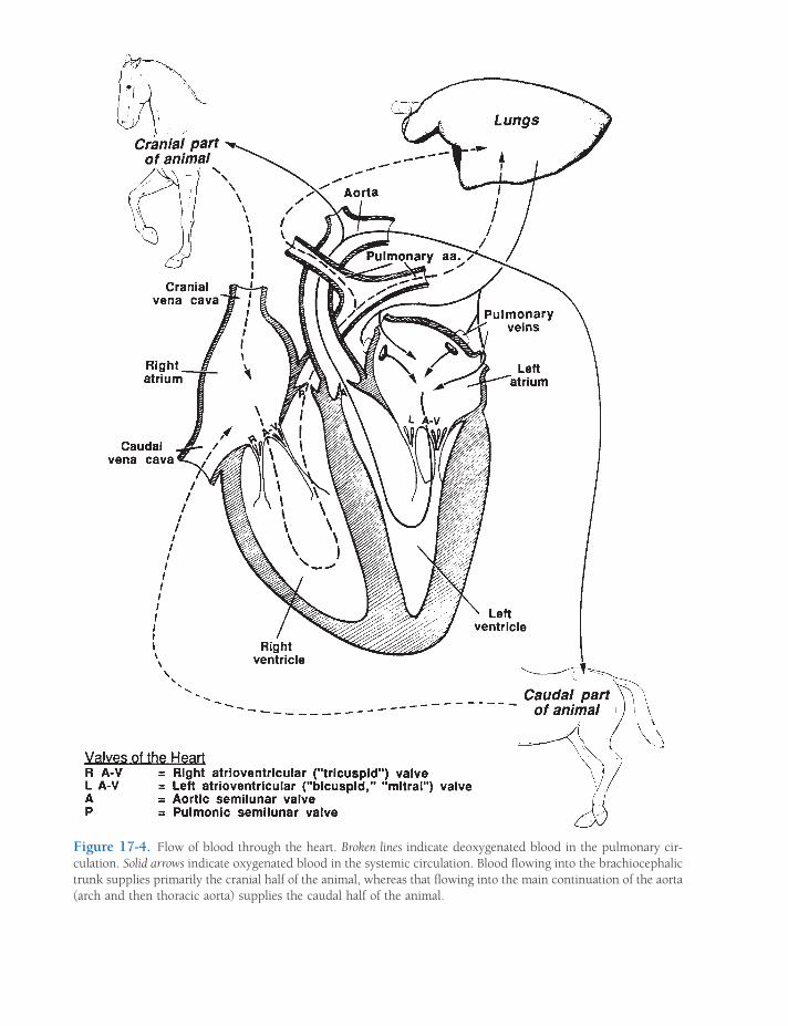
Figure 17-4. Flow of blood through the heart. Broken lines indicate deoxygenated blood in the pulmonary cir-culation. Solid arrows indicate oxygenated blood in the systemic circulation. Blood fl owing into the brachiocephalic trunk supplies primarily the cranial half of the animal, whereas that fl owing into the main continuation of the aorta (arch and then thoracic aorta) supplies the caudal half of the animal.
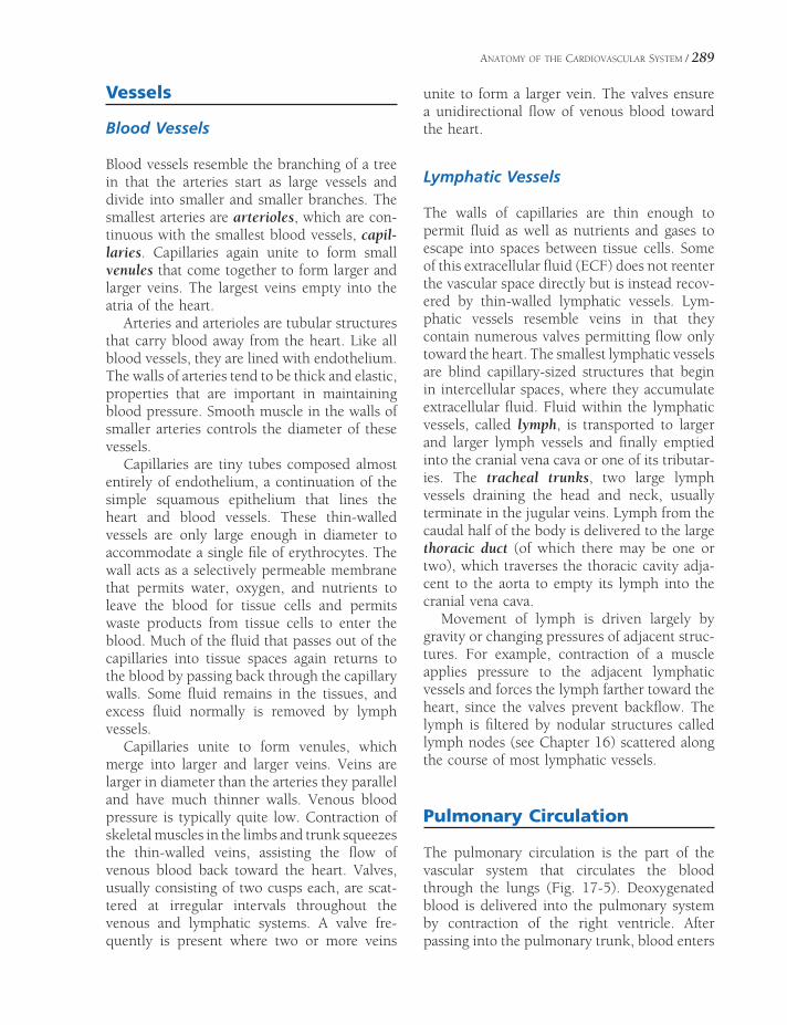
ANATOMY OF THE CARDIOVASCULAR SYSTEM / 289
Vessels
Blood Vessels
Blood vessels resemble the branching of a tree in that the arteries start as large vessels and divide into smaller and smaller branches. The smallest arteries are arterioles, which are con-tinuous with the smallest blood vessels, capil-laries. Capillaries again unite to form small venules that come together to form larger and larger veins. The largest veins empty into the atria of the heart.
Arteries and arterioles are tubular structures that carry blood away from the heart. Like all blood vessels, they are lined with endothelium. The walls of arteries tend to be thick and elastic, properties that are important in maintaining blood pressure. Smooth muscle in the walls of smaller arteries controls the diameter of these vessels.
Capillaries are tiny tubes composed almost entirely of endothelium, a continuation of the simple squamous epithelium that lines the heart and blood vessels. These thin-walled vessels are only large enough in diameter to accommodate a single fi le of erythrocytes. The wall acts as a selectively permeable membrane that permits water, oxygen, and nutrients to leave the blood for tissue cells and permits waste products from tissue cells to enter the blood. Much of the fl uid that passes out of the capillaries into tissue spaces again returns to the blood by passing back through the capillary walls. Some fl uid remains in the tissues, and excess fl uid normally is removed by lymph vessels.
Capillaries unite to form venules, which merge into larger and larger veins. Veins are larger in diameter than the arteries they parallel and have much thinner walls. Venous blood pressure is typically quite low. Contraction of skeletal muscles in the limbs and trunk squeezes the thin-walled veins, assisting the fl ow of venous blood back toward the heart. Valves, usually consisting of two cusps each, are scat-tered at irregular intervals throughout the venous and lymphatic systems. A valve fre-quently is present where two or more veins
unite to form a larger vein. The valves ensure a unidirectional fl ow of venous blood toward the heart.
Lymphatic Vessels
The walls of capillaries are thin enough to permit fl uid as well as nutrients and gases to escape into spaces between tissue cells. Some of this extracellular fl uid (ECF) does not reenter the vascular space directly but is instead recov-ered by thin-walled lymphatic vessels. Lym-phatic vessels resemble veins in that they contain numerous valves permitting fl ow only toward the heart. The smallest lymphatic vessels are blind capillary-sized structures that begin in intercellular spaces, where they accumulate extracellular fl uid. Fluid within the lymphatic vessels, called lymph, is transported to larger and larger lymph vessels and fi nally emptied into the cranial vena cava or one of its tributar-ies. The tracheal trunks, two large lymph vessels draining the head and neck, usually terminate in the jugular veins. Lymph from the caudal half of the body is delivered to the large thoracic duct (of which there may be one or two), which traverses the thoracic cavity adja-cent to the aorta to empty its lymph into the cranial vena cava.
Movement of lymph is driven largely by gravity or changing pressures of adjacent struc-tures. For example, contraction of a muscle applies pressure to the adjacent lymphatic vessels and forces the lymph farther toward the heart, since the valves prevent backfl ow. The lymph is fi ltered by nodular structures called lymph nodes (see Chapter 16) scattered along the course of most lymphatic vessels.
Pulmonary Circulation
The pulmonary circulation is the part of the vascular system that circulates the blood through the lungs (Fig. 17-5). Deoxygenated blood is delivered into the pulmonary system by contraction of the right ventricle. After passing into the pulmonary trunk, blood enters
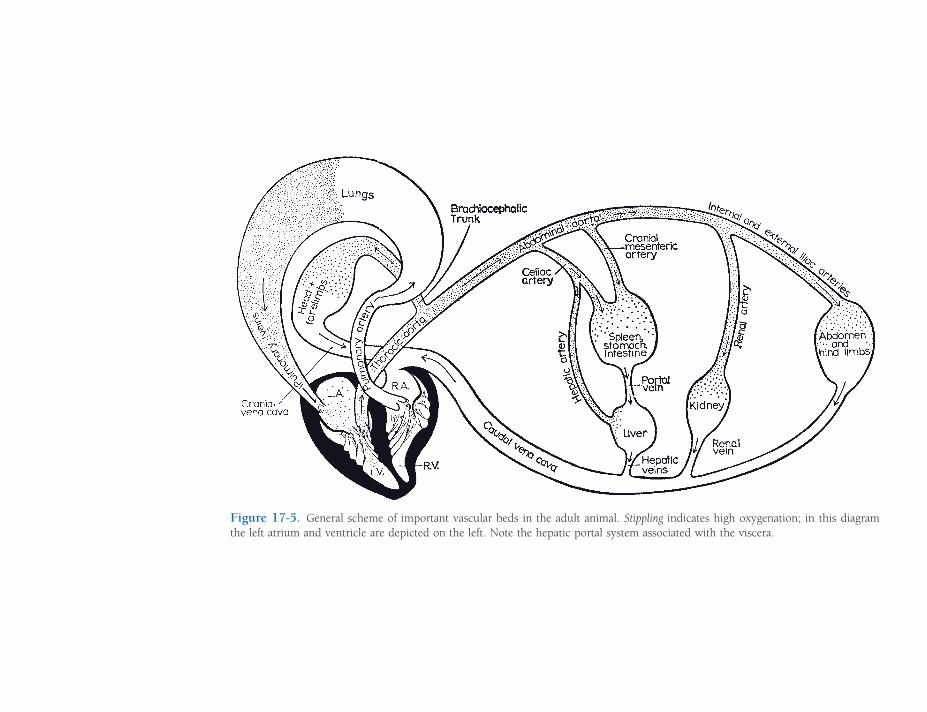
Figure 17-5. General scheme of important vascular beds in the adult animal. Stippling indicates high oxygenation; in this diagram the left atrium and ventricle are depicted on the left. Note the hepatic portal system associated with the viscera.
ANATOMY OF THE CARDIOVASCULAR SYSTEM / 291
the right pulmonary artery to go to the right lung and the left pulmonary artery to go to the left lung. Each pulmonary artery subdivides into lobar arteries going to individual lobes of the lungs. The lobar arteries again subdivide many times, fi nally forming arterioles that supply the extensive capillary beds of the lungs, where gaseous exchange takes place (see Chapter 19). After the blood passes through the capillary bed of the lungs, it enters venules, which combine to form pulmonary veins. These pulmonary veins, after leaving the lungs, deliver oxygenated blood to the left atrium, completing the pulmonary circulation.
As gases are exchanged, the color of the blood changes from the bluish maroon color of deoxygenated blood to the bright red of oxy-genated blood. In the adult the pulmonary cir-culation is the only place where deoxygenated
blood is found in arteries (which, by defi nition, carry blood away from the heart) and oxygen-ated blood is found in veins (returning blood to the heart).
Systemic Circulation
Systemic circulation refers to the movement of oxygenated blood to all areas of the body and the return of deoxygenated blood to the heart (Fig. 17-5). The following descriptions of the blood vessels of the systemic circulation are based mainly on the horse (Fig. 17-6). Animals having different digestive systems, such as ruminants, and those having more than one digit per limb of course have a somewhat dif-ferent arrangement of arteries and veins in asso-ciation with their specifi c anatomy.
Figure 17-6. Main arteries of the horse.

292 / ANATOMY AND PHYSIOLOGY OF FARM ANIMALS
Aorta
Aortic Arch. The left ventricle receives oxy-genated blood from the left atrium and pumps the blood throughout the systemic circulation by way of the largest artery, the aorta. The aortic valve, at the junction of the left ventricle and aorta, prevents backfl ow of blood from the aorta into the left ventricle when the ventricle relaxes.
Two large vessels arise from the aorta imme-diately distal to the aortic valve. These are the right and left coronary arteries, and they are the arterial blood supply for the myocardium. Most of the venous blood from the myocar-dium is returned to the right atrium by way of the coronary veins, which empty directly into the right atrium by way of the coronary sinus, adjacent to the opening of the caudal vena cava.
After emerging from the base of the heart, the aortic arch courses dorsad and then caudad, just ventral to the bodies of the thoracic verte-brae. The aorta continues as the thoracic aorta until it passes through the aortic hiatus of the diaphragm to become the abdominal aorta. Arteries that supply the head, neck, and tho-racic limbs branch from the aortic arch.
In horses and ruminants, the aortic arch gives rise to a single large brachiocephalic trunk, whose many branches distribute blood to the cranial half of the animal. The precise pattern of the main arterial branches is species dependent, but the following generalities can be made: (1) the main blood supply to the thoracic limbs arises as right and left subcla-vian arteries; (2) the right and left costocervi-cal trunks provide arterial blood to regions of the neck and cranial thoracic wall; and (3) right and left common carotid arteries, a main source of blood for the head and brain, arise together from a single bicarotid trunk.
Thoracic Aorta. The thoracic aorta passes caudad just ventral to the vertebral bodies. As it does so, pairs of segmental arteries arise from its dorsal aspect to supply the thoracic wall and epaxial muscles. Each of these intercostal arteries enters the corresponding intercostal
space, giving off a spinal branch that enters the vertebral canal to supply the spinal cord and spinal nerve roots. The continuation of the dorsal intercostal artery follows the caudal border of each rib ventrad. Other branches of the thoracic aorta supply parts of the esopha-gus, lungs, and diaphragm.
Abdominal Aorta. The aorta is called the abdominal aorta after it passes through the aortic hiatus of the diaphragm. Ventral to the last few lumbar vertebrae, it terminates by dividing into two external iliac arteries (sup-plying the pelvic limbs) and two internal iliac arteries (supplying the gluteal and perineal region). Some species have a median sacral artery, a small midline continuation of the aorta that continues ventral to caudal vertebrae as the median caudal artery. The accompany-ing median caudal vein (tail vein) at this site is often used for collection of blood from adult cattle.
The abdominal aorta features paired lumbar arteries arising from its dorsal side, supplying the abdominal wall and epaxial muscles and giving off spinal branches that supply the spinal cord and spinal nerve roots of the lum-bosacral region. Paired visceral branches provide arterial blood to the kidneys (renal arteries) and gonads (testicular or ovarian arteries). Three unpaired visceral branches supply nearly all the abdominal viscera. These are, from cranial to caudal, the celiac, cranial mesenteric, and caudal mesenteric arteries.
The celiac artery arises shortly after the aorta passes through the diaphragm. This is a large unpaired artery that supplies the stomach (left gastric artery), the spleen (splenic artery), and the liver (hepatic artery). The exact branch-ing pattern of this artery depends to a great extent upon the type of stomach; the ruminant has a much more complex distribution of the celiac artery than do animals with a simple stomach.
Immediately caudal to the celiac artery is the cranial mesenteric artery. This large, unpaired artery branches into a number of smaller arter-ies that supply blood to most of the small intes-tine and much of the large intestine. The

ANATOMY OF THE CARDIOVASCULAR SYSTEM / 293
number and distribution of the branches of the cranial mesenteric artery vary among species.
The caudal part of the large intestine and the rectum receive blood from a relatively small unpaired artery, the caudal mesenteric artery.
Arterial Distribution to the Head
Most of the structures of the face, head, and cranial neck are supplied by the right and left common carotid arteries, each of which runs craniad in a connective tissue sheath with the vagosympathetic trunk of the same side (Fig. 17-7A). This carotid sheath lies in a groove dorsolateral to the trachea. Branches of the common carotid arteries supply the thyroid gland and larynx. In the region of the larynx, the common carotid artery gives off the inter-nal carotid artery, a primary source of blood for the brain. The continuation of the common carotid artery is the external carotid artery, whose many branches supply the face, tongue, and structures of the oral and nasal cavities. The facial artery is convenient for taking a pulse as it passes across the mandible.
The internal carotid arteries or their deriva-tives enter into an anastomotic ring of vessels on the base of the brain called the cerebral arterial circle (formerly circle of Willis) (Fig. 17-7B). The cerebral arterial circle gives rise to arteries that supply the cerebral hemispheres and rostral parts of the brainstem. More caudal parts of the brainstem and the cerebellum receive most of their blood supply from branches of the basilar artery. This single ventral artery is formed by the joining of right and left verte-bral arteries. The robust vertebral arteries ascend from their origin in the thoracic inlet, run alongside the cervical vertebrae, enter the foramen magnum of the skull, and there coalesce into the basilar artery (coursing rostrad) and the ventral spinal artery (running caudad).
Arterial Distribution to the Thoracic Limb
The right and left subclavian arteries follow the same course on each side of the body and each
gives off similar branches. Within the thorax each subclavian artery gives off a number of branches that supply blood to the caudal part of the neck, much of the thoracic wall, and the dorsal part of the shoulder. The subclavian artery passes cranial to the fi rst rib on the respective side, passing into the axilla (armpit) of the thoracic limb, where it is called the axil-lary artery. The axillary artery enters the limb, becoming the brachial artery in the region of the brachium and then the median artery as it continues distal to the elbow. The largest ter-minal branch of the median artery in the horse is the medial palmar artery, which passes distad in the metacarpus to the fetlock, where it divides into medial and lateral digital arter-ies. In ruminants, the median artery is contin-ued in the manus as the palmar common digital artery (Fig. 17-8).
Arterial Distribution to the Pelvic Limb
The abdominal aorta terminates near the lum-bosacral junction in the two internal iliac arter-ies (and often a small, midline continuation called the median sacral artery). Each internal iliac artery and its many branches supply the region of the pelvis, the hip, and much of the genitalia.
Just cranial to the internal iliac arteries, the external iliac arteries arise and give rise to branches serving caudoventral parts of the abdominal wall and structures of the inguinal region (prepuce, scrotum, and/or mammary gland). These large arteries then continue into the pelvic limbs as the femoral arteries. The femoral artery descends on the medial aspect of the limb, giving branches to the large thigh muscles, and continues in the region of the caudal stifl e as the popliteal artery. After a very short course, the popliteal artery divides into cranial and caudal tibial arteries. The small caudal tibial artery supplies the muscles of the crus, or true leg. The cranial tibial artery is larger; it passes craniad between the tibia and fi bula and descends on the cranial side of the crus to the hock. Where this vessel lies on the

MaxillaryInternal Carotid
External Carotid
Common Carotid
Facial
Caudal Cerebral Artery
Rostral Cerebellar ArteryCaudal Cerebellar Artery
Ventral Spinal Artery
Rostral Cerebral Artery
Middle Cerebral Artery
Caudal Communicating Artery
Basilar Artery
A
B
Figure 17-7. Blood supply to the head and brain. A) The common carotid artery branches into a large external carotid artery supplying most of the head and the internal carotid artery, which enters the skull to supply the brain. B) Ventral view of the brain. Internal carotid arteries enter the cerebral arterial circle.
294

ANATOMY OF THE CARDIOVASCULAR SYSTEM / 295
Figure 17-8. Blood supply to the equine thoracic limb, medial view.
fl exor surface of the hock, it is referred to as the dorsal pedal artery. In horses, it continues distad as the dorsal (great) metatarsal a. III, running on the lateral side of the pes in the groove between the cannon bone and lateral splint. Ultimately, it passes to the plantar aspect of the distal cannon bone by crossing deep to the splint bone. At the equine fetlock it divides into medial and lateral digital arteries. In ruminants, the dorsal pedal artery continues distad on the dorsal aspect of the pes; the plantar side is supplied by a con-tinuation of the saphenous artery, a medial branch of the femoral artery (Fig. 17-9).
Veins
With some notable exceptions, veins accom-pany arteries of the same name. These “satellite” veins are always larger than their respective
arteries and frequently duplicated. For example, the brachial artery carrying blood to the forearm and digit may be accompanied by two or more brachial veins returning the blood to the heart. Some veins are superfi cial, visible in the subcu-taneous tissues, and these are particularly of interest as they may be accessed via venipunc-ture (introducing a needle into a vein). As indi-cated earlier, nearly all systemic veins eventually drain into either the cranial vena cava or caudal vena cava.
Cranial Vena Cava
The cranial vena cava drains the head, neck, thoracic limbs, and part of the thorax. Tributar-ies to the cranial vena cava include the jugular veins (internal and external), subclavian veins, and vertebral veins. The external jugular veins

296 / ANATOMY AND PHYSIOLOGY OF FARM ANIMALS
Caudal Vena Cava
The caudal vena cava is formed in the abdomen by the junction of the paired internal and exter-nal iliac veins. These drain the gluteal and peri-neal regions and the pelvic limbs, respectively. The caudal vena cava also receives lumbar veins, testicular or ovarian veins, renal veins, and various others from structures associated with the body walls. Just caudal to the point at which the caudal vena cava passes through the caval foramen of the diaphragm, it receives a number of short hepatic veins directly from the liver.
Portal System
A portal system is one in which a vessel divides into capillaries, recombines to form another
Figure 17-9. Arteries of the pelvic limb in the horse. Primary arterial supply to the pes of the horse is the dorsal metatarsal artery III.
drain the face and much of the head, while the internal jugular veins, if present, along with the vertebral veins drain most of the blood from the brain. Each subclavian vein receives venous blood from the same areas that are supplied by the subclavian artery and its branches (shoul-der, neck, and thoracic limbs). The azygos vein (the word azygos derives from the Greek word meaning “unpaired”) lies adjacent to the verte-bral column, receiving the segmentally arranged intercostal veins. In horses, the right azygos vein empties at the junction between cranial vena cava and right atrium. Ruminants some-times have both right and left azygos veins, but more usually have a single left azygos vein, which empties directly into the right atrium with the coronary sinus. The pig possesses a left azygous vein, which empties into the coronary sinus.

ANATOMY OF THE CARDIOVASCULAR SYSTEM / 297
vessel, and then redivides into a second capil-lary bed. The hypothalamohypophysial portal system was described in Chapter 12 in relation to the pituitary gland. In birds and in some reptiles and amphibians, part of the venous blood returning from the pelvic limbs enters the kidneys to form a renal portal system (see Chapter 30).
In the hepatic portal system, blood that has perfused the capillary beds of the viscera is brought to the liver by a single large vein, the portal vein, and then is redistributed into a second capillary bed within the substance of the liver (Fig. 17-5).
Tributaries to the portal vein include the gastric vein from the stomach, the splenic vein from the spleen, the mesenteric veins from the intestines, and the pancreatic veins from the pancreas. The portal vein enters the liver and immediately breaks up into smaller and smaller branches there, fi nally ending in the sinusoids of the liver. Here the blood comes into direct contact with cells of the liver. After being acted upon by the liver cells (see Chapter 21), the blood passes from the sinusoids of the liver into the liver’s venous system and eventually empties into the caudal vena cava.
Fetal Circulation
Throughout gestation, the fetus depends on the dam for the nutrients, water, and oxygen needed for growth and for the elimination of carbon dioxide and other waste products of fetal metab-olism. During fetal development, the lungs are collapsed and not aerated, and the pulmonary vascular beds have high resistance to blood fl ow. The fetal circulation therefore bypasses these pulmonary capillary beds. Immediately after birth, however, the newborn needs to direct its blood through the pulmonary vessels for oxy-genation. The heart and circulatory system are arranged in such an ingenious way that the cardiopulmonary circulation just moments after birth is profoundly different from that exhibited just prior to the fi rst breath (Fig. 17-10).
Because it is exchanging gas, supplying nutri-ents, and removing metabolic waste pro ducts,
the placenta must necessarily receive a large proportion of the fetus’s circulating blood.
It does so via two large umbilical arteries, extending from the caudal end of the abdomi-nal aorta through the umbilical cord to the placenta. After passing through the placental capillary bed, the blood is returned to the fetus by a single umbilical vein, which passes into the substance of the liver. Most of the highly oxygenated blood returning from the placenta in the umbilical vein is delivered directly into the caudal vena cava, bypassing the hepatic sinusoids via a fetal diversion, the ductus venosus.
Two features of the fetal heart allow blood to bypass the pulmonary circulation. During car-diogenesis, the wall between the two atria develops a fl utter valve. This aperture is called the foramen ovale, and its structure is such that the blood entering the right atrium (well oxy-genated, as a goodly portion of it is returning from the placenta) uses the one-way fl utter valve of the foramen ovale as a convenient pas-sageway from the right to the left atrium. This is one way blood bypasses the fetal lungs.
Second, blood fl owing from the right ven-tricle into the pulmonary trunk bypasses the pulmonary arteries through the ductus arterio-sus, which connects the pulmonary trunk and the aorta. In the fetus, the pressures in the right side of the heart are greater than those of the left side, since relatively little blood is returning from the lungs to the left side. As a result, the pressure is higher in the pulmonary trunk than the aorta, and blood therefore passes across the ductus arteriosus from the trunk to the aorta, bypassing the pulmonary circulation.
At birth, when the neonate takes its fi rst breath and infl ates its lungs, the resistance in the pulmonary capillary bed falls precipitously. The increase in oxygenation of the newborn’s blood constricts the ductus arteriosus. Within a few minutes, this formerly large vessel has shrunk drastically. Within the fi rst week of life, it closes completely, becoming a fi brous band, the ligamentum arteriosum, identifi able grossly between the pulmonary trunk and the aorta.
These changes abruptly increase blood fl ow to the now low-resistance pulmonary capillary
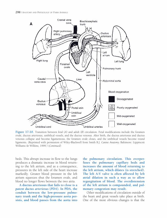
298 / ANATOMY AND PHYSIOLOGY OF FARM ANIMALS
Figure 17-10. Transition between fetal (A) and adult (B) circulation. Fetal modifi cations include the foramen ovale, ductus arteriosus, umbilical vessels, and the ductus venosus. After birth, the ductus arteriosus and ductus venosus collapse and become ligamentous, the foramen ovale closes, and the umbilical vessels become round ligaments. (Reprinted with permission of Wiley-Blackwell from Smith B.J. Canine Anatomy. Baltimore: Lippincott Williams & Wilkins, 1999.) (Continued)
beds. This abrupt increase in fl ow to the lungs produces a dramatic increase in blood return-ing to the left atrium, and as a consequence, pressures in the left side of the heart increase markedly. Greater blood pressure in the left atrium squeezes shut the foramen ovale, and blood no longer fl ows between the two atria.
A ductus arteriosus that fails to close is a patent ductus arteriosus (PDA). In PDA, the conduit between the low-pressure pulmo-nary trunk and the high-pressure aorta per-sists, and blood passes from the aorta into
the pulmonary circulation. This overper-fuses the pulmonary capillary beds and increases the amount of blood returning to the left atrium, which dilates (is stretched). The left A-V valve is often affected by left atrial dilation in such a way as to allow regurgitation of blood. The overdistension of the left atrium is compounded, and pul-monary congestion may result.
Other modifi cations of circulation outside of the heart and great vessels take place at birth. One of the most obvious changes is that the

ANATOMY OF THE CARDIOVASCULAR SYSTEM / 299
placenta loses its role as oxygenator and pro-vider of nutrients. The vasculature associated with it (umbilical arteries and veins) collapses and converts into fi brous cords (round liga-ments) that are sometimes identifi able grossly in the adult. The ductus venosus undergoes constriction as well. If the ductus venosus fails to close completely, blood returning from the
viscera via the portal vein may fi nd its way back to the systemic circulation (specifi cally, the caudal vena cava) without fi rst being pro-cessed in the liver. This abnormality consti-tutes a portosystemic shunt (sometimes called portocaval shunt) and can produce abnor-malities of metabolism, growth, and neural function.
Figure 17-10. Continued

PHYSIOLOGY OF THE HEART AND CIRCULATION
Basic Design and Function of the Cardiovascular System
The cardiovascular system consists of the heart and the many vessels through which blood fl ows. While the actual anatomy of the system makes it diffi cult to appreciate easily, the basic design of the system is a continuous loop of branching vessels with two pumps in the loop (Fig. 18-1). The loop design can be best under-stood by tracing the path of a single erythrocyte as it travels. When the erythrocyte is pumped out of the left side of the heart, it enters the aorta and passes into the systemic circulation. The systemic circulation is a subdivision of the cardiovascular system consisting of all vessels associated with all organs other than the parts of the lungs where exchange of gases—oxygen and carbon dioxide—takes place. When blood returns from the systemic circulation, it enters the right side of the heart. The right side of the heart pumps blood into the pulmonary circula-tion. The pulmonary circulation consists of vessels associated with the parts of the lungs where the exchange of gases takes place. From the pulmonary circulation, blood reenters the heart on the left side, and from here it is pumped out into the systemic circulation to begin the loop again. The loop design means that all com-ponents of the system must function together in a highly coordinated and integrated fashion to maintain blood fl ow throughout the system.
Basic Design and Function of the Cardiovascular System
Cardiac CycleSystole
Diastole
Heart Sounds and Murmurs
Echocardiography
Electrical Activity of the HeartSinoatrial Node and Heart Rate
Atrioventricular Node and Other
Specialized Conductive Cells in the Heart
Electrocardiography and Arrhythmias
Cardiac Output and Its RegulationVentricular Filling and Stroke Volume
Cardiac Contractility and Stroke Volume
Structure and Function of Blood VesselsMicroscopic Structure of Blood Vessels
Function of Blood Vessels
Regulation of Arterial Blood Pressure and Blood Volume
Neural Refl exes
Humoral Agents
Paracrine Agents
Cardiovascular Function During Exercise and Hypovolemia
Chapter 18
301

302 / ANATOMY AND PHYSIOLOGY OF FARM ANIMALS
For example, if the right side of the heart cannot pump an adequate amount of blood into the pulmonary circulation, the left side of the heart will not receive enough blood to maintain fl ow into the systemic circulation.
Blood fl ows through the vessels of the car-diovascular system because of a driving force gen erated by the contraction of the heart. Hydrostatic pressure, or mean blood pressure, in vessels is a measure of this driving force. (Chapter 2 explains hydrostatic pressure.) Blood fl ows from a point of high mean pressure to a point of low mean pressure. In the systemic circulation, mean blood pressure is higher in the arteries than in the capillaries and higher in the capillaries than in the veins, from which blood reenters the right side of the heart (Fig. 18-1). A series of one-way valves described in Chapter 17 regulates blood fl ow though the heart.
The driving force of blood pressure is neces-sary to overcome the vascular resistance pro-vided by the blood vessels. Any tube offers a resistance to the fl ow of liquid through it. The
resistance (R) to fl ow through a single tube depends on the length (L) of the tube, the radius (r) of the tube, and the character of the fl uid fl owing through the tube (viscosity, η). The resistance increases with the length of the tube, decreases as the radius of the tube increases, and increases with the viscosity of the fl uid. While all three factors can affect resis-tance, changes in the radius have the largest effect, as shown in the following formula. (Poi-seuille fi rst described this formula and the mathematical relations between these factors.)
RL
r= 8
4
ηπ
According to this formula alone, the vessels with the smallest radius, the capillaries, would have the greatest resistance. This is true for a single vessel, but it is not true when consider-ing the total combined resistance for the differ-ent types of vessels in the systemic circulation. Rather, the vessels on the arterial side of the circulation just before the capillaries have the greatest combined resistance. The total resis-
Figure 18-1. General design of cardiovascular system illustrating the systemic and pulmonary circulations. Pul-monary circulation is shown in black. (Reprinted with permission of Wiley-Blackwell from Reece W.O. Physiology of Domestic Animals. 2nd ed. Baltimore: Williams & Wilkins, 1997.)

PHYSIOLOGY OF THE HEART AND CIRCULATION / 303
tance at the level of the capillaries is less because of the extensive branching in capillary net-works. Branching of vessels tends to lower resistance, and the extensive branching of cap-illaries in their networks is responsible for a relatively low resistance at this point in spite of the small diameter of an individual capillary. The type of arterial vessel found just outside of a capillary network (before the extensive branching) is an arteriole, and these vessels contribute most to the total vascular resistance in the systemic circulation.
Transport is the ultimate function of the cardiovascular system. Blood is the transport medium; the heart provides the force for moving blood (i.e., pump function) around the circulation; and vessels provide a path for the movement and permit exchange between blood and interstitial fl uids at the level of the capil-laries. The rate of transport and exchange is usually determined by the rate of blood fl ow through the capillaries.
Cardiac Cycle
The cardiac cycle is one complete cycle of cardiac contraction and relaxation (heartbeat). The events of the cardiac cycle occur in a spe-cifi c sequence, and for descriptive purposes, the continuous cycle is divided into phases or periods marked by different events. Figure 18-2 illustrates the changes in blood pressures and volumes in the left atrium and left ventricle and pressure changes in the aorta during the cardiac cycle, and it identifi es some phases of the cycle. The sequence of changes in blood pressures and volumes in the right atrium, right ventricle, and pulmonary trunk are similar and occur in the same sequence, but the magnitude of pres-sure changes in the right ventricle and pulmo-nary trunk are much lower. The unique characteristics of the pulmonary circulation are discussed in Chapter 19.
Diastole (dilation, from the Greek dia, apart; stello, place or put) refers to the relaxation of a chamber of the heart just prior to and during the fi lling of that chamber. Systole (contrac-tion, from Gr. syn, together; stello, place) refers
to the contraction of a chamber of the heart that drives blood out of the chamber. The adjectives atrial and ventricular can be used with dias-tole or systole to describe the activity of specifi c cardiac chambers (e.g., atrial systole refers to atrial contraction in Figure 18-2). However, when the cardiac cycle is divided into only two general phases (diastole and systole) without specifying a chamber, it is generally assumed that division is based on ventricular activity. Thus, when the heart is said to be in systole or the systolic phase of the cardiac cycle, it is usually understood that this refers to ventricu-lar systole. (Note in Figure 18-2 the atrial con-tracts while the ventricle remains relaxed, i.e., ventricular diastole.)
Two distinct heart sounds can be heard during each cardiac cycle in all domestic species, and these are typically described as lub (fi rst sound, or S1) and dub (second sound, or S2). These sounds are separated by a short interval and followed by a longer pause (Figure 18-2). The pause increases with slower heart rates. These sounds are used clinically to divide the cardiac cycle into two phases, systole (ventricular) and diastole (ventricular). The fi rst sound marks the beginning of systole, and the second sound marks the beginning of diastole (Fig. 18-2).
Systole
As the ventricles begin their contraction, blood pressure increases in them. Almost immedi-ately, the pressure within each ventricle exceeds the pressure within their respective atria, and the pressure differences force the A-V valves closed. The fi rst heart sound is associated with closure of the right and left A-V valves (Fig. 18-2).
The ventricles continue to contract and pres-sure continues to increase during the early part of systole. At this point in the cycle, all four heart valves are closed, and all remain closed until pressure in the ventricles exceeds that in the arterial vessels that they supply (aorta for left ventricle and pulmonary trunk for right ventricle). The period of systole during which all valves are closed is the isovolumetric

Figure 18-2. Timing of various events of the cardiac cycle. Pressures, volumes, and times represent typical values for an adult human. These characteristics would be different among domestic animals of variable sizes and with different heart rates. However, the sequence of the events and the relative differences in pressures between the atrium, ventricle, and aorta would be the same. Note that the complete period for ventricular diastole would include the segment in this diagram labeled as atrial contraction. (Reprinted with permission from Tortora, G.J. and Derrickson, B. Principles of Anatomy and Physiology, 11th ed. Hoboken, NJ: John Wiley & Sons, 2006.)

PHYSIOLOGY OF THE HEART AND CIRCULATION / 305
contraction period, because during it the volume of each ventricle remains constant (Fig. 18-2).
When ventricular pressures exceed those in their respective arterial vessels, the semilunar valves open to permit ejection of blood. There is no sound associated with the opening of the semilunar valves. An initial rapid ejection phase of systole is followed by a reduced ejection phase, during which ventricular and arterial pressures fall (Fig. 18-2).
The elasticity of the aorta and pulmonary trunk maintains blood pressure in these vessels even though the ventricles begin to relax. When the blood pressures in these vessels are greater than the pressures in their associated ventricles, the pressure differences close the semilunar valves. The second heart sound is associated with closure of the aortic and pulmonary valves, and this sound is used to mark the end of systole (ventricular) and the beginning of dias-tole (ventricular) (Fig. 18-2).
Stroke volume is the volume of blood ejected from each ventricle during a single cardiac cycle. Normally, the stroke volumes for the right and left ventricles are the same. If these are not equal, blood tends to accumulate in either the systemic or pulmonary circulation.
Diastole
At the beginning of diastole, both the semilunar and A-V valves are closed, so the initial phase is termed isovolumetric relaxation. When ven-tricular relaxation reaches the point that atrial blood pressures exceed ventricular blood pres-sures, the pressure differences open the A-V valves. While the A-V valves are closed during systole and early diastole, blood continues to fl ow into the right and left atria from the sys-temic and pulmonary circulations, respectively. The accumulation of blood within the atria increases atrial blood pressure. When A-V valves open, much of the accumulated blood fl ows rapidly into the ventricles. Most ventricu-lar fi lling occurs during this period prior to any atrial contraction (Fig. 18-2).
Blood continues to fl ow into the atria throughout diastole, and because the A-V valves
are open, blood fl ows directly through the atria into the ventricles. As mentioned earlier, dias-tole is the phase of the cardiac cycle that length-ens most with slow heart rates, so slow heart rates provide a long period for ventricular fi lling.
Atrial contraction (atrial systole) occurs during ventricular diastole, forcing an additional volume of blood into the ventricles, but this amount is relatively small (perhaps 15%) com-pared to the volume already in the ventricles.
The volume of blood in each ventricle at the end of diastole is the end-diastolic volume (EDV). During systole, each ventricle ejects only a percentage of its EDV (Fig. 18-2), typi-cally 40–60%. The percentage of the EDV that is ejected is the ejection fraction.
Heart Sounds and Murmurs
The fi rst and second heart sounds are associ-ated with valve closures, but turbulent blood fl ow and vibrations of large vessels induced by the closures are believed to be the actual causes of the sounds. Third and fourth heart sounds may be heard in some normal horses and cattle with relatively slow heart rates. The third sound is associated with the rapid ventricular fi lling phase after the initial opening of the A-V valves, and the fourth sound is associated with atrial contractions (Fig. 18-2).
Heart murmur is a general term for any abnormal heart sound. Abnormalities in heart valves are a common cause of murmurs. Murmurs may occur when a valve fails to close completely (valvular insuffi ciency) and blood fl ow goes in the wrong direction at the wrong time. Murmurs may also occur when a valve fails to open completely (valvular stenosis) and blood is forced through a smaller than normal opening.
Echocardiography
Echocardiography is the use of ultrasound to image the heart and associated structures. It is a noninvasive procedure that provides dynamic images permitting visualization of structures in

306 / ANATOMY AND PHYSIOLOGY OF FARM ANIMALS
the heart as it contracts and relaxes. This pro-vides much more information on cardiac func-tion than can be obtained with a single static image, such as a radiograph.
Electrical Activity of the Heart
Like skeletal muscle, contraction of each cardiac muscle cell requires an action potential on the cell membrane (Fig. 18-3A), and the action potential brings about the release of calcium from intracellular stores. The calcium binds to regulatory proteins on the thin fi laments, and movement of the regulatory proteins permits an interaction between actin and myosin to bring about contraction (see Chapter 8). However, unlike skeletal muscle, each cardiac muscle cell is not innervated by a motor neuron that is responsible for eliciting an initial action poten-tial. In the heart, the initial action potential occurs spontaneously in a specialized group of myocardial cells found in the sinoatrial (SA)
node of the heart (Fig. 18-4), from which the action potential is propagated around the heart to bring about contraction of all cardiac muscle cells. The cell-to-cell propagation in the heart is possible because the intercalated disks provide an electrical connection between myo-cardial cells (Fig. 1–6C).
Sinoatrial Node and Heart Rate
Figure 18-3B shows a series of action potentials from SA node cells. A unique feature of the electrical activity of these cells is that the resting membrane potential is unstable. This instability permits SA node cells to depolarize spontane-ously to threshold, where an action potential is generated. The SA node (Fig. 18-4) is termed the pacemaker of the heart because each action potential that spontaneously develops in the SA node is propagated around the heart to stimu-late action potentials in all myocardial cells and produce a contraction.
A B
Figure 18-3A. Action potential of cardiac muscle contractile cell. Electrically gated calcium channels in cell membrane are open during the prolonged plateau phase of the action potential to permit calcium ions to enter from the extracellular fl uid. The entering calcium stimulates the release of more calcium from the sarcoplasmic reticulum. The initial depolarization phase involves electrically gated sodium channels, and potassium channels are involved with membrane repolarization.Figure 18-3B Four sinoatrial cell membrane potentials and action potentials to illustrate effects of sympathetic (S) and parasympathetic (PS) stimulation on a control heart rate. Sympathetic stimulation reduces the time period before the next action potential to increase heart rate. Parasympathetic stimulation prolongs the time period before the next action potential to reduce heart rate.

PHYSIOLOGY OF THE HEART AND CIRCULATION / 307
Both sympathetic and parasympathetic nerves innervate the SA node. By their actions on cells in the SA node, sympathetic nerves increase the rate of spontaneous action poten-tials and parasympathetic nerves reduce the rate (Fig. 18-3B). This is the means by which sym-pathetic stimulation increases heart rate and parasympathetic stimulation reduces heart rate. Parasympathetic nerves continuously inhibit the SA node in the heart of a resting animal, and this constant inhibition is responsible for the resting heart rate. During light exercise or with excitement, the parasympathetic inhibi-tion is fi rst reduced to permit an increase in heart rate. With greater excitement or more intense exercise, sympathetic stimulation increases, further increasing heart rate. Highly trained athletic animals, such as racing Thor-oughbreds, have relatively high levels of para-sympathetic stimulation to their hearts at rest.
Atrioventricular Node and Other Specialized Conductive Cells in the Heart
The atrioventricular node (A-V node) and the common bundle, or bundle of His, are also myocardial cells specialized for conducting action potentials. The A-V node is in the intra-atrial septum, and the common bundle extends from the A-V node into the ventricle (Fig. 18-4) through the fi brous connective tissue of the cardiac skeleton. The cardiac skeleton sepa-rates the cardiac muscle of the atria and ven-tricles, so the only direct electrical connection is through the A-V node and common bundle. The common bundle divides into several branches that rapidly propagate action poten-tials throughout the ventricle (Fig. 18-4). The individual cells that make up these branches are the Purkinje fi bers.
SA Node
AV NodeBundle Branches
Aortic Valve
Valve of the Pulmonary Trunk
Right AV Valve
Left AV Valve
Figure 18-4. Impulse generation and conduction system of the mammalian heart.
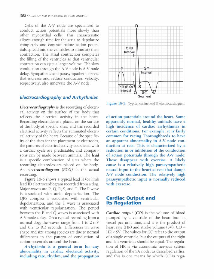
308 / ANATOMY AND PHYSIOLOGY OF FARM ANIMALS
Cells of the A-V node are specialized to conduct action potentials more slowly than other myocardial cells. This characteristic allows enough time for the atria to depolarize completely and contract before action poten-tials spread into the ventricles to stimulate their contraction. The atrial contraction completes the fi lling of the ventricles so that ventricular contraction can eject a larger volume. The slow conduction through the A-V node is A-V node delay. Sympathetic and parasympathetic nerves that increase and reduce conduction velocity, respectively, also innervate the A-V node.
Electrocardiography and Arrhythmias
Electrocardiography is the recording of electri-cal activity on the surface of the body that refl ects the electrical activity in the heart. Recording electrodes are placed on the surface of the body at specifi c sites, and the recorded electrical activity refl ects the summated electri-cal activity of the heart. Because of the specifi c-ity of the sites for the placement of electrodes, the patterns of electrical activity associated with a cardiac cycle are predictable, and compari-sons can be made between animals. The lead is a specifi c combination of sites where the recording electrodes are placed on the body. An electrocardiogram (ECG) is the actual recording.
Figure 18-5 shows a typical lead II (or limb lead II) electrocardiogram recorded from a dog. Major waves are P, Q, R, S, and T. The P wave is associated with atrial depolarization. The QRS complex is associated with ventricular depolarization, and the T wave is associated with ventricular repolarization. The period between the P and Q waves is associated with A-V node delay. On a typical recording from a normal dog, the waves range from 1 to 2 mV and 0.2 to 0.3 seconds. Differences in wave shape and size among species are due to normal differences in the pattern of conduction of action potentials around the heart.
Arrhythmia is a general term for any abnormality in cardiac electrical activity, including rate, rhythm, and the propagation
of action potentials around the heart. Some apparently normal, healthy animals have a high incidence of cardiac arrhythmias in certain conditions. For example, it is fairly common for racing Thoroughbreds to have an apparent abnormality in A-V node con-duction at rest. This is characterized by a reduction in or inhibition of the conduction of action potentials through the A-V node. These disappear with exercise. A likely cause is a relatively high parasympathetic neural input to the heart at rest that damps A-V node conduction. The relatively high parasympathetic input is normally reduced with exercise.
Cardiac Output and Its Regulation
Cardiac output (CO) is the volume of blood pumped by a ventricle of the heart into its vessel per unit time, and it is the product of heart rate (HR) and stroke volume (SV): CO = HR × SV. The values for CO refer to the output of a single ventricle, but the outputs of the right and left ventricles should be equal. The regula-tion of HR is via autonomic nervous system regulation of the SA node, as described earlier, and this is one means by which CO is regu-
P
Q
S
R
T
QRSInterval
S-TSegment
P-R (P-Q)Interval
Q-TInterval
Figure 18-5. Typical canine lead II electrocardiogram.

PHYSIOLOGY OF THE HEART AND CIRCULATION / 309
lated. However, SV, the other determinant of CO, is also subject to change and regulation. The two major factors that can change SV are ventricular fi lling and cardiac contractility.
Ventricular Filling and Stroke Volume
Figure 18-6 illustrates the relation between end-diastolic volume (EDV) and the SV of the subsequent ventricular contraction. SV increases as EDV increases until an optimal EDV is reached. After that point, further increases in EDV are associated with reductions in SV. The cellular basis for this relation is not fully under-stood, but stretching of cardiac muscle appears to induce changes in the physical relationship between thin and thick fi laments and in the affi nity of regulatory proteins for calcium ions. These changes are associated with stronger contractions until the muscle is overstretched. The relation between stretching of cardiac muscle and force of contraction is known as the Frank-Starling curve or Frank-Starling law of the heart. In normal resting animals, the Frank-Starling curve is less than optimal, so increases in EDV can produce increases in SV and CO.
Ventricular fi lling depends on fi lling pres-sure (the blood pressure in the veins and cardiac atria that force blood into the ventricles), time for fi lling, and ventricular compliance (the ease with which the ventricle relaxes during fi lling). The fi lling pressure in turn depends on blood volume and constriction of the smooth muscle in veins (venoconstriction). An increase in blood volume or venoconstriction tends to increase fi lling pressure and EDV. Slow heart rates provide more time for fi lling and tend to increase EDV. Decreases in ventricular compli-ance tend to reduce ventricular fi lling, because more pressure is required to distend the ven-tricle during fi lling.
The cardiac preload is the force on the heart muscle prior to contraction, and one measure of it is the amount that cardiac muscle is stretched prior to contraction. In intact animals, ventricular fi lling pressure and EDV are used as indicators of preload.
Cardiac Contractility and Stroke Volume
The contraction force generated by individual myocardial cells can also be changed by a mechanism that is independent of the length to which cardiac muscle is stretched prior to con-traction. This phenomenon is a change in cardiac contractility; it typically results from the direct action on myocardial cells of a hormone, neurotransmitter, or drug. Agents that can elicit changes in cardiac contractility are inotropes.
Norepinephrine and epinephrine are posi-tive inotropes, because both increase cardiac contractility. Norepinephrine and epinephrine both bind to β-adrenergic receptors on myo-cardial cells, and subsequent to this binding, both elicit increases in the availability of intra-cellular calcium in stimulated myocardial cells. The increased calcium, as well as other changes in intracellular metabolism brought about by the β-adrenergic receptor stimulation, pro-motes an increase in the force of contraction. Other inotropes use different membrane recep-tors, but the intracellular events usually involve
Str
oke
Vol
ume
End Diastolic Volume
Norepinephrine
A
B
Figure 18-6. Relation between end-diastolic volume (EDV) and stroke volume (SV) in a normal heart with (A) and without (B) sympathetic stimulation.

310 / ANATOMY AND PHYSIOLOGY OF FARM ANIMALS
calcium availability or the affi nity of intracel-lular proteins for calcium.
An indicator of myocardial contractility in the intact heart is the percentage of the EDV that is ejected during ventricular systole (i.e., ejection fraction). A typical value for ejection fraction in a resting animal is 40%. With sym-pathetic stimulation, this increases (Fig. 18-6), while with primary cardiac diseases it may reduce to 15–20%.
Structure and Function of Blood Vessels
Microscopic Structure of Blood Vessels
A single layer of endothelium, a simple squa-mous epithelium, lines all vessels. Capillaries are essentially endothelial tubes (Fig. 18-7), but they differ notably among themselves in cell-to-cell junctions and the structure of individual endothelial cells. Tight junctions connect the endothelial cells of some capillaries, and this forms a relatively impermeable barrier to non–lipid-soluble particles. Other capillaries appear to be quite porous because of openings in the endothelial cells themselves, and these are found where high rates of capillary exchange occur.
A wall with three layers, or tunics, charac-terizes all types of veins and arteries. From innermost to outermost, these layers are the tunica interna (intima), tunica media, and tunica externa (adventitia) (Fig. 18-7). The tunica interna consists of the lining endothe-lium, a subendothelial layer primarily consist-ing of connective tissue, and in some cases, an internal elastic membrane. The internal elastic membrane consists of elastin and is especially prominent in large arteries.
The tunica media of all veins and arteries contains smooth muscle. Arteries have a much thicker tunica media than veins of a correspond-ing size, and this characteristic can be used to differentiate between the two types of vessels.
Elastic arteries are a special type whose tunica media contains concentric elastic laminae.
The elastic properties of this type of artery permit vascular expansion and contraction during the various phases of the cardiac cycle. The aorta is an elastic artery that expands to increase its volume during ventricular systole and then rebounds during ventricular diastole to maintain relatively high arterial blood pres-sure. Arteries whose tunicae mediae consist pri-marily of smooth muscle are muscular arteries. An external elastic membrane is the outermost layer of the tunica media, but it is often diffi cult to visualize by light microscopy. Arterioles, the smallest of arteries, are found where arteries empty into a branching capillary network.
Some veins, and especially those in limbs below the level of the heart, have fl aplike valves that consist of folds of the tunica interna. The fold forms a cuplike pocket with the free edge of the fold directed toward the heart (Fig. 18-7). These one-way valves promote the fl ow of venous blood toward the heart when the vein is compressed.
Function of Blood Vessels
The arterial side of the circulation provides a ready supply of blood under relatively high hydrostatic pressure. Because of their relatively thick walls, arteries are not very compliant (i.e., do not distend easily with increases in pressure), so arterial pressure remains high as the heart pumps blood into the arteries. The arterioles at the end of the branching arterial network function as on-off valves to regulate the rate of blood fl ow from the arteries into capillary networks. Sympathetic vasoconstric-tor nerves innervate the smooth muscle in the wall of most arterioles, and this is one mecha-nism by which blood fl ow is regulated. However, the degree of constriction of arterio-lar smooth muscle is also subject to regulation by a large number of vasoactive agents, some locally produced (paracrine) and some that arrive via the systemic circulation. The para-crine agents (e.g., lactic acid, carbon dioxide, and adenosine) are typically vasodilators; they increase blood fl ow to the local area. Produc-tion of these agents increases when metabolism

PHYSIOLOGY OF THE HEART AND CIRCULATION / 311
Tunica interna(endothelium)
Elastic tissue
Tunica media(smooth muscle)
Tunica externa(connective tissue)
Artery Vein
Capillary
Arteriole
Blood flow
Venule
Figure 18-7. Section of small blood vessels showing the structure of their walls. A venous valve is also shown. (Reprinted with permission of Wolters Kluwer from Cohen B.J. and Wood D.L. Memmler’s The Human Body in Health and Disease. 9th ed. Baltimore: Lippincott Williams & Wilkins, 2000.)
by cells in the local area increases, providing a means to match metabolic rate with blood fl ow. That is, an increase in metabolism brings about an increase in blood fl ow. The process by which local mechanisms regulate local blood fl ow is autoregulation.
Capillaries are the site of exchange between blood and the interstitial fl uid that surrounds all cells. In most cases, this exchange is by simple diffusion (i.e., substances move down their concentration gradients). Gases (oxygen and carbon dioxide) and other lipid-soluble substances freely diffuse though capillary walls, but substances that are not lipid soluble, such as glucose, must diffuse through pores in the capillary wall. Exchange by diffusion does not necessarily require the movement of fl uid between the capillary and the interstitial space.
Oxygen, for example, can diffuse down its con-centration gradient from the plasma to metabo-lizing cells as blood fl ows in a capillary past the cells. As stated earlier, the rate of capillary exchange is primarily governed by the rate of blood fl ow into the capillaries. In resting tissues, blood fl ow occurs only through a small percent-age of the total capillaries at any one time. As metabolism and blood fl ow increase, the per-centage of capillaries being perfused increases.
Typically, there is a small net loss of fl uid from the plasma as it fl ows through most capil-lary networks. This fl uid is recovered via the lymphatics and ultimately returned to the blood where lymphatics enter large veins near the heart. A small number of plasma proteins are similarly lost from capillaries and returned via the lymph.

312 / ANATOMY AND PHYSIOLOGY OF FARM ANIMALS
The forces that govern fl uid movement at the capillary level are important clinically in that imbalances in these forces contribute to edema, an abnormal amount or collection of fl uid in the interstitial space. The primary factor that forces fl uid out of a capillary into the interstitial space is the blood pressure in the capillary. The primary force that tends to keep fl uid in capil-laries is the effective osmotic force (pressure) generated by plasma proteins, primarily albumin (Fig. 18-8). This pressure is also termed oncotic pressure. Plasma proteins generate an effective osmotic force because the protein concentra-tion in the interstitial fl uid is much lower than that of plasma. At the arterial end of a capillary, the blood pressure is higher than the oncotic pressure, so some fl uid is lost from the capillary, while at the venous end of a capillary the oncotic pressure is higher, so some fl uid moves into the capillary (Fig. 18-8). A slight imbal-ance between fl uid loss and fl uid gain by the capillaries gives rise to a net loss and provides fl uid for lymph formation (Fig. 18-8).
Like arteries, veins have smooth muscle in their walls, but the walls of veins are much thinner and more compliant. The compliance of venous vessels permits relatively large changes in the volume of blood in the veins with minimal changes in venous blood pressure. Thus, the venous side of the circulation functions as a low-pressure reservoir of blood. Constriction of venous smooth muscle promotes an increase in
blood fl owing back to the heart and an increase in cardiac fi lling pressure. This contributes to increases in cardiac output and the ability to perfuse capillaries for exchange.
Regulation of Arterial Blood Pressure and Blood Volume
Arterial blood pressure is a function of cardiac output (CO) and total peripheral vascular resistance (TPR), usually written as MAP = CO × TPR, where MAP is mean arterial pressure. To understand the basis for this function, recall that arteries are relatively noncompliant and function as a reservoir of blood under pressure. The pressure depends on the amount of blood being pumped into the reservoir (CO) and the rate at which blood is permitted to fl ow out of the reservoir (Fig. 18-9). Recall also that the resistance of the arterioles, which contribute the most to the total vascular resistance, regu-lates the rate of blood fl ow out of the arteries. Thus, the level of cardiac function and the degree of arteriolar constriction are the two determinants of MAP. Changes in MAP can be brought about by changes in cardiac function, the degree of arteriolar constriction, or some combination of these determinants. Any chemi-cal regulator (e.g., hormone or paracrine agent) or neural refl ex that affects cardiac activity or the smooth muscle of arterioles has the poten-
Figure 18-8. Microvascular fl uid movements. Arrows indicate direction of net fl uid movement. Note difference in relative amounts of protein molecules in plasma and interstitial fl uid. Interstitial fl uid with protein moves into lymphatics to form lymph.
Blood
flow
Arterial end
Net fluidmovement
Net fluid
movementBloodflow
Venous end
Lymphatic
Net fluidmovement
Protein
molecules
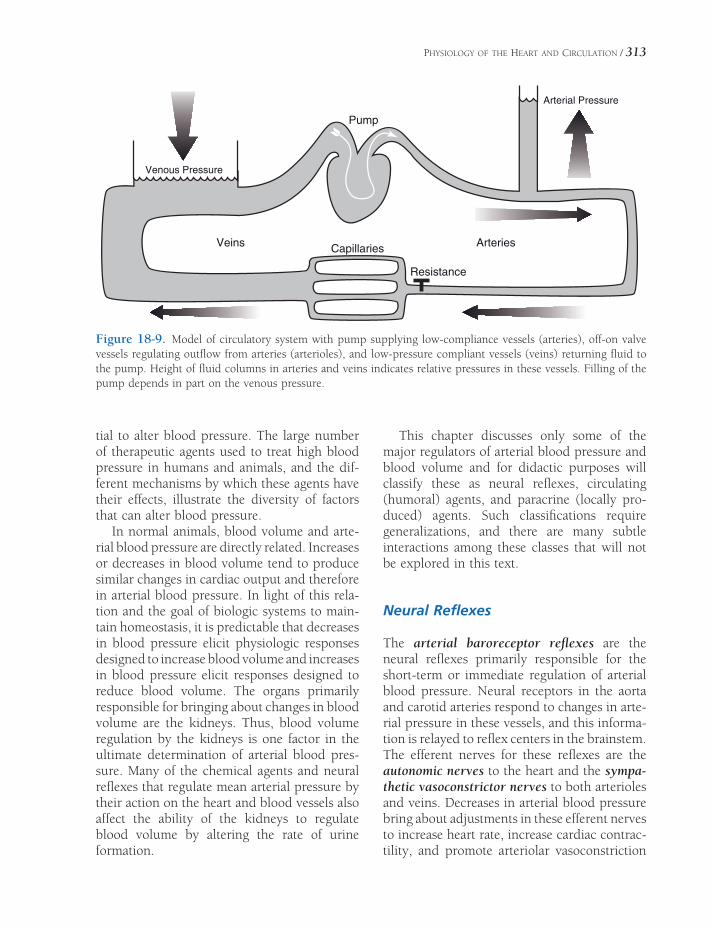
PHYSIOLOGY OF THE HEART AND CIRCULATION / 313
ArteriesVeinsCapillaries
Venous Pressure
Arterial Pressure
Pump
Resistance
Figure 18-9. Model of circulatory system with pump supplying low-compliance vessels (arteries), off-on valve vessels regulating outfl ow from arteries (arterioles), and low-pressure compliant vessels (veins) returning fl uid to the pump. Height of fl uid columns in arteries and veins indicates relative pressures in these vessels. Filling of the pump depends in part on the venous pressure.
tial to alter blood pressure. The large number of therapeutic agents used to treat high blood pressure in humans and animals, and the dif-ferent mechanisms by which these agents have their effects, illustrate the diversity of factors that can alter blood pressure.
In normal animals, blood volume and arte-rial blood pressure are directly related. Increases or decreases in blood volume tend to produce similar changes in cardiac output and therefore in arterial blood pressure. In light of this rela-tion and the goal of biologic systems to main-tain homeostasis, it is predictable that decreases in blood pressure elicit physiologic responses designed to increase blood volume and increases in blood pressure elicit responses designed to reduce blood volume. The organs primarily responsible for bringing about changes in blood volume are the kidneys. Thus, blood volume regulation by the kidneys is one factor in the ultimate determination of arterial blood pres-sure. Many of the chemical agents and neural refl exes that regulate mean arterial pressure by their action on the heart and blood vessels also affect the ability of the kidneys to regulate blood volume by altering the rate of urine formation.
This chapter discusses only some of the major regulators of arterial blood pressure and blood volume and for didactic purposes will classify these as neural refl exes, circulating (humoral) agents, and paracrine (locally pro-duced) agents. Such classifi cations require generalizations, and there are many subtle interactions among these classes that will not be explored in this text.
Neural Refl exes
The arterial baroreceptor refl exes are the neural refl exes primarily responsible for the short-term or immediate regulation of arterial blood pressure. Neural receptors in the aorta and carotid arteries respond to changes in arte-rial pressure in these vessels, and this informa-tion is relayed to refl ex centers in the brainstem. The efferent nerves for these refl exes are the autonomic nerves to the heart and the sympa-thetic vasoconstrictor nerves to both arterioles and veins. Decreases in arterial blood pressure bring about adjustments in these efferent nerves to increase heart rate, increase cardiac contrac-tility, and promote arteriolar vasoconstriction

314 / ANATOMY AND PHYSIOLOGY OF FARM ANIMALS
and venoconstriction. The overall effect tends to increase both CO and TPR so that blood pressure can be restored to its original level. Increases in arterial blood pressure above some original level should elicit reductions in cardiac activity and relaxation of the vessels. Inhibition of vasoconstrictor and venoconstrictor nerves is the mechanism by which the refl ex permits relaxation of the vascular smooth muscle.
Neural receptors in the atria of the heart respond to changes in the volume of blood fi lling the atria, and afferent information from these receptors is relayed to brainstem refl ex centers. The primary efferents involved in these refl exes are the sympathetic nerves to the kidneys. Increases in atrial fi lling bring about reductions in sympathetic nerve stimulation of the kidneys, and this permits an increase in the urinary excretion of sodium chloride and water. This tends to reduce blood volume and there-fore cardiac fi lling, cardiac output, and blood pressure. Reductions in atrial fi lling bring about increases in sympathetic nerve activity to the kidneys. This refl ex also affects the secretion of renin from the kidneys (discussed later).
Humoral Agents
Renin is an enzyme released from the kidneys, and its release is regulated in part by sympa-thetic nerve activity to the kidneys and arterial blood pressure in the vessels perfusing the kidneys. Increases in renal sympathetic nerve activity and/or reductions in arterial blood pressure to the kidneys elicit increases in renin secretion. Renin acts on a plasma protein sub-strate to produce a peptide, angiotensin I, which an enzyme converts to angiotensin II. Angiotensin II constricts vascular smooth muscle, so it tends to increase TPR and arterial blood pressure. Angiotensin II also reduces the urinary loss of sodium chloride and water by its action on tubules in the kidney and by pro-moting the release of aldosterone from the adrenal cortex. The overall biologic effect of angiotensin II is to increase arterial pressure and blood volume.
At rest, the plasma levels of epinephrine and norepinephrine are relatively low, so their effects on cardiac function and vascular smooth muscle are relatively minor. However, plasma levels can increase signifi cantly in highly stressed animals or in response to severe reductions in blood pressure or blood volume. In these conditions, circulating epinephrine and norepinephrine promote increases in cardiac function and con-strict vascular smooth muscle.
Atrial natriuretic peptides (ANPs) are released from the atria of the heart in response to increases in blood volume and atrial fi lling. ANPs promote an increase in the urinary excre-tion of sodium and water by a direct action on tubules in the kidney. ANPs also reduce TPR by relaxing arteriolar vascular smooth muscle that was constricted by vasoconstrictor agents.
Paracrine Agents
Endothelial cells lining many blood vessels produce and release nitric oxide, a local vaso-dilator. Nitric oxide release by endothelial cells is subject to regulation by a variety of agents, and the importance of its overall role in the regulation of TPR in normal animals is contro-versial. Bacterial endotoxins are lipopolysac-charide components of the bacterial cell wall that are released when the cell is lysed. These endotoxins stimulate macrophages throughout an animal’s body to produce and release unusually large amounts of nitric oxide. General overproduction of nitric oxide causes widespread vasodilation and a severe drop in arterial blood pressure. These cardiovascular changes are part of a clinical syndrome, endotoxic or septic shock.
Endothelial cells also secrete a peptide, endo-thelin, which constricts vascular smooth muscle of arterioles. As with nitric oxide, a variety of agents (e.g., angiotensin II and norepinephrine) are known to stimulate endothelin release. Interestingly, circulating vasoconstrictors tend to increase the release of this locally produced vasoconstrictor.

PHYSIOLOGY OF THE HEART AND CIRCULATION / 315
Cardiovascular Function During Exercise and Hypovolemia
Increases in cardiac output and rate of blood fl ow to skeletal muscle are needed during exer-cise to meet the increased metabolic needs of the active skeletal muscle. Several of the cardio-vascular adjustments during exercise are learned responses or behavior responses that begin just prior to or at the initiation of exercise. These are apparently initiated by the cerebral cortex, involve autonomic nerves, and include an increased heart rate and vasodilation of arteri-oles supplying skeletal muscle. Increased circu-lating levels of epinephrine may also contribute to these changes. After exercise has begun, appropriate vasodilation of arterioles supplying active skeletal muscles is primarily maintained by local metabolites produced by the active muscles. Typically, the increase in cardiac func-tion is greater than the degree of vasodilation, so arterial pressure rises slightly. Vascular resis-tance increases in other organs whose metabo-lism is not increased during exercise, and this prevents an increased blood fl ow to these organs. During exercise, the increased cardiac
output (increased heart rate and contractility) is maintained by sympathetic stimulation of the heart. This sympathetic stimulation is primarily maintained by the mechanisms originating in the cerebral cortex and by refl exes based on afferent information originating in the exercis-ing skeletal muscle.
Hypovolemia is abnormally decreased circu-lating fl uid volume (i.e., blood volume). It can occur rapidly, as a result of hemorrhage, or slowly, as a result of dehydration. Reduced blood volume results in reduced cardiac fi lling pressure and ventricular fi lling (i.e., reduced end-diastolic volume). Without adequate fi lling, SV falls, and therefore cardiac output can be insuffi cient to maintain capillary perfusion throughout the body. A severe reduction in cardiac output drastically lowers blood pres-sure, eliciting a strong response by the barore-ceptor refl ex. An accelerated heart rate is part of this response. The reduced cardiac output and the intense peripheral vasoconstriction resulting from the refl ex response both contrib-ute to the characteristic blanching of mucus membranes. The reduced stroke volume is the major cause of weakening of the pulse.


THE RESPIRATORY SYSTEM
Oxygen is a vital requirement of animals. An animal may survive for days without water
or for weeks without food, but life without oxygen is measured in minutes. Delivering oxygen and removing carbon dioxide (the product of cellular respiration) are the two major functions of the respiratory system. The processes involved with these functions related to gases include ventilation (movement of air in and out of the lungs), gas exchange between air and blood in the lungs, gas transport in blood, and gas exchange between blood and cells at the level of the tissues.
Secondary functions of the respiratory system include assistance in the regulation of the pH of the body fl uids, assistance in tem-perature control, and phonation (voice produc-tion). The role of the respiratory system in the regulation of the pH of blood and other body fl uids is closely associated with the ability of the respiratory system to remove carbon dioxide. If carbon dioxide accumulates in the blood because the respiratory system cannot remove it, blood pH falls; this is respiratory acidosis. Blood pH rises if the respiratory system removes more carbon dioxide than is appropriate and blood levels of carbon dioxide are lower than normal; this is respiratory alka-losis. The changes in carbon dioxide and pH are closely linked because of the chemical reac-tion shown in the equation below. The bicar-bonate ions and water molecules for this reaction are readily available in body fl uids.
Upper Respiratory TractNose
Paranasal Sinuses
Pharynx
Larynx
Trachea and Bronchi
ThoraxLungs
Pleura
Physiology of RespirationVentilation
Gas Exchange
Gas Transport in Blood
Control of Ventilation
Chapter 19
317
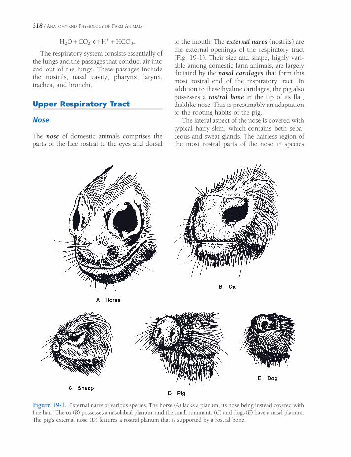
318 / ANATOMY AND PHYSIOLOGY OF FARM ANIMALS
H O CO H HCO2 2 3+ ↔ ++−
The respiratory system consists essentially of the lungs and the passages that conduct air into and out of the lungs. These passages include the nostrils, nasal cavity, pharynx, larynx, trachea, and bronchi.
Upper Respiratory Tract
Nose
The nose of domestic animals comprises the parts of the face rostral to the eyes and dorsal
to the mouth. The external nares (nostrils) are the external openings of the respiratory tract (Fig. 19-1). Their size and shape, highly vari-able among domestic farm animals, are largely dictated by the nasal cartilages that form this most rostral end of the respiratory tract. In addition to these hyaline cartilages, the pig also possesses a rostral bone in the tip of its fl at, disklike nose. This is presumably an adaptation to the rooting habits of the pig.
The lateral aspect of the nose is covered with typical hairy skin, which contains both seba-ceous and sweat glands. The hairless region of the most rostral parts of the nose in species
Figure 19-1. External nares of various species. The horse (A) lacks a planum, its nose being instead covered with fi ne hair. The ox (B) possesses a nasolabial planum, and the small ruminants (C) and dogs (E) have a nasal planum. The pig’s external nose (D) features a rostral planum that is supported by a rostral bone.

THE RESPIRATORY SYSTEM / 319
other than the horse contains no sebaceous glands but does have numerous sweat glands, which keep the region around the nostrils moist. This area is the planum nasale in the sheep and goat, planum rostrale in the pig, and planum nasolabiale in the cow. The grooves and bumps in the plana are distinctive enough to allow nose prints to be used for positive individual identifi cation.
The equine nose lacks a planum, being instead covered with short, fi ne hairs. The lateral wall of the external naris is fl exible, allowing for an enormous range of diameters. During exertion, the lateral wall is dilated, cre-ating a wider, lower-resistance passageway for the movement of air. In this, the nostril is aided by the presence of a short blind-ended diver-ticulum lateral to the true nasal cavity. This “false nostril” (nasal diverticulum) is probably a construct that aids in passive dilation of the nostrils during vigorous ventilation.
The nasal cavity is separated from the mouth by the hard and soft palates and separated into
two isolated halves by a median nasal septum. The rostral part of the septum is cartilaginous, whereas the most caudal part is created in part by a plate of bone. Each half of the nasal cavity communicates with the nostril of the same side rostrally and with the pharynx caudally by way of the choanae (caudal nares).
The nasal cavity is lined with mucous mem-brane that covers a number of scroll-like conchae (turbinate bones) arising from the bones of the lateral wall (Fig. 19-2). The two major conchae (dorsal concha and ventral concha) occupy rostral parts of the nasal cavity; caudal parts are fi lled with ethmoidal conchae. The vascular mucous membrane covering these conchae helps to warm and humidify inspired air. The mucous membrane investing the eth-moidal conchae is the olfactory epithelium. It contains the sensory endings of the olfactory nerve (cranial nerve I), which mediates the sense of smell (see Chapter 11).
The actual air space of each half of the nasal cavity is divided by the conchae into
Figure 19-2. Median section of the bovine head with nasal septum removed. (Reprinted with permission of Wiley-Blackwell from Reece W.R. Physiology of Domestic Animals. 2nd ed. Baltimore: Williams & Wilkins, 1997.)
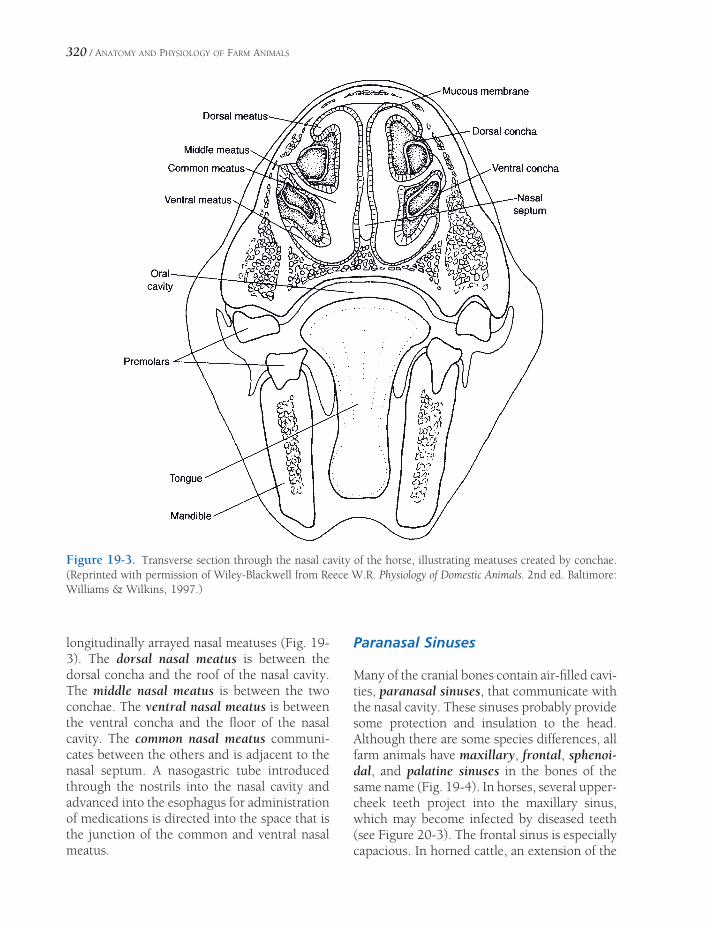
320 / ANATOMY AND PHYSIOLOGY OF FARM ANIMALS
longitudinally arrayed nasal meatuses (Fig. 19-3). The dorsal nasal meatus is between the dorsal concha and the roof of the nasal cavity. The middle nasal meatus is between the two conchae. The ventral nasal meatus is between the ventral concha and the fl oor of the nasal cavity. The common nasal meatus communi-cates between the others and is adjacent to the nasal septum. A nasogastric tube introduced through the nostrils into the nasal cavity and advanced into the esophagus for administration of medications is directed into the space that is the junction of the common and ventral nasal meatus.
Paranasal Sinuses
Many of the cranial bones contain air-fi lled cavi-ties, paranasal sinuses, that communicate with the nasal cavity. These sinuses probably provide some protection and insulation to the head. Although there are some species differences, all farm animals have maxillary, frontal, sphenoi-dal, and palatine sinuses in the bones of the same name (Fig. 19-4). In horses, several upper-cheek teeth project into the maxillary sinus, which may become infected by diseased teeth (see Figure 20-3). The frontal sinus is especially capacious. In horned cattle, an extension of the
Figure 19-3. Transverse section through the nasal cavity of the horse, illustrating meatuses created by conchae. (Reprinted with permission of Wiley-Blackwell from Reece W.R. Physiology of Domestic Animals. 2nd ed. Baltimore: Williams & Wilkins, 1997.)
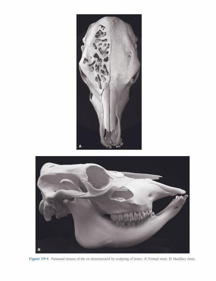
A
B
Figure 19-4. Paranasal sinuses of the ox demonstrated by sculpting of bones. A) Frontal sinus. B) Maxillary sinus.

322 / ANATOMY AND PHYSIOLOGY OF FARM ANIMALS
frontal sinus, the cornual diverticulum, extends well into the bony core of the horn (Fig. 13-7). Dehorning of animals older than 3 or 4 months of age usually exposes the interior of the frontal sinus and predisposes it to infection.
Pharynx
The pharynx is a common soft tissue conduit for food and air, lying caudal to the oral and nasal cavities. Openings into the pharynx include the two caudal nares (choanae), two auditory tubes from the middle ears, the oral cavity, the larynx, and the esophagus. The walls of the pharynx are supported by striated muscles whose actions assist in deglutition (swallowing) and phonation. In normal circumstances, food-stuffs and liquids are directed into the esopha-gus from the mouth (and vice versa during rumination) with a coordinated movement of pharyngeal and laryngeal muscles that excludes these substances from the airways.
Larynx
The larynx is the gatekeeper to the entrance of the trachea. It maintains a rigid, boxlike shape via a series of paired and unpaired cartilages that are moved relative to one another by stri-ated laryngeal muscles. The larynx’s primary function is to regulate the size of the airway and to protect it by closing to prevent substances other than air from entering the trachea. It is capable of increasing the diameter of the air passageway during forced inspiration (as during heavy exercise) and closing the opening during swallowing or as a protective mechanism to exclude foreign objects. Secondarily, the larynx is the organ of phonation (vocalization), hence its common name, voice box. Contraction of muscles in the larynx changes the tension on ligaments that vibrate as air is drawn past them; this vibration produces the voice.
Five mucous membrane–lined cartilages make up the larynx in most domestic animals (Fig. 19-5). The unpaired, spade-shaped epi-glottic cartilage (epiglottis), which lies just caudal to the base of the tongue, is mostly
elastic cartilage. During deglutition, movements of the tongue and larynx fold the epiglottis caudad so that it covers the entrance into the larynx.
The thyroid cartilage resembles a taco shell, consisting of two parallel plates, the laminae, on each side, joined on the ventral midline. In the human, the thyroid cartilage, or Adam’s apple, projects from the ventral aspect of the neck. The laminae of the thyroid cartilage have processes that articulate with other cartilages and give attachment to a number of muscles.
The cricoid cartilage is shaped like a signet ring, with the broad portion dorsal. The cricoid cartilage articulates with the thyroid cartilage and the two arytenoid cartilages (discussed later) cranially and attaches to the fi rst cartilagi-nous ring of the trachea caudally.
The paired arytenoid cartilages are irregular in shape, with processes that vary between species. The arytenoid cartilages of all species have a ventral vocal process, to which is attached the vocal ligament (vocal cord). The vocal ligaments mark the division between the vestibule, the laryngeal space cranial to them, and the infraglottic cavity, the space caudal to them (Fig. 19-5). The slitlike gap between the two vocal ligaments is the rima glottidis. Move-ment of the arytenoid cartilages changes the tension and angle of the vocal ligaments, closing the glottis or adjusting the pitch of the voice as the ligaments vibrate.
Horses and swine also possess a vestibular (ventricular) ligament cranial to the vocal liga-ment. An outpocketing of mucous membrane between the two ligaments forms a blind pouch called the lateral ventricle.
The vagus nerve (cranial nerve X) carries fi bers that innervate the muscles (motor) and the mucous membranes (sensory) of the larynx. Most of the muscles receive their motor inner-vation from the recurrent laryngeal nerve, which for embryological reasons branches from the vagus within the thorax and must return craniad along the trachea to reach the larynx. This very long course of the axons in the recur-rent laryngeal nerve (from the brainstem, where the neuronal cell bodies reside, down the neck into the thorax and back up the neck to the
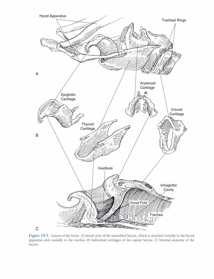
Figure 19-5. Larynx of the horse. A) lateral view of the assembled larynx, which is attached rostrally to the hyoid apparatus and caudally to the trachea. B) Individual cartilages of the equine larynx. C) Internal anatomy of the larynx.
Hyoid Apparatus
Epiglottic
A
B
C
Cartilage
Arytenoid
Cartilage
Cricoid
Cartilage
Tracheal Rings
Trachea
Vocal Fold
Thyroid
Cartilage
Vestibule
Infraglottic
Cavity

324 / ANATOMY AND PHYSIOLOGY OF FARM ANIMALS
larynx) makes them susceptible to trauma and metabolic diseases.
Injury to one of the recurrent laryngeal nerves will result in paralysis of most of the laryngeal muscles on the same side. Paraly-sis of the muscle that abducts the arytenoid cartilages and thereby increases the diameter of the airway (the dorsal cricoarytenoid muscle) results in a condition in horses called laryngeal hemiplegia or roaring. A horse that is a roarer cannot expand the rima glottidis during forceful inspiration and con-sequently has diffi culty bringing suffi cient air into the lungs when exercising. The fl accid vocal ligament fl utters as air moves rapidly past it, generating a loud, hoarse sound. Roaring usually is due to unilateral damage to the left recurrent laryngeal nerve,
as this nerve’s course is somewhat longer than that of the right recurrent laryngeal nerve.
Trachea and Bronchi
The trachea extends from the caudal end of the larynx to the bronchi (Fig. 19-6). It is formed by a series of C-shaped hyaline tracheal carti-lages that provide cross-sectional rigidity to resist collapse and are joined one to another by elastic annular ligaments that permit the trachea considerable fl exibility to follow move-ments of the neck. The dorsal side of the trachea is completed by connective tissue and the m. trachealis, a smooth muscle whose tone affects the diameter of the trachea.
Figure 19-6. Basic anatomy of trachea and principal bronchi.
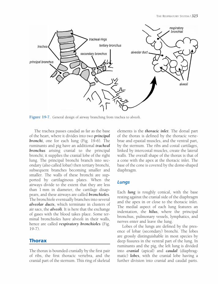
THE RESPIRATORY SYSTEM / 325
The trachea passes caudad as far as the base of the heart, where it divides into two principal bronchi, one for each lung (Fig. 19-6). The ruminants and pig have an additional tracheal bronchus arising cranial to the principal bronchi; it supplies the cranial lobe of the right lung. The principal bronchi branch into sec-ondary (also called lobar) then tertiary bronchi, subsequent branches becoming smaller and smaller. The walls of these bronchi are sup-ported by cartilaginous plates. When the airways divide to the extent that they are less than 1 mm in diameter, the cartilage disap-pears, and these airways are called bronchioles. The bronchiole eventually branches into several alveolar ducts, which terminate in clusters of air sacs, the alveoli. It is here that the exchange of gases with the blood takes place. Some ter-minal bronchioles have alveoli in their walls, hence are called respiratory bronchioles (Fig. 19-7).
Thorax
The thorax is bounded cranially by the fi rst pair of ribs, the fi rst thoracic vertebra, and the cranial part of the sternum. This ring of skeletal
elements is the thoracic inlet. The dorsal part of the thorax is defi ned by the thoracic verte-brae and epaxial muscles, and the ventral part, by the sternum. The ribs and costal cartilages, linked by intercostal muscles, create the lateral walls. The overall shape of the thorax is that of a cone with the apex at the thoracic inlet. The base of the cone is covered by the dome-shaped diaphragm.
Lungs
Each lung is roughly conical, with the base resting against the cranial side of the diaphragm and the apex in or close to the thoracic inlet. The medial aspect of each lung features an indentation, the hilus, where the principal bronchus, pulmonary vessels, lymphatics, and nerves enter and leave the lung.
Lobes of the lungs are defi ned by the pres-ence of lobar (secondary) bronchi. The lobes are grossly distinguishable in most species by deep fi ssures in the ventral part of the lung. In ruminants and the pig, the left lung is divided into cranial (apical) and caudal (diaphrag-matic) lobes, with the cranial lobe having a further division into cranial and caudal parts.
Figure 19-7. General design of airway branching from trachea to alveoli.
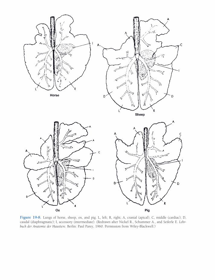
Figure 19-8. Lungs of horse, sheep, ox, and pig. L, left; R, right; A, cranial (apical); C, middle (cardiac); D, caudal (diaphragmatic); I, accessory (intermediate). (Redrawn after Nickel R., Schummer A., and Seiferle E. Lehr-buch der Anatomie der Haustiere. Berlin: Paul Parey, 1960. Permission from Wiley-Blackwell.)

THE RESPIRATORY SYSTEM / 327
The right lung in these animals is divided into four lobes: cranial and caudal lobes as on the left, plus a middle lobe between these and a ventrocaudal accessory lobe near the midline (Fig. 19-8). A more or less distinct gap between lobes along the ventral margin of the lungs is usually identifi able. This is the cardiac notch, and it tends to be larger on the right side (Fig. 19-9). At the cardiac notch, the heart makes contact with the thoracic wall, a fact that is exploited for echocardiogram imaging (wherein the ultrasound device is applied to the thoracic wall, where no lung tissue intervenes between it and the heart) and pericardiocentesis (intro-ducing a needle through the body wall to sample fl uid from the pericardial sac).
Both right and left lungs of the horse have cranial and caudal lobes, distinguished by the
cardiac notch. The right equine lung also has a small accessory lobe.
After an animal has taken one breath, the lungs always retain a signifi cant volume of air, even in pathologic conditions of col-lapsed lung. In the fetus, however, the lungs are nearly the consistency of liver, contain no air, and sink in water. Whether the lungs will sink or fl oat in water is a standard test to determine whether a newborn animal was born dead, in which case the lungs sink, or drew at least one breath, in which case the lungs fl oat.
Pleura
The thoracic cavity is lined by a serosa, the pleura. The smooth surfaces of the pleura are lubricated with a scant amount of serous fl uid, facilitating frictionless movement of the lungs during respirations. The pleura is attached to the bony and muscular elements of the thorax by endothoracic fascia, a thin connective tissue layer.
The pleura consists of two separate sacs, one surrounding each lung. The pleura that lines the thorax is the parietal pleura, and the pleura that covers the lungs is the visceral pleura. The pleural cavity, between parietal and visceral pleurae, is a potential space only. This pleural cavity normally contains nothing except a small amount of serous fl uid; conditions that intro-duce fl uid or gas (e.g., pus, blood, air) into the pleural space compress and may collapse the lung associated with that space.
The junction of the two pleural sacs near the midline of the thorax forms a double layer called the mediastinum in which are found the heart, great vessels, esophagus, and other midline structures. The caudal vena cava and right phrenic nerve (nerve innervating the right side of the diaphragm) are enclosed in a distinct fold of pleura, the plica venae cavae. The medi-astinum of cattle is thick and forms a complete barrier between the right and left pleural cavi-ties. In horses, parts of the mediastinum are thin, and openings between the two cavities either occur naturally or are readily created. For
Figure 19-9. Disposition of the left lung in the ox (above) and horse (below), shown in a cutaway view of the thorax. a, cranial lobe; a’, cranial part of cranial lobe (ox only); b, caudal part of cranial lobe (ox only); c, caudal lobe; d, diaphragm; e, heart.
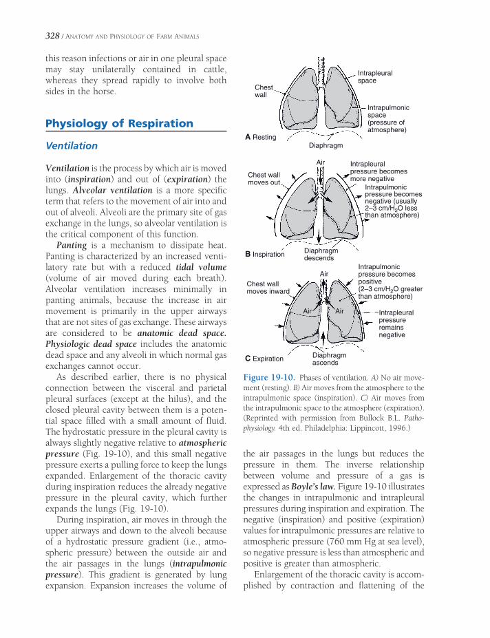
328 / ANATOMY AND PHYSIOLOGY OF FARM ANIMALS
this reason infections or air in one pleural space may stay unilaterally contained in cattle, whereas they spread rapidly to involve both sides in the horse.
Physiology of Respiration
Ventilation
Ventilation is the process by which air is moved into (inspiration) and out of (expiration) the lungs. Alveolar ventilation is a more specifi c term that refers to the movement of air into and out of alveoli. Alveoli are the primary site of gas exchange in the lungs, so alveolar ventilation is the critical component of this function.
Panting is a mechanism to dissipate heat. Panting is characterized by an increased venti-latory rate but with a reduced tidal volume (volume of air moved during each breath). Alveolar ventilation increases minimally in panting animals, because the increase in air movement is primarily in the upper airways that are not sites of gas exchange. These airways are considered to be anatomic dead space. Physiologic dead space includes the anatomic dead space and any alveoli in which normal gas exchanges cannot occur.
As described earlier, there is no physical connection between the visceral and parietal pleural surfaces (except at the hilus), and the closed pleural cavity between them is a poten-tial space fi lled with a small amount of fl uid. The hydrostatic pressure in the pleural cavity is always slightly negative relative to atmospheric pressure (Fig. 19-10), and this small negative pressure exerts a pulling force to keep the lungs expanded. Enlargement of the thoracic cavity during inspiration reduces the already negative pressure in the pleural cavity, which further expands the lungs (Fig. 19-10).
During inspiration, air moves in through the upper airways and down to the alveoli because of a hydrostatic pressure gradient (i.e., atmo-spheric pressure) between the outside air and the air passages in the lungs (intrapulmonic pressure). This gradient is generated by lung expansion. Expansion increases the volume of
the air passages in the lungs but reduces the pressure in them. The inverse relationship between volume and pressure of a gas is expressed as Boyle’s law. Figure 19-10 illustrates the changes in intrapulmonic and intrapleural pressures during inspiration and expiration. The negative (inspiration) and positive (expiration) values for intrapulmonic pressures are relative to atmospheric pressure (760 mm Hg at sea level), so negative pressure is less than atmospheric and positive is greater than atmospheric.
Enlargement of the thoracic cavity is accom-plished by contraction and fl attening of the
Intrapleuralspace
Chestwall
Intrapulmonicspace(pressure ofatmosphere)
DiaphragmA Resting
B Inspiration
C Expiration
Air Intrapleuralpressure becomesmore negative
Diaphragmdescends
Diaphragmascends
Intrapulmonicpressure becomesnegative (usually2–3 cm/H2O lessthan atmosphere)
Intrapleuralpressureremainsnegative
Intrapulmonicpressure becomespositive(2–3 cm/H2O greaterthan atmosphere)
Air
Air Air
Chest wallmoves inward
Chest wallmoves out
Figure 19-10. Phases of ventilation. A) No air move-ment (resting). B) Air moves from the atmosphere to the intrapulmonic space (inspiration). C) Air moves from the intrapulmonic space to the atmosphere (expiration). (Reprinted with permission from Bullock B.L. Patho-physiology. 4th ed. Philadelphia: Lippincott, 1996.)

THE RESPIRATORY SYSTEM / 329
dome-shaped diaphragm and a forward and outward movement of the ribcage by the con-traction of appropriate thoracic muscles. These are skeletal muscles innervated by somatic motor nerves. After inspiration, the pressure in the pleural cavity remains at its lowest point until expiration begins and the thoracic cavity begins to return to its original volume.
Expiration in a resting animal is a passive process that does not require muscle contrac-tion. Relaxation of the muscles contracted during inspiration permits the intrinsic elastic properties of the lungs and the thoracic wall to recoil to their original volume. The return to the original volume increases the intrapulmonic pressure so that it is greater than atmospheric pressure, and air is forced out of the lungs.
Forced expiration is an active process that forces more air from the lungs than would occur during a normal passive expiration. Forced expiration requires contraction of abdominal muscles to force viscera against the diaphragm and contraction of other muscles to pull the ribs caudad. Both of these actions reduce the size of the thoracic cavity and permit recoil of the lungs to a smaller volume than typical for resting expiration. This causes a further increase in intrapulmonic pressure and forces more air out than would occur with passive expiration. Even with the most forceful expiration, a resid-ual volume of air still remains in the lungs. Vital capacity is the maximal amount of air that can be expired after a maximal inspiration.
Both the upper airways and the air passages in the lungs resist airfl ow through them. Similar to a blood vessel, the resistance of an air passage to airfl ow is inversely proportional to its diam-eter. Most of the air passages in the lungs have smooth muscle in the walls, and the constric-tion or relaxation of this smooth muscle deter-mines the resistance to airfl ow. Much of this smooth muscle has β2-adrenergic receptors that produce smooth muscle relaxation when stim-ulated. Sympathetic stimulation during exer-cise reduces resistance to airfl ow and promotes alveolar ventilation.
Airway smooth muscle constricts in response to stimulation by histamine or leu-kotrienes. These substances are released
during allergic reactions from mast cells around airways. The resulting increase in airway resistance makes it more diffi cult to move air, and a more forceful skeletal muscle contraction is needed to move a given volume of air. This greatly increases the work of breathing. Heaves is a condition in horses that is characterized by labored or diffi cult breathing, and one potential cause is chronic exposure to environmental allergens, which results in chronic obstructive airway disease. The chronic need for increased skeletal muscle force may induce visible hypertro-phy of the abdominal muscles (heave line).
Surface tension, a property of fl uids, results from the cohesive forces that tend to pull or hold the molecules of a fl uid together. An example is the tendency of a layer of fl uid on a fl at surface to come together and form a bubble rather than remaining as a thin, fl at layer. A thin layer of fl uid lines the microscopic alveoli. The surface tension of this fl uid tends to draw the walls of alveoli together and col-lapse them. The alveolar fl uid contains the pul-monary surfactant, a combination of substances that reduces surface tension. The reduction in surface tension promotes alveolar stability and makes alveolar expansion during inspiration easier. Production of pulmonary surfactant does not begin until late in gestation. Neo-nates born prematurely may have insuffi -cient amounts of surfactant, which results in labored breathing.
In normal animals a soft rustling sound asso-ciated with air movements may be heard with a stethoscope. Abnormal lungs, for example lungs with abnormal amounts of fl uid in the airways, may produce exaggerated sounds termed rales. Pleuritis (pleurisy), or infl amma-tion of the pleura, may produce a rasping sound because of the roughened surfaces rubbing together. Hydrothorax (fl uid in the pleural cavity) may also result from pleuritis.
Gas Exchange
The oxygen and carbon dioxide concentrations in air can be described in two ways: partial

330 / ANATOMY AND PHYSIOLOGY OF FARM ANIMALS
pressures and percentages. Room air is approx-imately 21% oxygen and 0.3% carbon dioxide. The primary component of room air is the inert gas nitrogen (about 78%). The partial pressure of an individual gas in a mixture of gases is the product of the percentage of the individual gas in the mixture and the total barometric or atmospheric pressure. Thus, at sea level, where atmospheric pressure is 760 mm Hg, the partial pressure of oxygen in room air is approximately 160 mm Hg.
The partial pressure of an individual gas in a mixture is one factor that determines the amount of the gas that will dissolve in a liquid (such as blood plasma). The partial pressure of a gas in a mixture can be viewed as the driving force that moves molecules of an individual gas from the air into the liquid when the liquid is exposed to a gas mixture. Thus, because partial pressures depend on both total atmospheric pressure and the percentages of individual gases, both of these factors also determine the amount of an individual gas that can be dis-solved in a liquid. The unit of measurement of the amount of a gas dissolved in a liquid is in millimeters of mercury.
Gas Exchange in the Lungs. Gas exchange between the blood and alveolar air in the lungs occurs across the walls of alveoli. At its thinnest point, the alveolar wall barrier between blood plasma and alveolar air consists of the endothe-lial cell of pulmonary capillaries, a type I squa-mous epithelial cell lining the alveoli, and a fused basement membrane contributed by both cells (Fig. 19-11). Gases readily move back and forth across this very thin and fragile structure. Any abnormality that thickens this barrier (e.g., pulmonary edema with an accumula-tion of extracellular fl uid in the alveolar wall) can greatly reduce the effi ciency of exchange. Oxygen exchange is usually affected fi rst because its solubility is much less than that of carbon dioxide.
Exchange begins as soon as blood enters a pulmonary capillary from pulmonary arterial vessels and continues until equilibrium between alveolar air and plasma is reached. Figure 19-12 shows typical values for partial pressures of
oxygen and carbon dioxide in alveolar air and a pulmonary capillary. Plasma entering pulmo-nary capillaries from the pulmonary arteries contains the highest concentration of carbon dioxide and the lowest of oxygen. Because of the continuous gas exchange in the pulmonary capillaries, alveolar air contains less oxygen and more carbon dioxide than inspired air (PO2
of 160 mm Hg and PCO2 of 0.23 mm Hg for inspired air).
To be most effi cient, the rate of pulmonary artery blood fl ow into an area of the lung must be balanced with the rate of air movement in and out of the alveoli in the same area. To appreciate the importance of this balancing, consider an extreme case in which one lung is collapsed so that air movement is impossible, but the collapsed lung receives the same amount of blood fl ow as the infl ated normal lung. The lack of airfl ow in the collapsed lung means that
I
am
is
I
*
pc
II
is
I
II
II
is
Figure 19-11. Schematic representation of three adjacent alveoli. I- type I alveolar epithelial cell; II- type II alveolar epithelial cell; is- interalveolar septum; pc- pulmonary capillary; am- alveolar macrophage. Note the merger of the basal laminae of the pulmonary capil-lary and alveolus (*). (Reprinted with permission of Wiley-Blackwell from Dellmann H.D. Textbook of Vet-erinary Histology. 4th ed. Philadelphia: Lea & Febiger, 1993.)
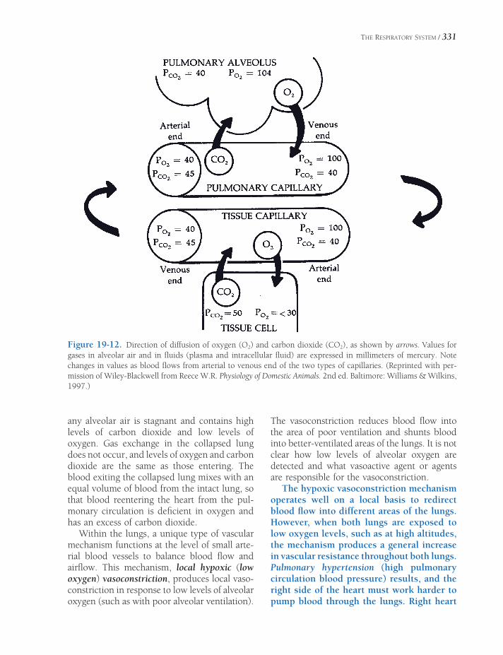
THE RESPIRATORY SYSTEM / 331
Figure 19-12. Direction of diffusion of oxygen (O2) and carbon dioxide (CO2), as shown by arrows. Values for gases in alveolar air and in fl uids (plasma and intracellular fl uid) are expressed in millimeters of mercury. Note changes in values as blood fl ows from arterial to venous end of the two types of capillaries. (Reprinted with per-mission of Wiley-Blackwell from Reece W.R. Physiology of Domestic Animals. 2nd ed. Baltimore: Williams & Wilkins, 1997.)
any alveolar air is stagnant and contains high levels of carbon dioxide and low levels of oxygen. Gas exchange in the collapsed lung does not occur, and levels of oxygen and carbon dioxide are the same as those entering. The blood exiting the collapsed lung mixes with an equal volume of blood from the intact lung, so that blood reentering the heart from the pul-monary circulation is defi cient in oxygen and has an excess of carbon dioxide.
Within the lungs, a unique type of vascular mechanism functions at the level of small arte-rial blood vessels to balance blood fl ow and airfl ow. This mechanism, local hypoxic (low oxygen) vasoconstriction, produces local vaso-constriction in response to low levels of alveolar oxygen (such as with poor alveolar ventilation).
The vasoconstriction reduces blood fl ow into the area of poor ventilation and shunts blood into better-ventilated areas of the lungs. It is not clear how low levels of alveolar oxygen are detected and what vasoactive agent or agents are responsible for the vasoconstriction.
The hypoxic vasoconstriction mechanism operates well on a local basis to redirect blood fl ow into different areas of the lungs. However, when both lungs are exposed to low oxygen levels, such as at high altitudes, the mechanism produces a general increase in vascular resistance throughout both lungs. Pulmonary hypertension (high pulmonary circulation blood pressure) results, and the right side of the heart must work harder to pump blood through the lungs. Right heart
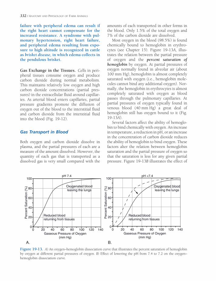
332 / ANATOMY AND PHYSIOLOGY OF FARM ANIMALS
failure with peripheral edema can result if the right heart cannot compensate for the increased resistance. A syndrome with pul-monary hypertension, right heart failure, and peripheral edema resulting from expo-sure to high altitude is recognized in cattle as brisket disease, in which edema collects in the pendulous brisket.
Gas Exchange in the Tissues. Cells in peri-ph eral tissues consume oxygen and produce carbon dioxide during normal metabolism. This maintains relatively low oxygen and high carbon dioxide concentrations (partial pres-sures) in the extracellular fl uid around capillar-ies. As arterial blood enters capillaries, partial pressure gradients promote the diffusion of oxygen out of the blood to the interstitial fl uid and carbon dioxide from the interstitial fl uid into the blood (Fig. 19-12).
Gas Transport in Blood
Both oxygen and carbon dioxide dissolve in plasma, and the partial pressures of each are a measure of the amount dissolved. However, the quantity of each gas that is transported as a dissolved gas is very small compared with the
amounts of each transported in other forms in the blood. Only 1.5% of the total oxygen and 7% of the carbon dioxide are dissolved.
Most oxygen in the blood (98.5%) is found chemically bound to hemoglobin in erythro-cytes (see Chapter 15). Figure 19-13A, illus-trates the relation between the partial pressure of oxygen and the percent saturation of hemoglobin by oxygen. At partial pressures of oxygen normally found in alveolar air (about 100 mm Hg), hemoglobin is almost completely saturated with oxygen (i.e., hemoglobin mole-cules cannot bind any additional oxygen). Nor-mally, the hemoglobin in erythrocytes is almost completely saturated with oxygen as blood passes through the pulmonary capillaries. At partial pressures of oxygen typically found in venous blood (40 mm Hg) a great deal of hemoglobin still has oxygen bound to it (Fig. 19-13A).
Several factors affect the ability of hemoglo-bin to bind chemically with oxygen. An increase in temperature, a reduction in pH, or an increase in the concentration of carbon dioxide reduces the ability of hemoglobin to bind oxygen. These factors alter the relation between hemoglobin saturation and the partial pressure of oxygen so that the saturation is less for any given partial pressure. Figure 19-13B illustrates the effect of
0 20 40 60 80 120 1401000
20
40
60
80
100
Gaseous Pressure of Oxygen(mm Hg)
Perc
ent S
atu
ration
(%
) Oxygenated bloodleaving the lungs
Reduced bloodreturning from tissues
0 20 40 60 80 120 1401000
20
40
60
80
100
Gaseous Pressure of Oxygen(mm Hg)
Perc
ent S
atu
ration (
%) Oxygenated blood
leaving the lungs
Reduced bloodreturning from tissues
A. B.
pH 7.4 pH <7.4
Figure 19-13. A) An oxygen–hemoglobin dissociation curve that illustrates the percent saturation of hemoglobin by oxygen at different partial pressures of oxygen. B) Effect of lowering the pH from 7.4 to 7.2 on the oxygen– hemoglobin dissociation curve.

THE RESPIRATORY SYSTEM / 333
lowering pH on hemoglobin saturation, and the effects of increases in temperature or carbon dioxide are similar. High temperature, low pH, and high carbon dioxide occur in tissues with high metabolic rates (e.g., exercising skeletal muscle). The effects of these factors on the rela-tion between hemoglobin and oxygen is useful, because more oxygen is liberated from hemo-globin and delivered to the metabolizing cells when blood passes through such areas.
Almost all of the carbon dioxide (93%) that enters the blood in the systemic circulation dif-fuses into erythrocytes (Fig. 19-14). Some of the carbon dioxide (23%) chemically combines with the hemoglobin in the erythrocytes to form carbaminohemoglobin. When the eryth-rocytes carrying the carbaminohemoglobin reach the pulmonary capillaries in the lungs, the reaction is reversed so that the carbon dioxide can diffuse into alveoli to be expired. Most (70%) of the carbon dioxide that enters the erythrocytes is converted to carbonic acid under the infl uence of the enzyme carbonic anhydrase. The carbonic acid rapidly dissoci-ates into a hydrogen ion and a bicarbonate ion
in the erythrocyte. The hydrogen ion is buff-ered by hemoglobin in the erythrocyte, and the bicarbonate ion leaves the erythrocyte and enters the plasma. It is in this form (bicarbonate ion in plasma) that most carbon dioxide is transported from the peripheral tissues by the blood to the lungs (Fig. 19-14). Within the lungs, the reactions are reversed so that carbon dioxide can be reformed and be expired from the alveoli.
Control of Ventilation
Contraction and relaxation of skeletal muscle generates the forces to move air in and out of the lungs. Although the skeletal muscles of res-piration can be consciously controlled, as illus-trated by voluntarily holding the breath, normal ventilation is almost entirely refl exive.
The respiratory refl ex centers consist mainly of three bilateral groups of nerve cells in the brainstem that have a defi nite effect on respira-tion when stimulated electrically. One of these, the medullary rhythmicity area, is responsible
Erythrocyte
Carbaminohemoglobin(23%)
Hemoglobin
Carbonic anhydrase
CO2 from cellularmetabolism
CO2
HCO3–
HCO3–
dissolved in
plasma (70%)
H2O + CO2
H2CO3
H+
CO2 dissolved in plasma(7%)
Figure 19-14. Gas exchange in systemic tissues and transport of carbon dioxide in the blood. Percentages refl ect the different modes by which carbon dioxide is transported from systemic tissues to the lungs.

334 / ANATOMY AND PHYSIOLOGY OF FARM ANIMALS
for setting respiratory rate, and it consists mainly of an inspiratory center. Neurons in this center are tonically active, fi ring at an inherent rhythmic rate by regular variations of their membrane potentials. Expiratory neurons are also in this area; however, they do not discharge spontaneously and so are normally active only during a forced expiration.
Stimulation of the inspiratory center leads to contractions of the diaphragmatic and inter-costal muscles via neural connections through the spinal cord and phrenic and intercostal nerves, respectively. Feedback circuits between the inspiratory center and the other two neural centers relax these muscles and allow for passive expiration. The interplay among these three centers provides for regular intermittent rhythmic breathing at rates appropriate for each species during eupnea (normal quiet breathing).
The tonic activity of the inspiratory center is regulated by neural input from a variety of sites. In resting animals, the most important neural input is from central chemoreceptors in the medulla of the brainstem. These receptors respond to hydrogen ion concentration changes in the interstitial fl uid of the brain and stimu-late the inspiratory center to increase ventila-tion when the hydrogen ion concentration increases. Because carbon dioxide from the blood readily diffuses into the interstitial fl uids of the brain, and in body fl uids is in equilib-rium with carbonic acid, an increase in blood carbon dioxide increases the hydrogen ion con-centration in the brain and stimulates ventila-tion. The effects of changes in blood carbon dioxide on ventilation are so pronounced that blood carbon dioxide is considered to be the
most important regulator of ventilation in most conditions.
Animals sometimes hyperventilate (breathe abnormally rapidly) during the induction of general anesthesia because they are excited. As a result of the hyperventilation, their blood carbon dioxide may be signifi cantly reduced. The low blood carbon dioxide con-centration means that the primary stimulus for normal ventilation is lost. These animals may undergo a period of apnea (cessation of breathing) until blood carbon dioxide levels are restored by metabolism.
Another group of chemoreceptors, the carotid and aortic bodies, also provide neural input to the inspiratory center. These periph-eral chemoreceptors detect changes in arterial blood hydrogen ion concentration and oxygen content. Increases in hydrogen ion concentra-tion or reductions in blood O2 content initiate neural inputs to increase ventilation. However, the effects of these peripheral chemoreceptors are less pronounced than those of the central chemoreceptors, so changes in blood hydrogen ion concentration or oxygen content must be severe to override the effect of blood carbon dioxide.
Further regulating the inspiratory center and breathing rhythmicity is a refl ex arc involving stretch receptors in the lung parenchyma, vis-ceral pleura, and bronchioles. These receptors are stimulated as the lung infl ates during inspi-ration, and afferent impulses are transmitted up the vagus nerves into the brainstem, where the inspiratory center is inhibited. This is the Hering-Breuer refl ex, which reinforces the action of the other center to limit inspiration and prevent overdistension of the lungs.

ANATOMY OF THE DIGESTIVE SYSTEM
Organization of the Digestive System
The digestive system (digestive tract) consists of a muscular tube lined with mucous mem-brane that is continuous with the external skin at the mouth and at the anus. Its primary func-tions are prehension, mastication, digestion, and absorption of food, and elimination of solid wastes. The digestive system reduces the nutri-tious constituents of the food to molecular com-pounds that are small enough to be absorbed and used for energy and for building other com-pounds for incorporation into body tissues.
Elements of the digestive system are the mouth, pharynx, esophagus, forestomach (ruminants), glandular stomach, small intes-tine, large intestine, rectum, and the accessory glands (salivary glands, liver, and pancreas).
Caudal to the diaphragm, the components of the digestive tract lie within the abdominal and pelvic cavities. Here they are invested with a simple squamous epithelium that is also called a mesothelium or serosa. Within these body cavities, the serosa is identifi ed as perito-neum. Like the pleura within the thoracic cavity, it is named according to the structures to which it is applied: where it lies directly on the organ, it is called visceral peritoneum, and where it invests the abdominal wall, it is parietal peri-toneum (see Fig. 1-9).
Parietal and visceral peritonea are continu-ous with one another through refl ections of the serosa that attach the organs to the body wall.
Organization of the Digestive SystemMouth
Teeth
Tongue
PharynxTonsils
EsophagusNonruminant StomachRuminant Stomach
Ruminoreticulum
Omasum
Abomasum
Small IntestineLarge Intestine
Ruminants
Pig
Horse
Peritoneal FeaturesAccessory Digestive Organs
Salivary Glands
Pancreas
Liver
Chapter 20
335

336 / ANATOMY AND PHYSIOLOGY OF FARM ANIMALS
These attachments collectively constitute the mesenteries, and they are named for the organ they suspend (discussed later). Blood vessels, lymphatics, and nerves travel within the mes-enteries, reaching the organs through them.
The wall of the digestive tract comprises four layers, or tunics. These are, from within outward: (1) the tunica mucosa, (2) the tunica submucosa, (3) the tunica muscularis, and (4) the tunica serosa, or (where organs lie outside body cavities) tunica adventitia (Fig. 20-1).
The tunica mucosa is the layer closest to the space (the lumen) inside the digestive tract. It has three histologic layers. The innermost layer consists of a stratifi ed squamous epithelium from the mouth to the level of the glandular part of the stomach; from this point to the anus,
the epithelium is of the simple columnar type. Underlying the epithelium of the tunica mucosa are a layer of connective tissue and a variably present smooth muscle layer.
The tunica submucosa is a layer of loose connective tissue in which are found blood vessels and nerves. In some locations, glands of the digestive tract can be found in the submu-cosa, as can lymphatic nodules.
As motility is important to the function of the digestive system, the tunica muscularis is generally well developed. In the horse, the cranial two-thirds of the tunica muscularis of the esophagus is striated muscle; in the pig, all but the most distal end of the esophagus is stri-ated; and in ruminants, the entire esophagus has striated muscle. From this point and distal,
Figure 20-1. Layers of a typical segment of gut. (Reprinted with permission of Wiley-Blackwell from Reece W.O. Physiology of Domestic Animals. 2nd ed. Baltimore: Williams & Wilkins, 1997.)
ANATOMY OF THE DIGESTIVE SYSTEM / 337
the muscle cells are smooth (involuntary) and are generally arranged in two layers. The deeper layer has fi bers that encircle the gut, and the more superfi cial muscle layer assumes a longi-tudinal arrangement.
The outermost tunic consists of the visceral peritoneum and scant connective tissue under-lying it. This is the tunica serosa. The thoracic and cervical parts of the esophagus are not suspended directly within a body cavity (the thoracic esophagus is surrounded by the tissues of the mediastinum) and therefore do not have a tunica serosa. Instead, the surrounding con-nective tissue in these locations constitutes a tunica adventitia. Likewise, the distal end of the rectum and anal canal are outside the pelvic cavity and surrounded by a tunica adventitia.
Mouth
The mouth is used primarily for holding, grind-ing, and mixing food with saliva but may also be used to manipulate the environment (through grasping of objects) and as a defensive and offensive weapon.
The entrance into the mouth is defi ned by the lips (labia), the appearance and mobility of which vary among species. The external parts of the lips are covered by typical haired skin, which changes to mucous membrane at the mucocutaneous junction of the labial margins. The upper lip of small ruminants is deeply grooved with a midline philtrum. Lips are densely innervated by sensory fi bers, making them very sensitive tactile organs. The lips of sheep, goats, and horses are soft and fl exible and aid in picking up food, whereas those of cattle and hogs are stiffer and less mobile.
The small space between the teeth and lips is the oral vestibule. The oral cavity proper lies deep to the teeth and is occupied primarily by the tongue. The oral cavity ends at a narrowing (the isthmus of the fauces) near the base of the tongue, where the digestive tract continues as the pharynx.
The dorsal wall of the oral cavity comprises the hard palate rostrally and the soft palate caudally. The hard palate is formed by horizon-
tal elements of the incisive, maxillary, and pala-tine bones, and its thick mucous membrane covering is characterized by prominent trans-verse folds called palatine rugae. The soft palate is a musculomucosal sheet that extends toward the base of the epiglottis (see Chapter 19). The equine soft palate is exceptionally long, and the horse is uniquely unable to actively lift the soft palate so as to permit passage of air from the oral cavity to the larynx. For this reason, horses are obligate nose breath-ers, breathing through the mouth only when the soft palate is displaced dorsad from its normal position ventral to the epiglottis.
Teeth
Teeth are arranged in two dental arcades, one associated with the mandible and one with the incisive and maxillary bones. Farm animals typi-cally have a gap in each arch between the front teeth (incisors) and the cheek teeth (see Fig. 4-4); such a physiologic gap is a diastema. The bit of the bridle lies in the horse’s diastema.
Mammals typically exhibit heterodonty. That is, they have various types of teeth that are specialized for different aspects of prehension and mastication. All domestic animals also are diphyodont. This means they develop a set of deciduous teeth (also called baby teeth or milk teeth) that fall out and are replaced with per-manent teeth. As growing teeth emerge from the gums, they are said to erupt. When their occlusal (grinding) surfaces meet those of the teeth in the opposing arcade, they are said to have come into wear. Eruption times of teeth are consistent enough to permit accurate aging of young animals by observing their dentition (see Tables 20-1 and 20-2). The age of animals with a full set of permanent teeth can be esti-mated through examination of the wear pat-terns of the occlusal surfaces.
A tooth is anchored by its root in a socket of bone called an alveolus. A connective tissue, the periodontium (also called periodontal membrane), fi rmly attaches the root to the sur-rounding bone in a specialized joint, the gom-phosis (see Chapter 6). The crown is the part

Table 20-1. Formulas and Eruption of Deciduous Teeth
Deciduous Teeth Horse Ox Sheep Pig
Formulas* 3 0 3 0 0 3 0 0 3 3 1 4
3 0 3 4 0 3 4 0 3 3 1 4
Eruption of Incisors
DI 1 birth to 1 week birth to 2 wk birth to 1 wk 2–4 wk
DI 2 4–6 wks birth to 2 wk birth to 1 wk 1.5–3 mo
DI 3 6–9 mo birth to 2 wk birth to 1 wk birth or before
DI 4 birth to 2 wk birth to 1 wk
Eruption of Canines
before birth
Eruption of Premolars
DP 1 birth to few days usually before birth 3.5–6.5 mo
DP 2 birth to 2 wk birth to few days usually before birth 7–10 wk
DP 3 birth to 2 wk birth to few days usually before birth 1–3 wk (upper)
1–5 wk (lower)
DP 4 birth to 2 wk 1–4 wk (upper)
2–7 wk (lower)
*The deciduous formula is shown for one side of the mouth only, with upper arcade represented above and lower arcade below. The fi rst number is the incisors; the second, canines; and the third is premolars.
Table 20-2. Formulas and Eruption of Permanent Teeth
Permanent Teeth Horse Ox Sheep Pig
Formulas* 3 1 3–4 3 0 0 3 3 0 0 3 3 3 1 4 3
3 1 3 3 4 0 3 3 4 0 3 3 3 1 4 3
Eruption of Incisors
I 1 2.5 yr 1.5–2 yr 1–1.5 yr 1 yr
I 2 3.5 yr 2–2.5 yr 1.5–2 yr 16–20 mo
I 3 4.5 yr 3 yr 1.5–3 yr 8–10 mo
I 4 3.5–4 yr 3.5–4 yr
Eruption of Canines
4–5 yr 9–10 mo
Eruption of Premolars
P 1 5–6 mo 2–2.5 yr 1.5–2 yr 12–15 mo
P 2 2.5 yr 1.5–2.5 yr 1.5–2 yr 12–15 mo
P 3 3 yr 2.5–3 yr 1.5–2 yr 12–15 mo
P 4 4 yr 12–15 mo
Eruption of Molars
M 1 9–12 mo 5–6 mo 3–5 mo 4–6 mo
M 2 2 yr 1–1.5 yr 9–12 mo. 8–12 mo
M 3 3.5–4 yr 2–2.5 yr 1.5–2 yr 18–20 mo
*The permanent formula is shown for one side of the mouth only, with upper arcade represented above and lower arcade below. The fi rst number is the incisors; the second, canines; the third is premolars; and the fourth is the molars.

ANATOMY OF THE DIGESTIVE SYSTEM / 339
Enamel
DentinPulp
Cementum
Gingiva
Peridontium
Infundibulum
Figure 20-2. Anatomy of typical brachyodont (left) and hypsodont (right) teeth.
of the tooth visible above the mucous mem-brane of the gum. Some teeth have a short crown separated from the root by a distinct neck. These teeth, of which ruminant incisors are an example, are described as brachyodont (from the Greek word brachy, short). In con-trast, equine incisors and cheek teeth have a tall, straight crown with no discernible neck. These teeth are described as hypsodont (Greek hypsi, high) (Fig. 20-2).
Most of the tooth’s substance is made up of a mineralized substance called dentin (sometimes spelled dentine), with a dental cavity at its center. The connective tissues, nerves, and blood vessels of the tooth reside in this cavity and constitute the dental pulp. It is dentin, incidentally, that constitutes the “ivory” of elephant tusks.
Superfi cial to the dentin is a layer of enamel, a white layer consisting of inorganic crystals. Enamel is the hardest substance in the body. It also is irreplaceable, as the cells that generate it (ameloblasts) are lost following formation of the
tooth, the only exception amongst farm animals being the tusks (canine teeth) of swine. Only the crowns of brachyodont teeth are covered with enamel, whereas nearly the entire hypsodont tooth has a layer of enamel, with only a small, short region of the tooth’s root lacking this layer. The enamel of hypsodont teeth is thrown into prominent folds on the grinding surfaces of these teeth, where they form characteristic crests (cristae enameli) and cups (infundibula).
Cementum is a thin, bonelike layer on the surface of the tooth. It covers only the root of the brachyodont tooth, but it extends from the root to cover the crown of the hypsodont tooth.
The front teeth are called incisors, and in dental formulas, they are designated by the letter I. The incisors are numbered starting from the midline. The fi rst pair of incisors is called I1, or centrals; the next pair I2, or fi rst intermedi-ates; next I3, or second intermediates; and the last and most lateral pair of incisors is called I4, or corners. In the nonruminants, only one pair of

340 / ANATOMY AND PHYSIOLOGY OF FARM ANIMALS
intermediate incisors is found. Ruminants lack incisors in the upper dental arcade. Instead, the mucous membrane in this region is modifi ed into a dense, keratinized dental pad, against which the lower incisors abut when the jaws are closed. Permanent incisors are preceded by a like number of deciduous teeth.
Canine teeth (abbreviated C) are also called eyeteeth, bridle teeth, tusks, and tushes. Rumi-nants lack canine teeth, and although they can be well developed in stallions, they are small or absent in mares and geldings. The canine teeth of pigs are large, especially in boars, and in this species they are usually called tusks. Porcine tusks are described as open rooted, meaning that they continue to grow throughout life. The lower tusk is generally much larger than its partner in the upper arcade. In pigs, the per-manent canines are preceded by analogous deciduous teeth; in the horse, the deciduous canines are often absent or so small that their crowns do not erupt.
Cheek teeth comprise premolars (P) and molars (M), which in herbivores are morpho-logically similar. Only premolars are preceded by deciduous teeth; the molars have no precur-sors. In the pig, the molars are larger than the premolars and have a fl atter occlusal surface, in keeping with their grinding function.
The fi rst premolar of horses is often absent, and when present, it is almost always seen only in the upper (maxillary) arcade. Unlike the other equine cheek teeth, the fi rst upper pre-molar is very small and does not come into wear. This tooth is called the wolf tooth, and some horse owners prefer to have it removed because of the perception that it may make unwanted contact with the bit.
In the young horse, only a small part of each cheek tooth is visible, as most of the crown lies developed but unerupted beneath the gum (Fig. 20-3). Throughout the horse’s life, these teeth continue to erupt, maintain-ing their intraoral height even as they are
Figure 20-3. Hypsodont teeth. The cheek teeth of the young horse are fully formed but largely within the bones of the face and mandible. The teeth slowly erupt over the life of the horse as the crowns are worn by chewing of coarse feedstuffs.

ANATOMY OF THE DIGESTIVE SYSTEM / 341
worn down by the coarse forage the horse eats. Equine cheek teeth are not open rooted; no new dental tissues are created after the tooth is initially formed. They are instead described as slowly erupting. Because of this phenomenon, horses are prone to develop sharp, elongated ridges of enamel on their cheek teeth. These are variously called points and hooks, and they may cause the horse considerable pain if they cut the soft cheeks, tongue, and gums. For this reason, it is con-sidered good husbandry to fi le down the points and hooks periodically, a procedure called fl oating the teeth that is done with a special rasp called a fl oat.
Deciduous and permanent dental formulas for domestic farm animals are shown in Tables 20-1 and 20-2.
Tongue
The tongue consists of a mass of muscle covered by mucous membrane. It is divided into a free apex at the rostral end, a meaty body, and a caudal root adjacent to the pharynx. The entire tongue is mobile through its muscular attach-ments to the hyoid apparatus and mandible. The muscles of the tongue (intrinsic muscles) have fi bers oriented in longitudinal, perpen-dicular, and transverse directions, permitting the tongue a wide range of movements. This is particularly evident in the ox, which uses its tongue as a prehensile organ (Fig. 20-4).
The tongue is covered with thick keratinized stratifi ed squamous epithelium. The surface is characterized by a large number of projections, the papillae, which are particularly well devel-oped on the dorsal surface (Fig. 20-5). Filiform, fungiform, and vallate papillae are found in all domestic animals, and foliate papillae are present in the horse, pig, and dog, but not in ruminants. Ruminants additionally have large conical papillae. The fi liform and conical papil-lae do not bear taste buds (cells specialized for gustation; see Chapter 11), but all other types of papillae do. Taste buds may also be found on the epiglottis, larynx, pharynx, and soft palate.
The fi liform papillae look somewhat hair-like. In the ox, they consist of a connective tissue core covered by a highly cornifi ed epithe-lial layer. These papillae are shorter and softer
Apex
Body
Root
5
3
1
2
4
A
B
C
D
6
Figure 20-4. Tongue of the ox, dorsal view. The pharynx and soft palate are transected and refl ected laterad. A, Vallate papillae; B, torus linguae with coni -cal papillae; C, fungiform papillae; D, fi liform papillae; 1, oropharynx; 2, tonsillar crypt; 3, cut surface of soft palate; 4, palatopharyngeal arch; 5, epiglottis; 6, opening into larynx.

A
B
C
Figure 20-5. A) Fungiform and fi liform papillae of the caprine tongue. The kera-tinized fi liform papillae have no taste buds. B) Vallate papilla of the caprine tongue. Note the deep groove adjacent to the papilla; taste buds occur in the mucosa of the papilla of this groove. C) Higher magnifi cation of a taste bud from an equine fungiform papilla. (Reprinted with permission of Wiley-Blackwell from Bacha, Jr. W.J. and Bacha L.M. Color Atlas of Veterinary Histology. Baltimore: Lippincott Williams & Wilkins, 2000.)

ANATOMY OF THE DIGESTIVE SYSTEM / 343
in the horse than in other domestic animals, giving the tongue of the horse its velvety feel. Interspersed amongst the fi liform papillae are fungiform papillae, so called because of their resemblance to tiny mushrooms.
Foliate papillae resemble the foliage or leaves of plants. They are found in the horse and pig (and only rarely in cattle) on the lateral margin adjacent to where the root of the tongue is connected to the soft palate by a mucous membrane fold, the palatoglossal arch.
Vallate papillae are large, circular projections surrounded by a deep groove. These papillae are arranged in a V shape on the caudal part of the tongue and demarcate the morphologic division between the body and the root of the tongue.
The body of the ruminant tongue has a prominent dorsal bulge, the torus lingua, which is thickly covered with prominent conical papillae. Similar cornifi ed projections cover the inside of the lips and cheeks.
Pharynx
The pharynx (pl. pharynges) is the common passage for food and air, is caudal to oral and nasal cavities, and is lined by mucous mem-brane and surrounded by muscles. The pharynx
can be arbitrarily divided into nasal (nasophar-ynx), oral (oropharynx), and laryngeal (laryn-gopharynx) portions, so named for their association with these regions.
The muscles of the walls of the pharynx are responsible for the orderly directing of air, food, and liquids in such a way that air from the nasal cavity is directed into the ventral larynx and food and liquids are directed into the dorsal esophagus. Thus, the paths of air and swal-lowed substances must cross in the pharynx; pharyngeal dysfunction can have severe conse-quences for the airway, which must be pro-tected from foodstuffs (Fig. 20-6).
The pharyngeal recess in the horse is a median niche at the caudodorsal angle of the nasopharynx (Fig. 20-7). The pig has a pharyn-geal diverticulum that opens into the dorsal wall of the pharynx near the beginning of the esophagus.
Care must be exercised not to enter this diverticulum while passing a stomach tube or giving medications via a balling gun.
Tonsils
Tonsils are more or less circumscribed aggrega-tions of lymphatic nodules found in association
Figure 20-6. Relationship of pharynx and mouth to larynx and esophagus during (A) normal respiration and (B) swallowing.

344 / ANATOMY AND PHYSIOLOGY OF FARM ANIMALS
with the mucous membranes of the mouth and pharyngeal region (Fig. 20-7). The histo-logic organization of tonsils is described in Chapter 16.
In humans, each of two obvious palatine tonsils lies in a pocket on the lateral wall of the pharynx ventral to the soft palate and adjacent to the base of the tongue (these are readily seen with a mirror as elongate nodules on each side of your throat, unless, of course, they have been removed). In the horse, ox, and sheep, these palatine tonsils are in about the same relative position, within the submucosa, and com-pletely covered by mucous membrane. In other words, the tonsils do not project into the pharynx at all in these animals. The ruminant palatine tonsil features a deep cleft, the tonsil-lar crypt, which increases the surface area through which the lymphatic tissue may come in contact with antigens (Fig. 20-4). In the pig the palatine tonsils lie in the substance of the soft palate.
The lingual tonsils consist of accumulations of lymphatic nodules in the base of the tongue. These tonsils are most prominent in the horse, ox, and pig.
The pharyngeal tonsil is an accumulation of lymphoid tissue in the submucosa of the dorsal pharyngeal wall of all domestic animals. Enlarge-
ment of the pharyngeal tonsil of humans creates the condition commonly called adenoids.
The openings of the auditory tubes (see Chapter 11) in the nasopharynx feature aggrega-tions of lymphatic nodules, the tubal tonsils.
Esophagus
The esophagus is a muscular tube extending from the pharynx to the stomach just caudal to the diaphragm. The end adjacent to the pharynx is kept closed by the m. cricopharyngeus, which passes from its origin on the cricoid cartilage over the dorsal aspect of the proximal esopha-gus. While not in the strictest sense a sphincter muscle, the action of the m. cricopharyngeus compresses the opening of the esophagus against the cricoid cartilage, making it function as a sphincter for this end of the esophagus.
From the pharynx, the esophagus runs dorsal to the trachea and usually inclines some-what to the left in the neck in the midcervical region. It again passes dorsal to the trachea when it enters the thorax and continues caudad between the trachea and the aorta through the mediastinum to pass through the diaphragm at the esophageal hiatus. Within the abdominal cavity, the esophagus joins the stomach.
Figure 20-7. Sagittal section of the equine head, showing locations of tonsils.

ANATOMY OF THE DIGESTIVE SYSTEM / 345
The mucosa of the esophagus is thrown into prominent longitudinal folds that permit con-siderable cross-sectional dilation of the lumen to accommodate the passage of a bolus of food. The epithelium is of the stratifi ed squamous type, being more or less keratinized in accordance with the roughness of the usual feedstuff.
The tunica muscularis of the esophagus con-sists of two layers that cross obliquely in the proximal esophagus, assume a spiral confi gura-tion in the midesophageal region, and form an inner circular and an outer longitudinal layer in the more distal parts. The muscle changes from striated to smooth in the caudal third of the esophagus in the horse and just cranial to the diaphragm in the pig; it is striated through-out its length in the ruminants. Esophageal muscles, both striated and smooth, are inner-vated by the vagus nerve.
Nonruminant Stomach
In nonruminants (horse and pig), the stomach is just caudal to the left side of the diaphragm. It is sometimes described in these species as a simple stomach. The old term monogastric is discouraged because it perpetuates the miscon-ception that ruminants possess more than one stomach, although the ruminant actually has a single stomach with multiple compartments.
The simple stomach is grossly subdivided into the cardia (entrance), fundus, body, and pyloric region (outfl ow); the pyloric region fea-tures a dense, palpable sphincter muscle called the pylorus that controls gastric emptying into more distal parts of the digestive tract (Fig. 20-8).
The esophagus joins the stomach at the cardia, a part of the stomach so named because of its proximity to the heart. The walls sur-rounding the cardia (where the lumen of the esophagus becomes continuous with that of the stomach) feature a thickening of the muscle that constitutes a functional sphincter, the cardiac sphincter. This muscle is especially well developed in the horse, where its strength and confi guration make it diffi cult or impossi-ble for the horse to vomit.
The cardia and pylorus are quite close together, giving the stomach a J shape. This arrangement results in a very short concave side between the cardia and pylorus, known as the lesser curvature, and a much longer convex side, the greater curvature. The large bulge near the cardia is the fundus. In the horse, the fundus is enlarged to create a blind sac, the saccus cecus, the mucosa of which is stratifi ed squamous and nonglandular. The porcine stomach features a similar albeit smaller out-pocketing called the gastric diverticulum; the mucosa of this feature of the pig stomach is of the typical glandular, columnar type.
The body of the stomach is the expansile part that is defi ned externally by the greater curva-ture. The size of the gastric body is determined largely by the degree of fi lling. It narrows as the stomach arcs ventrad and to the right, becoming the pyloric region. A very strong sphincter, the pylorus, regulates the outfl ow of the stomach in this region. In the pig (and in the equivalent region of the ruminant stomach), the pylorus features a muscular and fatty enlargement, the torus pyloricus. Its function is unknown.
The tunica muscularis of the stomach fea-tures three discontinuous layers of smooth muscle: an outer longitudinal, a middle circu-lar, and an inner oblique layer.
Fundus Saccus Cecus
CardiaEsophagus
Pylorus
DuodenumPyloric Region
Body
Figure 20-8. External anatomy of the equine simple stomach. Dorsal view.

346 / ANATOMY AND PHYSIOLOGY OF FARM ANIMALS
The lumen of the simple stomach features several histologically distinct regions whose names are similar to the gross parts of the stomach but that unfortunately do not directly correspond to these (Fig. 20-9). Immediately surrounding the cardia is an area of stratifi ed squamous epithelium called the esophageal region. This nonglandular region is limited in swine but is expanded in the horse, in which it lines the saccus cecus. It is the esophageal region of the stomach that is so markedly expanded in ruminants, where it lines the forestomach.
Exclusive of the esophageal region, the mucosa of the simple stomach is glandular. Grossly, the mucosa here is thrown into promi-nent gastric folds that allow the stomach volume to expand to accommodate meals. On the micro-scopic level, the columnar epithelium of the tunica mucosa undulates in deep infoldings that create depressions called gastric pits.
A transition from the stratifi ed squamous epithelium of the esophageal region to colum-nar epithelium in the glandular part of the stomach demarcates the beginning of the cardiac gland region. This transition is grossly obvious in the horse, where it is called the margo plicatus.
The cardiac glands that give this region its name are short, branched tubular glands whose major secretory product is mucus. The equine cardiac gland region is small, but it covers nearly half of the interior of the porcine stomach.
The fundic gland region lines much of the interior of the stomach (and certainly more than just the fundus). The typical gland is the fundic gland (also called the gastric gland proper). Fundic glands are simple tubular glands that open into the gastric pits, where they discharge their secretions.
The pyloric gland region corresponds more or less to the pyloric region of the simple stomach. The pyloric glands are histologically similar to the cardiac glands, and like them, they secrete mucus.
Enteroendocrine cells are scattered through-out the mucosa of the glandular stomach. These secrete hormones that affect the secretory and
muscular activity of the gut and its accessory organs (e.g., liver and pancreas).
Ruminant Stomach
The ruminant stomach is actually a single stomach modifi ed by marked expansion of the esophageal region into three distinct and volu-minous diverticula, the rumen, reticulum, and omasum, collectively known as the forestom-ach. These are lined with nonglandular strati-fi ed squamous epithelium and comprise a series of chambers where food is subjected to diges-tion by microorganisms before passing through the digestive tract to the smaller glandular portion of the stomach in the ruminant, the abomasum. The stomach of the ox is shown in Figures 20-9, 20-10, and 20-11.
Ruminoreticulum
Because of their functional and anatomic relat-edness, the reticulum and rumen are often col-lectively called the ruminoreticulum. The opening of the esophagus (the cardia) is about the level of the middle of the seventh intercostal space, and it opens into the dorsal space that is common to both the rumen and reticulum. The mucosa in the region of the cardia forms two heavy muscular folds that together create a groove extending from the cardia to omasum. This is the sulcus ruminoreticularis (variously called the esophageal, gastric, or reticular groove). In nursing ruminants, the act of suck-ling initiates a refl ex contraction of the muscu-lar walls of the sulcus, transforming it from a groove to a closed tube that connects the cardia with the omasum. By this refl ex, swallowed milk bypasses the ruminoreticulum and is instead delivered to the more distal parts of the stomach; this ensures that the milk will not be allowed to sour in the forestomach.
The reticulum is the most cranial compart-ment of the forestomach. Its mucosa is thrown up into intersecting ridges that give the reticu-lum its common name, the “honeycomb.” Foreign objects such as wire or nails that are

Figure 20-9. Stomach regions as defi ned by glandular type in the horse, pig, and ruminant. E, esophageal region; C, cardiac gland region; F, fundic gland region; P, pyloric gland region.
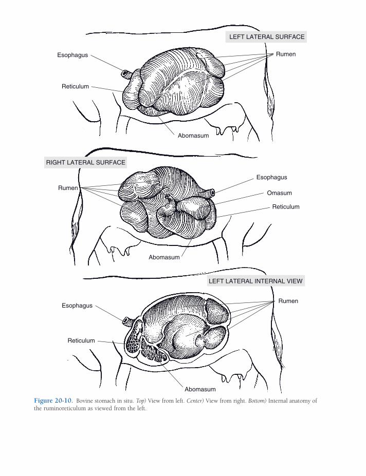
Esophagus
Reticulum
Rumen
Abomasum
LEFT LATERAL SURFACE
RumenOmasum
Esophagus
Abomasum
Reticulum
Rumen
Abomasum
Esophagus
Reticulum
RIGHT LATERAL SURFACE
LEFT LATERAL INTERNAL VIEW
Figure 20-10. Bovine stomach in situ. Top) View from left. Center) View from right. Bottom) Internal anatomy of the ruminoreticulum as viewed from the left.

ANATOMY OF THE DIGESTIVE SYSTEM / 349
Figure 20-11. Detailed anatomy of the bovine stomach. A) External anatomy from the left (top) and the right (bottom). B) Internal anatomy; grooves on the exterior of the stomach correspond to raised, muscular ridges (“pillars”) on the interior of the stomach.
swallowed typically will fall into and remain in the reticulum; contractions of this part of the forestomach may drive sharp objects through the wall of the stomach, leading to traumatic peritonitis or hardware disease (see Chapter 17). The location of the reticulum immediately caudal to the diaphragm places it opposite the heart, with only the muscular diaphragm between, so that these sharp objects may also be driven into pleural and pericardial spaces.
The reticulum and the rumen (colloquially known as the paunch) are divided ventrally by a thick, muscular ruminoreticular fold. The rumen extends from this fold to the pelvis and almost entirely fi lls the left side of the abdomi-nal cavity; its capacity depends on the size of the individual but in adult cattle ranges from 110 to 235 L (about 30 to 60 gallons).
The rumen is subdivided internally into compartments by muscular pillars, which cor-
respond to grooves visible on the exterior of the rumen (Fig. 20-10 and 20-11). Right and left longitudinal pillars (corresponding to right and left longitudinal grooves on the exterior) together with cranial and caudal pillars (exter-nally, cranial and caudal grooves) form a nearly complete constricting circle in the horizontal plane. These divide the rumen into dorsal and ventral sacs. The dorsal sac is the largest com-partment. The dorsal sac is continuous crani-ally with the reticulum over the ruminoreticular fold so that the two compartments share a dorsal space.
Caudally, the dorsal sac is further subdivided by the dorsal coronary pillars, which form an incomplete circle bounding the dorsal blind sac. The caudal part of the ventral sac forms a diverticulum, the ventral blind sac, separated from the rest of the ventral sac by the ventral coronary pillars.
350 / ANATOMY AND PHYSIOLOGY OF FARM ANIMALS
As in the rest of the forestomach, the mucous membrane lining the rumen is a nonglandular, stratifi ed squamous epithelium. The most ventral parts of both sacs of the rumen contain numerous feathery papillae up to 1 cm long, but papillae are almost entirely absent on the dorsal part of the rumen.
Omasum
The omasum is a spherical organ fi lled with muscular laminae (an estimated 90–130 in the bovine omasum) that lie in sheets, much like the pages of a book (giving the omasum its colloquial name, book stomach). The stratifi ed squamous mucous membrane covering the laminae is studded with short, blunt papillae. Each lamina contains three layers of muscle, including a central layer continuous with the tunica muscularis of the omasal wall.
The omasum lies to the right of the rumino-reticulum, just caudal to the liver, and in the ox makes contact with the right body wall. The omasum of the sheep and goat is much smaller than the bovine omasum and normally is not in contact with the abdominal wall. Food enters the omasum at the reticulo-omasal orifi ce, between the laminae, and goes on to the omasoabomasal orifi ce.
Abomasum
The abomasum (true stomach) is the fi rst glan-dular portion of the ruminant digestive system (Fig. 20-9). Its proximal portion is ventral to the omasum, and its body extends caudad on the right side of the rumen. The pylorus demar-cates the muscular junction of the stomach and small intestine, and like the porcine pylorus, it features an enlarged torus pyloricus.
The epithelium of the abomasum consists primarily of two glandular regions, equivalent to the fundic gland region (region of the proper gastric glands) and the pyloric gland region. The cardiac gland region in the abomasum is confi ned to a very small area adjacent to the omasoabomasal orifi ce.
Small Intestine
The duodenum is the fi rst of three divisions of the small intestine (Figs. 20-12 to 20-14). It is closely attached to the right side of the dorsal body wall by a short mesentery, the mesoduo-denum. The duodenum arises at the pylorus of the stomach and receives ducts from the pan-creas and liver in this region. It passes caudad on the right side of the abdominal cavity toward the pelvic inlet, then crosses to the left side caudal to the root of the great mesentery (dis-cussed later) and refl ects craniad to join the jejunum. The mesentery becomes considerably longer at this duodenojejunal fl exure, and the duodenum is attached at this site to the descend-ing colon by a serosal ligament, the duodeno-colic fold.
The transition between duodenum and the next portion of the small intestine, the jejunum, is defi ned by the marked increase in the length of the supporting mesentery. The jejunum is the longest part of the small intestine (e.g., as much as 28 m in the horse). Histologically, the jejunum is similar to the duodenum, although lymph nodules at the mucosal–submucosal junction may be more numerous.
The ileum is the short last part of the small intestine. It is distinguished from the jejunum by a fold of mesentery between it and the cecum. This ileocecal fold is found on the side of the intestine opposite the attachment of the mesentery (the antimesenteric side). The lumen of the ileum communicates with that of the large intestine at the ileal orifi ce; this junction is found in the right caudal part of the abdomi-nal cavity in all species. The ileal epithelium features numerous goblet (mucous) cells, and aggregates of lymph nodules in this region are more abundant than in other parts of the small intestine. Their especially prominent arrange-ment in the ileum has led to the use of the term Peyer’s patches to distinguish them.
The mesenteries that suspend the small intes-tine from the dorsal body wall can be named according to the part of the intestine supported, that is, mesoduodenum, mesojejunum, and mesoileum. The mesoduodenum is generally short, so the location of the duodenum is
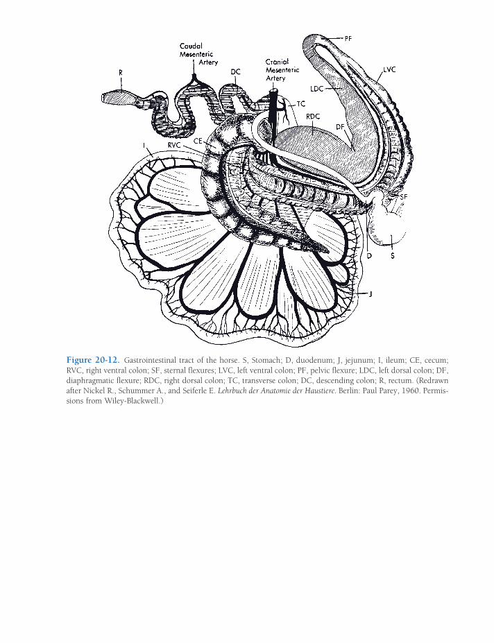
Figure 20-12. Gastrointestinal tract of the horse. S, Stomach; D, duodenum; J, jejunum; I, ileum; CE, cecum; RVC, right ventral colon; SF, sternal fl exures; LVC, left ventral colon; PF, pelvic fl exure; LDC, left dorsal colon; DF, diaphragmatic fl exure; RDC, right dorsal colon; TC, transverse colon; DC, descending colon; R, rectum. (Redrawn after Nickel R., Schummer A., and Seiferle E. Lehrbuch der Anatomie der Haustiere. Berlin: Paul Parey, 1960. Permis-sions from Wiley-Blackwell.)
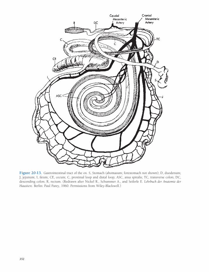
Figure 20-13. Gastrointestinal tract of the ox. S, Stomach (abomasum; forestomach not shown); D, duodenum; J, jejunum; I, ileum; CE, cecum; C, proximal loop and distal loop; ASC, ansa spiralis; TC, transverse colon; DC, descending colon; R, rectum. (Redrawn after Nickel R., Schummer A., and Seiferle E. Lehrbuch der Anatomie der Haustiere. Berlin: Paul Parey, 1960. Permissions from Wiley-Blackwell.)
352

Figure 20-14. Gastrointestinal tract of the pig. S, Stomach; D, duodenum; J, jejunum; I, ileum; CE, cecum; CC, ansa spiralis (coiled colon); TC, transverse colon; DC, descending colon; R, rectum. (Redrawn after Nickel R., Schummer A., and Seiferle E. Lehrbuch der Anatomie der Haustiere. Berlin: Paul Parey, 1960. Permissions from Wiley-Blackwell.)
353

354 / ANATOMY AND PHYSIOLOGY OF FARM ANIMALS
relatively fi xed. The mesenteries supporting the jejunum and ileum, on the other hand, are long and fanlike, so that many meters of intestine are connected to a small region of the dorsal body wall. The mesojejunum and mesoileum are often collectively called the great mesentery, and the narrow stalk by which they are attached to the body wall and through which blood vessels, nerves, and lymphatics reach the intes-tines is commonly called the root of the great mesentery. The length of the great mesentery permits considerable mobility of the intestinal mass.
Large Intestine
The large intestine consists of the cecum, a blind sac, and the colon, which consists of ascending, transverse, and descending parts. The descending colon terminates as the rectum and anal canal.
There is considerably more variation in the large intestine from one species to another than in the small intestine (Figs. 20-12 to 20-14). Most of this variation results from modifi ca-tions of the ascending colon. The transverse colon forms a short connection that runs trans-versely from distal ascending colon to proximal descending colon; it is invariably found running from right to left sides of the abdomen, just cranial to the root of the great mesentery. The descending colon is generally relatively straight, running caudad on the left side of the abdomen to the pelvic cavity, where it terminates as the rectum.
Ruminants
In the ruminant (Fig. 20-13) the cecum is about 12 cm in diameter, and when full, its blind end projects as far caudad as the pelvic inlet. Crani-ally, the cecum is continuous with the colon.
The proximal part of the colon is the ascend-ing colon. It is modifi ed into a series of three loops in the ruminant. The proximal loop (ansa proximalis) forms an S shape that leads to the spiral loop (ansa spiralis). The spiral colon
forms an orderly spiraling mass on the left face of the great mesentery. The fi rst portion of the spiral colon coils toward the center of the mesentery (centripetally), reverses direction at the central fl exure, then spirals away from the center (centrifugally). The last part of the ascending colon, the distal loop (ansa distalis) connects the spiral colon with the transverse colon. The transverse colon crosses from right to left, cranial to the cranial mesenteric artery, which supplies the small intestine, the cecum, and the ascending colon, and continues caudad as the descending colon to the rectum. An external anal sphincter of striated (i.e., volun-tary) muscle and an internal anal sphincter of smooth muscle characterize the walls of the most distal part of the gastrointestinal tract as it opens to the exterior of the animal at the anus.
The arrangement of the intestinal tract of the sheep and goat is similar to that of the ox. In small ruminants, however, the last centrifugal loop of the ansa spiralis lies much closer to the jejunum than in the ox.
Pig
The porcine cecum (Fig. 20-14) is a moderately large (1.5–2.2 L) blind sac that projects cranio-ventrad near the midline. The dorsal end of the cecum is continuous with the colon at the ileo-cecocolic junction, where the entrance of the ileum marks the division between the cecum and colon. Unlike those of most domestic species, the bulk of the porcine cecum lies to the left of midline, with its junction with the colon ventral to the left kidney.
The ascending colon of the pig, like that of the ruminant, presents a spiral arrangement of coils, although in the case of the pig the spiral loop is arranged in a cone shape rather than in a fl at plane. The transverse colon continues from its junction with the distal end of the spiral loop, passes forward, then crosses to the left side of the abdomen. The bowel continues caudad as the descending colon to the rectum. As in other animals, the rectum terminates at the anus.

ANATOMY OF THE DIGESTIVE SYSTEM / 355
Horse
The horse has the largest and most complex large intestine of any of the domestic animals (Fig. 20-12). The equine diet of grasses neces-sitates the assistance of microbes for digestion of celluloses, but unlike that of ruminants, the horse’s digestive system defers this fermenta-tion until ingested food reaches the cecum. For this reason, horses are often called postgastric fermentators.
The cecum in the horse is a comma-shaped structure extending from its base in the right side of the pelvic inlet to the fl oor of the abdominal cavity, where the apex lies just caudal to the diaphragm near the xiphoid car-tilage of the sternum. The ileum enters the cecum near its base at the ileal orifi ce. The cecum is the primary site of fermentation in the horse, and its average capacity is about 33 L (about 9 gallons).
The ascending colon of the horse is highly modifi ed and extremely capacious, for which reason it is commonly referred to as the great colon. The proximal part leaves the cecum and passes craniad along the right ventral abdomi-nal wall toward the sternal part of the dia-phragm, where it turns sharply to the left and proceeds caudad along the left ventral abdomi-nal wall toward the pelvic inlet. These fi rst parts of the large colon are known respectively as the right ventral colon, the sternal fl exure, and the left ventral colon. They are arranged like a horseshoe, with the toe forward and the branches directed caudad on either side of the apex of the cecum.
At the pelvic inlet, the left ventral colon turns sharply dorsad to form the pelvic fl exure. The colon then continues craniad as the left dorsal colon, located just dorsal to the left ventral colon. As it approaches the diaphragm (just dorsal to the sternal fl exure), it bends to the left as the diaphragmatic fl exure and then contin-ues a short distance caudad as the right dorsal colon. The right dorsal colon turns again to the left and crosses the midline in front of the root of the great mesentery as the transverse colon.
The descending colon in the horse (also called the small colon to distinguish it from
the great colon) is the direct continuation of the transverse colon. The descending colon is arranged in undulations within the mesoco-lon, much like the small intestine in the mesentery. The small colon, however, is some-what larger in diameter than the small intestine. The small colon is usually located near the middle of the caudal part of the abdominal cavity. It terminates within the pelvic cavity as the rectum.
Peritoneal Features
The parietal and visceral peritonea are continu-ous with one another at double folds of serosa called mesenteries (see above). In some loca-tions, visceral peritoneum refl ects off one region of the gut, spans a short distance, then merges onto the surface of nearby structures. Although these double folds of peritoneum are typically very thin, in this confi guration they are some-times referred to as ligaments. Some examples include the falciform ligament, which tethers the liver to the ventral midline; the renosplenic (nephrosplenic) ligament, spanning between the left kidney and spleen; and the hepatoduo-denal ligament, connecting the liver and proxi-mal duodenum.
Omentum refers to those parts of the perito-neum connecting the stomach with other struc-tures. It is divided into a lesser omentum, extending from the lesser curvature of the stomach to the liver, and the greater omentum, which is attached to the greater curvature of the stomach (and the comparable portion of the ruminant stomach). The greater omentum spreads like an apron from the stomach to cover most of the ventral aspect of the mass of intestine.
Accessory Digestive Organs
In addition to the numerous small glands located in the walls of the stomach and intes-tine, accessory glands include the salivary glands, the pancreas, and the liver.

356 / ANATOMY AND PHYSIOLOGY OF FARM ANIMALS
Salivary Glands
The salivary glands of domestic farm animals comprise three pairs of well-defi ned glands as well as scattered lobules of salivary tissue (minor salivary glands). The chief salivary glands are the parotid, mandibular, and sublin-gual (Fig. 20-15). The minor salivary glands include labial, buccal, lingual, and palatine glands.
The parotid salivary gland is located ventral to the ear in relation to the caudal border of the mandible. In most animals, the parotid sali-vary duct passes ventrad and craniad on the deep face of the caudal part of the mandible and crosses the cheek superfi cially just cranial to the masseter muscle. The duct then passes dorsad to penetrate the mucous membrane of the cheek near the third or fourth maxillary cheek tooth.
The mandibular salivary gland is usually located ventral to the parotid gland, just caudal to the mandible. The mandibular salivary duct passes forward along the medial side of the mandible to open ventral to the tongue on the sublingual caruncle, a small hillock of mucous membrane located on the fl oor of the mouth.
The sublingual salivary gland is located deep to the mucous membrane along the ventral side of the lateral surface of the tongue near the fl oor of the mouth. Numerous ducts pass directly from the gland to open into the fl oor of the mouth just ventrolateral to the tongue. With the exception of the horse, the sublingual salivary gland also has a monostomatic portion that empties at the sublingual caruncle on the fl oor of the mouth by way of a single sublingual duct that runs parallel to the mandibular duct.
The salivary glands are classifi ed as serous, mucous, or mixed. Serous glands secrete a watery clear fl uid, as compared with mucous glands, which secrete mucus, a viscous material that acts as a protective covering for the surface of mucous membranes. A mixed gland pro-duces both mucous and serous fl uids. The parotid salivary gland secretes primarily serous saliva; mandibular and sublingual glands are classifi ed as mixed glands in domestic farm
animals. Most of the minor salivary glands have a mucous secretion.
Pancreas
The pancreas is a compound gland that has both endocrine and exocrine portions. The exocrine portion of the pancreas produces sodium bicarbonate and digestive enzymes, which pass through the pancreatic ducts to empty into the duodenum close to the opening of the bile duct.
The endocrine portion of the pancreas con-sists of isolated groups of pale-staining cells scattered throughout the gland. These areas are called the pancreatic islets (formerly islets of Langerhans). They produce the hormones that pass directly into the bloodstream (see Chapter 12), most notably glucagon and insulin, which are the primary regulators of blood sugar levels.
Grossly, the pancreas is an irregularly lobu-lated organ that lies adjacent to the proximal duodenum and frequently abuts the stomach, the caudal vena cava, and caudal part of the liver as well. The pancreas has the appearance of aggregated nodules loosely connected to form an elongated gland lying parallel to the duodenum. The developing pancreas arises as two diverticula of the embryonic duodenum and therefore always begins as a bilobed organ connected to the lumen of the gut by two ducts. During organogenesis in many species, the duct systems of the two lobes intermingle and one of the two original connections to the gut lumen is lost; thus, a single duct is the normal condi-tion of the adult in these species. The disposi-tion of ducts of the pancreas is shown in Table 20-3.
In those species that possess it, the pancre-atic duct opens onto a small elevation within the duodenum in common with the bile duct from the liver. This is the major duodenal papilla. A short distance away, a smaller minor duodenal papilla marks the location of the accessory pancreatic duct. As small ruminants lack the accessory pancreatic duct, they also lack the minor duodenal papilla.
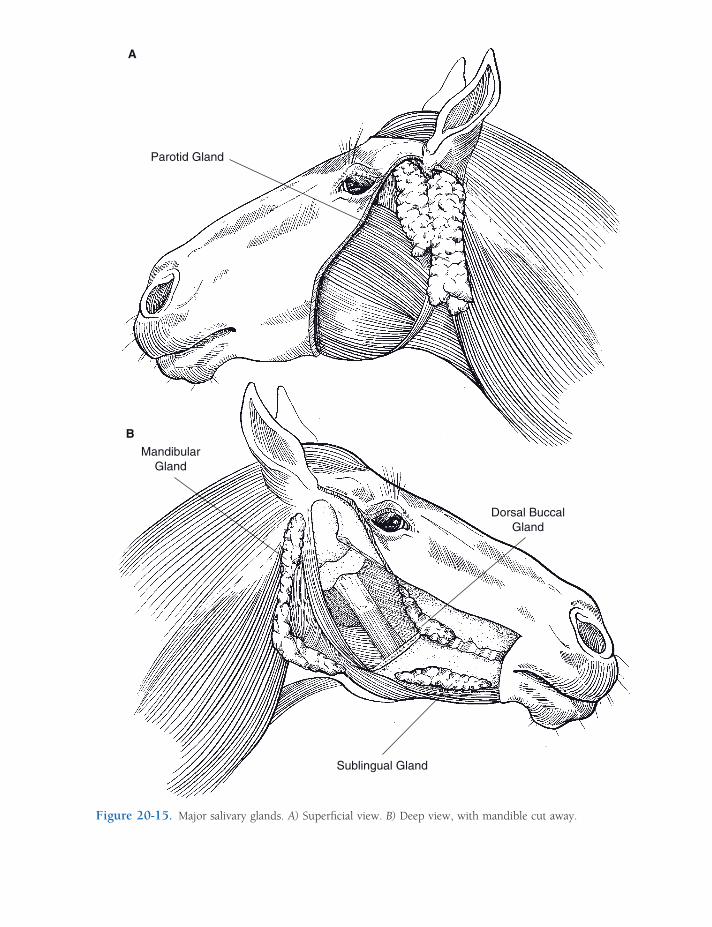
Parotid Gland
Dorsal BuccalGland
Sublingual Gland
MandibularGland
A
B
Figure 20-15. Major salivary glands. A) Superfi cial view. B) Deep view, with mandible cut away.
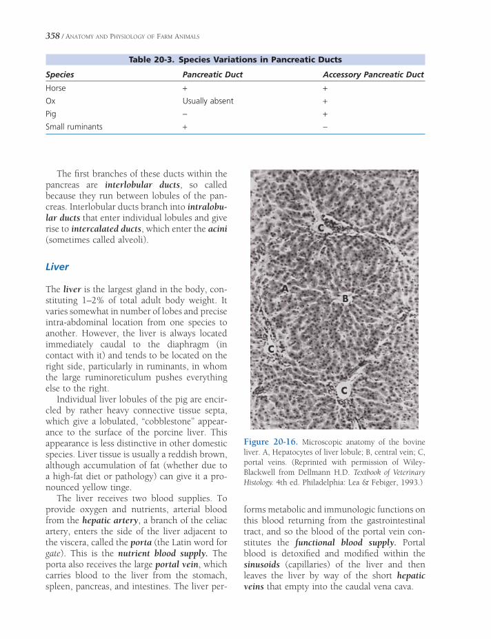
358 / ANATOMY AND PHYSIOLOGY OF FARM ANIMALS
The fi rst branches of these ducts within the pancreas are interlobular ducts, so called because they run between lobules of the pan-creas. Interlobular ducts branch into intralobu-lar ducts that enter individual lobules and give rise to intercalated ducts, which enter the acini (sometimes called alveoli).
Liver
The liver is the largest gland in the body, con-stituting 1–2% of total adult body weight. It varies somewhat in number of lobes and precise intra-abdominal location from one species to another. However, the liver is always located immediately caudal to the diaphragm (in contact with it) and tends to be located on the right side, particularly in ruminants, in whom the large ruminoreticulum pushes everything else to the right.
Individual liver lobules of the pig are encir-cled by rather heavy connective tissue septa, which give a lobulated, “cobblestone” appear-ance to the surface of the porcine liver. This appearance is less distinctive in other domestic species. Liver tissue is usually a reddish brown, although accumulation of fat (whether due to a high-fat diet or pathology) can give it a pro-nounced yellow tinge.
The liver receives two blood supplies. To provide oxygen and nutrients, arterial blood from the hepatic artery, a branch of the celiac artery, enters the side of the liver adjacent to the viscera, called the porta (the Latin word for gate). This is the nutrient blood supply. The porta also receives the large portal vein, which carries blood to the liver from the stomach, spleen, pancreas, and intestines. The liver per-
forms metabolic and immunologic functions on this blood returning from the gastrointestinal tract, and so the blood of the portal vein con-stitutes the functional blood supply. Portal blood is detoxifi ed and modifi ed within the sinusoids (capillaries) of the liver and then leaves the liver by way of the short hepatic veins that empty into the caudal vena cava.
Table 20-3. Species Variations in Pancreatic Ducts
Species Pancreatic Duct Accessory Pancreatic Duct
Horse + +Ox Usually absent +Pig − +Small ruminants + −
Figure 20-16. Microscopic anatomy of the bovine liver. A, Hepatocytes of liver lobule; B, central vein; C, portal veins. (Reprinted with permission of Wiley-Blackwell from Dellmann H.D. Textbook of Veterinary Histology. 4th ed. Philadelphia: Lea & Febiger, 1993.)

ANATOMY OF THE DIGESTIVE SYSTEM / 359
All domestic animals except the horse have a gallbladder for storage of bile. The liver’s digestive secretion, bile, leaves the liver through hepatic ducts, which join the cystic duct from the gallbladder to form the common bile duct, which then passes to proximal duodenum into the lumen to which it opens in common with the pancreatic duct on the major duodenal papilla (see above).
Microscopically, the morphologic unit of the liver is the hepatic lobule, a polygonal cylinder of liver cells (the hepatocytes) in the center of which is a central vein (Fig. 20-16). At the angles on the periphery, where adjacent hepatic lobules meet, are the portal triads, consisting of branches of the hepatic artery and portal vein (interlobular vessels), an interlobular bile duct, and lymphatics. These communicate with the spaces between sheets, or laminae, of hepato-cytes in the hepatic lobule; the spaces are the
hepatic sinusoids, and they are characterized by the lack of a basal lamina and numerous fenestrations in the endothelium, allowing free egress of blood constituents that then bathe the hepatocytes. Blood (both arterial and portal) fl ows from the portal canal through the sinusoids and is gathered by the central vein, the smallest tributary of the hepatic veins. In and around the sinusoids are fi xed macro-phages, which in this location are called Kupffer cells.
Between adjacent rows of liver cells is a tiny bile canaliculus, which is little more than a tube formed by grooves in the surfaces of the apposed liver cells. Bile produced by the hepa-tocytes is carried toward the periphery of the hepatic lobule by the bile canaliculi to the inter-lobular bile ducts located at the portal canal (notice that this net fl ow is opposite the direc-tion of blood fl ow).


PHYSIOLOGY OF DIGESTION
For normal metabolism, cells of an animal’s body need the three major classes of nutri-
ents (carbohydrates, proteins, and lipids) deliv-ered to them via the blood in their simplest forms (monosaccharides, amino acids, and fatty acids). Animals consume foodstuffs that contain these nutrients in more complex chemical and physical forms. It is the function of the gastro-intestinal tract to reduce the consumed food-stuffs to simpler molecules and to transfer them to the blood so that they can be delivered to the cells for metabolism. The processes of phys-ical and chemical breakdown of foodstuffs are termed mechanical and chemical digestion, respectively. In addition to monosaccharides, amino acids, and fatty acids, the gastrointesti-nal tract must absorb other essential minor nutrients (e.g., salts, vitamins), so that they are available to the cells of the body.
The gastrointestinal tract is essentially a long, smooth muscle tube extending from mouth to anus. The tube has two distinct layers of smooth muscle in its wall (circular and lon-gitudinal layers) and is lined with epithelia that function as selective barriers between the lumen and the body fl uids. The anatomic and functional characteristics of the mucosa and its epithelia vary greatly among segments of the intestine (Fig. 21-1). Indigestible substances or items (such as a coin) can pass through the tract without being altered and without affecting the
Pregastric PhysiologyPrehension and Chewing
Saliva and Salivary Glands
Swallowing
Ruminant ForestomachFermentative Digestion
Forestomach Motility
Reticular, or Esophageal, Groove
Omasum
Gastric PhysiologyGastric Glands and Secretions
Gastric Motility
Physiology of the Small Intestine, Exocrine Pancreas, and Liver
Small Intestine Secretions and Motility
Exocrine Pancreas
Liver Digestive Functions and
Secretion of Bile
Nutrient Absorption in the Small Intestine
Physiology of the Cecum and ColonCecum and Colon of the Horse
Rectum and Defecation
Chapter 21
361

362 / ANATOMY AND PHYSIOLOGY OF FARM ANIMALS
animal if they are not large enough to impair movement of the other contents.
The smooth muscle in the wall of the gas-trointestinal tract provides the force to move digesta through the tract; gastrointestinal motility is the general term used to describe the activity of this smooth muscle. Gastrointestinal motility is primarily regulated by three mecha-
nisms: (1) autonomic nervous system, (2) gas-trointestinal hormones, and (3) enteric nervous system.
Gastrointestinal hormones are released from endocrine cells in the epithelial lining of the gastrointestinal tract (enteroendocrine cells) and may stimulate or inhibit gastrointestinal smooth muscle. The release of these hormones
Figure 21-1. Cross-sections through various segments of the digestive tract. A and B, Esophagus with stratifi ed squamous epithelium. C, Small intestine with columnar epithelium and submucosal glands and aggregated lym-phatic nodules in some segments. D, Large intestine. E, tunica mucosa: epithelium; F, lamina propria; G, lamina muscularis; H, tela submucosa; I, tunica muscularis: circular layer; J, longitudinal layer; K, tunica serosa; L, tunica adventitia. (Reprinted with permission of Wiley-Blackwell from Dellmann H.D. and Eurell J. Textbook of Veterinary Histology. 5th ed. Philadelphia: Lippincott Williams & Wilkins, 1998.)

PHYSIOLOGY OF DIGESTION / 363
is usually in response to digesta in the lumen of the tract. Thus, these hormones are a means of local regulation that is coordinated with the ingestion and digestion of food.
The enteric nervous system consists of neural plexuses between layers of smooth muscle in the wall of the tract (Fig. 21-1). These plexuses contain complete neurons (dendrites, cell bodies, and axons) that can form complete neural and refl ex circuits in the wall of the tract so that neural regulation can be independent of external innervation. The presence of food and distension of gastrointestinal tract segments act as stimuli to initiate activity of the enteric nervous system. The three regulatory mecha-nisms (autonomic nervous system, gastrointes-tinal hormones, and enteric nervous system) also regulate secretions from glands in the wall of the gastrointestinal tract (Fig. 21-1) and the intestinal accessory organs (salivary glands, liver, and pancreas).
All three of these mechanisms may regulate a given intestinal segment or accessory organ, but the relative importance of each varies among the segments of the gastrointestinal tract and the accessory organs. For example, salivary secretion is almost entirely regulated by the autonomic nervous system, whereas gastroin-testinal hormones are primary in the initiation of bile secretion.
Pregastric Physiology
Prehension and Chewing
The act of bringing food into the mouth is prehension. The teeth, lips, and tongue are used as prehensile organs by domestic animals. The lips of the horse, the tongue of the cow and sheep, and the snout of the pig are used exten-sively in obtaining food.
The type of teeth, arrangement of jaws, and chewing (mastication) habits vary with the species and the food. Carnivorous animals have simple teeth and tear their food but do little grinding. Herbivorous animals have at least some hypsodont teeth; the upper jaw is wider than the lower jaw; and chewing of the food is
thorough. Mastication can be controlled volun-tarily, but the presence of food in the mouth will stimulate refl ex chewing.
Saliva and Salivary Glands
Saliva consists of water, electrolytes, mucus, and enzymes. The water and mucus soften and lubricate the ingesta to facilitate mastication and swallowing. Lysozyme is a salivary enzyme with antibacterial actions. The starch-digesting enzyme amylase is present in the saliva of omnivores (pig) and to a limited degree in horses but absent in ruminants and carnivores (dog).
Adult cattle may secrete up to 200 L of saliva per day as compared to 1–2 L per day for humans. This large volume maintains the fl uid consistency of the rumen contents, and com-ponents of the saliva may also prevent frothing of the rumen fl uid. Ruminant saliva has a rela-tively high pH and contains high concentra-tions of bases (bicarbonate and phosphate). These bases neutralize acids produced by fer-mentation in the rumen so that the pH in the rumen does not become too acidic.
Parasympathetic nerves are the efferent limbs of neural refl exes that regulate salivary secre-tion. Afferent inputs that stimulate salivary secretion include sight and smell of food, pres-ence of food in the oral cavity, and conditioned refl exes, where some event is associated with food and feeding. Conditioned refl ex control of salivation was the subject of the classic studies by Pavlov, who conditioned dogs to salivate at the sound of a bell.
Swallowing
Deglutition, the act of swallowing, is arbitrarily divided into three stages. The fi rst stage is passage of food or water through the mouth; the second is passage through the pharynx; and the third consists of passage through the esoph-agus into the stomach.
The fi rst stage of swallowing is under volun-tary control. After the food is chewed and mixed with saliva, a bolus (rounded mass of
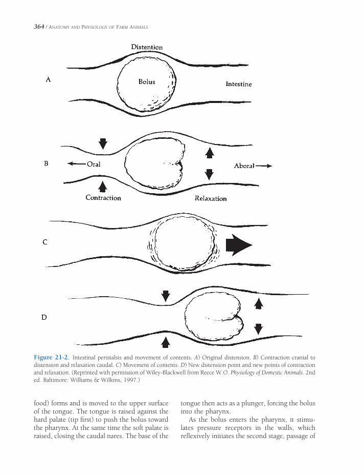
364 / ANATOMY AND PHYSIOLOGY OF FARM ANIMALS
Figure 21-2. Intestinal peristalsis and movement of contents. A) Original distension. B) Contraction cranial to distension and relaxation caudal. C) Movement of contents. D) New distension point and new points of contraction and relaxation. (Reprinted with permission of Wiley-Blackwell from Reece W.O. Physiology of Domestic Animals. 2nd ed. Baltimore: Williams & Wilkins, 1997.)
food) forms and is moved to the upper surface of the tongue. The tongue is raised against the hard palate (tip fi rst) to push the bolus toward the pharynx. At the same time the soft palate is raised, closing the caudal nares. The base of the
tongue then acts as a plunger, forcing the bolus into the pharynx.
As the bolus enters the pharynx, it stimu-lates pressure receptors in the walls, which refl exively initiates the second stage, passage of

PHYSIOLOGY OF DIGESTION / 365
the bolus through the pharynx. Respiration is refl exively inhibited, and the larynx refl exively closes and pulls up and forward. The base of the tongue folds the epiglottis over the laryn-geal opening as it moves back. The pharynx shortens, and a peristaltic (milking) action of the pharyngeal muscles forces the bolus into the esophagus.
The third stage of deglutition consists of refl ex peristalsis of the esophagus initiated by the presence of food in the esophagus. Peristal-sis consists of alternate relaxation and contrac-tion of rings of muscle in the wall coupled with regional contraction of longitudinal muscles in the area of the bolus (Fig. 21-2).
Peristalsis carries solid and semisolid food through the esophagus of the horse at 35 to 40 cm/second. Liquids travel about fi ve times as fast by a squirting action of the mouth and the pharynx.
Vomiting (emesis) is a protective response to remove potentially harmful ingesta from the stomach and upper small intestine. Vom-iting is a highly coordinated refl ex that is controlled by a refl ex center in the brain-stem. Drugs that stimulate this center to produce vomiting are termed emetics. The process begins with relaxation of the sphinc-ter between the stomach and upper small intestine and reverse peristalsis to move intestinal contents to the stomach. The move-ment of stomach contents into the esophagus and out of the mouth requires relaxation of the upper and lower esophageal sphincters together with an inspiratory movement against a closed glottis and forceful contrac-tion of abdominal muscles. Closure of the glottis and movement of the soft palate prevent regurgitated food from entering the trachea and nasal cavity, respectively.
Ruminant Forestomach
Fermentative Digestion
No mammal can directly digest the complex car-bohydrates that constitute plant cell walls (cel-lulose and hemicellulose), because mammals do
not produce the enzyme cellulase, which is nec-essary to break the unique chemical bonds in these compounds. The ruminant forestomach provides an excellent environment for the growth of bacteria, protozoa, and possibly other microbes that do produce cellulase. The action of cellulase on cellulose and hemicellulose produces mono-saccharides and simple polysaccharides, which are available for further microbial digestion.
The microbial digestion in the forestomach occurs in an anaerobic environment and is termed fermentative digestion. Volatile fatty acids (VFAs) are produced by fermentation of carbohydrates consumed by ruminants, includ-ing carbohydrates produced by the actions of microbial cellulase. The primary VFAs are acetic acid, propionic acid, and butyric acid. The VFAs are absorbed directly from the fore-stomach and are the major energy source for ruminants. VFAs are also used for synthesis of milk fat in lactating animals.
Methane and carbon dioxide are produced by fermentative digestion and accumulate as a gaseous layer above the ingesta in the rumen and reticulum. Bloat (acute tympany) results in enlargement of the rumen and reticulum, which in turn press on the thorax, inhibiting function of the heart and lungs. Bloat results from more gas being produced than is elimi-nated by eructation (belching). Eructation is particularly diffi cult if foam forms. A stomach tube may be passed into the rumen by mouth to remove the gas. If that is not possible, a trocar (sharp tube) may be passed into the rumen through the left fl ank.
Dietary protein consumed by ruminants is available fi rst to the microbes in the forestom-ach. The microbes may use the dietary protein to produce microbial proteins and promote microbial growth or to produce VFAs by fer-mentative digestion. Microbes can also produce microbial proteins from nonprotein nitrogen sources, such as urea and ammonia.
The microbes and by-products of microbial metabolism other than VFAs continue down the ruminant gastrointestinal tract from the fore-stomach. The microbial organisms, which grew in the forestomach, are a major source of dietary protein for ruminants. When the organisms

366 / ANATOMY AND PHYSIOLOGY OF FARM ANIMALS
enter the abomasum (true or glandular stomach of the ruminant) and the remainder of the tract, they are digested in a manner similar to diges-tion of protein sources in nonruminants. Bene-fi cial by-products of microbial metabolism include many water-soluble vitamins.
Forestomach Motility
The rumen and reticulum of the adult cow nor-mally undergo complicated sequences of con-tractions that are repeated at varying frequencies up to several times per minute. One pattern of contractions begins in the reticulum and spreads over both the dorsal and ventral sacs of the rumen (see Fig. 20-10). This series of contrac-tions mixes the contents to promote fermenta-tion and provide force to move liquifi ed digesta out of the forestomach and into the abomasum. A second pattern of contractions begins in the caudal portion of the dorsal sac and moves cra-nially. These contractions move gases toward the cranial part of the rumen for eructation. Rumen contractions can be felt by forcing the fi st into the upper left fl ank (paralumbar fossa). Pathologic condition of the rumen or morbidity associated with systemic diseases usually results in a decreased rate or com-plete cessation of rumen movements.
Rumination permits an animal to forage and ingest food rapidly and fi nish chewing later. It entails regurgitation of the food (returning it to the mouth) from the forestomach, remastica-tion (rechewing), reinsalivation (mixing with more saliva), and fi nally reswallowing.
Regurgitation is the only step of rumination that differs markedly from the initial mastica-tion, insalivation, and swallowing. Regurgita-tion is preceded by contraction of the reticulum, which presumably brings some of the heavier ingesta into proximity to the cardia. The sphinc-ter at the junction of the esophagus and fore-stomach (lower esophageal sphincter) relaxes as the bolus of food reaches it. An inspiratory movement with closed glottis follows. The neg-ative pressure produced in the thorax by this movement is transmitted to the relatively thin-walled esophagus, dilating the thoracic esopha-
gus and cardia. The lower pressure in the esophagus than in the rumen coupled with reverse peristalsis causes a quantity of material (semifl uid ingesta) to pass through the cardia into the esophagus and up to the mouth. The regurgitated material consists largely of rough-age and fl uid, with little if any concentrate. It is well known that whole kernels of corn may pass through the entire digestive tract with little change in physical appearance.
Cattle average about 8 hours a day ruminat-ing, with periods of activity scattered through-out the entire day. One rumination cycle requires about 1 minute, of which 3 to 4 seconds is used for both regurgitation and reswallowing. Rumi-nation appears to be largely refl exive, although the process can be interrupted or stopped vol-untarily. Both afferent and efferent portions of the refl ex are probably carried in the vagal nerves. Contact of roughage with the wall of the reticulum and near the cardia is likely the major stimulus for rumination.
Reticular, or Esophageal, Groove
In young ruminants, nursing and afferents from the pharynx appear to stimulate refl ex closure of the groove, which causes milk to bypass the rumen and reticulum and pass through the omasum directly to the abomasum. The paunchiness of bucket-fed calves usually is attributed to milk entering the rumen, where it is not properly digested. The use of buckets with nipples tends to prevent appreciable amounts of milk from entering the rumen. After weaning, fl uid drunk from open containers largely passes into the rumen and reticulum. The groove has no known function in adult animals, but refl ex closure in adults has been produced with sodium salts in cattle and copper sulfate in sheep.
Omasum
The omasum is nearly always found packed tightly with rather dry roughage in animals examined after death. The appearance of the omasal leaves, studded with short horny papil-

PHYSIOLOGY OF DIGESTION / 367
lae, suggests a burr type of grinder. Experimen-tal vagal stimulation elicits strong contractions of the omasal wall, but movement of the leaves is limited.
Gastric Physiology
Gastric Glands and Secretions
The term gastric juice refers to the combina-tion of substances secreted into the stomach lumen by gastric glands, also termed gastric pits because of their pitlike extension into the wall of the stomach (Fig. 21-3), and epithelial cells of the stomach mucosa. Gastric juice con-tains water, hydrochloric acid, mucus, intrinsic factor, pepsinogen (an inactive form of pepsin, a proteolytic enzyme), and the enzyme rennin. The regulation of gastric juice secretion has three phases, cephalic, gastric, and intestinal.
Stimulation of gastric secretions during the cephalic phase is in response to the sight, smell, or taste of food. These induce a neural response
that increases parasympathetic (vagal nerve) stimulation to the stomach, and this stimulates gastric secretions. The gastric phase begins when food enters the stomach.
The presence of food, especially proteins, stimulates the secretion of the hormones gastrin and histamine from cells in the gastric epithe-lium. Gastrin and histamine stimulate parietal cells in gastric glands to secrete hydrochloric acid (Fig. 21-3). Acetylcholine (parasympa-thetic neurotransmitter) also stimulates parietal cells to secrete hydrochloric acid, but all three regulators (gastrin, histamine, and acetylcho-line) must be present for the most effi cient hydrochloric acid secretion. The histamine receptors on parietal cells (H2 receptors) are different from those on cells involved in allergic reactions (H1 receptors). The spe-cifi c H2 receptor antagonists provide a means to reduce acid secretion with few side effects. The antihistamines used for allergies do not bind to H2 receptors and thus do not disturb digestion. The hormones cholecystokinin, gas-tric inhibitory peptide, and secretin inhibit hydrochloric acid secretion. These hormones are released from the duodenal epithelium in response to the presence of food in the duode-num. The release of these hormones that act to inhibit gastric function is part of the intestinal phase of gastric regulation.
The pH of gastric juice in mammals can be 2 or less. The low pH is protective in that most foreign microbes ingested with food cannot survive such an acidic environment. The low pH inhibits hydrochloric acid secretion to prevent it from becoming too acidic. Pepsino-gen (an inactive form of the enzyme pepsin and a component of gastric juice) is activated by the low pH. By its proteolytic activity, pepsin can activate more pepsinogen. The low pH also promotes the activity of pepsin, because the most favorable pH range for its proteolytic activity is 1.3 to 5. Chief or peptic cells (Fig. 21-3) secrete pepsinogen to begin protein digestion in the stomach, but protein digestion is completed in the small intestine by other digestive enzymes.
A layer of mucus covers the epithelial lining of the stomach and protects the epithelium
Gastric Lumen
Surface
Epithelial Cells
Mucous NeckCell
Parietal Cell
Chief Cell
Endocrine Cell
Figure 21-3. Gastric pit (gastric gland) in the lining of the stomach. Note the different cell types found in the epithelium lining the gland.

368 / ANATOMY AND PHYSIOLOGY OF FARM ANIMALS
from the low pH of the gastric fl uids. This mucus is produced by cells in the gastric glands (Fig. 21-3) and is secreted from there onto the surface of the epithelium. Mucus secretion is stimulated by prostaglandins, which are also produced locally in the wall of the stomach. Non-steroidal anti-infl ammatory drugs (such as aspirin and phenylbutazone) inhibit the synthesis of prostaglandins, and toxic doses of these agents are associated with gastric ulcers. It is presumed that a lack of mucus secretion contributes to the development of the ulcers.
Rennin is an enzyme in the gastric juice in the abomasum of young ruminants. Its func-tion is to coagulate milk and reduce its rate of passage through the gastrointestinal tract. Intrinsic factor, a carrier protein for vitamin B12, binds to the vitamin, and the resultant complex passes through the tract to the ileum, which absorbs the B12.
Gastric Motility
Gastric movements mix the ingesta with the gastric juice, continue mechanical digestion (to liquefy the digesta), and pass the digesta into the duodenum at a controlled rate. The stomach regularly produces peristaltic contractions, beginning in the region of the cardia and increas-ing in force as they travel over the stomach to the pyloric antrum (see Fig. 20-8). These mix and grind the food and force some through the pyloric sphincter into the duodenum. However, much of the food (and especially larger parti-cles) is held back to allow for more mixing and grinding. The ingesta forced through the pyloric sphincter, termed chyme, is a mushy, semisolid mixture of food, water, and gastric juice.
Similar to the regulation of gastric secre-tions, the regulation of gastric motility can be divided into cephalic, gastric, and intestinal phases. Stimulation during cephalic regulation occurs via the parasympathetic nerves, and this increases in response to sight, smell, or taste of food. The hormone gastrin stimulates overall gastric motility to promote mixing (gastric phase). The hormones cholecystokinin and
secretin and gastric inhibitory peptides promote a more forceful contraction of the pyloric sphincter to slow gastric emptying (intestinal phase). The inhibitory effect of the duodenal hormones (released in response to chyme enter-ing the duodenum) prevents the delivery of chyme to the duodenum too fast to be digested normally.
The stomach of a carnivore empties within a few hours, usually before the next meal. Other animals require many hours to empty the stomach. Both the horse and pig require a full day’s fast (24 hours) to empty a full stomach. The stomach of a nursing foal empties slowly, but in an adult pony liquid passes from the stomach to the cecum in 2 hours.
In addition to the typical pattern of stomach contraction when food is present, waves of peristaltic contractions may occur over the stomach as a slight ripple. These are produced by spontaneous electrical depolarizations, which sometimes induce action potentials, in the smooth muscle. These begin in the cardia region, and the waves of membrane depolariza-tion are termed gastric slow waves. In pro-longed fasting, the magnitude of the contractions becomes greater (hunger contractions). These are apparently a response to an increase in parasympathetic input during prolonged fasting. These reach maximum intensity in humans after about 3 days without food and weaken progressively thereafter. In the horse, hunger contractions may begin as early as 5 hours after eating, when the stomach still con-tains some food. The intensity of hunger con-tractions is related to the level of blood sugar. As the blood sugar level decreases, the intensity of hunger contractions increases.
Physiology of the Small Intestine, Exocrine Pancreas, and Liver
The small intestine is the primary site of chemi-cal digestion and absorption of nutrients. The exocrine secretions of the pancreas contain most of the enzymes for chemical digestion in the lumen of the small intestine, but the epi-

PHYSIOLOGY OF DIGESTION / 369
thelial cells that line the small intestine (entero-cytes) also have in their cell membranes enzymes that participate in the fi nal steps of chemical digestion. The primary digestive func-tion of the liver is to provide bile salts, which facilitate the enzymatic digestion of lipids. The liver is not a source of digestive enzymes.
Small Intestine Secretions and Motility
Intestinal juice is derived from intestinal glands in the wall of the small intestine. These include crypts or crypts of Lieberkuhn, scattered throughout the entire small intestine (Fig. 21-4),
and duodenal glands, which contribute mucus and are found only in the duodenum. The intes-tinal juice contains salts and water derived from blood capillaries in the wall of the intestine. The function of the secreted salts is unclear, but the water dilutes the chyme, which is usually hyper-tonic (higher osmolality than normal plasma). Food in the intestine stimulates secretion by these intestinal crypt glands.
The two primary types of movement by the small intestine are segmentation and peristal-sis. Segmentation movements, which occur when food is in the small intestine, are charac-terized by alternating local areas of contraction and relaxation (Fig. 21-5). These movements mix the digesta with intestinal juice and diges-tive enzymes and increase the contact between digesta and the epithelial surface of the small intestine. The increased contact provides more exposure to enzymes associated with epithelial cells and to the absorptive surface of the epi-thelial cells. Strong peristaltic contractions of the small intestine in fasting animals or several hours after a meal propel ingesta down the tract, presumably to clean the small intestine of undigested foodstuffs before the next meal.
Microvilli
Absorptivecell
Goblet cell
VilliCapillary network
Lacteal
Crypt ofLieberkuhn Lymph
vessel
Vein
Artery
Figure 21-4. Intestinal villi and crypts. (Reprinted with permission of Wolters Kluwer from Bullock B.L. Pathophysiology. 4th ed. Philadelphia: Lippincott, 1996.)
Figure 21-5. Segmentation pattern of gastrointestinal motility. The same segment of intestine is viewed at three different sequential times (A, B, and C). Regions of contractions at each time are identifi ed by arrows.
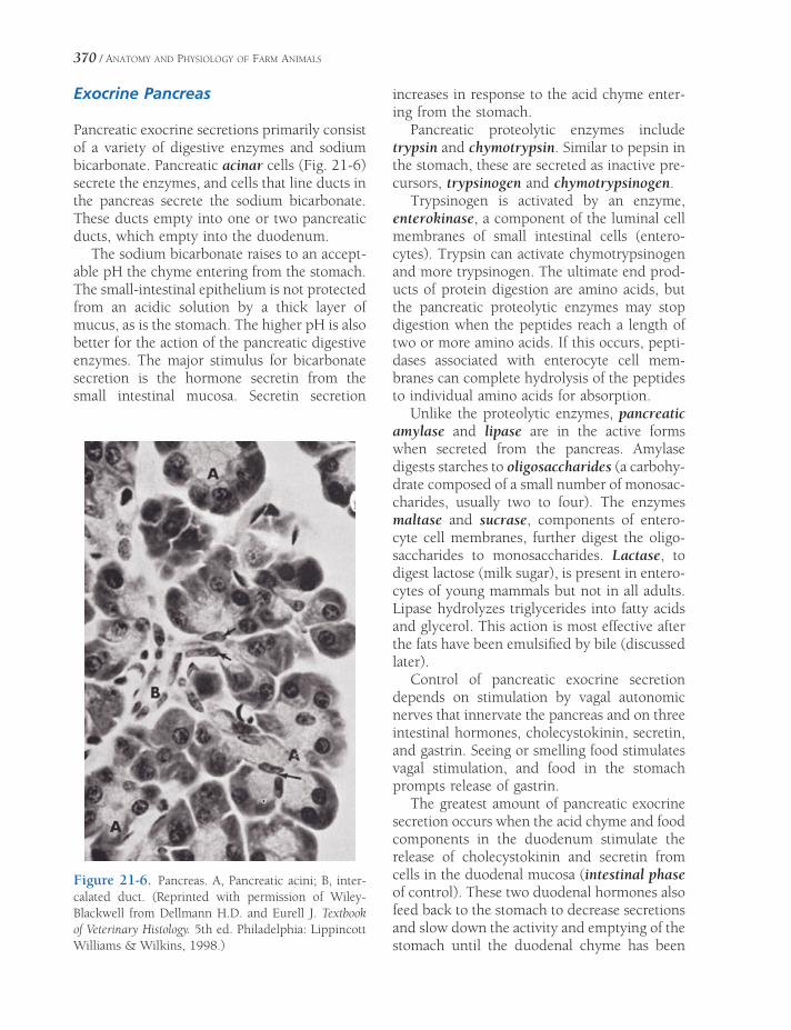
370 / ANATOMY AND PHYSIOLOGY OF FARM ANIMALS
Exocrine Pancreas
Pancreatic exocrine secretions primarily consist of a variety of digestive enzymes and sodium bicarbonate. Pancreatic acinar cells (Fig. 21-6) secrete the enzymes, and cells that line ducts in the pancreas secrete the sodium bicarbonate. These ducts empty into one or two pancreatic ducts, which empty into the duodenum.
The sodium bicarbonate raises to an accept-able pH the chyme entering from the stomach. The small-intestinal epithelium is not protected from an acidic solution by a thick layer of mucus, as is the stomach. The higher pH is also better for the action of the pancreatic digestive enzymes. The major stimulus for bicarbonate secretion is the hormone secretin from the small intestinal mucosa. Secretin secretion
increases in response to the acid chyme enter-ing from the stomach.
Pancreatic proteolytic enzymes include trypsin and chymotrypsin. Similar to pepsin in the stomach, these are secreted as inactive pre-cursors, trypsinogen and chymotrypsinogen.
Trypsinogen is activated by an enzyme, enterokinase, a component of the luminal cell membranes of small intestinal cells (entero-cytes). Trypsin can activate chymotrypsinogen and more trypsinogen. The ultimate end prod-ucts of protein digestion are amino acids, but the pancreatic proteolytic enzymes may stop digestion when the peptides reach a length of two or more amino acids. If this occurs, pepti-dases associated with enterocyte cell mem-branes can complete hydrolysis of the peptides to individual amino acids for absorption.
Unlike the proteolytic enzymes, pancreatic amylase and lipase are in the active forms when secreted from the pancreas. Amylase digests starches to oligosaccharides (a carbohy-drate composed of a small number of monosac-charides, usually two to four). The enzymes maltase and sucrase, components of entero-cyte cell membranes, further digest the oligo-saccharides to monosaccharides. Lactase, to digest lactose (milk sugar), is present in entero-cytes of young mammals but not in all adults. Lipase hydrolyzes triglycerides into fatty acids and glycerol. This action is most effective after the fats have been emulsifi ed by bile (discussed later).
Control of pancreatic exocrine secretion depends on stimulation by vagal autonomic nerves that innervate the pancreas and on three intestinal hormones, cholecystokinin, secretin, and gastrin. Seeing or smelling food stimulates vagal stimulation, and food in the stomach prompts release of gastrin.
The greatest amount of pancreatic exocrine secretion occurs when the acid chyme and food components in the duodenum stimulate the release of cholecystokinin and secretin from cells in the duodenal mucosa (intestinal phase of control). These two duodenal hormones also feed back to the stomach to decrease secretions and slow down the activity and emptying of the stomach until the duodenal chyme has been
Figure 21-6. Pancreas. A, Pancreatic acini; B, inter-calated duct. (Reprinted with permission of Wiley-Blackwell from Dellmann H.D. and Eurell J. Textbook of Veterinary Histology. 5th ed. Philadelphia: Lippincott Williams & Wilkins, 1998.)

PHYSIOLOGY OF DIGESTION / 371
degraded by the enzymes and adjusted in pH by the pancreatic bicarbonate.
Liver Digestive Function and Secretion of Bile
Liver cells (hepatocytes) are responsible for bile formation. Bile is a greenish-yellow salt solu-tion consisting primarily of bile salts, cho-lesterol, phospholipids (lecithins), and bile pigments (bilirubin). Hepatocytes synthesize the bile salts (primarily sodium salts of glyco-cholic and taurocholic acids) from cholesterol. These salts assist in digestion and absorption of lipids (triglycerides), and the production and secretion of these salts is the most important digestive function of the liver.
In an aqueous solution, such as the duode-nal chyme, lipids tend to clump together and form large droplets (recall the appearance of an oil and vinegar salad dressing after shaking the bottle). Such large lipid droplets present a small surface area for the action of the pancreatic lipases. Bile acids act as emulsifi ers to reduce droplet size and make the lipids more accessi-ble to the lipases. Lipases can function without bile salts, but lipid digestion is ineffi cient without them. Micelle is the term for the small droplets formed in the intestinal chyme that contain lipids, bile salts, and products of lipid digestion.
In all farm animals except the horse, bile is stored in the gallbladder. Since the horse has no gallbladder, the bile passes directly from the liver to the duodenum by way of the bile duct and its tributaries at a fairly continuous rate. The gallbladder stores bile for intermittent dis-charge into the duodenum and concentrates the bile by reabsorbing water from the stored bile. Cholecystokinin stimulates gallbladder contraction and the release of stored bile. Since food entering the duodenum stimulates the release of cholecystokinin, this coordinates the release of bile with the presence of food.
Most of the bile salts released from the liver remain mixed with the digesta as it passes into the terminal part of the small intestine (ileum). Here, enterocytes reabsorb bile salts, which
enter the blood. The reabsorbed bile salts are transported to the liver via the hepatic portal vein, and here hepatocytes take up the bile salts from the portal blood. These bile salts can then be secreted by the hepatocytes into bile for reuse. An increase in bile salts in portal blood, such as during the digestion of a meal, is the primary stimulus for bile salt secretion by hepa-tocytes. The recycling of bile salts between the digestive tract and the liver is enterohepatic circulation.
The liver is capable of synthesizing choles-terol, and the liver makes much of the choles-terol in bile. The liver can also eliminate excessive dietary cholesterol via the bile. Cho-lesterol is insoluble in water, but the bile salts and lecithin normally change it to a soluble form so that it can exist in the bile. However, sometimes cholesterol precipitates from the bile in the gallbladder or bile ducts, forming gallstones.
Nutrient Absorption in the Small Intestine
The small intestine is the major site of nutrient absorption. Most of the products of carbohy-drate, protein, and lipid digestion are absorbed as the digesta pass through the small intes -tine. The small intestine is also the primary site of absorption for vitamins, minerals, and water.
The epithelium lining the small intestine has structural features that increase the surface area for nutrient absorption. The mucosa is covered with villi (fi ngerlike projections) that extend into the lumen, and the individual enterocytes have microvilli on their cell membrane facing the lumen (Fig. 21-4).
Individual amino acids and monosaccha-rides (simple sugars) are the simplest products of protein and carbohydrate digestion, respec-tively. The cellular mechanisms of absorption of amino acids and monosaccharides (primarily glucose) are similar in that the transport across the cell membrane on the luminal surface involves sodium-linked co-transporters (see Chapter 2). The co-transporters all bind

372 / ANATOMY AND PHYSIOLOGY OF FARM ANIMALS
sodium, but different co-transporters are used by glucose and amino acids. At least fi ve co-transporters have been found to transport various amino acids. These are all characterized as secondarily active transport, for they depend on the gradient for sodium between the intra-cellular fl uid of the enterocytes and the fl uid in the lumen of the small intestine (see Chapter 2). Figure 21-7 summarizes the absorptive process for glucose.
Micelles formed in the lumen of the small intestine contain bile salts, triglycerides, cho-lesterol, and the products of lipase action on triglycerides (fatty acids and monoglycerides). Intestinal movements (segmentation) bring the micelles into contact with the microvilli of the enterocytes. The lipid-soluble products of lipid digestion and cholesterol can then diffuse from the micelle into the enterocytes (Fig. 21-8). Some long-chain fatty acids are also transported from micelles into the cell by specifi c sodium-linked co-transporter proteins in the cell membrane.
Tubular lumen
Proximal tubule cell
Interstitial fluid
Glucose
transporter for
facilitated
diffusion
Blood capillary
Na+
Na+
Na+G
Glucose-sodiumcotransporter
G
G
G
G
Na+
K+K+
ATPase
Within the enterocytes, absorbed monoglyc-erides and fatty acids are used to resynthesize triglycerides. The enterocyte packages the tri-glycerides and absorbed cholesterol together with intracellular proteins into a particle known as a chylomicron. The enterocytes secrete the chylomicrons into the interstitial fl uid, where they are absorbed into lymphatics (Fig. 21-8). The smallest of the lymphatic vessels that absorb the chylomicrons are lacteals (Fig. 21-4). The lymphatic drainage from the intestinal tract is ultimately added to the blood, and it is via this pathway that chylomicrons, containing absorbed lipids, reach the blood.
Sodium, potassium, phosphate, calcium, chloride, and other electrolytes are primarily absorbed in the small intestine by both active and passive mechanisms. With the exception of iron and calcium, the absorption of these min-erals is not regulated, so that most of what is consumed is absorbed. Iron absorption is reduced at the level of the enterocyte if body iron content is suffi cient. This reduction is
Figure 21-7. Sodium-linked co-transport of glucose. Similar mechanism found in proximal tubule cell (as labeled) and epithelial cell of small intestine. Co-transporter for sodium and glucose (G) is found on the portion of the cell membrane that lines the lumen of the tubule (or gut), and the Na-K-ATPase is found on the base and lateral sides of the cell.

PHYSIOLOGY OF DIGESTION / 373
primarily accomplished by an increase in an intracellular protein that binds iron in the enterocytes. The iron-containing enterocyte is lost from the body after it sloughs from the epithelial lining. The form of vitamin D pro-duced by the kidneys (calcitriol) increases calcium absorption by increasing calcium trans-port proteins in enterocytes. Calcitriol forma-tion by the kidneys is increased when blood calcium is low. To be absorbed, minerals such as calcium and phosphate must be in their ionized state. If the ratio of cations to anions is too high or too low, absorption can be reduced. For example, if the dietary content of the phos-phate (an anion) is too high relative to calcium (a cation), the excess phosphate binds the avail-able calcium to form calcium phosphate, and calcium absorption is impaired.
The small intestine of some neonatal (newborn) animals can absorb macromolecules,
including intact protein molecules from the colostrum. Colostrum, the fi rst milk of the horse, pig, and ruminants, contains the γ- globulins needed to produce passive immunity in the newborns of these animals. This receptive period lasts approximately 1 day in the horse and pig and up to 3 days in the ruminant.
Physiology of the Cecum and Colon
In carnivores the cecum and colon primarily absorb water and some electrolytes to reduce the volume and fl uidity of the digesta in the formation of feces. Feces are also stored in the terminal portions of the colon prior to their movement into the rectum for defecation. In omnivores (e.g., pig) and some herbivores (e.g., cattle and sheep) the cecum and colon are
Micelles containing bile salts, triglycerides,cholesterol, monoglycerides and fatty acids
Monoglyceride
Chylomicron
Cholesterol Fatty Acid
Chylomicron
Secretion of chylomicrons
Sites of transportby diffusion
Lacteal
Synthesis of
triglycerides andformation ofchylomicrons
Figure 21-8. Absorption of lipids and cholesterol by intestinal cell and subsequent formation and secretion of chylomicrons.
374 / ANATOMY AND PHYSIOLOGY OF FARM ANIMALS
also sites of some limited fermentation and microbial digestion. In cattle and sheep the cecum and colon are proportionally larger and more complex than in carnivores, but the fore-stomach is the much more important site of fermentative digestion in these herbivores.
Cecum and Colon of the Horse
The extremely large and complex cecum and great colon of the horse are the primary sites of fermentation and microbial digestion of cellu-lose. Roughage passes relatively quickly through the stomach and small intestine of the horse, but fermentative digestion and passage through the cecum and great colon may take days. Complex movements of the cecum and great colon mix the contents to promote fermentative digestion and to expose the contents to the epithelial surface for absorption of volatile fatty acids. The pelvic fl exure of the great colon and the junction between the great and small colons (see Fig. 20-12) are relatively small in diameter, and it appears that the passage of large particles of roughage is restricted. While these sites retain roughage so that it can be subjected to microbial digestion, the potential for impaction at these sites is also increased.
When consumed with roughage, some starches and sugars escape digestion in the equine stomach and small intestine and pass into the cecum and colon. Microbial digestion of these and the cellulose in the roughage pro-duces volatile fatty acids that can be absorbed and used for energy. Microbes in the cecum and colon can also use nonprotein nitrogen sources (urea) for the production of microbial proteins. However, these have limited nutritional value to the horse, because the gastrointestinal mech-anisms necessary to digest proteins and absorb the resulting amino acids are not readily avail-able in the cecum or colon. Some urea is made available to microbes by diffusing into the cecum and colon from the blood.
Fermentation and microbial digestion produce volatile fatty acids, which could lower the pH of the cecal and colonic contents to
potentially harmful levels. The colonic epithe-lium secretes bicarbonate ions to buffer the pH of the contents. Additional bicarbonate is secreted by the epithelium of the ileum, and this lowers the pH of the cecal contents.
Rectum and Defecation
Several times daily, strong and extensive mass movements of the colon move fecal material into the rectum. Distension of the rectum stim-ulates the need to defecate. The act of defeca-tion requires contractions of smooth muscle in the wall of the rectum, and these result from a spinal refl ex stimulated by distension of the rectum. Conscious control of defecation involves inhibition of the spinal refl ex and con-traction of the external anal sphincter, which is composed of skeletal muscle. Contraction of abdominal muscles increases intra-abdominal pressure, which also assists with emptying the rectum.
The variability in character and shape of feces among species is primarily a function of the structural and functional features of the more distal segments of the colon. In horses relatively strong segmentation contractions form the characteristic fecal balls.
Equine colic is a general term for any painful condition associated with the abdomen of the horse. Conditions associated with any segment of the gastrointestinal tract may result in colic, and clinical signs indicate that the degree of pain can vary from mild to severe. Causes of colic include ulcers; inappropriate diet, such as overload of grain or concentrates; infection; obstruc-tion due to parasites or foreign bodies; impaction of feed or foreign material, such as sand; twisted segments of intestine; and alterations in intestinal motility. Manage-ment, including parasite control, proper diet, access to water, and attention to environ-mental hazards, is helpful in preventing the incidence of colic. Some causes of colic can be readily treated medically, but other causes threaten life and require prompt surgery.
NUTRITION AND METABOLISM
Nutrition
The nutritional needs of animals vary greatly with their metabolic state. A rapidly growing, active young animal has much different nutri-tional needs from those of an older, more sed-entary animal. Both animals must consume certain essential nutrients (compounds needed for normal growth and/or survival that cannot be synthesized in adequate amounts in the body), but the amounts of these nutrients per unit of body weight and the relative amounts of specifi c nutrients vary.
Some essential nutrients are required in minute quantities and are toxic in large quanti-ties. For example, copper is an essential mineral, but excessive consumption over a short period leads to copper toxicity. Minerals that are needed in small quantities are often termed trace minerals. Vitamins function as coen-zymes for various biochemical reactions throughout the body and are also typically required in small amounts. Water-soluble vita-mins (B complex, biotin, C, folic acid, and niacin) are not stored in the body, but fat-soluble vitamins (A, D, E, and K) are stored in the liver and adipose tissue. Water-soluble vita-mins can be consumed in large amounts without signifi cant toxicities because the excess is rapidly excreted in the urine. Because they accumulate in the body, excessive consumption of fat-soluble vitamins can be harmful.
NutritionMetabolism
Absorptive State: Anabolism
Postabsorptive State: Catabolism
Energy Needs During Exercise
Blood Glucose in Ruminants
Ketosis
Chapter 22
375

376 / ANATOMY AND PHYSIOLOGY OF FARM ANIMALS
The nutritional and metabolic status of indi-vidual animals may be evaluated by determin-ing the balance for a given nutrient or type of nutrient. Balance is determined by comparing the amount consumed by the animal to the amount used or lost from the body. For example, because proteins are the primary nitrogen- containing nutrient, nitrogen balance is often used as an indicator of the status of protein metabolism. If an animal consumes more nitro-gen than it excretes, it is said to be in a positive nitrogen balance, indicating that the animal is synthesizing more body proteins than are being degraded and lost from the body. Young, growing animals are typically in positive nitrogen balance. During prolonged starvation, an animal is in negative nitrogen balance, for body proteins are broken down to provide energy, and the result-ing nitrogen is excreted in the urine.
Metabolism
The terms anabolic and catabolic are used to describe the overall metabolic state or status of an animal. Anabolism refers to a constructive process (e.g., synthesis of proteins from amino acids), while catabolism refers to a destructive process (e.g., degradation of proteins into indi-vidual amino acids). While anabolic and cata-bolic refer to the overall metabolic status, both processes are usually ongoing at the same time in an animal’s body. For example, even in a young, rapidly growing animal that is digesting a meal, absorbing nutrients, and synthesizing body proteins, some body proteins are degrad-ing at the same time. However, in this case, the rate of protein synthesis is greater than the rate of protein degradation, so the animal is in an anabolic state.
Several hormones contribute to the regula-tion of the balance between anabolic and cata-bolic processes, and the study of the hormones that regulate metabolism is termed metabolic endocrinology. A common feature of such hor-mones is that blood glucose concentrations participate in regulation of their secretion. This indicates that the maintenance of a minimal and constant source of glucose for energy is a
key factor in the overall endocrine control of metabolism.
To study the endocrine control of anabolism and catabolism, metabolic endocrinologists often contrast the period shortly following a meal, during which nutrients are being absorbed from the gastrointestinal tract (absorptive state), with a period during which there is no net absorption (postabsorptive state). During the absorptive state, blood levels of glucose, amino acids, and triglycerides (as part of chy-lomicrons) increase. The overall goals of the metabolic processes during this period appear to be to increase the use of these nutrients by cells of the body or store them so that they can be used later. Metabolic periods of ruminants differ from those of other animals, because nutrients are constantly being absorbed from the forestomach and passing from the fore-stomach down the remainder of their gastroin-testinal tract. This chapter discusses some specifi cs of ruminant metabolism after a more general overview.
Absorptive State: Anabolism
Figure 22-1 summarizes the overall fate of the major nutrients absorbed during the digestion of a meal, and these are described in more detail in the following paragraphs.
Glucose is the predominant product of car-bohydrate digestion in most animals, and fol-lowing a typical meal, blood glucose levels may rise to 150% of fasting levels. The increase in blood glucose is a major stimulus for the release of insulin from the pancreas, but increases in plasma amino acids during the digestion of a high-protein meal can also stimulate insulin release. Insulin affects carbohydrate, amino acid (protein), and lipid metabolism during the absorptive period, and it is considered the primary endocrine regulator of metabolism during anabolism.
Insulin stimulates the uptake of glucose by skeletal muscle cells, where it can be used for energy or stored as glycogen (essentially a polymer of glucose molecules). The liver also stores glucose as glycogen during the absorptive
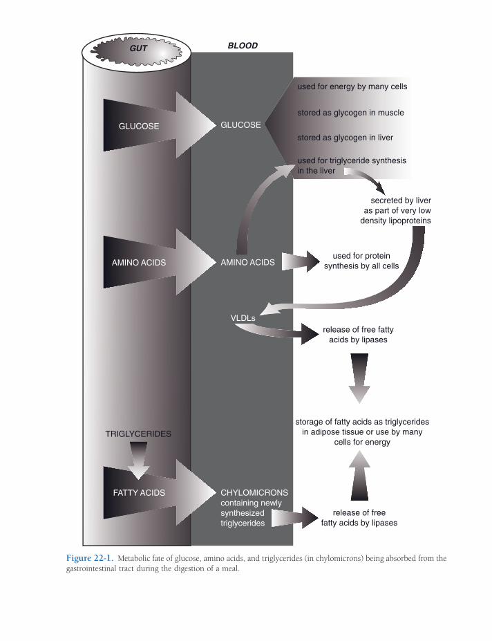
BLOOD
VLDLs
CHYLOMICRONScontaining newlysynthesizedtriglycerides
used for energy by many cells
stored as glycogen in muscle
stored as glycogen in liver
used for triglyceride synthesisin the liver
secreted by liveras part of very low
density lipoproteins
used for proteinsynthesis by all cells
release of free fattyacids by lipases
storage of fatty acids as triglyceridesin adipose tissue or use by many
cells for energy
release of freefatty acids by lipases
TRIGLYCERIDES
FATTY ACIDS
GUT
GLUCOSE GLUCOSE
AMINO ACIDS AMINO ACIDS
Figure 22-1. Metabolic fate of glucose, amino acids, and triglycerides (in chylomicrons) being absorbed from the gastrointestinal tract during the digestion of a meal.

378 / ANATOMY AND PHYSIOLOGY OF FARM ANIMALS
period, and this is also stimulated by insulin. Primarily because the mass of skeletal muscle is greater than the liver, much more glycogen (75% of the total) is formed and stored in skel-etal muscle. Blood glucose is also available for use by all cells of the body for energy during this period, but no other organ is capable of signifi cant glycogen storage.
Absorbed amino acids are immediately avail-able to all body cells for protein synthesis. Because all of the amino acids necessary for synthesis of a given protein must be available at the time of synthesis, it is imperative that animals have a balanced diet that contains all of the essential amino acids. Protein synthesis in many organs, including liver and skeletal muscle, is stimulated by insulin, so the increase in insulin following a meal also promotes protein synthesis during this period. However, this stimulatory effect on protein synthesis is minor compared to the effects of insulin on glucose metabolism (e.g., the rate of plasma protein production by the liver increases by only a small percentage after a meal).
The amounts of amino acids absorbed after a typical meal are more than can be effi ciently used by the body for protein synthesis. However, no metabolic pathways permit the various amino acids to be stored for later use the way glucose is stored as glycogen. Many of the excess amino acids are taken up by hepatocytes and enter metabolic pathways that result in triglyceride (lipid) formation. These pathways remove nitrogen-containing amino groups from the amino acids (deamination). Most of the resulting lipids are secreted by hepatocytes into the blood as part of lipoproteins (discussed later).
Deamination of amino acids is also part of a different metabolic pathway by which liver cells use amino acids to produce glucose. However, the hormone glucagon must be available to stimulate this pathway, and glucagon release from the pancreas is reduced by increases in blood glucose. Thus, during the period that blood glucose is elevated following a meal, the use of amino acids to produce glucose is sup-pressed. Gluconeogenesis is the term for the collective metabolic processes by which liver
cells produce glucose from non-carbohydrate substrates, such as amino acids and short-chain fatty acids. The liver and kidneys are the only organs that are capable of any signifi cant glu-coneogenesis, and the kidneys do so only in states of chronic acidosis.
During the absorptive state, the liver uses both excess glucose and amino acids as sub-strates for triglyceride (lipid) synthesis, and insulin stimulates these pathways. Some of the newly synthesized triglycerides are stored in the liver, but most are released into the blood in complex particles known as very low density lipoproteins (VLDLs). Lipoproteins are parti-cles that contain lipids, cholesterol, and pro-teins in various ratios. VLDLs are so named because their lipid content is high relative to their protein content. Because lipids are less dense than protein, the density of VLDL parti-cles is quite low.
Recall that chylomicrons are also circulating lipoproteins, but the triglycerides in these lipo-proteins were absorbed from the intestinal tract. As chylomicrons and VLDLs circulate throughout the body, they encounter lipoprotein lipase, an enzyme bound to endothelial cells that acts on their triglycerides to release free fatty acids. When the triglycerides are released within adipose tissue, the free fatty acids are available to adipose cells for the resynthesis and storage of lipids as triglycerides. In other organs, such as skeletal muscle, cells use the free fatty acids for energy. The synthesis and storage of triglycerides in adipose tissue is stimulated by insulin, which is typically elevated during the absorptive period.
After losing triglycerides by the action of lipoprotein lipase, some VLDLs undergo changes in the circulation and become a dif-ferent type of lipoprotein, low-density lipo-protein (LDL). LDLs contain a great deal of cholesterol, and cells throughout the body receive cholesterol from the blood by the endocytosis of LDLs. Cholesterol is a neces-sary component of cell membranes, and all cells need some cholesterol. However, abnor-mal increases in LDL levels are associated with an increased risk of cardiovascular disease in humans. Recall that the original
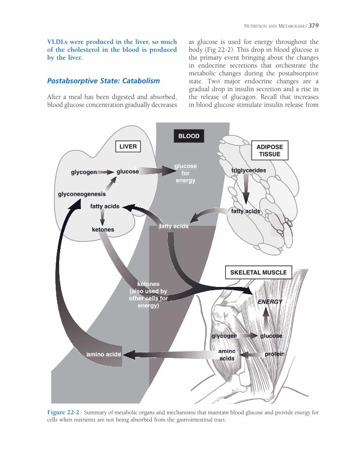
NUTRITION AND METABOLISM / 379
VLDLs were produced in the liver, so much of the cholesterol in the blood is produced by the liver.
Postabsorptive State: Catabolism
After a meal has been digested and absorbed, blood glucose concentration gradually decreases
as glucose is used for energy throughout the body (Fig 22-2). This drop in blood glucose is the primary event bringing about the changes in endocrine secretions that orchestrate the metabolic changes during the postabsorptive state. Two major endocrine changes are a gradual drop in insulin secretion and a rise in the release of glucagon. Recall that increases in blood glucose stimulate insulin release from
LIVER
glycogen
glyconeogenesis
glucose
fatty acids
ketones
glucosefor
energy
ketoonesketsed by(alsalso used
other ceellsr cells forenerrgy)
amino acids
rigiglycerg erideseririgrigtrtr
ty acy acidsy ac ssscidscidsfatty atty a
ENERGY
glucosegluglycogenn
aminoaacidsa
proteinp
ADIPOSETISSUE
BLOOD
SKELETAL MUSCLE
atty acidsfatty acidsfat
Figure 22-2. Summary of metabolic organs and mechanisms that maintain blood glucose and provide energy for cells when nutrients are not being absorbed from the gastrointestinal tract.

380 / ANATOMY AND PHYSIOLOGY OF FARM ANIMALS
β-cells, whereas decreases in blood glucose stimulate glucagon release from α-cells in pan-creatic islets.
During the absorptive period, when glucose and amino acids were being absorbed into the blood from the intestinal tract, insulin stimu-lated the synthesis of glycogen for glucose storage, proteins, and lipids (from any excess glucose and amino acids). As insulin levels decrease, its stimulatory effect on these syn-thetic (anabolic) processes is lost, and this is a major factor in changing the overall metabolic balance from anabolism to catabolism.
Glucagon stimulates the breakdown of gly-cogen (glycogenolysis) in the liver to provide glucose that the liver can release into the blood. Glycogenolysis is the initial process by which the liver derives glucose to add to the blood, but later the liver also releases glucose formed by gluconeogenesis, which is also stimulated by glucagon. The amino acids used for gluconeo-genesis are derived from the catabolism of body protein.
The maintenance of a minimal or fasting blood glucose level during this period has primary importance to neuronal function. Neurons do not have the metabolic processes to permit them to use fatty acids for energy, so they need a ready supply of glucose for cell energy. During fasting, the catabolism of adipose tissue increases the supply of fatty acids, which are used by cells other than neurons for energy. The increased use of fatty acids by other cells reduces the overall need for glucose and conserves it for use by neurons. This conservation of glucose is termed glucose sparing. The liver also metabo-lizes the circulating fatty acids to produce ketones, another cellular energy substrate. The ketones produced by the liver include acetone, acetoacetate, and β-hydroxybutyrate.
Two other hormones, growth hormone and glucocorticoids from the adrenal cortex, also contribute to the maintenance of blood glucose and other sources of energy during anabolic periods. Glucocorticoids do not increase in the circulation during a short fast, but a defi cit of glucocorticoids reduces the rate of liver gluco-neogenesis and mobilization of fatty acids from
adipose tissues. The effect of glucocorticoids on these processes during fasting is a permissive effect. Decreases in blood glucose stimulate the release of growth hormone, which increases the mobilization of fatty acids from adipose (lipoly-sis). Some tissues (e.g., skeletal muscle) can use the fatty acids for energy (glucose sparing), and the liver can further increase its production of ketones.
Energy Needs During Exercise
The increase in skeletal muscle metabolism during exercise can rapidly deplete the glyco-gen stores within skeletal muscle cells. In humans these stores are capable of providing energy only for an estimated 2 to 3 minutes of very intense exercise. To sustain exercise, other energy sources must be rapidly mobilized and delivered to working skeletal muscle. Circulat-ing levels of epinephrine and norepinephrine increase during exercise, and these catechol-amines have several actions that mobilize energy stores, including increased glycogenolysis in the liver and nonworking skeletal muscle and lipolysis in adipose tissue. Insulin levels are reduced and glucagon levels are increased during exercise, which promotes liver gluco-neogenesis and more lipolysis in adipose tissue. The decrease in insulin is not detrimental to working skeletal muscle, because glucose uptake by working muscle is less insulin dependent.
Anaerobic metabolism, by working skeletal muscles, raises the rate of lactic acid produc-tion. The lactate ion can diffuse into the blood from the skeletal muscle, and plasma levels of lactate increase during strenuous and prolonged exercise. The liver can use blood lactate for gluconeogenesis, and glucose can then be returned to the blood to maintain blood glucose levels.
Blood Glucose in Ruminants
Normal ranges for blood glucose levels in mature ruminants are lower than in other
NUTRITION AND METABOLISM / 381
animals, even other herbivores (cattle, 45 to 80 mg/dL; dog, 70 to 110 mg/dL; and horse, 60 to 110 mg/dL). The lower normal range for mature ruminants is associated with the rela-tively small amount of glucose-yielding carbo-hydrate digestion in their small intestine. Most of the carbohydrates that they consume undergo fermentative digestion in the forestomach and result in the production of short-chain volatile fatty acids (VFAs), which are absorbed directly from the forestomach.
Without glucose readily available via absorp-tion from the gastrointestinal tract, ruminants must have a continuous and a relatively high rate of gluconeogenesis in the liver to maintain the blood glucose level. Glucagon appears to be an important endocrine stimulant to maintain this rate of gluconeogenesis. Rising levels of amino acids and propionic acid (a VFA pro-duced in the rumen) can stimulate glucagon release, so presumably the continual absorption of these from the ruminant gastrointestinal tract can maintain glucagon secretion. Propionic acid is the one of the three major VFAs pro-duced in the rumen that can be used by the liver for gluconeogenesis.
Ketosis
Ketosis is a metabolic state characterized by an increase in blood ketones, a reduction in urine and blood pH, and ketones in the urine. The increase in the acidic ketones in the blood and urine are responsible for the changes in pH. Ketosis may occur when fatty acid mobilization from adipose tissue is elevated and glucose is defi cient. The defi ciency in glucose stimulates the release of glucagon and inhibits insulin release, and the increased ratio of glucagon to insulin promotes the formation of ketones by the liver from readily available fatty acids.
Ketosis may develop in dairy cattle at the peak of lactation, when the need for glucose to synthesize lactose (milk sugar) is maximal. The rapid use of glucose by the mammary glands reduces blood glucose and brings about these changes in glucagon and insulin. Ketosis may also develop as a result of type I diabetes mel-litus, in which the primary problem is a defi -ciency of insulin. In this case, the dominant effects of glucagon on fatty acid mobilization and ketone synthesis are primarily responsible for the development of the ketosis.


THE URINARY SYSTEM
The urinary system consists of two kidneys, two ureters, the urinary bladder, and the
urethra. The paired kidneys remove waste products from the blood, help regulate the composition of plasma, and perform certain hormonal functions. The system of tubules in each kidney coalesces into a single mucomus-cular tube, the ureter, which extends caudad to empty into the urinary bladder, a distensible reservoir for the storage of urine. When full, the urinary bladder discharges the urine through the urethra to the outside of the body.
Anatomy of the Kidney
The kidneys are paired reddish-brown organs that fi lter plasma and plasma constituents from the blood and then selectively reabsorb water and useful constituents from the fi ltrate, ulti-mately excreting excesses and plasma waste products. The kidneys of most animals are roughly bean-shaped, with the exceptions among domestic animals of the heart-shaped right equine kidney and the distinctively lobated kidneys of the ox (Figs. 23-1 and 23-2).
The kidneys are in the dorsal part of the abdominal cavity on each side of the aorta and caudal vena cava, just ventral to the fi rst few lumbar vertebrae. In most domestic animals, the right kidney is slightly more cranial than
Anatomy of the KidneyBlood and Nerve Supply
Ureters, Urinary Bladder, and UrethraMicturitionOverview of Function and Histology of
the KidneysGlomerular FiltrationProximal Tubule TransportConcentration and Dilution of Urine: Role
of the Loop of Henle and Collecting Duct Transport
Sodium Chloride and Water Reabsorption
by the Loop of Henle
Collecting Duct Transport and Antidiuretic
Hormone
Osmotic Regulation of Antidiuretic
Hormone
Polyuria and Polydipsia
Sodium, Potassium, and AldosteroneUrine Acidifi cationRegulation of Acid-Base Balance
Extracellular and Intracellular Buffers
Classifi cation of Alkalosis and Acidosis
and Compensation
Chapter 23
383

Lobe
Cortex
Medulla
Ureter
Pelvis
Renal crest
Lobe
Major calyx Ureter
Minor calyx
Papilla
Medulla
Cortex
Lobe
Papilla
Minor calyx
Major calyxUreter
Pelvis
Medulla
Cortex
A B
C
Figure 23-1. Right kidneys of the horse (A); the ox (B); and the sheep (C). 1, Renal artery; 2, renal vein; 3, ureter. (Reprinted with permission of Wiley-Blackwell from Reece W.O. Physiology of Domestic Animals. 2nd ed. Baltimore: Williams & Wilkins, 1997.)
Figure 23-2. Internal anatomy of kidneys. A) The renal cortex of the equine, ovine, and caprine kidney lacks visible divisions into individual lobes. The renal papillae are fused in these species into a longitudinal renal crest. B) The bovine kidney is grossly divided into lobes, each of which communicates with a minor calyx. C) The porcine kidney lacks external divisions into lobes, but the renal papillae of the medulla distinguish each lobe internally. (Reprinted with permission of Wiley-Blackwell from Dellmann H.D. Textbook of Veterinary Histology. 4th ed. Philadelphia: Lea & Febiger, 1993.)
THE URINARY SYSTEM / 385
the left, with the cranial pole of the right kidney lying snugly in a complementary fossa of the liver. The left kidney tends to be more pendu-lous, and in ruminants, the forestomach may push the left kidney to the right as far as the median plane or beyond, particularly when the rumen is full. The kidneys are described as being retroperitoneal in location, refl ecting their position outside the peritoneal cavity where they are more closely attached to the abdominal wall by fascia, vessels, and perito-neum than are most other abdominal organs. A tough connective tissue capsule surrounds the entire kidney.
The medial aspect of each kidney features a concavity, the hilus, where arteries and nerves enter the kidney, and the ureter, veins, and lymphatic vessels leave (Fig. 23-3). The wide origin of the ureter in the kidney is the renal pelvis. The renal pelvis receives urine from the collecting tubules of the kidney. The cavity in the kidney that contains the pelvis is the renal sinus. The bovine kidney does not have a renal pelvis, the ureter instead arising directly from the coalescence of individual calyces (discussed later).
The portion of the kidney immediately sur-rounding the renal pelvis is the renal medulla, which appears striated because of the radially arranged collecting tubules. In addition to col-lecting tubules, the medulla also contains some loops of Henle (descending and ascending loops). The medulla is surmounted peripher-ally by the renal cortex, in which reside the renal corpuscles, the histological units of fi ltra-tion. The cortex has a granular appearance because of the large number of these renal cor-puscles; also found in the cortex are proximal and distal convoluted tubules and other seg-ments of loops of Henle (discussed later).
The medulla and cortex are arranged in units called lobes, cone-shaped aggregates of renal tissue. The medullary portion of each lobe con-stitutes a renal pyramid, whose apex, the renal papilla, is directed toward the renal pelvis (Fig. 23-3). In the bovine kidney, each pyramid is associated with one of the grossly obvious lobes of the bovine kidney. In the pig and small rumi-nants, the adjacent cortices of individual lobes
are fused, so that the surface of the kidney appears smooth. The individual nature of the porcine lobes is revealed, however, through the persistence of discrete papillae projecting into the renal pelvis (Fig. 23-2). In the horse and small ruminants, the individual papillae, like the cortex, are fused. Consequently, they present as a single longitudinal ridge, the renal crest, projecting into the renal pelvis. Urine dis-charged from the collecting tubules of the renal crest is collected in the renal pelvis and from there is delivered to the ureter.
In the kidney of the ox and pig, individual pyramids project into minor calyces, cuplike diverticula of the common collecting space within the renal hilus. These in turn empty into major calyces. These major calyces in the porcine kidney empty into the renal pelvis, but the bovine kidney has no pelvis, and so the major calyces in this species empty directly into the ureter (Fig. 23-2).
Blood and Nerve Supply
Because of its important role in adjusting the composition of extracellular fl uid (including plasma), the blood supply to the kidney is much more extensive than the size of the organ would suggest. The two renal arteries may receive as much as one-fourth of the total cardiac output. Each renal artery enters the hilus of the kidney and divides into a number of relatively large branches, the interlobar arteries. These pass peripherally between pyra-mids almost to the cortex, where they bend abruptly and become arcuate arteries, which derive their name from the arched manner by which they pass along the junction between cortex and medulla (Fig. 23-3.)
Each arcuate artery gives off a number of interlobular arteries that extend into the cortex and in turn give rise to the afferent arterioles. Each afferent arteriole branches repeatedly to form a tufted capillary network called the glom-erulus, which is associated with the renal cor-puscle. The capillaries of the glomerulus coalesce into an efferent arteriole, which leaves each glomerulus (Fig. 23-4).

Figure 23-3. Kidney of the canid cut on the median plane (A) and a sagittal section off midline (B). The small ruminant kidney appears similar to this canine kidney. (Reprinted with permission of Wiley-Blackwell from Smith B.J. Canine Anatomy. Philadelphia: Lippincott Williams & Wilkins, 1999.)
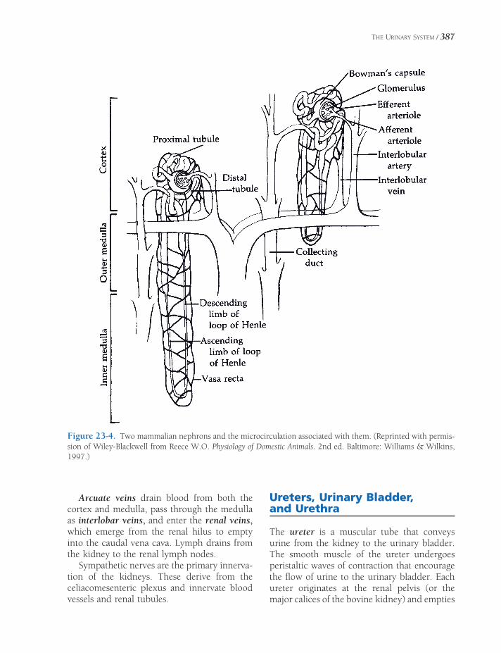
THE URINARY SYSTEM / 387
Figure 23-4. Two mammalian nephrons and the microcirculation associated with them. (Reprinted with permis-sion of Wiley-Blackwell from Reece W.O. Physiology of Domestic Animals. 2nd ed. Baltimore: Williams & Wilkins, 1997.)
Arcuate veins drain blood from both the cortex and medulla, pass through the medulla as interlobar veins, and enter the renal veins, which emerge from the renal hilus to empty into the caudal vena cava. Lymph drains from the kidney to the renal lymph nodes.
Sympathetic nerves are the primary innerva-tion of the kidneys. These derive from the celiacomesenteric plexus and innervate blood vessels and renal tubules.
Ureters, Urinary Bladder, and Urethra
The ureter is a muscular tube that conveys urine from the kidney to the urinary bladder. The smooth muscle of the ureter undergoes peristaltic waves of contraction that encourage the fl ow of urine to the urinary bladder. Each ureter originates at the renal pelvis (or the major calices of the bovine kidney) and empties

388 / ANATOMY AND PHYSIOLOGY OF FARM ANIMALS
into the urinary bladder near its neck at the trigone (Fig. 23-5). The manner in which the ureter passes obliquely through the wall of the urinary bladder creates a valve to prevent refl ux fl ow of urine to the kidney.
The urinary bladder is a hollow muscular organ that varies in size and position with the amount of urine it contains. The empty, con-tracted bladder is a thick-walled, piriform organ on the fl oor of the pelvic cavity. As it fi lls with urine, its wall thins, and it enlarges craniad toward and then into the abdominal cavity.
The neck of the bladder is continuous with the urethra caudally. The smooth muscle of the urinary bladder wall is arranged in three sheets; at the neck of the bladder these may form a smooth muscle sphincter that controls passage of urine into the urethra, although the precise role of the intramural muscle of the urinary bladder in creating a true sphincter is debated. The pelvic urethra extends from the urinary bladder across the fl oor of the pelvic canal to the ischial arch. In female animals, it opens onto the fl oor of the vaginal vestibule. In the male animal, it receives the ductus deferens and ducts from the accessory sex glands, then passes through the penis as the penile urethra. In both sexes, the pelvic urethra is surrounded by a true sphincter, the striated skeletal m. urethralis, over which the animal exercises voluntary control. The m. ure-thralis is innervated by the pudendal nerve.
The pelvis, ureter, bladder, and urethra are all lined with transitional epithelium. This epi-thelial lining is useful in these areas, where considerable distension of the lumen may occur. When these organs are empty, the lumen is small, the walls are thick, and the lining epi-thelial cells are piled deeply to form many layers. However, when the organs are distended, the lumen is enlarged, the walls are thinner, and a transition to a much lower stratifi cation of the lining occurs (Fig. 23-6). The urethra, like the urinary bladder, is lined with transi-tional epithelium; this changes to the typical stratifi ed squamous epithelium of mucous membranes at or near the urethral orifi ce at the tip of the penis of male animals or at its junc-tion with the vestibule of the vagina in the female animal.
Micturition
Micturition is the term for expulsion of urine from the bladder. It normally is a refl ex activity stimulated by stretch receptors in the bladder wall that respond to distension of the urinary bladder created by the constant infl ow of urine by way of the ureters. The bladder relaxes to accommodate a gradual increase in urinary volume until the stretch becomes great enough to stimulate refl ex centers in the spinal cord,
Figure 23-5. Kidneys, ureters, and urinary bladder in situ in the small rumi-nant. (Reprinted with permission of Wiley-Blackwell from Reece W.O. Physi-ology of Domestic Animals. 2nd ed. Balti-more: Williams & Wilkins, 1997.)

THE URINARY SYSTEM / 389
which in turn relax the smooth muscle sphinc-ter in the neck of the bladder and contract the muscle wall of the urinary bladder by way of sacral parasympathetic nerves. However, refl ex emptying of the bladder can be prevented by voluntary suppression of the refl ex and control of the m. urethralis surrounding the proximal urethra.
Overview of Function and Histology of the Kidneys
The urinary system is responsible for maintain-ing the relatively constant internal environment of the body fl uids. This is accomplished by the formation and excretion of urine of an appro-priate volume and composition. Urine forma-tion occurs in the kidneys, and by adjusting the volume and composition of urine in response to changes in dietary intake or metabolism, the kidneys regulate the body balance of water, various electrolytes, acids, and bases. The kidneys also excrete metabolic waste products in the urine, including the nitrogenous waste, urea, and a by-product of skeletal muscle metabolism, creatinine. Signs of kidney dis-eases include imbalances of water, electrolytes, acids, and bases and increases in blood levels of urea and creatinine.
Kidneys are composite organs that consist of thousands to millions of similar microscopic
functional units, the nephrons. Nephrons in all mammalian kidneys are similar in basic struc-ture and function, but the number of nephrons differs among mammals. Large animals have more nephrons per kidney than small animals (e.g., 4 million for cattle and 500,000 for dogs). Nephrons consist of a spherical structure (Bow-man’s capsule) that contains a capillary tuft (glomerulus) and a single long tubule connected to Bowman’s capsule (Fig. 23-4). Bowman’s capsule consists of two layers of cells. The inner (visceral) layer closely surrounds the glomerular capillaries, and the outer (parietal) layer is con-tinuous with the fi rst segment of the tubule. A Bowman’s capsule with its contained glomeru-lus is a renal corpuscle.
The single tubule is divided into segments based on differences in histological appearance, location in the kidney, and function. These seg-ments are named the proximal (convoluted) tubule, loop of Henle, and distal (convoluted) tubule (Fig. 23-4). The distal tubules of numer-ous nephrons connect to another tubular struc-ture found in the kidney, the collecting duct (tubule). Collecting ducts begin in the renal cortex, where they connect with distal tubules, and extend into and through the renal medulla (Fig. 23-4).
Three processes are involved in urine forma-tion: (1) glomerular fi ltration, (2) selective tubular reabsorption, and (3) selective tubular secretion (Fig. 23-7). As blood fl ows through
Figure 23-6. Transitional epithelium. A) Bladder full, wall distended. B) Bladder empty, wall relaxed.

390 / ANATOMY AND PHYSIOLOGY OF FARM ANIMALS
glomeruli, a large quantity of fi ltrate is formed and enters the urinary space of Bowman’s capsule. From here the fi ltrate fl ows through the tubules and collecting ducts, where tubular reabsorption and tubular secretion alter its volume and composition. Tubular reabsorption is the removal of substances from the tubular fl uid by the tubular cells; these substances are usually returned to the blood in the peritubu-lar capillaries. Tubular secretion is the addition of substances to the tubular fl uid by tubule cells. The secreted substances are produced in the tubule cells (e.g., hydrogen ion and ammonia) or taken up by the tubule cells from the blood in the peritubular capillaries (e.g., pharmaceuticals).
The renal microcirculation is unique in that glomerular capillaries are between two arterio-lar vessels rather than between an arteriole and a venule. Afferent arterioles lead into glomer-
uli, and efferent arterioles leave glomeruli (Fig. 23-4). Efferent arterioles from most glomeruli lead into capillary networks that surround tubules in the cortex (peritubular capillaries). Efferents from glomeruli deep in the cortex next to the medulla contribute blood to vessels that extend into the medulla. These vessels (vasa rectae) consist of straight descending branches (descending vasa rectae) that empty into medullary capillaries, which are drained by straight ascending vessels (ascending vasa rectae) (Fig. 23-4).
Near glomeruli, the walls of afferent arteri-oles contain specialized cells termed juxtaglo-merular (JG) or granular cells (Fig. 23-8). Secretory granules in these cells contain the enzyme renin. Renin is a component of the renin–angiotensin–aldosterone system (see Chapter 18), which is involved in the regula-tion of blood volume and blood pressure. The JG cells are part of a functional grouping of closely related structures, the juxtaglomerular apparatus. The juxtaglomerular apparatus con-sists of the JG cells, the macula densa, and extraglomerular mesangial cells (Fig. 23-8). The macula densa is a specifi c region of the wall of the distal tubule where the cellular nuclei
Figure 23-7. Urine formation. The glomerular fi ltrate enters the tubules, where some substances are removed by selective tubular reabsorption and others are added by selective tubular secretion. Reabsorption and secre-tion occur throughout the nephron and collecting ducts. (Reprinted with permission of Wiley-Blackwell from Reece W.O. Physiology of Domestic Animals. 2nd ed. Baltimore: Williams & Wilkins, 1997.)
Figure 23-8. The juxtaglomerular (JG) apparatus. Extraglomerular mesangial cells are found at the base of the glomerulus between the macula densa and the arte-rioles. (Reprinted with permission of Wiley-Blackwell from Reece W.O. Physiology of Domestic Animals. 2nd ed. Baltimore: Williams & Wilkins, 1997.)

THE URINARY SYSTEM / 391
appear to be bunched closely together. The segment of the distal tubule found here is part of the same nephron associated with the affer-ent arterioles (Fig. 23-7). The extraglomerular mesangial cells are between the macula densa and its associated JG cells.
Glomerular Filtration
The glomerular fi ltrate is the fl uid and fl uid constituents that pass from the blood plasma in the glomerulus into the urinary space of Bow-man’s capsule. The physical barriers through which the fi ltrate passes include (1) the capil-lary endothelium of the glomerulus, (2) the inner layer of Bowman’s capsule, and (3) a base-ment membrane (lamina) between these two cell layers. The glomerular endothelium is fenestrated (i.e., has openings or pores in the cells), so this part of the barrier is highly per-meable. Podocytes (cells of the inner layer of Bowman’s capsule) have cellular extensions that rest on the glomerular basement membrane, but slitlike pores between the extensions permit the passage of the fi ltrate (Fig. 23-9).
The glomerular fi ltration barrier acts much like a sieve, and all substances up to a molecu-lar weight of about 65,000 pass through the barrier. Blood cells are too large to pass, and only a small percentage of plasma proteins pass
through the barrier. Most other plasma con-stituents (e.g., glucose, amino acids, urea, cre-atinine, sodium, potassium, chlorine, and bicarbonate ions) readily cross the barrier, and their concentrations in the initial fi ltrate are about the same as in plasma. Proteinuria is the presence of abnormal amounts of protein in voided urine. Kidney diseases that localize in or primarily affect glomeruli are often associated with proteinuria or hematuria (blood in voided urine).
The forces determining the rate of move-ment of fl uid across the glomerular fi ltration barrier are the same as those that determine fl uid movement out of capillaries throughout the body. The effective fi ltration pressure (the pressure tending to force fl uid out of the capil-lary) is usually considered to be the difference between the blood (hydrostatic) pressure in the capillary and the osmotic pressure generated by the plasma proteins of the blood in the capil-laries. The hydrostatic pressure in the urinary space of Bowman’s capsule and the osmotic pressure generated by proteins in the fl uid in the space can also be factors, and these become important in disease states (e.g., blockage of the urinary tract or renal tubules).
In mammals glomerular fi ltration rate (GFR) and renal blood fl ow (RBF) remain relatively stable in normally hydrated animals in spite of minor short-term fl uctuations in arterial blood
Figure 23-9. The glomerular fi ltration barrier. E, Fenestrated glomerular capillary endothelium; EP, endothelial pore (fenestra); GBM, glomerular basement membrane; P, podocytes, or visceral layer of Bowman’s capsule with their fi ltration slits (FS); US, urinary space of Bowman’s capsule. (Reprinted with permission of Wiley-Blackwell from Dellmann H.D. Textbook of Veterinary Histology. 4th ed. Philadelphia: Lea & Febiger, 1993.)
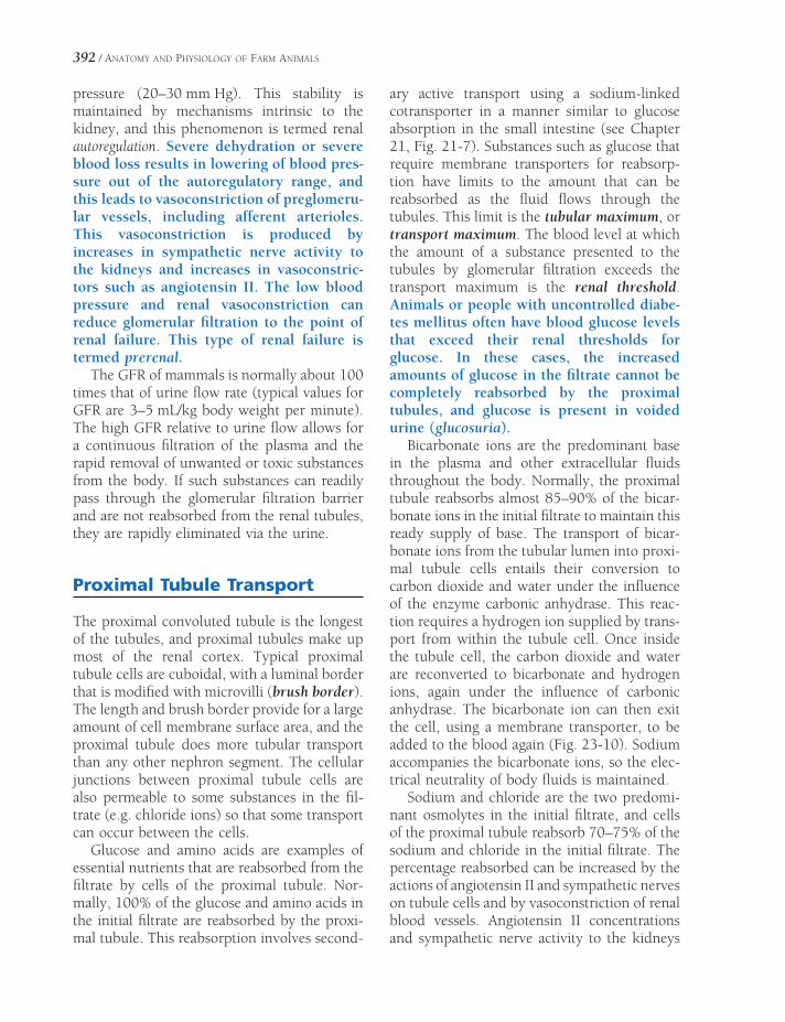
392 / ANATOMY AND PHYSIOLOGY OF FARM ANIMALS
pressure (20–30 mm Hg). This stability is maintained by mechanisms intrinsic to the kidney, and this phenomenon is termed renal autoregulation. Severe dehydration or severe blood loss results in lowering of blood pres-sure out of the autoregulatory range, and this leads to vasoconstriction of preglomeru-lar vessels, including afferent arterioles. This vasoconstriction is produced by increases in sympathetic nerve activity to the kidneys and increases in vasoconstric-tors such as angiotensin II. The low blood pressure and renal vasoconstriction can reduce glomerular fi ltration to the point of renal failure. This type of renal failure is termed prerenal.
The GFR of mammals is normally about 100 times that of urine fl ow rate (typical values for GFR are 3–5 mL/kg body weight per minute). The high GFR relative to urine fl ow allows for a continuous fi ltration of the plasma and the rapid removal of unwanted or toxic substances from the body. If such substances can readily pass through the glomerular fi ltration barrier and are not reabsorbed from the renal tubules, they are rapidly eliminated via the urine.
Proximal Tubule Transport
The proximal convoluted tubule is the longest of the tubules, and proximal tubules make up most of the renal cortex. Typical proximal tubule cells are cuboidal, with a luminal border that is modifi ed with microvilli (brush border). The length and brush border provide for a large amount of cell membrane surface area, and the proximal tubule does more tubular transport than any other nephron segment. The cellular junctions between proximal tubule cells are also permeable to some substances in the fi l-trate (e.g. chloride ions) so that some transport can occur between the cells.
Glucose and amino acids are examples of essential nutrients that are reabsorbed from the fi ltrate by cells of the proximal tubule. Nor-mally, 100% of the glucose and amino acids in the initial fi ltrate are reabsorbed by the proxi-mal tubule. This reabsorption involves second-
ary active transport using a sodium-linked cotransporter in a manner similar to glucose absorption in the small intestine (see Chapter 21, Fig. 21-7). Substances such as glucose that require membrane transporters for reabsorp-tion have limits to the amount that can be reabsorbed as the fl uid fl ows through the tubules. This limit is the tubular maximum, or transport maximum. The blood level at which the amount of a substance presented to the tubules by glomerular fi ltration exceeds the transport maximum is the renal threshold. Animals or people with uncontrolled diabe-tes mellitus often have blood glucose levels that exceed their renal thresholds for glucose. In these cases, the increased amounts of glucose in the fi ltrate cannot be completely reabsorbed by the proximal tubules, and glucose is present in voided urine (glucosuria).
Bicarbonate ions are the predominant base in the plasma and other extracellular fl uids throughout the body. Normally, the proximal tubule reabsorbs almost 85–90% of the bicar-bonate ions in the initial fi ltrate to maintain this ready supply of base. The transport of bicar-bonate ions from the tubular lumen into proxi-mal tubule cells entails their conversion to carbon dioxide and water under the infl uence of the enzyme carbonic anhydrase. This reac-tion requires a hydrogen ion supplied by trans-port from within the tubule cell. Once inside the tubule cell, the carbon dioxide and water are reconverted to bicarbonate and hydrogen ions, again under the infl uence of carbonic anhydrase. The bicarbonate ion can then exit the cell, using a membrane transporter, to be added to the blood again (Fig. 23-10). Sodium accompanies the bicarbonate ions, so the elec-trical neutrality of body fl uids is maintained.
Sodium and chloride are the two predomi-nant osmolytes in the initial fi ltrate, and cells of the proximal tubule reabsorb 70–75% of the sodium and chloride in the initial fi ltrate. The percentage reabsorbed can be increased by the actions of angiotensin II and sympathetic nerves on tubule cells and by vasoconstriction of renal blood vessels. Angiotensin II concentrations and sympathetic nerve activity to the kidneys
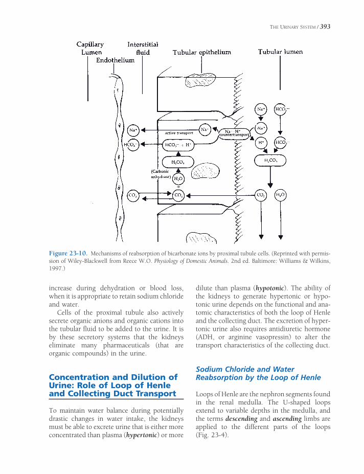
THE URINARY SYSTEM / 393
increase during dehydration or blood loss, when it is appropriate to retain sodium chloride and water.
Cells of the proximal tubule also actively secrete organic anions and organic cations into the tubular fl uid to be added to the urine. It is by these secretory systems that the kidneys eliminate many pharmaceuticals (that are organic compounds) in the urine.
Concentration and Dilution of Urine: Role of Loop of Henle and Collecting Duct Transport
To maintain water balance during potentially drastic changes in water intake, the kidneys must be able to excrete urine that is either more concentrated than plasma (hypertonic) or more
dilute than plasma (hypotonic). The ability of the kidneys to generate hypertonic or hypo-tonic urine depends on the functional and ana-tomic characteristics of both the loop of Henle and the collecting duct. The excretion of hyper-tonic urine also requires antidiuretic hormone (ADH, or arginine vasopressin) to alter the transport characteristics of the collecting duct.
Sodium Chloride and Water Reabsorption by the Loop of Henle
Loops of Henle are the nephron segments found in the renal medulla. The U-shaped loops extend to variable depths in the medulla, and the terms descending and ascending limbs are applied to the different parts of the loops (Fig. 23-4).
Figure 23-10. Mechanisms of reabsorption of bicarbonate ions by proximal tubule cells. (Reprinted with permis-sion of Wiley-Blackwell from Reece W.O. Physiology of Domestic Animals. 2nd ed. Baltimore: Williams & Wilkins, 1997.)
394 / ANATOMY AND PHYSIOLOGY OF FARM ANIMALS
The ascending limbs of the loops of Henle are relatively impermeable to water and have a thick portion that is the site of a great deal of sodium and chloride reabsorption. Sodium and chloride transport by the thick ascending limb uses a unique membrane transporter that cotransports sodium, chloride, and potassium into the cell from the lumen. This transport is sodium-linked in that the Na-K-ATPase pump on the opposite side of the cell maintains the low intracellular sodium concentration that permits the cotransporter to function. The net effect of this cellular transport is continuous addition of sodium and chloride to the intersti-tial fl uid of the medulla without any accompa-nying water (Fig. 23-11). Sodium chloride is also reabsorbed from the thin ascending limb of the loop (Fig. 23-11), but the mechanism responsible for this transport is controversial. The fl uid entering the loop of Henle is isotonic, and the fl uid exiting is hypotonic (Fig. 23-11). This change in tubular fl uid shows that the net effect of loop of Henle transport is to add more particles than water to the interstitial fl uids in the renal medulla.
Transport of sodium and chloride into the interstitium without water is the key factor in generation of hypertonic interstitial fl uid in the medulla (Fig. 23-11), and this hypertonic fl uid has an essential role in the ability to generate hypertonic urine. The transport of particles without water from the lumen of the loops also creates hypotonic fl uid in the tubule, and this is an essential step in the ability to generate hypo-tonic urine. However, regardless of the tonicity of the fi nal urine, the transport characteristics of the thick ascending limbs of the loops of Henle remain the same, so that hypertonic fl uid is gen-erated within the renal medulla and hypotonic fl uid is generated within the loop of Henle.
The descending limbs of the loops of Henle are relatively permeable to water but relatively impermeable to particles. As fl uid fl ows into and through the descending limbs, water is removed because of the osmotic gradient between the tubular lumen and the interstitial fl uids of the renal medulla (Fig. 23-11).
Because the ascending and descending limbs of the loops of Henle are relatively close together
in the medulla and because the tubular fl ows move in opposite directions, the combined effect of transport by ascending and descend-ing limbs produces an osmotic gradient in the interstitial fl uids of the renal medulla. Intersti-tial fl uid osmolality increases from the outer zones to the inner zones of the renal medulla (Fig. 23-11).
The countercurrent mechanism is any mech-anism that depends on streams of fl ow moving in opposite directions, and these are usually close to each other. The ascending and descend-ing loops of Henle form a countercurrent mechanism that amplifi es the osmolyte (sodium chloride) transport properties of the ascending limb of the loop of Henle. This countercurrent mechanism generates the osmotic gradient in the interstitial fl uids of the renal medulla.
The ascending and descending vasa recta also form a countercurrent exchange mecha-nism by permitting free exchange of solutes and water between the ascending and descending blood vessels. This exchange allows for blood fl ow into and out of the medulla without dis-rupting the gradient. There is a small net gain of both water and particles by the vasa rectae, and some of these are the water and particles that were reabsorbed from the loop of Henle (Fig. 23-11).
The maximal osmolality of the osmotic gra-dient in the renal medulla differs among species, and maximal urine concentration ability is determined by the maximal osmolality of the gradient (Fig. 23-11).
Collecting Duct Transport and Antidiuretic Hormone
Principal cells in the collecting ducts are the target cells for ADH. If ADH is not present, the luminal cell membrane of these cells is rela-tively water impermeable. ADH stimulates the insertion of water channels into these cell mem-branes to increase the overall water permeabil-ity of collecting ducts.
Collecting ducts begin in the renal cortex but extend into and through the renal medulla, where the interstitial fl uids are hypertonic.

THE URINARY SYSTEM / 395
CHYPOTONI
FlowFlow
CNa l
HYPERTONICCCeLoop of Henl
0012O /L
SO O C
m sm
I T NI
ClNa
ClNa
O CHYPERT NIasa ec aeV R t
080O /Lm sm
004LmOsm/
C xorteBloodBlooFlowFlow
300O /m sm L
H2O
H2O
H2O
H2O
H2O
p
gasa ec ae
Looof Henle
DescendinV R t
BloodBloloodloFlowFlowow
FlowwFlow
asa ec aeAscending
V R t
edu a
gt
M ll
CollectinDuc
Figure 23-11. Transport of sodium chloride and water by loop of Henle and vasa rectae. Arrows indicate direc-tion of net transport in the different tubular segments and vessels. The osmolarity of the interstitial fl uids in the cortex is 300 mOsm/L; the osmolarity of the interstitial fl uids in the medulla is shown as a gradient that increases from outer to inner medulla. Hypertonic, isotonic, and hypotonic refer to fl uid and plasma in tubules and vessels, respectively.
Also, because of the transport in the loop of Henle, the tubular fl uid entering the cortical portion of a collecting duct is dilute or hypo-tonic, and this is always true regardless of the water balance status of the animal. If ADH
is not present, the water permeability of the collecting duct is relatively low, and the hypo-tonic fl uid entering the collecting ducts passes through and is excreted as a hypotonic urine. Because water is not reabsorbed from the

396 / ANATOMY AND PHYSIOLOGY OF FARM ANIMALS
A. NO ANTIDIURETIC HORMONE B. ANTIDIURETIC HORMONE PRESENT
Dilute (HYPODilute (HYPOTONIC)ONIC)Large VolumeLarge Volume
Concentrated (HYPERConcentrated (HYPERTONIC)ONIC)Small VolumeSmall Volume
HYPOTONIC
CollectingDuct
Medulla
400400mOsm/LmOsm/L
800800mOsm/LmOsm/L
12001200mOsm/LmOsm/L
Cortex
HYPOTONIC
CollectingDuct
HOH
HOH
HOHHOHH
HOHH
HOHH
Figure 23-12. A) Distal tubule and collecting duct when ADH is absent. Water permeability of collecting is low, so tubular fl uid remains hypotonic (dilute). B) Distal tubule and collecting duct when ADH is present. Water per-meability of collecting duct is increased, so water is reabsorbed and tubular fl uid becomes hypertonic (concentrated).
water-impermeable collecting duct, the volume is also relatively large (Fig. 23-12A). If ADH is present, the water permeability of the collect-ing duct is increased, and water is reabsorbed because the osmolality inside the duct is less than that outside. As the tubular fl uid
passes through the medullary portion of the collecting duct, more and more water is reab-sorbed, and the osmolality of the tubular fl uid increases further. In these circumstances urine volume is low and urine osmolality is high (Fig. 23-12B).

THE URINARY SYSTEM / 397
Osmotic Regulation of Antidiuretic Hormone
ADH (or arginine vasopressin in most mam-malian species) release from the posterior pitu-itary can be regulated by changes in the ECF (extracellular fl uid) osmolality. Specifi c cells (osmoreceptors) in the hypothalamus monitor the osmolality of ECF. In response to increases in ECF osmolality, these cells stimulate increases in ADH release, which results in the excretion of a small volume of a hypertonic urine. The elimination of excess particles and conservation of water dilutes the ECF, which acts as a nega-tive feedback control to inhibit additional releases of ADH. Reductions in ECF osmolality inhibit ADH release, which results in the excre-tion of a relatively large volume of dilute urine. This eliminates any excess water.
Polyuria and Polydipsia
Polyuria is the passage of larger volumes of urine than normal. Animals that cannot gener-ate hypertonic urine when necessary become polyuric. Polydipsia is excessive thirst, and polyuric animals are often able to maintain water balance by increasing water intake. The increased intake is considered to be a sign of excessive thirst.
Polyuria and polydipsia (often abbrevi-ated PU/PD) may develop in animals with unregulated diabetes mellitus and signifi -cant increases in blood glucose. The renal tubules cannot reabsorb the abnormally large amounts of glucose in the glomerular fi ltrate, and the glucose that remains in the renal tubules exerts an osmotic effect to retain water in the tubules. Increased urine fl ow results, and the animal must increase water intake to maintain water balance.
Polyuria and polydipsia may also result when ADH is not available (e.g., pituitary tumor preventing its release) or the kidney does not respond appropriately to ADH. In either case the water permeability of the collecting ducts remains relatively low, and water cannot be reabsorbed from the collecting ducts into the
blood. Again, the affected animals must increase water intake to maintain water balance. This condition is diabetes insipidus.
Sodium, Potassium, and Aldosterone
Most of the sodium and potassium in the initial glomerular fi ltrate is reabsorbed by the proxi-mal tubule and the loop of Henle. However, the collecting duct is also capable of sodium and potassium transport, and it is here that the fi nal adjustments are made in the regulation of sodium and potassium balance. Aldosterone, a steroid hormone from the adrenal cortex, func-tions as a major regulator of sodium and potas-sium transport in the collecting duct.
Aldosterone acts on principal cells of the collecting ducts to promote their reabsorption of sodium when sodium must be retained to maintain sodium balance. The regulation of aldosterone secretion from the adrenal cortex relative to sodium balance is via the renin–angiotensin system. When sodium must be retained (such as with a low-salt diet or after loss of extracellular fl uid with sodium), the renin–angiotensin system is activated, and angiotensin II stimulates cells of the adrenal cortex to secrete aldosterone.
The concentration of potassium in plasma and other extracellular fl uids also regulates aldosterone secretion. Increases in potassium concentration directly stimulate cells of the adrenal cortex to secrete aldosterone. Aldoste-rone promotes potassium secretion by principal cells, and this tends to increase the urinary loss of potassium. The increased loss of potassium in the urine reduces plasma potassium, and thus potassium plasma concentration and potassium balance are maintained. Most potas-sium in excreted urine reaches the tubular fl uid by tubular secretion in the collecting duct.
Unregulated secretion of aldosterone by adrenal tumors can cause signifi cant reduc-tions in plasma potassium concentrations, and these can threaten life. Such individuals may also have moderate degrees of sodium retention and increases in ECF volume, but

398 / ANATOMY AND PHYSIOLOGY OF FARM ANIMALS
this is usually not as severe as the changes in plasma potassium. The more moderate changes in sodium balance are because sodium transport by other nephron seg-ments is regulated by other factors that can counterbalance the sodium-retaining effects of aldosterone.
Urine Acidification
Intercalated cells in the walls of collecting ducts are capable of the active transport of hydrogen ions into the tubular lumen to acidify the urine. This system can generate a difference between blood and urine of about 3 pH units, so if blood pH is 7.4, urine with a pH of 4.4 can be formed. The hydrogen ions to be secreted are generated in intercalated cells by the hydra-tion of carbon dioxide under the infl uence of carbonic anhydrase. A bicarbonate ion is also generated during this process, and these are secreted into the extracellular fl uids at the base of the cell, from where the bicarbonate can diffuse into the plasma (Fig. 23-13).
The secretion of hydrogen ions by interca-lated cells is regulated in part by the concentra-tions of carbon dioxide and bicarbonate ions in the plasma and other extracellular fl uids. If carbon dioxide concentration increases or if bicarbonate ion concentration decreases, the rate of hydrogen ion secretion accelerates, and urine becomes more acidic. The importance and relevance of this regulation by carbon
dioxide or bicarbonate to the regulation of overall acid-base balance is discussed in a later section of this chapter.
Some of the secreted hydrogen ions combine with HPO4
−2 ions in the tubular fl uid to form H2PO4
−. In this manner, HPO4−2 acts as an
intratubular buffer to reduce the concentration of free hydrogen ions and prevent the urine pH from dropping too low. The phosphate ions originally entered the tubular fl uid as part of the glomerular fi ltrate. Ammonia may also serve as an intratubular buffer (forming ammo-nium ions), and it is secreted into the tubular fl uid by the collecting duct.
Regulation of Acid-Base Balance
The pH of blood plasma and other extracellular fl uids is maintained relatively constant within a narrow range (about 7.35 to 7.45). It is important to maintain this constancy because enzyme activity and metabolic processes require close control of pH for optimal function. Three major mechanisms function together to prevent the development of an abnormal pH. These are (1) extracellular and intracellular chemical buffers, (2) ventilatory control of plasma carbon dioxide, and (3) urinary excretion of bicarbon-ate or an acid urine as needed.
Extracellular and Intracellular Buffers
A chemical buffer system acts to maintain a constant pH by either donating or removing free hydrogen ions in a solution (see Chapter 2).
The bicarbonate buffer system is quantita-tively the most important chemical buffer in blood plasma and other extracellular fl uids. Bicarbonate is the base in this system, and the acid is carbonic acid. However, because car-bonic acid is diffi cult to measure and in body fl uids it is in equilibrium with carbon dioxide, the levels of carbon dioxide are routinely used as indicators of carbonic acid levels.
The bicarbonate buffer system is the most important body buffer system from a physio-logic standpoint, because the concentrations of the components of the system can be rapidly
Intercalated CellPlasma
Urine
H+H+ HCO3
–
CO2
HCO3–
H2O CO2+
Carbonicanhydrase
Figure 23-13. Formation and secretion of hydrogen and bicarbonate ions by intercalated cells. Formation of hydrogen and bicarbonate from water and carbon dioxide in the cell is promoted by the action of intracel-lular carbonic anhydrase.

THE URINARY SYSTEM / 399
adjusted by changes in either renal excretion of bicarbonate or pulmonary ventilation to remove carbon dioxide. Bicarbonate is avidly reab-sorbed by renal tubules, but a renal threshold value permits rapid excretion of excess bicar-bonate. Also, plasma carbon dioxide levels are normally the primary regulator of ventilation, so these levels are also under constant control.
Figure 23-14 summarizes the roles of the urinary and respiratory systems in maintaining the concentrations of the components of the bicarbonate buffer system. Note that free hydro-gen ions and bicarbonate ions can be excreted in the urine, while carbon dioxide is excreted via the respiratory system. Because carbon dioxide is a potential acid (it is in equilibrium with carbonic acid) and can be excreted in expired air, it is termed a volatile acid. The respiratory system is responsible for excreting this potential volatile acid produced by cellular metabolism. Other acids in body fl uids, such as lactic acid, are not volatile, and the kidneys are responsible for excreting these non-volatile acids. The A− shown in Figure 23-14 represents the anion of non-volatile acids (e.g., lactate) that may be produced by cellular metabolism or absorbed from the gastrointestinal tract.
Both intracellular and extracellular proteins function as buffers; that is, these proteins are capable of accepting excess hydrogen ions or
donating free hydrogen ions to assist in the maintenance of a stable pH. Because of the large quantity of intracellular proteins in organs such as skeletal muscle, intracellular proteins account for a large percentage of the total buff-ering capacity in the body. However, intracel-lular buffers cannot be as easily regulated as the bicarbonate buffer system in the extracellular fl uid, and hydrogen ions from the extracellular fl uid must enter cells to be buffered by the intracellular proteins (Fig. 23-14). Hemoglobin proteins in erythrocytes are a major contributor to the total buffering capacity of whole blood.
Acids and bases are produced by normal cellular metabolism and are constantly being added to the ECF (Figure 23-14). Acids and bases are also added to the ECF by gastrointes-tinal absorption. Normally, these additions are balanced by the actions of the urinary and respiratory systems so that extracellular fl uid pH (Fig. 23-14) and the status of body buffer systems, including intracellular proteins, remain stable. However, the ability of these mecha-nisms to maintain a normal pH can be over-whelmed during metabolic disturbances or after the absorption of large amounts of acids or bases from the gastrointestinal tract.
Ruminal or lactic acidosis is seen in rumi-nants that ingest large amounts of carbohy-drates, usually grain, over a short period.
Urinary ExcretionExtracellular Fluid
Intracellular Fluid
GI Absorption
Cellular Metabolism
H+, HCO3–, A–
H+, HCO3–,
CO2, A–
H+, HCO3–
A–
H+,
HCO3–
A–
H+,
HCO3–
CO2
A–
Respiratory Excretion
CO2CO2
Figure 23-14. Primary factors affecting extracellular fl uid pH. The A− refers to a variety of anions such as lactate. The arrow between intracellular and the extracellular fl uids indicates that exchanges can occur in either direction, but this exchange is limited by the abilities of the different ions to cross cell membranes.

400 / ANATOMY AND PHYSIOLOGY OF FARM ANIMALS
The sudden ingestion of the concentrated carbohydrates alters the microbial popula-tion in the rumen, favoring the rapid growth of organisms that produce lactic acid. The rapid increase in lactic acid absorption into the blood overwhelms the ability of the pH regulatory systems, and systemic acidosis develops.
Classification of Alkalosis and Acidosis and Compensation
Alkalosis is a condition in which the pH of body fl uids (including blood) is abnormally high, and acidosis is a condition in which the pH is abnormally low. Acidosis or alkalosis can be classifi ed as being either metabolic or respi-ratory, to indicate the cause of the pH imbal-ance. The term respiratory refers to acid-base imbalances that come about as a result of primary or initial changes in carbon dioxide levels. The term metabolic refers to all other causes of acid-base imbalances.
When an acid-base imbalance arises and the buffering capacities of the extracellular and intracellular chemical buffers are overwhelmed, the body systems primarily responsible for acid-base balance (respiratory and urinary) should respond. This response may lead to a state of compensation, so that the acid-base imbalance is less severe than if the compensa-tion (or response) had not occurred. For example, chronic lung diseases may result in an accumulation of carbon dioxide and a respiratory acidosis. The kidneys compen-sate for the respiratory acidosis by produc-ing a more acidic urine and retaining base (bicarbonate). An increase in blood bicar-bonate concentration is an indicator of renal compensation for the primary respiratory acidosis.
Because of its importance as the major extra-cellular buffer system and because the compo-nents of the system can be readily determined in clinical laboratories, the status of the bicar-bonate buffer system is used to evaluate overall acid-base balance. The primary cause of acido-
sis or alkalosis is routinely diagnosed by evalu-ation of the bicarbonate buffer system.
Metabolic alkalosis is a primary increase in bicarbonate ions. Causes include excessive ingestion of alkaline salts or gastric vomiting, during which hydrochloric acid is lost from the body fl uids. The body compensates for meta-bolic alkalosis by hypoventilation to retain carbon dioxide and by increasing urinary bicar-bonate excretion.
Metabolic acidosis is a primary decrease in bicarbonate ions. Causes include (1) excessive loss of bicarbonate via diarrhea (e.g., calf scours); (2) excessive formation of metabolic acids as a result of metabolic disturbances (e.g., diabetes mellitus, starvation, and lactic acidosis during shock); (3) inability of the kidneys to excrete acid (e.g., renal failure); and (4) absorption of excess acid from the gastro-intestinal tract (e.g., ruminal acidosis). The body compensates for metabolic acidosis by hyperventilation and excretion of a more acid urine if possible.
Respiratory alkalosis is a primary defi cit in carbon dioxide (hypocapnia is low blood carbon dioxide). Causes include (1) exposure to high altitudes, where low oxygen tension causes hyperventilation; (2) a high fever, which may produce hyperventilation; and (3) infl am-mation or diseases of the brain, which stimulate the respiratory centers. The kidneys compen-sate for respiratory alkalosis by increasing excretion of bicarbonate ions in the urine.
Respiratory acidosis is a primary excess of carbon dioxide (hypercapnia is elevated blood carbon dioxide). Causes include pneumonia, pulmonary fi brosis, pulmonary edema, airway obstructions, and breathing air that is high in carbon dioxide. The kidneys compensate for the primary respiratory problem by excreting a more acid urine and secreting additional bicar-bonate into the blood. Note in Figure 23-13 that a bicarbonate ion is also produced when intercalated cells generate a hydrogen ion for urine acidifi cation, and that the bicarbonate ion can be secreted into the blood. Plasma levels of bicarbonate may increase as the compensatory response continues.
ANATOMY OF THE MALE REPRODUCTIVE SYSTEM
The organs and glands of the male reproduc-tive tract manufacture the male gamete
(spermatozoon [pl. spermatozoa]) and deliver it to the female reproductive tract. Embryo-logically, the reproductive system is closely related to the urinary system, developing the tubules and ducts of both interdependently. In the male adult, the urethra is a passage common to the urinary system and the reproductive tract.
The male reproductive system of mammals consists of two testes (testicles) in the scrotum, accessory organs including ducts and glands, and the penis. The testes produce spermatozoa (also called sperm) and testosterone (the male sex hormone). The scrotum provides a favor-able environment for the production and matu-ration of spermatozoa. The remaining structures assist the spermatozoa in reaching their ulti-mate goal (the ovum of the female) in a condi-tion conducive to fertilization of the ovum. These structures include the epididymis and ductus deferens, accessory sex glands (ampul-lary glands, vesicular glands, prostate, and bulbourethral glands), the urethra, and the penis. Figure 24-1 shows the comparative anatomy of the male reproductive organs of farm animals.
TestisEpididymisDuctus DeferensScrotumInguinal CanalDescent of the TestisCastrationAccessory Sex Glands
Ampullae
Vesicular Glands
Prostate Gland
Bulbourethral Glands
PenisPrepuceMuscles of the Male GenitaliaBlood and Nerve Supply of the Male
Genitalia
Chapter 24
401

402 / ANATOMY AND PHYSIOLOGY OF FARM ANIMALS
Figure 24-1. Schematic diagram comparing the reproductive anatomy of the boar, stallion, ram, and bull. T, testicle; U, urinary bladder; dd, ductus deferens; a, ampulla; vs, vesicular gland; p, prostate; b, bulbourethral gland; sf, sigmoid fl exure.
Testis
The testes vary somewhat between species in shape, size, and location (Fig. 24-1). In the horse, the long axis of each testis is nearly horizontal, and the testes are held close to the abdominal wall near the superfi cial (exter-nal) inguinal ring. The testes of the bull and small ruminants are near the sigmoid (S-shaped) fl exure of the penis; the long axis of each testis in these species is nearly vertical, so the ruminant scrotum is dorsoventrally elon-gate and pendulous. The testes of the boar are caudal to the sigmoid fl exure of the penis, just
ventral to the anus, a position described as perineal.
In spite of these positional differences, the essential structure of the testes in each of these species is the same. The spermatic cord, con-taining blood vessels, nerves, lymphatics, and the ductus deferens, suspends each individual testis within the scrotum. The spermatic cord and its testicle are doubly invested with perito-neum, a serosal sac referred to as the vaginal tunic (Latin vagina, sheath). This investment of the testis refl ects the fact that the fetal testis developed within the abdomen and reached its scrotal position by migrating through this

ANATOMY OF THE MALE REPRODUCTIVE SYSTEM / 403
serosa-lined cavity, carrying its serosal invest-ments with it (discussed later).
Each testis consists of a mass of coiled semi-niferous tubules (Fig. 24-2) surrounded by a heavy fi brous capsule called the tunica albu-ginea. A number of fi brous septa, also called trabeculae, pass inward from the tunica albu-ginea, dividing the testis into lobules and pro-viding a framework for support of the seminiferous tubules and the interstitial tissue that produces testosterone. The seminiferous tubules are the site of spermatogenesis, the formation of spermatozoa. The many seminifer-ous tubules deliver sperm into a network of tubules, the rete testis, which drains into the
efferent ductules. The efferent ductules coalesce into a single epididymal duct.
The connective tissue between the seminif-erous tubules contains the interstitial cells (Leydig cells). The interstitial cells secrete the male hormone testosterone when stimulated by the pituitary gonadotropin luteinizing hormone (LH) (see Chapters 12 and 25). Sustentacular cells (Sertoli cells) within the seminiferous tubules envelop developing spermatozoa and their precursors. The sustentacular cells nourish the developing sperm and mediate the effects of follicle-stimulating hormone (FSH) and testosterone on the germ cells (Figs. 24-3 and 24-4).
Figure 24-2. Internal anatomy of the testis. (Reprinted with permission of Wiley-Blackwell from Reece, W.O. Functional Anatomy and Physiology of Domestic Animals, 3rd ed. Baltimore: Lippincott, Williams & Wilkins, 2005.)

Figure 24-3. Illustration of seminiferous tubules surrounded by interstitial tissue. (Reprinted with per-mission of Wiley-Blackwell from Reece, W.O. Functional Anatomy and Physiology of Domestic Animals, 3rd ed. Baltimore: Lippincott, Wil-liams & Wilkins, 2005.)
Figure 24-4. Cells of the seminiferous tubule. Sertoli cells surround and support developing spermatozoa. (Reprinted with permission of Wiley-Blackwell from Reece, W.O. Functional Anatomy and Physiology of Domestic Animals, 3rd ed. Baltimore: Lippincott, Williams & Wilkins, 2005.)

ANATOMY OF THE MALE REPRODUCTIVE SYSTEM / 405
Epididymis
The epididymis is composed of the long, convo-luted epididymal duct that connects the efferent ductules of the testis with the ductus deferens. It appears as a fi rm, arcing appendage on one side of the testis along its long axis. The epididymis houses the spermatozoa as they mature before they are expelled by ejaculation. Spermatozoa are immature when they leave the testis and must undergo a period of maturation (usually 10–15 days) within the epididymis before they are capable of fertilizing ova. The epididymis is arbitrarily divided into a head (into which the efferent ductules empty), a body lying on the long axis of the testis, and a tail that is attached by ligaments directly to the testis and to the adjacent vaginal tunic. The duct of the epididy-mal tail continues as the ductus deferens, which conveys sperm from the testis to the urethra.
Ductus Deferens
The ductus deferens (formerly vas deferens) is a muscular tube that undergoes peristaltic con-tractions during ejaculation, propelling the sper-matozoa from the epididymis to the urethra. The ductus deferens leaves the tail of the epididymis, passes through the inguinal canal as a part of the spermatic cord, and within the abdomen returns caudad, separating from the neurovascular parts of the cord. As the two ductus deferentia approach the urethra, they approach one another and continue caudad between the rectum and urinary bladder, enclosed in a fold of perito-neum, the genital fold. The genital fold is embryologically homologous to the broad liga-ment suspending the uterus in females. In some male animals, a vestigial homologue of the tubes that in female animals becomes the uterus, the uterus masculinus, is present in the genital fold between the two ductus deferentia.
Scrotum
The scrotum is a cutaneous sac that conforms in size and shape to the testes it contains. The scrotal skin is thin, pliable, and relatively
hairless (except in some breeds of sheep, in which fl eece covers the scrotum). A layer of fi broelastic tissue mixed with smooth muscle fi bers, called the tunica dartos, lies immedi-ately under the skin. During exposure to cold the muscle fi bers of the tunica dartos contract and assist in holding the testes against the abdominal wall. The tunica dartos sends a fi brous sheet into the median plane between the two testes to form the scrotal septum, which divides the scrotum into two compart-ments, one for each testis. Deep to the tunica dartos are several layers of fascia, which are not easily separated. The internal abdominal oblique muscle contributes a slip of muscle that lies on the spermatic cord. This is the cremaster muscle, which also assists with drawing the testicle closer to the body wall when ambient temperatures are low or as a protective refl ex.
Inguinal Canal
The inguinal canal is a passage from the abdominal cavity to the exterior of the body that extends from the deep inguinal ring to the superfi cial inguinal ring (Fig. 24-5). The deep (internal) inguinal ring is a space or potential space between the caudal border of the internal abdominal oblique muscle and the deep face of the aponeurosis (fl at tendon) of the external abdominal oblique muscle. The superfi cial (external) inguinal ring is merely a slit in the aponeurosis of the external abdominal oblique muscle. In addition to the spermatic cord, the canal allows passage of the external pudendal artery and a sensory nerve that serves the ingui-nal region of the abdominal wall. This accounts for the existence of the inguinal canal in female animals.
The inguinal canal is normally a potential space, large enough only to permit passage of the spermatic cord and inguinal vessels and nerves. If the internal ring and canal are too capacious, a loop of intestine may pass through the canal into the scrotum, produc-ing an inguinal hernia.
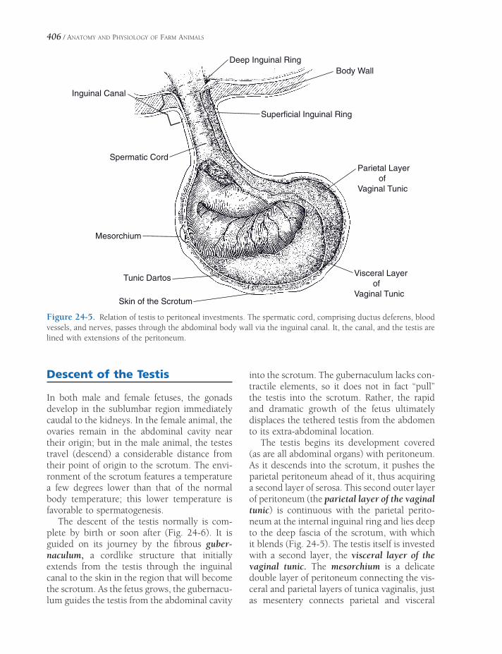
406 / ANATOMY AND PHYSIOLOGY OF FARM ANIMALS
Deep Inguinal Ring
Superficial Inguinal Ring
Body Wall
Parietal Layer ofVaginal Tunic
Visceral Layer ofVaginal Tunic
Tunic Dartos
Skin of the Scrotum
Mesorchium
Spermatic Cord
Inguinal Canal
Figure 24-5. Relation of testis to peritoneal investments. The spermatic cord, comprising ductus deferens, blood vessels, and nerves, passes through the abdominal body wall via the inguinal canal. It, the canal, and the testis are lined with extensions of the peritoneum.
Descent of the Testis
In both male and female fetuses, the gonads develop in the sublumbar region immediately caudal to the kidneys. In the female animal, the ovaries remain in the abdominal cavity near their origin; but in the male animal, the testes travel (descend) a considerable distance from their point of origin to the scrotum. The envi-ronment of the scrotum features a temperature a few degrees lower than that of the normal body temperature; this lower temperature is favorable to spermatogenesis.
The descent of the testis normally is com-plete by birth or soon after (Fig. 24-6). It is guided on its journey by the fi brous guber-naculum, a cordlike structure that initially extends from the testis through the inguinal canal to the skin in the region that will become the scrotum. As the fetus grows, the gubernacu-lum guides the testis from the abdominal cavity
into the scrotum. The gubernaculum lacks con-tractile elements, so it does not in fact “pull” the testis into the scrotum. Rather, the rapid and dramatic growth of the fetus ultimately displaces the tethered testis from the abdomen to its extra-abdominal location.
The testis begins its development covered (as are all abdominal organs) with peritoneum. As it descends into the scrotum, it pushes the parietal peritoneum ahead of it, thus acquiring a second layer of serosa. This second outer layer of peritoneum (the parietal layer of the vaginal tunic) is continuous with the parietal perito-neum at the internal inguinal ring and lies deep to the deep fascia of the scrotum, with which it blends (Fig. 24-5). The testis itself is invested with a second layer, the visceral layer of the vaginal tunic. The mesorchium is a delicate double layer of peritoneum connecting the vis-ceral and parietal layers of tunica vaginalis, just as mesentery connects parietal and visceral
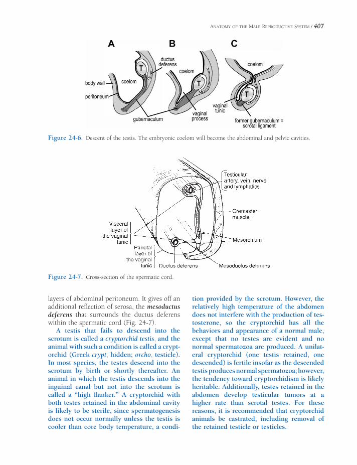
ANATOMY OF THE MALE REPRODUCTIVE SYSTEM / 407
layers of abdominal peritoneum. It gives off an additional refl ection of serosa, the mesoductus deferens that surrounds the ductus deferens within the spermatic cord (Fig. 24-7).
A testis that fails to descend into the scrotum is called a cryptorchid testis, and the animal with such a condition is called a crypt-orchid (Greek crypt, hidden; orcho, testicle). In most species, the testes descend into the scrotum by birth or shortly thereafter. An animal in which the testis descends into the inguinal canal but not into the scrotum is called a “high fl anker.” A cryptorchid with both testes retained in the abdominal cavity is likely to be sterile, since spermatogenesis does not occur normally unless the testis is cooler than core body temperature, a condi-
tion provided by the scrotum. However, the relatively high temperature of the abdomen does not interfere with the production of tes-tosterone, so the cryptorchid has all the behaviors and appearance of a normal male, except that no testes are evident and no normal spermatozoa are produced. A unilat-eral cryptorchid (one testis retained, one descended) is fertile insofar as the descended testis produces normal spermatozoa; however, the tendency toward cryptorchidism is likely heritable. Additionally, testes retained in the abdomen develop testicular tumors at a higher rate than scrotal testes. For these reasons, it is recommended that cryptorchid animals be castrated, including removal of the retained testicle or testicles.
Figure 24-6. Descent of the testis. The embryonic coelom will become the abdominal and pelvic cavities.
Figure 24-7. Cross-section of the spermatic cord.

408 / ANATOMY AND PHYSIOLOGY OF FARM ANIMALS
Castration
Castration is a term usually applied to removal of the testes of the male animal, although tech-nically it can apply to ovariectomy (removal of the ovaries) of the female animal as well. Cas-tration of male animals is practiced primarily to prevent those of inferior quality from repro-ducing. Except for colts that exhibit unusual quality meriting reproduction, most male horses are castrated to improve their tractability and utility as performance animals. Early cas-tration also improves the quality of meat animals by inhibiting undesirable secondary sex char-acteristics (notably the failure to develop mar-bling of muscle). Table 24-1 lists the common terms for intact and castrated male animals.
Sometimes a horse is referred to as being proud cut, meaning that the horse behaves like a stallion in spite of having been cas-trated. It has been believed in some circles that this results from the failure to remove all of the epididymis and that this part of the reproductive tract somehow produces suffi -cient testosterone to give the horse the attri-butes of a stallion. This belief is common but erroneous; the epididymis is entirely incapable of producing male sex hormones. It is much more likely that one or both testes were cryptorchid and that only the epididy-mis was removed (it can look remarkably like a small testicle), leaving the cryptorchid testis to produce testosterone. Alternatively, a colt castrated correctly but relatively late in life may have already acquired physical and behavioral characteristics of an intact stallion, characteristics that may not be
wholly extinguished by removal of the testes.
Vasectomy refers to transection (usually with ligation and/or removal of a section) of the ductus deferentia (formerly called the vas def-erens, hence the name of the procedure). This procedure prevents delivery of spermatozoa from the epididymides but has no effect on the production of the male hormones. The behav-ior and appearance of the vasectomized animal, therefore, are that of the intact male. Vasectomy is sometimes used experimentally or to produce teaser animals used to identify females in heat.
Accessory Sex Glands
The male accessory sex glands produce the bulk of the ejaculate, or semen, the medium for transport of sperm. Semen provides favor-able conditions for nutrition of sperm and acts as a buffer against the natural acidity of the female genital tract. The accessory sex glands include the ampulla of the ductus deferens, vesicular gland, prostate gland, and bulboure-thral gland. Except for the prostate, these glands are paired. There is considerable variation in shape and size of the various accessory sex glands among species, but the relative location is similar in all animals (Figs. 24-1 and 24-8).
Ampullae
The ampullae are glandular enlargements asso-ciated with the terminal parts of the ductus deferentia. They are well developed in the stal-
Table 24-1. Common Terms for Male Animals
Species Intact Adult Male Castrated Male
Horse Stallion Gelding
Ox Bull Steer
Pig Boar Barrow
Sheep Ram Wether
Goat Buck (billy) Wether
Chicken Rooster Capon

ANATOMY OF THE MALE REPRODUCTIVE SYSTEM / 409
lion, bull, and ram and absent in the boar. Glands of the ampullae empty into the ductus deferentia and contribute volume to the semen.
Vesicular Glands
The vesicular glands (formerly called seminal vesicles) are paired glands associated with the genital fold. In most domestic species, each vesicular gland merges with the ipsilateral ductus deferens, creating the short ejaculatory duct, which empties into the pelvic urethra. In the boar, the vesicular glands open into the urethra separately from the ductus deferentia. The vesicular glands of the stallion are hollow, pear-shaped sacs; those of the bull, ram, and boar are lobulated glands of considerable size.
Prostate Gland
The prostate gland is an unpaired gland that more or less surrounds the pelvic urethra. In farm animals the prostate gland comprises various combinations of diffuse and compact parts extending along the pelvic urethra under cover of the urethral muscle. The multiple ducts of the prostate gland open in two parallel
rows, one on each side of the lumen of the urethra. The prostate produces an alkaline secretion that gives semen its characteristic odor. In older intact male animals, the prostate may become enlarged and interfere with urination.
Bulbourethral Glands
The bulbourethral (formerly Cowper’s) glands are paired glands on either side of the pelvic urethra just cranial to the ischial arch but caudal to the other accessory glands. Bulbourethral glands are especially large in the boar.
Penis
The male organ of copulation, the penis, may be divided into three general areas: the glans, or free extremity; the main portion, or body; and the two crura, or roots, that attach to the ischial arch of the pelvis (Fig. 24-9).
The bulk of the penile body’s internal struc-ture is composed of paired columns of erectile tissue, the corpora cavernosa. Each corpus cav-ernosum is replete with blood sinusoids divided by sheets of connective tissue called trabeculae. These are derived from the tunica albuginea,
ductusdeferens
ampulla
vesicular gland prostate gland bulbourethral gland
penile urethra
ureter
urinary bladdder
Figure 24-8. Accessory sex glands of the bull.

Prostate Gland
Bulbourethral Gland
Seminal Vesicle
Ampulla ofDuctus Deferens
CorpusCavernosum
Urethra
Corpus Spongiosum
Corpus Spongiosum
Corpus Cavernosum
Right TesticularArtery and Vein
Rectum
Bladder
Bulbourethral Gland
Prostate GlandRight Ductus Deferens
Transection of the penis, midshaft
Accessory Reproductive Glands; dorsal view
MALE REPRODUCTIVE ORGANS
A.
C.
B.
Figure 24-9. Anatomy of the equine penis. A) Lateral view, showing relationship of penis to body wall and pelvis. B) Dorsocaudal view of attachment of penis to pelvis and accessory sex glands. C) Cross-section of equine penis, midshaft.

ANATOMY OF THE MALE REPRODUCTIVE SYSTEM / 411
a heavy, fi broelastic capsule surrounding the penis. In species with a fi broelastic penis (rumi-nants and swine), the trabeculae form the bulk of the penis, and as a consequence, in these species the penis is fi rm when not erect. The stallion has a musculocavernous penis, with the blood sinusoids predominating over connective tissue. The equine penis therefore is fl accid when not erect.
The two crura of the penis are the proximal parts of the corpora cavernosa. They originate on the caudal surface of the ischial arch, one on each side of the symphysis of the pelvis. The ventral midline groove between the corpora cavernosa contains the penile urethra and an associated unpaired body of erectile tissue, the corpus spongiosus. The proximal continuation of the corpus spongiosum is the bulb of the penis, which lies between the crura. In most animals, the corpus spongiosum penis is con-tinuous distally with the erectile tissue of the glans.
In dressing a carcass for human consump-tion, the penis is removed. Removal of the penile crura leaves two round marks on the ischial arch. These are called pizzle eyes, and their presence is used to identify a dressed carcass as male.
The glans penis shows considerable varia-tion from species to species. The horse and sheep both have a free portion of the urethra, the urethral process, that projects beyond the glans. The bull and ram have a helmet-shaped glans, and the external urethral opening of the bull opens into a twisted groove. The penis of the boar has a twisting cranial extremity and only a small glans (Fig. 24-10).
Erection of the penis occurs when more blood enters the penis by way of the arterial supply than leaves by the veins. The increased blood volume enlarges the penis and makes it turgid. In the stallion, whose penis is musculo-cavernous, the penis becomes much larger in all dimensions upon erection. The fi broelastic penis (as found in ruminants and swine) does not increase much in diameter during erection. Instead, the chief effect of erection of the penis in these animals consists of lengthening the penis by straightening the sigmoid fl exure,
much as a bent garden hose tends to straighten when water pressure increases.
Prepuce
The prepuce is an invaginated fold of skin sur-rounding the free extremity of the penis. The outer surface is fairly typical skin, while the inner mucous membrane consists of a preputial layer lining the prepuce and a penile layer cov-ering the surface of the free extremity of the penis. The prepuce of the horse makes a double fold, so two concentric layers surround the penis when it is retracted. The prepuce of the pig has a diverticulum dorsal to the preputial orifi ce. It accumulates urine, secretions, and dead cells, which contribute to the typical odor of a mature boar.
Figure 24-10. Free end of the penis. A1) Bull penis prior to intromission; A2) bull penis following intro-mission. Spiraling of the glans is a normal consequence of erection. (Reprinted with permission of Wiley- Blackwell from Hafez E.S.E and Hafez, B. Reproduction in Farm Animals. 7th ed. Philadelphia: Lippincott, Williams & Wilkins, 2000.)

412 / ANATOMY AND PHYSIOLOGY OF FARM ANIMALS
Muscles of the Male Genitalia
The urethral muscle (m. urethralis) is the stri-ated muscle associated with the pelvic portion of the urethra. It completely encircles the urethra of the goat and horse but surrounds only the ventral and lateral aspects of the bovine, porcine, and ovine urethra. The ure-thral muscle forms the voluntary sphincter of the urinary bladder; during ejaculation, waves of peristaltic contractions in it help propel the semen through the urethra.
The extrapelvic continuation of the urethral muscle is a striated muscle called the bulbo-spongiosus muscle. It surrounds the urethra and bulb of the penis. The bulbospongiosus muscle of the horse extends from the penile bulb between the crura of the penis along the entire urethra to the glans penis. In other animals it covers the urethral bulb and extends only a short distance along the penile urethra. The bulbospongiosus muscle continues the action of the urethral muscle in emptying the urethra by peristaltic contractions.
The ischiocavernosus muscles are paired striated muscles that cover the superfi cial aspect of the crura of the penis. The two muscles converge from their origins on the lateral sides of the ischial arch toward the body of the penis. When these muscles contract, they pull the penis dorsocraniad against the bony pelvis, aiding erection by compressing much of the venous drainage from the penis.
The retractor penis muscles are paired smooth muscles that take origin from the ventral aspect of the fi rst few caudal vertebrae. These muscles pass ventrad on each side of the anal canal, then continue on the midline of the penis superfi cial to the urethra.
Blood and Nerve Supply of the Male Genitalia
The testis derives its blood supply from the testicular artery, which branches directly from the abdominal aorta a short distance caudal to the renal artery of the same side. Within the spermatic cord, the testicular artery assumes a tortuous arrangement. The artery is embedded in the likewise convoluted mass of the testicular vein, the pampiniform plexus. The artery of the ductus deferens from the internal pudendal artery supplies the ductus deferens and may also provide some arterial blood to the epididy-mis and testis.
The external pudendal artery, a branch of the deep femoral artery, passes through the inguinal canal and supplies the scrotum and prepuce. In most domestic species, a branch of the internal iliac artery, the internal pudendal artery, supplies most of the penis, urinary bladder, urethra, and accessory sex glands. The blood supply to the penis of the horse is more extensive than in other species, with additional contributions by the obturator artery, which passes through the obturator foramen of the pelvis, and the external pudendal artery after it passes through the inguinal canal.
The autonomic innervation of the testis is composed of sympathetic nerves. These auto-nomic nerve fi bers accompany the testicular artery. Sympathetic input affects activity in the blood vessels and smooth muscle fi bers. The dorsal nerve of the penis is a continuation of the pudendal nerve, which is derived from ventral branches of sacral nerves. It passes along the dorsum of the penis to ramify in the glans penis. Sensory fi bers from the glans provide the afferent limb of refl exes for erection and ejaculation.
PHYSIOLOGY OF MALE REPRODUCTION
Seminiferous Tubules and Spermatogenesis
Seminiferous Tubules
The epithelium lining the seminiferous tubules contains two cell types, the sustentacular, or Sertoli cells, and the developing spermatozoa and their precursor germ cells (Fig. 25-1).
The large sustentacular cells extend from the base of the epithelium to the lumen of the seminiferous tubules. Their shape is highly irregular because they encircle the developing germ cells (Fig. 25-1). Sustentacular cells secrete a fl uid that bathes the developing germ cells and assists with the transport of sperma-tozoa from the tubules into the rete testis after their release from the sustentacular cells. One component of this fl uid, androgen-binding protein, transports androgens from their site of synthesis in the testis to the epididmyis, where they are required for maturation of the sperma-tozoa. Interstitial cells (Leydig cells) in the connective tissue between seminiferous tubules secrete testosterone, the primary androgen originating in the testicles.
The composition of the interstitial fl uid between sustentacular cells and within the lumen of the seminiferous tubules differs from typical interstital fl uid. The differences are due in part to a selectively permeable barrier (blood–testis barrier) between these fl uids and fl uid
Seminiferous Tubules and SpermatogenesisSeminiferous Tubules
Germ Cells and Spermatogenesis
Spermatozoa Morphology and
Spermatogenesis
Rates and Timing of Spermatogenesis
EpididymisSemen and Semen TechnologyHormones of Male Reproduction
Endocrine Regulation of Testicular
Function
Testosterone and Its Effects
Erection and Ejaculation
Chapter 25
413

414 / ANATOMY AND PHYSIOLOGY OF FARM ANIMALS
outside the tubules. The cell junctions between adjacent sustentacular cells and between myoid cells that surround the tubules contribute to this barrier (Fig. 25-1). The luminal concentra-tions of cellular secretions, such as androgen-binding proteins and androgens, are higher than in typical extracellular fl uid because this functional barrier sequesters them.
Germ Cells and Spermatogenesis
Spermatogenesis is the term for all processes involved in the formation of mature male gametes from the most undifferentiated germ cells. It includes several mitotic cell divisions followed by two meiotic cell divisions, during which the chromosome number is reduced from diploid to haploid. This series of cell divisions is termed spermatocytogenesis (Fig. 25-2).
Some of the cells resulting from the mitotic cell divisions of the most undifferentiated germ cells remain at the base of the epithelium to maintain the supply of stem cells. Others begin the sequence of cell divisions (mitotic followed by meiotic divisions) and developmental changes to become spermatozoa. The mitotic cell divisions double the number of cells at each step, so a single spermatogonium gives rise to many spermatozoa.
Meiosis entails two cell divisions and occurs only during the development of gametes in the testis and ovary. Prior to the fi rst division, the DNA is replicated in a manner similar to that of mitotic cell division (see Chapter 2). This replication results in chromosomes that consist of two identical chromatids. In preparation for the fi rst meiotic division, homologous chromo-somes pair up along the middle of the cell. (Homologous chromosomes are the similar
Figure 25-1. Location and microscopic anatomy of seminiferous tubules. Note the irregular border of the sus-tentacular (Sertoli) cells and their relation to the developing germ cells. (Reprinted with permission of Wiley- Blackwell from Hafez E.S.E. and Hafez B. Reproduction in Farm Animals. 7th ed. Philadelphia: Lippincott Williams & Wilkins, 2000.)
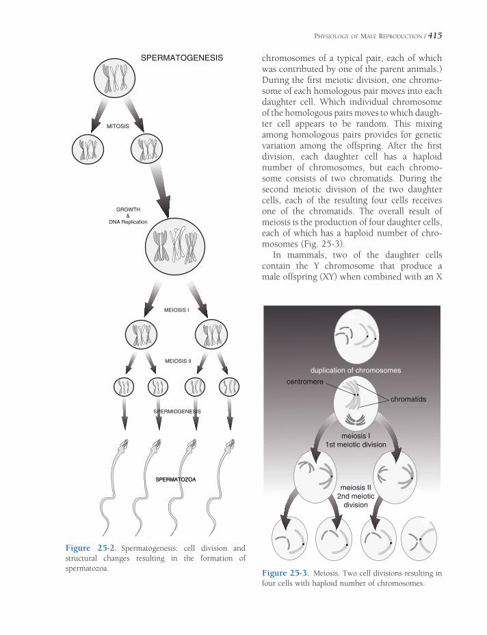
PHYSIOLOGY OF MALE REPRODUCTION / 415
chromosomes of a typical pair, each of which was contributed by one of the parent animals.) During the fi rst meiotic division, one chromo-some of each homologous pair moves into each daughter cell. Which individual chromosome of the homologous pairs moves to which daugh-ter cell appears to be random. This mixing among homologous pairs provides for genetic variation among the offspring. After the fi rst division, each daughter cell has a haploid number of chromosomes, but each chromo-some consists of two chromatids. During the second meiotic division of the two daughter cells, each of the resulting four cells receives one of the chromatids. The overall result of meiosis is the production of four daughter cells, each of which has a haploid number of chro-mosomes (Fig. 25-3).
In mammals, two of the daughter cells contain the Y chromosome that produce a male offspring (XY) when combined with an X
Figure 25-2. Spermatogenesis: cell division and structural changes resulting in the formation of spermatozoa.
duplication of chromosomes
chromatids
centromere
meiosis I1st meiotic division
meiosis II2nd meiotic
division
2N = 4
Figure 25-3. Meiosis. Two cell divisions resulting in four cells with haploid number of chromosomes.
SPERMIOGENESIS
MEIOSIS I
MEIOSIS II
SPERMATOGENESIS
MITOSIS
GROWTH
DNA Replication&
SPERMATOZOASPERMATOZOA

416 / ANATOMY AND PHYSIOLOGY OF FARM ANIMALS
chromosome from an ovum. The other two contain the X chromosome to produce female offspring (XX) when united with the X chromosome –containing ovum. However, in avian species, all spermatozoa contain the same sex chromosome, known in these species as the Z chromosome. Female birds carry one Z and a second sex chromosome, the W chromosome. An ovum may contain either a W or Z chromo-some, so the ovum is the gamete that deter-mines the sex of the offspring in birds.
When homologous chromosomes are paired in preparation for the fi rst meiotic division, crossing over may occur. During this process, similar regions of chromosomes may be exchanged between homologous chromosomes. Such exchanges further increase genetic vari-ability among the offspring, for they may now inherit chromosomes that are different from either parent chromosome.
Spermatid is the term for the cells resulting from the second meiotic division in the semi-niferous tubules. Spermatids undergo a series of functional and structural changes to become spermatozoa, and this process is termed sper-miogenesis (Fig. 25-2). There is no further cel-lular division after second meiotic division, so there is no further increase in cell number after that division.
Spermatozoa Morphology and Spermatogenesis
When fi rst produced, spermatids are round immobile cells, while fully formed spermatozoa are mobile and consist of a head and tail (Fig. 25-4). The head contains the nucleus, primarily consisting of condensed genetic material (DNA), and an acrosome, a membranous sac that lies immediately under the plasma membrane at the tip of the head and extends down over the nucleus (Fig. 25-5). The tail has a central core of microtubules and fi laments that provide motility. The middle piece of the tail contains a dense collection of mitochondria that provide energy for spermatozoan motility.
The acrosome is essentially a membranous sac of hydrolytic enzymes, including acrosin
Figure 25-4. Comparison of the spermatozoa of farm animals and other vertebrates. (Reprinted with permis-sion of Wiley-Blackwell from Hafez E.S.E. and Hafez B. Reproduction in Farm Animals. 7th ed. Philadelphia: Lippincott Williams & Wilkins, 2000.)
and hyaluronidase. Some of these enzymes are released from the acrosome during fertilization and facilitate the fusion of the male and female gametes. The release and exposure of these enzymes during fertilization is termed the acro-some reaction.
The conversion of the round spermatid to the elongated spermatozoon with its acrosome containing head and tail requires reshaping of the original spermatid, synthesis and packaging of acrosomal enzymes, organization of micro-tubules, and elimination of excess cytoplasm and cell membrane. The sustentacular cells, in which the developing spermatozoa are embed-ded, assist with some of these conversions. For example, sustentacular cells phagocytose resid-ual bodies of excess cytoplasm and membrane after the release of fully formed spermatozoa.
Rates and Timing of Spermatogenesis
Daily production of spermatozoa has been esti-mated as 4.4 × 10⁹ in the ram and 2 × 10⁹ in the bull. Eight ejaculations of the bull within an hour reduced the semen from 4.2 mL at the fi rst collection to 2.9 mL at the eighth collec-tion, and the number of spermatozoa was cor-respondingly reduced from 1.7 billion to 98 million per milliliter. These data indicate that

PHYSIOLOGY OF MALE REPRODUCTION / 417
normal animals produce adequate spermatozoa even when used often for breeding. However, these data also suggest that several days of sexual rest may increase numbers of spermato-zoa in animals whose numbers are abnormally low. A correlation between daily production and testicular size has been found for several species.
The time required for spermatogenesis (from spermatogonium to fully formed and released spermatozoa) varies with species, but it is a matter of weeks to months rather than days. In most farm animals, this period is about 2 months.
Epididymis
Fully formed spermatozoa released from the seminiferous tubules pass through the rete testis into the epididymis. The epididymides are the major sites of storage of spermatozoa, and most spermatozoa are contained in the tail of each epididymis.
Spermatozoa entering the head of the epi-didymis from the rete testis are immotile and incapable of fertilization. During their passage through the epididymis, spermatozoa acquire motility and become capable of fertilization. Presumably, these two characteristics are closely linked. The functional changes in spermatozoa that occur during their passage through the epididymis depend in part on epididymal secre-tions and testosterone in the epididymal fl uids. Some minor morphologic changes (e.g., changes in nuclear chromatin and the acrosome) may also occur during the passage through the epididymis.
Semen and Semen Technology
Semen consists of spermatozoa suspended in the fl uid secretions of the male accessory sex organs. The fl uid portion of semen (seminal plasma) functions as a transport medium for the spermatozoa, and it contains a variety of substances including various electrolytes, fruc-tose, citric acid, and sorbitol. The fructose (a sugar) is a potential source of energy for the spermatozoa.
Semen is collected and evaluated as part of protocols to evaluate the fertility of breeding males, but no single characteristic of semen or spermatozoa is accepted as a perfect gold stan-dard for predicting the fertility of a given sample of semen. Some characteristics of semen that are evaluated and appear to have some correla-tion with potential fertility when considered together are (1) concentration of spermatozoa per milliliter of semen, (2) motility characteris-tics of spermatozoa, and (3) shape of the sper-matozoa. The concentrations of spermatozoa per milliliter vary among species, and this should be considered when evaluating semen.
Figure 25-5. A sagittal section of a bovine sperm head showing the various anatomic subdivisions. (Reprinted with permission of Wiley-Blackwell from Hafez E.S.E. and Hafez B. Reproduction in Farm Animals. 7th ed. Philadelphia: Lippincott Williams & Wilkins, 2000.)
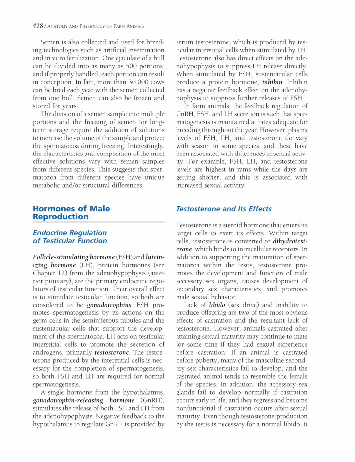
418 / ANATOMY AND PHYSIOLOGY OF FARM ANIMALS
Semen is also collected and used for breed-ing technologies such as artifi cial insemination and in vitro fertilization. One ejaculate of a bull can be divided into as many as 500 portions, and if properly handled, each portion can result in conception. In fact, more than 30,000 cows can be bred each year with the semen collected from one bull. Semen can also be frozen and stored for years.
The division of a semen sample into multiple portions and the freezing of semen for long-term storage require the addition of solutions to increase the volume of the sample and protect the spermatozoa during freezing. Interestingly, the characteristics and composition of the most effective solutions vary with semen samples from different species. This suggests that sper-matozoa from different species have unique metabolic and/or structural differences.
Hormones of Male Reproduction
Endocrine Regulation of Testicular Function
Follicle-stimulating hormone (FSH) and lutein-izing hormone (LH), protein hormones (see Chapter 12) from the adenohypophysis (ante-rior pituitary), are the primary endocrine regu-lators of testicular function. Their overall effect is to stimulate testicular function, so both are considered to be gonadotrophins. FSH pro-motes spermatogenesis by its actions on the germ cells in the seminiferous tubules and the sustentacular cells that support the develop-ment of the spermatozoa. LH acts on testicular interstitial cells to promote the secretion of androgens, primarily testosterone. The testos-terone produced by the interstitial cells is nec-essary for the completion of spermatogenesis, so both FSH and LH are required for normal spermatogenesis.
A single hormone from the hypothalamus, gonadotrophin-releasing hormone (GnRH), stimulates the release of both FSH and LH from the adenohypophysis. Negative feedback to the hypothalamus to regulate GnRH is provided by
serum testosterone, which is produced by tes-ticular interstitial cells when stimulated by LH. Testosterone also has direct effects on the ade-nohypophysis to suppress LH release directly. When stimulated by FSH, sustentacular cells produce a protein hormone, inhibin. Inhibin has a negative feedback effect on the adenohy-pophysis to suppress further releases of FSH.
In farm animals, the feedback regulation of GnRH, FSH, and LH secretion is such that sper-matogenesis is maintained at rates adequate for breeding throughout the year. However, plasma levels of FSH, LH, and testosterone do vary with season in some species, and these have been associated with differences in sexual activ-ity. For example, FSH, LH, and testosterone levels are highest in rams while the days are getting shorter, and this is associated with increased sexual activity.
Testosterone and Its Effects
Testosterone is a steroid hormone that enters its target cells to exert its effects. Within target cells, testosterone is converted to dihydrotest-erone, which binds to intracellular receptors. In addition to supporting the maturation of sper-matozoa within the testis, testosterone pro-motes the development and function of male accessory sex organs, causes development of secondary sex characteristics, and promotes male sexual behavior.
Lack of libido (sex drive) and inability to produce offspring are two of the most obvious effects of castration and the resultant lack of testosterone. However, animals castrated after attaining sexual maturity may continue to mate for some time if they had sexual experience before castration. If an animal is castrated before puberty, many of the masculine second-ary sex characteristics fail to develop, and the castrated animal tends to resemble the female of the species. In addition, the accessory sex glands fail to develop normally if castration occurs early in life, and they regress and become nonfunctional if castration occurs after sexual maturity. Even though testosterone production by the testis is necessary for a normal libido, it

PHYSIOLOGY OF MALE REPRODUCTION / 419
is not testosterone that directly affects neurons within the brain to produce a normal libido. Within neurons, testosterone is converted to estradiol, an estrogen, and it is this estradiol that actually stimulates the appropriate neurons. Anabolic steroids (discussed next) used to promote growth cannot be converted to estra-diol and thus these do not increase the libido.
Anabolic steroids are synthetic com-pounds used to increase net protein synthe-sis and skeletal muscle mass. In this manner, these compounds are similar to endogenous androgens such as testosterone. Because of their similarity to testosterone and endoge-nous androgens, anabolic steroids also promote the development of secondary sexual characteristics and exert a negative feedback effect on the hypothalamic–pitu-itary axis. As a result of this negative feed-back, endogenous testosterone production and spermatogenesis are suppressed. It is not clear whether these can return to normal levels if animals receive anabolic steroids for prolonged periods.
Erection and Ejaculation
Penile erection is a neural refl ex initiated by appropriate tactile stimulation of the penis, visual or environmental stimuli (such as a female in estrus), or as a result of learned behavior. For example, an erection may begin when a breeding stallion is being led to a breed-
ing area but before a mare is present. This is believed to be a learned response. Similarly, it is believe that learned responses may be respon-sible for lack of libido in certain males that have undergone trauma (e.g., a stallion kicked by a mare) or that associate mating with some other painful or unpleasant event.
Penile erection requires vasodilation within the penis, which results in an increased blood fl ow into the penis. Parasympathetic nerves innervating blood vessels within the penis initi-ate the vasodilation. Studies in experimental animals suggest that the parasympathetic nerves release acetylcholine, which in turn stimulates endothelial cells lining the blood vessels to release nitric oxide. Nitric oxide acts directly on vascular smooth muscle to bring about vasodilation.
Prior to ejaculation, spermatozoa are moved from storage sites in the epididymides through the ductus deferentia to the pelvic urethra. This movement is termed emission and is assumed to result from contractions of smooth muscle in the wall of these tubular structures. Within the pelvic urethra, secretions of the accessory sex glands are mixed with the spermatozoa. Ejaculation of the mixture (semen) through the penile urethra is associated with more contrac-tions of the epididymides and ductus deferentia and additional contractions by muscles of the penis surrounding the penile urethra. Emission and ejaculation are autonomic refl exes involv-ing both the sympathetic and parasympathetic divisions.

ANATOMY OF THE FEMALE REPRODUCTIVE SYSTEM
The mammalian female reproductive tract produces the female gamete (ovum, pl.
ova), delivers it to a site where it can be fertil-ized by the male gamete (spermatozoon), pro-vides an environment for the development and growth of the embryo, and expels the fetus when it is capable of survival outside of the mother’s body. The female organs of reproduc-tion include two ovaries, two uterine tubes (also called oviducts), the uterus, the vagina, and the vulva (Fig. 26-1). The ovum is released from the ovary and enters the open end of the uterine tube. Fertilization normally occurs within the uterine tube during passage of the ovum from the ovary to the uterus. Within the uterus, the fertilized ovum, now a zygote, develops into an embryo and then into a fetus and fi nally passes out of the uterus through the vagina and vulva as a newborn (neonate). Table 26-1 compares the anatomies of the reproduc-tive tracts in adult nonpregnant farm animals.
Ovaries
The ovaries, like the testes in the male, are the primary organs of reproduction in the female. The ovaries are both endocrine and cytogenic (cell producing) as they produce hormones, which are released directly into the blood stream, and ova, which are released from the surface of the ovary during ovulation (see Chapter 27).
OvariesUterine TubesUterusVaginaVestibule and VulvaBlood and Nerve Supply of the Female
Reproductive Tract
Chapter 26
421

422 / ANATOMY AND PHYSIOLOGY OF FARM ANIMALS
The ovaries are paired glands usually found in the lumbar region of the abdominal cavity, a short distance caudal to the kidneys. Like all abdominal organs, the ovaries are covered with peritoneum. They are suspended from the body wall by a refl ection of this serous membrane, the mesovarium, the most cranial part of the peritoneal investments of the female genital tract.
In most species, the ovaries are somewhat ovoid (Fig. 26-2). In the mare, however, the ovaries have a bean shape because of a defi nite ovulation fossa, an indentation in the attached border of the ovary. Ovaries of the sow usually appear lobulated because of the numerous devel-oping ova; the sow is the only common farm animal that typically produces a litter, rather than one or two offspring per pregnancy.
FEMALE REPRODUCTIVE TRACT
Uterine Horns
BroadLigament
Vaginal Fornix
Urethral Opening
MARE COW
Vestibule
Vagina
Cervix
Ovary
Uterine Tube
Uterine Horns
BroadLigament
UterineBody
UterineBody
Vaginal Fornix
Figure 26-1. Anatomy of the female reproductive tract.
When palpated through the wall of the rectum, an ovary feels solid because of the large amount of connective tissue that makes up the stroma of the gland. Normal size of the ovary varies considerably from species to species, and even within a species there is some variation. For example, the ovary of the mare may be less than 2.5 cm in diameter when no developing ova are present or as large as 10 cm with many developing ova.
The ovary is invested in a dense connective tissue capsule, the tunica albuginea. The medulla, or central portion, of the ovary is the most vascular part, while the cortex, or outer portion, consists largely of dense, irregular con-nective tissue interspersed with follicles (devel-oping ova; see Fig. 27-1) and interstitial cells, which have an endocrine function.
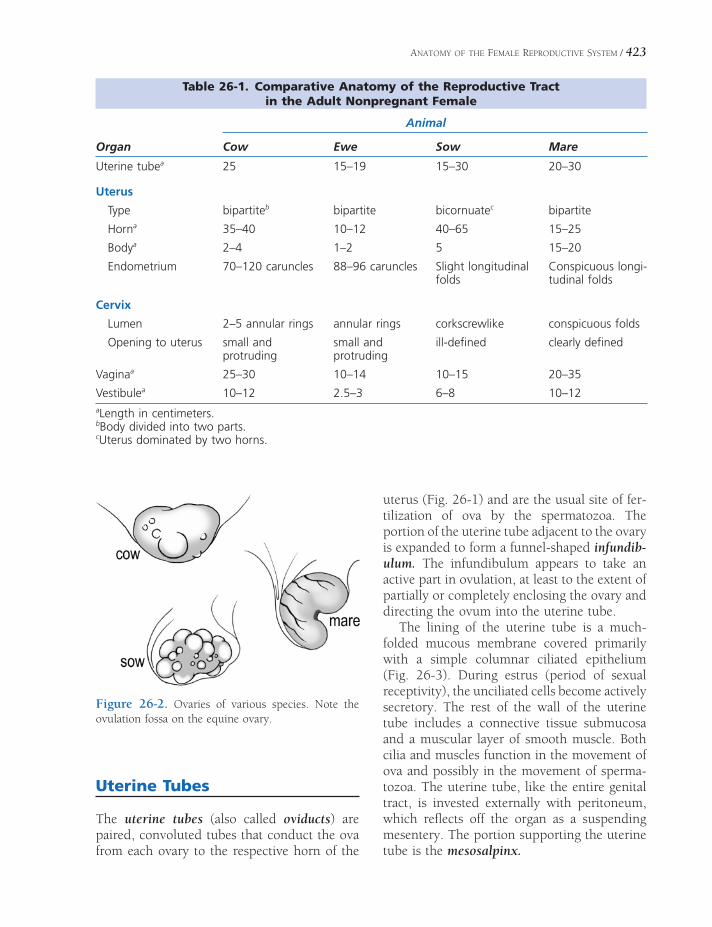
ANATOMY OF THE FEMALE REPRODUCTIVE SYSTEM / 423
Figure 26-2. Ovaries of various species. Note the ovulation fossa on the equine ovary.
Table 26-1. Comparative Anatomy of the Reproductive Tract in the Adult Nonpregnant Female
Animal
Organ Cow Ewe Sow Mare
Uterine tubea 25 15–19 15–30 20–30
Uterus
Type bipartiteb bipartite bicornuatec bipartite
Horna 35–40 10–12 40–65 15–25
Bodya 2–4 1–2 5 15–20
Endometrium 70–120 caruncles 88–96 caruncles Slight longitudinal folds
Conspicuous longi-tudinal folds
Cervix
Lumen 2–5 annular rings annular rings corkscrewlike conspicuous folds
Opening to uterus small and protruding
small and protruding
ill-defi ned clearly defi ned
Vaginaa 25–30 10–14 10–15 20–35
Vestibulea 10–12 2.5–3 6–8 10–12aLength in centimeters.bBody divided into two parts.cUterus dominated by two horns.
Uterine Tubes
The uterine tubes (also called oviducts) are paired, convoluted tubes that conduct the ova from each ovary to the respective horn of the
uterus (Fig. 26-1) and are the usual site of fer-tilization of ova by the spermatozoa. The portion of the uterine tube adjacent to the ovary is expanded to form a funnel-shaped infundib-ulum. The infundibulum appears to take an active part in ovulation, at least to the extent of partially or completely enclosing the ovary and directing the ovum into the uterine tube.
The lining of the uterine tube is a much-folded mucous membrane covered primarily with a simple columnar ciliated epithelium (Fig. 26-3). During estrus (period of sexual receptivity), the unciliated cells become actively secretory. The rest of the wall of the uterine tube includes a connective tissue submucosa and a muscular layer of smooth muscle. Both cilia and muscles function in the movement of ova and possibly in the movement of sperma-tozoa. The uterine tube, like the entire genital tract, is invested externally with peritoneum, which refl ects off the organ as a suspending mesentery. The portion supporting the uterine tube is the mesosalpinx.
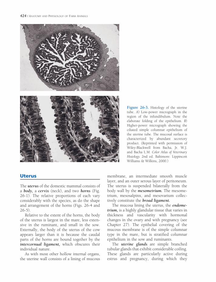
424 / ANATOMY AND PHYSIOLOGY OF FARM ANIMALS
Figure 26-3. Histology of the uterine tube. A) Low-power micrograph in the region of the infundibulum. Note the elaborate folding of the epithelium. B) Higher-power micrograph showing the ciliated simple columnar epithelium of the uterine tube. The mucosal surface is characterized by abundant secretory product. (Reprinted with permission of Wiley-Blackwell from Bacha, Jr. W.J. and Bacha L.M. Color Atlas of Veterinary Histology. 2nd ed. Baltimore: Lippincott Williams & Wilkins, 2000.)
Uterus
The uterus of the domestic mammal consists of a body, a cervix (neck), and two horns (Fig. 26-1). The relative proportions of each vary considerably with the species, as do the shape and arrangement of the horns (Figs. 26-4 and 26-5).
Relative to the extent of the horns, the body of the uterus is largest in the mare, less exten-sive in the ruminant, and small in the sow. Externally, the body of the uterus of the cow appears larger than it is because the caudal parts of the horns are bound together by the intercornual ligament, which obscures their individual nature.
As with most other hollow internal organs, the uterine wall consists of a lining of mucous
membrane, an intermediate smooth muscle layer, and an outer serous layer of peritoneum. The uterus is suspended bilaterally from the body wall by the mesometrium. The mesome-trium, mesosalpinx, and mesovarium collec-tively constitute the broad ligament.
The mucosa lining the uterus, the endome-trium, is a highly glandular tissue that varies in thickness and vascularity with hormonal changes in the ovary and with pregnancy (see Chapter 27). The epithelial covering of the mucous membrane is of the simple columnar type in the mare, but is stratifi ed columnar epithelium in the sow and ruminants.
The uterine glands are simple branched tubular glands that exhibit considerable coiling. These glands are particularly active during estrus and pregnancy, during which they
A
B

ANATOMY OF THE FEMALE REPRODUCTIVE SYSTEM / 425
Figure 26-4. Comparative shapes of animal uteri. The female dog (bitch) is provided for comparison.
produce a fl uid colloquially known as uterine milk. These glands are scattered throughout the endometrium of the uterus, except in rumi-nants, in which the caruncles are nonglandular. Caruncles are mushroomlike projections from the inner surface of the uteri of ruminants; they provide a site of attachment for the fetal mem-branes (see Chapter 28).
The cervix of the uterus projects caudally into the vagina. The cervix is a heavy, smooth muscle sphincter that is tightly closed except during estrus and parturition. During estrus the cervix relaxes slightly, permitting spermatozoa to enter the uterus. In ruminants, and to some extent in sows, the inner surface of the cervix
is arranged in a series of circular ridges or rings, sometimes called annular folds (Fig. 26-6). The cervix of the mare is relatively smooth and projects prominently into the vagina, which surrounds the cervix as a deep vaginal fornix.
The tunica muscularis is the muscular portion of the uterine wall, commonly called the myometrium. It consists of a thick inner circular layer of smooth muscle and a thinner outer longitudinal layer of smooth muscle, separated by a vascular layer. During pregnancy, the amount of muscle in the uterine wall increases dramatically, both in cell size (hyper-trophy) and in cell numbers (hyperplasia).
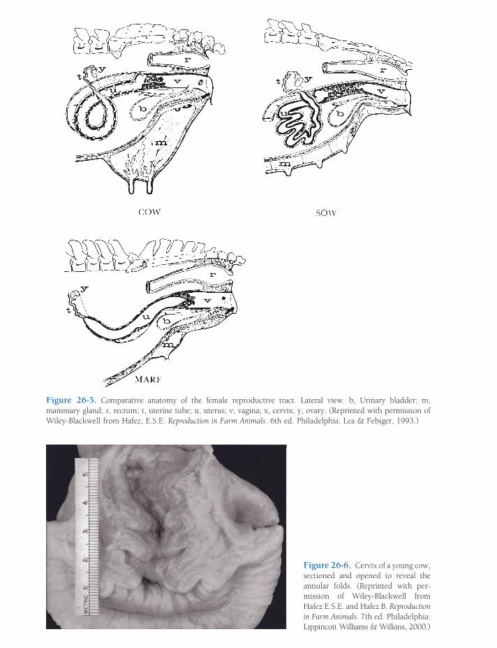
Figure 26-6. Cervix of a young cow, sectioned and opened to reveal the an nular folds. (Reprinted with per-mission of Wiley-Blackwell from Hafez E.S.E. and Hafez B. Reproduction in Farm Animals. 7th ed. Philadelphia: Lippincott Williams & Wilkins, 2000.)
Figure 26-5. Comparative anatomy of the female reproductive tract. Lateral view. b, Urinary bladder; m, mammary gland; r, rectum; t, uterine tube; u, uterus; v, vagina; x, cervix; y, ovary. (Reprinted with permission of Wiley-Blackwell from Hafez, E.S.E. Reproduction in Farm Animals. 6th ed. Philadelphia: Lea & Febiger, 1993.)
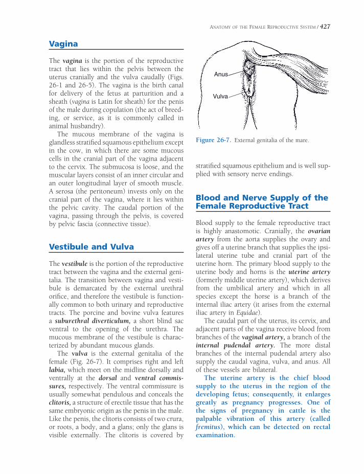
ANATOMY OF THE FEMALE REPRODUCTIVE SYSTEM / 427
Vagina
The vagina is the portion of the reproductive tract that lies within the pelvis between the uterus cranially and the vulva caudally (Figs. 26-1 and 26-5). The vagina is the birth canal for delivery of the fetus at parturition and a sheath (vagina is Latin for sheath) for the penis of the male during copulation (the act of breed-ing, or service, as it is commonly called in animal husbandry).
The mucous membrane of the vagina is glandless stratifi ed squamous epithelium except in the cow, in which there are some mucous cells in the cranial part of the vagina adjacent to the cervix. The submucosa is loose, and the muscular layers consist of an inner circular and an outer longitudinal layer of smooth muscle. A serosa (the peritoneum) invests only on the cranial part of the vagina, where it lies within the pelvic cavity. The caudal portion of the vagina, passing through the pelvis, is covered by pelvic fascia (connective tissue).
Vestibule and Vulva
The vestibule is the portion of the reproductive tract between the vagina and the external geni-talia. The transition between vagina and vesti-bule is demarcated by the external urethral orifi ce, and therefore the vestibule is function-ally common to both urinary and reproductive tracts. The porcine and bovine vulva features a suburethral diverticulum, a short blind sac ventral to the opening of the urethra. The mucous membrane of the vestibule is charac-terized by abundant mucous glands.
The vulva is the external genitalia of the female (Fig. 26-7). It comprises right and left labia, which meet on the midline dorsally and ventrally at the dorsal and ventral commis-sures, respectively. The ventral commissure is usually somewhat pendulous and conceals the clitoris, a structure of erectile tissue that has the same embryonic origin as the penis in the male. Like the penis, the clitoris consists of two crura, or roots, a body, and a glans; only the glans is visible externally. The clitoris is covered by
Anus
Vulva
Figure 26-7. External genitalia of the mare.
stratifi ed squamous epithelium and is well sup-plied with sensory nerve endings.
Blood and Nerve Supply of the Female Reproductive Tract
Blood supply to the female reproductive tract is highly anastomotic. Cranially, the ovarian artery from the aorta supplies the ovary and gives off a uterine branch that supplies the ipsi-lateral uterine tube and cranial part of the uterine horn. The primary blood supply to the uterine body and horns is the uterine artery (formerly middle uterine artery), which derives from the umbilical artery and which in all species except the horse is a branch of the internal iliac artery (it arises from the external iliac artery in Equidae).
The caudal part of the uterus, its cervix, and adjacent parts of the vagina receive blood from branches of the vaginal artery, a branch of the internal pudendal artery. The more distal branches of the internal pudendal artery also supply the caudal vagina, vulva, and anus. All of these vessels are bilateral.
The uterine artery is the chief blood supply to the uterus in the region of the developing fetus; consequently, it enlarges greatly as pregnancy progresses. One of the signs of pregnancy in cattle is the palpable vibration of this artery (called fremitus), which can be detected on rectal examination.
428 / ANATOMY AND PHYSIOLOGY OF FARM ANIMALS
Venous drainage of the female reproductive tract is via veins that are satellite to the arteries and that ultimately drain to the caudal vena cava. In ruminants, the ovarian artery and the uterine vein run close together, providing a venoarterial pathway for chemical messengers from the uterus to reach the ovary on the same side. This relationship underlies certain physi-ologic phenomena of ruminant reproduction (see Chapter 27).
Sympathetic autonomic innervation to the female reproductive tract arrives from the caudal mesenteric ganglion via hypogastric nerves. Parasympathetic fi bers arise from sacral spinal cord segments and travel to the repro-ductive organs in the pelvic nerves. The puden-dal and perineal nerves, somatic nerves from sacral spinal cord segments, provide motor and sensory innervation, respectively, to the exter-nal genitalia.
THE OVARY AND ESTROUS CYCLES
The ovaries are the source of mature female gametes (ova) and hormones necessary for
reproduction. Oogenesis is formation of ova. Estrus (heat) is the period of sexual receptivity in the female. An estrous cycle is the interval from the beginning of one estrus to the begin-ning of the next.
Animals that have only one estrous cycle per year are called monestrous animals, while those that have several estrous cycles per year are polyestrous. Many animals have successive periods of estrus if a pregnancy is not estab-lished and maintained. The timing of these periods is often determined by seasonal changes in length of day, so these animals are seasonally polyestrous. The relatively long period of inac-tivity in seasonally polyestrous animals is termed anestrus and is not properly a part of the sexual cycle.
Oogenesis
In the fetus, primordial germ cells migrate from the yolk sac to the developing ovaries, where a single layer of follicular cells surrounds a germ cell destined to become an ovum. The central germ cell (now termed an oogonium) enlarges and begins meiosis. (Recall that meiosis entails two cell divisions during which the diploid number of chromosomes is reduced by half to the haploid number.) The oogonium does not complete meiosis; it stops in the fi rst
OogenesisSecondary Follicles
Hormones and Follicular Development
OvulationLuteinizing Hormone Surge
Spontaneous and Reflex Ovulators
Corpus LuteumPhases of the Estrous Cycle
Proestrus
Estrus
Metestrus
Diestrus and Anestrus
Puberty
Specifics of Selected Estrous CyclesMare
Cow
Ewe
Sow
Chapter 27
429

430 / ANATOMY AND PHYSIOLOGY OF FARM ANIMALS
prophase before the fi rst division. At this stage, the developing ovum is a primary oocyte, and the combination of a primary oocyte and its surrounding cuboidal follicular cell (granulosa cell) layer is a primary follicle (Fig. 27-1). At birth, the ovaries of most domestic species contain hundreds of thousands of primary fol-licles waiting to continue their development. What determines which of the thousands of primary follicles is selected to develop further during a specific estrous cycle is unknown.
In contrast to spermatogenesis, which pro-duces four spermatozoa from each primary germ cell, the maturation of the primary oocyte results in only one mature ovum and three rudimentary cells, called polar bodies. In most animals, the fi rst of the two meiotic divisions is complete, resulting in the formation of the fi rst polar body, before or immediately after ovulation (the discharge of an oocyte from a follicle).
Secondary Follicles
In all animals, multiple primary follicles typi-cally begin further development during a single estrous cycle. In monotocous animals (animals not bearing litters and normally having only one offspring per gestation, such as the mare and cow), one follicle usually develops more rapidly than others, and only one ovum is released at ovulation. The rest of the developing follicles regress and form atretic follicles. Polytocous animals, such as carnivores and swine, which normally produce two or more offspring per gestation, usually have several fol-licles that develop and ovulate at approximately the same time. The ova may all come from one ovary, or some may come from each ovary.
The further development of primary follicles includes enlargement of the oocyte and replica-tion of the surrounding follicular cells. The replicating follicular cells become several layers
Figure 27-1. Sagittal section of an ovary showing the origin, growth, and ovulation of a follicle and a corpus luteum developing at the site of a follicle that has ovulated. A, Primary follicle; B, growing follicle; C, Graafi an or tertiary follicle; D, ovulation; E, corpus luteum. (Reprinted with permission of Wiley-Blackwell from Reece W.O. Physiology of Domestic Animals. 2nd ed. Baltimore: Williams & Wilkins, 1997.)

THE OVARY AND ESTROUS CYCLES / 431
thick, and this surrounding group of cells is a granulosa. The granulosa cells secrete glyco-proteins that cross-link to form a protective shell, the zona pellucida, around the oocyte (Fig. 27-1). Cytoplasmic processes of granulosa cells penetrate the zona to permit communica-tion and exchange between them and the oocyte. The initial development to this point is independent of hormonal stimulation by gonadotrophins (follicle-stimulating hormone [FSH] and luteinizing hormone [LH]).
The developing follicle is termed a second-ary follicle when the oocyte has enlarged and is surrounded by a developing granulosa. A theca, consisting of layers of cells immediately surrounding the granulosa, also fi rst develops late during the secondary follicle stage.
Hormones and Follicular Development
Gonadotrophin-releasing hormone (GnRH) is released from the hypothalamus to promote the release of both FSH and LH from the adenohy-pophysis. The release of GnRH can be modu-lated by steroid (estradiol and progesterone) and peptide (inhibin) hormones from the ovary, but its basal release is determined by neural inputs to the hypothalamus. The basal release of GnRH is assumed to be pulsatile, for this is the type of release seen in ovariectomized animals. The pulsatile nature is physiologically important, because continuous infusions of GnRH do not result in the continuous release of FSH and LH.
The granulosa and theca of secondary folli-cles develop cellular receptors for FSH and LH, respectively, and become responsive to these hormones. From this point, the coordinated effects of FSH and LH are both needed for normal follicular development. Under the infl u-ence of LH, thecal cells proliferate and produce androgens (androstenedione and testosterone) that diffuse into the granulosa. FSH promotes further granulosa cell proliferation, the devel-opment of cellular enzymes necessary for the conversion of androgens to estrogens (estradiol), and the secretion of several other paracrine agents necessary for follicular development.
The cellular secretions accumulate among the granulosa cells, and ultimately a fl uid-fl lled cavity (antrum) can be identifi ed. The develop-ing follicles are tertiary follicles (also known as vesicular or Graafian follicles) when an antrum can be identifi ed among the granulose cells (Fig. 27-1). Theca surrounding tertiary follicles have two layers, the theca externa and theca interna (Fig. 27-1). The internal layer is highly vascular and contains thecal cells with cellular characteristics of steroid-producing cells. The theca externa primary consists of connective tissue.
The estrogen produced by the granulosa cells acts as a paracrine agent on the developing follicle and also enters the systemic circulation to affect other sites throughout the body (Fig. 27-2). Locally, the estrogen acts on granulosa cells to increase FSH and LH receptors, and, together with these gonadotrophins, it pro-motes further granulosa cell replication, growth, and secretion. The overall effect is that locally produced estrogens promote the development of the follicle from which they are being pro-duced. This is characterized as a local positive feedback effect of the estrogens. This positive feedback effect is one factor in the selection process that determines which of the develop-ing follicles will ultimately produce the ovum and ovulate. A second factor is that circulating estrogens have a negative feedback effect on FSH secretion from the adenohypophysis. The decrease in FSH during this period contributes to the atresia of more slowly developing folli-cles. The selection process also involves another follicular hormone, inhibin, discussed later.
Estrogens from developing follicles are also necessary to prepare the follicles and the hypo-thalamic–adenohypophyseal axis for ovulation. Within the ovary, estrogens promote an increase in LH receptors in thecal cells so that these cells increase their production of androgens and appropriately respond to LH at the time of ovulation. Circulating estrogens promote an increase in LH within the adenohypophysis and condition the hypothalamic–adenohypophyseal axis so that the short-term large LH release (termed LH surge) necessary for ovulation can be delivered (Fig. 27-2).

432 / ANATOMY AND PHYSIOLOGY OF FARM ANIMALS
Figure 27-2. Ovarian, uterine, and hormonal changes during the estrous cycle of the cow. The scale indicates days 1 to 21 of the cycle. Relative blood levels of progesterone (***), estrogen(_), FSH( ), and LH (___)are shown. (Reprinted with permission of Wiley-Blackwell from Dellmann H.D. and Eurell J. Textbook of Veterinary Histology. 5th ed. Baltimore: Lippincott Williams & Wilkins, 1998.)
Non–litter-bearing animals typically have one or two follicles per estrous cycle that develop faster and grow larger than the rest. These are dominant follicles. In primates there is typically only one dominant follicle per estrous cycle, and it provides the ovum. The development of the dominant follicle is acceler-ated after the corpus luteum (discussed later) from the previous estrous cycle has regressed (luteolysis). The phase of the estrous cycle in primates during which there is no corpus luteum and the dominant follicle is developing is the follicular phase of the estrous cycle. The luteal phase is the part of the cycle during which a corpus luteum is intact and secreting progesterone. Formation of a corpus luteum and its functions are discussed later in this chapter.
In domestic animals that typically have only one or two offspring per pregnancy, large domi-nant follicles may develop while a corpus luteum remains intact. These dominant follicles may or
may not ovulate. Mature follicles that do not ovulate undergo atresia if a corpus luteum remains intact. At that point, another dominant follicle begins to develop rapidly so that ovula-tion can occur soon after luteolysis. Because dominant follicles may develop while a corpus luteum remains intact, the estrous cycles of large domestic animals are considered to have over-lapping follicular and luteal phases.
Inhibins are peptide hormones secreted by granulosa cells of developing follicles. Circulat-ing levels of inhibins increase with follicular development, and inhibins have a negative feedback effect on FSH release from the adeno-hypophysis. By this means, a developing domi-nant follicle can suppress the development of competing follicles in non–litter-bearing animals. In litter-bearing animals, the com-bined negative feedback effect of inhibins from multiple follicles can suppress other follicles to prevent litter sizes from becoming inappropri-ately large. Inhibins from developing follicles

THE OVARY AND ESTROUS CYCLES / 433
apparently do not suppress LH secretion neces-sary for ovulation.
Ovulation
In mature follicles just prior to ovulation, ova are usually seen surrounded by a halo of granu-losa cells (cumulus) that are continuous with granulosa cells lining the fl uid-fi lled antrum (Fig. 27-3). The large, thin-walled follicles bulge from the ovarian surface (Fig. 27-4).
The primary oocyte, which remains in an arrested stage of meiosis during follicular devel-opment, undergoes the fi rst meiotic division to produce a secondary oocyte and the fi rst polar body just before ovulation in most species. The fi rst polar body is extruded from the ovary with the secondary oocyte. In the mare, this fi rst meiotic division occurs just after ovulation.
Luteinizing Hormone Surge
In most species, LH release from the adenohy-pophysis increases sevenfold to tenfold during the 24 hours prior to ovulation. After reaching its peak, LH release rapidly decreases and plasma levels return to preovulatory levels (Fig. 27-2). This short-term change in LH release is the LH surge. As discussed earlier, the LH surge depends on changes in the hypophyseal– adenohypophysis axis and an increase in ade-nohypophysis content of LH induced by the rapid rise in production of estrogens by large, mature follicles. The extremely high levels of
Figure 27-3. Bovine oocyte shortly before ovulation. Oocyte is surrounded by zona pellucida (a), the corona radiata (b), a single layer of unique granulosa cells immediately adjacent to the zona, and a layer of granu-lose cells termed the cumulus oophorus (c). The cumulus appears to have separated from the granulosa cells lining the antrum of the follicle (e). The theca interna (g) is also visible. (Reprinted with permission of Wiley-Blackwell from Dellmann H.D. and Eurell J. Textbook of Veterinary Histology. 5th ed. Baltimore: Lippincott Williams & Wilkins, 1998.)
Figure 27-4. Ovary of sow with mature tertiary fol-licles just before ovulation (a) and a site where a follicle has just ovulated (b). A surface vessel is also visible (g). (Reprinted with permission of Wiley-Blackwell from Dellmann H.D. and Eurell J. Textbook of Veterinary Histology. 5th ed. Baltimore: Lippincott Williams & Wilkins, 1998.)
434 / ANATOMY AND PHYSIOLOGY OF FARM ANIMALS
LH promote the fi nal development of the primary oocyte and its progress though the fi rst meiotic division. This prepares the oocyte for ovulation.
Granulosa cells also respond to the LH surge by transforming from estrogen-producing cells to progesterone-producing cells. This is part of luteinization, the transformation of granulosa cells to luteal cells (cells of a corpus luteum). This process begins prior to ovulation, so estro-gen levels are decreasing and progesterone levels are increasing at ovulation. Under the infl uence of the LH surge, granulosa cells also acquire the ability to synthesize prostaglandins, thromboxanes, and leukotrienes. These agents induce a local response similar to infl ammation that will weaken the wall of the follicle and promote its rupture.
Spontaneous and Reflex Ovulators
The LH surge and ovulation occur in most domestic species (mare, cow, ewe, and sow) independent of copulation, and these species are spontaneous ovulators. In these species, the preovulatory increase in estrogens from devel-oping follicles is the primary event that brings about ovulation. The female animals of some species (rabbit, ferret, mink, camel, llama, and alpaca) usually require copulation for ovula-tion. These are induced ovulators. In these species, the fi nal preovulatory surge of GnRH, and subsequent LH surge, is apparently depen-dent on a neural refl ex elicited by vaginal stim-ulation. Induced ovulators have characteristic estrous cycles and follicular development, but mature follicles regress if copulation does not occur.
Corpus Luteum
The ovarian corpus luteum (pl. corpora lutea) is a temporary endocrine organ with progester-one as its primary secretory product. A corpus luteum forms at the site of each ovulated follicle (Fig. 27-1), so litter-bearing animals may have multiple corpora lutea on an individual ovary.
Sometimes during ovulation small blood vessels rupture, and the cavity of the ruptured follicle fi lls with a blood clot, a corpus hemor-rhagicum. Whether or not a corpus hemor-rhagicum forms, the granulosa cells lining the empty follicular cavity begin to multiply under the infl uence of LH and form a corpus luteum, or yellow body. The granulosa cells also con-tinue to undergo luteinization. Most luteal cells are derived from granulosa cells, but some cells in the corpus luteum are derived from the theca interna.
Although a mature follicle and a fully formed corpus luteum are about the same size, they can be differentiated by sight or palpation. The follicle is a sac fi lled with fl uid that has the appearance and feel of a blister, while the corpus luteum looks and feels solid (Fig. 27-5).
Blood progesterone levels increase as corpora lutea grow and develop after ovulation (Fig. 27-2). When corpora lutea are fully developed, progesterone secretion is maximal and plasma levels stabilize. If fertilization of the ova does not occur and a pregnancy is not established, the corpora lutea spontaneously regress, with a relatively rapid decrease in plasma progester-one (Fig. 27-2). Corpus luteum regression entails apoptotic death of luteal cells, their removal, and the replacement of the corpus luteum with connective tissue forming a corpus albicans. If a pregnancy is established, mater-nal recognition of pregnancy occurs, and regression of the corpus luteum is prevented. This process and the role of the corpus luteum during pregnancy are discussed in more detail in Chapter 28.
The basic function of progesterone during this part of the estrous cycle is to prepare for a pregnancy. Progesterone increases uterine gland secretion and inhibits uterine motility to promote implantation and maintain any preg-nancy (Fig. 27-2). Progesterone also promotes mammary gland development. High levels of progesterone act on the hypothalamic– adenohypophyseal axis to inhibit further LH secretion.
If a successful pregnancy is not established, the corpora lutea must undergo regression

THE OVARY AND ESTROUS CYCLES / 435
Figure 27-5. Part of a mature corpus luteum (sow). Two types of cells, large luteal cells (a) and small luteal cells (b) can be identifi ed. A blood vessel (c) enters from the periphery and trabeculae (arrows). The ovarian epithelium (d) and adjacent tunica albuginea (e) are also shown. (Reprinted with permission of Wiley-Blackwell from Dellmann H.D. and Eurell J. Textbook of Veterinary Histology. 5th ed. Baltimore: Lippincott Williams & Wilkins, 1998.)
(luteolysis) to permit the animal to continue the estrous cycle. The humoral signals between the uterus and ovary that initiate or inhibit luteoly-sis differ among species. In most domestic species (mare, cow, ewe, sow), prostaglandin F2α (PGF2α) is the humoral signal used by the nonpregnant uterus to stimulate luteolysis. The nonpregnant uterus increases PGF2α synthesis; releases are increased after ovulation at times appropriate for the species (e.g., 10 days for sows and 14 days for ewes); and luteolysis occurs shortly thereafter (Fig. 27-2).
Luteolysis can be induced in cattle by administering analogs of PGF2a at any point in the estrous cycle as long as a corpus luteum is intact and functioning. The removal of the corpus luteum permits rapid development of new follicles and ovulation in about 3 days. The use of PGF2a to induce ovulation and estrus at a predictable time is a management tool to synchronize the estrus cycles of groups of animals.
Phases of the Estrous Cycle
The basic pattern of the estrous cycle is the same in all domestic animals, but some species differ-ences are found in specifi c parts of the cycle. Some specifi cs about relevant farm species are summarized in Table 27-1. The estrous cycle may be divided into several phases based on behavioral changes or structural changes in inter-nal and external genitalia. These phases are called proestrus, estrus, metestrus, and diestrus.
Proestrus
The fi rst phase (proestrus) of the estrous cycle is the building-up phase. During this phase the ovarian follicle (under the infl uence of FSH and LH) enlarges and begins to secrete estrogens. In polyestrous species, proestrus usually begins within a day or two of regression of the corpus luteum from the previous cycle.

436 / ANATOMY AND PHYSIOLOGY OF FARM ANIMALS
Estrogens absorbed from the follicles into the blood stimulate increases in vascularity and cell growth of the tubular genitalia in prepara-tion for estrus and pregnancy. Late in proestrus the vaginal wall thickens, and the external geni-talia may increase in vascularity (e.g., swelling and redness) in preparation for copulation. In some species, the vulva may discharge mucus late in proestrus.
Estrus
Estrus, the period of sexual receptivity, is pri-marily initiated by the elevation in estrogens from mature follicles just prior to ovulation. In most domestic species, ovulation occurs within a day or two after the onset of behavioral estrus, which is about the end of behavioral estrus. Progesterone from preovulatory follicles, devel-oping corpora lutea, or corpora lutea from pre-vious cycles also promotes behavioral estrus in some species.
Metestrus
The end of sexual receptivity marks the begin-ning of metestrus, the postovulatory phase dominated by corpus luteum function. During this period, serum estrogens decrease and pro-gesterone increases. A fully developed corpus luteum has a notable infl uence on the uterus. The endometrial lining of the uterus thickens; uterine glands enlarge; and uterine muscles show increased development. The external genitalia return to their state before estrus as plasma estrogens decrease.
Diestrus and Anestrus
Polyestrous animals have a short period of inac-tivity before the proestrus phase of the next cycle. This is diestrus. Animals with long periods between cycles or polyestrous animals that stop cycling (e.g., due to change in season) enter a long period of inactivity termed anestrus. For example, sheep have a short diestrus while cycling during a breeding season but enter anes-trus if pregnancy is not established during the breeding season. During anestrus the uterine tubes, uterus, and vagina shrink and remain small until the next breeding season.
Puberty
Puberty in female animals can be defi ned as the fi rst estrus accompanied by ovulation. The endocrine basis for puberty in female animals is the development of the hypothalamic mecha-nisms responsible for GnRH release. The ade-nohypophysis is capable of releasing FSH and LH before GnRH becomes available to stimu-late their release. Great variations in the timing of puberty can be found within a single species, depending on climate, level of nutrition, and heredity.
Specifics of Selected Estrous Cycles
Mare
Puberty begins at 10 to 24 months, with an average onset of about 18 months.
Table 27-1. Average Ages or Times of Reproductive Parameters for Selected Species
Animal Onset of Puberty Age of First Service Estrous Cycle Estrus Gestation
Mare 18 mo 2–3 yr 21 d 6 d 336 d
Cow 1–2 yr 1–2 yr 21 d 18 hr 282 d
Ewe 8 mo 1–1.5 yr 17 d 1–2 d 150 d
Sow 7 mo 8–10 mo 21 d 2 d 114 d

THE OVARY AND ESTROUS CYCLES / 437
Length of Estrous Cycle. Cycle lengths varying from 7 to 124 days have been reported. However, average lengths are about 21 to 22 days. The abnormally long cycles undoubtedly include a number of skipped periods or cycles.
Length of Estrus. The average length of estrus in the mare is approximately 6 or 7 days. Estrus tends to shorten from spring to midsummer. Early in the breeding season, through March and April, estrus tends to be irregular and long, frequently with no ovulation. From May to July the periods become shorter and more regular, with ovulation as a normal part of the cycle. Ovulation usually occurs 1 to 2 days before the end of estrus.
Time of Breeding. Fertility rises during estrus to a peak 2 days before the end of estrus, then falls off abruptly. Mares with longer estrous periods should be bred on the third or fourth day and again 48 to 72 hours later. If heat is longer than 8 to 10 days, it is better to wait until the next heat period. Mares with regular short heat periods throughout the year can be successfully bred at any time of the year.
Early in the breeding season, some mares show intense sexual desire during long heat periods but do not ovulate. These mares prob-ably will not conceive until their heat periods become shorter and more regular. Other mares may have only silent heat periods, in which ovulation occurs but no sexual desire is evident. Many of these mares conceive if the time for breeding can be identifi ed by rectal palpation and appearance of the vulva, vagina, and cervix.
Foal heat (or postpartum estrus) may occur 1 to 2 weeks after foaling. Ovulation may occur during this period of estrus, and conceptions are possible if mares are bred during a foal heat.
Histologic (tissue) changes in the lining of the genitalia of the mare during the estrous cycle approximate the general pattern found in all mammals. However, these changes are not distinctive enough to make a vaginal smear useful in determining the stage of the estrous cycle.
Cow
In cattle, puberty varies considerably with the breed and level of nutrition. Holstein heifers show fi rst estrus at an average of 37 weeks of age on a high level of nutrition, 49 weeks on a medium level, and 72 weeks on a low level of feeding. Puberty appears to occur when the heifer is about two-thirds of her adult body size, measured by height and length rather than weight.
Length of Estrous Cycle. The estrous cycle averages 20 days for heifers and 21 to 22 days for mature cows.
Length of Estrus. The estrous period in the cow may be defi ned as the time she will stand when mounted by another cow or bull. This standing heat averages about 18 hours in both dairy and beef cows, somewhat less in heifers.
The normal range is 12 to 24 hours. Ovula-tion normally occurs about 10 to 14 hours after the end of estrus in the cow.
Time of Breeding. Conception has occurred in cattle bred as early as 34 hours before ovula-tion and as late as 14 hours after ovulation. It has been suggested that bovine spermatozoa must be present for at least 6 hours in the uterus or uterine tubes of the cow before they are capable of fertilizing an ovum. For artifi cial insemination, cows that come into standing heat in the morning are bred the same after-noon, and cows that come into standing heat in the afternoon are bred the next morning.
Bleeding from the vulva occurs in a high percentage of heifers and cows 1 to 3 days after the end of estrus. This phenomenon is metes-trus bleeding; fertility is reduced if breeding is done during bleeding. However, fertility is not impaired if breeding has taken place before the bleeding.
Ewe
Puberty usually occurs during the fi rst breeding season (usually fall) after ewes are 4 to 12

438 / ANATOMY AND PHYSIOLOGY OF FARM ANIMALS
months of age if the ewes are well fed. If ewes fail to reach puberty during their fi rst potential breeding season, they may be over 12 months old before reaching puberty.
Breeding Season. The ewe is probably the best example of a seasonally polyestrous animal, with a long period of anestrus followed by a breeding season that may vary from 1 to 20 consecutive estrous cycles. The length of breed-ing season appears to be related to the severity of climate in which the breed developed. In severe climates, a suitable lambing period is restricted; hence, the breeding season is like-wise restricted so that lambing occurs only during the favorable time (Scotch Blackface is an example). Breeds developed in milder cli-mates may lamb successfully over a longer period, so the breeding or sexual season is also extended (e.g., Merino sheep).
Length of Estrous Cycle. The average estrous cycle in the ewe is 16.5 to 17.5 days. Unusually long or short cycles tend to appear during the early and later parts of the sexual season, rather than during the middle part.
Length of Estrus. Duration of estrus averages about 30 hours, with a range for most ewes between 24 and 48 hours. The ram may be attracted during proestrus and metestrus as well as estrus, but the ewe will accept him only
during the actual estrous period. Ovulation occurs near the termination of estrus, and two or three ovulations may occur in the same estrous period.
Time of Breeding. The best time for breeding ewes is middle to late estrus.
Sow
Sexual maturity in the gilt usually occurs about 7 months of age.
Length of Estrous Cycle. The average estrous cycle in swine is about 21 days, with a range of 18 to 24 days considered normal.
Length of Estrus. The estrous period may range from 15 to 96 hours, with an average duration between 40 and 46 hours. The fi rst estrus after weaning is usually longer and may average 65 hours; it occurs about 7 to 9 days after weaning of the piglets.
Many sows exhibit infertile estrus 1 to 3 days following parturition. In nearly all of these animals, ovulation does not occur. It has been suggested that this heat may be caused by estro-gen from some source other than the ovary. Ovulation occurs during the latter part of estrus, about the second day of the cycle. At each period, 10 to 25 ova are shed, with an average of about 16 for most breeds.

PREGNANCY AND PARTURITION
Pregnancy is the condition of a female animal while young are developing within her
uterus. The interval, the gestation period, extends from fertilization of the ovum to the birth of the offspring. It includes fertilization, the union of the ovum and sperm; early embry-onic development in the lumen of the female reproductive tract; implantation of the embryo in the uterine wall; placentation, the develop-ment of fetal membranes; and continued growth of the fetus.
Normal gestation periods vary greatly from species to species (see Table 27-1), and there is considerable variation between individuals within each species. If the young are carried throughout a normal gestation period, it is a full-term pregnancy. A premature birth is deliv-ery of a viable fetus before fetal development is complete. Termination of pregnancy with deliv-ery of a nonviable fetus is abortion.
Fertilization
Spermatozoa Transport and Viability
In most cases, fertilization occurs in the uterine tube next to the ovary; during natural copula-tion, spermatozoa are deposited in the vagina (most species) or uterus via the cervix (mare,
FertilizationSpermatozoa Transport and Viability
Gamete Fusion and Early Embryonic
Development
Implantation and PlacentationHormones of Pregnancy
Progesterone
Equine Chorionic Gonadotrophin
Relaxin
Pregnancy DiagnosisParturition
Late Gestation
Initiation of Parturition
Oxytocin
Fetal Presentations and DeliveryDystocia
Chapter 28
439

440 / ANATOMY AND PHYSIOLOGY OF FARM ANIMALS
sow, and bitch). Even though ejaculated sper-matozoa are motile, the major factor in the transport of spermatozoa to the site of fertiliza-tion is muscular activity of the tubular genitalia following insemination. The time required for spermatozoa to travel to the site of fertilization in a cow is about 2.5 minutes, but the fi rst arriving spermatozoa typically do not accom-plish fertilization. Based on calculated speed of bull spermatozoa, it would take 1.5 hours for them to swim this distance. Less than 0.5% of the ejaculated spermatozoa reach the site of fertilization.
Oxytocin, a peptide hormone from the neu-rohypophysis (see Chapter 12), promotes mus-cular activity of the female tubular genitalia to assist with spermatozoa transport. It is released in the cow during natural mating and during artifi cial insemination, presumably as a result of a neural refl ex initiated by physical stimula-tion of the female tract.
Spermatozoa must remain in the female reproductive tract for some period after ejacula-tion before they are capable of fertilization. The process that occurs here to convert nonfertile spermatozoa to fertile spermatozoa is termed capacitation. Capacitation includes changes in or removal of components of the outer acrosome and plasma membranes so that acrosomal enzymes can later be released and activated. Part of the natural capacitation process requires exposure of the spermatozoa to female reproductive tract secretions, but capacitation of spermatozoa can be done in vitro using experimentally derived protocols and solutions.
Under normal conditions, viability and sur-vival times of spermatozoa in the female repro-ductive tract are only a matter of hours. Length of fertility in the female tract is as follows: ewe, 30 to 48 hours; cow, 28 to 50 hours; mare, 144 hours. Motility may last somewhat longer than fertility. The limited viability of spermatozoa means that insemination must occur within hours of ovulation so that viable spermatozoa are present when ova arrive for fertilization. In most species, female sexual receptivity begins some hours prior to ovulation, so that this is possible.
Gamete Fusion and Early Embryonic Development
At ovulation, a zona pellucida, a relatively thick membranous structure consisting of cross-linked glycoproteins, surrounds the vitelline membrane (cell membrane or plasma mem-brane) of the ovum. In most cases, a variable number of granulosa cells surround the zona, and this layer is termed the cumulus oopho-rous. The zona is believed to be a product of the innermost layer of granulosa cells. Micro-villi from the vitelline membrane of the ovum penetrate the zona, as do processes from the granulosa cells. The fi rst polar body, which results from the fi rst meiotic division, also accompanies the ovulated ovum within the zona (see Chapter 27).
The zona pellucida is a semipermeable mem-brane that helps protect the ovum and that has receptor sites for attachment of spermatozoa during fertilization. A specifi c protein, ZP3, in the zona serves as a binding site for spermato-zoa. The precise structure of this protein varies among species, and this variation is the primary reason that spermatozoa from one species cannot bind to and fertilize ova from other species.
During or just after binding and attachment to the zona, spermatozoa undergo a series of events termed the acrosome reaction (Fig. 28-1). As part of this reaction, locally released acrosome enzymes digest a passage through the zona. This passage permits spermatozoa to swim their way to the vitelline membrane of the ovum, which is accomplished in a matter of minutes. Multiple spermatozoa may attach to the zona of a single ovum, even though only one spermatozoon will ultimately be responsi-ble for fertilization.
After penetration of the zona, the cell mem-brane of the single spermatozoon that will accomplish fertilization attaches to and fuses with the vitelline membrane of the ovum (Fig. 28-1). This initiates the second meiotic division by the ovum, which results in formation of the second polar body. The fusion of the sperma-tozoon with the ovum also stimulates release of cytoplasmic granules by the ovum, whose con-

PREGNANCY AND PARTURITION / 441
tents bring about changes in the chemical nature of the zona pellucida. These changes act to prevent penetration by other spermatozoa. This, together with changes in the vitelline membrane, prevents polyspermy, entry of more than one spermatozoon into the ovum. When these mechanisms fail and polyspermic fertil-ization occurs, the typical result is early embry-onic death. This is believed to be rare in most domestic species when fertilization occurs in vivo, but the incidence of polyspermy is higher when fertilization is done in vitro.
A maternal pronucleus is formed in the ovum by the enclosure of the maternal chro-mosomes in a nuclear membrane. The head of the sperm enlarges, becoming the male pronu-cleus. The two pronuclei come together and fuse membranes, forming one cell with the genetic material of both parents. The new cell is ready for cleavage and formation of a morula. Cleavage and the subsequent development into a morula and blastula are described in detail in Chapter 3.
During early development, the embryo is not attached to the epithelium lining the female reproductive tract. During this period, embryos obtain their nourishment from fl uids and nutri-ents secreted by glands in the walls of the reproductive organs (e.g., endometrial glands in the walls of the uterus). Progesterone stimu-lates secretion by these glands, and blood levels of progesterone are relatively high during this period, as it is being secreted from ovarian corpora lutea.
Maternal recognition of pregnancy is detec-tion of a developing embryo, which prevents regression of the progesterone-secreting corpora lutea. Several mechanisms have been identifi ed in different species, but in general, the mecha-nisms involve secretory products from the developing embryo. These products (e.g., pro-teins or steroids) act locally within the repro-ductive tract. In most cases, the embryonic secretory products inhibit the uterine secretion of prostaglandin F2α (PGF2α). Recall that uterine secretion of PGF2α is the key hormonal signal
Figure 28-1. Sequence of events from initial binding of spermatozoon to zona pel-lucida to fusion between plasma membranes of spermatozoon and oocyte: A, spermato-zoa arrive at site of fertilization; B, initial binding of spermatozoon to zona pellucida; C, acrosome reaction; D and E, penetration of zona pellucida; F, fusion of plasma mem-branes of spermatozoon and oocytes; G, the cumulus matrix. (Reprinted with permission of Wiley-Blackwell from Hafez E.S.E. and Hafez B. Reproduction in Farm Animals. 7th ed. Philadelphia: Lippincott Williams & Wilkins, 2000.)
442 / ANATOMY AND PHYSIOLOGY OF FARM ANIMALS
that causes leutolysis in most domestic species. In litter-bearing animals, a minimal number of developing embryos appears to be required to recognize pregnancy and prevent regression of the corpora lutea. This number is about four for the sow.
Embryos develop to the blastula stage while still enclosed in a zona pellucida (see Chapter 3). The zona is shed (hatching) prior to attach-ment of the embryo to the uterine wall for placentation. The outermost layer of cells of the blastula is the trophoblast, and it is from these cells that the fetal membranes will be formed.
Early embryonic death (death of the embryo prior to attachment to the uterine wall) is responsible for a signifi cant number of reproductive failures in domestic animals. Some studies report that up to 30% of fertil-ized embryos die before developing into fetuses. Possible causes of embryonic death include inherited lethal factors, infections, nutritional defi ciencies, inappropriate levels of maternal hormones, and defects in the ovum or spermatozoa before fertilization.
Implantation and Placentation
Implantation is attachment of a blastula to the uterine epithelium and penetration of the epi-thelium by embryonic tissue. The degree of penetration by embryonic tissues varies among species. In most domestic animals, the degree of penetration is much less than in rodents and primates, whose penetration extends into the connective tissue beneath the epithelium. Implantation in domestic animals is considered to be noninvasive and primarily the result of formation of cell-to-cell junctions between embryonic tissues and the uterine epithelium. These junctions involve binding of membrane proteins in embryonic tissues to receptors on maternal epithelium. After fertilization, attach-ment occurs in the sow at about 11 days, in the ewe about 16 days, in the cow about 35 days, and in the mare about 55 days.
Developing embryos migrate (i.e., move about) within the lumen of the uterus before
implantation. In litter-bearing animals, this migration permits the spacing of embryos so that each has adequate room for development and ensures that each uterine horn contains some embryos. Contact, or paracrine commu-nication, between embryos and the uterine epi-thelium is necessary for pregnancy recognition, and in litter-bearing animals, each uterine horn must contain embryos to permit this recogni-tion. Embryos of non–litter-bearing species also migrate within the uterus before attachment. In the mare, migration back and forth between uterine horns before attachment is necessary to prevent luteolysis and loss of the developing embryo.
Placentation is development of the extraem-bryonic membranes, or placenta. The placenta is an arrangement of membranes with sites for exchanges between the maternal and fetal cir-culations so that nutrition from the dam can reach the fetus and waste products from the fetus can be transferred to the dam. In domestic animals, the terms fetal membranes and placenta are used interchangeably, although technically the fetal membranes are called the fetal placenta to distinguish them from maternal components of the placenta. In some species a portion of the endometrium is also shed at parturition. This is the maternal placenta, or decidua. The fetal placenta includes the chorion, allantois, amnion, and vestigial yolk sac.
The chorion, the outermost membrane, is in contact with the maternal uterine endome-trium. The next layer (moving from outermost inward), the allantois, is a continuous layer that encloses a sac, the allantoic cavity (Fig. 28-2). The chorion and the outer layer of the allantois fuse to form the chorioallantois (Fig. 28-2). The amnion is the innermost membrane, closest to the fetus. It is a fl uid-fi lled cavity that contains the fetus (Fig. 28-2). The amnion is fused with the inner layer of the allantois. The allantoic cavity, sometimes called the fi rst water bag, is continuous with the cranial extremity of the bladder by way of the urachus, which passes through the umbilical cord. The fl uid-fi lled amniotic cavity or sac is sometimes called the second water bag. The terms fi rst and second water bag refer to fetal membranes at the time

PREGNANCY AND PARTURITION / 443
of parturition, when the allantoic sac is expelled fi rst and the amniotic sac, second.
Branches of the umbilical arteries and veins run through the connective tissue between the allantois and chorion. These vessels are an important part of the fetal circulation. The umbilical arteries and their branches carry unoxygenated blood from the fetus to the pla-centa, and the umbilical vein carries oxygen-ated blood from the placenta to the fetus.
As a general principle, blood from the fetus never mixes with blood from the dam. However, the two circulations are close enough at the junction of chorion and endometrium to permit oxygen and nutrients to pass from the maternal blood to the blood of the fetus and waste prod-ucts to pass from the fetal blood into the blood-stream of the dam.
The relationship between the fetal and maternal tissues at the histologic (microscopic) site of exchange is a basis for classifi cation of mammalian placenta. The tissue of the mater-nal side is usually named fi rst, then the fetal side. The placenta of most domestic mammals (sow, mare, ewe, and cow) is classifi ed as epi-theliochorial. In this type, the chorion of the fetus is in direct contact with the epithelium of the uterus of the dam (Fig. 28-3). (The placenta of ruminants may also be termed a synepithe-liochorial placenta because a number of fetal trophoblast cells fuse with endometrial cells to form binucleate cells.) A hemochorial type of
placenta, in which fetal vessels and chorion are invaginated into pools of maternal blood, is found in humans and some rodents. An endo-theliochorial type, in which the chorion is in direct contact with the endothelium of blood vessels of the dam, is found in carnivores. Mammalian placenta can also be classifi ed at the gross anatomic level based on the distribu-tion of the microscopic sites of exchange. In the mare and sow, extensions of the chorion (cho-rionic villi) project into crypts scattered over the entire endometrium, and this placental type is termed diffuse (Fig. 28-4). Ruminants have a cotyledonary type of placental attachment
Figure 28-2. Equine fetus in the pla-centa. The chorioallantois is the outer allantois plus the chorion. The chorion is associated intimately with the endome-trium. The inner allantois is fused with the amnion. (Reprinted with permission of Wiley-Blackwell from Reece W.O. Physiol-ogy of Domestic Animals. 2nd ed. Baltimore: Williams & Wilkins, 1997.)
Maternal Tissue Fetal Tissue
endothelial cell
red blood cell
UterineEpithelium
FetalEpithelium
Figure 28-3. Epitheliochorial type of placenta.
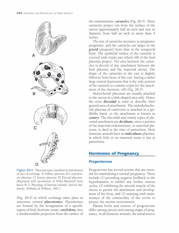
444 / ANATOMY AND PHYSIOLOGY OF FARM ANIMALS
Figure 28-4. Placental types classifi ed by distribution of sites of exchange. A) Diffuse placenta. B) Cotyledon-ary placenta. C) Zonary placenta. D) Discoid placenta. (Reprinted with permission of Wiley-Blackwell from Reece W.O. Physiology of Domestic Animals. 2nd ed. Bal-timore: Williams & Wilkins, 1997.)
(Fig. 28-4) in which exchange takes place at structures termed placentomes. Placentomes are formed by the invagination of a specifi c region of fetal chorionic tissue, cotyledons, into a mushroomlike projection from the surface of
the endometrium, caruncles (Fig. 28-5). These caruncles project out from the surface of the uterus approximately half an inch and vary in diameter from half an inch to more than 4 inches.
The size of caruncles increases as pregnancy progresses, and the caruncles are larger in the gravid (pregnant) horn than in the nongravid horn. The epithelial surface of the caruncle is covered with crypts into which villi of the fetal placenta project. The area between the carun-cles is devoid of any attachment between the fetal placenta and the maternal uterus. The shape of the caruncles in the ewe is slightly different from those of the cow, having a rather large central depression that is the only portion of the caruncle to contain crypts for the attach-ment of the chorionic villi (Fig. 28-5).
Hemochorial placentas are usually attached to the uterus in a disk-shaped area only. Hence the term discoidal is used to describe their general area of attachment. The endotheliocho-rial placenta of carnivores is attached in a gir-dlelike band, so the attachment is known as zonary. The discoidal and zonary types of pla-cental attachment are deciduate, since a portion of the maternal endometrium, or maternal pla-centa, is shed at the time of parturition. Most domestic animals have an indeciduate placenta, in which little or no maternal tissue is lost at parturition.
Hormones of Pregnancy
Progesterone
Progesterone has several actions that are essen-tial for maintaining a normal pregnancy. These include (1) providing negative feedback to the hypothalamus to inhibit any further estrous cycles, (2) inhibiting the smooth muscle of the uterus to permit the attachment and develop-ment of the fetus, and (3) assisting with main-tenance of the contractility of the cervix to protect the uterine environment.
Plasma levels and sources of progesterone differ among species and among stages of preg-nancy. In all domestic animals, the initial source

PREGNANCY AND PARTURITION / 445
of the necessary progesterone is the corpus luteum, and in some species (e.g., cow and sow), the corpus luteum remains the primary source throughout pregnancy. In other species (e.g., mare and ewe), the initial corpus luteum can be removed after secondary sources produce enough progesterone to maintain pregnancy. These sources include secondary or accessory corpora lutea and the placenta in the mare; the placenta is the secondary source in the ewe.
Equine Chorionic Gonadotrophin
The mare appears to be unique among domes-tic species in that the equine placenta is the source of a protein hormone that acts similarly to the luteinizing hormone, the source of which is the pituitary. Secretion of equine chorionic gonadotrophin (eCG, formerly known as preg-nant mare serum gonadotrophin, or PMSG) begins after about a month of gestation and continues until about 4 months of gestation. During this period, follicular development occurs on the ovary of the pregnant mare, and eCG promotes the luteinization of these folli-cles. These accessory corpora lutea provide sec-ondary sources of progesterone.
Trophoblastic cells of fetal origin found in specialized structures termed endometrial cups
are the source of the eCG. These structures are seen as small, raised circular areas with a central depression on the endometrial surface of the pregnant uterus. The trophoblastic cells are found in association with the endometrium in these areas.
Relaxin
Relaxin is a protein hormone secreted by the corpus luteum in some species (sow and cow) and the placenta in others (bitch and mare). The primary function of relaxin is preparation for parturition and ultimately lactation. Relaxin contributes to the opening of the cervix and the relaxation of the muscles and ligaments associ-ated with the birth canal to facilitate the passage of the fetus. In some species, peak secretion of relaxin occurs just prior to parturition. Gradual increases in relaxin during the latter part of gestation also facilitate mammary gland devel-opment to prepare for lactation.
Pregnancy Diagnosis
If accurate records of estrus periods and breed-ing dates are available, the earliest indication of pregnancy in most animals is the failure to have
Figure 28-5. Placental attachments of cow, ewe, and mare. Villi from chorioallantois (black) invaginates into crypts in maternal uterine epithelium (stippled) in caruncles in the cow and the ewe and in diffuse locations in the mare.
446 / ANATOMY AND PHYSIOLOGY OF FARM ANIMALS
another estrous cycle at the expected time. Such an absence of estrus is not, however, proof of pregnancy. A nonpregnant animal may miss an estrous cycle because of failure of the corpus luteum to regress normally or some other reproductive abnormality. An animal may also have a delay of one or two estrous cycles if an initial conception is followed by inability to sustain that pregnancy.
Palpation of the reproductive tract via the rectum in the mare and cow can be useful for pregnancy diagnosis and estimation of the stage of pregnancy. In the cow, the presence of a corpus luteum in an ovary and a slight enlarge-ment of one uterine horn as compared to the other suggest an early pregnancy. At about 3 months of pregnancy in the cow, fetal mem-branes and caruncles become palpable, and the uterine artery on the side with the fetus is slightly larger than the vessel on the other side. Pregnancy diagnosis by rectal palpation is con-sidered to be more diffi cult in the mare than in the cow, but an early diagnostic feature is a bulge in the uterus due to the development of the amniotic sac.
Ultrasonography is used to diagnose preg-nancy in a variety of domestic species, includ-ing cattle, horses, sheep, goats, llamas, and swine. The earliest time for verifi cation of preg-nancy is dictated in part by the size of the ges-tational sac, which in turn varies among species. In general, the times vary between about 2 weeks for mares to about 5 weeks for ewes. In large animals, the ultrasound probe can be inserted rectally so that it is closer to the uterus.
Parturition
Parturition (labor), the act of giving birth to young, marks the termination of pregnancy. Parturition may be divided into three stages. The fi rst stage consists of uterine contractions that gradually force the fetus and fetal mem-brane to the cervix. The duration of this stage is a matter of hours in most species (e.g., 2 to 6 in the cow and ewe, 1 to 4 in the mare, and 2 to 12 in the sow).
In the second stage, actual delivery of the fetus occurs. Passage of parts of the fetus through the cervix into the vagina along with rupture of one or both water bags refl exively initiates actual straining or contraction of the abdominal muscles. The combination of uterine contraction and abdominal contraction forces the fetus through the birth canal.
The third stage of parturition consists of delivery of the placenta, which normally follows the fetus almost immediately.
Late Gestation
To facilitate delivery of the fetus, muscles and ligaments of the birth canal relax shortly before parturition. The vulva swells, and a mucus dis-charge may be present. Muscles on both sides of the tail head may relax and appear lowered or depressed. Mammary glands enlarge and may secrete a milky material for a few days prior to parturition. As the time of parturition becomes imminent, animals may become rest-less, seek seclusion, and increase the frequency of attempts to urinate. The bitch and sow often try to build a nest.
An important endocrine change during late gestation in most species is in the ratio of estro-gen to progesterone. Progesterone is high rela-tive to estrogen during most of gestation, but this ratio changes during late gestation, with estrogen increasing relative to progesterone. Estrogen promotes the development of con-tractile proteins in the smooth muscle cells of the uterus and gap junctions between these cells. These uterine changes increase the force that the uterus can generate for delivery. The timing of the changes in estrogen and pro-gesterone relative to parturition varies among species.
Initiation of Parturition
In domestic animals, an endocrine signal from the fetus appears to initiate parturition. Plasma levels of glucocorticoids (e.g., cortisol) increase in most domestic species, except for the mare,

PREGNANCY AND PARTURITION / 447
shortly prior to parturition. The fetal adrenal cortex is the source of these glucocorticoids, and the adrenal cortical secretion is in response to adrenocorticotropic hormone (ACTH) from the fetal adenohypophysis. An increase in sen-sitivity of the adrenal cortex to ACTH is part of the reason for the increased secretion of gluco-corticoids during this period.
The rising fetal glucocorticoids affect the placenta and the maternal uterus. In some species, the glucocorticoids increase estrogen production by the placenta so that plasma estrogens increase relative to progesterone. This brings about the changes in the uterine smooth muscle described earlier. Glucocorticoids and estrogens act synergistically to promote uterine synthesis and secretion of PGF2α.
Increases in PGF2a have multiple effects that can contribute to the onset of parturition, and some view these as the fi nal link in the chain of events initiating parturition. In species whose primary corpus luteum remains and is a signifi -cant source of progesterone (e.g., cow and sow), the rise in PGF2a can bring about leutoly-sis and remove this source of progesterone. Recall that progesterone suppresses the activity of uterine smooth muscle and maintains cervi-cal tone, so a reduction in progesterone is appropriate to facilitate delivery. PGF2a also directly stimulates the contraction of uterine smooth muscle to move the fetus into the birth canal and directly promotes dilation of the cervix.
Oxytocin
The entrance of a fetus into the birth canal brings about a refl ex increase in oxytocin secre-tion from the neurohypophysis. Oxytocin acts directly on uterine smooth muscle to enhance uterine contractions and promote delivery. Extracts of the neurohypophysis are used clini-cally to stimulate contractions of the fatigued uterus during prolonged labor.
In the mare, plasma levels of oxytocin gradually rise during the latter stages of gesta-tion and then increase greatly as parturition begins.
Fetal Presentations and Delivery
The calf is normally presented front feet fi rst with the head extended and the nose between the front feet (Fig. 28-6). The dorsum of the calf is in contact with the sacrum of the dam. This position, called cranial (anterior) presentation, takes advantage of the natural curvature of the birth canal of the dam and the curvature of the fetus. A caudal (posterior) presentation with the hind feet fi rst, hocks up, occurs frequently enough in cattle to be considered normal.
Uterine contractions force the fetal placenta (water bags) against the cervix of the uterus, and ultimately these water bags rupture. At about the same time, abdominal muscles begin to contract forcefully to expel the fetus through the birth canal.
The contraction of abdominal muscles, called straining, is a refl ex response to stimuli from the presence of parts of the fetus within the vagina and vulva of the dam. Straining can also be readily evoked by the insertion of a hand and arm into the vulva and vagina, for example when attempting to deliver a calf.
The legs of a foal are longer than those of a calf, and a foal is carried to a larger extent in the body of the uterus, while a calf is carried almost
Cranial Presentationof the Calf
Figure 28-6. Cranial or anterior presentation of calf.

448 / ANATOMY AND PHYSIOLOGY OF FARM ANIMALS
entirely in one horn of the uterus. Presentation of a foal is essentially the same as that of a calf. With pigs and dogs, the young are carried in both horns of the uterus and may be presented either cranially or caudally with equal facility.
In species that typically have a single off-spring, the placenta, or afterbirth, is delivered soon after birth, but it may accompany the fetus or, rarely, precede it. In the litter-bearing sow, the placenta for each fetus is typically delivered still attached to the fetus and may completely surround the fetus. In these cases, immediate removal of the placenta from the nostrils of the newborn is essential for life; usually the sow does it. A placenta is consid-ered to be retained pathologically if an abnor-mally long time elapses between birth of the young and delivery of the placenta. The pla-centa of the cow and ewe should be delivered within 24 hours, and the placenta of the mare should be delivered within 2 to 3 hours.
Retention of the placenta is a signifi cant problem in dairy cattle. The incidence seems to be higher in cattle than other species and higher in dairy than in beef breeds. Manual removal of a retained placenta has been a common method of treatment, but more con-servative treatment (e.g., no manual removal, with antibiotics to prevent infection) are also advocated, and their use is supported by research studies.
Dystocia
Normal parturition with no complications is by far the most common occurrence in domestic animals. Dystocia is diffi cult birth; in some
cases, birthing is not possible without assis-tance. In general, from the onset of labor, cows should calve within 8 hours, ewes should lamb within 2 hours, and mares should foal within 2 to 3 hours. Sows should average one offspring per hour until farrowing (i.e., birthing of a litter) is complete.
In large animals (cow, mare, and ewe), improper fetal presentation is a common cause of dystocia, and typically with abnormal posi-tioning of the limbs or head (e.g., head rotated up and ventrum of neck presented or head rotated down and dorsum of neck presented). Correction of such a presentation may be nec-essary for delivery of the fetus. Correction usually entails repelling the fetus into the uterus away from the pelvic inlet so that the fetus can be manipulated and the position of its limbs and head changed.
Excessive size of the fetus relative to the size of the birth canal of the dam may also produce dystocia. This is a common cause of dystocia in fi rst-calf heifers and in relatively small cows mated with much larger bulls. Excessive fetal size may also produce problems in ewes with single lambs and sows with small litters. Even though the presentation may be normal in these cases, excessive traction may be required to deliver the newborn, and this may damage both the fetus and the dam.
A cesarean section (surgical removal of the fetus) may be necessary in some cases of dys-tocia. Successful surgeries produce a viable fetus and preserve the reproductive capacity of the dam. An embryotomy (surgical dismember-ment of the fetus to permit its passage through the birth canal) may be necessary in some cases to save the life of the dam.
ANATOMY AND PHYSIOLOGY OF
THE MAMMARY GLANDS
The mammary glands (also called mammae) are modifi ed sudoriferous (sweat) glands
that produce milk for the nourishment of off-spring. They develop from bilateral thickenings of ventrolateral ectoderm of the embryo, the so-called milk lines, which are more correctly referred to as mammary ridges. In carnivores and the sow, the mammary glands develop throughout the axillary to inguinal extent of the ridges, as is appropriate for species that typi-cally deliver multiple fetuses. However, in most other domestic animals, only the inguinal mammary glands develop, usually two (e.g., mares, ewes, and does) or four (e.g., cows). In the anthropoid apes and the elephant, only two pectoral mammary glands develop.
Each gland is composed of a system of ducts connecting masses of secretory epithelium sur-rounded by connective tissue and fat and sup-ported in a fi broelastic capsule. The proportion of secretory parenchyma to connective tissue is hormonally dictated; during lactation, the mammary gland’s secretory tissues increase in volume. After the end of lactation (when the dam is dry), the secretory tissues regress, and connective tissue constitutes a greater percent-age of the gland.
In ruminants and horses, individual glands are associated so closely to one another that they are commonly referred to as a single udder. Even so, the individual nature of glands of the udder is readily appreciated by the presence of
Mammary Glands of the CowSuspensory Apparatus
Blood Supply
Lymphatic Vessels
Microscopic Anatomy of the Mammary Gland
Mammary Glands of SwineMammary Glands of Sheep and GoatsMammary Glands of the HorsePhysiology of Lactation
Composition of Milk
Milk Secretion
Lactogenesis
Galactogenesis
Milk Ejection or Letdown
Colostrum
Cessation of Lactation
Chapter 29
449

450 / ANATOMY AND PHYSIOLOGY OF FARM ANIMALS
a single teat (papilla) for each gland. A single (as in ruminants) or multiple (as in the mare and sow) duct system may discharge milk at the tip of each teat.
Embryonic ectoderm invaginates along the mammary ridge to become the duct system of individual mammary glands. These invaginations (mammary buds) will ulti-mately be associated with an individual teat. In all species more buds initially develop than will persist, and these extra buds regress promptly after appearing. Not uncommonly, though, some extra buds persist and produce supernumerary teats. These extra teats are usually small and not associated with a well-developed gland; because they can interfere with milking, they are usually removed from the udders of cows and does.
The mammary glands of domestic species have a great deal in common, but inasmuch as the udder of the dairy cow has been so dramati-cally developed to produce milk far beyond that necessary to nourish the cow’s offspring, it is discussed in detail.
Mammary Glands of the Cow
The udder of the cow comprises four individual glands, referred to as quarters. The skin of the udder is covered with fi ne hair; however, the teat is completely hairless. The right and left halves of the udder each consist of a cranial (front) quarter and a caudal (hind) quarter. Each side of the udder is almost completely independent of the other insofar as blood supply, nerve supply, and suspensory apparatus are concerned (discussed later).
Ventrally, the two halves of the udder are demarcated by a longitudinal furrow, the inter-mammary groove, which corresponds to a median septum of connective tissue dividing left and right halves. Because of the relative isolation of each side, half of the udder can be removed surgically without damaging the other half, as might be done to treat an aggressive tumor. The two quarters in each half are sepa-rate from one another as far as the gland tissue and duct system are concerned. Thus, all the
milk from one teat is produced by the glandular tissue of that quarter. The vasculature, nerve supply, and lymphatic drainage, however, are common to both quarters of a given half.
The parenchyma of the lactating mammary gland consists of secretory tissue and the ducts of the gland (Fig. 29-1). The secretory units, the alveoli, are lined by a simple epithelium that varies from columnar to cuboidal in height. The alveoli are the chief structures for actual milk production, although the initial portion of the associated duct is also lined with secretory epithelium.
The various small initial ducts converge to form larger ducts, and these converge to form yet larger ones, all of which eventually termi-nate in a large single basin, the lactiferous sinus. The lactiferous sinus is sometimes described as being divided into a large cavity within the quarter itself, the pars glandularis (gland cistern), and a smaller cavity within the associated teat called the pars papillaris (teat cistern) (Fig. 29-1). The demarcation between gland cistern and teat cistern frequently is marked by a circular ridge (annulus) that con-tains a vein and some smooth muscle fi bers.
The wall of the empty cistern contains numerous overlapping longitudinal and circu-lar folds that are obliterated through expansion of the wall when it is full of milk. There may also be diverticula (pockets) in the wall of the gland cistern.
The teat cistern is continuous with the exte-rior of the teat through a narrow opening in the end of the teat, the papillary duct (commonly called streak canal or teat canal), which opens at the ostium papillae. The bovine streak canal is about 8.5 mm long, and its lumen is nor-mally closed by epithelial folds that project inward from the wall of the streak canal, leaving only a star-shaped potential opening. A sphinc-ter of smooth muscle fi bers surrounds the streak canal at the distal end of the teat.
Suspensory Apparatus
The udder of a lactating dairy cow can weigh as much as 60 kg, so the organ is supported by
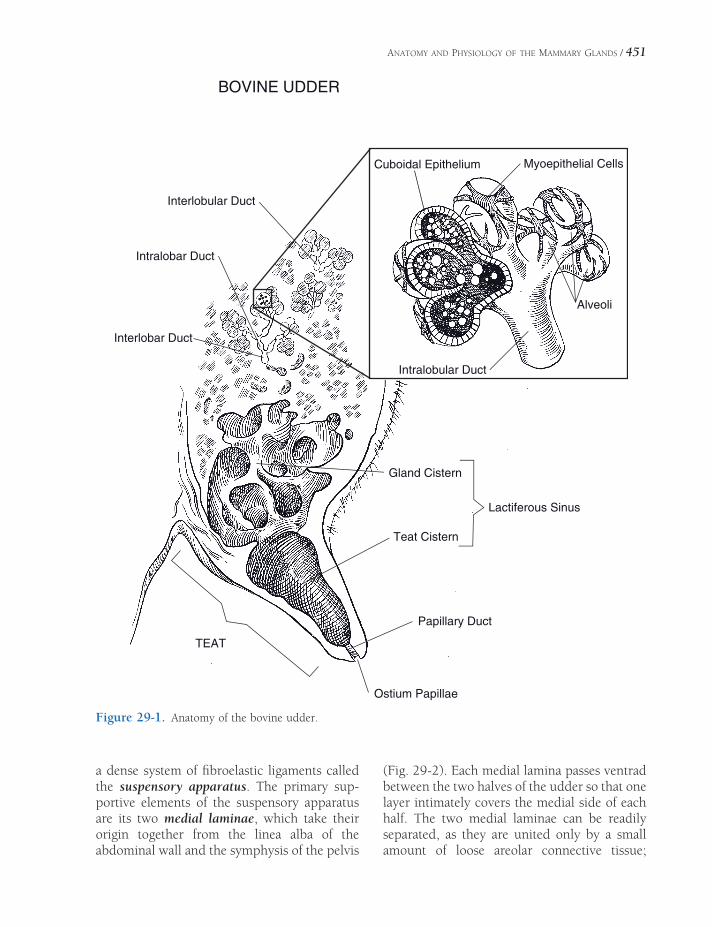
ANATOMY AND PHYSIOLOGY OF THE MAMMARY GLANDS / 451
BOVINE UDDER
Interlobular Duct
Intralobar Duct
Interlobar Duct
Cuboidal Epithelium Myoepithelial Cells
Alveoli
Intralobular Duct
Gland Cistern
Teat Cistern
Lactiferous Sinus
Papillary Duct
Ostium Papillae
TEAT
Figure 29-1. Anatomy of the bovine udder.
a dense system of fi broelastic ligaments called the suspensory apparatus. The primary sup-portive elements of the suspensory apparatus are its two medial laminae, which take their origin together from the linea alba of the abdominal wall and the symphysis of the pelvis
(Fig. 29-2). Each medial lamina passes ventrad between the two halves of the udder so that one layer intimately covers the medial side of each half. The two medial laminae can be readily separated, as they are united only by a small amount of loose areolar connective tissue;

452 / ANATOMY AND PHYSIOLOGY OF FARM ANIMALS
practically no vessels or nerves pass through the medial ligament from one half of the udder to the other. Proximally (close to the body wall), the laminae are thickest. As they descend, they give off sheets of connective tissue that diverge from the midline and interdigitate into the parenchyma of the udder so that the two medial laminae are thinnest near the intermam-mary groove.
The lateral laminae of the suspensory appa-ratus are composed largely of dense white fi brous connective tissue, making them less elastic than the medial laminae, which are mostly elastic connective tissue. The cranial
part of the lateral lamina derives from the apo-neurotic tissues of the body wall near the exter-nal inguinal ring and more caudally from the regions of the pelvic symphysis and prepubic tendon (the tendon of insertion of the m. rectus abdominis). From its origins, the lateral lamina passes ventrad and around the lateral side of each half of the mammary gland, meeting the medial lamina at the cranial and caudal aspects of each half. Like the medial lamina, the lateral lamina is thick close to the body wall and thins progressively ventrally as it gives off sheets of connective tissue into the substance of the gland.
Blood Supply
The blood supply to the udder is primarily through the external pudendal artery (Fig. 29-3), a branch of the pudendoepigastric trunk. The external pudendal artery passes downward through the inguinal canal in a more or less tortuous manner and divides into cranial and caudal branches that supply the front- and hindquarters on the same side as the artery. A small artery that may be single or paired (as determined by chance) is the ventral perineal artery, which continues from the internal pudendal artery and passes downward from the vulva just deep to the skin on the median line. The perineal artery usually supplies a small amount of blood to the caudal part of both halves of the udder.
The venous drainage from the udder is largely by way of a venous circle at the base of the udder, where it attaches to the abdominal wall. This venous circle is formed from the main veins that drain the udder. The external pudendal vein of each side receives blood from both the cranial and caudal quarters of the same side. Cranially, each external pudendal vein is continuous with the caudal superfi cial epigastric vein and caudally with the perineal vein. An anastomosis between the two caudal superfi cial epigastric veins just at or in front of the udder completes the venous circle. The caudal superfi cial epigastric vein passes forward in a sagittal plane lateral to the midline on the
Medial SuspensoryLamina
LateralSuspensoryLamina
Figure 29-2. Suspensory apparatus of the cow. Udder is shown in transverse section through hindquarters.

Internal Iliac A. Abdominal Aorta
External Iliac A.
Pudendoepigastric
Trunk
Milk WellSubcutaneous
Abdominal V.
Mammary Lymph Vessels
Superficial Inguinal
Lymph Node
External Pudendal A.
External Pudendal V.
Cranial Vena Cava
Internal Thoracic V.
External Pudendal
A. & V.
ARTERIAL AND VENOUS SUPPLY TO THE UDDERARTERIAL AND VENOUS SUPPLY TO THE UDDER
Ventraal Perineal
A.. & V.
Deep
Femoral A.
Figure 29-3. Arterial and venous blood supply to the bovine udder. Vessels other than aorta and cranial vena cava are paired.
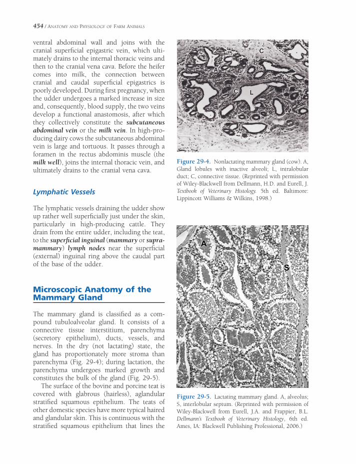
454 / ANATOMY AND PHYSIOLOGY OF FARM ANIMALS
ventral abdominal wall and joins with the cranial superfi cial epigastric vein, which ulti-mately drains to the internal thoracic veins and then to the cranial vena cava. Before the heifer comes into milk, the connection between cranial and caudal superfi cial epigastrics is poorly developed. During fi rst pregnancy, when the udder undergoes a marked increase in size and, consequently, blood supply, the two veins develop a functional anastomosis, after which they collectively constitute the subcutaneous abdominal vein or the milk vein. In high-pro-ducing dairy cows the subcutaneous abdominal vein is large and tortuous. It passes through a foramen in the rectus abdominis muscle (the milk well), joins the internal thoracic vein, and ultimately drains to the cranial vena cava.
Lymphatic Vessels
The lymphatic vessels draining the udder show up rather well superfi cially just under the skin, particularly in high-producing cattle. They drain from the entire udder, including the teat, to the superfi cial inguinal (mammary or supra-mammary) lymph nodes near the superfi cial (external) inguinal ring above the caudal part of the base of the udder.
Microscopic Anatomy of the Mammary Gland
The mammary gland is classifi ed as a com-pound tubuloalveolar gland. It consists of a connective tissue interstitium, parenchyma (secretory epithelium), ducts, vessels, and nerves. In the dry (not lactating) state, the gland has proportionately more stroma than parenchyma (Fig. 29-4); during lactation, the parenchyma undergoes marked growth and constitutes the bulk of the gland (Fig. 29-5).
The surface of the bovine and porcine teat is covered with glabrous (hairless), aglandular stratifi ed squamous epithelium. The teats of other domestic species have more typical haired and glandular skin. This is continuous with the stratifi ed squamous epithelium that lines the
Figure 29-4. Nonlactating mammary gland (cow). A, Gland lobules with inactive alveoli; L, intralobular duct; C, connective tissue. (Reprinted with permission of Wiley-Blackwell from Dellmann, H.D. and Eurell, J. Textbook of Veterinary Histology. 5th ed. Baltimore: Lippincott Williams & Wilkins, 1998.)
Figure 29-5. Lactating mammary gland. A, alveolus; S, interlobular septum. (Reprinted with permission of Wiley-Blackwell from Eurell, J.A. and Frappier, B.L. Dellmann’s Textbook of Veterinary Histology, 6th ed. Ames, IA: Blackwell Publishing Professional, 2006.)
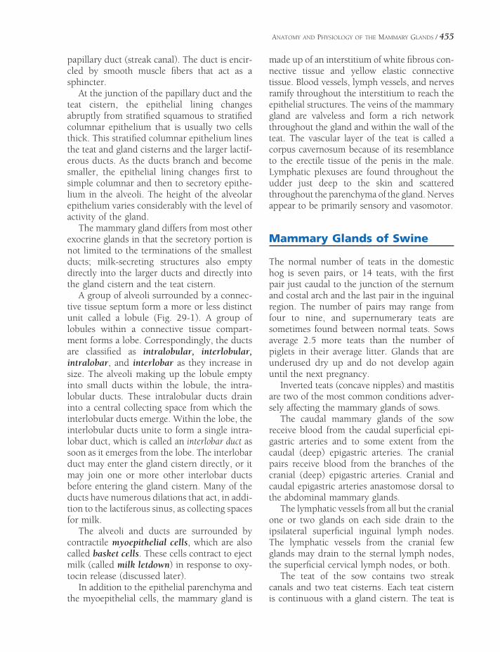
ANATOMY AND PHYSIOLOGY OF THE MAMMARY GLANDS / 455
papillary duct (streak canal). The duct is encir-cled by smooth muscle fi bers that act as a sphincter.
At the junction of the papillary duct and the teat cistern, the epithelial lining changes abruptly from stratifi ed squamous to stratifi ed columnar epithelium that is usually two cells thick. This stratifi ed columnar epithelium lines the teat and gland cisterns and the larger lactif-erous ducts. As the ducts branch and become smaller, the epithelial lining changes fi rst to simple columnar and then to secretory epithe-lium in the alveoli. The height of the alveolar epithelium varies considerably with the level of activity of the gland.
The mammary gland differs from most other exocrine glands in that the secretory portion is not limited to the terminations of the smallest ducts; milk-secreting structures also empty directly into the larger ducts and directly into the gland cistern and the teat cistern.
A group of alveoli surrounded by a connec-tive tissue septum form a more or less distinct unit called a lobule (Fig. 29-1). A group of lobules within a connective tissue compart-ment forms a lobe. Correspondingly, the ducts are classifi ed as intralobular, interlobular, intralobar, and interlobar as they increase in size. The alveoli making up the lobule empty into small ducts within the lobule, the intra-lobular ducts. These intralobular ducts drain into a central collecting space from which the interlobular ducts emerge. Within the lobe, the interlobular ducts unite to form a single intra-lobar duct, which is called an interlobar duct as soon as it emerges from the lobe. The interlobar duct may enter the gland cistern directly, or it may join one or more other interlobar ducts before entering the gland cistern. Many of the ducts have numerous dilations that act, in addi-tion to the lactiferous sinus, as collecting spaces for milk.
The alveoli and ducts are surrounded by contractile myoepithelial cells, which are also called basket cells. These cells contract to eject milk (called milk letdown) in response to oxy-tocin release (discussed later).
In addition to the epithelial parenchyma and the myoepithelial cells, the mammary gland is
made up of an interstitium of white fi brous con-nective tissue and yellow elastic connective tissue. Blood vessels, lymph vessels, and nerves ramify throughout the interstitium to reach the epithelial structures. The veins of the mammary gland are valveless and form a rich network throughout the gland and within the wall of the teat. The vascular layer of the teat is called a corpus cavernosum because of its resemblance to the erectile tissue of the penis in the male. Lymphatic plexuses are found throughout the udder just deep to the skin and scattered throughout the parenchyma of the gland. Nerves appear to be primarily sensory and vasomotor.
Mammary Glands of Swine
The normal number of teats in the domestic hog is seven pairs, or 14 teats, with the fi rst pair just caudal to the junction of the sternum and costal arch and the last pair in the inguinal region. The number of pairs may range from four to nine, and supernumerary teats are sometimes found between normal teats. Sows average 2.5 more teats than the number of piglets in their average litter. Glands that are underused dry up and do not develop again until the next pregnancy.
Inverted teats (concave nipples) and mastitis are two of the most common conditions adver-sely affecting the mammary glands of sows.
The caudal mammary glands of the sow receive blood from the caudal superfi cial epi-gastric arteries and to some extent from the caudal (deep) epigastric arteries. The cranial pairs receive blood from the branches of the cranial (deep) epigastric arteries. Cranial and caudal epigastric arteries anastomose dorsal to the abdominal mammary glands.
The lymphatic vessels from all but the cranial one or two glands on each side drain to the ipsilateral superfi cial inguinal lymph nodes. The lymphatic vessels from the cranial few glands may drain to the sternal lymph nodes, the superfi cial cervical lymph nodes, or both.
The teat of the sow contains two streak canals and two teat cisterns. Each teat cistern is continuous with a gland cistern. The teat is

456 / ANATOMY AND PHYSIOLOGY OF FARM ANIMALS
hairless, but hair is found at the base of the teat and on the gland.
Seedy-cut bacon is due to inclusion of pigmented mammary tissue in the cut, giving the appearance of small seeds in the bacon. Black seedy cut is due to invagination of pigmented epithelium at the time of mammary gland formation. It occurs only in dark-colored glands. Red seedy cut may occur in any color sow or gilt past puberty, as it is due to infl ammation of mammary glands associated with the estrous cycle.
Mammary Glands of Sheep and Goats
The udders of the ewe and the doe differ from that of the cow in that each half of the udder has only one teat, one streak canal, one teat cistern, and one gland cistern. One half of the ovine and caprine udder resembles one quarter of the bovine udder. The teat is sparsely covered with fi ne hair. Each half of the udder of the ewe is craniomedial to the inguinal sinus (a pouch of skin lined with scent glands) of the same side. Supernumerary teats in the ewe do not appear to have separate gland tissue, as is fre-quently found in the cow. The sphincter muscle around the papillary duct is poorly developed, so closure is affected by elastic tissue in the end of the teat.
Mammary Glands of the Horse
The mammary glands of the mare consist of one teat on each side attached to half of the udder. Each teat has two streak canals and two
teat cisterns, each of which is continuous with a separate system of ducts and alveoli (occasionally a gland has a third set of ducts). There appears to be no communication between ducts or cisterns within the same half of the udder.
The suspensory apparatus is arranged as in the cow, although it is not as robust. The udder and teats of the mare are covered with thin, fi ne hair and with numerous sebaceous and sweat glands. There is a difference of opinion regard-ing the presence or absence of rudimentary teats in the stallion and gelding. They have been described in the skin forming the cranial part of the prepuce, but their presence here has also been denied. Apparently no adequate embryologic study of this subject has been reported.
Physiology of Lactation
Composition of Milk
Milk contains all of the nutrients necessary for survival and initial growth of mammalian neo-nates. The nutrients in milk include sources of energy (lipids and carbohydrates), proteins to provide amino acids, vitamins, minerals (ash) for electrolytes, and water. The relative amounts of these nutrients in milk vary among species (Table 29-1).
Diet and the stage of lactation also affect the composition of milk. Diets high in non-fi ber carbohydrate sources of energy are associated with increases in the percentage of lipids in the milk. Diets high in protein promote a slight increase in the percentage of protein in the milk, but this effect is much less than the effect
Table 29-1. Typical Values for Constituents of Milk in Grams per Liter
Species Lipids Lactose Protein Total Minerals (ash) Calcium
Cow 38 48 37 7.0 1.3
Mare 16 50 24 4.5 1.0
Ewe 70 40 60 8.0 1.9
Sow 80 46 58 8.5 2.0
Doe 40 45 35 7.8 1.2
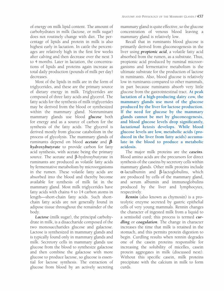
ANATOMY AND PHYSIOLOGY OF THE MAMMARY GLANDS / 457
of energy on milk lipid content. The amount of carbohydrates in milk (lactose, or milk sugar) does not routinely change with diet. The per-centage of lipids and protein in milk is also highest early in lactation. In cattle the percent-ages are relatively high in the fi rst few weeks after calving and then decrease over the next 3 to 4 months. Later in lactation, the concentra-tions of lipids and proteins again increase as total daily production (pounds of milk per day) decreases.
Most of the lipids in milk are in the form of triglycerides, and these are the primary source of dietary energy in milk. Triglycerides are composed of three fatty acids and glycerol. The fatty acids for the synthesis of milk triglycerides may be derived from the blood or synthesized within the mammary gland. Nonruminant mammary glands use blood glucose both for energy and as a source of carbon for the synthesis of the fatty acids. The glycerol is derived mostly from glucose catabolism in the process of glycolysis. The mammary glands of ruminants depend on blood acetate and b-hydroxybutyrate to provide carbon for fatty acid synthesis, with acetate being the primary source. The acetate and β-hydroxybutyrate in ruminants are produced as volatile fatty acids by fermentative metabolism by microorganisms in the rumen. These volatile fatty acids are absorbed into the blood and thereby become available for synthesis of milk fat in the mammary gland. Most milk triglycerides have fatty acids with chains 4 to 14 carbon atoms in length—short-chain fatty acids. Such short-chain fatty acids are not generally found in adipose tissue throughout the remainder of the body.
Lactose (milk sugar), the principal carbohy-drate in milk, is a disaccharide composed of the two monosaccharides glucose and galactose. Lactose is synthesized in mammary glands and is typically found only in mammary glands and milk. Secretory cells in mammary glands use glucose from the blood to synthesize galactose and then combine the galactose with more glucose to produce lactose, so glucose is essen-tial for lactose synthesis. The extraction of glucose from blood by an actively secreting
mammary gland is quite effective, so the glucose concentration of venous blood leaving a mammary gland is relatively low.
Recall that in ruminants blood glucose is primarily derived from gluconeogenesis in the liver using propionic acid, a volatile fatty acid absorbed from the rumen, as a substrate. Thus, propionic acid produced by ruminal microor-ganisms and fermentative metabolism is the ultimate substrate for the production of lactose in ruminants. Also, blood glucose is relatively low in ruminants compared to other mammals, in part because ruminants absorb very little glucose from the gastrointestinal tract. At peak lactation of a high-producing dairy cow, the mammary glands use most of the glucose produced by the liver for lactose production. If the need for glucose by the mammary glands cannot be met by gluconeogenesis, and blood glucose levels drop signifi cantly, lactational ketosis develops. While blood glucose levels are low, metabolic acids (pro-duced in the liver from fatty acids) accumu-late in the blood to produce a metabolic acidosis.
The major milk proteins are the caseins. Blood amino acids are the precursors for direct synthesis of the caseins by secretory cells within mammary glands. Other milk proteins include α-lactalbumin and β-lactoglobulins, which are produced by cells of the mammary gland, and serum albumin and immunoglobulins produced by the liver and lymphocytes, respectively.
Rennin (also known as chymosin) is a pro-teolytic enzyme secreted by gastric epithelial cells of very young mammals. Rennin changes the character of ingested milk from a liquid to a semisolid curd; this process is termed cur-dling or coagulation. The change in character increases the time that milk is retained in the stomach, and this permits protein digestion to begin. Curdling results when rennin degrades one of the casein proteins responsible for increasing the solubility of micelles, casein protein aggregates in milk (discussed next). Without this specifi c casein, milk proteins precipitate with the calcium in milk to form curds.

458 / ANATOMY AND PHYSIOLOGY OF FARM ANIMALS
Milk Secretion
The epithelial cells lining the alveoli of mammary glands are the cells primarily respon-sible for the secretion of milk. The appearance of these cells varies as they synthesize and release the lipids, proteins, and lactose of milk. After the cells actively secrete the constituents of milk and the lumen of the alveoli are fi lled with milk, the epithelial cells shrink and are described as a simple low-cuboidal epithelium (Fig. 29-5). At this stage, their secretory activity is relatively low. Shortly after the stored milk is removed, the epithelial cells increase their secretory activity and begin to refi ll the alveoli. Early in the secretory phase the cells assume a more columnar appearance and then gradually reduce to cuboidal as milk fi lls the alveoli. Small, apparently nonfunctioning alveoli can be found in dry mammary glands, and there is a relative increase in the amount of interstitial loose connective tissue (Fig. 29-4).
Milk lipids are synthesized and packaged into secretory droplets, which are extruded from the luminal surface of the cell into the alveoli (Fig. 29-6). As they are released, a mem-
brane covering derived from the cell membrane of the epithelial cell encases the lipid droplets. The alveolar secretory cells also produce secre-tory vesicles that contain both milk proteins (caseins) and lactose (Fig. 29-6). As caseins are synthesized and packaged in these vesicles, they self-aggregate into particles termed micelles. The inclusion of one specifi c type of casein (κ-casein) in this aggregation increases the solubility of the micelle so that milk pro-teins remain in solution after their release from the cell. The lactose within the secretory vesicle generates an osmotic force to draw water into the vesicle from the cytosol of the cell. The secretory vesicles, each containing a mixture of micelles, lactose, and water, are transported to the apical surface of the cell and released into the alveoli by exocytosis (Fig. 29-6). Because of the various mechanisms by which lipids, pro-teins, and lactose are secreted from alveolar cells, milk is considered to be a combination of apocrine and merocrine secretions.
Lactogenesis
Lactogenesis is the establishment of milk secre-tion, and galactopoiesis is the continued pro-duction of milk by the mammary glands. Growth and development of the secretory epithelium and the ductile network of the mammary glands must precede lactogenesis. The initial extensive development of the mammary gland is usually associated with puberty (the beginning of sexual maturity) and the subsequent cyclic changes in the ovarian hormones, estrogen and progester-one. Estrogen particularly promotes the growth of the ductile system at each estrus, while pro-gesterone, acting with estrogen, is required for growth and anatomic development of secretory alveoli. Normal secretion of growth hormone and glucocorticoids are also required for com-plete mammary gland development.
During pregnancy, prolonged exposure of the mammary glands to progesterone promotes a more extensive anatomic development of the secretory alveoli. While progesterone stimu-lates anatomic development, it inhibits the functional development of the secretory epithe-
Lipid Droplets
Nucleus Rough Endoplasmic Reticulum
Lumen
Figure 29-6. Secretion of milk lipids, milk proteins, and lactose by epithelial cells lining the alveoli of the mam mary glands. Proteins and lactose are together in secretory vesicles that are released by exocytosis (arrows).

ANATOMY AND PHYSIOLOGY OF THE MAMMARY GLANDS / 459
lium. Progesterone also inhibits the production of intracellular enzymes necessary for normal milk secretion. This inhibitory effect of proges-terone is lost just prior to parturition, and this is one factor promoting lactogenesis.
Prolactin, a protein hormone, is unique in that its release from the adenohypophysis is primarily regulated by a humoral inhibitor factor from the hypothalamus. In the absence of this inhibitory factor (which is believed to be the catecholamine dopamine), there is a con-tinuous and relatively high rate of prolactin release. In most domestic animals, blood levels of prolactin gradually increase late in gestation, with an abrupt increase at parturition. During late gestation, prolactin receptors in mammary glands also increase under the infl uence of rising estrogen. Prolactin promotes both ana-tomic and functional development of the secre-tory epithelium of mammary glands to promote milk secretion, so the abrupt rise at parturition is appropriate for lactogenesis. Under the infl u-ence of prolactin, secretory cells lining alveoli produce intracellular enzymes necessary for milk secretion. The functional development of alveolar secretory cells is also enhanced by glu-cocorticoids, which increase in the blood prior to parturition in most species.
The placenta of ruminants produces a protein hormone, placental lactogen, or chorionic somatomammotropin, which is similar in struc-ture and function to prolactin. Placental lactogen production increases in late gestation in rumi-nants and is believed to be more responsible for mammary gland development in these species than prolactin from the adenohypophysis.
Galactogenesis
The continuation of lactation requires stimuli to promote milk production and removal or inhibition of stimuli that retard milk produc-tion. Stimulation of the nipples (teats) by either milking or suckling elicits an abrupt increase in blood levels of prolactin. The increased secretion of prolactin is the result of a neural refl ex mediated through the hypothalamus that regulates prolactin release from the adenohy-
pophysis. The increase in prolactin is relatively short in duration (minutes to an hour). This periodic and relatively brief surge of prolactin is essential to maintain normal lactation in most species, but prolactin does not appear to be an essential regulator of lactation in cows. Supple-mentation with prolactin does not increase milk secretion in cows, and lactation is main-tained in cows in spite of severe reductions in blood levels of prolactin.
Growth hormone appears to be especially important in cows, whose blood levels of growth hormone are directly correlated with the maintenance and level of milk production. Growth hormone supplementation increases milk production in cows by 10–40%. Growth hormone supplementation in cattle is associ-ated with a variety of physiologic changes that promote milk production. These include increased mobilization of body energy stores and alterations in overall protein metabolism in other organs to provide substrates for milk secretion, increased food intake, increased nutrient absorption from the gastrointestinal tract, and improved effi ciency of the conversion of nutrients to milk by the mammary gland. It is presumed that many of the effects of growth hormone are mediated via insulin-like growth factors (IGFs), whose production is increased by growth hormone (see Chapter 12). Even though growth hormone appears to be a primary regulator of milk production in cows, milking or suckling does not produce an imme-diate release of growth hormone in the lactating cow.
Routine milking or suckling to remove milk from the mammary glands is essential for con-tinued milk production. When milking is stopped abruptly (probably the best way to dry up a cow), a number of changes in the udder occur. At the end of 24 hours, the alveoli become distended to a maximum, and the cap-illaries are full of blood. Between 36 and 48 hours, there is a decrease in the number of patent (open) capillaries, and the alveoli do not respond to intravenous oxytocin. A protein that inhibits milk production, feedback inhibitor of lactation (FIL), is apparently produced in the mammary gland as milk is produced. This
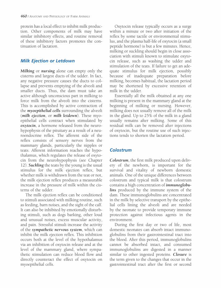
460 / ANATOMY AND PHYSIOLOGY OF FARM ANIMALS
protein has a local effect to inhibit milk produc-tion. Other components of milk may have similar inhibitory effects, and routine removal of these inhibitory factors promotes the con-tinuation of lactation.
Milk Ejection or Letdown
Milking or nursing alone can empty only the cisterns and largest ducts of the udder. In fact, any negative pressure causes the ducts to col-lapse and prevents emptying of the alveoli and smaller ducts. Thus, the dam must take an active although unconscious part in milking to force milk from the alveoli into the cisterns. This is accomplished by active contraction of the myoepithelial cells surrounding the alveoli (milk ejection, or milk letdown). These myo-epithelial cells contract when stimulated by oxytocin, a hormone released from the neuro-hypophysis of the pituitary as a result of a neu-roendocrine refl ex. The afferent side of the refl ex consists of sensory nerves from the mammary glands, particularly the nipples or teats. Afferent information reaches the hypo-thalamus, which regulates the release of oxyto-cin from the neurohypophysis (see Chapter 12). Suckling the teats by the young is the usual stimulus for the milk ejection refl ex, but whether milk is withdrawn from the teat or not, the milk ejection refl ex produces a measurable increase in the pressure of milk within the cis-terns of the udder.
The milk ejection refl ex can be conditioned to stimuli associated with milking routine, such as feeding, barn noises, and the sight of the calf. It can also be inhibited by emotionally disturb-ing stimuli, such as dogs barking, other loud and unusual noises, excess muscular activity, and pain. Stressful stimuli increase the activity of the sympathetic nervous system, which can inhibit the milk ejection refl ex. This inhibition occurs both at the level of the hypothalamus via an inhibition of oxytocin release and at the level of the mammary gland, where sympa-thetic stimulation can reduce blood fl ow and directly counteract the effect of oxytocin on myoepithelial cells.
Oxytocin release typically occurs as a surge within a minute or two after initiation of the refl ex by some tactile or environmental stimu-lus, and the plasma half-life of oxytocin (a small peptide hormone) is but a few minutes. Hence, milking or suckling should begin in close asso-ciation with stimuli known to stimulate oxyto-cin release, such as washing the udder and stimulation of the teats. If failure to get an ade-quate stimulus for milk ejection, possibly because of inadequate preparation before milking, becomes habitual, the lactation period may be shortened by excessive retention of milk in the udder.
Essentially all the milk obtained at any one milking is present in the mammary gland at the beginning of milking or nursing. However, milking does not usually remove all of the milk in the gland. Up to 25% of the milk in a gland usually remains after milking. Some of this residual milk can be removed after injections of oxytocin, but the routine use of such injec-tions tends to shorten the lactation period.
Colostrum
Colostrum, the fi rst milk produced upon deliv-ery of the newborn, is important for the survival and vitality of newborn domestic animals. One of the unique differences between colostrum and typical milk is that colostrum contains a high concentration of immunoglobu-lins produced by the immune system of the dam. These immunoglobulins are concentrated in the milk by selective transport by the epithe-lial cells lining the alveoli and are needed by the neonate to provide temporary immune protection against infectious agents in the environment.
During the fi rst day or two of life, most domestic neonates can absorb intact immuno-globulins from their gastrointestinal tract into the blood. After this period, immunoglobulins cannot be absorbed intact, and consumed immunoglobulins are digested in a manner similar to other ingested proteins. Closure is the term given to the changes that occur in the gastrointestinal tract after the fi rst or second
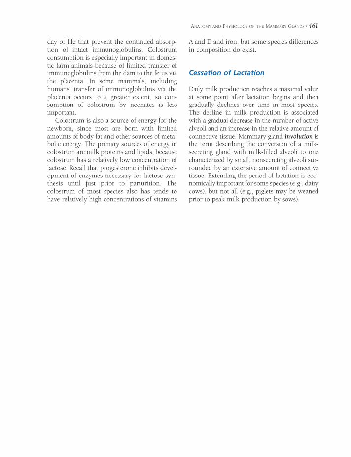
ANATOMY AND PHYSIOLOGY OF THE MAMMARY GLANDS / 461
day of life that prevent the continued absorp-tion of intact immunoglobulins. Colostrum consumption is especially important in domes-tic farm animals because of limited transfer of immunoglobulins from the dam to the fetus via the placenta. In some mammals, including humans, transfer of immunoglobulins via the placenta occurs to a greater extent, so con-sumption of colostrum by neonates is less important.
Colostrum is also a source of energy for the newborn, since most are born with limited amounts of body fat and other sources of meta-bolic energy. The primary sources of energy in colostrum are milk proteins and lipids, because colostrum has a relatively low concentration of lactose. Recall that progesterone inhibits devel-opment of enzymes necessary for lactose syn-thesis until just prior to parturition. The colostrum of most species also has tends to have relatively high concentrations of vitamins
A and D and iron, but some species differences in composition do exist.
Cessation of Lactation
Daily milk production reaches a maximal value at some point after lactation begins and then gradually declines over time in most species. The decline in milk production is associated with a gradual decrease in the number of active alveoli and an increase in the relative amount of connective tissue. Mammary gland involution is the term describing the conversion of a milk-secreting gland with milk-fi lled alveoli to one characterized by small, nonsecreting alveoli sur-rounded by an extensive amount of connective tissue. Extending the period of lactation is eco-nomically important for some species (e.g., dairy cows), but not all (e.g., piglets may be weaned prior to peak milk production by sows).


POULTRY
The term poultry refers to domesticated birds used for the production of meat and
eggs. Chickens are the most common poultry worldwide, followed by turkeys in the United States and ducks and geese elsewhere. Because poultry can be maintained in a relatively small space and are extremely effi cient in converting plant protein to animal protein, they are a sig-nifi cant source of animal protein in developing countries, as well as the rest of the world. In the United States, poultry are primarily housed in large facilities under closely regulated envi-ronments. Elsewhere, poultry are often raised in small groups in close proximity to humans and other animals. Under such circumstances, poultry can be an important source of zoonotic diseases.
The domestic chicken (Gallus gallus or Gallus domesticus) and turkey (Meleagris gallopavo) are in the order Galliformes, whereas domestic ducks (Anas platyrhynchos and Cairina mos-chata) and geese (Anser anser and Anser cygnoi-des) are Anseriformes. All are in the superorder, Carinatae. In this chapter, the term gallina-ceous will refer to chickens and turkeys, and fowl will refer to all domesticated birds. The emu, rhea, and ostrich are in a separate super-order of fl ightless birds, Ratitae. Table 30-1 lists terms commonly used for poultry of different genders, ages, and reproductive status.
Studies of domestic chickens are the basis for most of the avian physiology in this chapter,
IntegumentBody DesignSkeleton and BoneMusculatureGastrointestinal SystemRespiratory System
Ventilation and Gas Exchange
Cardiovascular SystemLymphatic SystemUrinary SystemFemale Reproductive System
Egg Formation and Oviposition
Male Reproductive SystemSex ChromosomesReproduction and Photoperiods
Chapter 30
463

464 / ANATOMY AND PHYSIOLOGY OF FARM ANIMALS
and much of this information (e.g., urinary excretion of uric acid, basics of respiratory function) could be applied to all birds. However, turkeys and chickens raised by producers in the United States have been derived by intense genetic selection for egg and/or meat produc-tion, and these domesticated birds have some anatomical and physiological characteristics that are quite different from their wild ances-tors. For example, almost all breeding of domestic turkey hens is done by artifi cial insemination because of the poor breeding per-formance of the male domestic turkey.
Integument
The skin of poultry is similar to mammalian skin in that it is an organ composed of an over-lying stratifi ed squamous epithelium and the underlying vascular dermis. Avian skin is typi-cally very thin. It lacks sweat glands entirely. Most of the skin is not particularly well vascu-larized, but in birds incubating eggs brooding patches may develop on the breast. In these areas, the skin thickens, becomes more vascu-lar, and feathers are lost. The brooding patch provides the incubating eggs with intimate contact with the warm skin of the parent bird.
The presence of feathers is the hallmark of class Aves. Like the hairs of mammals, feathers are keratinous in nature and grow from an epi-dermal follicle that encircles a dermal core.
The vascular feather pulp, an extension of dermal tissue into the shaft of the feather, is
prominent only during growth. Trauma that breaks the shaft of a large growing feather (a so-called blood feather) may lead to consider-able hemorrhage.
There are two main types of feathers seen in poultry: contour feathers, which comprise the fl ight feathers and those that cover the body, and the fl uffy down feathers. Both types possess a semirigid shaft with multiple barbs arising from it forming the vane. Contour feathers are characterized by the orderly interlocking nature of the barbs, whereas down feathers lack this systematic linking of barbs.
Feathers are not uniformly distributed on the body of the bird, occurring instead in well-described feather tracts. Some locations on the bird’s body are relatively free from contour feathers. These are the apteria (singular apterium), which in domestic fowl are espe-cially notable under the wings. Feathers are replaced in most birds once or twice a year, and in most species the molt proceeds in a sequen-tial way so as not to render the bird fl ightless due to insuffi cient fl ight feathers. Ducks and geese (Anseriformes), however, are unable to fl y for several weeks during their molt.
Chickens of breeds selected for high egg production may molt after a typical 12-month laying period. However, some birds may molt during this laying period, and low-producing hens often stop egg production if they molt at this time. Restriction of food and water and decreasing periods of light will induce an entire fl ock to stop laying and begin molting (a forced molt). During a forced molt, the reproductive system regresses and circulating levels of
Table 30-1. Terms Commonly Used for Domestic Fowl of Different Ages and Genders
Species Male Female Juvenile Other
Chicken Rooster or Cock Hen Chick Capon—castrated maleCockerel—immature malePullet—immature female
Turkey Tom Hen Poult
Duck Drake Hen or Duck Duckling
Goose Gander Goose Gosling
Swan Cob Pen Cygnet

POULTRY / 465
prolactin, luteinizing hormone (LH), estro-gen, and progesterone decrease. After a forced molt, and after photoperiods and diets are corrected, hens typically enter a new laying period with improved egg pro-duction. The use of forced molts as a man-agement tool has become controversial in part due to the restriction of food and water required to induce the molt.
Although avian skin is nearly glandless, domestic fowl have a large, well-developed sebaceous gland that opens dorsal to the caudal vertebrae. This bilobed gland, the uropygial gland, opens onto a raised papilla between the tail feathers. The oily secretion of the uropygial gland is distributed over the feathers by the preening behavior of the bird. It is especially large in water fowl, in which it has the critical function of waterproofi ng the feathers.
The feet and legs (shanks) of most domestic fowl are covered with scales that are thickened epidermal patches. A few varieties of chickens have been bred with feathered legs (e.g., the Brahma). The spur of male chickens and turkeys (gallinaceous birds) is used as a weapon in fi ghting. It has a bony center and is surrounded with epidermis, but it is not considered a “toe.” The three forward-facing digits (numbers two through four) are joined by webs that are largest and most obvious in the Anseriformes.
The face of gallinaceous birds is decorated variously by modifi cations of the skin that probably function at least in part in sexual display. The comb is placed on the dorsal midline and exhibits considerable variation in appearance among different breeds. Paired wattles hang from the ventral face, and the ear lobes decorate the side of the head near the external ear. The heads of turkeys are essen-tially featherless and covered with irregular bumps called caruncles. In addition to the comb and wattles, turkeys have a fi ngerlike snood that hangs from the dorsal aspect of the face down across the beak.
The yellowish color of the skin and associ-ated structures of most domestic chickens is due to an accumulation of a carotenoid pigment, xanthophyll, which is derived from dietary items such as corn. This pigment also contrib-
utes to the yellow color of an egg yolk, and when a hen begins laying, the yellowish color of the skin and associated structures is lost as pigment is deposited in the eggs. Color is lost in sequence from the skin around the vent (anus), eye ring, beak, and shanks, thus a hen with pale shanks has been laying for some time. Turkeys cannot accumulate carotenoid pigments in the skin and hence have a white skin.
Body Design
Birds are clearly distinct from domestic mam-mals in the overall construction of their bodies (Fig. 30-1); the thoracic limb of all birds, whether capable of fl ight or not, is modifi ed into a non–weight-bearing wing. Additionally, all birds, even relatively poor fl yers, have skeletal, cardiopulmonary, and muscular modifi cations that refl ect their common ancestry in a fl ighted progenitor. The body cavity has no muscular diaphragm, being instead incompletely parti-tioned into a number of compartments by connective tissue sheets. These are typically per-forated by a number of natural apertures that allow communication between the compart-ments and with peripheral air sacs.
Bones of birds are higher in mineral concen-tration than are mammals, and the medullary cavities of many long bones and some fl at bones are variably displaced by air-fi lled spaces that make the bones lightweight for their size and strength. Bones modifi ed in this way are called pneumatic bones. Because of the role of the thoracic limb as a wing, the thoracic girdle is extremely well developed, providing rigid scaf-folding for the muscles of fl ight. The pelvic limbs are very strong for the size of the animal and brought craniad toward the body’s center of gravity (Fig. 30-1). Both of these character-istics refl ect the fact that the pelvic limbs in birds have assumed all the responsibility of supporting the body weight when the bird is not in fl ight. Overall, the skeleton shows a ten-dency toward fusion between small bones that in mammals constitute complex joints (e.g., carpus and tarsus).

466 / ANATOMY AND PHYSIOLOGY OF FARM ANIMALS
Figure 30-1. The muscular system of the chicken. (Reprinted with permission of Wiley-Blackwell from McCracken, T.O., Kainer, R.A., and Spurgeon, T.S. Spurgeon’s Color Atlas of Large Animal Anatomy. Baltimore: Lippincott Williams & Wilkins, 1999.)
Skeleton and Bone
The avian vertebral column comprises verte-brae divided into cervical, thoracic, lumbar, sacral, and caudal divisions as in mammals, but vertebrae in the thoracic region are partially fused into the notarium, and in the lumbosa-cral region 14 to 15 vertebrae are fused into the synsacrum. The last few caudal vertebrae are fused into the pygostyle, which provides a solid mass for stability of the tail feathers. Cervical vertebrae are increased in number relative to mammals, accounting for the remarkable fl exi-bility of the avian neck; chickens, turkeys, and
ducks have 14 and geese 17 cervical vertebrae (See Fig. 30-2.)
Each thoracic limb possesses a bony attach-ment to the axial skeleton (in contrast to the muscular synsarcosis of the mammalian tho-racic limb). This shoulder girdle comprises the scapula, the coracoid (which is a bridge between the shoulder joint and the sternum), and the clavicle. The right and left clavicles in birds are fused into a single bone, commonly called the wishbone but in anatomical terms is called the furcula. The furcula acts as a bony strut to maintain distance between the two shoulders.

POULTRY / 467
Figure 30-2. The skeleton of the chicken. (Reprinted with permission of Wiley-Blackwell from McCracken, T.O., Kainer, R.A., and Spurgeon, T.S. Spurgeon’s Color Atlas of Large Animal Anatomy. Baltimore: Lippincott Williams & Wilkins, 1999.)
In the antebrachium, the radius is consider-ably smaller than the ulna. These bones articu-late with the two bones of the carpus. The distal row of carpal bones have become fused with the proximal end of the metacarpals, and so these long bones of the metacarpal region are known as carpometacarpal bones, of which there are three. There are three digits. The fi rst is often called the pollux and like the “thumb” of mammals possesses two phalanges. Digit two
is robust, with two well-developed phalanges and one vestigial phalanx. The third digit is very small and comprises only a single phalanx.
There is considerable diversity in the appear-ance of the pelvis in birds, but like the mam-malian pelvis, three bones (ilium, ischium, and pubis) contribute to its construction. Like the thoracic limb, the pelvic limb shows a reduc-tion in the number of individual bones of the
468 / ANATOMY AND PHYSIOLOGY OF FARM ANIMALS
tarsus. Fusion of the proximal row of tarsal bones with the tibia creates the tibiotarsus. The fi bula is fused with the tibiotarsus in its distal half. The distal tarsal bones are fused to a single weight-bearing metatarsal bone; this is there-fore correctly referred to as the tarsometatar-sus in birds. Poultry possess four digits; digit one projects in a medioplantar direction. The other three toes fan out in a forward direction. Digit one has two phalanges, digit two has three, digit three has four, and digit four has fi ve.
The sternum of domestic poultry is dramati-cally modifi ed for fl ight. As the pectoral muscles of birds are massively developed, the sternum (their primary attachment site) is correspond-ingly enlarged. The large, shieldlike mass of the sternum is further increased for muscle attach-ment by the presence of a large midline keel or sternal crest. The cranial sternum, especially, is adorned with projections that serve as articulat-ing processes and sites for muscle attachment.
Ten to 14 days prior to the beginning of a laying period, chickens develop a type of bony tissue unique to birds and crocodilians in the shaft of long bones such as the femur. This medullary bone consists of interlacing bony spicules, with blood sinuses and marrow between the spicules. Medullary bone may replace up to 75% of the hematopoietic tissue in laying hens, but it is absent in non-laying hens and males. Medullary bone formation is stimulated by the effects of estrogen on osteo-blasts, and just prior to the beginning of an egg production period, estrogen levels are relatively high as ovarian follicles mature and begin secreting estrogen. Medullary bone provides a ready source of calcium that can be mobilized later for use in eggshell production. Eggshell formation and calcium metabolism will be dis-cussed in more detail in a later section of this chapter.
Musculature
The most remarkable variations in the skeletal muscles of birds are not surprisingly associated with the musculature of the wing. The m. pec-
torales produce the downbeat of the wings that is essential to provide lift during fl ight. These muscles also constitute the breast of the bird bred for meat.
Gastrointestinal System
The complete gastrointestinal system of poultry is illustrated in Figure 30-3.
A distinguishing characteristic of the class Aves is the modifi cation of the jaws into a beak, the shape of which is adapted to the foraging behaviors and diet of individual species. Galli-naceous birds have pointed beaks for picking up individual food items, and Anseriformes have a fl attened, spoon-shaped beak that is somewhat more fl exible and is adapted for straining through water for foodstuffs. Beaks are featherless, covered by a thick keratinous epidermis.
The mouth of birds differs from that of mammals in that the palate is incompletely fused; this results in a longitudinal opening between oral and nasal cavities called the choanal cleft. The tongue is variable in shape, conforming as it does to the contours of the oral cavity. In domestic species, the tongue lacks musculature and is not much more than a bone (entoglossal bone) covered with a thick, corni-fi ed mucous membrane.
The pharynx begins caudal to the tongue. The pharyngeal fl oor is characterized by a raised laryngeal mound, which features the aditus laryngeus (laryngeal slit). The opening into the esophagus is caudal and somewhat dorsal to this. Within the neck, the esophagus features a distensible dilation called the crop or ingluvies. It is a small fusiform enlargement in ducks and geese, but is extremely well devel-oped in gallinaceous birds. In these birds, the crop lies on the left side of the neck cranial to the furcula, and when full of food, it is readily palpable and visible. Like the esophagus, the crop is lined by a keratinized stratifi ed squa-mous epithelium.
Within the body cavity, the esophagus leads to a bipartite stomach. The fi rst chamber is the proventriculus or glandular stomach, which

Figure 30-3. The gastrointestinal tract of the chicken. (Reprinted with permission of Wiley-Blackwell from McCracken, T.O., Kainer, R.A., and Spurgeon, T.S. Spurgeon’s Color Atlas of Large Animal Anatomy. Baltimore: Lippincott Williams & Wilkins, 1999.)
470 / ANATOMY AND PHYSIOLOGY OF FARM ANIMALS
provides pepsin and hydrochloric acid for enzy-matic digestion. The proventriculus is demar-cated clearly by a constriction from the second chamber, the gizzard (muscular stomach or ventriculus muscularis). It largely fi lls the lower left of the body cavity. The highly keratinized mucosa on the inside of the gizzard, the cuticle, forms a thick, leathery sheet, which is removed during processing for human consumption.
Crops and gizzards are unique to avian digestive systems and contribute to the effi -ciency with which many wild and domestic birds can utilize whole, intact seeds, including grains, as part of their diet. The crop provides for temporary storage after swallowing, and the gizzard provides a muscular force to grind and crush foodstuffs so that they can be more effectively digested. Hard particles, such as grit or gravel, consumed with food con-tribute to the ability of the gizzard to prop-erly grind intact seeds, and a source of such particles should be part of the diet of birds fed this diet. Grit is not necessary for diets that are fi nely ground or in mash form. Neither the crop nor the gizzard secretes enzymes to contribute to enzymatic digestion of foodstuffs.
The small intestine of birds is not remark-ably different from that of mammals. The duo-denum forms a distinctive loop with the pancreas sandwiched between descending and ascending parts. The pancreas communicates with the lumen of the ascending duodenum through three ducts in gallinaceous birds and usually via two (sometimes three) in Anseri-formes. The ascending limb of the duodenum also receives the two bile ducts. These carry bile from the liver directly (the hepatoenteric duct) and from the gall bladder (the cysticoenteric duct). The liver of domestic birds consists of two lobes, simply referred to as the right and left lobes. These lie in the ventral body cavity against the sternum and body wall. The diges-tive functions of the liver, small intestine, and pancreas are similar to those for mammals.
Foie gras (literally fat liver in French, pro-nounced fwah′ grah) is a food product made from the liver of ducks or geese. It has been made uniquely creamy and (to some palates)
delicious by the forced-feeding of the bird on corn. Because of the role of the liver in digestion, this force-feeding results in accu-mulation of fat in the liver, which becomes markedly enlarged as a result. The produc-tion of foie gras is controversial, as many people perceive that the practice produces a state of ill health for the birds, and the force-feeding technique may result in injury or death of the bird.
As in mammals, the longest segment of the small intestine is the jejunum. In many adult domestic birds, the jejunum retains a remnant of the embryonic connection to the yolk sac, Meckel’s diverticulum, which assumes an immune function after retraction of the yolk sac before hatching.
The short ileum terminates at the large intes-tine, a point demarcated clearly in domestic birds by the presence of paired ceca. These blind-ended diverticula of the colon are 10–25 cm in ducks, chickens, and turkeys, and 22–34 cm in geese. The paired ceca of domestic birds and wild birds that eat seeds (granivores) provide a site for the digestion of fi ber by micro-organisms. The resulting volatile fatty acids are absorbed from the ceca. Cecal digestion is of little importance in domestic fowl fed highly digestible feeds. The colon itself is rela-tively short and straight, terminating at a sphincter interposed between the large intestine and the cloaca, an expanded region that is common to the gastrointestinal and urogenital tracts.
The cloaca is partially divided into three named portions by encircling ring folds of muscle covered with mucosa. Receiving the ter-minus of the colon is the coprodeum, a dilated region that temporarily holds feces. It is divided by a ring fold from the next compartment, the urodeum. The urodeum features the openings of the two ureters in the dorsal aspect of the lumen. Just lateral to these are openings for the seminal ducts in male animals. In most female domestic birds, only the left oviduct persists in development, and this oviduct opens lateral to the left ureter. Unlike for mammals, oviduct refers to the entire tubular portion of the female reproductive tract in birds.

POULTRY / 471
The last portion of the cloaca is the procto-deum. The unpaired bursa of Fabricius (cloacal bursa) opens into the dorsal aspect of the proc-todeum. This bursa (for which the B-cells of the immune system are named) is lined by lympho-reticular tissue and is largest in the juvenile animal, gradually becoming smaller as the bird ages. In Anseriformes, the proctodeum also houses the male copulatory organ.
Absorption of water and electrolytes in the ceca, rectum, and coprodeum contribute to overall fl uid and electrolyte balance. As dis-cussed in the “Urinary System” section, birds have a limited ability to form hypertonic urine, so conservation of water and electrolytes may include absorption from the relatively dilute urine after it enters the gastrointestinal tract. Urine from the urodeum is moved through the tract to the ceca by reverse peristalsis. Water absorption appears to be secondary to the absorption of sodium, and sodium absorption is promoted by aldosterone.
The anus of the bird is often referred to as the vent. It presents as a horizontally oriented slit on the exterior of the body.
Respiratory System
The avian respiratory system is dramatically different from the mammalian counterpart in that the lungs are part of a unidirectional circuit that includes a series of nonrespiratory (that is, lacking gas exchange surfaces) air sacs (Fig. 30-4), and airfl ow is created by movements of the sternum rather than through contraction of a muscular diaphragm. The arrangement is an especially effi cient way of extracting oxygen from air, in keeping with the extreme respira-tory demands created by fl ight.
The avian nasal cavity is widely open to the oral cavity via the choanal cleft, but is other-wise not functionally different from the mam-malian nose. The oral end of the trachea is guarded by the larynx, which in birds com-prises only cricoid and arytenoid cartilages. The opening into the larynx, the aditus laryn-gis, is situated in a prominence caudal to the tongue called the laryngeal mound. The avian
larynx is a protector of the airway and is not the organ of phonation in birds.
The avian trachea is constructed of tracheal rings that, unlike the C-shaped rings of the mammalian trachea, are complete cartilaginous circles that are joined by annular ligaments, themselves so short that adjacent rings may overlap. The trachea bifurcates into the two principal bronchi within the body cavity. The distal end of the trachea and the fi rst portion of the bronchi are modifi ed into a uniquely avian organ, the syrinx. The syrinx is the avian organ of phonation. Male ducks and swans possess an additional dilation off the left side of the syrinx, believed to be a resonator.
The lungs of birds are unlobed and lie adja-cent to and deeply imprinted by the thoracic vertebrae. The primary bronchus passes through the lung and at its caudal border is continuous with the abdominal air sac (Fig. 30-4). Within the lung, it gives off many small groups of sec-ondary bronchi, which themselves give rise to hundreds of parabronchi. Most of these are arranged in tightly parallel loops, and others form more irregular loops. These parabronchi are the site of gas exchange. The secondary bronchi are also continuous with other air sacs of the body cavity (Fig. 30-4).
Air sacs are blind-ended expansions of the respiratory tree the walls of which are com-posed of connective tissue covered externally by serosa. They are lined by a simple squamous epithelium and are relatively poorly vascular-ized. In many locations, the air sacs surround and are attached to the organs within the body cavity. Some air sacs have diverticuli that extend into the medullary cavity of bones; bones thus modifi ed are said to be pneumatic bones. Domestic fowl, in which fl ight is somewhat limited, have fewer pneumatic bones than species with good fl ight capabilities. Fractures of pneumatic bones can lead to air sac infection.
Depending on whether or not certain com-partments are considered separate sacs, most domestic birds are considered to have eight or nine named air sacs. These include cervical, clavicular, cranial, and caudal thoracic and abdominal air sacs; some of these are paired.

Figure 30-4. Schematic of the avian respiratory system. The trachea (unlabeled) ends at the syrinx near the top of this fi gure. 1, Clavicular sac; 2, lateral diverticulum of clavicular sac; 3, subscapular diverticulum; 4, axillary diverticulum; 5, cervical sac; 6, primary bronchus; 7–9, secondary bronchi. (Reprinted with permission of Wolters Kluwer from Rosskopf, W. and Woerpel, R. Diseases of Cage and Aviary Birds, 3rd ed. Philadelphia: Williams & Wilkins, 1996.)

POULTRY / 473
Ventilation and Gas Exchange
Similar to domestic mammals, air movement into and out of the respiratory tract of birds requires skeletal muscle contraction. However, as noted above, avian lungs are not contained in a closed thoracic cavity and change little in size during ventilation. Gas exchange by diffu-sion does occur within the avian lung, but air moves through the lungs as a result of pressure differences between the air passages within the lungs and the avian air sacs.
Like domestic mammals, birds have skeletal muscles that, based on their function, are con-sidered to be either inspiratory or expiratory muscles. Contraction of inspiratory muscles in birds results in a cranial and ventral movement of the sternum and a cranial movement of ribs of the thoracic cage. These movements enlarge the body cavity in which the air sacs are located. The enlargement results in a lowering of atmo-spheric pressure within both the cranial and caudal air sacs, and the resulting pressure gra-dient promotes the movement of outside air into the air sacs via the trachea and upper airways (Fig. 30-4). Contraction of expiratory muscles moves the sternum and ribs in the opposite direction to reduce the size of the body cavity, which increases atmospheric pres-sure within the air sacs and promotes an outward movement of air. Unlike domestic mammals, in which expiration at rest is typi-cally a passive event associated with relaxation of inspiratory muscles, a normal respiratory cycle in resting birds involves the appropriately timed cyclic contraction and relaxation of both inspiratory and expiratory muscles. Move-ments of the sternum alone are suffi cient to support ventilation, and these movements can be performed manually to maintain ven-tilation in overly anesthetized birds. When restraining birds, care must be taken not to overly restrict the movements of the sternum, for this would signifi cantly impair ventilation.
Air moves into both cranial and caudal air sacs during inspiration and out of both during expiration (Fig. 30-4). However, during both inspiration and expiration, air also fl ows
through the lungs in a caudal to cranial direc-tion via parabronchi. The unidirectional airfl ow through the lungs during both phases of the respiratory cycle is a unique feature of airfl ow patterns in the avian respiratory tract and con-tributes to the effi ciency of gas exchange (described in a later section.) The unidirec-tional fl ow in the lungs may be maintained by local differences in airway branching patterns, airway resistances, fl ow velocities, and other factors, but no anatomical valves have been found.
Air capillaries are the smallest blind-ending airway of a branching system of airways extend-ing from a parabronchus. Figure 30-5 is a sim-plifi ed model of the branching system and its relationship to blood vessels for gas exchange. The connecting air passages permit gas exchange by diffusion between the lumen of a parabron-chus and the surrounding air capillaries (Fig. 30-5). This exchange occurs throughout both phases of a respiratory cycle, as air fl ows in the same direction through the parabronchus during both inspiration and expiration as described earlier.
Air capillaries are surrounded by a mesh-work of blood capillaries, a relationship similar to that for alveoli and pulmonary capillaries of the mammalian lung. In Figure 30-5, blood in the vessels entering the lungs is “venous” blood that has a relatively high partial pressure of CO2 (PCO2) and a low partial pressure of oxygen (PO2). These vessels branch so that each air capillary is perfused with venous blood with similar values of PCO2 and PO2. In this model, gas exchange can occur at all gas capillary/vas-cular capillary pairs, even though the partial pressures of O2 and CO2 are quite different in the various gas capillaries. Due to gas exchange between air and blood at each gas capillary/vas-cular capillary pair and diffusional mixing between the multiple gas capillaries and the single parabronchus, PO2 and PCO2 within the lumen of the parabronchus will decrease and increase, respectively, as air fl ows from begin-ning to end of a parabronchus. Thus, air capil-laries at the end of a parabronchus will contain air with a relatively low O2 and a relatively high CO2 compared to the air in the fi rst air capillary
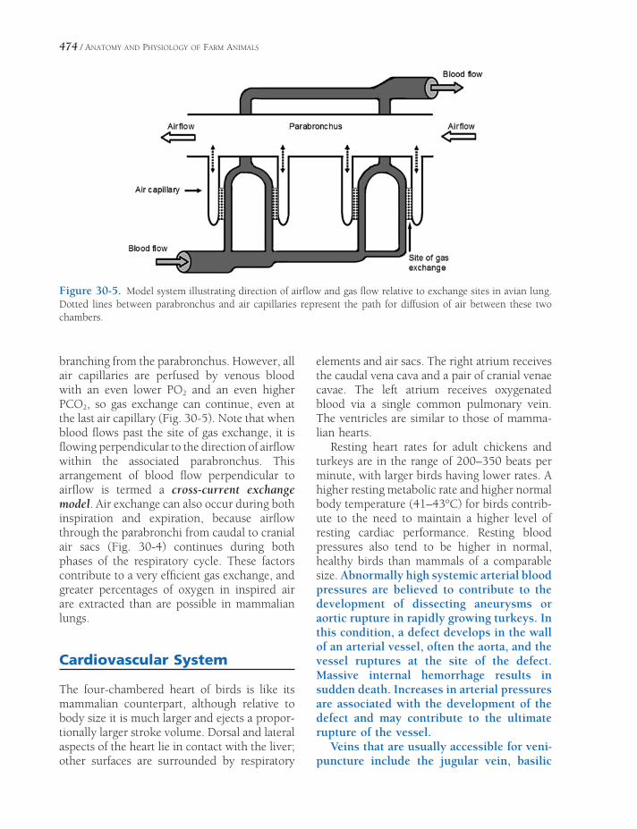
474 / ANATOMY AND PHYSIOLOGY OF FARM ANIMALS
Figure 30-5. Model system illustrating direction of airfl ow and gas fl ow relative to exchange sites in avian lung. Dotted lines between parabronchus and air capillaries represent the path for diffusion of air between these two chambers.
branching from the parabronchus. However, all air capillaries are perfused by venous blood with an even lower PO2 and an even higher PCO2, so gas exchange can continue, even at the last air capillary (Fig. 30-5). Note that when blood fl ows past the site of gas exchange, it is fl owing perpendicular to the direction of airfl ow within the associated parabronchus. This arrangement of blood fl ow perpendicular to airfl ow is termed a cross-current exchange model. Air exchange can also occur during both inspiration and expiration, because airfl ow through the parabronchi from caudal to cranial air sacs (Fig. 30-4) continues during both phases of the respiratory cycle. These factors contribute to a very effi cient gas exchange, and greater percentages of oxygen in inspired air are extracted than are possible in mammalian lungs.
Cardiovascular System
The four-chambered heart of birds is like its mammalian counterpart, although relative to body size it is much larger and ejects a propor-tionally larger stroke volume. Dorsal and lateral aspects of the heart lie in contact with the liver; other surfaces are surrounded by respiratory
elements and air sacs. The right atrium receives the caudal vena cava and a pair of cranial venae cavae. The left atrium receives oxygenated blood via a single common pulmonary vein. The ventricles are similar to those of mamma-lian hearts.
Resting heart rates for adult chickens and turkeys are in the range of 200–350 beats per minute, with larger birds having lower rates. A higher resting metabolic rate and higher normal body temperature (41–43°C) for birds contrib-ute to the need to maintain a higher level of resting cardiac performance. Resting blood pressures also tend to be higher in normal, healthy birds than mammals of a comparable size. Abnormally high systemic arterial blood pressures are believed to contribute to the development of dissecting aneurysms or aortic rupture in rapidly growing turkeys. In this condition, a defect develops in the wall of an arterial vessel, often the aorta, and the vessel ruptures at the site of the defect. Massive internal hemorrhage results in sudden death. Increases in arterial pressures are associated with the development of the defect and may contribute to the ultimate rupture of the vessel.
Veins that are usually accessible for veni-puncture include the jugular vein, basilic

POULTRY / 475
vein (sometimes called the superfi cial ulnar vein or simply the “wing vein,” owing to its location on the medial side of the brachium), and the medial metatarsal vein.
Lymphatic System
Avian lymph vessels are less numerous and less widely distributed than the analogous struc-tures in mammals. They run satellite to blood vessels and most ultimately converge on paired thoracic ducts that, in turn, empty into the paired cranial venae cavae. Only Anseriformes possess true encapsulated lymph nodes; in these species, the cervicothoracic lymph nodes are found within the thoracic inlet and the lumbar lymph nodes ventral to the vertebral column in the region of the kidneys. Lymph nodules are found abundantly associated with most organs and in the walls of the gastrointes-tinal tract. These are especially prominent in the ceca of chickens, where they may be referred to as cecal tonsils.
The cloacal bursa has already been men-tioned as a diverticulum of the proctodeum. It is well developed in young birds and disappears following sexual maturation. During its short existence, it is an important lymphatic organ, being the maturation site for B-lymphocytes.
The avian spleen lies in contact with the right lobe of the liver adjacent to the gizzard. In addition to its important role in erythrocyte physiology, the spleen produces lymphocytes that are clustered in sheaths around splenic arterioles (the so-called white pulp of the spleen).
Urinary System
Avian kidneys lie on the ventral surface of the synsacrum. Each kidney is divided into cranial, middle, and caudal lobes created by external grooves associated with large vessels that cross the renal surface (Fig. 30-6). Collecting ducts empty directly into the ureter adjacent to the cranial lobe of the kidney; there is neither renal pelvis nor renal calyces as in other domestic
species. Right and left ureters terminate in the dorsal part of the urodeum.
Uniquely, birds possess a renal portal system in addition to the expected renal arterial blood supply (one renal artery per lobe) and renal venous return (Fig. 30-6). This system, consist-ing of cranial and caudal renal portal veins, receives venous blood from the caudal body and distributes it into a second capillary system in the interlobular spaces.
The presence of the renal portal system has been used as a justifi cation for avoiding the caudal body as a site for injection of medications in birds. One reason for this avoidance is that the drugs may be more rapidly excreted when injected here than is desirable due to their being immediately transported to and removed by the kidney. Another concern is that drugs that are poten-tially nephrotoxic (damaging to the kidneys) may be more so when they are fi rst distrib-uted undiluted to the kidneys via the portal system.
As in mammals, avian kidneys are composed of nephrons that perform glomerular fi ltration, selective tubular reabsorption, and selective tubular secretion in the process of urine forma-tion. However, avian kidneys contain two dif-ferent types of nephrons, and one type is markedly different in structure compared to nephrons of mammalian kidneys. This unique type of nephron is termed a reptilian-type (RT) nephron and is similar to those found in kidneys of reptiles. Renal corpuscles of RT nephrons are found in the lobules of avian kidneys, and a renal tubule connects each renal corpuscle to a collecting duct also found in the lobule (Fig. 30-7). However, the tubules of the RT nephrons do not include a segment that corresponds to a loop of Henle, and the tubules of the RT nephrons are contained in the outer zones of the lobules, where they connect to collecting ducts (Figure 30-7). Avian kidneys also contain mammalian-type (MT) nephrons that do have segments that correspond to the loops of Henle in mammalian kidneys, and these loops extend downward into medullary cones that support the lobules (Figure 30-7). These tubular loops of MT nephrons in avian kidneys function

476 / ANATOMY AND PHYSIOLOGY OF FARM ANIMALS
Figure 30-6. The avian genitourinary tract (male).(Reprinted with permission of Wolters Kluwer from Rosskopf, W. and Woerpel, R. Diseases of Cage and Aviary Birds, 3rd ed. Philadelphia: Williams & Wilkins, 1996.)
similarly to loops of Henle in mammalian kidneys and contribute to the formation of an osmotic gradient within the interstitial fl uids of the medullary cone. However, the magnitude of the gradient is much less in birds. Tubular segments corresponding to proximal tubules and distal tubules can be found in both MT and RT nephrons, and these segments in birds perform secretory and reabsorptive functions similar to nephrons in mammalian kidneys.
Glomerular fi ltration rate (GFR) remains relatively stable in normal mammals despite
mild to moderate changes in hydration status. However, in birds, moderate dehydration, such as with periods of water restriction, is associated with signifi cant reductions in GFR. Some suggest that this is primarily due to vasocon-striction of vessels supplying the RT nephrons.
To affect water balance in response to changes in water intake, the water permeability of col-lecting ducts in mammalian kidneys is regu-lated by antidiuretic hormone. Collecting ducts in avian kidneys respond similarly to arginine vasotocin (AVT), the avian antidiuretic
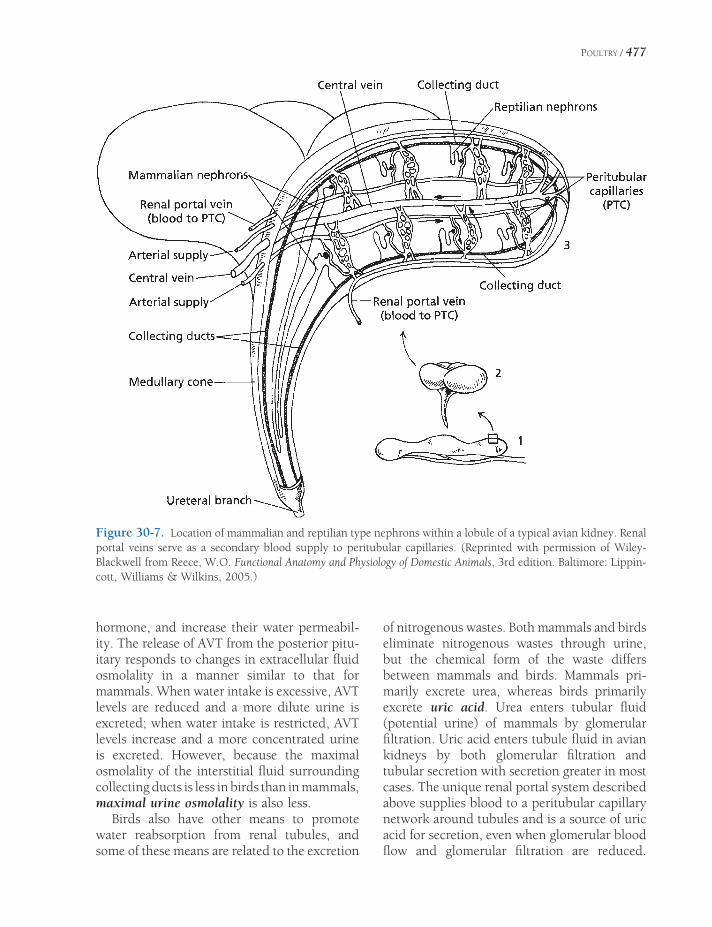
POULTRY / 477
hormone, and increase their water permeabil-ity. The release of AVT from the posterior pitu-itary responds to changes in extracellular fl uid osmolality in a manner similar to that for mammals. When water intake is excessive, AVT levels are reduced and a more dilute urine is excreted; when water intake is restricted, AVT levels increase and a more concentrated urine is excreted. However, because the maximal osmolality of the interstitial fl uid surrounding collecting ducts is less in birds than in mammals, maximal urine osmolality is also less.
Birds also have other means to promote water reabsorption from renal tubules, and some of these means are related to the excretion
of nitrogenous wastes. Both mammals and birds eliminate nitrogenous wastes through urine, but the chemical form of the waste differs between mammals and birds. Mammals pri-marily excrete urea, whereas birds primarily excrete uric acid. Urea enters tubular fl uid (potential urine) of mammals by glomerular fi ltration. Uric acid enters tubule fl uid in avian kidneys by both glomerular fi ltration and tubular secretion with secretion greater in most cases. The unique renal portal system described above supplies blood to a peritubular capillary network around tubules and is a source of uric acid for secretion, even when glomerular blood fl ow and glomerular fi ltration are reduced.
Figure 30-7. Location of mammalian and reptilian type nephrons within a lobule of a typical avian kidney. Renal portal veins serve as a secondary blood supply to peritubular capillaries. (Reprinted with permission of Wiley-Blackwell from Reece, W.O. Functional Anatomy and Physiology of Domestic Animals, 3rd edition. Baltimore: Lippin-cott, Williams & Wilkins, 2005.)

478 / ANATOMY AND PHYSIOLOGY OF FARM ANIMALS
Because urea is not a major nitrogenous waste in birds, measure of blood urea nitro-gen (BUN) levels cannot be used as indica-tors of renal function. Plasma uric acid levels can be used, but because most uric acid enters tubular fl uid by secretion, signifi cant renal tubular dysfunction must occur before plasma uric acid increases.
Within renal tubules of avian kidneys, unique, extremely small (3–13 μm), spherical concretions or particles form, and these contain uric acid, albumin, and inorganic ions. The accumulation of multiple osmotic particles into a single particle effectively reduces the osmolal-ity of the tubular fl uid and permits additional water reabsorption. Albumin is readily avail-able in the tubular fl uid of birds for it contains signifi cantly more plasma protein than in mammals.
As discussed earlier, urine may be moved from the urodeum by reverse peristalsis to the ceca, and additional absorption of water and electrolytes may occur in the coprodeum, rectum, and ceca. Uric acid in the urine entering the cloaca may precipitate on feces, and these are seen as a white pasty part of the droppings.
Female Reproductive System
The production of eggs for human consump-tion is a primary reason for keeping poultry worldwide. Among female birds, both right and left ovaries and oviducts are present embryo-logically, but in chickens, turkeys, and domes-tic geese the right organs regress early in development and only the left side develops (both sides persist and develop in ducks). The ovaries are located cranioventral to the corre-sponding kidney, and the size varies with the reproductive status of the bird. Prior to the onset of the fi rst laying period, the small ovary is smooth; as puberty is approached, it begins to develop a granular, then cobblestone appear-ance as follicles enlarge in preparation for ovu-lation. Just prior to “coming into lay,” the ovary will resemble a bunch of grapes.
The mature ovum is more-or-less equivalent to the yolk of the egg; it will be released at
ovulation and received into the expanded ter-minus of the oviduct, the infundibulum (Figure 30-8). It is in the infundibulum that fertiliza-tion will take place if the bird is allowed to breed. The oviduct is variable in size, being maximally expanded and lengthened during the lay (60–70 cm long in the hen) and regress-ing in size during broodiness and the molt. It possesses grossly identifi able sections (the magnum, isthmus, uterus, and vagina) that cor-relate with the important tasks of producing the albumen (the “white” of the egg) and the laying down of the egg shell. The oviduct terminates in a slitlike aperture in the urodeum, next to the opening of the ureter (Figure 30-8).
Egg Formation and Oviposition
The number of eggs laid consecutively by wild birds during any period of egg laying is termed a clutch. The number of eggs per clutch for a wild bird is probably related to the ability of the species to incubate the eggs and raise the hatchlings. The time period between clutches for wild birds may extend to months or years. Under typical management practices for fl ocks of domestic chickens used for egg production, eggs do not remain with hens after the eggs are laid. However, hens typically lay a number of eggs consecutively for several days and then interrupt laying for at least 1 day. Some refer to this pattern as a sequence of eggs, rather than a clutch. During a 12-month laying period, hens may produce more than 300 eggs by laying multiple sequences (clutches).
The time period between ovulation and ovi-position (expulsion of the egg) for the domestic hen is about 24–26 hours. Ovulation of the next egg occurs after oviposition of the preced-ing egg, so only one egg is in the oviduct at a time. As in mammals, ovulation is preceded by a sudden, rapid rise in luteinizing hormone (LH), and this hormone surge is believed to be responsible for changes in the mature follicle about to be released from the ovary. Four to 6 hours prior to the LH surge, plasma progester-one levels peak at the highest levels seen in a laying cycle, and this peak appears to be neces-

POULTRY / 479
Figure 30-8. The avian genitourinary tract (female). (Reprinted with permission of Wolters Kluwer from Rosskopf, W. and Woerpel, R. Diseases of Cage and Aviary Birds, 3rd edition. Philadelphia: Williams & Wilkins, 1996.)
sary for the subsequent surge. The source of this progesterone appears to be the largest and most mature ovarian follicle. Unlike mammals, there is no corpus luteum formed on an ovary at the site of an ovulated follicle. Progesterone receptors are found in various sites throughout the oviduct, where progesterone may promote secretions and muscular contractions for egg development and transport, respectively.
For fertilization to occur, spermatozoa must reach the egg before it enters the tubular segment of the infundibulum where the fi rst albumen layer will be secreted to surround the
egg. Spermatozoa may be temporarily stored in the infundibulum so that they can be readily available for the relatively short period that fer-tilization is possible. In the domestic hen, the egg is in the infundibulum for about 15 minutes. Spermatozoa are also stored in specialized tubules in the uterovaginal region for longer periods. In the domestic hen, spermatozoa remain viable for 7–14 days when stored here. Spermatozoa are released from this site in asso-ciation with ovipostion of each egg and migrate to the infundibulum to be available to fertilize the next egg. Ovulation of the next egg occurs

480 / ANATOMY AND PHYSIOLOGY OF FARM ANIMALS
about 30–45 minutes after oviposition of the previous egg.
Albumen (egg white) secretion around the yolk begins in the infundibulum, but the major-ity is secreted by the magnum. Albumen pri-marily consists of water, protein, and minerals and is a source of these nutrients for the devel-oping embryo. Some of the proteins also have antimicrobial properties and protect against microorganisms. The chalazae are two twisted, fi berlike structures that extend from opposite sides of the yolk to each end of the egg (Fig. 30-9). These are formed from fi bers in the inner layers of the albumen when the yolk rotates as it passes through the oviduct.
The eggshell consists of four layers that were applied to the outer surface of the albumen. From inner to outer, the layers are (1) shell membranes, (2) mammillary cores, (3) matrix, and (4) cuticle. The combination of the mam-millary cores and matrix has also been termed the testa. The innermost shell membranes permit the exchange of gases and water, but these mem-branes are not permeable to albumen. As eggs age, the inner and outer shell membranes tend to separate at the large end of the egg, and an air cell is formed (Fig. 30-9). The size of an air cell increases as an egg continues to
age. Egg candling, shining a beam of light through the intact egg to view the interior, is used to determine the size of the air cell, and this is one measure of egg quality.
The mineral component of the eggshell, pri-marily calcium carbonate, is deposited within the mammillary core and matrix layers. The mammillary cores and matrix layers contain proteins, carbohydrates, and mucopolysaccha-rides, which provide a framework for the depo-sition of calcium carbonate crystals (i.e., calcifi cation). Several different proteins that contribute to this framework have been identi-fi ed, and experiments suggest that the appro-priate production of these proteins by the shell gland is a key factor in determining shell quality. Gene expression studies indicate that the pro-duction of these proteins is infl uenced by hor-monal factors, such as progesterone, and that rates of gene expression vary greatly as the egg passes through the shell gland. The outermost cuticle layer contains lipid, prevents evapora-tion, is water repellent, and functions as a barrier to microorganisms.
Eggshell formation in the uterus or shell gland of the domestic chicken requires about 17–20 hours. During this period, approxi-mately 2–2.5 grams of calcium are transported
Figure 30-9. Basic internal structure of a typical egg. (Reprinted with permission of Wiley-Blackwell from Reece, W.O. Functional Anatomy and Physiology of Domestic Animals, 3rd ed. Baltimore: Lippincott, Williams & Wilkins, 2005.)

POULTRY / 481
from the blood by the uterus and deposited in the eggshell as calcium carbonate crystals (calcite). Approximately 95% of the total weight of an eggshell is calcium carbonate. Plasma calcium levels decrease during the period of eggshell formation and then rebound before the next period of eggshell formation begins. During periods of active eggshell formation, calcium is added to the blood by intestinal absorption and mobilization of medullary bone. If dietary calcium is adequate, the majority of the necessary calcium can be obtained during this period by intestinal absorption. However, a net loss of medullary bone calcium will occur during the hours of active eggshell formation, even with adequate calcium intakes. Replace-ment of the lost medullary bone can be done with calcium gained by intestinal absorption during the hours that active eggshell formation is not occurring. Vitamin D stimulates both the intestinal absorption of calcium and transport of calcium by the shell gland (uterus). Parathy-roid hormone also promotes calcium transport by the shell gland for eggshell formation.
Osteoclasts are responsible for the mobiliza-tion of calcium from medullary bone, so the increased mobilization during eggshell forma-tion refl ects an increased osteoclastic activity. Osteoclasts also mobilize calcium from lamellar cortical bone throughout the skeleton, and a net loss of calcium from cortical bone does occur in high-producing laying chickens who have been laying for extended periods. Osteo-porosis, a progressive decrease in mineral-ized bone leading to bone fragility and increased risk for fractures, is recognized in laying fl ocks and is believed to contribute to the syndrome cage layer fatigue, in which birds are found paralyzed in their cage. Numerous fractures may be found involving the ribs and long bones of such birds.
Calcium carbonate deposition within the eggshell also requires the localized formation of carbonate ions within the shell gland. This gen-eration of carbonate ions is dependent on car-bonic anhydrase activity within the gland. Under the infl uence of carbonic anhydrase, CO2 from blood perfusing the gland is used to produce carbonate ions. Laying chickens sub-
jected to abnormally high environmental temperatures (heat stress) typically have declines in egg production and in the shell quality of eggs that are produced. The decline in shell quality is associated with a reduc-tion in blood PCO2, as a result of hyperven-tilation induced by heat stress. The lower CO2 is believed to adversely affect the ability of the shell gland to produce carbonate ions. Heat-stressed birds also reduce food intake, which would also contribute to the decline in egg quantity and quality.
Arginine vasotocin (AVT) stimulates uterine contractions during oviposition. Like oxytocin in mammals, AVT is released from the posterior pituitary, and its plasma levels rise signifi cantly shortly before and peak during oviposition. Unlike mammals, birds have one small peptide hormone, AVT, that functions in both the regu-lation of urine osmolality and the regulation of uterine contractions.
Male Reproductive System
Testes in domestic birds are normally paired and located cranioventral to the kidneys (Fig. 30-6). Like the ovaries, testes are smaller outside of breeding seasons and will increase markedly in size during breeding periods. The microscopic appearance of an avian testis is similar to a mammalian testis in that it primar-ily consists of seminiferous tubules surrounded by an interstitium that contains Leydig cells. The process of spermatogenesis in the tubules is similar to that for mammals and is promoted by FSH and LH. However, unlike in mammals, spermatogenesis occurs in avian testes found normally within the body cavity, where they are maintained at normal core body temperature.
The ductus deferens (deferent duct) conveys sperm from the testis to a raised papilla on the lateral aspect of the urodeum. Male birds do not possess accessory sex glands; the ejaculate is composed of sperm and scant additional secretions produced by the testes and the walls of the ducts.
On the ventral fl oor of the proctodeum of roosters and male turkeys there lies a copulatory

482 / ANATOMY AND PHYSIOLOGY OF FARM ANIMALS
organ, the phallus, which is nonprotrusible (i.e., does not form a protruding copulatory organ). The phallus has erectile tissue within it, and during copulation the engorged tissue forms a groove that conveys the ejaculate from the openings of the deferent ducts to the surface of the phallus. During the act of breeding, the proctodeum is everted and the phallus pressed against the vent of the female so as to facilitate the transfer of ejaculate.
Drakes and ganders (and, incidentally, male swans and ostriches) have a protrusible phallus that, when erect, is about 5 cm long. Its tip is spiraled and is introduced into the cloaca of the female during copulation.
Sex Chromosomes
Unlike mammals, in birds the female is the gender that is the heterogametic sex (carrying one copy of each of two different sex chromo-somes). Female birds carry Z and W sex chro-mosomes, and males carry two copies of the Z chromosome. Normally, females can contribute either Z or W to progeny, whereas males can contribute only Z.
Reproduction and Photoperiods
Reproductive processes in all female birds are subject to regulation by changes in photoperi-ods (period of light exposure per day). Increas-
ing periods of light, such as during the spring, induce hormonal changes in wild female birds to initiate egg production and mating behavior. Longer periods of light are associated with the release of luteinizing hormone-releasing hormone (LHRH) from the hypothalamus, which in turn stimulates the release of LH from the anterior pituitary.
Breeds of domestic chickens selected for high egg production will begin egg laying if they reach maturity during either spring (increasing day length) or fall (decreasing day length). However, chickens raised during the spring reach maturity and begin laying at earlier ages. Egg size is reduced when smaller, younger chickens begin laying, so producers often manipulate photoperiods to delay the beginning of laying until birds are of an appropriate size. After laying has begun, reductions in photoperiods will lower egg production, as compared to egg produc-tion with a constant photoperiod.
Increasing periods of light are associated with testicular enlargement and development in birds that are seasonal breeders. Male domes-tic chickens (roosters) will reach sexual matu-rity and begin spermatogenesis when raised with either a long photoperiod (14 or more hours of light per day) or a short photoperiod (such as 8 hours per day). However, spermato-genesis is delayed with shortened photoperi-ods. The beginning of spermatogenesis is associated with increasing levels of FSH and LH. Mature males must be maintained with appropriate photoperiods for maximal fertility.
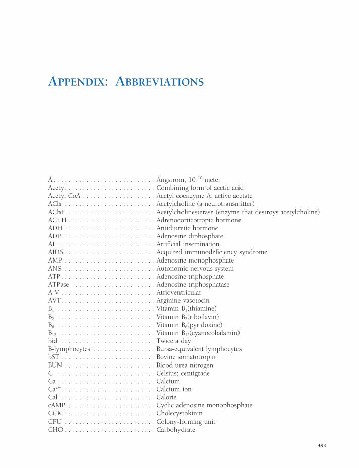
APPENDIX: ABBREVIATIONS
Å . . . . . . . . . . . . . . . . . . . . . . . . . . . . Ångstrom, 10−10 meterAcetyl . . . . . . . . . . . . . . . . . . . . . . . . Combining form of acetic acidAcetyl CoA . . . . . . . . . . . . . . . . . . . . Acetyl coenzyme A, active acetateACh . . . . . . . . . . . . . . . . . . . . . . . . . Acetylcholine (a neurotransmitter)AChE . . . . . . . . . . . . . . . . . . . . . . . . Acetylcholinesterase (enzyme that destroys acetylcholine)ACTH . . . . . . . . . . . . . . . . . . . . . . . . Adrenocorticotropic hormoneADH . . . . . . . . . . . . . . . . . . . . . . . . . Antidiuretic hormoneADP. . . . . . . . . . . . . . . . . . . . . . . . . . Adenosine diphosphateAI . . . . . . . . . . . . . . . . . . . . . . . . . . . Artifi cial inseminationAIDS . . . . . . . . . . . . . . . . . . . . . . . . . Acquired immunodefi ciency syndromeAMP . . . . . . . . . . . . . . . . . . . . . . . . . Adenosine monophosphateANS . . . . . . . . . . . . . . . . . . . . . . . . . Autonomic nervous systemATP. . . . . . . . . . . . . . . . . . . . . . . . . . Adenosine triphosphateATPase . . . . . . . . . . . . . . . . . . . . . . . Adenosine triphosphataseA-V . . . . . . . . . . . . . . . . . . . . . . . . . . AtrioventricularAVT. . . . . . . . . . . . . . . . . . . . . . . . . . Arginine vasotocinB1 . . . . . . . . . . . . . . . . . . . . . . . . . . . Vitamin B1(thiamine)B2 . . . . . . . . . . . . . . . . . . . . . . . . . . . Vitamin B2(ribofl avin)B6 . . . . . . . . . . . . . . . . . . . . . . . . . . . Vitamin B6(pyridoxine)B12 . . . . . . . . . . . . . . . . . . . . . . . . . . Vitamin B12(cyanocobalamin)bid . . . . . . . . . . . . . . . . . . . . . . . . . . Twice a dayB-lymphocytes . . . . . . . . . . . . . . . . . Bursa-equivalent lymphocytesbST . . . . . . . . . . . . . . . . . . . . . . . . . . Bovine somatotropinBUN . . . . . . . . . . . . . . . . . . . . . . . . . Blood urea nitrogenC . . . . . . . . . . . . . . . . . . . . . . . . . . . Celsius; centigradeCa . . . . . . . . . . . . . . . . . . . . . . . . . . . CalciumCa2+ . . . . . . . . . . . . . . . . . . . . . . . . . . Calcium ionCal . . . . . . . . . . . . . . . . . . . . . . . . . . CaloriecAMP . . . . . . . . . . . . . . . . . . . . . . . . Cyclic adenosine monophosphateCCK . . . . . . . . . . . . . . . . . . . . . . . . . CholecystokininCFU . . . . . . . . . . . . . . . . . . . . . . . . . Colony-forming unitCHO . . . . . . . . . . . . . . . . . . . . . . . . . Carbohydrate
483

484 / ANATOMY AND PHYSIOLOGY OF FARM ANIMALS
Cl . . . . . . . . . . . . . . . . . . . . . . . . . . . ChlorineCl−. . . . . . . . . . . . . . . . . . . . . . . . . . . Chloride ioncm . . . . . . . . . . . . . . . . . . . . . . . . . . CentimeterCNS . . . . . . . . . . . . . . . . . . . . . . . . . Central nervous systemCO . . . . . . . . . . . . . . . . . . . . . . . . . . Carbon monoxide; Cardiac outputCO2 . . . . . . . . . . . . . . . . . . . . . . . . . Carbon dioxideCoA . . . . . . . . . . . . . . . . . . . . . . . . . Coenzyme A (acyl activation)CP . . . . . . . . . . . . . . . . . . . . . . . . . . . Creatine phosphateCRH . . . . . . . . . . . . . . . . . . . . . . . . . Corticotropin-releasing hormoneCSF . . . . . . . . . . . . . . . . . . . . . . . . . . Cerebrospinal fl uid; colony-stimulating factorCu. . . . . . . . . . . . . . . . . . . . . . . . . . . CopperD. . . . . . . . . . . . . . . . . . . . . . . . . . . . Vitamin D (antirachitic vitamin)dB . . . . . . . . . . . . . . . . . . . . . . . . . . . Decibel, a unit of sound intensityDNA . . . . . . . . . . . . . . . . . . . . . . . . . Deoxyribonucleic acidDOPA . . . . . . . . . . . . . . . . . . . . . . . . DihydroxyphenylalanineDPG . . . . . . . . . . . . . . . . . . . . . . . . . DiphosphoglycerideECF. . . . . . . . . . . . . . . . . . . . . . . . . . Extracellular fl uideCG . . . . . . . . . . . . . . . . . . . . . . . . . Equine chorionic gonadotropin (see PMSG)ECG . . . . . . . . . . . . . . . . . . . . . . . . . ElectrocardiogramEDTA . . . . . . . . . . . . . . . . . . . . . . . . Ethylene diaminetetraacetic acidEPP . . . . . . . . . . . . . . . . . . . . . . . . . . End-plate potentialEPSP . . . . . . . . . . . . . . . . . . . . . . . . . Excitatory postsynaptic potentialER . . . . . . . . . . . . . . . . . . . . . . . . . . . Endoplasmic reticulumF . . . . . . . . . . . . . . . . . . . . . . . . . . . . FahrenheitFAD . . . . . . . . . . . . . . . . . . . . . . . . . Oxidized adenine dinucleotideFADH . . . . . . . . . . . . . . . . . . . . . . . . Reduced adenine dinucleotideFe . . . . . . . . . . . . . . . . . . . . . . . . . . . IronFe2+ . . . . . . . . . . . . . . . . . . . . . . . . . . Ferrous iron (low valence)Fe3+ . . . . . . . . . . . . . . . . . . . . . . . . . . Ferric iron (high valence)FFA . . . . . . . . . . . . . . . . . . . . . . . . . Free fatty acidsFSH . . . . . . . . . . . . . . . . . . . . . . . . . Follicle-stimulating hormoneg . . . . . . . . . . . . . . . . . . . . . . . . . . . . GramGABA . . . . . . . . . . . . . . . . . . . . . . . . γ-Aminobutyric acidGALT . . . . . . . . . . . . . . . . . . . . . . . . Gut-associated lymphatic tissueGFR . . . . . . . . . . . . . . . . . . . . . . . . . Glomerular fi ltration rateGH . . . . . . . . . . . . . . . . . . . . . . . . . . Growth hormoneGHRH . . . . . . . . . . . . . . . . . . . . . . . . Growth hormone–releasing hormoneGHIH . . . . . . . . . . . . . . . . . . . . . . . . Growth hormone–inhibiting hormone (somatostatin)GI . . . . . . . . . . . . . . . . . . . . . . . . . . . GastrointestinalGIP . . . . . . . . . . . . . . . . . . . . . . . . . . Gastric inhibitory peptideGnRH . . . . . . . . . . . . . . . . . . . . . . . . Gonadotropin-releasing hormoneH+ . . . . . . . . . . . . . . . . . . . . . . . . . . . Hydrogen ionH & E . . . . . . . . . . . . . . . . . . . . . . . . Hematoxylin and eosin stainHb . . . . . . . . . . . . . . . . . . . . . . . . . . HemoglobinHbO2 . . . . . . . . . . . . . . . . . . . . . . . . OxyhemoglobinhCG . . . . . . . . . . . . . . . . . . . . . . . . . Human chorionic gonadotropinHCl . . . . . . . . . . . . . . . . . . . . . . . . . . Hydrochloric acidHCO3
− . . . . . . . . . . . . . . . . . . . . . . . . . Bicarbonate ionH2CO3 . . . . . . . . . . . . . . . . . . . . . . . Carbonic acid

APPENDIX: ABBREVIATIONS / 485
HDL . . . . . . . . . . . . . . . . . . . . . . . . . High-density lipoproteinHg . . . . . . . . . . . . . . . . . . . . . . . . . . MercuryH2O . . . . . . . . . . . . . . . . . . . . . . . . . Water (HOH)H2O2 . . . . . . . . . . . . . . . . . . . . . . . . . Hydrogen peroxideHPO4
= . . . . . . . . . . . . . . . . . . . . . . . . . Monobasic phosphate ionH3PO4 . . . . . . . . . . . . . . . . . . . . . . . . Phosphoric acidI . . . . . . . . . . . . . . . . . . . . . . . . . . . . IodineICSH . . . . . . . . . . . . . . . . . . . . . . . . . Interstitial cell–stimulating hormone (LH)IDL . . . . . . . . . . . . . . . . . . . . . . . . . . Intermediate-density lipoproteinIg . . . . . . . . . . . . . . . . . . . . . . . . . . . ImmunoglobulinIGF . . . . . . . . . . . . . . . . . . . . . . . . . . Insulin-like growth factorIM . . . . . . . . . . . . . . . . . . . . . . . . . . . IntramuscularIP . . . . . . . . . . . . . . . . . . . . . . . . . . . IntraperitonealIPSP . . . . . . . . . . . . . . . . . . . . . . . . . Inhibitory postsynaptic potentialIU . . . . . . . . . . . . . . . . . . . . . . . . . . . International unitIV . . . . . . . . . . . . . . . . . . . . . . . . . . . IntravenousK . . . . . . . . . . . . . . . . . . . . . . . . . . . PotassiumKcal . . . . . . . . . . . . . . . . . . . . . . . . . KilocalorieL . . . . . . . . . . . . . . . . . . . . . . . . . . . . literLDH . . . . . . . . . . . . . . . . . . . . . . . . . Lactate dehydrogenaseLDL. . . . . . . . . . . . . . . . . . . . . . . . . . Low-density lipoproteinLH . . . . . . . . . . . . . . . . . . . . . . . . . . Luteinizing hormoneM . . . . . . . . . . . . . . . . . . . . . . . . . . . Molar concentrationMAO . . . . . . . . . . . . . . . . . . . . . . . . . Monoamine oxidasemEq . . . . . . . . . . . . . . . . . . . . . . . . . Milliequivalentmg . . . . . . . . . . . . . . . . . . . . . . . . . . Milligram (10−3 gram)μm . . . . . . . . . . . . . . . . . . . . . . . . . . Micrometer (10−6 meter)mL . . . . . . . . . . . . . . . . . . . . . . . . . . Milliliter (10−3 of a liter)mm . . . . . . . . . . . . . . . . . . . . . . . . . . Millimeter (10−3 meter)mM . . . . . . . . . . . . . . . . . . . . . . . . . . MillimolemOsm . . . . . . . . . . . . . . . . . . . . . . . MilliosmolemRNA . . . . . . . . . . . . . . . . . . . . . . . Messenger ribonucleic acidmsec . . . . . . . . . . . . . . . . . . . . . . . . . Millisecond (10−3 second)MSH . . . . . . . . . . . . . . . . . . . . . . . . . Melanocyte-stimulating hormonemv . . . . . . . . . . . . . . . . . . . . . . . . . . Millivolt (10−3 volt)MW . . . . . . . . . . . . . . . . . . . . . . . . . Molecular weightN . . . . . . . . . . . . . . . . . . . . . . . . . . . NitrogenNa . . . . . . . . . . . . . . . . . . . . . . . . . . . SodiumNaCl . . . . . . . . . . . . . . . . . . . . . . . . . Sodium chloride, table saltNAD . . . . . . . . . . . . . . . . . . . . . . . . . Oxidized nicotinamide adenine dinucleotideNADH . . . . . . . . . . . . . . . . . . . . . . . Reduced nicotinamide adenine dinucleotideNaHCO3 . . . . . . . . . . . . . . . . . . . . . . Sodium bicarbonateNaOH . . . . . . . . . . . . . . . . . . . . . . . . Sodium hydroxideNa+–K+–ATPase . . . . . . . . . . . . . . . . Sodium- and potassium-activated adenosine triphosphataseNE . . . . . . . . . . . . . . . . . . . . . . . . . . NorepinephrineNF . . . . . . . . . . . . . . . . . . . . . . . . . . National Formularyng . . . . . . . . . . . . . . . . . . . . . . . . . . . Nanogram (10−9 gram)NH−
2 . . . . . . . . . . . . . . . . . . . . . . . . . Amino groupNH3 . . . . . . . . . . . . . . . . . . . . . . . . . Ammonia
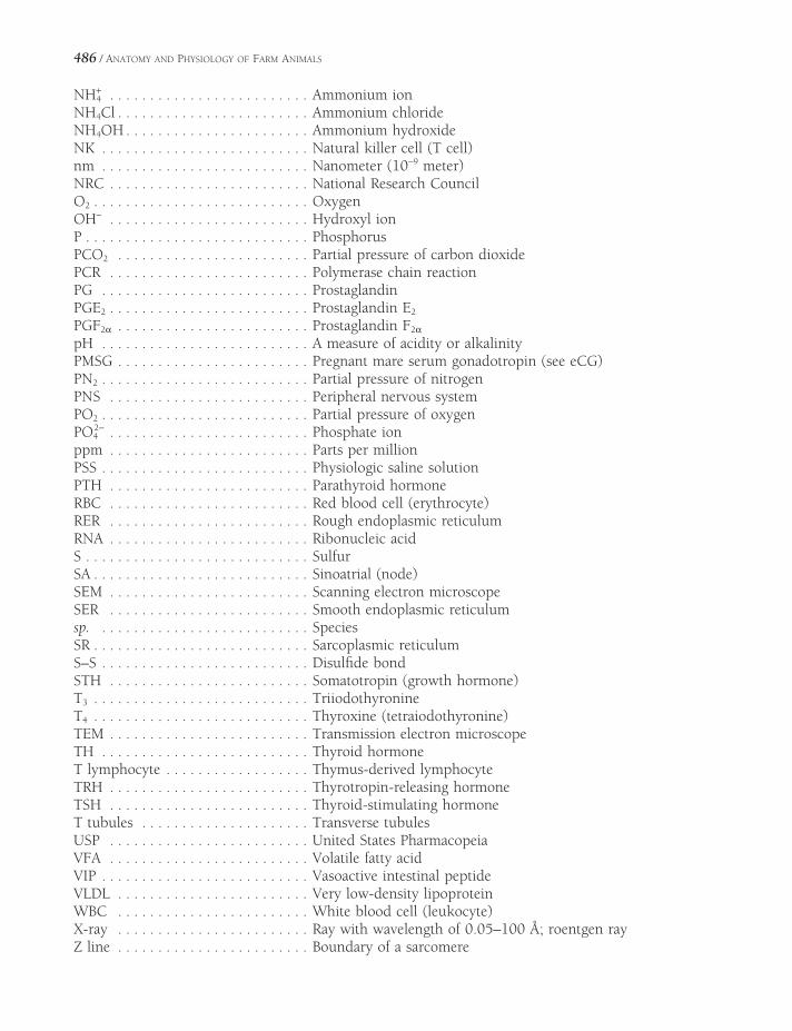
486 / ANATOMY AND PHYSIOLOGY OF FARM ANIMALS
NH+4 . . . . . . . . . . . . . . . . . . . . . . . . . Ammonium ion
NH4Cl . . . . . . . . . . . . . . . . . . . . . . . . Ammonium chlorideNH4OH . . . . . . . . . . . . . . . . . . . . . . . Ammonium hydroxideNK . . . . . . . . . . . . . . . . . . . . . . . . . . Natural killer cell (T cell)nm . . . . . . . . . . . . . . . . . . . . . . . . . . Nanometer (10−9 meter)NRC . . . . . . . . . . . . . . . . . . . . . . . . . National Research CouncilO2 . . . . . . . . . . . . . . . . . . . . . . . . . . . OxygenOH− . . . . . . . . . . . . . . . . . . . . . . . . . Hydroxyl ionP . . . . . . . . . . . . . . . . . . . . . . . . . . . . PhosphorusPCO2 . . . . . . . . . . . . . . . . . . . . . . . . Partial pressure of carbon dioxidePCR . . . . . . . . . . . . . . . . . . . . . . . . . Polymerase chain reactionPG . . . . . . . . . . . . . . . . . . . . . . . . . . ProstaglandinPGE2 . . . . . . . . . . . . . . . . . . . . . . . . . Prostaglandin E2
PGF2α . . . . . . . . . . . . . . . . . . . . . . . . Prostaglandin F2α
pH . . . . . . . . . . . . . . . . . . . . . . . . . . A measure of acidity or alkalinityPMSG . . . . . . . . . . . . . . . . . . . . . . . . Pregnant mare serum gonadotropin (see eCG)PN2 . . . . . . . . . . . . . . . . . . . . . . . . . . Partial pressure of nitrogenPNS . . . . . . . . . . . . . . . . . . . . . . . . . Peripheral nervous systemPO2 . . . . . . . . . . . . . . . . . . . . . . . . . . Partial pressure of oxygenPO4
2− . . . . . . . . . . . . . . . . . . . . . . . . . Phosphate ionppm . . . . . . . . . . . . . . . . . . . . . . . . . Parts per millionPSS . . . . . . . . . . . . . . . . . . . . . . . . . . Physiologic saline solutionPTH . . . . . . . . . . . . . . . . . . . . . . . . . Parathyroid hormoneRBC . . . . . . . . . . . . . . . . . . . . . . . . . Red blood cell (erythrocyte)RER . . . . . . . . . . . . . . . . . . . . . . . . . Rough endoplasmic reticulumRNA . . . . . . . . . . . . . . . . . . . . . . . . . Ribonucleic acidS . . . . . . . . . . . . . . . . . . . . . . . . . . . . SulfurSA . . . . . . . . . . . . . . . . . . . . . . . . . . . Sinoatrial (node)SEM . . . . . . . . . . . . . . . . . . . . . . . . . Scanning electron microscopeSER . . . . . . . . . . . . . . . . . . . . . . . . . Smooth endoplasmic reticulumsp. . . . . . . . . . . . . . . . . . . . . . . . . . . SpeciesSR . . . . . . . . . . . . . . . . . . . . . . . . . . . Sarcoplasmic reticulumS–S . . . . . . . . . . . . . . . . . . . . . . . . . . Disulfi de bondSTH . . . . . . . . . . . . . . . . . . . . . . . . . Somatotropin (growth hormone)T3 . . . . . . . . . . . . . . . . . . . . . . . . . . . TriiodothyronineT4 . . . . . . . . . . . . . . . . . . . . . . . . . . . Thyroxine (tetraiodothyronine)TEM . . . . . . . . . . . . . . . . . . . . . . . . . Transmission electron microscopeTH . . . . . . . . . . . . . . . . . . . . . . . . . . Thyroid hormoneT lymphocyte . . . . . . . . . . . . . . . . . . Thymus-derived lymphocyteTRH . . . . . . . . . . . . . . . . . . . . . . . . . Thyrotropin-releasing hormoneTSH . . . . . . . . . . . . . . . . . . . . . . . . . Thyroid-stimulating hormoneT tubules . . . . . . . . . . . . . . . . . . . . . Transverse tubulesUSP . . . . . . . . . . . . . . . . . . . . . . . . . United States PharmacopeiaVFA . . . . . . . . . . . . . . . . . . . . . . . . . Volatile fatty acidVIP . . . . . . . . . . . . . . . . . . . . . . . . . . Vasoactive intestinal peptideVLDL . . . . . . . . . . . . . . . . . . . . . . . . Very low-density lipoproteinWBC . . . . . . . . . . . . . . . . . . . . . . . . White blood cell (leukocyte)X-ray . . . . . . . . . . . . . . . . . . . . . . . . Ray with wavelength of 0.05–100 Å; roentgen rayZ line . . . . . . . . . . . . . . . . . . . . . . . . Boundary of a sarcomere

BIBLIOGRAPHY
Bullock, B.L., and R.L. Henze, eds. Focus on Pathophysiology. Philadelphia, Lippincott, Williams & Wilkins, 2000.
Carlson, B.M. Patten’s Foundations of Embryol-ogy. 4th ed. New York, McGraw-Hill, 1981.
Cheville, N.F. Introduction to Veterinary Pathol-ogy. 2nd ed. Ames, Iowa State University Press, 1999.
Cohen, B.J., and D.L. Wood. Memmler’s The Human Body in Health and Disease. 9th ed. Baltimore, Lippincott, Williams & Wilkins, 2000.
Cunningham, J.G. and B.G. Klein, eds. Textbook of Veterinary Physiology. 4th ed. St. Louis, Elsevier, 2007.
De Lahunta, A. Veterinary Neuroanatomy and Clinical Neurology. 2nd ed. Philadelphia, W.B. Saunders, 1983.
Dellman, H.D., and J.A. Eurell. Textbook of Veterinary Histology. 5th ed. Baltimore, Lippincott, Williams & Wilkins, 1998.
Eurell, J.A. and Frappier, B.L. Dellmann’s Text-book of Veterinary Histology, 6th ed. Ames, IA, Blackwell Publishing Professional, 2006.
Dorland, W. A. Dorland’s Illustrated Medical Dictionary. 30th ed. Philadelphia, W.B. Saunders, 2003.
Dyce, K.M., W.O. Sack, and C.J.G. Wensing. Textbook of Veterinary Anatomy. 3rd ed. Philadelphia, W.B. Saunders, 2002.
Adams, H.R., ed. Veterinary Pharmacology and Therapeutics. 8th ed. Ames, Iowa State University Press, 2001.
Alberts, B., A. Johnson, J. Lewis, M. Raff, K. Roberts, and P. Walter. Molecular Biology of the Cell. 4th ed. New York, Garland Pub-lishing, 2002.
Ashdown, R.R., S.H. Done, and S.W. Barnett. Color Atlas of Veterinary Anatomy. Vol. 1. The Ruminants. London/Boston, Mosby-Wolfe, 1996.
Ashdown, R.R., S.H. Done, and S.A. Evans. Color Atlas of Veterinary Anatomy. Vol. 2. The Horse. London/Boston, Mosby-Wolfe, 1996.
Bacha, W.J., Jr. and L.M. Bacha. Color Atlas of Veterinary Histology. 2nd ed. Baltimore, Lippincott Williams & Wilkins, 2000.
Baumel, J., ed. Handbook of Avian Anatomy: Nomina Anatomica Avium. 2nd ed. Cam-bridge, MA, Nuttall Ornithological Club, 1993.
Berne, R.M., and M.N. Levy. Principles of Physi-ology. 3rd ed. St. Louis, Mosby, 2000.
Bertone, J. and C.M. Brown. The 5-Minute Veteri-nary Consult—Equine. Baltimore, Lippincott, Williams & Wilkins, 2001.
Boron, W.F. and E.L. Boulpaep. Medical Physiol-ogy. 2nd ed. Philadelphia, W.B. Saunders, 2009.
Budras, K.D., W.O. Sack, and S. Röck. Anatomy of the Horse: An Illustrated Text. 3rd ed. Hannover, Germany, Schlütersche, 2001.
487
488 / ANATOMY AND PHYSIOLOGY OF FARM ANIMALS
Elliott, W.H., and D.C. Elliott. Biochemistry and Molecular Biology. 2nd ed. New York, Oxford University Press, 2001.
Getty, R. Sisson and Grossman’s Anatomy of the Domestic Animals. 5th ed. Philadelphia, W.B. Saunders, 1975.
Hafez, B., and E.S.E. Hafez. Reproduction in Farm Animals. 7th ed. Baltimore, Lippin-cott, Williams & Wilkins, 2000.
Jones, T.C., R.D. Hunt, and N.W. King. Veteri-nary Pathology. 6th ed. Baltimore, Williams & Wilkins, 1997.
Kainer, R.A. and T.O. McCracken. Horse Anatomy: A Coloring Atlas. 2nd ed. Love-land, CO, Alpine Publications, 1998.
Kaneko, J.J., J.W. Harvey, and M.L. Bruss, eds. Clinical Biochemistry of Domestic Animals. 5th ed. San Diego, CA, Academic Press, 1997.
Lewis, L.D. Feeding and Care of the Horse. 2nd ed. Philadelphia, Lippincott, Williams & Wilkins, 1996.
McCracken, T.O., R.A. Kainer, and T.L. Spur-geon. Spurgeon’s Color Atlas of Large Animal Anatomy: The Essentials. Baltimore, Lippin-cott, Williams & Wilkins, 1999.
Merck Veterinary Manual. 9th ed. Rahway, N.J., Merck & Co., Inc., 2005 and on-line at http://www.merckvetmanual.com/mvm/index.jsp.
Nickel, R., A. Schummer, and E. Seiferle. The Anatomy of the Domestic Birds. New York, Springer-Verlag, 1977.
Nickel, R., A. Schummer, and E. Seiferle. The Viscera of the Domestic Mammals. New York, Springer-Verlag, 1979.
Nickel, R., A. Schummer, and E. Seiferle. The Anatomy of the Domestic Animals, Vol. 1. The Locomotor System of the Domestic Mammals. New York, Springer-Verlag, 1986.
Pasquini C., T. Spurgeon, S. Pasquini. Anatomy of Domestic Animals. 7th ed. Pilate Point, TX, Sudz Publishing, 1995.
Pineda, M.H. Veterinary Endrocrinology and Reproduction. 5th ed. Ames, Iowa State University Press, 2001.
Plumb, D.C. Plumb’s Veterinary Drug Handbook. 5th ed. Ames, IA, Blackwell Publishing, 2005.
Pollard, T.D., and W.C. Earnshaw. Cell Biology. Philadelphia, W.B. Saunders, 2004.
Porth, C.M. Pathophysiology: Concepts of Altered Health States. 6th ed. Philadelphia, Lippin-cott, Williams & Wilkins, 2002.
Radostits, O.M. Herd Health: Food Animal Pro-duction Medicine. 3rd ed. Philadelphia, W.B. Saunders, 2001.
Radostits, O.M., C. Gay, D. Blood, and K. Hinchcliff. Veterinary Medicine: A Textbook of the Diseases of Cattle, Sheep, Pigs, Goats, and Horses. 9th ed. London/New York, W.B. Saunders, 2000.
Reece, W.O. Physiology of Domestic Animals. 2nd ed. Baltimore, Williams & Wilkins, 1997.
Reece, W.O. Functional Anatomy and Physiology of Domestic Animals. 3rd ed. Baltimore, Lippincott, Williams & Wilkins, 2005.
Reed, S.M. and W.M. Bayly, eds. Equine Internal Medicine. Philadelphia, W.B. Saunders, 1998.
Rhoades, R. and R. Pfl anzaer. Human Physiology. 4th ed. Pacifi c Grove, CA, Thomson Learn-ing, Inc., 2003.
Rosskopf, W. and R. Woerpel. Diseases of Cage and Aviary Birds. 3rd ed. Philadelphia, Williams & Wilkins, 1996.
Scanes, C.G., G. Brant, and M.E. Ensminger. Poultry Science. 4th ed. Upper Saddle River, N.J., Pearson, Prentice Hall, 2004.
Smith, B.J. Canine Anatomy. Philadelphia, Williams & Wilkins, 1999.
Smith, B.P., ed. Large Animal Internal Medicine. 3rd ed. St. Louis, Mosby, 2002.
Stashak, T.S., ed. Adams’ Lameness in Horses. 5th ed. Philadelphia, Lippincott Williams & Wilkins, 2002.
Swenson, M.J. and W.O. Reece, eds. Dukes’ Physiology of Domestic Animals. 11th ed. Ithaca, N.Y., Comstock Publishing Associ-ates, 1993.
Tortora, G.J. and B. Derrickson. Principles of Anatomy and Physiology. 11th ed. Hoboken, N.J., John Wiley & Sons, 2006.
INDEX
nervous impulse, 173–174, 174myelinated axon, 174neurons, 37, 130–131skeletal muscle contraction, 134, 134–135smooth muscle contraction, 142, 145, 145
Active immunity, 277Active transport, 34–35, 35Acute infl ammation, 272Acute infl ammatory response, 272–273Acute tympany, 365Adduction, 90, 91Adductor, 111Adenohypophysis, hormones of, 210t, 213, 215Adenosine triphosphatase (ATPase)
skeletal muscle and, 132smooth muscle and, 144
Adenosine triphosphate (ATP), 22muscle fatigue and, 140replenishment of, 138rigor mortis and, 142
Adenyl cyclase, 38–39Adhesion, intercellular, 29, 30, 31Adipocytes, 12Adipose tissue, 12ADP (adenosine diphosphate), 41Adrenal cortex, hormones secreted, 210t, 217Adrenal gland, 208, 216, 216
hormones of, 217Adrenaline, 177Adrenergic receptors, 177, 183tAdrenocorticotropic hormone (ACTH), 216Afferent arterioles, 385, 387Afterbirth, 448
A band, 132Abbreviations, 483–486Abdominal aorta, 292Abdominal cavity, 15Abducens nerve, 167, 168tAbduction, 90, 90Abductor, 111Abomasum, 348, 346Absorption, defi ned, 18Absorptive state, 376Accessory nerve, 167, 168tAccommodation, 202Acetic acid, 365Acetyl coenzyme A (acetyl CoA), 22Acetylcholine (ACh), 134
anticholinesterase and, 141curare and, 141skeletal muscle contraction, 134smooth muscle and, 146
Acetylcholinesterase, 135Achondroplasia, 85Acid-base balance, 398–400Acidifi cation of urine, 398Acidosis, 400Acids, defi ned, 24Acinar gland, 9, 10Acquired immune response, 272Acromegaly, 216Acrosome reaction, 416Actin, 21, 134, 136, 137Action potential
defi ned, 37heart, 146–147, 306, 306
489
Page numbers in italics denote fi gures; those followed by a t denote tables.

490 / ANATOMY AND PHYSIOLOGY OF FARM ANIMALS
Agonists, 111, 177Agranulocytes, 13, 263Air capillaries and gas exchange, 473–474, 474Air sacs, 471, 472Albinism, 226Albuminoids, 21Albumins, 21,
in blood, 258tplasma, 265
Aldosterone, 210t, 217,314, 397Alkalosis, 266, 400Alkyl phosphates, 141All-or-none law, 139Allantois, 442, 443Allergen, 275Alpha-adrenergic receptors, 183tAlveolar ducts, 325, 325Alveolar gland, 9, 10Amino acids, 20, 20, 21
metabolism of, 377, 379, 378–380Ammonium, 398Amnion, 442, 443Amniotic cavity, 52, 53, 443Amplitude, hearing and, 194Ampulla, 193Ampullae, 408, 409Amylase, 363
pancreatic, 370Anabolic steroids, 419Anabolism, 222, 376–378Anaplasmosis, 262Anatomic dead space, 328Anatomy
comparative, 4descriptive terms for, 5–6, 5tgeneral body plan,
domestic mammals, 14–17poultry, 465–466, 466
gross, 4systematic, 4nomenclature for systemic anatomy, 4
Anconeus, 115t, 117Androgen-binding protein, 413Androgens, bones and, 80Anemia, 13, 262Anencephaly, 153Anesthesia, epidural, 161Anestrus, 429, 436Angina pectoris, 190Angiotensin II, 314, 397Annular cartilage, 192Annular folds, 425, 426Annular ligaments, 89Annulus fi brosus, 92
Antagonists, 111, 182Anterior chamber, 202, 203Anterior, defi ned, 5Antibodies, secretion of, 274, 274Anticholinesterases, 141Anticoagulant, 265Antidiuretic hormone (ADH), 210t, 215, 394–396,
396Antigen-presenting cells (APCs), 276Antigens, 271Antihemophilic globulin, 268tAorta, 286–287, 288, 291, 292Aortic arch, 292Aplasia, 18Aponeuroses, 110Apoptosis, 50Apteria, 464Aqueous humor, 201Arachnoid, 159, 160Arcuate arteries, 385, 386Arcuate veins, 387Areolar connective tissue, 12Arginine vasotocin (AVT)
avian kidney, 476–477oviposition, 481
Arrector pili muscle, 227, 228Arrhythmia, 308Arterial baroreceptor refl exes, 313Arterial blood pressure, 312Arteries, 310, 311Arterioles, structure of, 310, 311Arthritis, 101, 102tArthrology, 4t, 87Articular cartilage, 62, 63, 88, 89Articular cavity, 88, 89Arytenoid cartilages, 322–324, 323, 471Atlantal bursa, 112Atlanto-occipital joint, 92Atlantoaxial joint, 92Atmospheric pressure, 328, 328Atrial natriuretic peptides (ANPs), 314Atrioventricular node (A-V), 307, 307Atrioventricular valve, 285, 286
systole and, 303, 304Atrium, 285, 286, 287, 288, 298, 304Atrophy
bone, 82defi ned, 18neurogenic, 134
Atropine, 182Auditory ossicles, 192Auditory tube, 192, 193Auricle, 192, 193, 285Auricular cartilage, 192

INDEX / 491
Autoimmune disorders, 271Autonomic nervous system, 181Autoregulation, 311, 392Axial skeleton
bones of, 65tdefi ned, 64joints of, 92bird, 466
Axillary artery, 293, 295Axillary lymph node, 279, 281tAxillary nerve, 164, 165tAxon, 14, 14
anatomy of, 151, 152collaterals, 151, 152hillock, 151, 152myelinated, 153, 153pain and, 188proprioception and, 189regeneration and, 184
Azygos vein, 296
B lymphocytes, 274, 274B memory cells, 274, 275Bacterial endotoxins, endotoxic shock and nitric
oxide, 314Balance
mechanisms of, 196–198nutrient, 376
Ball and socket joints, 91Barrow, 408tBasal metabolic rate, 220Basal nuclei, 157Base, defi ned, 24Basilar artery, 293Basilar membrane, 174Basophils, 13, 259, 264
numbers in selected animals, 260tBelching, 365Bicarbonate, 24, 333
buffer in acid-base balance, 398in plasma, 258treabsorption, kidney, 392
Bicarotid trunk, 292Biceps brachii, 108, 117, 165tBiceps femoris, 107, 108, 122t, 124Bicipital bursa, 112Bicipital bursitis, 102tBicuspid valve, 285Bile, 359, 371
salts, 369, 371Bilinogens, 262Bilirubin, 262, 371Biliverdin, 262Binocular fi eld, 204, 204
Biotechnology, 46Bipolar neurons, 151, 152Bladder, urinary, 387–388, 388Blastocele, 52Blastula, 52, 52Blind spot, 204, 205Blister, 225Bloat, 365Blood, 257–265
coagulation, 266- 269, 267, 268tcomponents of, 12, 260t, 263components of plasma, 258terythrocytes, 258–260, 259, 260, 260tfunctions of, 257–258gas transport in, 332–333, 332, 333hematopoiesis, 258–259, 259hemostasis, 266leukocytes, 12, 263–265, 260tpH, 266plasma, 258, 263, 265–266platelets, 12, 263, 260ttyping, 262vessels, 289volume, 258
Blood pressure, 302, 304Blood urea nitrogen (BUN), in blood, 258t, 389,
478Body wall, 15, 15, 55, 56Bog spavin, 98, 102tBolus, 363, 364, 369Bone, 12
pneumatic, 64, 465, 471spavin, 102tspurs, 103
Botulinum, 141Bovid and bovine (see Bull and Cow)Bowed tendon, 102tBowman’s capsule, 389, 390Boyle’s law, 328Brachial artery, 293, 295Brachial plexus, 164–165, 165tBrachialis, 117, 118, 165tBrachiocephalic trunk, 292Brachiocephalicus, 107, 108, 112, 113, 114t, 116,
126Brachyodont, 339, 339Brain, 156–159, 156, 157, 159Breeding
seasonal, 429, 438time of, 437, 438
Brisket disease, 147, 332Bronchi, 324–325, 324, 325Bronchial lymph node, 281tBronchioles, 325

492 / ANATOMY AND PHYSIOLOGY OF FARM ANIMALS
Brooding patch, 464Buccinator, 107, 108, 126, 127Buffer, defi ned, 24Bulb of the hoof, 230, 231Bulbospongiosus muscle, 412Bulbourethral glands, 409, 409, 410Bull (See Cow for bovid in general)
color vision, 203common terms for intact and castrated, 408tlymphocenters, 281tnervous system, 150reproductive system anatomy, 402, 402,
408–412, 409, 411spermatogenesis, 416, 418
Bundle of His, 307Bursa, defi ned, 112Bursa of Fabricius, 277, 471Bursitis, 112Butyric acid, 365
Calcitonin, 218, 210tCalcium
bone, 82cardiac muscle contraction, 147as a coagulation factor, 267, 268, 268tdiet insuffi cient in, 198–199in milk, 456tskeletal muscle contraction, 135–138, 136, 137smooth muscle contraction, 143–144, 144
Calcium channel blockers, 32, 144Calcium channels, 144, 145, 175, 175, 306Calmodulin, 144, 144Calyces, 384, 385Cancellous bone, 12, 78Cancer
cell growth, 50eye, 199, 226immune system and, 271
Canine teeth, 108, 340Cannon bone, 71t, 72, 73, 238Capacitation, 440Capillary
exchange, 312, 312function of, 311structure of, 310, 311
Capon, 408t, 464tCapped elbow, 102t, 112Capped hock, 102t, 112Caprine arthritis-encephalitis (CAE), 101Carbaminohemoglobin, 260Carbon dioxide
acid-base balance and, 317, 398, 399alveolar air and, 330, 331control of ventilation and, 334
hemoglobin and, 260, 333transport of, 333, 333
Carbonic anhydraseerythrocyte, 333, 333eggshell formation, 481proximal tubule, 392urine acidifi cation and intercalated cell, 398
Carboxyhemoglobin, 261Cardiac contractility, 309Cardiac cycle, 303–305, 304Cardiac hypertrophy, 147Cardiac muscle, 14, 146Cardiac output (CO), 308–310Cardiac preload, 309Cardiac skeleton, 307Cardiovascular system, 283–316
arterial distribution head, 293, 294arterial distribution pelvic limb, 293, 296arterial distribution thoracic limb, 293, 295blood pressure regulation, 312–314cardiac cycle, 303–305, 304cardiac output, 308–309conduction system, 306–308, 306, 307design and function, 301electrocardiogram, 308, 308exercise, 315fetal circulation, 297–300, 298, 299hypovolemia, 315lymphatic vessels, 311–312, 312portal system, 296–297pulmonary circulation, 289, 301systemic circulation, 291, 301valves, 285–287, 287, 303–305
Carotenoid pigment, 465Carpal glands, 228Carpal hygroma, 112Carpitis, 102tCarpus, 93, 94, 95Cartilage, 12Cartilaginous joints, 88–89Caruncles
mammalian female reproductive system, 423t, 425, 444, 445
turkeys, 465Caseins, 457Castration, 408Catabolism, 376, 379–380Cataracts, 204Catecholamines, defi ned, 177Caudal cerebral artery, 294Caudal colliculi, 158Caudal, defi ned, 5, 6Caudal mesenteric artery, 293Caudal neuropores, 153
INDEX / 493
Caudal superfi cial epigastric vein, 452Caudal tibial artery, 293Caudal vena cava, 296Caval foramen, 296Cavities, body, 15–16Cecum
paired of bird, 470–472equine, 108, 351, 355ox, 352, 354physiology of mammalian, 373–374swine, 353, 354
Celiac artery, 292Celiac lymph node, 281tCell
adhesion molecules, 29–30biotechnology and, 46chemical composition of, 19–24cytoplasm and cytoplasmic organelles, 39–41,
40division of, 48–50, 49DNA and DNA replication, 43–45, 43, 44growth, regulation of, 18, 50membrane, 28–29microscopic study of, 25–27nucleus, 42–43receptors, 38–39resting potential and action potentials, 37RNA: transcription and translation, 45–46, 46transport across membranes, 31–36
Cell-mediated immunity, 275–276Cellular response, 272Cellulase, 365Cellulose, digestion and, 365Celom, 56Cementum, 339, 339Central chemoreceptors, 334Central nervous system, defi ned, 156Centrioles and centrosome, 42Cerebellum, 150, 156, 156, 157, 158
afferent information and, 163Cerebral arterial circle, 293Cerebral cortex, 156
sensory information, 185–205Cerebral peduncles, 158Cerebrospinal fl uid (CSF), 158, 159Cerebrum, 150, 156, 156Cerumen, 23, 192Cervix, 422, 423t, 424–425, 426Cesarean section, 448Channel, voltage gated, defi ned, 31, 32Chemical digestion, defi ned, 361Chemical senses, defi ned, 190Chemokines, defi ned, 273Chemoreceptors, 187
Chemotactic factors, 263Chestnuts, 233Chewing, 363Chicken
blood cell and platelet numbers, 260tcastration, name for, 408t, 464tcommon names for, 464tvertebral formula for, 69t
Chloride, in blood, 258tChoanae, 319Choanal cleft, 468, 471Cholesterol
absorption and transport of, 373–374, 373cell membrane and, 28, 29liver and, 371–372steroid hormones and, 208, 208
Chondrocytes, 12, 80, 81Chondroma, 85Chorda tympani, 190, 194Chordae tendineae, 285, 287Chorion, 442–443, 443Choroid, 442–443, 443Choroid plexus, 158–159Chromaffi n cells, 56t, 182, 217Chromatids, 48–49, 414–415, 415Chromatin, 27, 42Chromoproteins, 21Chronic infl ammatory responses, 272Chronic obstructive airway disease, 329Chylomicron, 372, 373Chymotrypsin, 370Ciliary body, 202, 202Circadian rhythms, 205Circumduction, 91Cisternae, 40Cleavage, 52Cleidobrachialis and cleidocephalicus, 116Clitoris, 427Cloaca, 470–471, 478, 469, 476Clonal selection, 274Clones, 48Clostridium botulinum, 141Clostridium tetani, 139Clutch, 478Coagulation, 266–268, 267
international nomenclature, factors, 268tCoat color, horses, 233–234Cochlear duct, 193, 194–196, 195Cochlear nerve, 195–196Cochlear nuclei, 196Coelom, 54, 56, 407Colic, 190, 374Collagens, 21Collateral ligaments, 89, 97, 245, 249

494 / ANATOMY AND PHYSIOLOGY OF FARM ANIMALS
Collecting tubules, 385, 389, 389, 390, 391–398, 395, 396
Colliculi, 158, 205Colon
anatomy, 354avian, 470equine, 108, 351, 355, 374ruminants, 354swine, 353
Colostrum, 460–461Comb, 465Comminuted fracture, 83, 83Common calcaneal tendon, 98, 107, 121Common carotid arteries, 292–293Common nasal meatus, 320Compact bone, 12, Plate I, 62, 78Complement, 273Complete fracture, 83, 83Complexus, 108, 109, 728Composite joints, 91Concentric contraction, 142Conchae, 65, 65t, 319–320, 320Conductivity, defi ned, 19Cones, 203Conjunctiva, 199
conjunctivitis, 199Connective tissue, 11–13Contractile proteins, 21Conus arteriosus, 285Convergence, in neural networks, 176, 176Coprodeum, 470–471, 478Coracoid, 71, 71t, 466, 467Coracobrachialis, 114t, 117, 165tCorium, 225, 229–232, 232, 239–242, 242, 244Corneal refl ex, 179Cornifi ed tissues, 239–244Coronary arteries, 292Coronary band, 230, 231, 239–240, 244Coronary sinus, 292, 296Coronary veins, 292Corpora nigra, 203Corpus albicans, 434Corpus hemorrhagicum, 434Corpus luteum, 434–435, 435Corticospinal tract, 163Cornual diverticulum, 232, 232, 322Costocervical trunks, 292Cotyledonary placenta, 444Coumarin, 269Countercurrent mechanism, 394Cow (Also see Bull)
breeding time, 437brisket disease, 147, 322cancer eye, 199, 226
cervix, 423t, 426dewclaws, 74, 231, 232–233epaxial muscles, 127–129, 129estrous cycle, 437horns, 65, 230–232, 232iridial granules, 203lungs, 326lymphatic system of, 279lymph nodes, 281tmajor muscles, 109mammary glands, 450–454, 451, 452, 453, 454milking, 215nitrate poisoning, 261ovaries, 423puberty onset, 436trumination, 366saliva, 363uterus, 424–426, 426
Cranial cervical ganglion, 171Cranial, defi ned, 5, 6Cranial mesenteric artery, 292Cranial tibial artery, 293Cranial vena cava, 295–296, 453Creatine phosphate (CP), 138, 139tCreatinine, 389
in blood, 258tCremaster muscle, 405Cretinism, 220Cricoid cartilage, 322, 323Crista ampullaris, 197, 198Crop (see Ingluvies), 468–470, 469Cross-current exchange, 474Crossing over, 416Crown, 337–338Cryptorchid testis, 407Crypts of Lieberkuhn, 369, 369Cumulus oophorous, 433, 440Cupula, 197, 198Curare, 141Curb, 102tCurdling, 457Cutaneous muscles, 111Cuticle, 226, 470, 480Cyclic adenosine monophosphate (cAMP), 39Cytokine, 272Cytoskeleton, 39, 41Cytotoxic T cells, 274, 276
Deafness, 196Deciduous teeth, 337Deep cervical lymph node, 279, 281tDeep, defi ned, 6Defenses, nonspecifi c, 272–273Degenerative joint disease (DJD), 101–102
INDEX / 495
Deglutition, 127, 363–364Dehydration, kidneys and, 392, 476Deltoideus, 107, 108, 114t, 116Dendrites, 151, 152Dendritic cells, 276Denervation, atrophy, 134Dense regular connective tissue, 11, 11Dental arcades, 337Dentin, 339Dermal papillae, 225Dermis, 12, 224, 225–226Desmosomes, 30Dewclaws, 74, 231, 232–233Diabetes mellitus, 204, 318, 392, 397, 400Diacylglycerol (DAG), 211Diaphragm, 130, 328Diaphysis, 62, 63Diastema, 337Diastole, 303, 304, 305Diazinon, 141Dicumarol, 269Diencephalon, 155–158, 155, 157, 168tDiestrus, 436Diffuse placenta, 444Digastricus, 108, 126Digestive system
absorption, 371–373accessory organs, 355–359, 357, 358, 358t,
370–371cecum and colon, 351, 352, 352, 354–355,
373–374esophagus, 344–345fermentative, 365–366forestomach motility, 366–367gastric glands, 367–368gastric motility, 368peritoneum, 335pharynx, 343, 343prehension and chewing, 363rectum and defecation, 374reticular groove, 346ruminant stomach anatomy, 346–350, 347, 348,
349saliva, 363simple stomach, anatomy, 345–346, 345, 347small intestine, secretions and motility, 369–373swallowing, 363–364small intestine, anatomy, 350–354, 351, 352,
353tongue, 127, 168t, 190, 190, 341–342, 341, 342,
363Digital arteries, 293–296, 296Digital cushion, 230, 239, 242Dihydropyridine receptor, 135, 136
Dioptric media, 205Diphyodont, 337Directional terms, 5, 6Discoidal placenta, 444Dislocation, 101, 102Distal convoluted tubule, 385, 387Distal, defi ned, 5, 6Distal interphalangeal joint, 93, 96, 120, 239Distal sesamoid bones, 71t, 74Divergence, in neural networks, 176, 176DNA ligase, 48DNA replication, 43–44, 44Dog
blood cell and platelet numbers, 260texternal nares, 318uterus, 425spinal cord, 162vertebral formula for, 69t
Dopamine, 177, 210t, 214milk production and, 459
Dorsal, defi ned, 5, 6Dorsal funiculus, 155, 161, 163Dorsal metatarsal artery III, 296Dorsal nasal meatus, 320Dorsal pedal artery, 296Dorsal root ganglia, 161, 161Dorsolateral motor system, 163Ductus arteriosus, 297–298, 298Ductus deferens, 402, 405, 406, 476, 481Ductus venosus, 297, 298Duodenum, 345, 350, 351, 352, 353Dura mater, 159–161, 160Dystocia, 184
Eccentric contraction, 142Echocardiography, 305Ectoderm, 53, 54, 55, 56, 56tEdema, 269Effective fi ltration pressure, 391Effective osmotic force, 265, 312Effector organs, 151Efferent arteriole, 385, 387Egg
internal structure, 480, 480white (albumen), formation and components,
480Eggshell, formation and components, 480–481Eicosanoids, 208–209, 266Ejaculate, 408Ejaculation, 419Ejection fraction, 305Elastic arteries, 310Elastins and elastic tissue, 21Elbow joint, 60, 72, 73
496 / ANATOMY AND PHYSIOLOGY OF FARM ANIMALS
Electrolytesdefi ned, 24intracellular, 24plasma values, 258t
Ellipsoid joint, 91Embryology
defi ned, 51differentiation, 54–56germ layers, 52–56, 53, 54, 55, 56tneurulation, 53–54, 54
Embryonic death, 441, 442Embryonic stem cells, 52, 57Embryotomy, 448Emesis, 365Emetics, 365Emulsifi ers, 371Enamel, 56, 339, 339Encapsulated receptors, 186End-diastolic volume (EDV), 305, 309, 309End-plate potential, 135Endocardium, 285Endochondral ossifi cation, 80Endocrine system, 207–222
adenohypophysis, 213, 213–214, 215–220glands, 207, 208, 210–211thormone receptors, 209–211hormones, 210–211thypothalamopituitary axis, 213–215, 213negative and positive feedback regulation,
212–213, 214neurohypophysis, 213, 215peptide hormones, effects of, 211–212steroid hormones, effects of, 212trophic hormones, 214
Endocytosis, 18, 19Endoderm, 53, 56tEndolymph, 194Endomysium, 106Endoplasmic reticulum, 28, 40, 40Endorphins, 188Endosteum, 62, 64, 79Endotheliochorial placenta, 444Enteroendocrine cells, 346Enterohepatic circulation, 371Enterokinase, 370Eosinophils, 13, 259, 240, 264
numbers in selected animals, 260tEntoglossal bone, 468Epiblast, 52Epicardium, 284–285Epidermal pegs, 225Epidermis, 224–225, 224, 225Epididymal duct, 403Epididymis, 403, 405
Epidural space, 161Epiglottic cartilage, 322, 323Epikeras, 232Epimysium, 106Epinephrine, 171, 177, 210t, 309, 380Epiphyseal cartilage, 63Epiphysis, 62, 62Epiphysis cerebri, 222Epithalamus, 157Epithelial tissue, 7, 8Equid or Equine (see Horse)Equine chorionic gonadotrophin (eCG), 445Erection, 411–412, 411, 419Erector spinae, 127Ergots, 233Erysipelas, 101Erythrocytes, 258–259, 259, 260, 263
numbers in selected animals, 260tErythropoiesis, 261–262Erythropoietin, 259, 261Esophageal groove, 366Esophageal hiatus, 344Esophagus, 344–345Essential amino acids, 378Esthesiology, 4tEstrous cycle
length of, 436tphases of, 435–436specifi cs for species, 436–438
Estrus, length of, 436tEthmoidal conchae, 319Eupnea, 334Excitatory postsynaptic potential (EPSP), 175Excitatory synapses, 175Exercise
cardiovascular function during, 315energy and, 380
Exocrine glands, 9types of, 10
Exocytosis, 19, 19Exons, 45Exostoses, 85, 103Expiration, 328–329, 328Extension, 90–91, 90Extensor carpi obliquus, 107, 108, 115t, 119, 165tExtensor carpi radialis, 107, 108, 115t, 118, 119,
165tExtensor carpi ulnaris, 115t, 119Extensor digitorum communis, 115t, 119Extensor digitorum lateralis, 115t, 119, 123t, 125Extensor digitorum longus, 123t, 125, 166tExtensor postural thrust refl ex, 179External acoustic canal, 192External carotid artery, 293, 294
INDEX / 497
External, defi ned, 6External epithelial root sheath, 226External iliac arteries, 292External iliac veins, 296External pudendal artery, 412, 452External pudendal vein, 452Extracellular fl uid (ECF), 20Extraocular eye muscles, 127, 168t, 200–201, 201Eyeball, 201–204, 202Eyelid, third, 199
Facial artery, 293Facial nerve, 126Facilitated diffusion, 31–33, 32False joint, 101Fasciculi, 153Fast twitch muscle, 132Fat tissue, 12, Plate IFatty acids, 22, 22Feathers, 464–465Feedback, negative and positive regulation,
212–213Femoral arteries, 293Femoral nerve, 164Fermentative digestion, 365–366, 374Ferritin, 262Fertilization, 439–441Fetal circulation, 297–300, 298, 299Fetal lungs, 297–298, 298Fetal presentations, 447–448, 447Fibrin, 267Fibrin stabilizing factor, 268tFibrinogen, 268tFibrocartilage, 12Fibrous joints, 87–88, 88Fibrous tunic, 202Fight-or-fl ight response, 171, 181–182Fistulous withers, 102t, 112Flaccid paralysis, 141, 178, 256Flexor carpi radialis, 107, 115t, 118, 119, 165tFlexor carpi ulnaris, 107, 115t, 118, 119, 165tFlexor digitorum profundus, 115t, 120, 123tFlexor digitorum superfi cialis, 115t, 120, 123t,
166tFluid mosaic model, 28, 29Fluids, serous, 269Foal heat, 437Follicle-stimulating hormone (FSH), 210–211t,
432birds, 481, 482follicular development, 431GnRH, 418male reproduction, 403, 418
Follicular phase, 432
Foramen ovale, 297–298, 298Forced expiration, 329Forestomach
anatomy, 346–350, 347, 348, 349digestion in, 365–366motility, 366
Fractures, 83–84, 83, 84Fremitus, 427Frequency, hearing and, 194Frog, 230, 242, 242, 250Furcula, 71, 466, 467
G proteins, 38, 39Galactopoiesis, 458Gallbladder, 359, 371Gallinaceous, 463Gallstones, 371Gamma aminobutyric acid (GABA), 177Ganglia, 153, 154Ganglion cells, 203Gap junctions, 30, 31Gas exchange, 301, 301–302Gas transport, 329–332, 331Gastric glands, 367, 367Gastric juice, 367Gastric motility, 368Gastric phase, 367, 368Gastric vein, 297Gastrin, 367, 368, 370Gastrocnemius, 107–109, 111, 121, 123t, 125Gastrointestinal motility, 182, 362Gastrulation, 53, 53Gelding, 340, 408tGenetic engineering, 46Genetic mutations, 44Genioglossus, 127Germ layers
development of, 52–53origin of tissues, 56t
Gestation, 436t, 439, 446Gigantism, 216Ginglymus joints, 91Gizzard, 469, 70, 475Gland cistern, 450, 451, 455Glands (see Endocrine system)Glaucoma, 203Glial cells, 151, 184Gliding, 90, 91Globe, 201–204, 202Globulins, plasma, 265Glomerular fi ltration, 391, 391–392, 475–477Glomerulus, 385, 386, 389, 390Glossopharyngeal nerve, 190–191Glottis, 322, 324, 365

498 / ANATOMY AND PHYSIOLOGY OF FARM ANIMALS
Glucagon, 211t, 221–222, 378–381Glucocorticoids, 210t, 216–217, 380, 446Gluconeogenesis, 183t, 211t, 217, 378–381, 457Glucose, 23
absorptive state and, 376–378, 377blood plasma levels, 258tenergy, skeletal muscle and, 139tinsulin and, 222glucosuria, 392postabsorptive state, 379–380, 379ruminants and, 381–382transport, 33, 35, 41, 372, 372
Glucose sparing, 380Glutamate, 177, 191Gluteobiceps, 109, 121, 122t, 124Gluteus medius, 122t, 124Glycogen, 23
absorptive state, 376–378, 377ATP replenishment skeletal muscle and, 138,
139tpost-absorptive state, 370–380, 379
Glycogenolysis, 138, 183t, 211t, 380Glycolysis, 23, 138, 457Glycolytic pathway, 22, 41, 141Glycoproteins, 21, 40, 431, 440Glycosaminoglycans (GAGs), 77Goat
castrated and intact, name for, 408tmammary glands, 456sebaceous glands, 228, 229vertebral formula for, 69twool, 234–235
Golgi apparatus, 39–40, 39Golgi tendon organs, 189Gomphosis, 87Gonadotrophin-releasing hormone (GnRH), 210t,
418, 431, 434, 436Gonadotrophins, 418, 431Gracilis, 121, 122t, 124, 166tGranulocytes, 13, 263Granzymes, 273Gray matter, 153–154, 156, 157, 161Growth factors, 50
insulin-like, 80, 215, 459Growth hormone, 210–211t, 215–216, 380
bones and, 80lactation and, 459
Guard hairs, 226–227, 234Gustation, 190–191Guttural pouch, 192, 194Gyri, 156–157
Hair, 56t, 142, 224, 226–228, 227color in horses, 233–234
Hair bulb, 226Hair cells in ear, 195–196, 197Hair follicle, 224, 226, 227Harderian gland, 199Haversian system, 79Hearing, 192–196Heaves, 329Helicotrema, 195Hemagglutination, 262Hemal nodes, 280–281Hematocrit, 262–263, 263Hematopoiesis, 258–259, 259Hematopoietin, 259Hematoxylin, 26Hematuria, 391Hemicellulose, digestion and, 365Hemochorial placenta, 443–444Hemoglobin, 13, 21, 260–261
percent saturation of, 332–333, 332Hemoglobinemia, 262Hemoglobinuria, 262Hemolysis, 33, 262Hemostasis, 266Heparin, 264, 268Hepatic portal system, 297Hepatic veins, 296, 358Hepatocytes, 358, 359, 371, 378Hering-Breuer refl ex, 334Heterodonty, 337Hilus, 278, 325, 385–387, 386Hinge joints, 91Histamine
basophils and, 264eosinophils and, 264gastric secretions, 367immunoglobins and, 275nonspecifi c response, 273respiration and, 329
Histochemistry, 4, 26, 26Histones, 21Hog, vertebral formula for, 69tHolocrine gland, 11Homeokinesis, 9, 169Homeostasis, 9, 18Hoof wall, 102t, 230, 239–242, 244, 246Hoofs, 56t, 230, 231, 232–233, 239–240, 239Horizontal plane, 6Horner’s syndrome, 194Horns, 230–233, 232Horse
adrenal gland, 216blood cell and platelet numbers, 260tcastrate and intact, name for, 408tcheek teeth, 340

INDEX / 499
coat color, 233–234colic, 190, 374colostrum, 373corium, 228–230, 239–242, 244corpora nigra, 203dental formula and eruption of teeth, 338tergots, 233external nares, 318female reproduction
anatomy, 422–425, 422, 423t, 425, 426, 427equine chorionic gonadotrophin (eCG), 445luteolysis, 435placenta, 443, 444–445, 445pregnancy and parturition, 443–448timing of various events, 436t, 436–437
foot, 237–242gastrointestinal tract of, 321, 324, 351, 355, 374heaves, 329hoof, 230, 238–242, 239hyoid apparatus, 68, 68, 323hyperkalemic periodic paralysis (HyPP), 136kidney, 383, 384laminae of hoof, 240–242, 242, 243, 244, 245larynx, 68, 191, 323lungs, 326muscles
deeper of, 108epaxial of, 129head of, 126superfi cial of, 107nasal cavity and meatuses, 320
navicular disease, 102t, 249pelvic limb
bones of, 75joints of, 97, 98, 99, 100muscles of, 121
penis, 402, 410, 411reciprocal apparatus, 253, 255roaring, 324salivary glands, 357skeleton of, 60skull, 66sleep, 251, 256stay apparatus, 247, 250–256, 251, 252, 254shoeing, 242, 250sweating, 228thoracic limb
bones of, 72joints of, 93, 94, 95, 96medial splint, 73muscles of, 118
vertebral formula for, 69tHuman, vertebral formula for, 69tHumoral response, 272
Hunger contractions, 368Hunter’s bump, 96–96, 97, 102tHuntington’s chorea, 157Hyaline cartilage, 12, 63, 88Hydrocephalus, 159Hydrostatic pressure, 33, 302, 310, 328, 391Hydrothorax, 329Hyoid apparatus, 65t, 68, 68, 323Hyperalgesia, primary, 188Hyperextension, 90, 91Hyperimmune serum, 265Hyperkalemic periodic paralysis (HyPP), 136Hyperplasia, defi ned, 18Hypertension, pulmonary, 331Hypertonic solution, 33, 369Hypertrophy, defi ned, 18Hypoblast, 52–53, 53Hypodermis, 225, 226Hypogastric nerve, 171, 428Hypoglossal nerve, 168tHypophysis, 158Hypoplasia, 18Hypothalamopituitary axisHypothalamus, 213–215, 214
fi ght-or-fl ight response and, 182hormones secreted, 210t
Hypothyroidism, 220Hypotonic solution, 33Hypovolemia, 315Hypsodont, 339–340, 339, 340, 363
I band, 132, 134, 138Icterus, 262IgA, 275tIgE, 275, 275t
allergic response and, 275IgG, 275, 275tIgM, 275tIleum, 350–354, 351, 352, 353Iliosacral lymph node, 281tImmune serum, 265Immune system, 271–278
active and passive immunity, 277B lymphocytes, 274–275cell-mediated immunity, 275–277immunoglobins, 275, 275timmunological surveillance, 277–278lymphatic system, 278–282lymphocytes, origin, 277specifi c immune response, 273–274spleen, 281–282T cells, 275–277thymus, 277, 279tonsils, 282

500 / ANATOMY AND PHYSIOLOGY OF FARM ANIMALS
Implantation, 53, 434, 442Incisors, 337–340, 338tIncus, 192, 193Induced ovulators, 434Infl ammation, 272Infraorbital pouch, 228, 229Infraspinatus, 108, 114t, 116, 134, 165tInfundibulum, 213, 339, 423, 424, 478–480Ingluvies, 468Inguinal canal, 128–129, 405, 405, 406, 407Inguinal hernia, 405–406Inguinal pouch, 228, 229Inhibin, 418, 432Inhibitory postsynaptic potential (IPSP), 175–176Innate responses (see Nonspecifi c defenses), 272Inositol triphosphate (IP3), 211Inotropes, 309Insensitive laminae, 230, 240, 244Inspiration, 324, 328–329Inspiratory center, 334Insulin, 21, 211t, 221–222, 356, 376–381Insulin-like growth factors (IGFs), 80, 215, 459Integral proteins, 28, 29Integumentary system, 223–235
chestnuts, 233coat color, horses, 233–234dermis, 225–226, 225dewclaws, 232–233, 231epidermis, 224–225, 224ergots, 233glands, 228hair, 226–228, 227hoofs, 230, 231horns, 230–232, 232
hypodermis, 225, 226wool, 234–235
Intercalated disks, 146–147, 306Intercapital ligament, 92Intercostal lymph node, 279, 281tInterdigital pouch, 228, 229Interleukin-2, 259, 277Interlobular arteries, 385, 386Internal carotid artery, 293, 294Internal, defi ned, 6Internal epithelial root sheath, 226Internal iliac arteries, 292, 293, 412, 427, 453Internal pudendal artery, 412, 427, 452International nomenclature, coagulation factors,
268tInterphalangeal joint, 93, 102t, 120, 238Interstitial cells, 403, 413, 418, 422Interstitial fl uid, 20, 21, 258, 312Interventricular foramina, 158Intervertebral disk (IVD), 12, 88, 92, 102t, 163
Intestinal villi, 369Intracapsular ligaments, 89, 89Intracellular fl uid (ICF), 20, 331, 399Intracellular signaling, 38–39, 39Intramembranous ossifi cation, 81–82Intratubular buffer, 398Intrinsic clotting pathway, 267–268, 267Introns, 45Involuntary muscle, 142Involution, mammary glands, 461Iridial granules, 203Iridocorneal angle, 203Iris, 168t, 183t, 202–203, 202Irritability, defi ned, 19Ischiatic lymph node, 281tIschiocavernosus muscle, 412Isometric contraction, 142Isotonic contraction, 142Isovolumetric contraction period, 303–305, 304Isovolumetric relaxation, 304, 305
Jaundice, 262Jejunum, 350–354, 351, 352, 352, 470Joint capsule, 88–93, 89, 102t, 112Joint effusion, 88Joint receptors, 189Jugular veins, 289, 295–296
Keel, 467, 468Keratin, 9, 21, 224, 225, 227, 232Ketosis, 381Kinases, 39Krebs cycle, 41, 138Kupffer cells, 359
Labia, 337, 427Labor, 446–447Lacrimal apparatus, 199–200, 200Lactase, 370Lacteals, 269, 269, 372Lactic acid, ATP replenishment and, 138–139, 139t
muscle fatigue and, 140Lactic acidosis, 399, 400Lactiferous sinus, 450, 451, 455Lactose, 23, 370, 381, 456t, 457–458, 458Lamina externa, 64Lamina interna, 64Laminitis, 102t, 240–242, 246Lanolin, 23, 235Larynx, 322–324, 323Lateral collateral ligament, 97–98, 98, 112t, 245,
249Lateral, defi ned, 5, 6Lateral funiculus, 155, 161, 163

INDEX / 501
Lateral laminae, 452Lateral vestibulospinal tract, 163, 198Latissimus dorsi, 107, 108, 114t, 116, 165tLeft azygos vein, 296Lens, 201–204, 202Lenticular sclerosis, 204Leukocytes, 13, 259, 260t (numbers), 263, 263Leukotrienes, 22, 208–209, 217, 275, 329Levator palpebrae superioris, 201Libido, 418Ligament, defi ne, 12Ligamentum arteriosum, 297–298, 299Ligand, defi ned, 38Ligand-gated channels, defi ned, 38Limberneck, 141Limbus, 202Lipases, 22Lipid inclusion, 28Lipids, 21–23, 22
absorption, 372, 373milk, 456t, 457
Lipoproteins, 22, 377–379low-density (LDL), 378very low density (VLDL), 377, 378–379
Lips, 127Liver
anatomy, 358–359, 358cholesterol and, 371, 378–379digestion and, 371glucose and, 377, 379, 377–381glycogen storage, 377, 379, 379, 381
Local hypoxic vasoconstriction, 331Local positive feedback effect, 431Lockjaw, 139–140Longissimus, 127, 129Longitudinal fi ssure, 156, 156Longus capitis, 108, 128Longus colli, 128Loop of Henle, 385, 387
urine concentration, 393–396, 395, 396Loose connective tissue, 11, 12Low-density lipoproteins (LDLs), 378–379Low ringbone, 93, 103Lower motor neuron (LMN), 178Lumbar lymph node, 281t, 475Lumbar veins, 296Lumbosacral plexus, 150, 165, 166tLuteal cells, 434–435, 435Luteal phase, 432Luteinizing hormone (LH), 210t
follicular development, 431–433ovulation and, 433–434poultry and, 465, 478, 481–482testicular function, 418
Luteinizing hormone-releasing hormone (LHRH)avian, 482
Luteolysis, 432, 435Luxation, defi ne, 101Lymph, formation, 312, 312Lymph node, anatomy, 278–281, 280Lymphatic capillaries, 312, 312Lymphatic system, 278–282
cow, 279lymph node anatomy, 278–281, 280spleen, 281–282, 282tonsils, 282
Lymphocenter, 280, 281Lymphocytes, 13, 264–265
B, 274–275, 274numbers in selected animals, 260torigin, 258–259, 259specifi c immune response, 272T cells, 274, 275–277
Lymphoid stem cells, 259, 277Lysosome, 19, 40, 41Lysozyme, 272, 363
Macrophages, 259, 261, 264, 273, 276Macula, 196, 197, 205Major histocompatibility complex (MHC), 274,
276Malathion, 141Malleus, 192, 193Maltase, 370Mammalian-type nephrons, 475–476, 477Mammary gland, 449–450
blood supply, 452–454, 453colostrum, 460–461lactogenesis, 458milk composition, 456t, 456–457milk ejection, 460milk secretion, 458, 458microscopic anatomy of, 454–455, 454oxytocin secretionsuspensory apparatus, 450–452, 452
Mandibular lymph node, 279, 280, 281tMandibular salivary gland, 356, 357Manus, 237, 238Marrow, 7, 56t, 62, 79, 81, 261Masseter, 107, 109, 126, 126, 139Mast cells, 264, 273–275, 275t, 329Mastication, 126, 337, 363Maternal recognition of pregnancy, 434, 441Measurements, metric linear, 25tMechanical digestion, defi ned, 361Mechanoreceptors, 186–187
hearing, 195skin, 189

502 / ANATOMY AND PHYSIOLOGY OF FARM ANIMALS
Medial collateral ligaments, 89, 97–98, 245, 249Medial, defi ned, 5, 6Medial laminae, 451–452Medial palmar artery, 293, 295Medial splint, 73, 100Median artery, 293, 295Median caudal vein, 292Median nerve, 164Median plane, defi ned, 5, 6Median sacral artery, 292, 293Mediastinal lymph node, 281tMedulla oblongata, 153–158, 168Medullary bone, 468Medullary cavity, 62, 62, 471Medullary rhythmicity, 333–334Megakaryocytes, 263Meiosis, 50, 414–415, 415, 429Melanin, 226Melanocyte stimulating hormone (MSH), 226Melanocytes, 226Melatonin, 157, 205, 222Membrane potential, resting, 36–37Membrane receptors, 38, 39Membranous labyrinth, 194–198Memory T cells, 274, 276–277Meninges, 159–161, 160Menisci, 89, 89, 97, 98Meckel’s diverticulum, 470Merocrine gland, 10–11Mesencephalic aqueduct, 158–159, 159Mesencephalon, 155–156, 157, 158Mesenteric vein, 297Mesentery, 336, 336, 354Mesoderm, origin of tissues, 53–54, 53–56, 56tMesotendon, 112, 113Mesothelium, 15–16, 56t, 335Mesovarium, 422, 424Metabolic acidosis and alkalosis, 402Metabolic endocrinology, 376Metabolism
anabolism, 376–377anaerobic, 138, 365, 380catabolism, 379–380defi ned, 19
Metacarpophalangeal joint, 74, 93, 96, 120, 238Metalloproteins, 21Metaphysis, 62, 63–64, 80, 81Metencephalon, 156, 158Metestrus, 436, 438Methemoglobin, 261Metric linear measurements, 25, 25tMicrofi laments, 41–42Microtome, 26Microtubules, 39–42, 42
Micturition, 128, 388–389Midbrain, 155–158, 157, 168tMidsagittal plane, 5, 6Milk letdown, 455Milk vein, 454Mineralocorticoids, 210t, 217Mitochondrion, 28, 40, 40–41Mitosis, 48–50, 49Mitral valve, 285Modiolus, 194–195Molars, 338t, 340Molt, 464–465
forced, 464–465Monestrous, 429Monocular fi eld, 204, 204Monocyte-macrophage system, 261Monocytes, 13, 259, 264, 273
numbers in selected animals, 260tMonosaccharides, 23Monosodium glutamate (MSG), 191Morula, 52, 52, 441Motility
gastrointestinal, 145, 171, 183t, 336, 362forestomach, 366gastric, 368segmentation and peristalsis, 369, 369
Motor cortex, 163, 180Motor nerves, 169, 329Motor units, 139Mucus, 21, 346, 356, 367–370Multipolar neurons, 151, 152Multiunit smooth muscle, 142, 146Murmur, heart, 305Muscarinic receptors, 146, 177, 182, 183tMuscle spindles, 179, 189Muscle tone, 142, 178, 256Muscules, anatomy
cardiac muscle, 13, 14, 106, 146–146chicken, 466grouping by function, 111–112, 111head, 126–127horse (equine), 107, 108, 118, 121, 126, 129ox (bovine), 109, 118, 121, 129pelvic limb, 120–125, 122t, 123treproductive system, male, 409–410, 410respiration, 130thoracic limb, 113–120, 114t, 115ttrunk and neck, 127–130
Muscle, types, 13–14, 13, 105–106cardiac, 13, 146–147skeletal, 13, 106–110, 131–142, 132–140smooth, 13, 142–146, 144, 145
Musculocutaneous nerve, 114t, 115t, 150, 164, 165tMyelencephalon, 158
INDEX / 503
Myelin sheath, 14, 152, 153–154, 174Myelodysplasia, 154Myocardium, 285, 292Myofi brils, 131–134, 132Myofi lament, 132, 134, 145Myometrium, 215, 425Myosin, 21, 132–139, 132, 133, 137
phosphorylation of, 144
Nares, external, 318, 318Nasal cartilage, 318Nasal cavity, 319–320, 319, 320, 343, 365, 471Nasal meatus, 320, 320Nasal septum, 319–320Nasolacrimal duct, 200, 200Natural killer (NK) cells, 264, 273Navicular bursa, 102t, 112, 249Navicular disease, 102t, 249Nephron, 387, 389, 390
reptilian and mammalian types, 475–476, 477Nerve, defi ned, 14Nervous system, 130–131
anatomybrain and meninges, 156–161, 156, 157, 159,
160, 167brachial plexus, 164–165, 164, 165tbull, 150lumbosacral plexus, 164, 165, 166tmicroscopic, 151–153, 152, 153spinal cord, 161–163, 161, 162
autonomic, 169–171, 170, 181–183, 183tcentral, 14, 140, 151, 156–164cranial nerves, 165–168, 167, 168tdevelopment of, 153–156, 154, 155enteric, 171movement, control of, 177–181, 178neurotransmitters, 177regeneration and repair, 183sensory receptors, 186–187, 186, 187synaptic transmission, 175–177, 175ventricular system, 196–199, 197
Nervous tissue, 14, 14Neural crest, 154, 154Neural groove, 54, 54, 55, 153–154, 154Neural plate, 53–54, 54, 153, 154Neural refl exes, blood pressure and, 313–314, 315Neural tube, 54, 54, 153–154, 154Neurectoderm, 54Neurogenic atrophy, muscle fi ber size and, 134Neuroglia, 14, 151, 154, 222Neurohypophysis, 210t, 213–215, 213, 447, 460Neuromuscular junction, 134–135, 135, 141Neuron, 14, 14
anatomy of, 149, 151–153, 152, 153
Neuropores, 153Neurotransmitter, 134, 175, 175, 177
autonomic, 146, 182–183Neurulation, 53–54, 54Neutropenia and neutrophilia, 264Neutrophils, 13, 259, 263–264
nonspecifi c response, 272numbers in selected animals, 260t
Nictitating membrane, 199Nitrate poisoning, 261Nitric oxide, 183t, 207, 314, 419Nitrogen balance, 376Nociceptors, 187
pain and, 188Node of Ranvier, 174, 174Nonencapsulated receptors, 186Nonspecifi c defenses, 271–273Nonsteroidal antiinfl ammatory drugs (NSAIDS),
209Norepinephrine, 38, 146, 171, 177, 182, 217
as an inotrope, 309Notarium, 466, 467Notochord, 53–54, 54Noxious stimuli, 187–189Nuclear envelope and pores, 27, 28, 42Nucleolus, 27, 28Nucleoproteins, 21Nucleotides, 43–48, 43Nucleus, 7, 7, 28, 40
DNA replication, 43–45structure of, 42–43
Nucleus pulposus, 92, 102Nursing, 346, 366, 460Nystagmus, 198–199
Obliquus capitis, 128Obliquus dorsalis, 200Obliquus externus abdominis, 128Obliquus internus abdominis, 128Obliquus ventralis, 201Obturator artery, 412Obturator foramen, 74, 76Obturator nerve, 122t, 150, 164, 164t, 184Ocular adnexa, 199–201, 200, 201Oculomotor nerve, 203Olfaction, 191–192, 191Olfactory epithelium, 319, 319Olfactory nerve, 65, 191, 319Oligodendrocytes, 14, 168, 184Oligosaccharides, 21, 370Omasum, 347–349, 350, 366–368Omentum, 355Omotransversarius, 107, 109, 113, 114tOncotic pressure, 312

504 / ANATOMY AND PHYSIOLOGY OF FARM ANIMALS
Oogenesis, 50, 429–433Open fracture, 83Opsonin and opsonization, 273Optic disk, 203Optic nerve, 155, 155, 168t, 201, 202–205, 203Optic vesicles, 155Oral vestibule, 337Orbicularis oculi, 107, 109, 111, 126, 127Orbicularis oris, 126, 127Organophosphates, 141Osmoreceptors, 397Osmosis, 33–34, 34Osselets, 102tOssifi cation, bones, 80–82, 81Osteoarthritis, 101–103, 102tOsteoblasts, 12, 63, 78–85, 80, 468Osteoclasts, 64, 79, 82, 218, 481Osteocytes, 77–79, 78, 82Osteodystrophy, 85Osteogenic sarcoma, 85Osteoid tissue, 12, 78, 80–81, 84Osteoma, 85Osteomalacia, 85Osteomyelitis, 85Osteon, 12, 64, 78–79, 78Osteonal canals, 64Osteophytes, 103Otolithic membrane, 196–198, 197Ovarian artery, 427–428Ovarian veins, 296Ovaries, 421–423, 423, 429–430, 429, 478, 479Oviducts, 422–423, 478–481, 479Oviposition, 478–479Ovulation, 421–423, 430, 433–434, 478–479Ovulation fossa, 422, 423Ox (See Cow or Bull)Oxygen
diffusion of, 31, 330–332, 331hemoglobin and, 260–261, 332–333
Oxygen debt, 138–139Oxyhemoglobin, 260Oxytocin, 210t, 211, 460
parturition, 215, 447
Pacemaker and pacemaker cells, 147, 306Pain, 163, 187–189, 188
referred, 190Palate, 191, 337, 341, 468Palmar common digital artery, 293, 295Palmar, defi ned, 5, 6Palmar ligaments, 89, 107, 118, 245, 248, 249Palpebrae, 199–200Palpebral fi ssure, 127, 199Palpebral refl ex, 179
Pampiniform plexus, 412Pancreas
endocrine, 208, 221–222, 222exocrine, 370, 370gross anatomy, 356–358, 358t
Pancreatic vein, 297Panting, 328Papillae, tongue, 190, 190, 341, 342, 341–343Papillary muscles, 285Parabronchi, 471, 472, 474
gas exchange from, 473–474Paracrine effect, described, 207Paralysis, fl accid, described, 178Paranasal sinuses, 320–322, 321Parasympathetic nerves, 181–182, 183tParasympathetic postganglionic nerve fi bers, 146Parathion, 141Parathyroid hormone (PTH), 210t, 220–221
bone, 182chicken, 481
Paravertebral ganglia, 169Parietal peritoneum, 15, 16, 335, 406Parietal serous membrane, 16, 16Parkinson’s disease, 57, 157, 180–181Parotid lymph node, 279, 281tParotid salivary glands, 107, 109, 126, 159, 356,
357Pars glandularis, 450, 451Pars papillaris, 450, 451Partial pressure, defi ned, 330Parturition, 446–448Passive immunity, 275t, 277Patella, 60, 61, 64, 74, 75, 97, 121
fi xation of, 102t, 256Patellar tendon refl ex, 178, 178–179Patent ductus arteriosus (PDA), 298Pectineus, 121, 122t, 124, 166Pectoral nerve, 150, 165Pectoralis muscles
chicken, 468profundus, 114t, 116, 117superfi cialis, 114t, 117
Pelvic cavity, 15, 74Pelvic limb
bones of, 71t, 74–76, 75, 76joints of, 93–100, 98, 99, 100muscles of, 120–125, 121, 122–123tnerves of, 150, 164, 165, 166t
Pelvic urethra, 388, 409Penile urethra, 388, 409Penis, 402, 409–412, 410, 411, 419Pennate muscles, 106–110Pepsinogen, 367Peptide hormones, cellular effects of, 39, 211–212

INDEX / 505
Perforating canals, 79Perforin, 273, 277Pericardial sac, 15, 283–284, 284, 327Pericarditis, 269, 284Pericardium, 16, 284–285, 284Perilymph, 194–195Perimysium, 106Perineal nerves, 428Periodontium, 88, 337Periople, 230–232, 240, 244Periorbita, 201Periosteum, 62, 63–64, 78, 79, 89Peripheral chemoreceptors, 334Peripheral nervous system (PNS), 151, 164, 169Peripheral proteins, 29, 29Peripheral vision, 204, 204Peristalsis, 364, 365, 369, 471Peritoneum, 15–16, 15, 335Peritonitis, 190, 269, 349Permanent teeth, 337, 338tPermeability, described, 31Peroneal nerve, 123t, 150, 164, 166tPeroneus tertius, 107, 108, 109, 123t, 125, 253Peyer’s patches, 350pH, defi ned, 24Phagocytosis, 19, 19Phalanges, 71t, 72, 74, 75, 93, 238, 241, 467Phallus, avian, 481–482Pharynx, 191, 319, 322, 343, 343Pheromones, 192Philtrum, 337Phonation, 127, 322, 471Phospholipids, 22–23Phosphoproteins, 21Photoperiods
avian reproduction and, 482haircoats and, 228seasonal breeders and, 205
Photopigment, 203Photoreceptors, 187, 203, 205, 222Physeal fracture, 83Physiologic dead space, 328Pia mater, 159, 160Pig
backfat, 226carpal glands, 228, 229colostrum, 373dewclaws, 72, 74, 232–233endocrine glands, 208external nares, 318–319, 318female reproductive system anatomy, 422, 423t,
423, 425, 426, 433, 435gastrointestinal tract of, 353hoof and foot, 230, 231
liver and pancreas of, 358, 358tlungs, 325, 326male reproductive system anatomy, 409–412,
411mammary glands, 455–456pelvic limb, 75, 75puberty and extrous cycle, 438reproductive parameters, 436tstomach, regions of, 347teeth, formulas and eruption of, 338tterms for males, 408tthoracic limb, 72, 73–74tusks, 340
Pigment, melanin, 226Pineal gland, 157, 208, 222Pinna, 192Pituitary gland, 158, 213–214, 213
hormones secreted, 210tPivot joints, 91Placenta, 52, 442–445, 444, 445, 447–448
retention of, 448Placental lactogen, 459Plane joints, 91Planes, defi ned, 5, 6Plantar, defi ned, 5, 6Planum nasale, 319Planum nasolabiale, 319Planum rostrale, 319Plasma, 12–13, 20, 258t, 263, 265Plasma cells, 275, 275t, 278Plasminogen and plasmin, 269Plasticity, 143Platelet adhesion and aggregation, 266Platelet plug, 266Platelets, 13, 263, 266–268
numbers in selected animals, 260tPleura, 15–16, 284, 284, 327–328Pleuritis, 329Pluripotent stem cells, 259, 259Podocytes, 391, 391Polar bodies, 430Poll evil, 102t, 112Polydipsia, 397Polyestrous, 429Polymorphonuclear leukocytes, 13, 263Polyspermy, 441Polyuria, 397Pons, 156–159, 157, 159Popliteal artery, 293, 296Popliteal lymph node, 279, 281tPopliteus, 123t, 125, 166tPortal systems, 213–214, 213, 296–297, 475, 477Portal vein, 297, 300, 358, 358Postabsorptive state, 376, 379–381, 379
506 / ANATOMY AND PHYSIOLOGY OF FARM ANIMALS
Posterior chamber, 202, 203Postpartum estrus, 437Postsynaptic neuron, 151, 152, 175Postural refl ex, 179Poultry
defi ned, 463common terms for, 464tgastrointestinal system of, 469muscular system of, 466skeleton of, 467
Pregnancydiagnosis of, 427, 445–446dystocia, 448fertilization, 439–441fetal presentations and delivery, 447–448hormones of, 444–445implantation, 442late gestation, 446maternal recognition of, 441parturition, 446–447
Prehension, 363Premolars, 338t, 340–341Prepuce, 411Pressure, blood, 302, 312–323Presynaptic neuron, defi ned, 151Pretectal nuclei, 205Primary auditory cortex, 196Primary follicle, 430, 430Primary hyperalgesia, 188Primary oocyte, 430Primary visual cortex, 205Primitive streak, 53, 53Primordial germ cells, 429Proctodeum, 471, 475, 479, 481–482Proestrus, 435–436Progesterone, 431–434, 432, 444–445, 478–479Prolactin, 210t, 459, 465Pronation, defi ned, 6Pronator teres, 115t, 117, 165tProne, defi ned, 6Pronucleus, 48, 441Propionic acid, 365, 381, 457Proprioception, 163, 189Proprioceptors, 189Prosencephalon, 155, 157Prostacyclin, 207, 266, 268Prostaglandins, 22, 208–209, 368Protamines, 21Proteinuria, 391Prothrombin, 267, 268t, 269Proud cut, 408Proventriculus, 468–470, 469Proximal convoluted tubules, 385, 387, 392–393,
393
Proximal, defi ned, 5, 6Proximal interphalangeal joint, 93, 238Proximal sesamoid bones, 60, 61, 93, 96, 238,
245–253, 247, 248Pseudostratifi ed epithelial tissue, 8, 9Pseudounipolar neuron, 151, 152Pterygoideus, 126Puberty, 436, 436t, 478Pudendal nerves, 150, 164, 388, 412, 428Pulmonary circulation, 283, 288, 289–291, 290,
301–302, 302characteristics of, 331–332
Pulmonary edema, 330Pulmonary hypertension, 331–332Pulmonary surfactant, 329Pulmonary valve, 285, 286, 305Pulp
dental, 339, 339feather, 475red and white of spleen, 281, 282
Pupil, 183t, 203, 205Purkinje fi bers, 14, 307Pus, 264Pygostyle, 466, 467Pyrogen, 273
Quadratus femoris, 123t, 124, 166tQuadriceps femoris, 121, 122t, 124–125, 166t
refl ex and, 179Quittor, 102t, 239
Radial nerve, 115t, 150, 164, 165tRapid ejection phase, 304, 305Rathke’s pouch, 213–214Reciprocal apparatus, 253, 255Recombinant human erythropoietin (rhEPO), 261Rectum, 351, 352, 353, 354
defecation, 374Rectus abdominis, 130, 452Rectus capitis, 128Rectus femoris, 108, 122t, 124–125Red blood cells, 12–13, 260, 260tRed marrow, 62Red pulp, 281, 282Referred pain, 190Refl ex
arc, 178, 178, 188autonomic nervous system and, 181–182center, 178–178vestibular, 198–199
Regurgitationblood, 298food, 366
Reinsalivation, 366
INDEX / 507
Relaxin, 96, 445Remastication, 366Remodeling, bone, 79, 82Renal
anatomy, 383–385, 384, 386, 387portal system, birds, 475, 476threshold, 392vessels, 385–387, 386, 387
Renin, 314, 390, 397Rennin, 367–368, 457Reproduction, cellReproductive system, female
anatomy of, 421–428, 422, 423t, 425, 426corpus luteum, 430, 434–435, 435estrous cycles, 435–438, 436toogenesis and ovulation, 429–434, 430, 432,
433parturition, 446–448, 447poultry, 478–481, 478, 482pregnancy, 439–446, 443, 444, 445
Reproductive system, maleanatomy of, 401–412, 402, 409, 410descent of the testis, 406–407, 407erection and ejaculation, 419hormones and, 418–419poultry, 476, 481–482semen and semen technology, 417–418spermatogenesis, 404, 414–417, 414, 415spermatozoa, 416, 416, 417
Reptilian-type nephrons, 475–476, 477Residual volume, 329Resistance, vascular, 302–303Respiratory acidosis, 317, 400Respiratory alkalosis, 317, 400Respiratory bronchioles, 325, 325Respiratory system
external nares and nose, 318–320, 318, 319gas exchange, 329–332, 330, 331gas transport in blood, 332–333, 332, 333larynx, 322–324, 323lungs, 325–327, 326, 327pleura, 327trachea and bronchi, 324–325, 324, 325ventilation, 328–329, 328, 333–334
Resting membrane potential, 36–37, 37Restriction nucleases, 48Reticular connective tissue, 12Reticular groove, 346Retina, 187, 202, 203Retractor bulbi, 168t, 200, 201Retractor penis muscle, 412Retrobulbar fat, 201Retropharyngeal lymph node, 279, 280, 281tRetrovirus, 48
Rhinencephalon, 156–157Rhombencephalon, 155–156Rhomboideus, 107, 108, 113, 114t, 116, 117,
128Ribosomes, 40, 40Ribs, 65t, 70–71Rickets, 82, 85, 221Right azygos vein, 296Rigor and rigor mortis, 142Ring bone, 102tRNA polymerase II, 45, 46RNA (ribonucleic acid), 24, 43RNA, transcription and translation, 45–46, 46Roaring, 324Rods and cones, 203Root, toothRostral bone, 337–341Rostral colliculi, 158, 205Rostral, defi ned, 6Rotation, 91Rubrospinal tract, 163Ruminants, blood glucose in, 380–381Rumination, 366Ruminoreticulum, 346–350
Sacculus, 193, 194balance and, 196–198
Sacroiliac joint, 74, 93, 96–97, 97Saddle joint, 91Sagittal plane, 5, 6Saliva, 191, 272, 363Salivary glands, 107, 168t, 183t, 356, 356, 363Saphenous artery, 295Sarcolemma, 131, 132Sarcomere, 133, 134Scala tympani, 195, 195Scala vestibuli, 195, 195Scanning electron microscope, 27Schleiden, Matthias, 18Schwann cells, 14, 153–154, 153, 174Schwann, Theodor, 18Sciatic nerve, 123t, 150, 164, 166tSclera, 202, 202Scrotum, 401, 405–407, 406Scutiform cartilage, 192Seasonal breeding, 405, 429, 482Sebaceous glands, 11, 225, 228, 229Second polar body, 440Secondary follicles, 430–431Secretory vesicle, 19, 28Sedatives, 177Seedy-cut bacon, 456Segmentation, 369, 369Semen, 408, 417–418
508 / ANATOMY AND PHYSIOLOGY OF FARM ANIMALS
Semicircular ducts, 193, 194, 196, 198Semilunar valve, 285, 287, 305Semimembranosus, 107, 108, 121, 122t, 124–125,
166tSeminiferous tubules, 403, 403, 404, 413–414,
414, 481Semitendinosus, 107, 108, 121, 122t, 124–125,
166tSense organs
chemical senses, 190–192hearing and balance, 192–199, 193, 195, 197sensory receptors, 186–187somatosensation, 187–189visceral sensations, 189–190vision, 199–206, 200, 201, 202, 204
Sensorineural deafness, 196Sensory nerves, defi ned, 151Sensory receptors, 185–187, 186Sensory tracts, 163Septic shock, 314Serosa, 15, 327, 335–337, 362Serotonin, 222, 266Serous fl uids, 269Serous membrane, 15–16, 16Serratus ventralis, 107, 109, 113, 114t, 116, 117,
250Sertoli cells, 403, 404, 413, 414Serum, 265Sesamoidean ligaments, 245, 248, 249Shedding, hair, 228Sheep
external nares, 318female reproductive system
comparative anatomy, 423testrous cycle parameters, 436t, 437–438placental attachment, 445
horns, 230–232kidney, 384lungs, 326male reproductive system
penis, 411males, terms for, 408tmammary glands, 456sebaceous glands, 228, 229teeth, formulas and eruption, 338tvertebral formula for, 69twool, 234–235
Shoeing, horse, 242, 250Shoulder joint, 72, 466
muscles acting on, 113–117Sidebone, 239, 241Simple fracture, 83Simple joints, 91Single motor unit, 139
Single-unit smooth muscle, 145–146Sinoatrial (SA) node, 183t, 306–307, 306, 307Sinus hairs, 227Sinuses, 64, 65, 468
dural, 161paranasal, 320–322, 321
Sinusoids, 214, 297, 358–359, 409, 411Skeletal muscle (see Muscule types)Skeletal system, 59–64
appendicular, 71–76comparison of thoracic and pelvic limbs,
71tpelvic limb, 74–76, 75thoracic limb, 71–74, 72, 73joints of, 92–100, 94–100
axial, 64–71bones of, 65t, 66, 67, 68, 69, 69tjoints of, 92
bony features, 63tbovine, 61equine, 60fractures and fracture healing, 83–85, 83, 84joints
classifi cation of, 87–89, 88, 89movements of, 90–91, 90synovial, types, 91
Skin, 12, 15, 223–226, 224, 225, 464–465Skull, 64–68, 65t, 66, 67Sliding fi lament model, 137, 138Slow twitch muscle, 132Slow-wave electrical activity, smooth muscle, 145,
145Small intestine, 350–354, 351, 352, 353
neonates, 373nutrient absorption, 371–373, 372, 373poultry, 469, 470secretions and motility, 369, 369
Smell, 168t, 191–192, 191Sodium-potassium pump, 34–35, 35Sole, 230, 231, 242, 242Somatopleure, 54, 56Somatosensation, 185–186, 187–189, 188Somites, 54, 55Sounds, heart, 303, 304, 305Snood, 465Specifi c immune response, 273–274Spermatic cord, 402, 405, 406, 407Spermatid, 416Spermatogenesis, 403, 414–416, 415
poultry, 481, 482timing of, 416
Spermatozoa, 401, 404, 415, 416, 416, 417fertilization, 440–441, 441, 479
Spheroid joints, 91

INDEX / 509
Sphincters, defi ned, 111Sphingomyelin, 174Spina bifi da, 154Spinal cord, 150, 160, 161–163, 161, 162Spinal nerves, 70, 161, 162, 164–165Spinal refl ex, 178–179, 178Spinocerebellar tract, 163Spinothalamic tract, 163Spiral ganglion, 195–196, 195Spiral organ, 195–196, 195Splanchnopleure, 54, 56Spleen, 279, 281–282, 282, 475Splenius, 107, 126, 128Splints, 73, 73, 102t, 238Spontaneous ovulators, 434Sprain, 101, 102tSpur, 465Stapedius, 193–194Stapes, 192, 193Stay apparatus, 250
pelvic limb, 253–256, 254–256thoracic limb, 250–253, 251, 252
Steer, 408tStem cells, 53–53
lymphoid, 259, 277pluripotent, 258–259, 259
Sternal lymph node, 281tSternocephalicus, 107, 109, 128Sternohyoideus, 126, 128Sternothyroideus, 128Sternum, 60, 61, 65t, 70–71, 114, 116Steroids, 23Stifl e joint, 60, 74, 91, 97, 98, 99Stifl ing, 102tStocking up, 250Stoicism, 189Stomach, 367–368, 367
nonruminant, 345–346, 345, 347poultry, 468–469ruminant, 346–350, 347–349
Strain, 101, 102tStraining, 447Strap muscles, 106Stratifi ed epithelium, defi ned, 9Stratum basale, 9, 224–225, 224Stratum corneum, 9, 224–225, 224Stratum granulosum, 9, 224–225, 224Stratum lucidum, 9, 224–225, 224Stratum spinosum, 9, 224–225, 224Stress-relaxation, 143Striated muscle tissue, 13–14, 13Stroke volume, 305, 308–310Subarachnoid space, 158–159, 159, 161Subclavian vessels, 292, 293, 295–296
Subcutaneous abdominal vein, 454Subiliac lymph node, 279, 281tSublingual salivary gland, 356Subluxation, 101, 102tSubscapularis, 114t, 117, 165tSuckling, 215, 346, 459, 460Sucrase, 370Sudoriferous glands, 228Sulci, 156, 242Summation, 139, 140, 176Superfi cial bursa, 112Superfi cial cervical lymph node, 279,281tSuperfi cial, defi ned, 6Superfi cial digital fl exor, 107, 112, 115t, 118, 120,
121, 248Superfi cial inguinal lymph node, 279, 281t, 453,
454Superior, defi ned, 5–6Supination, defi ned, 7Supine, defi ned, 6–7Suprachiasmatic nucleus, 205Suprascapular nerve, 114t, 134Supraspinatus, 108, 112, 114t, 116, 165tSupraspinous bursa, 112Surface tension, 329Suspensory ligaments, 107, 109, 120, 202Suture, 87, 88Swallowing, 322, 343, 363–365Sweat glands, 228Sweet clover poisoning, 269Sympathetic nerves, 169–170, 181
fi ght-or-fl ight response and, 182Sympathetic postganglionic nerve fi bers, 146,
169–170Sympathetic trunk, 170, 171Sympathetic vasoconstrictor nerves, 310, 313Symphyses, 88, 88Synapse, 151
synaptic transmission, 175–177, 175, 176Synaptic cleft, 175Synchondrosis, 88, 88Syndesmosis, 87Synergists, 111, 111Synostosis, 88Synovial fl uid, 89, 101Synovial joints, 88–89, 89
types of, 91Synovial sheath, 89–90Synovitis, 102tSynsacrum, 466, 467Syrinx, 471, 472Systemic circulation, 283, 290, 291–297, 301–302,
302Systole, 303–305, 304

510 / ANATOMY AND PHYSIOLOGY OF FARM ANIMALS
T cells, 275–277, 274Tactile hairs, 226–227Tapetum, 202Target organ, 209Tarsal glands, 199Tarsal plate, 199Tarsus (hock) joint, 60, 64, 71t, 75, 76, 98, 100Taste, 190–191, 190
buds, 190, 341–343, 342Teat, 449–450Teat cistern, 450, 451Tectorial membrane, 195, 195Teeth, 337–341, 340
anatomy of, 337–338, 339formulas and eruption tables, 338t
Telencephalon, 155–157, 155Telodendrion, 151, 152Temporal summation, 139, 140, 176Temporalis, 106, 127Temporomandibular joint, 91–92Tendon, 10–11, 11Tenosynovitis, 90, 102t, 113, 120, 243Tensor fasciae antibrachii, 115t, 117Tensor tympani, 192–193Teres major, 114t, 116Teres minor, 114t, 116Testes, 401–404, 402, 406
descent of, 406–407, 407internal anatomy, 403–404, 403, 404seminiferous tubules, 403, 404
Testicular artery, 410, 412Testosterone, 413, 418–419Tetany, 139–140, 140Thalamus, 157Theca, 431, 433Thirst, excessive, 397Thoracic cavity, 15Thoracic duct, 279, 289, 475Thoracic limb
bones of, 60, 61, 71–74, 71t, 72compared to pelvic, 71tjoints of, 92–93, 94–96joints movements, 90–91, 90muscles of, 113–120, 114t, 115t, 116, 118
Thoracolumbar fascia, 107, 109, 116, 129Thorax, 70, 116Thorough-pin, 102tThreshold voltage, 37, 173–174Thrombocytes, 13, 263Thromboxane A
2(TXA
2), 266
Thromboxanes, 208–209Thrombus, 263, 267Thymic hormones, 277Thymocytes, 282
Thymus, 277, 282Thyroglobulin, 218–219, 219Thyroid cartilage, 322, 323Thyroid, hormones secreted, 210t, 218–220,
218Thyroxine, 218–220Tibial nerve, 122t, 123t, 150, 164, 166tTibialis cranialis, 123t, 124, 166tTidal volume, 328Tight junctions, 30Tissue
connective, 11–13, 11defi ned, 7epithelial, 7–11, 8, 10muscle, 13–14, 13nervous, 14, 14
Tissue plasminogen activator, 269Tissue thromboplastin, 268, 268tTissues
gas exchange in, 332, 331germ layer origin of, 54–57, 56
Tone, muscle, 142Tongue, 341–343, 341, 342Tonic neck refl ex, 179Tonsils, 282Total peripheral vascular resistance (TPR), 303,
312Trace minerals, 375Trachea, 324–325, 324, 325Tracheal cartilage, 324Trachealis, 324Tract, spinal cord, 14, 153, 163Tranquilizers, 177Transcription, 45–46, 46Transferrin, 261–262Transitional epithelium, 8, 9, 388, 389Translation, 45–46, 47Transmembrane proteins, 28–29, 29Transverse plane, 5, 6Transverse tubules (T systems), 131, 132, 134
skeletal muscle contraction, 134–135, 136Transversospinalis, 128Trapezius, 107, 109, 113, 114t, 116, 128Tricarboxylic acid cycle, 41Triceps brachii, 107, 109, 111–112, 111, 115t,
117histochemistry, 26innervations, 165t
Tricuspid valve, 295Trigeminal nerve, 126, 190–191Triglycerides, 22, 22
metabolism of, 376–379, 377milk, 457
Triiodothyronine, 217–218, 218

INDEX / 511
Trochanteric bursitis, 102tTrochlear nerve, 168tTrophoblast, 52–53, 53, 442, 443, 445Tropomyosin, 136–137, 137Troponin, 136–137, 137Trypsin, 370Tubuloalveolar gland, 9, 10Tumors, 50
adrenal gland of, 397bone of, 85chromaffi n cells of, 217lymph nodes and, 280testicular, 407
Tunica adventitia, 336, 337, 362Tunica albuginea, 403, 403, 422, 435Tunica dartos, 405Tunica externa, 310, 311Tunica interna, 310, 311Tunica media, 310, 311Tunica mucosa, 336, 336, 362Tunica muscularis, 336, 336–337, 362Tunica serosa, 336, 337, 362Tunica submucosa, 336, 336, 362Tympanic cavity, 192–194, 193
Udder, 449–452, 451, 452blood supply, 452–454, 453mare, 456sheep and goats, 456
Ulnar nerve, 115t, 150, 164, 165tUmbilical vessels, 297–300, 298, 299, 443Undercoat, 226, 234Ungual cartilages, 108, 238–239, 240, 241, 250Ungulates, 230Unipolar neurons, 151, 152Unitary smooth muscle, 142Upper motor neuron, 178Urachus, 442Urea, 258, 365, 374, 389, 477–478Ureter, 9, 383, 384, 386, 387–388, 388, 409
avian, 475, 476, 479Urethra, 9, 383, 388, 388, 401, 409, 410Urethral muscle, 412Urethral process, 411Urethralis, 388–389, 412Uric acid, avian kidneys, 477–478Urinary bladder, 9, 183t, 383, 388, 388, 389
female anatomy, 426male anatomy, 409, 410
Urinary systemkidney
anatomy of, 383–387, 384, 386, 387, 388overview of function and histology, 387,
389–391, 390
glomerular fi ltration, 391–392, 390, 391proximal tubule transport, 392–393, 393urine acidifi cation and acid-base balance,
398–400, 398, 399urine concentration and dilution, 393–397,
395, 396urine sodium, potassium and aldosterone,
397–398micturition, 388ureters, 384, 386, 387–388, 388urethra, 387–388, 388urinary bladder, 387–388
Urobilinogen, 262Urodeum, 470, 471, 478, 479Uropygial gland, 465Uterine artery, 427, 446Uterine glands, 424, 436Uterine tubes, 421, 423Uterus, 424–426, 424
cervix, 425, 426comparative anatomy, 423t, 425, 426
Utriculus, 193, 193–194balance and, 196–198
Uvea, 202–203
Vaccination, 265, 274Vagina, 388, 422, 426, 427Vaginal artery, 427Vaginal fornix, 425Vaginal tunic, 402, 406, 406Vagosympathetic trunk, 171, 293Vagus nerve, 150, 168t, 171Valves, heart, 285–288, 286, 287, 288, 304–305Valvular insuffi ciency, 305Vascular resistance, 302–303, 312–313Vascular tunic, 201–203Vasectomy, 408Vasoactive agents, 310Vasodilators, 310Vasopressin, 210–211t, 215, 393, 397Vastus intermedius, 122t, 125Vastus lateralis, 108, 122t, 125Vastus medialis, 122t, 125Veins, 295, 310, 311, 312, 313Venous valve, 310, 311Vent, 465, 471, 482Ventilation, 328–329, 329
control of, 333–334Ventral, defi ned, 5, 6Ventral funiculus, 155, 161, 163Ventral nasal meatus, 320, 320Ventral perineal artery, 452, 453Ventral root, 161, 161, 163–164Ventral spinal artery, 293, 294
512 / ANATOMY AND PHYSIOLOGY OF FARM ANIMALS
Ventriclesbrain, 157, 158–159, 159heart, 285, 286–288, 303–305, 304
Ventricular fi lling, 309, 309Ventricular septum, 285Ventromedial motor system, 163, 198Vertebral arteries, 70, 293Vertebral column, 68–70, 466Vertebral formulas, 69tVery low density lipoproteins (VLDLs), 377,
378–379Vesicular glands, 409Vestibular apparatus, 193, 194, 196–198, 197Vestibular ganglion, 198Vestibular membrane, 195, 195Vestibular nerve, 196–198, 197Vestibular nuclei, 198–199Vestibular refl exes, 198–199Vestibular system, 196Vestibular window, 192, 193Vestibule
female reproductive tract, 422, 423t, 427larynx and, 322, 323oral, 337
Vestibulocochlear nerve, 168t, 193, 194Vestibulocollic refl ex, 198Vestibulospinal refl ex, 199Villi, 369, 371Visceral pericardium, 16, 284, 284Visceral peritoneum, 15, 16, 335–337, 336Visceral pleura, 16, 327Visceral sensations, 189–190Vision
extraocular eye muscles, 200–201, 201globe, 201–204, 202lacrimal apparatus, 199–200, 200ocular adnexa, 199pathways of the brain, 205–206visual fi eld and light path, 204, 204
Vital capacity, 329Vitamins, 375
Vitelline membrane, 440–441Vitreous body, 201, 202Vitreous chamber, 201Vocal ligament, 322, 324Volatile fatty acids (VFAs), 365, 374, 470
ruminants and, 365, 381, 457Volkmann’s canals, 79Voltage-gated calcium channels, 144, 175, 175,
306Voltage-gated sodium channels, 173–174, 306Vomeronasal organ, 191, 192Vomiting, 179, 365Vulva, 427, 427
Warfarin, 269Water
bags, 446–447balance, 389, 393, 476–478cellular and body content, 20–21collecting duct and reabsorption of, 394–396,
395, 396plasma, 265
Wattles, 465Waxes, 23Wether, 408tWhite blood cells, 13, (see Leukocytes)White matter, 153White pulp, 281, 282Wind puffs, 102t, 249Withdrawal refl ex, 179Wool, 21, 227, 234–235Wool fat, 23Wright’s stain, 26, 263, 265
Yellow marrow, 62
Z line, 133, 134, 137Zona pellucida, 430, 431, 433, 440–442, 441Zonary placenta, 444ZP3, 440Zygote, 51, 52, 421

PLATE 1: TISSUE TYPES
(Numbers 1, 3, 7, 10, and 12 are from Bacha & Wood, Color Atlas of Veterinary Histology, Lea & Febiger, 1990, used with permission of Wiley-Blackwell. Numbers 2, 4, 5, 6, 8, 9, and 11 are courtesy of Sandra Pitcaithley, DVM.)
1. Simple cuboidal epithelial lining the collecting tubules of the kidney. 2. Simple columnar epithelium of the colonic mucosa. 3. Pseudostratifi ed columnar epithelium characteristic of respiratory epithelium. Note ciliated
surface. 4. Stratifi ed squamous epithelium, nonkeratinized. 5. Transitional epithelium of the urinary bladder. 6. Dense, regular connective tissue in a longitudinal section of tendon. 7. Areolar (loose) connective tissue from mesentery. Note the fi ne elastic fi bers and thicker
collagen fi bers. A capillary runs from top to bottom in this view. Most cells outside the capillary are fi broblasts.
8. Adipose (fat) tissue. 9. Hyaline cartilage.10. Bone in cross-section. Osteocytes reside in small lacunae in the concentric circles of the
Haversian system.11. Skeletal muscle in longitudinal section.12. Smooth muscle in the wall of a medium-sized artery. The lumen of the artery is at the top of
the micrograph; muscle cells lie just beneath the endothelium of the vessels. Darker pink is the elastic tunica adventitia outside the muscle.

1
2
3

4
5
6

7
8
9

10
11
12

PLATE 2: TYPES OF BLOOD CELLS
(Numbers 1, 2, 3, and 4, are courtesy of Sandra Pitcaithley, DVM. Numbers 3, 5, 6, 7, and 8 are from Bacha & Wood, Color Atlas of Veterinary Histology, Lea & Febiger, 1990, used with permission of Wiley-Blackwell.)
1. Canine red blood cells (erythrocytes). In the center of the fi eld are small, dark-staining, enucleate platelets.
2. Canine neutrophil.3. Equine basophil (left) and neutrophil (right). Small platelets and red blood cells are also seen.4. Equine eosinophil.5. Bovine eosinophil.6. From left to right: neutrophil, monocyte, and lymphocyte.7. From left to right: two lymphocytes and a monocyte.8. Low-power micrograph of feline blood showing a variety of blood cell types: 1) red blood cell,
2) eosinophil, 3) lymphocyte, 4) monocyte, 5) neutrophil, 6) platelets.
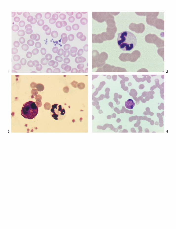
1
3
2
4

22
11
33
44
6655
5
7
6
8