anatomy and physiology of pharynx
TRANSCRIPT
ANATOMY AND PHYSIOLOGY OF
PHARYNX
DR AARYA SERIN
Pharynx is a conical fibromuscular tube 12-14 cm long extendS from base of the skull
(basiocciput and basisphenoid) to the lower border of cricoid cartilage.
The width is 3.5 cm at its base and this narrows to 1.5 cm at pharyngo-oesophageal junction which is the narrowest part of digestive tract apart from the appendix.
STRUCTURE OF PHARYNGEAL WALL From within outwards it consists of four layers:
1. Mucous membrane 2. Pharyngeal aponeurosis (pharyngobasilar fascia) 3. Muscular coat 4. Buccopharyngeal fascia
1. Mucous membrane It lines the pharyngeal cavity and is continuous
with mucous membrane of eustachian tubes, nasal cavities, mouth, larynx and oesophagus.
The epithelium is ciliated columnar in the nasopharynx and stratified squamous elsewhere.
There are numerous mucous glands scattered in it.
2. Pharyngeal aponeurosis (pharyngobasilar fascia) It is a fibrous layer lines the muscular coat It is thick near the base of skull but is thin and
indistinct inferiorly. It fills up the gap left in the muscular coat near the
base of skull.
3. Muscular coat It consists of two layers of muscles with three muscles
in each layer. (a) External layer: contains superior, middle and inferior constrictor muscles. (b) Internal layer: contains stylopharyngeus, salpingopharyngeus and palatopharyngeus muscles.
4. Buccopharyngeal fascia It covers outer surface of the constrictor
muscles Upper part prolonged forwards to cover the
buccinator muscles Above the upper border of superior
constrictor, it blends with pharyngeal aponeurosis.
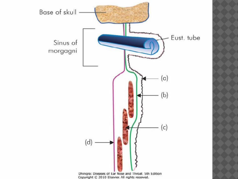
KILLIAN'S DEHISCENCE Inferior constrictor muscle has two parts;
thyropharyngeus with oblique fibres cricopharyngeus with transverse fibres.
Between these two parts exists a potential gap called Killian's dehiscence.
It is also called the "gateway of tears" as perforation can occur at this site during oesophagoscopy.
Also the site for herniation of pharyngeal mucosa in cases of pharyngeal pouch.
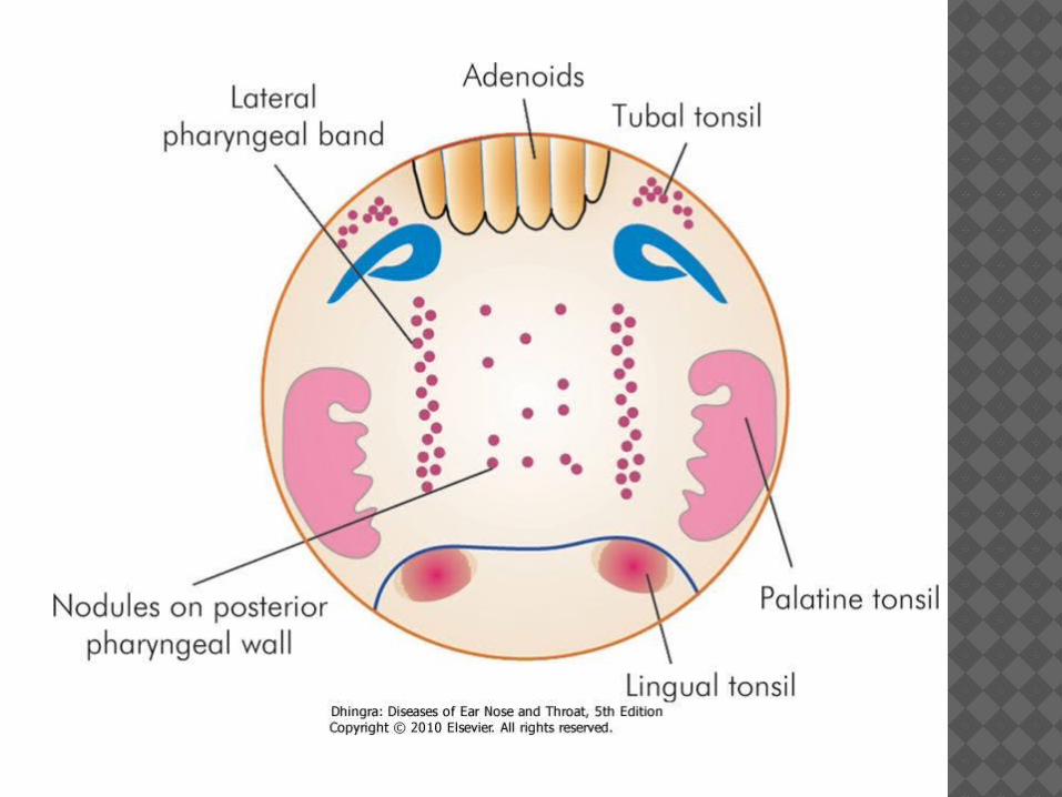
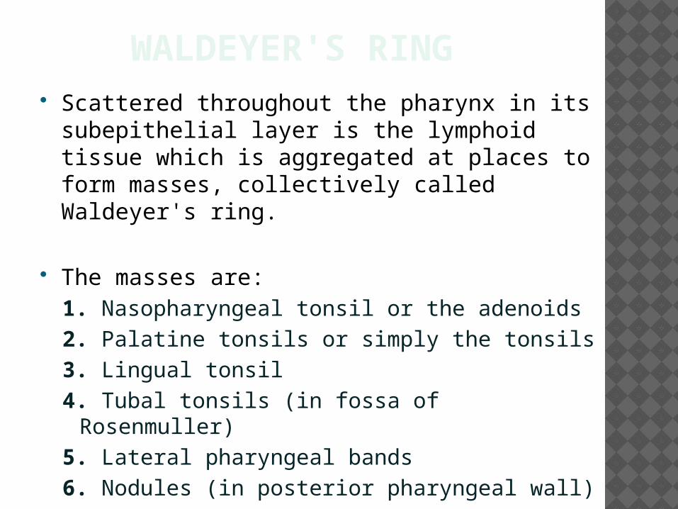
WALDEYER'S RING Scattered throughout the pharynx in its
subepithelial layer is the lymphoid tissue which is aggregated at places to form masses, collectively called Waldeyer's ring.
The masses are: 1. Nasopharyngeal tonsil or the adenoids 2. Palatine tonsils or simply the tonsils 3. Lingual tonsil 4. Tubal tonsils (in fossa of Rosenmuller) 5. Lateral pharyngeal bands 6. Nodules (in posterior pharyngeal wall)
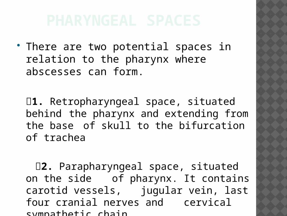
PHARYNGEAL SPACES There are two potential spaces in relation
to the pharynx where abscesses can form.
1. Retropharyngeal space, situated behind the pharynx and extending from the
base of skull to the bifurcation of trachea
2. Parapharyngeal space, situated on the side of pharynx. It contains carotid vessels,
jugular vein, last four cranial nerves and cervical sympathetic chain
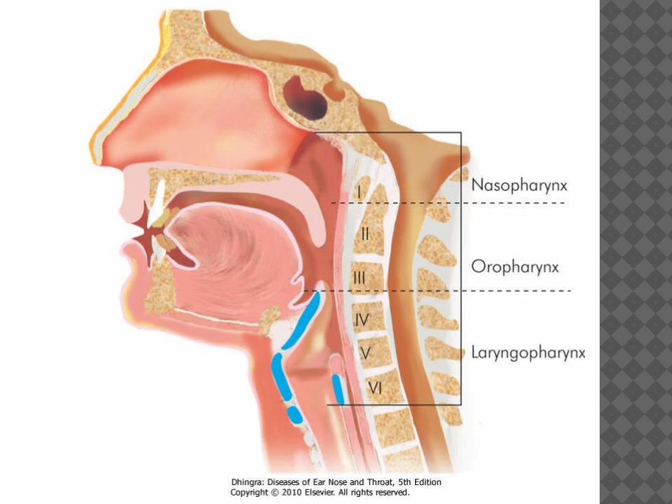
DIVISIONS OF PHARYNX
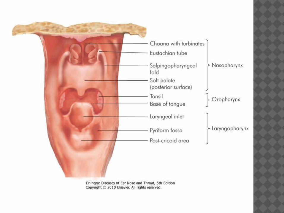
Anatomically, pharynx is divided into three parts : 1. Nasopharynx 2. Oropharynx 3. Hypopharynx or Laryngopharynx
NASOPHARYNX (EPIPHARYNX)
Applied Anatomy
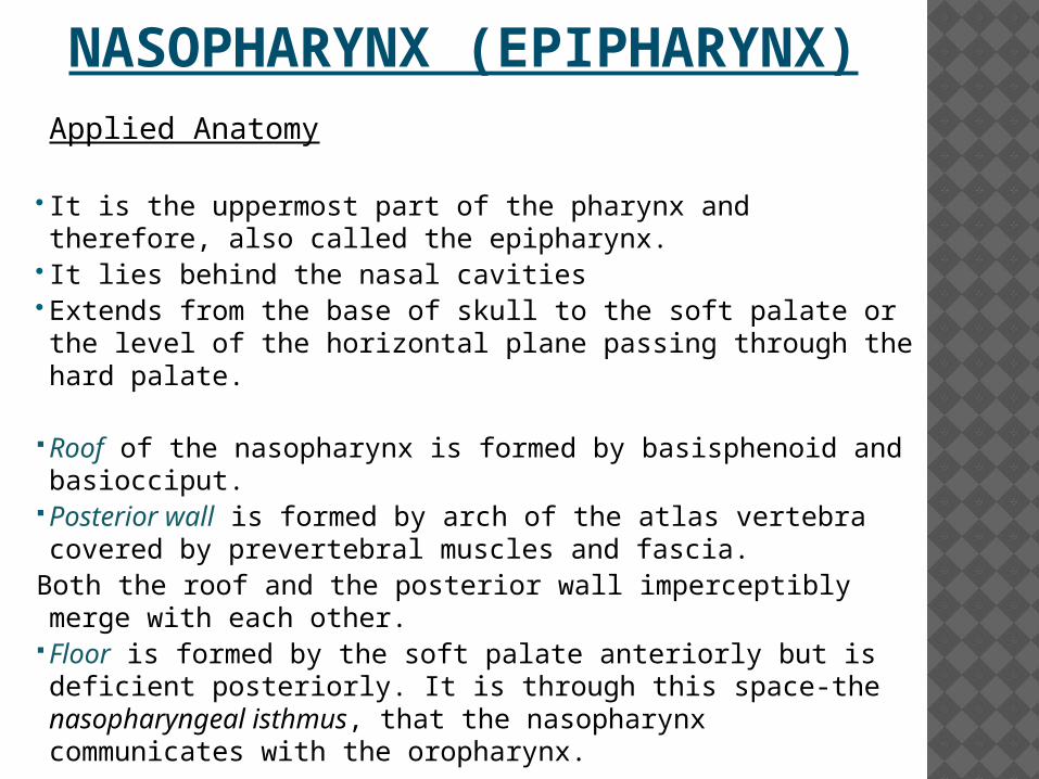
It is the uppermost part of the pharynx and therefore, also called the epipharynx.
It lies behind the nasal cavities Extends from the base of skull to the soft palate or the level
of the horizontal plane passing through the hard palate.
Roof of the nasopharynx is formed by basisphenoid and basiocciput.
Posterior wall is formed by arch of the atlas vertebra covered by prevertebral muscles and fascia.
Both the roof and the posterior wall imperceptibly merge with each other.
Floor is formed by the soft palate anteriorly but is deficient posteriorly. It is through this space-the nasopharyngeal isthmus, that the nasopharynx communicates with the oropharynx.
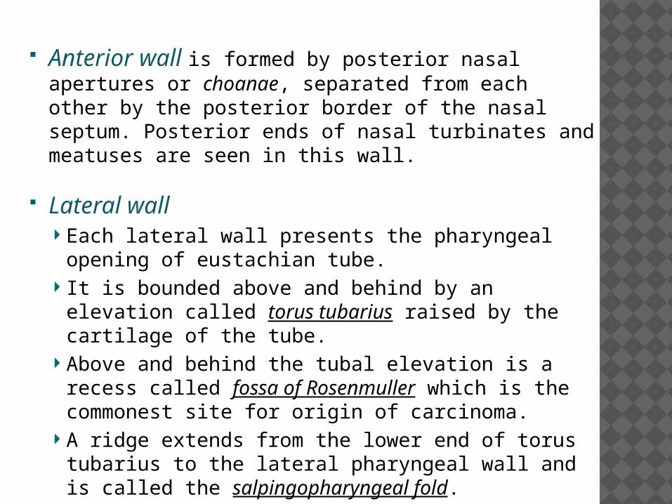
Anterior wall is formed by posterior nasal apertures or choanae, separated from each other by the posterior border of the nasal septum. Posterior ends of nasal turbinates and meatuses are seen in this wall.
Lateral wallEach lateral wall presents the pharyngeal opening of eustachian tube.It is bounded above and behind by an elevation called torus tubarius raised by the cartilage of the tube. Above and behind the tubal elevation is a recess called fossa of Rosenmuller which is the commonest site for origin of carcinoma. A ridge extends from the lower end of torus tubarius to the lateral pharyngeal wall and is called the salpingopharyngeal fold.
NASOPHARYNGEAL TONSIL (ADENOIDS)
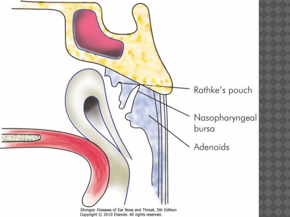
It is a subepithelial collection of lymphoid tissue at the junction of roof and posterior wall of nasopharynx
It increases in size up to the age of six years and then gradually atrophies.
RATHKE'S POUCH
It is represented clinically by a dimple above the adenoids and is reminiscent of the buccal mucosal invagination, to form the anterior lobe of pituitary.
A craniopharyngioma may arise from it.
TUBAL TONSIL
It is collection of subepithelial lymphoid tissue situated at the tubal elevation.
It is continuous with adenoid tissue and forms a part of the Waldeyer's ring.
When enlarged due to infection, it causes eustachian tube occlusion.
SINUS OF MORGAGNI
It is a space between the base of the skull and upper free border of superior constrictor muscle.
Through it enters (i) the eustachian tube(ii) the levator veli palatini(iii) tensor veli palatini and(iv) ascending palatine artery-branch of the facial artery
PASSAVANT'S RIDGE It is a mucosal ridge raised by fibres of
palatopharyngeus.
It encircles the posterior and lateral walls of nasopharyngeal isthmus.
Soft palate, during its contraction, makes firm contact with this ridge to cut off nasopharynx from the oropharynx during the deglutition or speech.
EPITHELIAL LINING OF NASOPHARYNX
Functionally, nasopharynx is the posterior extension of nasal cavity.
It is lined by pseudostratified ciliated columnar epithelium.
LYMPHATIC DRAINAGE
Lymphatics of the nasopharynx, adenoids and pharyngeal end of eustachian tube drain into upper deep cervical nodes either directly or indirectly through retropharyngeal and parapharyngeal lymph nodes.
Lymphatics of the nasopharynx may also cross midline to drain into contralateral lymph nodes.
FUNCTIONS OF NASOPHARYNX
1. Acts as a conduit for air. 2. It ventilates the middle ear and equalises air
pressure on both sides of tympanic membrane. This function is important for hearing.
3. Elevation of the soft palate against posterior pharyngeal wall and the Passavant's ridge helps to cut off nasopharynx from oropharynx. This function is important during swallowing, vomiting, gagging and speech.
4. Acts as a resonating chamber during voice production.
5. Acts as a drainage channel for the mucus secreted by nasal and nasopharyngeal glands.
OROPHARYNX
Applied Anatomy Extends from the plane of hard palate above to
the plane of hyoid bone below. It lies opposite the oral cavity with which it
communicates through oropharyngeal isthmus. The latter is bounded above by the soft palate;
below, by the upper surface of tongue, and on either side, by palatoglossal arch (anterior pillar).
BOUNDARIES OF OROPHARYNX
Posterior wall It is related to retropharyngeal space and lies opposite the second and upper part of the third cervical vertebrae.
Anterior wall It is deficient above, where oropharynx communicates with the oral cavity, but below it presents:
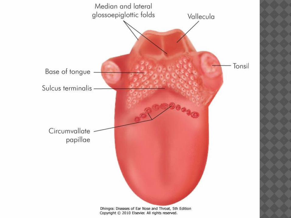
(a) Base of tongue, posterior to circumvallate papillae.
(b) Lingual tonsils, one on either side, situated in the base of tongue.
(c) Valleculae. They are cup-shaped depressions lying between the base of tongue and anterior surface of epiglottis. Each is bounded medially by the median glossoepiglottic fold and laterally by pharyngoepiglottic fold.They are the seat of retention cysts.
Lateral wall
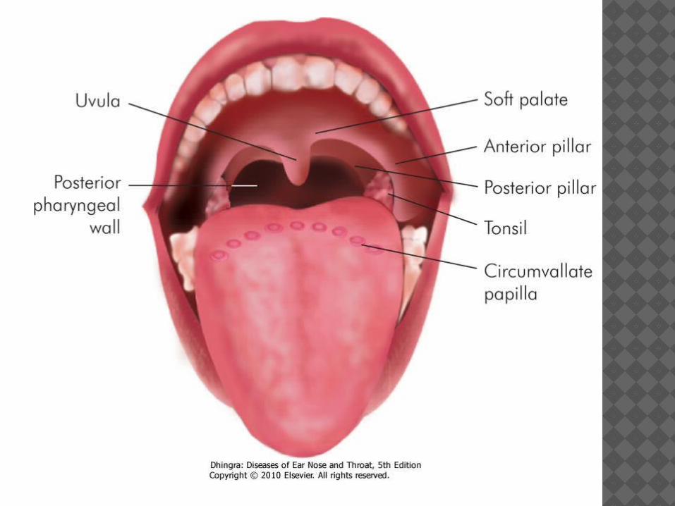
It presents: (a) Palatine (faucial) tonsil(b) Anterior pillar (palatoglossal arch) formed by
the palatoglossus muscle. (c) Posterior pillar (palatopharyngeal arch) formed
by the palatopharyngeus muscle.
Both anterior and posterior pillars diverge from the soft palate and enclose a triangular depression called tonsillar fossa in which is situated the palatine tonsil.
Lymphatic Drainage
Lymphatics from the oropharynx drain into upper jugular chain particularly the jugulodigastric (tonsillar) node.
The soft palate, lateral and posterior pharyngeal walls and the base of tongue also drain into retropharyngeal and parapharyngeal nodes and from there to the jugulodigastric and posterior cervical group.
The base of tongue may drain bilaterally.
FUNCTIONS OF OROPHARYNX
1. As a conduit for passage of air and food.2. Helps in the pharyngeal phase of
deglutition. 3. Forms part of vocal tract for certain speech
sounds. 4. Helps in appreciation of the taste. 5. Provides local defence and immunity
against harmful intruders into the air and food passages. This function is subserved by subepithelial masses of lymphoid tissues scattered as Waldeyer's ring.
HYPOPHARYNX (LARYNGOPHARYNX)
Applied Anatomy It is the lowest part of the pharynx and lies behind and
partly on the sides of the larynx. Its superior limit is the plane passing from the body of
hyoid bone to the posterior pharyngeal wall. Its inferior limit is lower border of cricoid cartilage
where hypopharynx becomes continuous with oesophagus.
Hypopharynx lies opposite the 3rd, 4th, 5th, 6th cervical vertebrae.
Clinically, it is subdivided into three regions-the pyriform sinus, post-cricoid region and the posterior pharyngeal wall.
1. Pyriform sinus (fossa)It lies on either side of the larynx and extends from pharyngoepiglottic fold to the upper end of oesophagus.It is bounded laterally by the thyrohyoid membrane and the thyroid cartilage and medially by the aryepiglottic fold, posterolateral surfaces of arytenoid and cricoid cartilages.Foreign bodies may lodge in the pyriform fossa. Internal laryngeal nerve runs submucosally in the lateral wall of the sinus and thus is easily accessible for local anaesthesia.
2. Post-cricoid region It is the part of the anterior wall of laryngopharynx between the upper and lower borders of cricoid lamina.It is a common site for carcinoma in females suffering from Plummer-Vinson syndrome.
3. Posterior pharyngeal wall It extends from the level of hyoid bone to the level of cricoarytenoid joint.
LYMPHATIC DRAINAGE
Pyriform sinus is richly supplied by lymphatics which exit through the thyrohyoid membrane and drain into the upper jugular chain.
Lymphatics of the posterior wall lateral pharyngeal or parapharyngeal nodes and thence to the deep cervical lymph nodes.
Lymphatics of post-cricoid region parapharyngeal nodes but may also drain into nodes of supraclavicular and paratracheal chain.
Rich lymphatic network of pyriform fossae explains the high frequency with which nodal metastases are seen in carcinoma of this region.
FUNCTIONS OF HYPOPHARYNX
Laryngopharynx, like oropharynx, is a common pathway for air and food, provides a vocal tract for resonance of certain speech sounds and helps in deglutition.
There is a coordination between contraction of pharyngeal muscles and relaxation of cricopharyngeal sphincter at the upper end of oesophagus.
Lack of this coordination, i.e. failure of cricopharyngeal sphincter to relax when pharyngeal muscles are contracting causes hypopharyngeal diverticulum.