anatomy and systematics of dendroseris (sensu lato)
TRANSCRIPT

Made in United States of AmericaReprinted from BRrrroNlk
Vol. 19, No. 2, April—June, 1967
pp. 99—121
ANATOMY AND SYSTEMATICS OF DENDROSERIS (SENSU LATO)
SHERWIN CARLQuIsT
Cariquist, S. (Rancho Santa Ana Botanic Garden, Claremont, Calif.) Anatomyand Systematics of Dendroseris (sensu lato). Brittonia 19: 99—121. 1967.—Anatomical study of the genus Dendroseris (Compositae: Cichorieae), endemic to theJuan Fernandez Islands, was undertaken to determine if segregate genera werevalid. Other questions include the significance of pith bundles and of receptacularbristles, and whether the ancestors of these peculiar rosette trees and shrubs werewoody or herbaceous. Anatomical evidence, when added to that from grossmorphology, suggests that the segregation as distinct genera of Rea, Phoenicoseris,and Hesperoseris is probably not justified, and that they are better treated assubgenera of Dendroseris. Differences in pollen morphology, floral trichomes,achene and leaf anatomy provide good species characteristics. These all appear,respectively, as variations on a basic plan, and Dendroseris can be envisagedas derived from a common stock in the Juan Fernandez Islands. The genus is notparticularly primitive within the family or tribe. The presence of additionalbundles in the flowers of D. litoralis may be interpreted as related to gigantism.The separate corolla lobes in D. gigantea probably do not represent a vestigeof an actinomorphic condition. Pith bundles in Dendroseris may have been presentin ancestors; in any case, they seem likely to have increased in prominence withincrease in stem diameter. Available evidence seems to favor the interpretationof growth forms in Dendroseris as derived from an herbaceous ancestry.
INTRODUCTION
The genus Dendroseris (Compositae, tribe Cichorieae), is an endemic of the JuanFernandez Islands, Chile. This genus has attracted attention because of its range ofpeculiar growth forms. Sparsely branched rosette trees, palmiform rosette trees,and succulent rosette shrubs are prominently represented. The species of Dendroserisare among the woodiest of the tribe Cichorieae, a tribe notable for woody genera onislands almost exclusively: Thamnoseris (Desventuradas Islands, Chile), Munzothamnus (San Clemente Island, California), and Sonchus sect. Dendrosonchus(Canary Islands) are conspicuous among these. I have been interested in the anatomyof Dendroseris because of the possibility that anatomical studies will elucidate thephylogeny and relationships of this genus and its peculiar growth forms. AlthoughSkottsberg (1956, p. 361) averred that Dendroseris and other Juan Fernandez rosettetrees are relicts, the opposing view, stated by Darwin (1859), has been the onewhich seems supported by recent comparative investigations.
The genus Dendroseris is worthy of comparative anatomical study for other reasons.The diversity of form within the genus is great, and although Skottsberg (1922)regarded it as a single genus, he later (1951) decided to recognize four genera:Dendroseris, Rca, Phoenicoseris, and Hesperoseris. Skottsberg’s decision in favor ofthe segregate genera was influenced by Erdtman’s (1952) work on pollen (also summarized in Skottsberg, 1951). Are the differences in pollen morphology wideenough to warrant recognition of segregate genera, or are they in reality variationson a single basic pattern? Study of anatomical features is needed to support orrefute segregate genera. With regard to other considerations, Dendroseris containsexcellent examples of loss of dispersibility (Carlquist, 1966). This phenomenon hasso far been described only in terms of gross morphology; anatomical study canreveal the underlying basis of this evolutionary tendency.
This study was inspired by material provided directly and indirectly by the lateDr. Carl Skottsberg. He provided me in 1956 with an excellent collection of liquid
BRnT0NIA 19: 99—121. Apr—Jun. 1967.
99

100 B1UTTONIA [voL. 19
•1
_1 I .‘.‘L? “lr(A )T1
-
- -.
1
& . -
• .
+
FIGs. 1 3 F G. 1. Dendroseris mic,antha. Longisectiori of shoot apex, bowing natuie of leafprirnordia and trichornes. F&. 2. Dendro ens rnicrcsntha Transection of prirnar) tern, bowingtwo phloern strands in pith. Fic. 3. Dendroseris macrophylla. Portion of leaf transection, showinmidvein in upper larnina. daxial face at iight FIG. 1, 3, X 102, Fin,. 2, >< 145.
r :1

1967 CARLQUIST I DENDROSERIS 101
preserved Juan Fernandez Cornpositae. This material was the basis for my earlierstudies on Centaurodendron and Yunquea (1958), and is permitting similar studieson Robinsonia, Rhetinodendron. and Symphyochaeta. Dried specimens were sentto me by Dr. Skottsberg, and other specimens collected by him also were used inthe present study.
For the sake of simplicity, the taxonomic conclusions of this study may he givenhere so that the nomenclature of the species mentioned can reflect those conclusions.Anatomical studies do show certain characters which might be used to supportsegregate genera. Indeed, any anatomical study is likely to demonstrate more characters than were known before such a study. However, I am impressed with thefact that all expressions, either in gross morphology or anatomy, point to thelikelihood that in Dendroseris we are dealing with variations on a single basic pattern. In oth’er words, this evidence does not conflict with a hypothesis that theJuan Fernandez Cichorieae all stem from a single ancestral stock. The diversitywithin Dendroseris is indeed remarkable, but recognition as a single genus, in accordance with Stebbins (1953), emphasizes the adaptive radiation which has occurred,an adaptive radiation paralleling that of other insular genera. These considerationsappear to me to override the convenience of using segregated genera. Indeed, Iwould like to use the names of segregate genera as a means of referring to naturalgroupings within Dendroseris. I followed this practice earlier (1960, 1965, 1966),as did Kunkel (1957) and Skottsberg (1953). However, the goals of taxonomy appear better served by use of the following scheme:
Subg. Dendroseris: D. litoralis, B. macrant/ia, D. macrophylla, D. marginata.Subg. Schizoglossum’: D. gigantea.Subg. Phoenicoseris Skottsb.: D. berteriana, B. pinnata, B. regia.Subg. Rca (Bert.) Skottsb.: D. inicrantha, D. neriifolia, D. pruinata.
Only D. gigantea. D. macrophylla, and D. regia occur on Más Afuera, where theyare endemic. The remainder are endemic to Más a Tierra or islets offshore from Mása Tierra. Floristic information on Dendroseris has been given by Skottsberg (1922,1929, 1951). Data on ecology are offered by Skottsberg (1928, 1953) and Kunkel(1957), and Skottsberg (1956) has reached phytogeographical conclusions aboutDendroseris. Illustrations of habit or morphological details of Dendroseris may befound in all of these papers, as well as in Carlquist (1965, 1066). Wood anatomy ofthe Juan Fernandez Cichorieae has been surveyed earlier (Carlquist, 1960), and consequently data are not included here.
MATERIALS AND METHODS
The specimens upon which studies were based are listed below. Specimens listedfrom Stockholm (S) document the liquid-preserved portions sent me by Dr. Skottsberg. Specimens from American herbaria provided portions which were treated inorder to provide anatomical preparations. In addition to the kindness of Dr.Skottsberg, who provided dried collections (RSA) in addition to the liquid-preservedspecimens, I would like to acknowledge the generosity of the curators of the U.S.National Herbarium.
Dendroseris berteriana (Dcne.)Hook. & Am.: Skottsberg 28-11-1955 (S); D.gigantea Johow: Skottsberg 436 (RSA): D. litoralis Skottsb.: Skottsberg 30-X11-1954, vegetative (5): Skottsberg 3-111-1954, floral (S); D. macrant/ia (Bert. &
Dendroseris subgenus Schiog1ossum (Skottsberg) Cariquist, comb. nov. Rea sect. Schizoglossum Skottsb., Nat. Hist. Juan Fernandez and Easter Island 2: 201. 1922. HesperoserisSkottsberg, Nat. Hist. Juan Fernandez and Easter Island 2: 788. 1951.

102 BRITTONIA jVOL. 19
V
/;:
J7 6‘1-
9(FIGs, 4—9. Dendroseris litoralis. Sections of stem and petiole to show vascularization. FIG. 4.
Stem, petiole, and portion of leaf blade, disassembled to show venation features. Fins. 5—7. Enlarged portions of leaf midvein, middle region of petiole, and basal portion of petiole, respectively.Xylem shown in black, phloem unpatterned, collenchyma stippled. Fin. 8. Radial longisectionof stem, showing vasculature associated with three nodes. Note pith bundles. FIn. 9. Transection.Further explanation in text. FIG. 4, X 1.2; FIGs. 5—7. X 5.5; Fin. 8, X 1.4; Fin. 9, X 1.6.

19671 CARLQUIST: DENDROSERIS 103
Dcne.)Skottsb.: Skottsberg 330 (RSA); D. macrophylla D. Don: Skottsberg 12-11-1955 (S); Skottsberg 362, floral (US); D. marginata (Bert. & Dcne.)Hook. & Am.:Skottsberg 3-XII-1916 (US); D. micrantha Hook. & Am.: Skottsberg 20-111-1955,vegetative (S); Skottsberg 567, floral (RSA); D. pinnata (Bert. & Dcne.)Hook. &Am.: Skottsberg 5-111-1955, vegetative (5) H. Weber in 1937, floral (RSA) D.pruinata (Johow)Skottsb.: Skottsberg 20-111-1955, vegetative (S); Shottsberg 79,floral (US); D. regia Skottsb.: Skottsberg 570, vegetative (RSA).
No material of D. neriifolia was studied. Dendroseris regia has been collectedsterile only. Fully mature achenes are apparently not known in D. gigantea, D.macrant/ia, nor D. inacrophylla.
The fixation of liquid-preserved material proved suitable for all but embryologicalstudies. Conventional methods were used for preparing paraffin sections, whichwere mounted and stained with a safranin—fast green combination, correspondingto Northen’s modification of Foster’s tannic acid—ferric chloride method (Johansen,1940). Dried material for anatomical studies was treated with 2.5% aqueous NaOHto expand tissues and to clear. Portions were then embedded, sectioned, and stainedaccording to the above techniques. These sections provided reliable, if sometimesunattractive, preparations. Other portions of the NaOH-treated material wereprepared as whole mounts, using safranin as a stain and Canada balsam as amounting medium. This technique also was employed for pollen grains. Sectionsof pollen grains proved useful, and these were prepared according to the usualparaffin techniques: sections 5—6 thick of pollen-containing flowers were obtained.
ANATOMICAL DESCRIPTIONS
Vascularization of the Shoot. In Figs. 1—16 are shown details of vascularbundles and their arrangement in stems and leaves. The complexity of vascularization seems related to the large size of stems and leaves. The pith region widens outmarkedly only a short distance below the shoot apex (Fig. 1). In all species ofDendroseris, vascularization is present in pith. This takes the form of bundles whichcontain phloem only, as in D. inicrantha (Fig. 2), or both xylem and phloem. asin D. litoralis (Figs. 4, 8, 9). Stem material was available for D. berteriana, D.lit oralis, D. macrophylla, D. micrantha, and D. pruinata. Of these, only D. litoralishas vessel elements and libriform fibers in pith bundles of the stem. The remainderhave phloem bundles only. In D. herteriana, xylem was observed in bundles of pithof the inflorescence axis, however. In all cases, the pith vascularization takes theform of transverse plates related to nodes (Figs. 8, 9, 14). These plates are opennetworks in which bundles join and diverge in various patterns (Figs. 9, 15). Fromthis network, bundles may run from one plate to the one above and below (Figs. 8,14). In addition, the periphery of the pith contains many vertical bundles (Figs.2, 8, 9, 14, 15). Some of these are phloem strands which are very small and havebeen omitted from the drawings. Bundles do not run from one plate to the nextif a species contains transverse pith diaphragms (each containing one or two platesof vascularization) separated by transverse pockets of air. Dendroseris berterianahas such pith diaphragms; pith is “hollow” according to Skottsberg (1951) in subg.Dendroseris and Piwenicoseris, and presumably such pith diaphragms as observedin my material of D. berteriana are meant.
In the cortex of sterns, numerous leaf traces are ordinarily visible, because nodesare multilacunar and crowded. How are leaf traces and the bundles of the vascularcylinder of the stem related to the pith bundles? As far as can be determined, thecylinder bundles are collateral, not bicollateral, in structure, although bicollateralbundles are characteristic of petioles and major veins of leaves (Figs. 3, 5—7, 11—13).In D. litoralis, plates of pith bundles are clearly related to bundles of the vascular

104 BRITTONIA [VOL. 19
0L
16
\1,°
15 °
Fins. 10—16. Dendroseyjs micrantha. Sections of stem and petiole to show vascularization.Conventions as in Figs. 4—9. Fm. 10. Stem, petiole, and portion of leaf blade, disassembled toshow venation features. FIGs. 11—13. Enlarged portions of petiole, midrib of lower leaf, midribmidway along leaf, corresponding to sections, as indicated by gaps, in Fig. 10. Fm. 14. Radiallongisection of stem, showing vascularization relating to four nodes. Fm. 15. Transection, showing pith bundles and leaf traces. Fm. 16. Four successive stages in the departure of a leaf trace.A pair of bundles other than the leaf trace is shown in each stage. Relationship between a strandof phloem in the pith (first stage) and internal phloem of the leaf trace (stage at far right) isindicated. Fm. 10, X 1.8; Fins. 11—13, X 6.8; Fm. 14, X 1.8; Fin. 15, )< 2.0; Fm. 16, X 6.8.

1967] CARLQUIST: DENDROSERIS 105
cylinder at nodal regions At the same levels at which some bundles extend outwardas leaf traces, other traces turn inward to vascularize the pith (Fig. 8, upper right).In other cases, phloem strands from the pith can be seen to extend outward throughthe leaf gaps (Fig. 9, below) and enter the petioles as phloem strands on the upperfaces of petiolar bundles (Fig. 8, lower right, middle left). This is also true inD. micrantha (Figs. 15, 16). In addition, strands of phloem from bundles adjacentto the leaf gap may contribute phloem to the adaxial side of a leaf trace (Fig. 16).
Leaf traces depart as a simple arc, which may be seen at the lowest levels of thepetiole. Further out (Fig. 13), additional bundles are evident. These form aconspicuous upper series in the petiole, as in Fig. 7. Such upper bundles are mostlyinverted or otherwise abnormally oriented. Ontogenetic studies of apical meristemsshowed that these additional bundles are the product of an adaxial meristem. Sucha meristem can be seen in the bulging adaxial portions of leaf primordia such asthose shown in Fig. 1. The additional bundles join the normal set in the lowerpetiole. Multiplication of bundles in the normally oriented set occurs by ordinarydivarications (such a branching is shown in Fig. 13).
The supernumerary bundles increase in prominence distally along the petiole (Figs.6, 12) and both normal and supernumerary bundles are seen in the midribs of leaves(Figs. 3, 5, 11). Not all the supernumerary bundles have both xylem and phioem.Many are merely phloem strands (Figs. 3, 5—7, 12—13). Some phloem strandsare associated with the adaxial faces of bundles, which thereby become bicollateral.Part of these phloem strands enter the petiole from the stem in the ways indicatedabove in connection with pith bundles. Others arise de novo in the petiole.
Bundles inverted in orientation are limited to petiole, midrib, and secondary veinsin the leaf. Smaller veins in the lamina are entirely normal in orientation. Thisresults from the fact that inverted bundles alter in orientation as they divergefrom main veins into the lamina.
The significance of pith bundles in Cichorieae has bewildered various workers.For example, Worsdell (1919) considered them as vestiges of a formerly more complete and complex vascularization. This hypothesis, although appealing in itsdirectness, is not supported by any evidence which could not just as easily beinterpreted in the reverse fashion. Moreover, one would ultimately have to inventan explanation for the origin of such a complex vascularization from the normaldicotyledonous pattern in ancestors of Cichorieae.
If one compares the systematic occurrence of pith bundles in Cichorieae (cf.Metcalfe & Chalk, 1950) to a phylogenetic classification of the tribe (Stebbins, 1953),one notes that the occurrence of pith bundles characterizes genera at every level ofphylogenetic specialization. The abundance of pith bundles in the Juan FernandezCichorieae is almost in direct proportion to the diameter of the pith. Conditionslike those of the Juan Fernandez Cichorieae were reported by Worsdell in severalgenera—Tragopogon, for example. More instances could doubtless be uncovered.Complex nodal vascularization of this sort occurs in genera of other tribes ofCornpositae as well (Worsdell, 1919; Davis, 1961).
While no purpose would be served by attempting to use the limited data of thepresent study as a basis for an all-inclusive hypothesis, one may note the correlationin Dendroseris between occurrence of pith bundles and both width of pith and relatively poor secondary growth. Cichorieae are notable among Compositae in theirlack of secondary growth, and vascularization of pith regions might compensate forpaucity in secondary xylem conducting elements. Another possibility is a relationship to the broad, multilacunar nodal regions characteristic of Cich’orieae. The factthat the above correlations are not perfect in all groups in angiosperms at largedoes not alter the possibility that in particular groups these factors might be operative.

106 BRITTONIA IVOL. 19
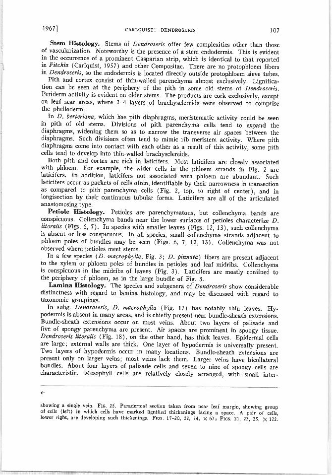
24 25FIGs. 1/—25 Sections of leaes. Fin. 17. Dendrmeri rnacrophvlla Fin. 18. Dendroseric
litoralis. Dark streaks in mesophyll at left and right are laticifers. Fi 19. Dendroseris ,nacrantha.The ein at right is one of the secondarie. Fios. 20—21 Dendroceris gigantea. Fm. 20. Portionof lamina, low poer Fm. 21. Lower surface of leaf, showing a trichome at higher magnification.Fios. 22—2 . Dendro ens pinnata. Fm. 22 Portion of lamina, showing three veins. Fin. 23.Enlargement of vein shown at left in Fig. 22; fibers are visible above xylem in vein; a laticiferis just below the phloem pole. Fins. 24—25. Dendro enir pruinata. Fin 24. Section of lamina,
19671 CARLQTJIST: DENDROSERIS 107
Stem Histology. Stems of Dendroseris offer few complexities other than thoseof vascularization. Noteworthy is the presence of a stem endodermis. This is evidentin the occurrence of a prominent Casparian strip, which is identical to that reportedin Fitchia (Carlquist, 1957) and other Cornpositae. There are no protophloem fibersin Dendroseris, so the endodermis is located directly outside protophloem sieve tubes.
Pith and cortex consist of thin-walled parenchyma almost exclusively. Lignification can be seen at the periphery of the pith in some old stems of Dendroseris.Periderm activity is evident on older stems. The products are cork exclusively, excepton leaf scar areas, where 2--4 layers of brachysclereids were observed to comprisethe phelloderm.
In D. berteriana, which has pith diaphragms, meristematic activity could be seenin pith of old sterns. Divisions of pith parenchyma cells tend to expand thediaphragms, widening them so as to narrow the transverse air spaces between thediaphragms. Such divisions often tend to mimic rib meristem activity. Where pithdiaphragms come into contact with each other as a result of this activity, some pithcells tend to develop into thin-walled brachysclereids.
Both pith and cortex are rich in laticifers. Most laticifers are closely associatedwith phloern. For example, the wider cells in the phloern strands in Fig. 2 arelaticifers. In addition, laticifers not associated with phloem are abundant. Suchlaticifers occur as packets of cells often, identifiable by their narrowness in transectionas compared to pith parenchyma cells (Fig. 2, top, to right of center), and inlongisection by their continuous tubular forms. Laticifers are all of the articulatedanastomosing type.
Petiole Histology. Petioles are parenchymatous, but collenchyma bands areconspicuous. Collenchyma bands near the lower surfaces of petioles characterize D.litoralis (Figs. 6, 7). In species with smaller leaves (Figs. 12, 13), such collenchymais absent or less conspicuous. In all species, small collenchyma strands adjacent tophloem poles of bundles may be seen (Figs. 6, 7, 12. 13). Collenchyma was notobserved where petioles meet stems.
In a few species (D. macro ph’vlla, Fig. 3: D. pinnata) fibers are present adjacentto the xylem or phloem poles of bundles in petioles and leaf midribs. Collenchyrnais conspicuous in the midribs of leaves (Fig. 3). Laticifers are mostly confined tothe periphery of phloem, as in the large bundle of Fig. 3.
Lamina Histology. The species and subgenera of Dendroseris show considerabledistinctness with regard to lamina histology, and may be discussed with regard totaxonomic groupings.
In subg. Dendroseris, D. macrophylla (Fig. 17) has notably thin leaves. Hypodermis is absent in many areas, and is chiefly present near bundle-sheath extensions.Bundle-sheath extensions occur on most veins. About two layers of palisade andfive of spongy parenchyma are present. Air spaces are prominent in spongy tissue.Dendroseris litoralis (Fig. 18), on the other hand, has thick leaves. Epidermal cellsare large; external walls are thick. One layer of hypodermis is universally present.Two layers of hypodermis occur in many locations. Bundle-sheath extensions arepresent only on larger veins; most veins lack them. Larger veins have bicollateralbundles. About four layers of palisade cells and seven to nine of spongy cells arecharacteristic. Mesophyll cells are relatively closely arranged, with small inter-
showing a single vein. FIG. 25. Paradermal section taken from near leaf margin, showing groupof cells (left) in which cells have marked lignified thickenings facing a space. A pair of cells,lower right, are developing such thickenings. Fics. 17—20, 22, 24, X 67; Fins. 21, 23, 25, )< 122.

108 BRITTONIA VOL. 19
/ 29 30 / V
Icm
28
26
:‘Th
Fins. 26—33. Floral venation in Dendroseris. Fins. 26—28. Dendroseris litoralis. Fit;. 26. Venation of an entire flower; adaxial edge of flower, upper portion of ligule turned so as to be seenin face view. Outlines of flower in narrow lines, veins in wider lines. Style and stamen tracesare typical for Compositac and have been omitted . Similar conventions apply to other drawings.Fin. 27. Achenc, showing five-veined condition. Fin. 28. Enlarged portion of corolla throat,

1967 CARLQUIST: DhNDROSEIUS 109
cellular spaces. Although laticifers “wandering” in mesophyll occur in all species ofDendroseris, they are especially conspicuous in D. litoralis. The leaves of D.macrantha (Fig. 19) and D. marginata are essentially identical, in these two species,leaves are relatively thick. A single hypodermis layer is universally present. Bundle-sheath extensions are present on almost all veins. About four palisade and sevenspongy layers are present.
The fact that leaves of D. macrophylla are thin and have large air spaces andlittle hypodermis seems related to the occurrence of these plants in shady foresthabitats. Contrasting conditions in D. litoralis would connote the open sunny lowland habitats that species inhabits. Variations in constitution of mesophyll with relation to sun or shade may be expected in these and other Dendroseris species.
The sole species of subg. Schizoglossum, D. gigantea (Figs. 20, 21) has notablythin leaves. A single hypodermis layer is present. On virtually all veins, bundle-sheath extensions occur. About two palisade and five spongy layers are present.On the ahaxial epidermis, uniseriate non-glandular trichomes may be found (Fig. 21).These trichomes are essentially the same as those which occur on corolla-lobe tipsin this species (Fig. 36).
In subg. Phoenicose;is, D. pinnata (Figs. 22, 23) and D. regia are alike in leafanatomy. A single hypodermis layer is present; bundle-sheath extensions are present on larger veins only. On larger veins (about half the veins observed), one ora few fibers are present above the xylem pole. Major veins are bicollateral. Leavesof D. berterZana conform to the same pattern, but bicollateral bundles were notobserved. About three layers of palisade and six of spongy tissue characterize speciesin subg. Phoenicoseris.
In subg. .Rea, leaves are exceptionally thick. The pattern is the same for bothD. pruinata (Fig. 24) antI D. micrantha. Beneath the epidermis is a single layerof large hypodermis cells. Bundle-sheath extensions characterize the majority ofveins. About five layers of palisade and six to eight of spongy tissue are present.
The term “palisade” has been used above despite the fact that these cells arenearly isodiarnetric in shape. Trichomes are mentioned above only for D. gigantea.The difference between this species and the others is merely one of preservation oftrichomes into the mature leaves. As one might expect, leaf primordia of all speciesbear uniseriate non-glandular trichomes, as shown for D. micrantha (Fig. 1). Noglandular trichomes were observed in Dendroseris.
In all species examined, peculiar sclereids were observed in leaf mesophyll, asshown in Fig. 25. These sclereids differ from normal ones in having thickeningsonly on walls facing similarly thickened walls. Layering is conspicuous in thethickenings. These sclereids occur in small or large nests, in various portions of theepidermis and mesophyll. Their patterns of thickening appear as a response to aninjury. Their distribution in the leaf also suggests this. Kunkel’s (1957) reportof galls in the same position—near leaf margins—in D. pinnata as I have observedin all species confirms the interpretation that these sclereid nests are pathologicalin origin.
Secondary Xylem. Wood anatomy of the Juan Fernandez Cichorieae has beenanalyzed earlier (Cariquist, 1960). Additional comments here are unnecessary.
showing the anomalous girdle of bundles and veinlets which extend short distances above andbelow this girdle. FiG. 29. Dendroseris macrantha. Tip of corolla. FIG. 30. Dendroscris larginata. Tip of corolla. Fic. 31. Dendroseris pinnata. Venation of entire flower. Fic. 32. Dendroseris gigantea. Venation of entire flower. Fin. 33. Dendroseris pruinaia. Venation of entireflower. 1 cm scale applies to Figs. 26, 27, 29, 30. 5 mm scale applies to Figs. 31, 32, 33. 2 mmscales applies to Fig. 28.

110 BRITTONIA 1V0L. 19
36
1 frcTh
yr I : r
J
r39 40 41 42 43
FIGs. 34—50. FIGs. 34—38. Tips of corolla lobes, viewed from exterior surface, to show trichomesand sclereids. Heavy lines indicate sclerification; epidermal relief is indicated on cells at tips oflobes. FIG. 34. Dendroseris macrophylla. FIG. 35. Dendroseris litoralis. FIG. 36. Dendroserisgigantea. Fxc. 37. Dendroseris pinnata. FIG. 38. Dendroseris pruina.ta. Fsos. 39—43. Surfacesof corolla tubes (edge at left in each) showing trichomes. FIG. 39. Dendroseris macrantha. Fin.40. Dendroseris rnacrophylla. FIG. 41. Dendroseris gigantea. FIG. 42. Dendroseris pinnafa. FIG.43. Dendroseris pruinata. FIGS. 44—50. Views of pollen grains. One of the three colpi and germ

19671 CARLQUIsT: DENDROSERIS 111
Involuere. The writer noted earlier (1966) that in Dendroseris loss of dispersibility is not merely a matter of achene morphology, but extends to the natureof involucres as well. The anatomical features basic to this phenomenon are worthyof investigation.
Involucral bracts in D. berteriana (Fig. 51) open out somewhEt at maturity,mostly at the tips. Tn Fig. 51 are seen the central portion of an intermediate bract(left), the lateral portion of an outer bract (right). These bracts are essentiallymature. Ground tissue of the bracts is parenchyma, although the large cells(center) have lightly lignified walls which would offer a modest degree of rigidity.A few epidermal cells on the inner faces of bracts of D. berteriana have lignifiedthickenings. In involucres of most Compositae, the mechanism for reflexing ofbracts is a fibrous inner bract face coupled with a parenchymatous outer face;when the latter dries and shrinks, the bract turns outward. In D. berteriana, thisscheme is poorly fulfilled, for the inner face is not strongly rigid, and in some bractssclerenchyma in the outer face of the bract would hinder shrinkage, so that thebract would tend to remain in the same position as when it was fresh.
In subg. Dendroseris, the broad involucres containing large fruits are completelyenfolded by the bracts when heads dry. This behavior is understandable whenbract anatomy is studied. In D. litoralis (Fig. 52), ground tissue of bracts is
exclusively thin-walled parenchyma, except for some suggestion of collenchymatousthickenings. Collenchyma does not influence bract rigidity appreciably upon drying.Thus, bracts have no mechanism to insure their folding outward when an involucrematures and dries. Lacking sclerenchyma, bracts can weather and fray easily, however, so that achenes would eventually be released.
In D. micrantha (Fig. 53), bracts have an anatomy similar to those of D.litoralis, although they are much thinner. Mature bracts of D. micrantha haverelatively thin-walled cells exclusively; some cells suggest lignification of walls intheir staining reactions.
All bracts in Dendroseris have only a single arc of collateral bundles, althoughadditional phloem strands are present in larger bracts. Laticifers are notably wideand prominent in involucral bracts, and a number may be seen (especially adjacentto phloem poles of bundles) in Figs. 51—53.
In subg. Dendroseris, receptacles of heads bear conspicuous bristles; they arepresent, but much smaller, in subg. Phoenicoseris. In D. litoralis (Fig. 63, right),these bristles are large, terete, and of various lengths. No vascularization was observed, although presence of laticifers was established in larger bristles, and association of a few laticifers with sieve tubes would not he unexpected. Cells of receptacularbristles remain thin-walled and no lignification was observed. Th’e function ofreceptacular bristles in Dendroseris is not evident. They do not seem to aid dispersal—in fact, one might imagine that they would hinder it. Their form andanatomy does not suggest that they are vestiges of true receptacular bracts. Theymay (in subg. Dendroseris. at least) be a by-product of the gigantism so evident inthe species in which these bristles are conspicuous.
Floral Venation. The venation of flowers in Dendroseris is basically that designated “Cichorieae—simplified” in a phylogenetic summary of floral venation of
pores shown in center of each of the entire grains. In sections of pollen grain walls (frommicrotome sections), spines and stratification of exine are shown. Intine indicated by line atbottom, nexine by densely stippled strip, sexine by more sparsely stippled area. FiGs. 44—45.Dendroseris litoralis. FiGs. 46—47. Dendroseris pinnata. “Para-apertural depression” shown atleft in Fig. 47. Fin. 48. Dendroseris gigantea. Fins. 49—50. Dendroseris prisinata. Scales of magnification apply to all figures within the horizontal rows in which they occur.

112 BRITTOI\IA 1V0L. 19
• .
.- b4•J
-.ti
49j 4
J 1 ‘f± -vc. rc-i5
55 56f GS. 1 56. Fius. 51—53. Transections of inlolucral bract , outer faces at iight. FIG. 51
Dendro e;i berteriana. T o successic bracts shown. FIG. 52. Dendroseri.s 1itorali. FIG. 5.Dendroseris niirantha. FIGs. 54— 6. Section of mature achenes. FIGs. 54—55. Dendrosericinzcrantha FIG. 54. TranSection of achene a em is present at the angle, above, but at theinside face 01 the ss’all, not in the fiber . FIG. 55. Lonisection of achene, showing folds in

1967] CARLQUIST: DENDPOSERIS 113
Compositae (Carlquist, 1961). Deviations from this pattern in Dendroseris includepresence of additional corolla bundles—particularly at tips of corolla lobes, andin the corolla throat. In each style branch there is only a single vein and thus apair of veins in the undivided portion of the style. Stamens contain a single traceeach; each stamen trace parallels a corolla vein, joining it at the base of the corollatube.
In .D. litoralis (Fig. 26), there are basically five veins in the corolla tube, four orfive (Fig. 27) in the achene. At the base of the achene, more than a single veinconnects with receptacle venation—a fact which probably is related to the largesize of achenes in subg. Dendroseris, for the other subgenera have only a singlevascular bundle connecting achene to receptacle. The ovule trace, however, is unbranched despite the large size of the ovule; the trace outlines the odd, broad triangular shape of the ovule, a shape which foreshadows the large, wide embryo. Dendroseris litoralis has markedly winged achenes. However, the achene veins do notextend into the wings, but are close to the locule (Fig. 63, left). Wings are formedrelatively late in the ontogeny of the achene. At the tip of the corolla, median veinsoccur in most lobes. These are short and often irregular in placement and attachment to other veins. In the throat of the corolla (Fig. 28), a peculiar girdle ofbundles is present, interconnecting the five main veins of the corolla tube. Fromthe girdle, supernumerary veins extend downwards and upwards—mostly the latter.Some of these veins extend well up into the ligule, so that more than the minimalsix veins can be found there.
In other species of subg. Dendroseris, venation is essentially the same. Variationsin veins at the corolla-lobe tips occur, however. Degrees in presence and absence ofmedian veins are shown for D. macrant/rn (Fig. 29) and D. marginata (Fig. 30).
In subg. Pizoenicoseris, D. pinnata (Fig. 31) demonstrates the simplified typeof venation without any alteration. Veins below corolla-lobe sinuses may join atvarious distances from the sinus. Four veins are usually present in the achene wall.
Floral venation of species of subg. Rea follows the same pattern, as shown for D.pruinata in Fig. 33.
in subg. Schizoglossum, D. gigantea (Fig. 32) is notable for having a characterat odds with a key character of Cichorieae: corolla lobes are separate, the ventralsinus only a little longer than the other sinuses. Corolla-lobe veins parallel the lobes,but unite just below the sinuses. If separation of lobes in D. gigantea were a vestigeof an actinomorphic condition, one would expect that (1) additional veins would bepresent, especially in the achene, as in Dubyaea (Stebbins, 1940: Carlquist, 1961);(2) veins would extend separately in pairs beneath the sinuses instead of joiningimmediately below the sinuses, suggesting greater separateness of lobes, as in Glossarion (Cariquist, 1961); and (3) vestiges of median veins in corolla lobes wouldbe present, as in Glossarion or Dubyaea (Carlquist, 1961). None of these conditions,nor other criteria of primitive venation in Compositae, are satisfied by D. gigantea.One may assume that separateness of lobes in D. gigantea is merely an indicationthat lobe primordia fuse at a slightly later stage in ontogeny than in other Cichorieae.Such a change in ontogeny could easily occur in a relatively specialized genus.
Similar considerations apply to the peculiarities of venation in subg. Dendroseris.Segments of median veins at lobe tips are present in an irregular fashion, as in thecultivated Fleizanthus annuus (Carlquist, 1961). Likewise, the girdle of bundles inthe corolla throat of subg. Dendroseris is unrelated to any primitive pattern of
achene wall. Fifl. 56. Dendroseris berteriana. Transection of achene; vein is dark patch interiorto the angle below. Fios. 51—53, X 75; Fios. 54—56, 7< 127.

114 BRITTONTA [VOL. 19
FIGs. 57-62. Sections of mature achenes. FIGs. 57—58. Dendroseris pinnata. Portions ofcotyledons shown below in each. FIG. 57. Transection. FIG. 58. Longitudinal section, showingfolds in achene wall. FIGs. 59—62. Dendroseris marginata. FIGs. 59, 60. Transections. Fig. 59shows a portion with ridge-like expansions of the achene wall, Fig. 60 shows a portion in which
,.y;t
&/$_u

19671 CARLQUIST: DENDROSERIS 115
venation and must be considered an innovation. Such an innovation would beexpected, for the very large size of flowers in subg. Dendroseris is undoubtedly aninstance of gigantism, as are the flowers of the commercial sunflower.
Floral Histology. The nature of trichomes at the tips of corolla lobes provesan interesting characteristic of species in Dendroseris, reminiscent of similar distinctions in an unrelated genus of Compositae, Fitcl’tia (Cariquist, 1957). In D.macrophylla (Fig. 34), D. macrant/ia, and D. marginata, lobe-tips bear uniseriatenon-glandular trichomes. Some of these have transverse or diagonal tip cells, somehave thick lignified walls, and some have distorted shapes. In D. litoralis (Fig. 35),trichomes are fewer and less scierenchymatous; some are club-shaped. In D.gigantea, the hairiness of leaves is paralleled by the abundance of trichomes atcorolla-lobe tips (Fig. 36). These trichomes have lignified walls, are two to fourcells in length, and are not distorted in shape. In subg. Phoenicoseris (e.g., D.pinnata, Fig. 37) and subg. Rea (D. pruinata, Fig. 38) the uniseriate trichomes areabsent. A few of the rounded cells characteristic of lobe-tips in all species bear thicklignified walls in these two subgenera, however.
Trichdmes on the corolla tube show similar variations. In D. macrant/ta (Fig. 39)and D. marginata, large multiseriate non-glandular trichomes are present. These aremostly quadriseriate or triseriate at bases; they are tapered distally, terminatingin one to three files of cells. Corolla-tube trichomes of D. litoralis (Fig. 40) and D.niacrophylla are similar, but smaller. In D. gigantea (Fig. 41), trichomes areabundant, but narrow, nlostly biseriate or triseriate. In all species of the subgeneraPhoenicoseris and Rea, corolla-tube trichomes are sparse, short, and rounded inshape. As shown for D. pinnata (Fig. 42) and D. pruinata (Fig. 43), these trichornesare often biseriate, the files rarely more than two cells in length. In no case wereglandular trichomes observed on flowers.
Pollen Grains. Erdtman (1952) investigated pollen grains of Dendroserissynoptically, and provided a key to the species. This key was later reworked interms of the segregate genera (Skottsberg, 1951). Pollen grains are figured here(Figs. 44—50) because the features reported have not hitherto been illustrated. Erdtman’s findings were confirmed by my observations.
Pollen grains of subg. Dendroseris are notably large, as with D. litoralis (Figs. 44,45), with respect to both entire grains and spines. As noted by Erdtman, sexine isconnected with nexine by tenuous processes in this subgenus; spines, which arerelatively long and borne on conical protuberances, are roughly equidistant fromeach other. No “para-apertural depressions” are present.
The subgenera Schizoglossum, Phoenicoseris, and Rca agree in relatively smallsize of grains, and in presence of para-apertural depressions, as shown in Figs. 46and 49. In D. pruinata (Fig. 49), however, a population of pollen grains showsvarious degrees of presence or absence of para-apertural depressions. An intermediate condition is illustrated. The para-apertural depressions take the form ofvery thin areas of sexine (Fig. 47, left). The three subgenera seem close. They canbe differentiated in terms of the protuberances and the spines these bear: conical,tipped by a spine more than 3 long in subg. Phoenicoseris (Fig. 47); rounded,tipped by short spines in subg. Schizoglossum (Fig. 48); conical, tipped by veryshort spines or none at all in subg. Rea (Fig. 50).
no such alterations are present. FR;s. 61—62. Longisections. Fig. 61 shows, from left to right,fibers, brachysciereids with circular pits, and thick-walled parenchyma. Fig. 62 shows, at higherpower, epidermis, 1—2 layers of parenchyma, and brachysclereids with prominent circular pits.Fins. 57—61, X 135. Fin. 62, >< 245.

116 BRITTONIA 1V0L. 19
FIGS. 63—66. Sections of achenes of Dendroseris litoralis. FIG. 63. Transection of immatureachenc, showing formation of the winged margin. Vein is at left. At right, two receptacularbristles. FIG. 64. Transection of mature achene. corresponding to the view shown in Fig. 63.

1967] CARLQUIST: DENDROSERIS 117
All species of Dendroseris have the same basic colpus conformation: relativelyshort, widened at the ends. The spines contain a central channel which is relatedto the lacunose structure of the outer exine. Outer sexine is composed of minuterods in a lacunose background; the inner sexine is apparently unscuiptured. Noteshould be made of the fact that aside from depression areas, spines are by no meansexactly equidistant in any species. Germ pores are covered by a thick intine layerwhich is surfaced by a very thin layer of sexine.
Despite the “key characters” of pollen grains within Dendroseris, the distinctionsall seem to be variations on a single basic type. There is every reason to believe thatthe diversity has been derived from a single ancestral stock.
Fruit Anatomy. Fruits in Dendroseris exhibit considerable diversity in formand anatomy. The simplest type may be seen in the subgenera Rea (Figs. 54—55)and Scizizoglossum. In D. micrantiza, the achene wall is about 6 cell-layers thick,except at the angles. Some cells in the achene wall are thin-walled parenchyma,others are prominently pitted brachysclereids. At intervals strands of fibers occur.Veins are located toward the inside of the achene wall in thin-walled parenchyma.When seen in longitudinal section (Fig. 55), the achene wall is prominently buckled;this convolution occurs prior to maturation of fibers.
The above pattern characterizes species of subg. Phoenicoseris also, althoughthese tend to have more prominent fibers in achene walls, as seen for D. berteriana(Fig. 56). This species has tannin accumulations in the epidermis. These featurescan also be seen, more prominently, in D. pinnata (Figs. 57, 58). The cells of theepidermis are large, densely filled with tannins. The achene wall consists wholly offibers except for the innermost layers, which are parenchymatous and collapsedat maturity. As seen in longisection, the achene wall has conspicuous folds and bulges.
The achenes of subg. Dendroseris are more elaborate in structure. In D. marginata(Figs. 59—62), achene walls are thick. The achene surface is raised into ridges(Figs. 59, 61) in some areas, although other portions lack them (Fig. 60). Achenesshould probably not be termed “winged” in this species. Non-ridged portions of thewall contain two or three layers of parenchyma beneath the epidermis, followed bybrachysclereids, then fibers, and finally the thin-walled parenchyrna, collapsed atmaturity, lining the achene locule. Where ridges occur, parenchyma and brachysclereids form more numerous layers and are radially elongate. Brachysclereids (Fig.62) bear pits conspicuous for their uniformly circular outline and equal spacing.
In D. litoralis (Figs. 63—66) and other species of subg. Dendroseris (exceptingD. marginata), achenes are winged. Wings are dorsiventral, not lateral, in orientation.These wings are the product of meristematic action much like that which produceslaminae on leaf primordia. As seen in Fig. 63, files of cells (oriented horizontallyin this photograph) indicate recent divisions extending the width of the wing, justas “sheet meristem” activity widens a lamina. Before lignification of fibers obscuresveins, a few vessels, sieve tubes, and laticifers may be seen in achenes. At maturity(Figs. 64—66), a pattern similar to that of D. marginata is visible. Epidermal cellsare large, and do not contain tannin accumulations. Within the epidermis are aboutthree layers of parenchyma. Within the parenchyma are one (or two) layers of
Dark cells are fibers. Fig. 65. Longisection of achene, showing ovule (left half of photograph)and achene wall, in which fibers are abundant. FIG. 66.A longisection similar to that of Fig. 65.showing histology. Tn the ovule tissue (left half of photograph), numerous large crystals canbe seen. In the achene wall, the left half is composed of fibers the right half of shorter cells,the innermost of which arc brachysclereids with circular pits. Fn;. 63, X 135; Fics. 64, 65. X 110;Fic. 66. X 180.

118 BRITTONIA [VOL. 19
thin-walled brachysciereids with prominent circular pits, as shown in Fig. 66. Numerous layers of fibers underlie the brachysciereids. Lining the locule of the achene arethin-walled parenchyma cells which are collapsed (dark line, center, in Figs. 65 and 66).Ovule cells (Figs. 65, 66, left) are not persistent; they collapse as the embryo enlarges.Prior to collapse, large prismatic crystals are evident (Fig. 66, left). The surfaces ofachenes in D. litoralis are irregular, although not as markedly buckled as in D. pinnata.
Achenes of Dendroseris are notable in their lack of trichomes. Trichomes are common on achenes of many Compositae; their absence in Dendroseris is one aspectwhich limits dispersibility. The function of hairs on achenes of Compositae is eitherto help eject them from the head (biseriate “Zwillingshaare” flex outward whenmature and dry, pushing achenes up from the receptacle) or to help achenes catchon fur or feathers. Pappus bristles in many Cichorieae flex outward; this actionpermits ejection of achenes from the head, or provides a flotation mechanism, orboth: the familiar example of Taraxacum is typical of this mechanism. Pappusbristles do not reflex in Dendroseris; they lack the differential thickenings at basesof bristles which induce this action. Gigantism is especially notable in the body ofachenes in subg. Dendroseris, and to a lesser extent in the other subgenera. Pappusbristles are not proportionately increased in size or number, so there would be anet loss of dispersibility even if bristles could reflex in a normal manner (Carlquist,1966).
Embryos in Dendroseris, especially in subg. Dendroseris, are notably large, withbroad cotyledons (Skottsberg, 1922). Increase in cotyledon size also results in irregular folding of cotyledons. This was observed in D. litoralis, D. marginata, andD. pinnata. Distortions in shape of achene walls, such as buckling and convolutions,do not seem the result of increase in embryo volume. Rather, such malformationsare probably a by-product of loss of dispersibility, tolerable because achenes ofDendroseris lost efficiency of dispersal prior to development of these shapes. Ifvarious factors assure that dispersal will be restricted to short distances only, malformed achenes have no selective disadvantage (Carlquist, 1966).
TAXONOMY
The species of Dendroseris as recognized by Skottsberg (1922, 1951) have beenaccepted here without revision. Some species characters are evident in anatomicaldata, particularly in leaf anatomy. More numerous characters are evident, however,at the level of subgenera. Both gross morphology and anatomy of Dendroseris arepresented in Table I. The distinctions assembled in Table I, however, do not include any expressions constant throughout Dendroseris as a whole.
Most of the character expressions in Table I distinguish one of the subgenera fromthe other three. In the majority of cases, such separations divide subg. Dendroserisfrom the other three subgenera. In some features, these distinctions underline thegigantism so typical of subg. Dendroseris—heads, flowers, achenes, and pollen grains,for example. The basis for this gigantism would be interesting to know. It evidentlydoes not lie in polyploidy. Cytological investigations by Stebbins, Jenkins, & Walters(1953) showed that the somatic chromosome number 2n = 36 occurs not only in aspecies of subg. Dendroseris (D. macrophylla) but also in a species of subg.Phoenicoseris (D. pinnata). The same number has also been counted in D. litoralisof subg. Dendroseris (Raven, unpubl.), and in D. pruinata of subg. Rea (Soibriget al. 3849, Más a Tierra, Puerto Ingles, GH, US. I am grateful to Dr. Soibrig for permission to publish his count here). This number suggests that Dendroseris as a wholemay be a polyploid derivative from the basic number x 9 which is present in sub-tribes related to Dendroseridinae.

1967] CARLQUIST: DEND0SERIS 119
TABLE I
CSIARACTERISTICs DIFFERENTIATING SUBGENERA ix D endroseris.
Dendroseris Schizoglossum Phoenkoseris Rea
Habit Branched Rosette shrub Palmiform Rosette shrubrosette tree
Pith Hollow Solid Hollow Solid(diaphragms) (diaphragms)
Leaves Entire, ± ovate Entire, elongate Pinnate, elongate Entire, elongateThin to thick, gla- Thin, hairy Thick, glabrous Very thick,
brous glabrous
Inflorescence Heads few, large Heads many, small Heads many, small Heads many, small
Head Receptacle bristly No bristles Receptacle bristly No bristles
Achenes Winged, large, Non-winged, small, Non-winged, small, Non-winged, small,thick-walled thin-walled thin-walled thin-walled
Corollas Large, orange Small, white Small, white Small, whiteLigules entire Ligules 5-fid Ligules entire Ligules entireSupernumerary Venation simplified Venation simplified Venation simplified
veins in corollaTrichomes at lobe Trichomes at lobe No trichomes at No trichomes at
tips tips lobe tips lobe tipsTube hairs large Tube hairs medium Tube hairs few, Tube hairs few,
short short
Pollen Grains large Grains small Grains small Grains smallNo depressions Depressions Depressions Depressions ±Protuberances coni- Protuberances round, Protuberances coni- Protuberances coni
cal, spines long spines short cal, spines short cal, spines veryshort
Sexine to nexine No processes No processes No processesprocesses
One might, on the basis of the data in Table I, more easily recognize two segregategenera than four, if segregate genera were desirable. Subgenera, however, seem quiteadequate for the purpose of expressing the variation pattern of the Juan FernandezCichorieae. Although two subgenera might be an acceptable taxonomic solution,the four subgenera seem advisable because each of the species groups thus emphasized possesses several clearly defined and significant characters.
Recognition of a single genus, Dendroseris, seems the most satisfying treatmentbecause divergences such as those listed in Table I can be explained as variations ona single basic pattern. Moreover, the characters common to all the species are notexpressed in Table I. Characteristics in which expressions appear to be variations ona single plan include trichome types on corollas, venation of flowers, nature ofachene-wall anatomy, vascularization pattern of stem and leaf, and involucral bractanatomy. Presence of hypodermis, bundle-sheath extensions, and palisade cellsisodiametric in shape are features common to leaves of all species. Pollen grains arebasically alike, most notably in the shape of furrows and the fiber structure of exine.
This basic group of unifying characters suggests that a single ancestral stock canbe envisioned for all the species of Dendroseris. Seen thus, the characteristics ofsubgenera and species are evidences of adaptive radiation not unlike that whichcharacterizes other insular genera, such as Dubautia (Compositae) and Cvanea(Campanulaceae) in the Hawaiian Islands, or Aconium (Crassulaceae) and Sonchussect. Dendrosonchus (Compositae) in the Canary Islands.

120 BRITTONIA VOL 19
PHYLOGENETIC CONCLUSIONS
The relationships between Dendroseris and other Cichorieae have not been exploredhere. The placement of this genus in a separate subtribe, Dendroseridiriae, has beenfollowed by various authors. This treatment stresses in no small measure peculiaritiesrelated to growth form and geographical isolation. A relationship between Dendroseridinae and Stephanomeriinae can be envisioned. Stebbins (1953) placesThamnoseris, a genus of succulent shrubs endemic to the Desventurados Islands,Chile, in Stephanomeriinae. Munzothamnus, endemic to San Clemente island (California), must also be placed in Stephanomeriinae; its chromosome number is thesame as that of Stephanoineria, to which it was once referred (Stebbins, Jenkins, &Walters, 1953).
Neither Dendroseridinae nor Stephanomeriinae are notably primitive or advancedwithin Cichorieae. The conclusion that Dendroseris is a relict (Skottsberg, 1956)does not seem tenable. The growth form cannot be regarded as antique, but aspecialization related to the insular condition. If rosette trees and rosette shrubson oceanic islands were ancient forms, one would expect them in more primitivefamilies. None of the relatively primitive families on the Juan Fernandez Islandsdemonstrate such growth forms there. On the other hand, most of the rosette treesand rosette shrubs are Compositae: Robinsonia, Rhetinodendron, and Symphyochaeta (Senecioneae); Centaurodendron and Yunquea (Cynareae). These can beshown to be specialized offshoots of mainland relatives. Centaurodendron andFunquea appear derived from Centauraea sect. Plectocephalus, for example (Carlquist, 1958).
The evidence from wood anatomy is very pertinent in this regard. Studies onanatomy of Cichorieae (Carlquist, 1960) indicate that Dendroseris wood is essentially that of an herb; it is too highly juvenilistic to be considered directly derivedfrom woody ancestors. Cichorieae are probably a group which have lost the abilityto form extensive secondary xylem. The limited xylem accumulation and succulentstems of .Dendroseris seem best explained as the responses of herbs to climaticallymild and relatively uniform conditions, as well as to ecological opportunities notpresent on continents but in the disharmonic floristic assemblage of an oceanicisland. This hypothesis has been suggested earlier for the Juan Fernandez rosettetrees by the writer (Cariquist, 1965). The idea that in dicotyledons at large, woodyspecies are more primitive than herbs has gained the acceptance it deserves. Onemust stress, however, that in individual groups of dicotyledons, considerable flexibility is involved, and herbaceous species can increase accumulation of secondaryxylem under certain conditions. This idea is not in conflict with the hypothesis ofwoody ancestry for the major groups of dicotyledons, for reasons discussed elsewhere(Cariquist, 1962). if one assumes that the many genera and species of peculiarherb-like rosette trees on oceanic islands are relictual. one must hypothesize now-vanished rosette trees on continental areas; such continental relatives, moreover,must be imagined to have vanished during the relatively short times in which florasof oceanic islands have developed, according to geological evidence. Not only is therelict hypothesis awkward under these conditions, it is not supported by an increasingfund of comparative taxonomic, floristic, and anatomical information concerning thenature of herb-like rosette trees such as Dendroseris.
LiTERATURE CITED
Cariquist, S. 1957. The genus Fitchia (Compositae). Univ. California Pubi. Bot. 29: 1—144.• 1958. Anatomy and systematic position of Centaurodendron and Yunquea (Corn
positae). Brittonia 10: 78—93.• 1960. Wood anatomy of Cichorieae (Compositae). Trop. Woods 112: 65—91.

1967] CARLQUIST: DENDROSERIS 121
• 1961. Comparative Plant Anatomy. New York: Holt, Rinehart & Winston. lx + 146 pp.• 1962. A theory of paedomorphosis in dicotyledonous woods. Phytomorphologv 12:
30—45.• 1965. Island Life .A Natural History of the Islands of the World. New York: Natural
History Press. vii + 451 pp.• 1966. The biota of long-distance dispersal. II. Loss of dispersibility in Pacific
Compositae. Evolution 20: 30—48.Darwin, C. 1859. On the Origin of Species by Means of Natural Selection. London: John
Murray. ix + 502 pp.Davis, E. L. 1961. Medullary bundles in the genus Dahlia and their possible origin. Amer. Jour.
Bot. 48: 108—113.Erdtman, G. 1952. Pollen Morphology and Plant Taxonomy. Vol. I. Angiosperms. Stockholm:
Ajmqvist & Wiksell. xii + 539 pp.Johansen, D. A. 1940. Plant Microtechnique. New York: McGraw-Hill Book Co. xi + 523 pp.Kunkel, C. 1957. tber einige endemische Kompositen der Flora fernandeziana. Ber. Schweiz.
Bot. Ges. 67: 428—457.Metcalfe, C. R. & L. Chalk. 1950. Anatomy of the Dicotyledons. 2 vols, Oxford: Clarendon
Press. lxiv + 1500 pp.Skottsberg, C. 1922. The phanerogams of the Juan Fernandez Islands. Nat. Hist. Juan Fer
nandez and Easter Island 2: 95—240.• 1928. Pollinationsbiologie und Samenverbreitung auf den Juan Fernandez-Inseln.
Nat. Hist. Juan Fernandez and Easter Island 2: 503—547.• 1929. Notes on some recent collections made in the Islands of Juan Fernandez. Acta
Horti Gothob. 4: 155—171.• 1951. A supplement to the pteridopbytes and phanerogams of Juan Fernandez and
Easter Island. Nat. Hist. Juan Fernandez and Easter Island 2: 763—792.• 1953. The vegetation of the Juan Fernandez Islands. Nat. Hist, Juan Fernandez and
Easter Island 2: 793—960.• 1956. Derivation of the flora and fauna of Juan Fernandez and Easter Island. Nat.
Hjst. Juan Fernandez and Easter Island 1: 193—438.Stebbins, G. L., Jr. 1940. Studies in the Cichorieae. Dubyaea and Soroseris, endemics of the
Sino-Himalayan Region. Mem. Torrey Bot. Club 19(3): 1—76.1953. A new classification of the tribe Cichorieae, family Compositae. Madroño 12:
65—81.
J. A. Jenkins, & Maria A. Walters. 1953. Chromosomes and phylogeny in theCompositae, tribe Cichorieae. Univ. California Publ. Bot. 26: 401—430.
Worsilell, W. C. 1919. The origin and meaning of medullary (intraxylary) phloem in the stemsof dictotyledons. II. Compositae. Ann. Bot. 33: 421—458.