anatomy for sport & exercise science · ☐perimysium ☐endomysium ☐fascicle . ......
TRANSCRIPT
1
ANATOMY for Sport & Exercise Science
Unit 1
1.1 The Skeletal System
Name :
2
UNIT SPECIFICATION
Skeletal System - Bones
Anatomical Language
☐Anatomical standing position (point of reference)
☐Anterior
☐Posterior
☐Lateral
☐Medial
☐Proximal
☐Distal
☐Superior
☐Inferior
☐Peripheral
☐Superficial
☐Deep
☐Supine
☐Prone
Long Bone anatomy
☐Periosteum
☐Bone minerals
☐Bone marrow
☐Epiphysis
☐Growth plates
☐Diaphysis
☐Cancellous bone
☐Compact bone
☐Articular cartilage.
Bony landmarks
☐Notches
☐Fossa
☐Condyles
☐Borders
☐Processes
☐Tuberosity
Process of bone growth & ossification
☐Osteoclasts
☐Osteoblasts
☐Osteocytes
☐Growth plates
☐Epiphysis
☐Bone remodelling
☐Minerals - calcium, vitamin D
Skeletal bones
☐Cranium
☐Clavicle
☐Ribs
☐Sternum
☐Scapula
☐Humerus
☐Radius
☐Ulna
☐Carpals
☐Metacarpals
☐Phalanges
☐Pelvis (ilium, ischium, pubis, iliac crest)
☐Vertebral column (cervical, thoracic, lumbar, sacrum, coccyx, curves of the spine)☒
☐Femur
☐Patella
☐Tibia
☐Fibula
☐Tarsals
☐Calcaneus
☐Metatarsals
☐Bones of axial skeleton
☐Bones of appendicular skeleton
Types of bones
☐Long
☐Short
☐Flat
☐Sesamoid
☐Irregular
Skeletal system function
☐Supporting framework
☐Protection
☐Attachment for skeletal muscle
☐Source of blood cell production
☐Store of minerals
☐Movement

3
Skeletal System - Joints
Ligaments
☐Role and function
Joints
☐Fibrous (fixed)
☐Cartilaginous (slightly moveable)
☐Synovial (freely moveable)
Types of synovial joint
☐Ball and socket
☐Condyloid
☐Gliding
☐Saddle
☐Hinge
☐Pivot
Synovial Structure
☐Joint capsule
☐Bursa
☐Articular cartilage
☐Synovial membrane
☐Synovial fluid
☐Ligaments
Range of movement at joints
☐Shape of bones and use
Muscular System
Types of muscles tissue
☐Cardiac (non-fatiguing, involuntary).
☐Skeletal (fatiguing, voluntary).
☐Smooth (involuntary).
Types of muscle fibres
☐Type I
☐Type IIa
☐Type IIx
Skeletal muscle anatomy
☐Epimysium
☐Perimysium
☐Endomysium
☐Fascicle
Neuromuscular junction
☐Impulse & action potential
☐NM junction
☐Neurotransmitter
Sliding filament theory
☐Calcium ions
☐Myofibril
☐Sarcomere
☐Actin
☐Myosin
☐Cross-bridges
☐H zone
☐Z line
☐A band
☐I band
☐Troponin
☐Tropomyosin
☐ATPase
☐ATP
Types of muscle contraction
☐Isometric
☐Concentric
☐Eccentric
Muscle fibre recruitment
☐Recruitment at different levels of intensity of exercise
Muscles
☐Deltoids (posterior, anterior, medial)
☐Medial and lateral shoulder rotators
☐Biceps brachii
☐Triceps brachii
☐Wrist flexors
☐Wrist extensors
☐Forearm supinators
☐Forearm pronators
☐Sternocleidomastoid
☐Pectoralis major
☐Rectus abdominis
☐Obliques
☐Transverse abdominis (TVA)

4
☐Quadriceps (rectus femoris, vastus medialis, vastus lateralis, vastus intermedius)
☐Iliopsoas
☐Tibialis anterior
☐Erector spinae
☐Trapezius
☐Rhomboids
☐Latissimus dorsi
☐Gluteals (gluteaus maximus, gluteaus medius, gluteaus minimus)
☐Hamstrings (biceps femoris, semitendinosus, semimembranosus)
☐Gastrocnemius
☐Soleus
Antagonist muscle pairs
☐Agonist
☐Antagonist
☐Synergist
☐Fixator.
Movement Analysis
Types of movement
☐Flexion (horizontal flexion, hip flexion, shoulder flexion, plantarflexion, dorsiflexion, lateral flexion)
☐Extension (hyper-extension, horizontal extension, hip extension, shoulder extension)
☐Abduction
☐Adduction
☐Rotation (medial and lateral)
☐Circumduction
☐Pronation
☐Supination
☐Elevation
☐Depression
☐Protraction
☐Retraction
Planes of movement
☐Description of planes
☐Types of movement in each plane
Phases appropriate to the movement
☐Preparation
☐Execution
☐Follow through
Body sections for analysis
☐Upper body
☐Trunk
☐Lower body
Bones involved in the movement
☐Type of bone
Muscles involved in movement (muscle action)
☐Role / function of antagonistic pairs
☐Role / function of synergist muscles
☐Role / function of fixator muscles
Joints involved in movement
☐Type of joint
☐Bones forming each joint
☐Range of movement permitted at each joint.
Types of movements
Planes of movement
Movement efficiency
☐Dynamic (balanced) stability at joints and mobility at other joints
☐Kinetic chain
☐Transfer of movement across body segments
☐Transfer of loads and maintain force
☐Muscle balance
☐Mechanical efficiency
5
Functions of the Skeleton
Types of Bone
Type Structure Function Examples

6
Bone (Endochondral) Formation, Growth & Re-modelling What does endochondral mean? What does osteogenesis mean? Describe the process of bone growth. Use the diagram below and table provided. Ensure you use and understand the following terminology in your answer:
Osteoblasts Osteocytes Minerals (calcium) Epiphysis Diaphysis Bone re-modelling Osteoclasts Vitamin D Growth plates
Figure 1 - Process of Endochondral Osteogenesis
7
Process of Osteogenesis (Bone Growth & re-modelling)
Stage Description A
B
C
D
E

8
Long Bone Anatomy
9
Long Bone Anatomy
Long Bone Anatomy Description
Periosteum
Bone minerals
Bone marrow
Epiphysis
Growth plates
Diaphysis
Cancellous bone
Compact bone
Articular cartilage
Ligaments

10
Axial Skeleton
Appendicular skeleton
11
Writing Style Activity
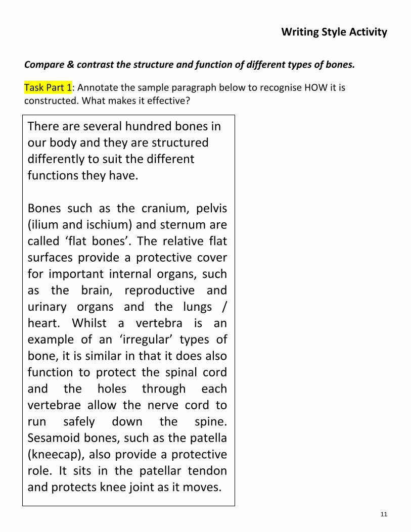
Compare & contrast the structure and function of different types of bones. Task Part 1: Annotate the sample paragraph below to recognise HOW it is constructed. What makes it effective?
There are several hundred bones in our body and they are structured differently to suit the different functions they have. Bones such as the cranium, pelvis (ilium and ischium) and sternum are called ‘flat bones’. The relative flat surfaces provide a protective cover for important internal organs, such as the brain, reproductive and urinary organs and the lungs / heart. Whilst a vertebra is an example of an ‘irregular’ types of bone, it is similar in that it does also function to protect the spinal cord and the holes through each vertebrae allow the nerve cord to run safely down the spine. Sesamoid bones, such as the patella (kneecap), also provide a protective role. It sits in the patellar tendon and protects knee joint as it moves.

12
Writing Style Activity
Compare & contrast the structure and function of different types of bones. Task Part 2: Now write a paragraph yourself in the box, thinking about the things discussed.
PEER Annotations
13
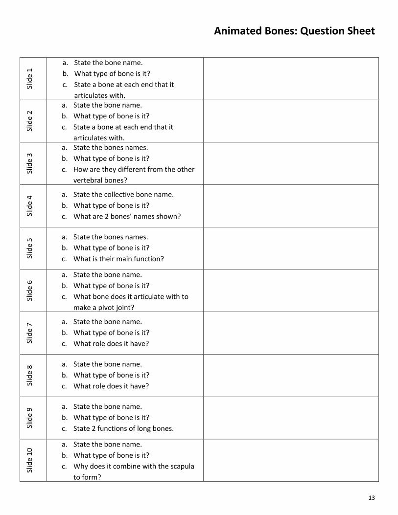
Animated Bones: Question Sheet
Slid
e 1
a. State the bone name. b. What type of bone is it? c. State a bone at each end that it
articulates with.
Slid
e 2
a. State the bone name. b. What type of bone is it? c. State a bone at each end that it
articulates with.
Slid
e 3
a. State the bones names. b. What type of bone is it? c. How are they different from the other
vertebral bones?
Slid
e 4 a. State the collective bone name.
b. What type of bone is it? c. What are 2 bones’ names shown?
Slid
e 5 a. State the bones names.
b. What type of bone is it? c. What is their main function?
Slid
e 6
a. State the bone name. b. What type of bone is it? c. What bone does it articulate with to
make a pivot joint?
Slid
e 7 a. State the bone name.
b. What type of bone is it? c. What role does it have?
Slid
e 8 a. State the bone name.
b. What type of bone is it? c. What role does it have?
Slid
e 9 a. State the bone name.
b. What type of bone is it? c. State 2 functions of long bones.
Slid
e 10
a. State the bone name. b. What type of bone is it? c. Why does it combine with the scapula
to form?
14
Classification of Joints Make detailed notes about the THREE different classifications of joints. Give examples. Consider :
Structure Function Range of movement Stability
15
Anatomy of a Typical Synovial Joint Structures Structure – what is it like? Function – What job does it do?
Ligament
Synovial Membrane
Synovial Fluid
Articular Cartilage
Joint Capsule
Menisci
Tendon
Pad of Fat
Bursae
Draw and label a simple
diagram of a typical synovial
joint.

16
Types of Movement at Joints Could you demonstrate and define the following joint movements? Do you know where these can occur? ☐Flexion
− ☐horizontal flexion − ☐hip flexion − ☐shoulder flexion − ☐plantarflexion − ☐dorsiflexion − ☐lateral flexion
☐Extension
− ☐hyper-extension − ☐horizontal extension − ☐hip extension − ☐shoulder extension
☐Abduction ☐Adduction ☐Rotation
− ☐medial rotation − ☐lateral rotation
☐Circumduction ☐Pronation ☐Supination ☐Elevation ☐Depression ☐Protraction ☐Retraction
17
Types of Synovial Joints Make detailed notes about the THREE different classifications of joints. Give examples.
Consider : Structural differences Planes and Axes of movement Types of movement possible
Locations in the body Functional differences Sporting examples
Types of Synovial
Joint Details
18
Animated Joints: Question Sheet Sl
ide
1
a. State the classification of joint b. State the type of joint. c. Give 2 examples of this joint d. State 2 possible joint movements
Slid
e 2
a. State the classification of joint b. State the type of joint. c. Give 1 examples of this joint d. State 2 possible joint movements
Slid
e 3
a. State the classification of joint b. State the type of joint. c. Give 2 examples of this joint d. State 2 possible joint movements
Slid
e 4 a. State the classification of joint
b. State the type of joint. c. Give 2 examples of this joint
Slid
e 5
a. State the classification of joint b. State the type of joint. c. Give 2 examples of this joint d. State 1 possible joint movement
Slid
e 6
a. State the classification of joint b. Give 2 examples of this joint c. What is another name for this
classification of joint?
Slid
e 7
a. State the classification of joint b. Give 2 examples of this joint c. What is another name for this
classification of joint?
Slid
e 8
a. What is A? b. What is B? c. What is C? d. What is D?
Slid
e 9 a. State the classification of joint
b. State the type of joint. c. Name the 2 bones.
Slid
e 10
a. State the classification of joint b. A section of vertebral column? c. B section of vertebral column? d. C section of vertebral column?
19
Writing Style Activity Compare and contrast the structural and functional characteristics for the 5 regions of the spine – consider the 2 different types of synovial and the cartilaginous joints in the spine.
Task: Complete the written answer to this question.
A significant function of the role of the vertebral column is protection of the vital spinal cord that runs within it. The cord is very delicate and vulnerable to damage which would be critical to anyone. This function is similar the 3 main areas of the vertebral column (cervical spine – 7 vertebrae, thoracic spine – 12 vertebrae and lumbar spine – 5 vertebrae) and they are all structured with a built in hole to allow for the cord to run though each individual vertebra. The vertebral column is a versatile and complex system of joints that allows movement in the back for several sporting actions. Movements that occur at the back are flexion and extension, lateral flexion and extension and rotation of the trunk and neck. The vertebrae, in isolated pairs, allow minimal movement and are generally categorised as a form of ‘cartilaginous joint’. This is because the vertebrae of the cervical, thoracic and part of the lumbar spine all have …….

20

21