anatomy. in man. - dwc.knaw.nl · in the spina scaplilae j according 10 ihe latter ihe spina in the...
TRANSCRIPT

Anatomy. "Tlte Developmenl of t!te Slwuldel'-blade in Man". By O. H. DIJKSTRA. (Communicated by Prof. L. BOLK).
(Communieated at the meeting of Mareh 24, 1923).
Unlike the development of Ihe clavicula that of Ihe scapula has received comparalively little attenlion. TlJe lextbooks of analomy (CUNNINGHAlIl, GKGENnAu~:R, RAUBER-KoPSCH, MERKEL, POIIUER-CHARPY, TESTUT) conlain only genel'al nolions such as Ihe informalion that the ossification of the shoulder-blade begins in the vicinity of the colltull scapulae at Ihe end of the second or in Ihe beginning of lhe Ihird monlh. POIRIER and CRARPY speak of an incipieut ossification bel ween the 40th and 50th day . BAUDKU:BEN reports a periostal ossiticalion (such as occllrs wilh Ihe bones of tlle cl'anial "auIt) beside and IInder the spina scapulae at the end of the 10th week.
BRrc!!: alone enters into more details in QUAINS'S Elements of Anatomy . According to his descl"iption the mdiment of Ihe shoulder-blade i!f in Ihe 6til week entit'ely cartilaginolls, pl'Oc. acromialis and proc. coracoïdens are present, but the spina Rcapulae is wanling. (Nevertheless BUYCE reproduces Ihe diagram of LEWIS I), iu w hich a spina is really indicated). In Ihe 8tll week ossiticalion begins wilh a centre near the collllm scaplllae, developilrg inlo a trianglliar plale, at whose uppel' margin Ihe spina appears in the 3rel month as a low ridge. At birth coracoid and aCl'omion, margo vertebl'alis and the margin of Ihe spina al'e slill made up of cartilage. This descrip~iofl by BRym: agrees faidy weIl with fhe one we find in BRoMAN's textbook of Embryology and in Ihat of KEIBI~J. and MAJ,L, in which RARm:EN deals with this subject. BROMAN, like BRYCJ4:, stal es lhat 110 spina is to be found at the cartilaginolls scapula. Nonetheless he reproduces t.he figllre of LEWJS, iu which there is indeed a spina. KOLLl\1ANN, SCHENCK, MINOT, PARKER do not speak of the fit'sl development of the shollldel'-blade and only dweil on stadia of advlI.nced ossification. In H~;RTWIG'S Entwickelungsgeschichte BUAUS alld also H~:RTWIG himself report a separate cenh'e of ossification in the spina scaplilae j according 10 Ihe latter Ihe spina in the neonatus ' still consisls of carlilage sometimes j according to KÖLJ.lKKR (qlloted by BADE, Arch. f. mikro Anal. LV) this is even always the case.
I) Am. Journ. Anat. Vol. l. 1901-'02.
298
The most detailed l'epOl't cOllceming the development of Ihe shoulderblade is that by 1:3 RYCl<: and BROMAN. From Iheil' figlll'es it is evident that they del'ive theil' data fl'om LEWIS, who Pllblished in the American JOllmal of AJlatomy (Vol. I 1901 - '02) a minute description of the developmeJlt of the arm in man. Broadly stated his data agl'ee with those of BRYC ~: , mentioll ed above. They diffel', however, as to the spilla scaplliae. ACCOl'dillg to LEWIS the spina probably takes ol'igin in the lIpper mal'gin of the scapula . This mal'go superior thickens and then splits into tt medial aJld a lateml lip. The medial lip is the future mal'go supel'iol', Ihe laleml ane is the first beginning of the spinascapulae,
HAGI~ N 1) describes a shollldel'-blade of all embl'Yo 17 mmo in length, The spina scapulae is absent , the pl'OC . coracoïdeus is large, the proc. aCJ'omialis smal\. The latter slatemellt cannot be reconeiled with LEWIS'S com m Uil icalion, wh ich, on I he con tl'ary, speaks of a I'elati valy large pl'OC. aCI'omiali s.
Thi s I'eview of th e lil em tllre would not be complete without IJlen tion i ng lhe j n tel'esl i ng sludy by R UTH I~ RFOIlD ' ) w ho en tered into mally detail s of th e developll1ellt of the shollldel'-blarle. Like Ll<:WIS he consll'llcted wax models of the skeleton of the shollldel'-gi I'd Ie, aJld i. a , foulld Iha t Ihe spina seapulae ol'igina les iJl very early ossilieation of del'ivates of caI'lilage ce ii s, sitllated belweell M. supl'a- and infl'aspiJlatus,
li'l'om this review ij, is c leal' t.hal OUI' kllowledge of the modus of development of t.he sholllder- blade in lllall is slill limiled, The share in t.he initial stages of developmenl is described ' diffel'ently, Conflicting' views are held as 10 Ihe genesis of the spilla and fl'orn the cOlltents of thi s paper il will be seen Ihat these al'e not the only points of contl'ovel'sy.
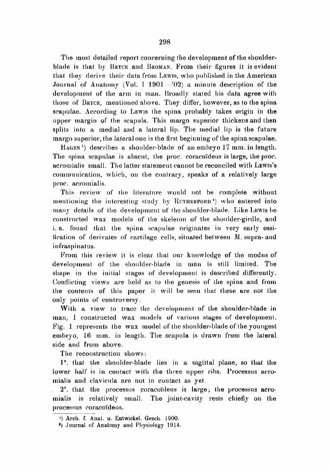
With Et view 10 tmce Ihe developrnent of the shoulder-blade in man, I constructed wax models of "al'iolls stages of development. Fig. 1 I'epl'esellts the wax model of the shollldel'-blade of Ihe youJlgest embryo, 16 mmo in lenglh. The scapllia is dl'awn frOnt the latel'al side and fl'om above .
The reconstruction shows: JO. that ~he shollldel'-blade lies iJl a sagittal plane, sa that the
lower half is in conlact with the thl'ee upper I'ibs. Pl'ocessus acl'omialis and c1avicllla are not in contact as yet.
2" , that the pl'OCel3Sus ~ol'aco'jdells is large; the processus acromialis is rel a tively smal\. The joillt-cavity l'ests chiefly on the processus eOI'acoïdeus.
1) Arch. f. Anat. u. Enlwickel. Gesch . 1900. ,) Journalof Anatomy and Physiology 1914.
299
3'. There is no indication of a spina scaplliae. The margo superior is neither thickelled nol' split into two labia.
~'ig. 1. Fig. 2.
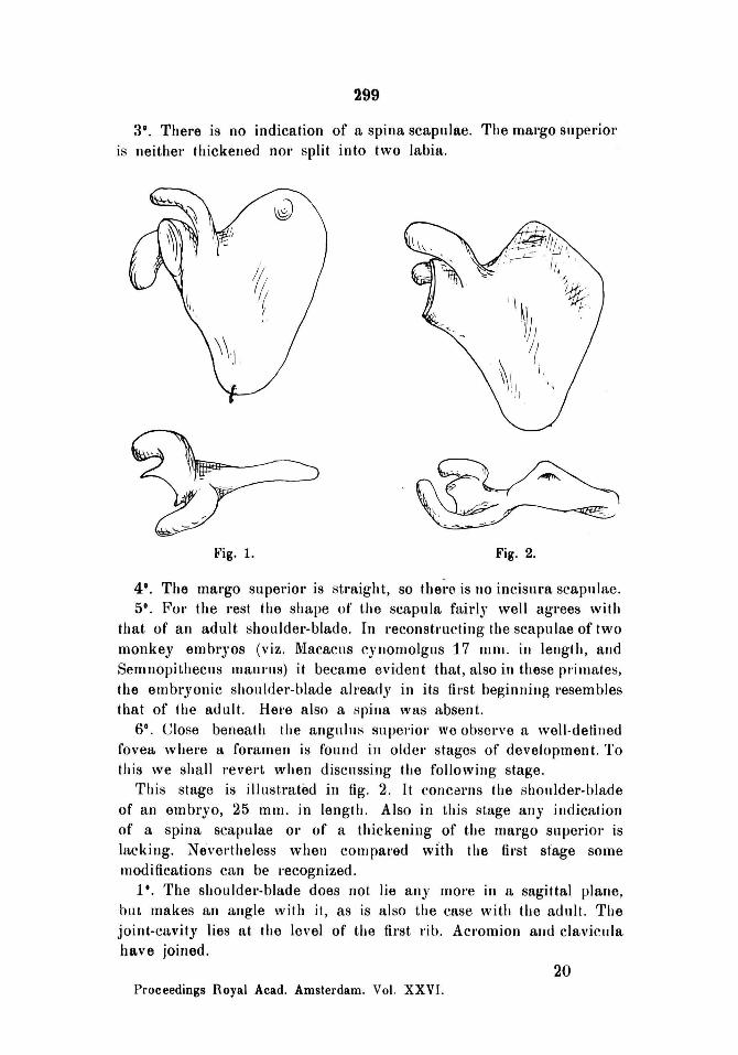
4'. The mal'go superiol' is stl'aight, so thel'{\ is 110 incisllra scapnlae. 5°. For the rest the shape of' the scapula fairi)' weil ag rees with
that of an adult shoulder-blade. In I'econstl'llcting the scaplliae of two monkey embl'Yos (viz. Macacns c)'lIomolgns 17 mnl. ill lengt h, alld Semnopithecus mali nIs) it became evident that, a1so in these pr-imateti, the embl'yonic shonldel'-blade already in its {hst heginllillg l'esembles that of the adult. Hel'e also a spilla was absent.
6°. Close beneath the angllillti superior we obsel'Ve a well ·delilled fovea whel'e a foramen is found in oldel' stages of deveJopment. To this we sIrall revert when discllssing Ihe followillg stage.
Tlris stage is illustratêd in fig. 2. II concerns tlre sholllder-blade of an embryo, 25 mmo in lengtil. Also in tltis stage an)' indicafion of a spina scapulae Ol' of a thickening of tlte margo supel"Ïol' is lacking. Nevedheless when cOlllpal'ed wlth Ihe fil'st stage some moditications can be recognized.
1'. The shouldel'-blade does not lie any more in a sagittal plane, but makes an angle with it, as is also tlte case with the adult. The joint-cavity lies at Ihe level of the first rib. Acromion allo clavicllia have joined.
20 Proceedings Royal Acad. Amsterdam. Vol. XXVI.

300
2°. The processus coraeoïoeus has eomparatively decreased, the proeesslls acrornialis, 011 Ihe other hand, has increased . lt appears,
.then, thaI Ihe pl'ocessus coraeoïdeus, whiclt is pbylogelletically the oldest part, is most strollgly de"eloped in the youngest stage, whereas Ihe processus acromialis. whieh is phylogenetically younger, I~omes mOre to the fOI'e in tlle ol der stages. The joint-cavily now lies for the greater parI on the plallum scapulae.
3°. The margo verlebmlis cOllsists of a shorter uppel' pOI,tioll and a longer I O\<\'e I' pOI,tioll. They are al an obtuse angle 10 eaeh other.
4'. The pol'tion of the scapula from which aftel'wards the fossa supraspinata develops, makes an angle with Ihe fulure suhspillal portion. This deviation of Ihe uppel' part, wbich also occurs ill the adult shoulder-blade (since fossa supra- and infraspillata do 1I0t lie in one and the same plane), bad not yel taken place in Ihe 16 mm . emlJl'Yo.
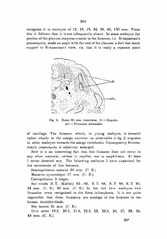
5°. In the c .. anial pal't of the sboulder blade a rommen OCCIll'S under t.he anguilIs superior, whicb extends at Ihe costal plane of the scapula as a gl'oo,'e along Ibe margo supel'ior in the direclion of the joint-cavity. In fig . 3 we give a cI'oss-section of tbis fOl'amell, whieh is filled with eonnective tissue.
The existence of th is foramen is no doubt surprising j yet it was not entirely unknown, as all'eady RUTHI!:RFORD bas described il (I. c.). However, according to this author it proceeds in a groo,'e, whidl reaches as far as the mal'go vel'tehralis . Now, ill all the sel'Ïal sections ill whieh I also met wilh a gl'oove as weil as wilh the fOl 'amen, it proceeoed along the margo supel'Ïor ill tlle direction of thejoint-cavity.
RUTH~;RFORD explains thi :; foramell as follow s. He eonsiders the part of Ihe scapula, I~ranial to Ihe foramen (resp. groove), as a sepal'ate piece of cartilage, which he terms praescapuIa, and which, according to his ac~onnt, is conneeted by a strand of mesenchyma tissue witl! tbe sternal balf of Ihe clavicula. In this way be believes an innel' sbollldel'-git'dle to ha,ve developed, while he supposes ' the aeromion-clavicula to bnild up the outer girdle. He adduees val'ioug arguments to pl'o,'e Ibis; howevel', Ihey are weak . In my judgment the hy pothesis is of riO valne, becanse a cbrmect.ion of the so-called pI'aescapula with the sternal balf of the clavicula does not occur. At all events in my preparations I never found a cell-strand like the one described by RUTHERFORD.
This rommen is not presellt in all cases, lts development also ditfers with val'ious indi vidnals, as showll by Ihe following data. I could establish its presellce eithel' as, ft Irue fOl'amen, Ol' as a deep groove in human embryos of the length of 16, 17.5, 18, 19.6, 21, 22, 25 (see tig. 3), 26, 27, 56, alld 90 mmo 011 the olher hand I did not
301
l'ecognize it in embryos of 12, 18, 18, 24, 26, 40, 120 mm. ' Fl'om this it follows that it is not infrequently absent. In some embryos the portion of the planum scapulae crallial to the fommen, i'-e. RUTHERFORD'S praescapula, made all allgle with the rest of the plallllm, a fact that lends support to RUTHERFORD'S view, viz , that it is really a Heparate piece
~~ig. 3. Homo 25 mmo transverse. Sc = Scapula; Acr = Processus acrOlpill.lis.
of cartilage. The fOl'amen which, in yonng embl'yos, is sitllated mther closely to the margo superior, as obsei'vahle in fig. 3, migl'ates in older embryos towards Ihe margo vertebmlis . Consequentl)' RU'I'HERFORD'S praescapuia is relatively enlarged.
Now it is an illtel'f'sting fact that t.his fommen does I10t occur in aIl'y olhel' mammal, neilher in reptiles, nor in l1.mphihians. AI leasi I never detected any. The following embryos I havo examilled fol' tho OCCUI'l'ence of Ihis foramen.
Semllopithecus malll·us 20 mm . (C. R.)
Mncacus cynomolgus 17 mmo (C. R.) Cercopit hecus 2 stages. Sus sCl'ofa N. T. (Keibel) 83- 85, N. T . 88, N. T. 88, N. T. 91,
24 mm. lC. R.) 26 mm. (C. R.) In the lasl two embryos two fOl'amina wel'e I'ecognized in Ihe fossa infmspinata. It is not quite impossible that these fOl'amina are analoga of lhe foramell in the human shouldet'-blade.
Bos tanrus 21 mmo (C. R.) Ovis aries 19.5, 20.5, 21.5, 22.5, 23, 23.5, 26, 27, 29, 35,
45 mm, (C. R.)
20*

302
Cards familiari s 12, 12, 22, 23.5 mmo (C. R.) Sciurus vnlgaris 12, 30 mmo (C. l{ .) Mus deCUmarlllS 11.5, t 2,13, 13, 13~ 2, 14.5, 16, 18,20,22 mm. (C. R.) Lepus cunicllius 17, 20 mmo (C. R) Spel'mophillus cilillllS 15 mm. (C. R.) Rousettlls amplexicaudalus 7.5, 10.5, 11, 11, 11.5, 12, 12, 14.5,
15.5, 16, 18 mmo (C. R.) Talpa europea 8.5, 9, 9, 10, 12, 13, 16.5, 20 mmo (C. R). Perameles obesula 50 mOl . (C. R.) Perameles spec. 38 mmo (C. R.) Dasyul'us viverri/lus 19.6, 33, 36, 40, 53, 63 mm o (C. R.) Sminthopsis cl'assicaudalus 13. 25 mm. (C. R.) Phascalogale pennicillala 37 mmo (C. R .) TI'ichoSlll'US vulpecula 32 mm. (C. R.) Didelphys cancl'ivora, 4 embl'Jos of 25 mmo lengtIl. Lacerta agilis N. T. (Kei bel) 117, 118, 120, 123, 123, 124,
125, 126. Calotes iubatus, length of Ihe head 5 1
/. Olm., 7 mm . Lagysoma 27.5 mm o Hemidactylus fren . ' Ienglh of the head 4.5 mm o Salamandl'8 mac. 11, 13, 15, 16, 16, 24 mmo Pipa Americana, J 2 mmo Rana . 2 embl'Yos .
So fa I' as I am able to judge fOl'amina in adult shoulder-blades occur ollly with Homo and with various Edenlala, in whirh they are always formed by bl'idging of Ihe IncisUl'a scaplllae, and wilh Delphinus delphis. In Ihe lattet' the chal'acter of the foramen is not kllown. RUTHKRFORD (I. c.) has described it.
A conceivable con/lection , that might exisl betwet'n Ihe praescapuia of RUTHERFORD and Ihe altachmenl of Ihe clavicula (nol only Ihe sternal half of Ihe clavicula, as l{UTHERFORD supposed) 10 Ihe margo superior scaplllae, aA it oeCUJ's in reptiles, echidna and ol'IJithorynchus. eOllld not be ascel'tained, since a connedioll of Ihe praescapuIa of RUTHERFonv to the aCl'Omial pari of Ihe clavicula could not be delected eilher.
lt appears, then, th at the fOl 'amen, pl'esent in the majol'Ïly of human embl'yos in Ihe ('ranial pal't of Ihe shoulder-blade, does not occur in olhel' vertebl'ates, (excep! i/l Delphinus ' delphis, whieh, howe\'el', is of such a pronollneed specifieity Ihat Ihis foramen eannot be looked upon as a homologue of that of man). Neithel' did I find a ny attachment of the praeseapllla of RUTHERFORD to ani other'
303
skelelal bone. The aigllificance of this fOl'amen is unknown as yet. As to the ossifiration of the scapula my expel'Ïence pl'oved it not
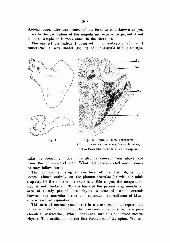
to be so simple as is l'epl'esented in the literatUl'e, The earliest ossi ficatioll I obsened in aH elll bryo of 40 m lil, I
constructed a wax model (fig, 4) of the 'scapula of this embryo,
'I l I,
Fig,4. ~'ig, 5. Homo 40 mmo Transvel'sal.
Cor = Processus coracoideus; Hu = Humerusj
Acr = Processus acromialis; Sc = Scapula,
Like rhe preceding lIlodel this also is viewed from above and fl'om the dorso-Iateml side. Wh at tllis reeollstmcted model shows IJS may follow here:
Tlle joint-eavity, lying at the level of the fh'st l'ib .. is now located almoat entir'ely on the planum scapulae (as with the adult scapula), Of the spinR not a tmce is visible as yet, tlte mal'go superior is not thickened . To the basi s of the processus aeromialis an area of closely pacl<ed llI esenchyma is attached, which extellds b'etween the IDnscular tissue alld separ'ates the rudiment of Musc, snpra-, and infmspinatns.
This area of mesellchyma is cut in a cross sectioll as represented in fig. 5. Behind Ihe root of the pl'oressus acromialis begilIs a perichondrial ossification, \vhich continues into this condensed mesenchyma. This ossification is the fh'st format.ion of the spina. We see,

304
thet'efore, that it is formed by a pel'ichondrial ossification, fOl' although no ossifying pel'ichondl'Ïnm is visible here, the fact that the bone is fOI'med from the sUI'l'ounding mesenchyma co-ossifying with cartilage, established the charactel' of the ossification. In fig. 5 we gi"e a cross section of Ihis tlI'st stage of the spina.
I have not been able 10 recogllize two centl'es of ossification in the cartilaginous scapllla, descl'ibed by RAMBAUD and RENAU (quoted by POIRIER I), which, according to these authors, arise bet ween the 40th and 50th day and fuse in the third month.
In the scapula of an older embryo (56 mmo in lenglh) Ihis perichondl'Ïal ossificalion appeal's 10 be largely exlellded. The margo anterior scapulae is almost I'eached. Tbe cartilage of the planullI
éh-.
~----------------~~~~
Fig. 6. Homo 56 mMo Transversal. Hu = Humerus; Cl = Clavicula; Cor = Processus coracoideus; Acr = Proces sus acromialis;
Sp = Spina scapulae; Sc = Scapula.
scapulae, however, has been distinclly calcified over a considel'able area ah·eady. The mal'ked enlal'gement of the spina seaplliae is shown iJl tig. 6 . Besides the spina tbis figul'e also shows pal'! of the foramen described above. The spina is fOl'med by a. gl'Owth of bone between
I) POJRIER el CHARPY, Traité d'Anatomie humaine.

305
M. supra- and in frRspinat us, between acrornion and planum scaplliae. It cannot be denied, however, that in the rnesenchyrna, ill which this bone develops. vel'Y yOllng cat·tilage-cells are noticeable here and there. These cells, howe"el', have no intermediate mattel' as .vet; they al'e little ditfel'entiated and it is difficult to distinguish thern from the rnesenchyrna-cells: So it is evident that besides bone-cells also cal'tilage-cells develop in the mesenchJma.
In an embl'Jo of 90 mrn. enchondl'ial as weil as pel'Ïchondrial ossitication takes place, the boundal'Y between the two being no
Fig. 7. Homo 90 mm, Margo anteriol' scapulae · transversal.
Acr
Pl. Sc. Fig. 8. Homo 90 mmo Scapula transversal Acr. = Processus acromialis J,c. = Joint-cavity, Pl, Sc, = Planum
scapulae,
longer percei vable. The peeulial' chal'acter of the pel'ichondrial ossitication along the margo antel'iol' is remal'kable. In the place of the formation of compact bone, which in othel' cases occurs with pel'ichondrial o~sification e.g. that of the long bones, we see here a bony fl'amewol'k encircled by rnesenchyma, Fig. 7 shows a cross section thl'Ough the margo anterior.
The study of this object (embryo of 90 mrn.) shows remarkable pecularities of the growth of the spina scaplllae. In the mesenchJma between M. supra- and infl'aspinatlls a distinct cartilage is now rècognizable, lt is quite independent of the other mass of cartilagè
306
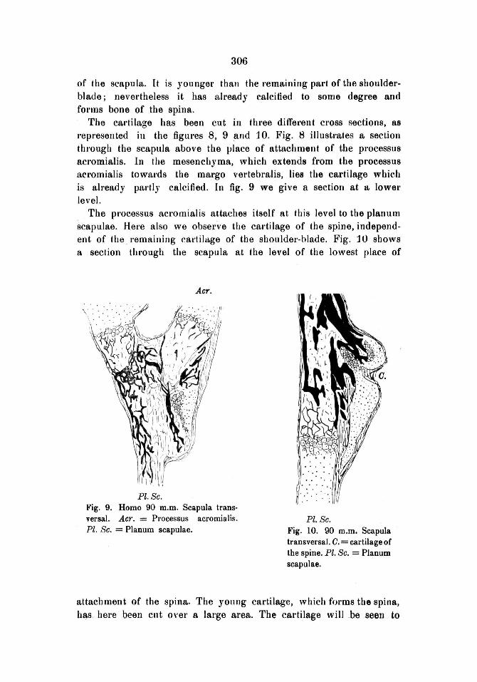
of the scapula. It is youngel" thall the remaining part of the shoulderblade ; nevel"theless it has already calcitied to some degree and fOl'lIlS bone of the spina.
The cal,tilage has been cut in thl"ee different cross sections, as I'epr'esented in the figur'es 8, 9and 10. Fig. 8 i1lustrates a section through the scapllla above the vlace of attachment of the processus aCl'omialis. In the mesenchyma, which extends from the processus acromialis towal'ds the margo vel'tebmlis, lies the cartilage whir.h is al ready pal,tlJ calcified. In fig. 9 we give a sectiort at lt lower level.
The pl'ocessus acrornialis attaches itself at this level to the planum scapulae. Here also we observe the cal'tilage of the spine, independent of the. I'emailling cal'tila.ge of the shonlder-hlade. Fig. 10 shows a section tlll'ough the scapula at the level of the lowest place of
ACT.
Pl. Sc. Fig. 9. Homo 90 m.m. Scapula trans· versa\. Acr. = Processus acromialis. Pl. Sc. = Planum scapulae.
Pl. Sc. Fig. 10. 90 m.m. Scapula transversal. C. = cartilage of the spine. Pl. Sc. = Planum scapulae.
aUachment of the spina. The yonng cal·tilage, which fOl'ms the spina, has here been cnt o\'er a large area. The cal'tilage willbe seen . to

307
be pal,tly calcified, while bone has been fOl'med, uniting witt. this calcilied area,
80 while Ihe first begillning of the spina is fOl'med by perichondl'ial bone in the mesenchyma between M, supl'a-, and illfraspillatus, its furthel' developmellt is etfected by chondrial bone, whi('h ol,jginates in the younger eal'tilage. This cal'tilage has been generated betweell the afore-said museles hy the same mesenchyma.
A peculiar feature is still to be observed at the shoulder-blade of the embryo of 90 mmo Bone is developed at the margo superior as weil enchondrially as perichondrially . In the mesençhyma t.hat. forma Ihe pel'ichondl'Ïal bone, and into which Ihis bone extends over some distance, there al'e two cartilaginous nuclei, made up of Ihe sllme YOllng tissue from whieh tlle cartilage of the spina has been built up. Fig. 11 shows in Cl'OSS seclion these nuclei, which are 1I0t in contact with the remuining eal'tilage of the shoulder-blade, These cartilage-islets appeal' to be al ready caleified and ossified hel'e and thel'e. It is impossible to draw a bOllndary-line between the bOlle formed in thi!l process and the pel'ichondrial bone of the scapula. This ossifying ' process, in which (besides the ellchondrial oS8ific8olion of the scapula) both perichondrial and chondrial ossificalion of a cf\rtilage nucleus, situated olltside the perichondrial bone, are present, ag rees completely wilh the fOl'mation of the spina scapulae. This is stl'iking, since the spina scapulae and the definilive margo su peri or are I he I wo pal'ts of t he shoulder-blade, w hieh are missing in Ihe lirsl rudiment of Ihe cartilaginous scapllia. This deficiency "ertebral of the place destined fOl' I he future incislll'e, is indeed accounted for by the faet that the margo supel'ior in young embryos is still straight and displays no incisure. The missing pal'ts are appal'ently supplied by the perichondl'ial bone that reaches far into the mesenehyma, together with Ihe bone formed by the afol'esaid cartilage-nllelei. At the shouldel'-blade of an embryo of120 mm' in length, in which Ihe ossification had eonsiderably advanced, the ineisure was indeed present.
Of course, the question arises, how the e80rtilage of tlle spina as weil as the cal,tilage nuclei are fUl'ther developing. In both plaees the cartilage is soon trallSfOl'llled completely into bone. ]n an embl'yo of 120 mm, onlJ a very few remnants of the cal'tilage of the spina were still left. The rest had been ossified.
Aftel' this Ihe development of Ihe shoulder-blade pl'oceeds in the way described in the text-books of embryology.
Now let us l'eviaw once rnOl'e the CUlTent opinions of the development of the spina scapulae, It will be seen, then, that howe\'er

308
divet'gent they may be, most of them cannot be deemed incolTect, when we bear in mind that they concern different stages, '
Fig. 11.
Homo 90 m.m.
RUTHItRE'ORD'S \"iew of the ,-ery early ossification of cartilaginous cells is no doubt correct, bilt holds good only fol' youug stadia. Neithel' is the conception of HERTWIG and BRAUS abollt a separate centre of ossification quite inronect, since there is a stage in which an independent cartilage is fOl'ming bone.BARDELEBEN's
record about an ossitication under and beside the spina cannot altogether be disqualified either, but it only applies to a brief stage of development. However, ossificatioll like th at of the bon es of the cranial vault does not occU[' in the development of the shouJder-blade, In the neonatus a few cal,tilage may possibly
sometimes be fou~d at the spina (BRYCE), but it is certain that the spina scapulae in the new-bol'H child does not con sist of cartilage.
Margo superior scapulae (KÖLLIKER and HERTWIG advocate the opposite transversal.
view). LEWIS'S conceptioll, howe\"er, (doubling of tlle mal'go superior) is altogether wrong. The diagram bOlTowed from L!!:wls by HROMAN, BRYc!!: and BAltDEEN l'epresents a faulty reconstruction of the shoulder-blade.