anatomy of ear(ext & middle)
TRANSCRIPT

ANATOMY OF ANATOMY OF EAREAR
Presenter :Dr.Razal M Sherif
Moderator :Dr.Joythi swarup

EAREAR
EXTERNAL EARMIDDLE EARINNER EAR
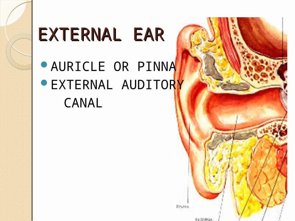
EXTERNAL EAREXTERNAL EAR
AURICLE OR PINNAEXTERNAL AUDITORY CANAL
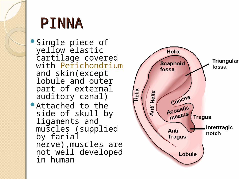
PINNAPINNASingle piece of yellow
elastic cartilage covered with Perichondrium and skin(except lobule and outer part of external auditory canal)
Attached to the side of skull by ligaments and muscles (supplied by facial nerve),muscles are not well developed in human
INCISURA TERMINALIS is a gap between the superior part of tragus and root of helix, which is devoid of cartilage having only fibrous tissue
◦ Endaural approach-inscion made on this area will not cut through the cartilage in surgery of EAC or Mastoid
PINNAPINNA Contd.Contd.
APPLIED ANATOMYAPPLIED ANATOMY
Tragal cartilage, perichondrium from tragus, concha, fat from lobule – reconstruction surgery for middle ear
Conchal cartilage – correct depressed nasal bridge
Composite graft of skin & cartilage from pinna – repair the defects of nasal ala
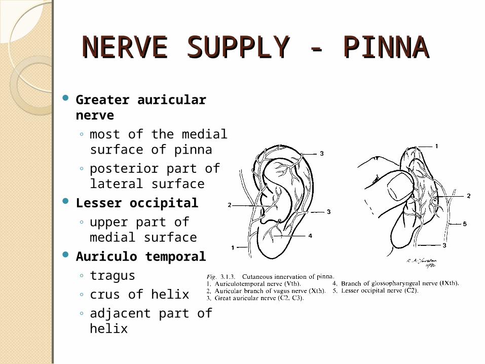
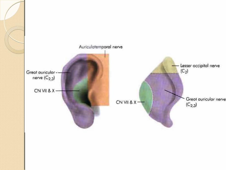
NERVE SUPPLY - PINNANERVE SUPPLY - PINNA
Greater auricular nerve◦ most of the medial surface
of pinna◦ posterior part of lateral
surface Lesser occipital
◦ upper part of medial surface
Auriculo temporal◦ tragus◦ crus of helix◦ adjacent part of helix

Auricular branch of vagus◦ concha◦ corresponding eminence on medial surface
Facial nerve◦ distributed with fibers of auricular branch of vagus◦ concha◦ retroauricular groove
NERVE SUPPLY – PINNA Contd

EXTERNAL AUDITORY CANALEXTERNAL AUDITORY CANAL
Extends from bottom of concha to tympanic membrane
24 mm (along post wall)not a straight tubeouter part(cartilaginous) directed upwards,
backwards & mediallyinner part (bony) directed downwards,
forwards & mediallypinna pulled upwards, backwards &
laterally(make it straight)
EXTERNAL AUDITORY CANALEXTERNAL AUDITORY CANAL
CARTILAGINOUSBONY
CARTILAGINOUS PARTCARTILAGINOUS PARTOuter 1/3rd & 8mm canalContinuation of cartilage which forms the
frame work of pinna
◦Fissures of Santorini through them parotid or superficial mastoid
infection can appear in the canal or vice versa
Skin covering the cartilaginous canal is thick and contains appendages like 1.CERUMINOUS GLANDS(modified sweat gland),which secrets cerumen (wax) 2.PILOSEBACEOUS GLANDS 3. HAIR is only confined to the outer canal & therefore furuncles are seen only in the outer 1/3rd of canal

BONY PARTBONY PARTInner 2/3rd & 16mmSkin lining the bony canal in thin & continuous
over the tympanic membrane Devoid of skin appendages(Hair and
ceremonious GlandsAbout 6mm lateral to tympanic membrane ,
bony meatus presents as narrowing called ISTHMUS
Foreign body lodged medial to isthmus, get impacted & are difficulty to remove

Anteroinferior part of deep meatus, beyond the isthmus, presents a recess - Anterior recess which acts as a cesspool for discharge & debris
Antero inferior part of bony canal may present a deficiency in children up to age of 4 or sometimes in adults permitting infection to & from parotid (Foramen of Huschke)

NERVE SUPPLY - EACNERVE SUPPLY - EACAuriculo temporal nerve(V3)◦anterior wall & roof
Auricular branch of vagus (X)◦posterior wall & floor
Posterior wall of auditory canal also receives sensory fibres of CN VII through auricular branch of vagus

TYMPANIC MEMBRANETYMPANIC MEMBRANEForms partition between
EAC & middle earObliquely set – 45deg
with floor of EACPosteriosuperior part
more lateral than Anterioinferior part
9-10 mm tall8-9 mm wide0.1 mm thick

PARS TENSAPARS FLACCIDA (SHRAPNEL’S
MEMBRANE)
TYMPANIC MEMBRANETYMPANIC MEMBRANE

PARS TENSAPARS TENSA
Forms most of Tympanic Membrane Periphery is thickened to form a fibro
cartilaginous ring – ANNULUS TYMPANICUS, which fits in tympanic sulcus
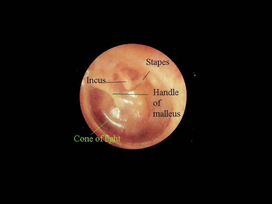
Central part of pars tensa is tented inwards at the level of tip of malleus – UMBO
Bright Cone of Light – seen radiating from the tip of malleus to periphery in anterioinferior quadrant
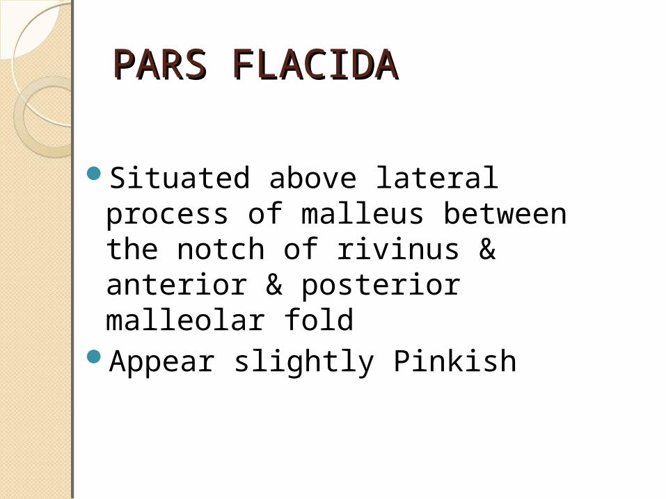
PARS FLACIDAPARS FLACIDA
Situated above lateral process of malleus between the notch of rivinus & anterior & posterior malleolar fold
Appear slightly Pinkish
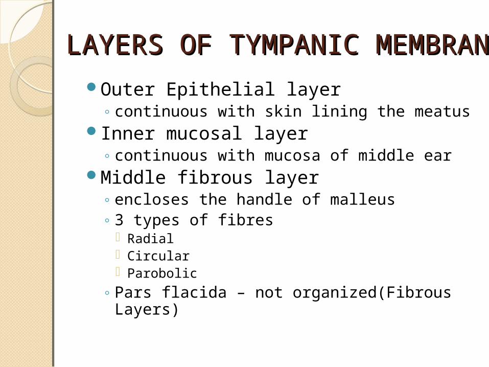
LAYERS OF TYMPANIC MEMBRANELAYERS OF TYMPANIC MEMBRANEOuter Epithelial layer◦ continuous with skin lining the meatus
Inner mucosal layer◦ continuous with mucosa of middle ear
Middle fibrous layer◦ encloses the handle of malleus◦ 3 types of fibres
Radial Circular Parobolic
◦ Pars flacida – not organized(Fibrous Layers)

NERVE SUPPLY - TMNERVE SUPPLY - TMAURICULO TEMPORAL NERVE (V3)◦Anterior half of lateral surface
AURICULAR BRANCH OF VAGUS (X)◦Posterior half of lateral surface
TYMPANIC BRANCH OF CN IX (JACOBSON NERVE)◦Medial surface
ANATOMY OF EAR Video Presentation

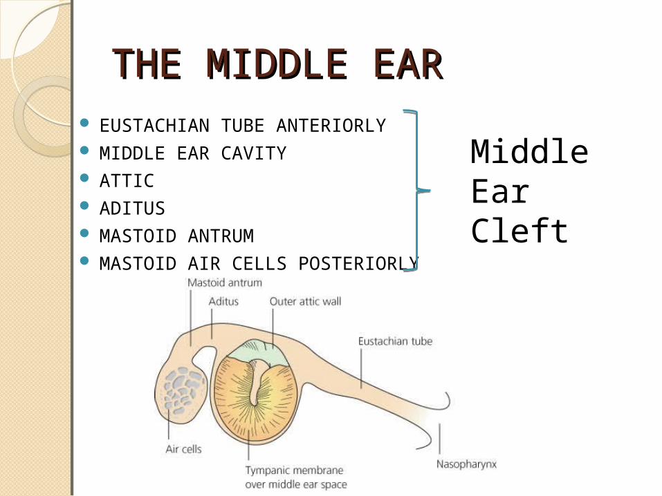
THE MIDDLE EARTHE MIDDLE EAR EUSTACHIAN TUBE ANTERIORLY MIDDLE EAR CAVITY ATTIC ADITUS MASTOID ANTRUM MASTOID AIR CELLS POSTERIORLY
Middle Ear Cleft

MIDDLE EAR CAVITYMIDDLE EAR CAVITYROOF◦Thin Plate Of Bone – Tegmen Tympani◦Separates Middle Ear From Middle Cranial
Fossa◦Incomplete Ossification – Allow Infection To
Middle cranial fossa

FLOOR◦Thin bone separates cavity from jugular bulb
LATERAL WALL◦Main Part – Tympanic Membrane ◦Superiorly Area Of Bone – Scutum◦Inferiorly – Tympanic Bone Separates
Tympanic Cavity From Medial Part Of TMJ

ANTERIOR WALL◦Narrow inferiorly◦Thin plate of bone separates the cavity from
internal carotid artery.◦2 openings in upper part of anterior wall◦Above – opening for tensor tympani◦Below – opening for Eustachian tube
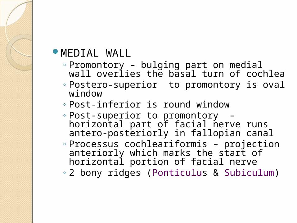
MEDIAL WALL◦ Promontory – bulging part on medial wall overlies the
basal turn of cochlea◦ Postero-superior to promontory is oval window ◦ Post-inferior is round window◦ Post-superior to promontory – horizontal part of
facial nerve runs antero-posteriorly in fallopian canal◦ Processus cochleariformis – projection anteriorly
which marks the start of horizontal portion of facial nerve
◦ 2 bony ridges (Ponticulus & Subiculum)
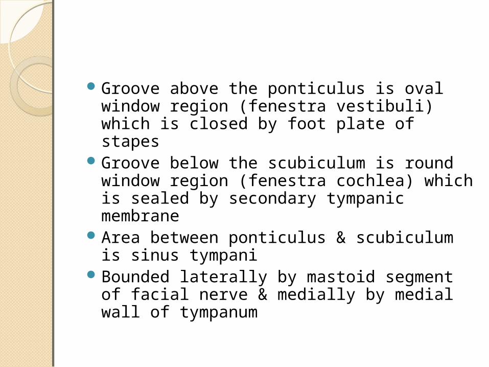
Groove above the ponticulus is oval window region (fenestra vestibuli) which is closed by foot plate of stapes
Groove below the scubiculum is round window region (fenestra cochlea) which is sealed by secondary tympanic membrane
Area between ponticulus & scubiculum is sinus tympani Bounded laterally by mastoid segment of facial nerve &
medially by medial wall of tympanum

FACIAL RECESS BOUNDARIES◦ LATERALLY – postero superior part of anulus◦ SUPERIORLY – short process of incus in fossa incudis◦MEDIALLY – vertical part of facial nerve
Explored during posterior tympanotomy procedure which is widely used during ci & combined approach tympanoplasty
POSTERIOR WALL
◦ SUPERIORLY – aditus ad antrum which leads from epitympanum to mastoid antrum
◦ INFERIOR TO ADITUS – small conical projection – pyramid transmits the stapedial tendon to its insertion at the neck of stapes
◦ below pyramid & lateral to it is opening for chorda tympani

DIVISIONS OF MIDDLE EAR DIVISIONS OF MIDDLE EAR CAVITYCAVITYEPITYMPANUM
◦ part of middle ear above the malleolar fold◦ contains head of malleus, body of incus, ossicular
ligament & mucosal fold◦ prussacs space – situated in attic
laterally – pars flaccida medially – head & neck of malleus anterosuperiorly – lateral malleolar ligament inferiorly – ant & post malleolar fold
◦ site of formation of primary acquired cholesteotoma
MESOTYMPANUM◦Part lying medial to pars tensa & is major air
filled space◦Contains – long process of incus, stapes,
mucosal fold
HYPOTYMPANUM◦Part lying below the lower margin of tympanic
membrane◦Contains globus body & bulge produced by
jugular bulb◦Floor may be deficient sometimes & thus
jugular bulb may project into tympanic cavity
PROTYMPANUM
◦Portion of middle ear around the tympanic orifice of et
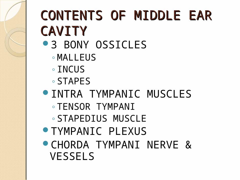
CONTENTS OF MIDDLE EAR CONTENTS OF MIDDLE EAR CAVITYCAVITY3 BONY OSSICLES◦MALLEUS◦INCUS◦STAPES
INTRA TYMPANIC MUSCLES◦TENSOR TYMPANI◦STAPEDIUS MUSCLE
TYMPANIC PLEXUSCHORDA TYMPANI NERVE & VESSELS

MASTOID PROCESS & AIR MASTOID PROCESS & AIR CELLSCELLSPart of Temporal boneAt Birth – No Mastiod ProcessStarts developing at the end of first year 3 TYPES ◦CELLULAR◦SCLEROTIC◦DIPLOEIC
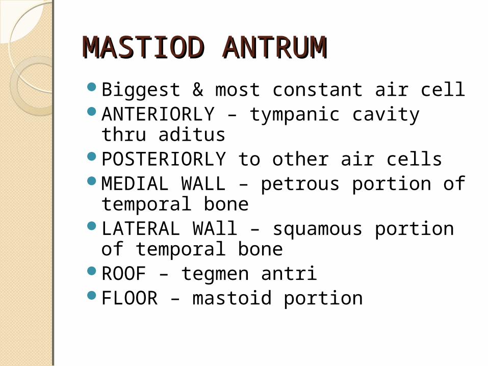
MASTIOD ANTRUMMASTIOD ANTRUMBiggest & most constant air cellANTERIORLY – tympanic cavity thru aditusPOSTERIORLY to other air cellsMEDIAL WALL – petrous portion of temporal
boneLATERAL WAll – squamous portion of temporal
boneROOF – tegmen antriFLOOR – mastoid portion
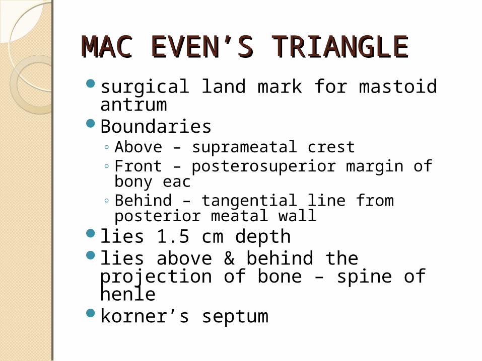
MAC EVENMAC EVEN’’S TRIANGLES TRIANGLEsurgical land mark for mastoid antrumBoundaries◦ Above – suprameatal crest◦ Front – posterosuperior margin of bony eac◦ Behind – tangential line from posterior meatal wall
lies 1.5 cm depthlies above & behind the projection of bone –
spine of henlekorner’s septum

VARIOUS GROUPS OF AIR CELLSVARIOUS GROUPS OF AIR CELLSMASTOID AIR CELLSMASTOID AIR CELLS LOCATIONLOCATION
PERISINUS CELLSPERISINUS CELLS SIGMOID SINUSSIGMOID SINUS
PERILABYRINTHINE CELLSPERILABYRINTHINE CELLS LABYRINTH(SUPRA, INFRA, RETRO)LABYRINTH(SUPRA, INFRA, RETRO)
TEGMEN CELLSTEGMEN CELLS TEGMEN TYMPANITEGMEN TYMPANI
ZYGOMATIC CELLSZYGOMATIC CELLS ROOT OF ZYGOMAROOT OF ZYGOMA
PETROUS CELLSPETROUS CELLS PETROUS APEXPETROUS APEX
PERITUBAL CELLSPERITUBAL CELLS ETET
RETROFACIAL CELLSRETROFACIAL CELLS VERTICAL SEGMENT OF FACIAL NERVEVERTICAL SEGMENT OF FACIAL NERVE
TIP CELLSTIP CELLS MASTOID TIP(LATERAL & MEDIAL)MASTOID TIP(LATERAL & MEDIAL)
PERIANTRAL CELLSPERIANTRAL CELLS MASTOID ANTRUMMASTOID ANTRUM
SQUAMOUS CELLSSQUAMOUS CELLS SQUAMOUS PART OF TEMPORAL BONESQUAMOUS PART OF TEMPORAL BONE

