anatomy of thethorax and … · anatomyofthethoraxand shouldergirdledisplayedby ... the anatomical...
TRANSCRIPT

ANATOMY OF THE THORAX ANDSHOULDER GIRDLE DISPLAYED BYMAGNETIC RESONANCE IMAGINGJames D. Collins, MD, Marla L.Shaver, MD, Poonam Batra, MD, and Katheleen Brown, MDLos Angeles, California
In 1971, radiographic anatomy of the humanbody was added to the gross anatomy courseat UCLA.1 Radiographic contrast studies andplain anatomical displays were formulated intoteaching packages for all organ systems. Resi-dents presented each package to first-yearmedical students in the dissection laboratoryto augment the teaching of anatomy. In Novem-ber 1984, magnetic resonance imaging wasinstituted in the radiology department. Imagingthe chest produced coronal and axial planeswhich displayed the muscles and soft tissuesof the thorax. In 1986, the authors presentedtheir study of MR anatomy of the chest andshoulder girdle to the American Association ofAnatomists.2The purpose of this presentation is to dem-
onstrate the anatomy of the thorax and shoul-der girdle as displayed by magnetic resonance,correlated with regional anatomy, with empha-sis on soft tissue structures.
Key Words * gross anatomy * MRI * thorax * chest*shoulder girdle * MRI soft tissues * medical student
teaching * chest wall * MRI nerves
From the Department of Radiological Sciences, UCLA Schoolof Medicine, Los Angeles, California. Requests for reprintsshould be addressed to Dr James D. Collins, Department ofRadiological Sciences, UCLA Medical Center, Los Angeles, CA90024.
Magnetic resonance imaging (MRI) of soft tissuesdisplays normal and abnormal anatomic images of thesurface and connective tissues that cannot be imagedwith conventional radiographic techniques. The adventof the cursor lines, depicting the area of tissue selectedfor imaging, enables the radiologists at UCLA to offera new modality for teaching anatomy. Cursor lines maybe rotated 180 degrees on the axial, the coronal, andsagittal planes of anatomy. The images may bemagnified and displayed without reconstruction.When the cursor lines are placed on the lateral chest
wall of the mid-coronal MR of the chest, the pleuralsurface and adjacent muscles of the chest wall aredemonstrated in the oblique coronal plane. Applied tothe anatomical planes of the thorax, MR imagingdisplays anatomy that assists the teaching of medicalstudents, anatomists, and radiologists. David Maxwellfrom the UCLA Department of Anatomy prepared atemplate list of anatomical structures to determine if theauthors' team could demonstrate the thoracic anatomywith MRI. Volunteers were chosen for imaging. Theimages and prepared list were matched beyond expec-tations.
MATERIALS AND METHODSAll images were recorded with a spin echo ofTE = 28
and TR = 500, using a 0.3 Tesla Fonar permanentmagnet. Axial (transverse), coronal, and sagittal planeswere selected to image the anatomy for comprehensivelearning. Anatomical landmarks were maintained fororientation. Images chosen for display were labeled
26 JOURNAL OF THE NATIONAL MEDICAL ASSOCIATION, VOL. 83, NO. 1

TABLE. ANATOMICAL NOMENCLATURE DIRECTORY CHEST AND SHOULDER GIRDLE
F. First of the Series1. Pectoralis major muscle2. Pectoralis minor muscle3. Trapezius muscle4. Spine of the scapula5. Clavicle6. Supraspinatus muscle7. Deltoid muscle8. Serratus anterior muscle9. External oblique muscle
10. Latissimus dorsi muscle11. Deltoid muscle (posterior)12. Infraspinatus muscle13. Subclavius muscle14. Acromion process15. Body of the scapula16. Coracoid process17. Teres major muscle18. Teres minor muscle19. Coracobrachialis muscle20. Triceps muscle (long head)21. Triceps muscle (lateral head)22. Brachialis muscle23. Subscapularis muscle24. Rhomboid muscle25. Humerus26. Humerus (head)27. Intertubercular sulcus
28. Aorta29. Pulmonary artery30. Trachea31. Aorta (descending)32. Subclavian artery33. Vertebral artery34. Brachial plexus35. Mandible36. Spinalis thoracis muscle37. Brachiocephalic artery38. Subcutaneous tissue
(lymphatics, etc)39. Diaphragm40. Liver41. Spleen42. Stomach43. Right lung44. Left lung45. Vertebral body (thoracic)46. Right pulmonary artery47. Intervertebral disc space48. Thoracic duct49. Brachial artery50. Intercostal artery51. Intercostal muscle52. Superior vena cava53. Right atrium54. Left atrium
55. Brachial vein56. Axillary artery57. Median nerve58. posterior humeral circumflex
artery59. Scalenus anterior muscle60. Ulnar nerve61. Subracromial bursa62. Radial nerve63. Scalenus medius muscle64. Biceps muscle65. Right primary bronchus66. Left primary bronchus67. Ligamentum arteriosum68. Bronchial arteries69. Axillary nerve70. Inferior vena cava71. Spinal cord72. Rib73. Spinalis thoracis and cervicis
muscles74. Left ventricle75. Pericardial fat pad (right)76. Pericardial fat pad (left)77. Subclavian vein78. Longus collis muscle79. Biceps muscle (short head)80. Biceps muscle (long head)
JW-,~~ ~ ~ - ..b:~~~~~~~~~~~~~~~~~~~. ..... .. : ^ .Figure 1. Thoracic duct (arrows) crosses theangle between the internal jugular vein andthe subclavian vein. Aorta (28), subclavian(77).
with numbers and assigned to a directory (Table) toassist location of the anatomical structure on eachimage. Serial images were enlarged to enhance visualassociation with the gross anatomy prior to dissection.All images were labeled, and mounted on cleartransparent plastic panels. The panels were then placedon large radiograph view boxes and backlighted fordisplay. Images selected for presentation are displayedfrom the mid-coronal and posterior coronal planes.Oblique sagittal images were positioned from the leftmid-coronal image.
RESULTSThe reader should refer to the nomenclature directory
(Table) for Figures 1 through 13 for specific structures.Not every structure is labeled in each figure, and thestructures labeled are intended to orient and guide. Ingeneral, the lungs, blood vessels, joint capsules, tendoninsertions, and scarred tissues have low signals (black)in MR Imaging.The vascular supply of nerves are continuous like a
spider web enveloping the nerve sheaths.6 Small nerveshave a relative intermediate high signal in fatty tissue.
JOURNAL OF THE NATIONAL MEDICAL ASSOCIATION, VOL. 83, NO. 1 27

MRI OF THE THORAX AND SHOULDER GIRDLE
(black arrows). Pectoral nerves (white ar-rows).
Nerves are composed of phospholipids and displayed asa high signal. When the nerves are embedded in fattytissue; the blood supply marginates the nerve from thefat.
Magnetic resonance imaging detects the visualdisplay of the nerve and locates the low signal of theblood supply. Lymph nodes have a relative gray signaland are located in specific anatomical locations. Chyle(fat) has a high signal and allows location of thethoracic duct. These features are important for under-standing the anatomy displayed in the following MRimages. Figures 1 through 8 demonstrate the surfaceanatomy of the chest wall and shoulder girdle. Figure 1is the first of the series and orients the reader to theimage positions.The high signals (white) demonstrate deposited fat
and nerve signals.7'8 The low signals (black) demon-strate a variety of vascular structures coursing throughthe mediastinum and muscular tissue. Bones are
marginated by low signals of the periostium, cortex,and cartilage. Cartilage may be located when sufficientfat surrounds the margins. In Figyure 1, the thoracic duct
38
Figure 3. Note the low signals (white arrows)marginating the high signals of the nerves.
crosses the left subclavian artery and internal jugularvein, and the ligamentum arteriosum joins the aorta andpulmonary artery. The vagus nerve is a plexus ofintermediate signals within the high signals of fatsurrounding the aorta, left lateral wall of the trachea, leftcarotid artery, and pulmonary artery.
Serial images two through eight display the muscularstructures as they appear from the left medial oblique tothe lateral oblique position. The spine of the scapulaseparates the supraspinatus from the infraspinatus musclein Figure 2. The trapezius muscle attaches to the scapulaand clavicle. The low signals of blood vessels andintermediate high signals of the nerves overlay themuscles. The axillary artery, axillary vein, and brachialplexus are positioned in the axillary fat between thesubscapularis muscle and the pectoralis muscles.
Figure 3 images the teres major and minor muscles.The subclavius muscle is inferior to the clavicle. Theanterior deltoid muscle appears superior to the pector-alis muscle. The triceps muscles are inferior to thesubcutaneous tissue of the chest wall. The axillary
28 JOURNAL OF THE NATIONAL MEDICAL ASSOCIATION, VOL. 83, NO. 1
MRI OF THE THORAX AND SHOULDER GIRDLE
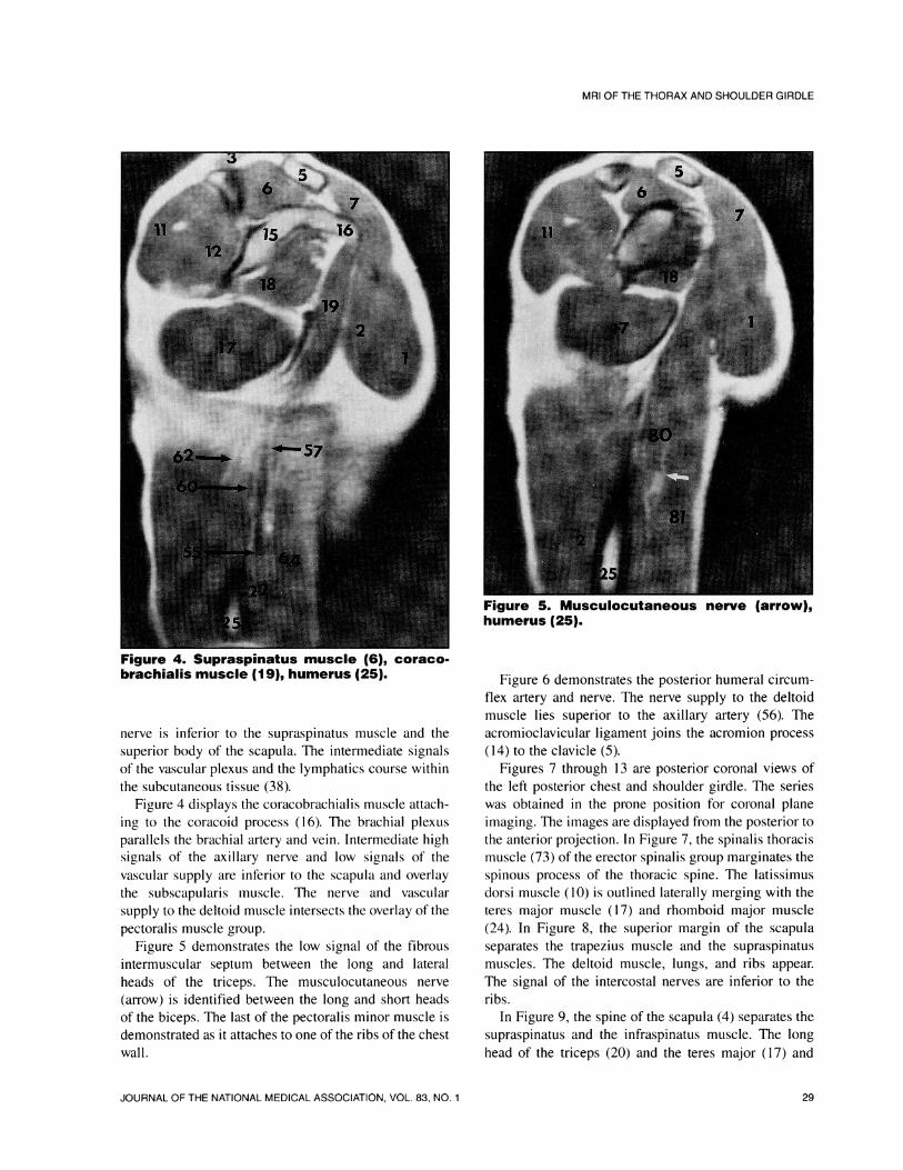
Figure 4. Supraspinatus muscle (6), coraco-brachialis muscle (19), humerus (25).
nerve is inferior to the supraspinatus muscle and thesuperior body of the scapula. The intermediate signalsof the vascular plexus and the lymphatics course withinthe subcutaneous tissue (38).
Figure 4 displays the coracobrachialis muscle attach-ing to the coracoid process (16). The brachial plexusparallels the brachial artery and vein. Intermediate highsignals of the axillary nerve and low signals of thevascular supply are inferior to the scapula and overlaythe subscapularis muscle. The nerve and vascularsupply to the deltoid muscle intersects the overlay of thepectoralis muscle group.
Figure 5 demonstrates the low signal of the fibrousintermuscular septum between the long and lateralheads of the triceps. The musculocutaneous nerve(arrow) is identified between the long and short headsof the biceps. The last of the pectoralis minor muscle isdemonstrated as it attaches to one of the ribs of the chestwall.
l~~~~~
Figure 5. Musculocutaneous nerve (arrow),humerus (25).
Figure 6 demonstrates the posterior humeral circum-flex artery and nerve. The nerve supply to the deltoidmuscle lies superior to the axillary artery (56). Theacromioclavicular ligament joins the acromion process(14) to the clavicle (5).
Figures 7 through 13 are posterior coronal views ofthe left posterior chest and shoulder girdle. The serieswas obtained in the prone position for coronal planeimaging. The images are displayed from the posterior tothe anterior projection. In Figure 7, the spinalis thoracismuscle (73) of the erector spinalis group marginates thespinous process of the thoracic spine. The latissimusdorsi muscle (10) is outlined laterally merging with theteres major muscle (17) and rhomboid major muscle(24). In Figure 8, the superior margin of the scapulaseparates the trapezius muscle and the supraspinatusmuscles. The deltoid muscle, lungs, and ribs appear.The signal of the intercostal nerves are inferior to theribs.
In Figure 9, the spine of the scapula (4) separates thesupraspinatus and the infraspinatus muscle. The longhead of the triceps (20) and the teres major (17) and
JOURNAL OF THE NATIONAL MEDICAL ASSOCIATION, VOL. 83, NO. 1 29

MRI OF THE THORAX AND SHOULDER GIRDLE
.......
_ w#s S ~ ~ ~ ....... ..
Figure 6. Acomo prcs.:.! 4..),. potro
W:.:'..........,_
iW64,_il- _t( ^
j - § "g S ! !|36
Figure 7 Spinals thoaci muscl mergewit th overlying trpzu mscle
Figure 8. Spinal nerves (arrows) communicatewith spinal ganglia (bar arrows) and intercos-tal nerves.
_ 12 | 4~~~~~~~~~~~~~~~~~~~~~~~.. .. .... ...XrtFisv. 18 ~
Figure 9. Spinal cord (71), serratus anteriormuscle (8), rhomboid muscle (24), right lung(43).
minor muscles (18) intersect. The serratus anteriormuscle lies parallel to the latissimus muscle (10). InFigure 10, the intercostal arteries arise from the medialaspect of the thoracic aorta and course across thethoracic vertebra. The hemiazygous vein (arrow) joinsthe azygous vein which parallels the aorta. Nerve rootsexit to the left of the first thoracic vertebral body.Figure 11 demonstrates the left atrium (54) in the arms
30 JOURNAL OF THE NATIONAL MEDICAL ASSOCIATION, VOL. 83, NO. 1
30

MRI OF THE THORAX AND SHOULDER GIRDLE
Figure 10. The intrca ar (
Figur 1. Th vau nev branches (arwove th let prmr brncus
ofth righ (65 ad letpiay(6)bocu. Th
chs In Fiur 1, th thrccdc s h ihlna
cirufe scpua nev is ineior to th coaci
pigroes (16). The cotracobrchali muscle (19) inter-ssects wthteremorajorh hmuscle.oTh reigh pulonrywartery (46) cryossstherighnn. etpimr rnhs
The_superiorpulmonary vens enter theleft atrium
-4 '#!_ _ _ , _
.>1_ * S_d.g : . .
fromtheaorta The hemiazygos ven(arwjis th ayos vein.
Fiur I.Th vags nerv brnhsarwovr the lef prmr rnhs
ofthe rih .65 and lef prmr 6brnhshvau ere(rrw race infriro th umnr
set it tee nijr muce.ITh Igh umnr
atry ii6crse th rih ndlf riaybrnhsThe.suero plmonar ven ene heftim
/ 5f 8
it _
1 !~4Figure 12. The coracobrachialis muscle (19)intersects with the teres major muscle. Tho.racic duct (arrow).
Figure 13. The region of the coronary sinus(arrow) and the left coronary artery.
In Figure 13, the left ventricle lies on the bed of theleft diaphragm. The convexed left auricle seems tomerge with the coronary sinus (arrow) crossing the leftventricle. The coracobrachialis muscle forms a conflu-ence with the teres major and pectoralis major muscles(1). The trapezoid ligament is superior to the coracoidprocess. The high signal of the pericardial recess liesbetween the pulmonary artery and aorta (28). Focal highsignals of the fat on the left lung represent ribcostochondral cartilage. Vertebral arteries are demon-strated as the brachiocephalic and subclavian arteriesarch laterally.
JOURNAL OF THE NATIONAL MEDICAL ASSOCIATION, VOL. 83, NO. 1 31
31

MRI OF THE THORAX AND SHOULDER GIRDLE
DISCUSSION AND CONCLUSIONChest and shoulder girdle anatomy as displayed by
MRI has allowed the authors' team to prepare exhibitsfor teaching surface anatomy to medical students,visiting physicians, and residents in radiology2-57-9 Inthe past, the experienced radiologist interpreted theplain film by recognizing changes in the normal densityof the presented anatomy. The displayed abnormaldensity represented a possible differential diagnosticlist. Applying linear tomography to the area of theabnormal density usually revealed patterns suggesting adiagnosis.
The correlation' of the radiographic abnormality tothe clinical history was, in most cases, convincing to theclinician. However, the fascial planes of anatomy werenot always clearly identified by plain radiograph study.Computerized axial tomography (CAT) added an extrastudy demonstrating axial density display of organsystems.'I Reconstruction was often required to displaythe sagittal plane and contrast injections were given tooutline vascular structures and the intestinal tract.Computerized tomography (CT) did not always clearlydisplay the separation of vasc-ular structures and tumormasses. Contrast was injected into the patients toseparate vascular structures from mass densities. Thecontrast did not entirely distinguish between anabnormal density and a blood vessel of the samedensity. Proton densities separate organ systems.Magnetic resonance separates proton densities withinorgan systems.
Multiplane imaging of MR does not require recon-struction. Increasing the signal-to-noise ratio with waterbags sharpens the details of images and allowsidentification of small tissues. 1'12 Positive and negativemode imaging supported by visual display techniquescan be used to image without contrast materials.8"13These various techniques are possible in many MRUnits. The authors applied these techniques in ourstudies of disease. Radiological imaging is an importantadjunct to teaching anatomy. Magnetic resonance
imaging is a powerful tool in identifying small tissuesfor teaching and for clinical studies.
Literature Cited1. Clemente CD. Anatomy: A regional atlas of the human
body. 3rd ed. Baltimore, MD: Urban & Schwartzenberg; 1987.2. Collins D, Batra P, Brown K, Shaver M. Anatomy of the
thorax and shoulder girdle as displayed by magnetic reso-nance. Anat Rec. 1986;214(3):24A.
3. Collins JD. Shaver ML, Batra P. Brown K. Anatomy ofthe abdomen, back and pelvis as displayed by MRI: Part One.J Natl Med Assoc. 1989;81(6):680-684.
4. Collins JD, Shaver ML, Batra P, Brown K. Anatomy ofthe abdomen, back and pelvis as displayed by MRI: Part Two.J Natl Med Assoc. 1989;81(7):809-813.
5. Collins JD, Shaver ML, Batra P, Brown K. Anatomy ofthe abdomen, back and pelvis as displayed by MRI: Part Three.J NatI Med Assoc. 1989;81 (8);857-861.
6. Sunderland S. Blood supply of the nerves to the upperlimb In man. Archives of Neurology and Psychology.1945;53:91 -106.
7. Collins JD, Shaver ML, Batra P, Brown K. Why can wesee nerves on MRI? Presentation at the American Associationof Anatomists 100th Annual Meeting; 1987 and the 92ndAnnual Convention of the National Medical Association; 1987.
8. Collins JD, Shaver ML, Batra P. Brown K. Nerves onmagnetic resonance imaging. J Natl Med Assoc.1989;81 (2):129-134.
9. Collins JD, Shaver ML, Batra P, Brown K. Anatomy ofthe upper and lower extremity muscle and tendon insertions asdisplayed by magnetic resonance imaging. Anat Rec.1 988;224(4):24A.
10. Collins JD, Batra P, Brown RK, Winter J, King W.Computerized chest tomography in asbestos workers sus-pected of having pleural disease. J Natl Med Assoc.1 987;79(3):273-277.
11. Cameron L, Ord VA, Fullerton GD. Characterization ofproton NMR relaxation times in normal and pathological tissuesby correlation with other tissue parameters. Magn ResonImaging. 1984;2:97-106.
12. Collins JD, Shaver ML, Kovacs BJ, et al. Enhancingmagnetic resonance images using water bags. J Natl MedAssoc. 1990;3:197-200.
13. Masih S, Bakhda RK, Collins JD. Pelvic fused kidneys:Magnetic resonance imaging and intravenous pyelogram corre-lation. J Natl Med Assoc. 1988;(8):925-927.
32 JOURNAL OF THE NATIONAL MEDICAL ASSOCIATION, VOL. 83, NO. 1