anatomy of visual pathway
TRANSCRIPT


It starts from retina, optic nerves, optic
chiasma, optic tracts, lat.geniculate
bodies, optic radiations & visual cortex.

2nd cranial nerve.
Starts from optic disc & travels upto
chiasma where the 2 nerves meet.
Backward continuation of retinal nerve
fibre layer

Contains axons originating from ganglion
cells of retina,& also afferent fibres of
pupillary light reflex.
optic nerve is comparable to sensory
tract.
It is not covered by neurilemma.
Fibres of optic N.about million are very
fine of 2 um
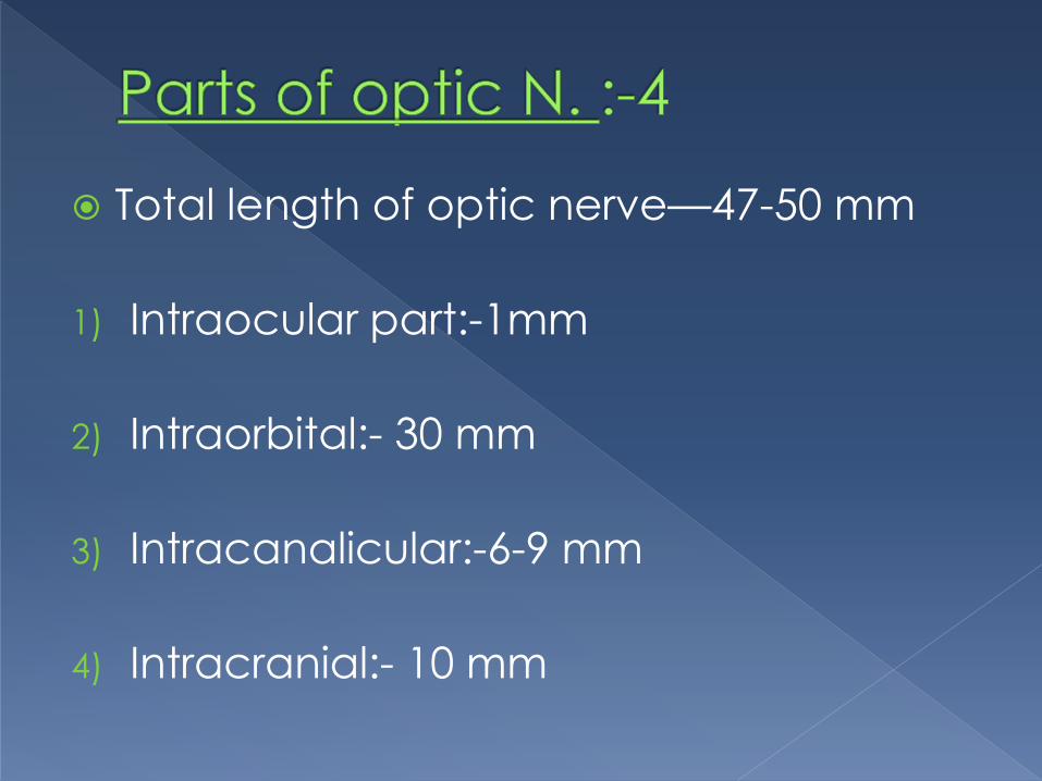
Total length of optic nerve—47-50 mm
1) Intraocular part:-1mm
2) Intraorbital:- 30 mm
3) Intracanalicular:-6-9 mm
4) Intracranial:- 10 mm

Intraocular:- from optic disc pierces
sclera and choroid converting into
“lamina cribrosa”.
at the back of eye ball it becomes
continuous with intraorbital part.

Intraorbital part: from back of the eyeball to optc foramina.
It is sinious to give scope for eye movements.
Posteriorly it is closely associated with annulus of zinn & origin of 4 rectimuscles.
Sup.rectus muscle fibres are adherent to the nerve fibre sheath so very painful movement will be manifested in retro bulbar neuritis.

Intra canalicular:- closely related to
ophthalmic artery lies inferolateral to it &
crosses obliquely over it,enters the orbit
lies on its medial side.
Sphenoid&post.ethmoidal sinuses lies
medial to it seperated by thin lamina,so
if infection of these sinuses will lead to
retrobulbar neuritis.

Intra cranial part:-lies above the cavernous sinus &meets ts fellow part over diaphragma sellae to form optic chiasma.
Meningeal sheaths: piamater, dura&arachnoid covering the brain continuous over optic N.
Subarachnoid and subdural spaces also continuous along with brain

Flat structure
Anterio posteriorly 8mm
Horizontally 12 mm
Lies over tuberculum&diaphragma sellae
Fibres of nasal halves of retina cross here.

Cylindrcal bundles of nerve fibres running
outwards and backwardsfrom postero
lateral aspect of the optic chiasma.
Each optic tract has fibres from temporal
half of retina of same eye & nasal half of
opposite eye .
Each optic tract ends in lateral
geniculate body.
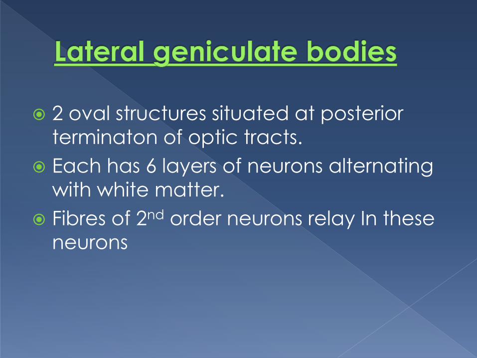
2 oval structures situated at posterior
terminaton of optic tracts.
Each has 6 layers of neurons alternating
with white matter.
Fibres of 2nd order neurons relay In these
neurons

Extend from lateral geniculate bodies to
visual cortex & consists of 3rd order
neurons of visual pathway.

Located on medial aspect of occipital
lobe,above and below the calcarine
fissure.
Subdivided into 2 parts
Visuosensory (striate area 17)
Visuopsychic area (peri striate area 18;
para striate area 19)
Receives the radiations


Mainly supplied by pial network of
vessels except orbital part of optic nerve
Optic nerve supplied by axial system
derived from central retinal artery.
Pial network composed by internal
carotid A., middle cerebral A.,
Ant.choroidal A.,Post.cerebral A.,deep
optic artery.


Surface layer of optic disc by capillaries of retinal arterioles.
Prelaminar region by centripetal branches of peri papillary choroid with some contribution from vessels of lamina cribrosa.
Lamina cribrosa by post.ciliaryarteries&arterial circle of zinn.
Retro laminar part by centrifugal branches from central retinal artery & centripetal from choroidal arteries, central retinalA, &ophthalmic.A

Lesions of optic N.:-loss of vision/blindness
common causes:opticatrophy,traumatic
avulsion of opticN., indirect optic
neuropathy´ optic neuritis.
Near reflex intact.

Lesions through proximal part of opticN:
ipsilateral blindness,contralateral hemi
anopia,absence of light reflex on the
affected side and consensual on the
contralateral side.
---near reflex intact.
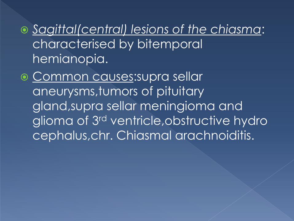
Sagittal(central) lesions of the chiasma:
characterised by bitemporal
hemianopia.
Common causes:supra sellar
aneurysms,tumors of pituitary
gland,supra sellar meningioma and
glioma of 3rd ventricle,obstructive hydro
cephalus,chr. Chiasmal arachnoiditis.
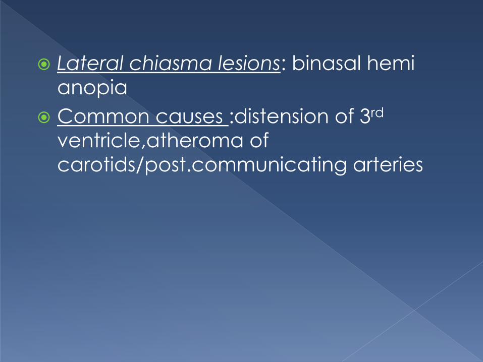
Lateral chiasma lesions: binasal hemi
anopia
Common causes :distension of 3rd
ventricle,atheroma of
carotids/post.communicating arteries

Lesions of optic tract: incongruous homonymous hemianopia associated with contralateral hemianopic pupillaryreactions.
May lead to decending optic atrophy with ipsilateral hemiplegia& contralateral3rd nerve paralysis.
Common causes:syphiliticmeningitis/gumma,TB/tumors of optic thalamus,post.cerebral/sup.cerebellar A.

Lesions of lat.geniculate body:
homonymous hemi anopia with sparing
of pupllary reflexes & may end in partial
optic atrophy.
Lesions of optic radiations:
Total involvement lead to complete
homonymous hemi anopia(some times
sparing macula)

Sup.fibres of optic radiation(lesions of
parietal lobe) causes inf.quadrantic
hemianopia (pie on floor)
Inf.fibres of optic radiation(lesions of
temporal lobe) causes sup.quadrantic
hemi anopia(pie in the sky)
Lesions not produce optic atrophy
Common lesions:vascular
occlusions,primary & secondary
tumors,trauma

Lesions of the visual cortex:
Occlusion of post.cerebral A which
supplying ant.part of cerebral cortex
causes congruous homonymous hemi
anopia sparing macula.
Congruous homonymous macular
defect occur in tip ofoccipital cortex
following head injury/gun shot injuries.
Pupillary reflexes normal& optic atrophy
doesn’t occur in these lesions
