anatomy: trachea chris van zylkhc. 2 trachea: landmarks begins at lower border of cricoid cartilage...
TRANSCRIPT
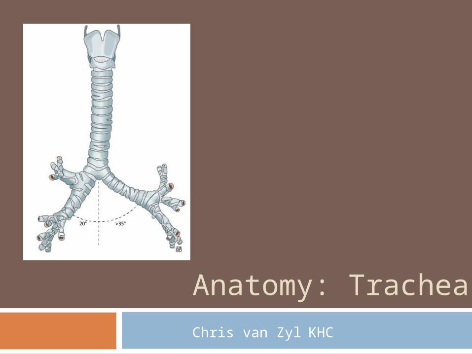
Anatomy: Trachea
Chris van Zyl KHC
2
Trachea: Landmarks
Begins at lower border of cricoid cartilage / C6
Extends to Carina Right of the midline Sternal angle T4 on inspiration / T6 on expiration
Lined by ciliated columnar epithelium 15 cm long / 2cm in diameter 15 – 20 incomplete rings of cartilage
Bridged post. by trachealis muscle
3
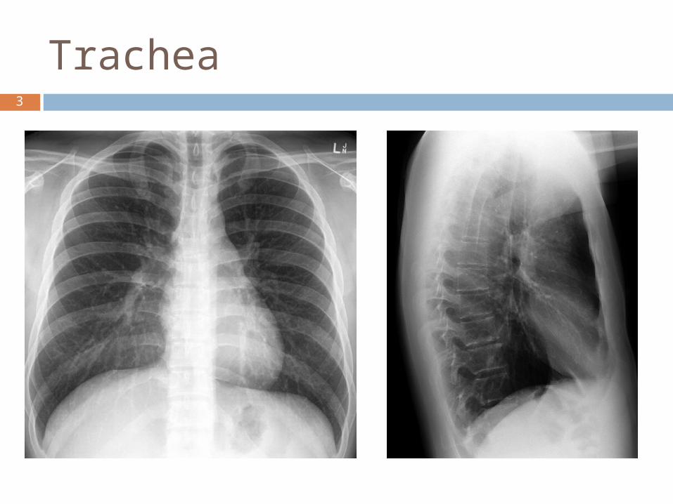
Trachea
4
Trachea
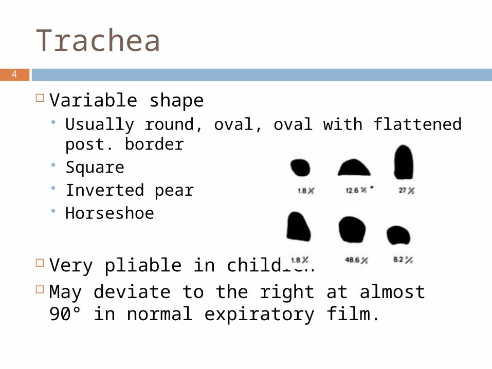
Variable shape Usually round, oval, oval with flattened post.
border Square Inverted pear Horseshoe
Very pliable in children May deviate to the right at almost 90° in
normal expiratory film.
5
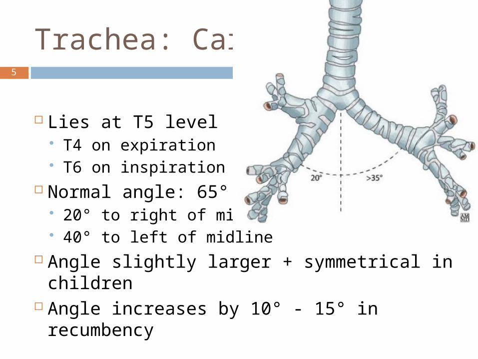
Trachea: Carina
Lies at T5 level T4 on expiration T6 on inspiration
Normal angle: 65° 20° to right of midline 40° to left of midline
Angle slightly larger + symmetrical in children
Angle increases by 10° - 15° in recumbency
6
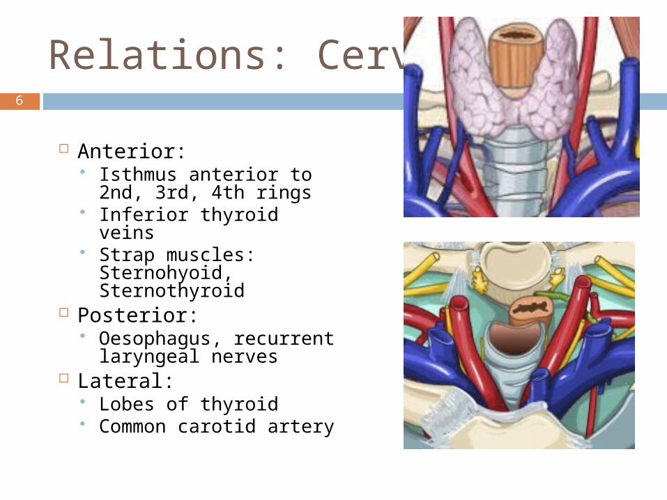
Relations: Cervical
Anterior: Isthmus anterior to
2nd, 3rd, 4th rings Inferior thyroid veins Strap muscles:
Sternohyoid, Sternothyroid
Posterior: Oesophagus, recurrent
laryngeal nerves Lateral:
Lobes of thyroid Common carotid artery
7
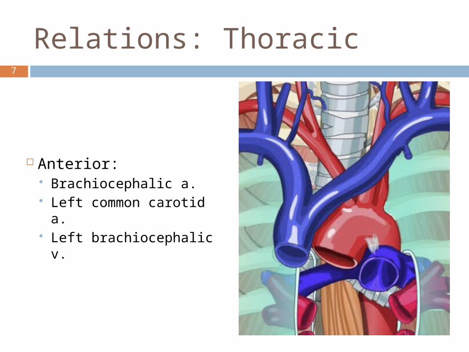
Relations: Thoracic
Anterior: Brachiocephalic a. Left common carotid a. Left brachiocephalic v.
8
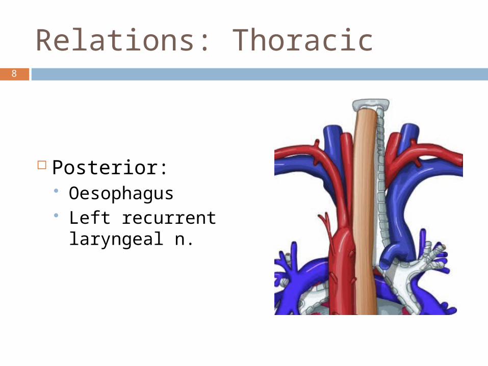
Relations: Thoracic
Posterior: Oesophagus Left recurrent
laryngeal n.
9
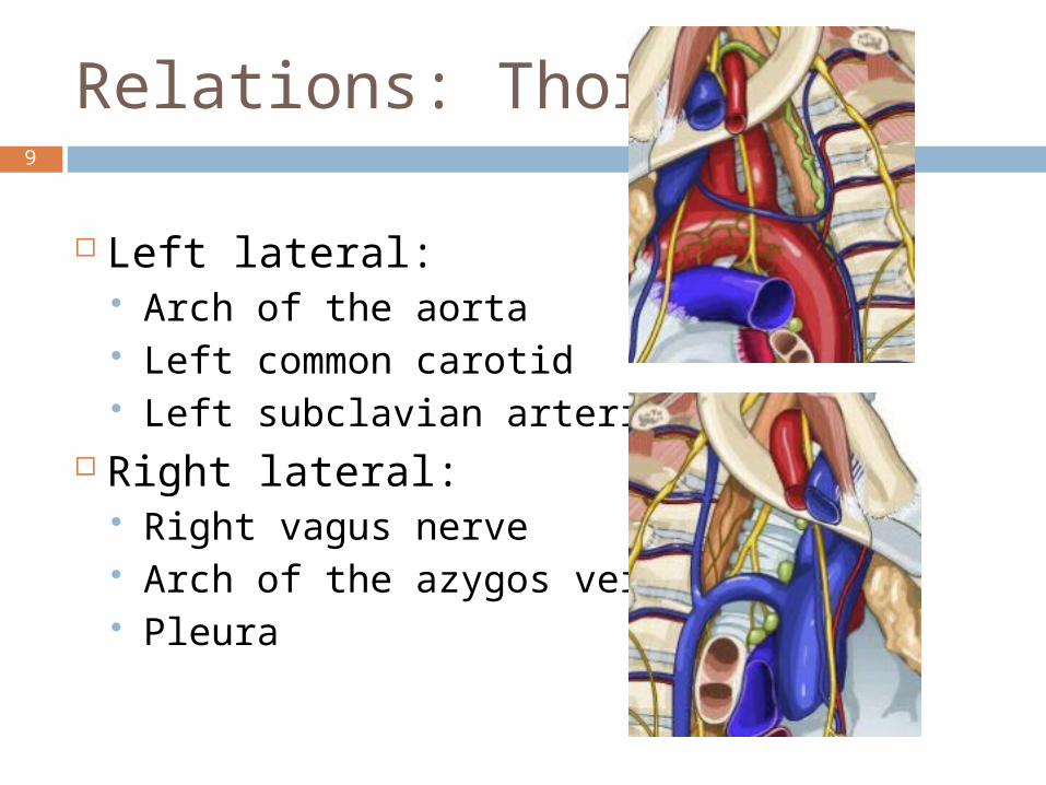
Relations: Thoracic
Left lateral: Arch of the aorta Left common carotid Left subclavian arteries
Right lateral: Right vagus nerve Arch of the azygos vein Pleura
10
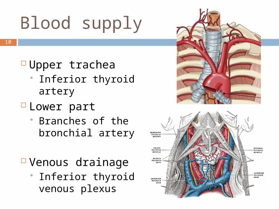
Blood supply
Upper trachea Inferior thyroid
artery Lower part
Branches of the bronchial artery
Venous drainage Inferior thyroid
venous plexus
11
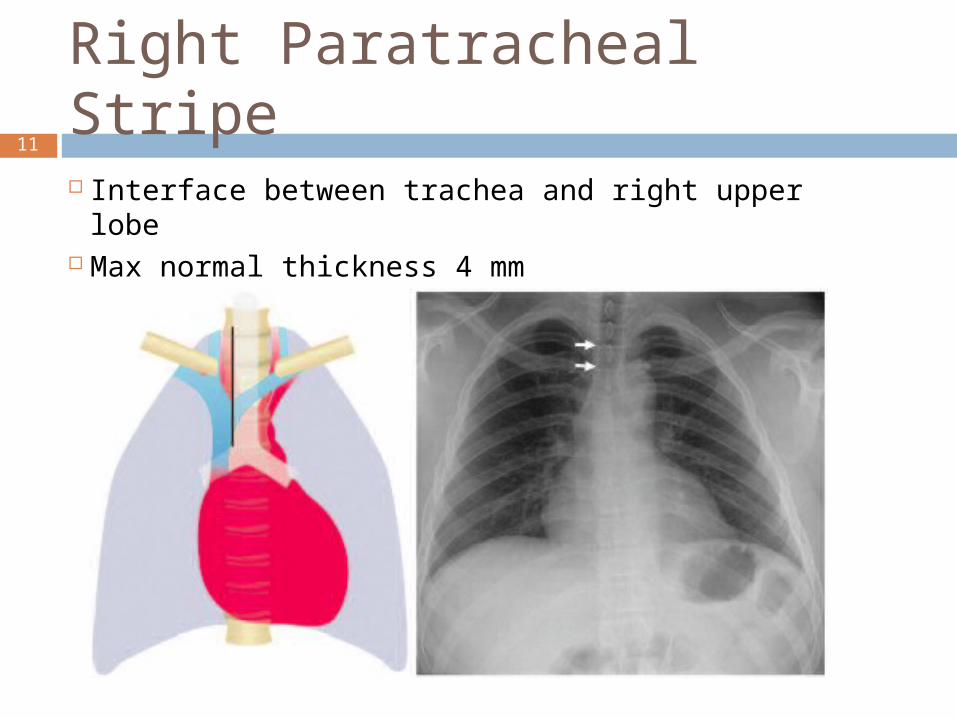
Right Paratracheal Stripe
Interface between trachea and right upper lobe Max normal thickness 4 mm
12
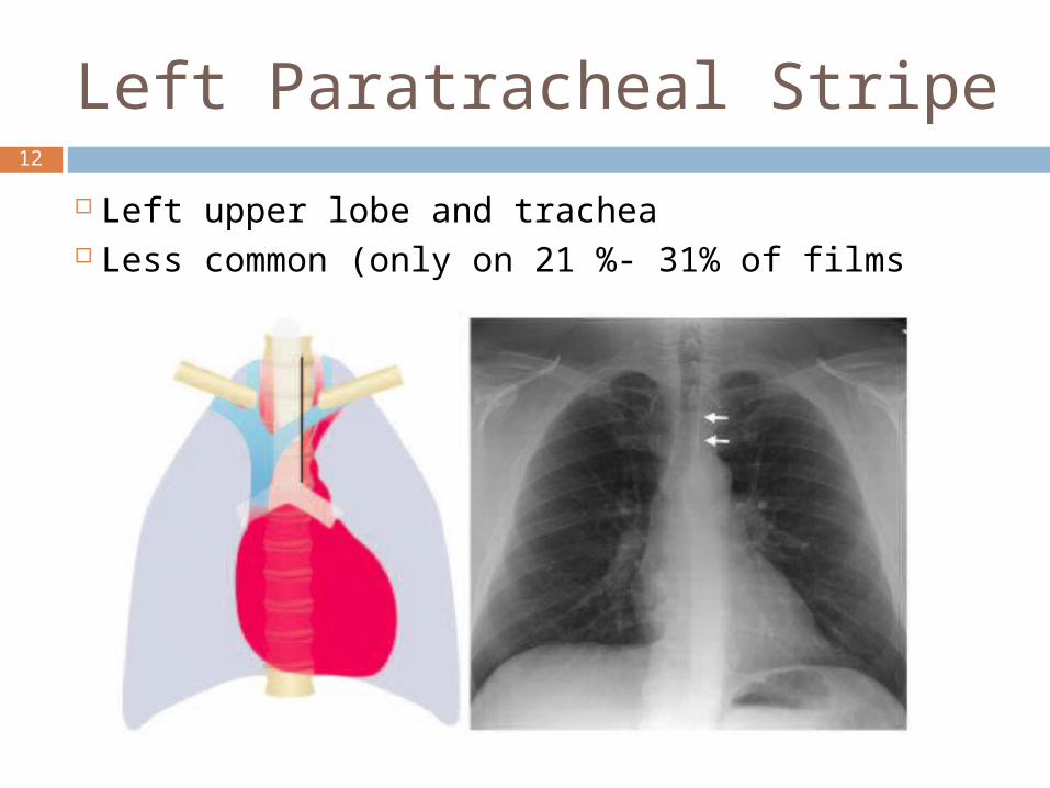
Left Paratracheal Stripe
Left upper lobe and trachea Less common (only on 21 %- 31% of films
13
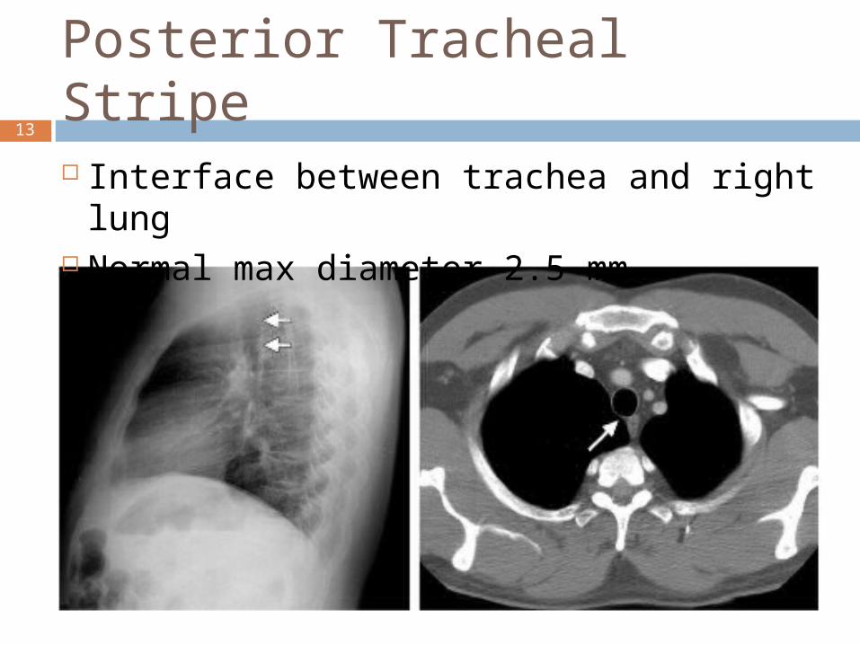
Posterior Tracheal Stripe
Interface between trachea and right lung Normal max diameter 2.5 mm
14
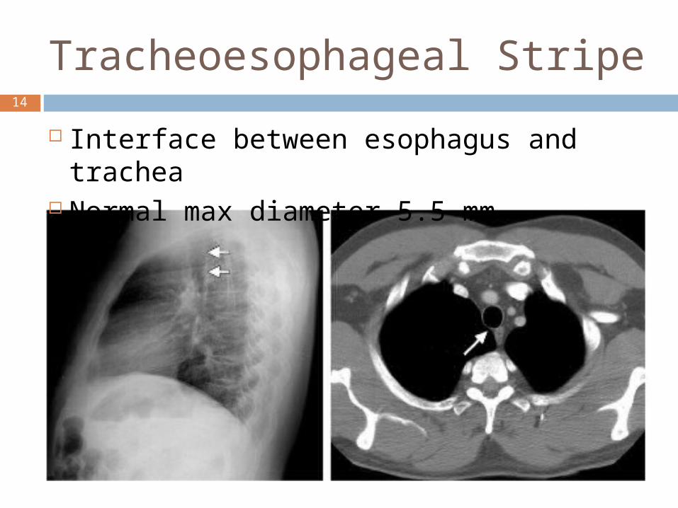
Tracheoesophageal Stripe
Interface between esophagus and trachea
Normal max diameter 5.5 mm
15
References
Lines and Stripes: Where Did They Go? Jerry M. Gibbs, Chitra A. Chandrasekhar, Emma C.
Furgasson, Sandra A. A. Oldham
Applied Radiological Anatomy Paul Butler, Adam W. M. Mitchell, Harold Ellis
Anatomy for Diagnostic Imaging Stephanie Ryan, Michelle McNicholas, Stephen
Eustace Third Edition