anesthetic management of neonates with expanding ... · expanding congenital pulmonary lesions:...
TRANSCRIPT

PBLD - Table # 11
Expanding Congenital Pulmonary Lesions: Urgent Resection of Congenital Lobar Emphysema
(CLE) and Congenital Cystic Adenomatoid Malformation (CCAM) in Neonates
Lianne L. Stephenson, M.D. Assistant Professor of Anesthesiology University of Wisconsin, Madison, WI
Christian Seefelder, M.D. Assistant Professor of Anaesthesia Children’s Hospital Boston, MA
Goals
1. Understand the various types of congenital pulmonary lesions and their terminology
2. Understand the physiologic implications of the various congenital pulmonary lesions
3. Understand the management options including fetal surgery, EXIT procedure, postnatal stabilization, emergency surgery or delayed surgery
4. Understand the presentation and problems of large congenital cystic adenomatoid malformation (CCAM) and expanding congenital lobar emphysema (CLE)
5. Understand the anesthetic concerns, anesthetic approach, and perioperative management of large CCAMs and expanding CLEs
2
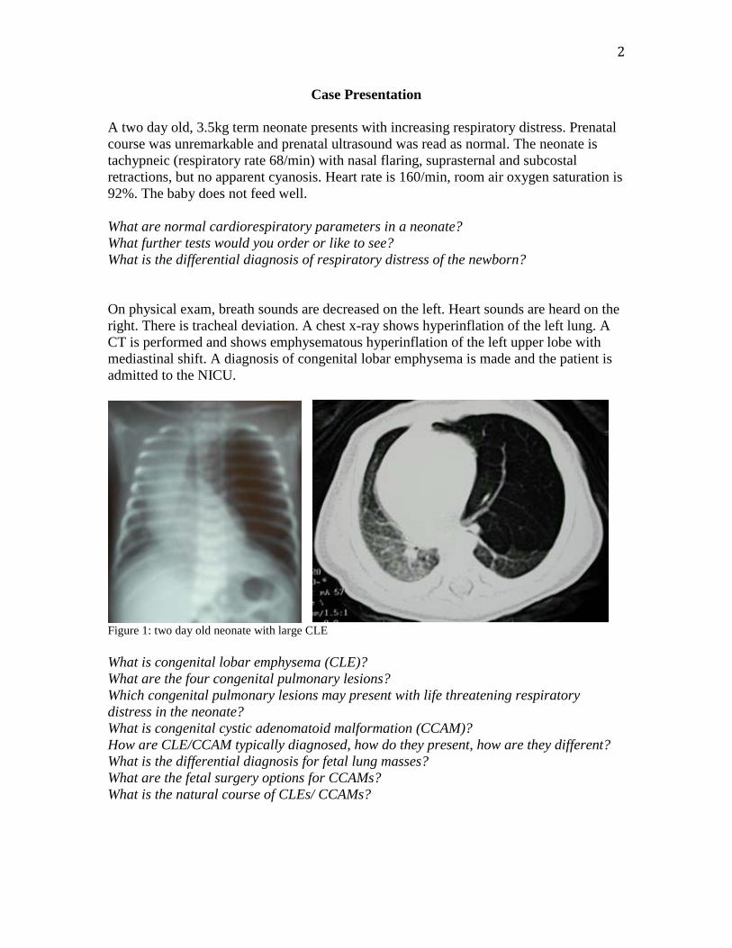
Case Presentation A two day old, 3.5kg term neonate presents with increasing respiratory distress. Prenatal course was unremarkable and prenatal ultrasound was read as normal. The neonate is tachypneic (respiratory rate 68/min) with nasal flaring, suprasternal and subcostal retractions, but no apparent cyanosis. Heart rate is 160/min, room air oxygen saturation is 92%. The baby does not feed well. What are normal cardiorespiratory parameters in a neonate? What further tests would you order or like to see? What is the differential diagnosis of respiratory distress of the newborn? On physical exam, breath sounds are decreased on the left. Heart sounds are heard on the right. There is tracheal deviation. A chest x-ray shows hyperinflation of the left lung. A CT is performed and shows emphysematous hyperinflation of the left upper lobe with mediastinal shift. A diagnosis of congenital lobar emphysema is made and the patient is admitted to the NICU.
Figure 1: two day old neonate with large CLE What is congenital lobar emphysema (CLE)? What are the four congenital pulmonary lesions? Which congenital pulmonary lesions may present with life threatening respiratory distress in the neonate? What is congenital cystic adenomatoid malformation (CCAM)? How are CLE/CCAM typically diagnosed, how do they present, how are they different? What is the differential diagnosis for fetal lung masses? What are the fetal surgery options for CCAMs? What is the natural course of CLEs/ CCAMs?

3
The baby is placed on 1 l/min oxygen by nasal cannula and the oxygen saturation is stable at 95%. The neonatologist would like to intubate the baby for respiratory distress. They also noted a heart murmur and the hemoglobin is 9. Blood is not typed and screened. The surgeon wants to take the baby to the operating room for emergent thoracotomy and resection of CLE. The parents ask you about the anesthetic risk in “such a small” baby. Would you encourage intubation of this patient with the diagnosis of CLE vs. CCAM? What are the concerns with positive pressure ventilation in CLE vs. CCAM? What additional preoperative workup would you like to see? Would you like to transfuse the baby preoperatively? When is CLE/CCAM resection an emergency? When would you bring the baby to the operating room? What is your discussion with the parents? What do you put on the consent? The baby arrives in the operating room awake, alert, comfortable, breathing with 1 l/min nasal cannula, SaO2 95%, HR 160/min, NIBP 85/55, RR 70/min, retracting. The surgeon wants to perform a bronchoscopy before the thoracotomy. She wants to proceed rapidly to decompress the chest and suggests that you do not waste time on lines. What is the indication for bronchoscopy? Rigid or flexible? What is your anesthetic plan for monitoring, lines, ventilation, analgesia? How would you induce the patient? What anesthetic options for maintaining spontaneous ventilation do you have? Would your anesthetic plan be different if you induced a two day old vs. a one 1 year old with CLE?
Figure 2: Right middle lobe CLE in a 1 y/o

4
The baby does well with induction and is breathing spontaneously. During bronchoscopy, the patient coughs and starts to desaturate. After resolution of this, the surgeon asks whether you could provide lung isolation for the surgery, and if you can achieve lung isolation and the baby is stable with it, she would consider a thoracoscopic left upper lobectomy for CLE. How do you prevent or manage coughing or laryngospasm during bronchoscopy while maintaining spontaneous respirations? What are your options for lung isolation and single lung ventilation in a 3.5 kg baby? What are the physiologic considerations of one lung ventilation in a neonate compared to an older child or adult? The baby is stable and breathing spontaneously with sevoflurane, but becomes hypotensive (43/25). The first blood gas returns: pH: 7.19/pCO2 70/pO2 144/HCO3 26.7/BE -2.8 on FiO2 50%. Hgb 8.9. With incision, the baby moves, breath-holds and desaturates. After the chest is opened and the emphysematous lobe delivered, you administer muscle relaxant and ventilate with pressure control ventilation. During the resection, the end-tidal CO2 continues to rise and the oxygen saturation decreases to 88%. How do you prevent or manage hypotension in the spontaneously breathing, deeply anesthetized neonate? What changes in oxygenation, ventilation and blood gases do you expect in a spontaneously breathing, anesthetized neonate in lateral decubitus position? What changes do you tolerate, when and how do you intervene? At what point would you be comfortable using muscle relaxation and converting to positive pressure ventilation? What are common reasons for difficulty ventilating and oxygenating during thoracic surgery in neonates and small infants? The resection is completed with minimal blood loss. The NICU calls to inquire about the plan for postoperative ventilation and pain management. The muscle relaxant is reversed. The patient is breathing spontaneously with adequate oxygenation and ventilation. What are the options for postoperative analgesia? What are the considerations regarding postoperative respiratory management?

5
Putting it all together: 2 day old term infant with respiratory distress 1. Admission: to the ICU 2. Workup: CXR, CT, labs 3. Diagnosis: CLE 4. OR urgency: within 24 hours 5. Induction: inhalation induction with sevoflurane, low dose remifentanil 6. Intubation: lidocaine to the cords, 3.0 LoPro cuffed ETT, no lung isolation 7. Monitoring/lines: a-line 8. Ventilation: spontaneous ventilation/PSV until chest open, then PCV 9. Analgesia: remifentanil intraop, epidural catheter placement at the end 10. Postoperative respiratory management: extubated in the operating room 11. Postoperative disposition: ICU Putting it all together: neonate with prenatally diagnosed giant CCAM 1. Prenatal: diagnosis of CCAM, fetal intervention debated 2. Delivery: unscheduled due to premature labor 3. Presentation: immediate severe respiratory distress 4. Intubation: immediately in the delivery room 5. Admission: to the NICU for stabilization 6. NICU lines: a-line, Foley, NGT 7. Workup: CXR, ABG, labs 8. Ventilation: unable to provide adequate ventilation/oxygenation: HFOV 9. OR urgency: emergently taken to OR 10. Ventilation in the OR: HFOV/PCV 11. Analgesia: iv opioids 12. Postoperative management: transition to conventional ventilation, transfer to
NICU, opioid analgesia
6
DISCUSSION POINTS The following are discussion points for the PBLD and do not intend to cover the topic comprehensively. They may in fact contain controversial statements to provoke discussion during the PBLD. 1. PREOP CONSIDERATIONS AND CLINICAL BACKGROUND: What are normal cardiorespiratory parameters for a term infant? BP: mean systolic 65-85
mean diastolic 45-55 HR: 100-150bpm RR: 50/min SaO2: 95% What are causes for respiratory distress in the neonate? 1. Airway: obstruction at various levels: choanal atresia, macroglossia, laryngomalacia, vocal cord paralysis, subglottic stenosis, laryngeal web, tracheomalacia, vascular ring, mediastinal mass, intrathoracic tumors, large congenital pulmonary anomalies, foreign body, tracheo-esophageal fistula, laryngeal cleft 2. Chest: intrathoracic space occupying lesions: congenital diaphragmatic hernia, congenital lobar emphysema, congenital cystic adenomatoid malformation, intrathoracic tumors, pneumothorax, abdominal distention parenchymal lung disease: respiratory distress syndrome, pneumonia, RSV, bronchiolitis, aspiration 3. Cardiovascular: congenital heart disease, PPHN 4. Neurologic: increased intracranial pressure, Arnold Chiari, brainstem compression 5. “Metabolic”: acidosis, sepsis, hypoglycemia, metabolic disorders, drugs

7
What are the four types of congenital pulmonary malformations? 1. Congenital lobar emphysema (CLE) 2. Congenital cystic adenomatoid (pulmonary) malformation (CCAM) 3. Bronchopulmonary sequestration (BPS) 4. Bronchogenic cyst (5. Mixed forms: sequestration/CCAM/CLE) What is a congenital lobar emphysema (CLE)? CLE is a congenital anomaly characterized by overdistention of the lung. It may be associated with anomalies of the bronchial cartilage (reduced or absent bronchial cartilage resulting in intrinsic bronchial narrowing and bronchomalacia) or external bronchial compression from various causes resulting in air trapping. CLE can be hypoalveolar (reduced number of alveoli) or polyalveolar (increased number of alveoli). Typically only one lobe is involved (left upper lobe: 41%, right middle lobe: 34%, right upper lobe: 21%). CLE may be diagnosed during prenatal ultrasound (fluid filled distended lobe), but most commonly is detected in neonates when progressive distention causes symptoms from compression of the remaining ipsilateral as well as the contralateral lung, mediastinal shift and tracheo-esophageal compression. If less severe, CLE may not present until later in infancy or childhood. Associated anomalies such as congenital heart disease are present in 10% of patients with CLE. What is a congenital cystic adenomatoid malformation (CCAM)? CCAM is a congenital anomaly of the lung resulting from abnormal fetal lung development. There is increased cell proliferation but decreased apoptosis, resulting in adenomatoid proliferation and cyst formation. The lesion is connected to the airway, but a normal intrapulmonary bronchial system is missing. Blood supply is from the pulmonary circulation. CCAM classifications are based on the cyst size (Type I: cysts 2-10 cm; Type II: cysts 0.5-2 cm; Type III: microcystic, grossly solid) as well as other histologic features. CCAMs may be diagnosed by prenatal ultrasound and can regress or increase in size. Large CCAMs may be associated with fetal hydrops and demise, or with respiratory distress at birth. Small CCAMs may remain asymptomatic and present later in life or are found incidentally. CCAMs represent up to 25% of congenital lung abnormalities and may be associated with areas of pulmonary sequestration and lobar emphysema. Associated (renal, intestinal, bony, cardiac) anomalies are present in up to 25% of patients with CCAM.
8
What is the differential diagnosis of fetal lung masses? 1. Congenital pulmonary malformations 2. Congenital diaphragmatic hernia (CDH)
a. posterolateral (Bochdalek) b. anteromedial (Morgagni) c. paraesophageal hernia
3. Tumors a. neuroblastoma b. teratoma c. pulmonary blastoma d. thymic tumor
4. Vascular malformation 5. Esophageal duplication cyst How does congenital cystic adenomatoid malformation (CCAM) present? 1. polyhydramnios 2. fetal hydrops with fetal death 3. respiratory distress at birth 4. symptomatic after birth 5. asymptomatic and incidental finding
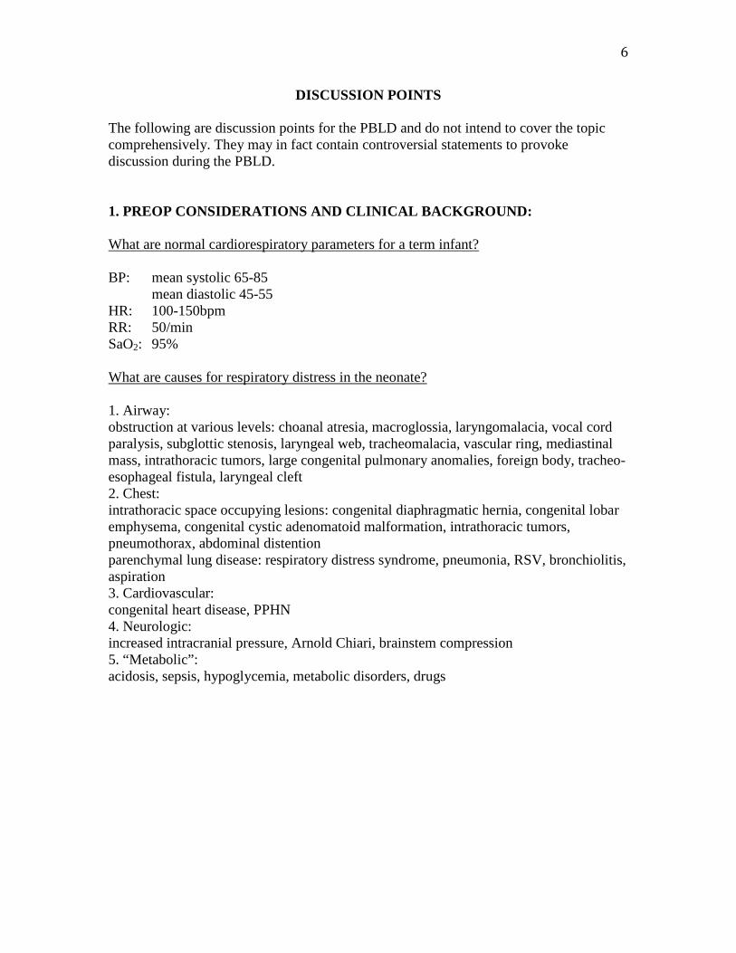
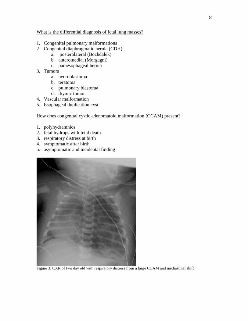
Figure 3: CXR of two day old with respiratory distress from a large CCAM and mediastinal shift

9
What is the natural course of CCAMs? 1. fetal hydrops and death 2. continued intrauterine growth 3. stable intrauterine size 4. involution 5. malignant transformation 6. complications:
a. infection b. bleeding c. pneumothorax
From: Calvert JK et al. Outcome of antenatally suspected congenital cystic adenomatoid malformation of the lung: 10 years’ experience 1991-2001. Arch Dis Child Fetal Neonatal Ed 2006; 91:F26-F28

10
What are the indications and options for fetal intervention in CCAM? Indications: 1. hydrops fetalis 2. growing lesions 3. large lesions Surgical options: 1. laparotomy, hysterotomy, open fetal surgery: CCAM resection/debulking 2. fetoscopic procedures 3. percutaneous procedures: drainage of cysts, placement of shunts 4. Ex utero intrapartum therapy (EXIT procedure): intervention while on placental
support a. intubation b. ECMO c. thoracotomy and resection
What are the respiratory management options after delivery of a neonate with a lung mass? 1. Maintain spontaneous respirations if cardiorespiratory status is adequate 2. Provide noninvasive respiratory support for mild respiratory compromise
a. blow-by oxygen b. nasal cannula oxygen c. high flow nasal cannula d. nasal CPAP e. mask CPAP f. mask BIPAP
3. Intubate if in severe respiratory distress a. endotracheal tube
i. uncuffed ii. cuffed
b. intubate awake c. intubate asleep
i. without muscle relaxation ii. with muscle relaxation
1. succinylcholine: advantage of rapid return of spontaneous respiration
2. non-depolarizing muscle relaxants: concerns of positive pressure ventilation for duration of action
4. Ventilation after intubation a. spontaneous
i. T-piece ii. PEEP
iii. CPAP iv. neonatal pressure support ventilation

11
b. controlled ventilation i. pressure control ventilation
ii. volume control ventilation iii. HFOV
5. ECMO support for severe respiratory or cardiopulmonary decompensation 6. Emergency surgery to decompress the lung What are the surgical options for CLE? When do you take the baby to the operating room? 1. Emergency thoracotomy within hours for the neonate in severe distress or with
progressive decompensation despite intervention in setting of unlikely improvement by non-surgical management
2. Urgent surgery within days for the symptomatic patient in respiratory distress who is stable after intervention without expected further improvement and with risk of deterioration
3. Elective surgery within days to months for the stable neonate and infant with moderate symptoms in no distress
4. No surgery and conservative management for the asymptomatic patient with small lesions?
From: Ulku R, Onat S, Ozçelık C. Congenital lobar emphysema: Differential diagnosis and therapeutic approach. Pediatrics International (2008) 50, 658–661

12
2. INTRAOP CONSIDERATIONS: What are the general concerns regarding anesthesia for a neonate? 1. patient size, equipment, infrastructure 2. line placement: difficulty, time delay? 3. immaturity of all organ systems 4. presence of other congenital malformations What are the anesthetic concerns for resection of a large or expanding CCAM or CLE? Size of the lesion: a. difficulty oxygenating b. difficulty ventilating c. hemodynamic effects of lesion d. surgical challenges Surgical technique: 1. thoracotomy 2. sternotomy 3. thoracoscopy 4. bronchoscopy Intraoperative concerns: 1. position of the patient
a. lateral b. supine (sternotomy)
2. access to the patient will be difficult for the anesthesiologist 3. access to the lesion by the surgeon will result in
a. additional ventilation/oxygenation problems i. positioning
ii. retraction iii. compression iv. tracheobronchial kinking v. tube movement
b. additional hemodynamic effects i. venous return
ii. aortic compression iii. heart retraction iv. mechanical arrhythmias v. bleeding

13
What anesthetic options for maintaining spontaneous ventilation do you have? 1. inhalation anesthetics 2. remifentanil 3. other opioids 4. propofol 5. ketamine 6. dexmedetomidine What are your options for lung isolation and single lung ventilation in a 3.5 kg baby? 1. double lumen tube: Marraro Double Lumen Tube for neonates and infants 2. double endotracheal tube intubation 3. bronchial blocker
a. Fogarty catheter (available in 3,4,5 F) b. Arndt Endobronchial Blocker (smallest 5 F)
4. mainstem intubation 5. none (surgical retraction)
From: Pawar DK, Marraro GA. One lung ventilation in infants and children: experience with Marraro double lumen tube. Pediatr Anesth 2005; 15: 204–208 and: Marraro G. Selective endobronchial intubation in paediatrics: the Marraro Paediatric Bilumen Tube. Paediatr Anaesth 1994; 4: 255-258 What are the physiologic considerations of one lung ventilation in a neonate compared to an adult? 1. Higher oxygen consumption 2. Immature alveoli 3. Increased chest wall compliance impedes the oxygenation of the healthy dependent
lung unlike adults

14
Figure 4: Extraluminal Arndt Endobronchial Blocker is being positioned with the help of an intraluminal fiberoptic bronchoscope in a 7 month old. What are the options for intraoperative analgesia? 1. Inhalation anesthetics? 2. Opioids 3. Local anesthesia (preincision infiltration) 4. Regional anesthesia (preincision epidural) 3. EXTUBATION AND POST-OP CARE: What are the considerations regarding postoperative pain management? 1. Opioids 2. Epidural
a. Placement preop b. Placement postop
3. Other local/regional a. Local anesthetic infiltration b. Wound catheters c. Intercostal blocks, paravertebral blocks
4. NSAIDS a. In neonates?
5. Acetaminophen/paracetamol

15
What are considerations regarding postoperative respiratory disposition? 1. Postoperative return to the NICU. 2. Patients on ECMO:
a. Remain on ECMO b. ECMO decannulation in the operating room at the end of the resection
3. Postoperative ventilation a. Continuation on HFOV b. Transition to conventional ventilation in the operating room or in the NICU
4. Extubation in the operating room or early extubation in the NICU
References 1. Adzick NS: Management of fetal lung lesions. Clin Perinatol 2009; 36:363-76 2. Adzick NS: Open fetal surgery for life-threatening fetal anomalies. Semin Fetal Neonatal
Med 2009 3. al-Salem AH, Adu-Gyamfi Y, and Grant CS: Congenital lobar emphysema. Can J
Anaesth 1990; 37:377-9 4. Arora MK, Karamchandani K, Bakhta P, et al: Combination of inhalational, intravenous,
and local anesthesia for intubation in neonates with congenital lobar emphysema. Paediatr Anaesth 2006; 16:998-9
5. Calvert JK, et al. Outcome of antenatally suspected congenital cysticadenomatoid malformation of the lung: 10 years’ experience 1991-2001. Arch Dis Child Fetal Neonatal Ed 2006; 91:F26-28
6. Cote CJ: The anesthetic management of congenital lobar emphysema. Anesthesiology 1978; 49:296-298
7. Goto H, Boozalis ST, Benson KT, et al: High-frequency jet ventilation for resection of congenital lobar emphysema. Anesth Analg 1987; 66:684-6
8. Gupta R, Singhal SK, Rattan KN, et al: Management of congenital lobar emphysema with endobronchial intubation and controlled ventilation. Anesth Analg 1998; 86:71-3
9. Guruswamy V, Roberts S, Arnold P, et al: Anaesthetic management of a neonate with congenital cyst adenoid malformation. Br J Anaesth 2005; 95:240-2
10. Hammer GB: Single-lung ventilation in infants and children. Paediatr Anaesth 2004; 14:98-102
11. Iodice F, Harban F, and Walker I: Anesthetic management of a patient with bilateral congenital lobar emphysema. Paediatr Anaesth 2008; 18:340-1
12. Marraro G: Simultaneous independent lung ventilation in pediatric patients. Crit Care Clin 1992; 8:131-45
13. McCartney CJ and Johnston G: Anaesthetic management of a 6-week-old child with unilateral pulmonary interstitial emphysema. Paediatr Anaesth 2000; 10:325-8
14. Mei-Zahav M, Konen O, Manson D, et al: Is congenital lobar emphysema a surgical disease? J Pediatr Surg 2006; 41:1058-61
15. Moideen I, Nair SG, Cherian A, et al: Congenital lobar emphysema associated with

16
congenital heart disease. J Cardiothorac Vasc Anesth 2006; 20:239-41 16. Pariente G, Aviram M, Landau D, et al: Prenatal diagnosis of congenital lobar
emphysema: case report and review of the literature. J Ultrasound Med 2009; 28:1081-4 17. Pawar DK and Marraro GA: One lung ventilation in infants and children: experience with
Marraro double lumen tube. Paediatr Anaesth 2005; 15:204-8 18. Raghavendran S, Diwan R, Shah T, et al: Continuous caudal epidural analgesia for
congenital lobar emphysema: a report of three cases. Anesth Analg 2001; 93:348-50 19. Rahman N and Lakhoo K: Comparison between open and thoracoscopic resection of
congenital lung lesions. J Pediatr Surg 2009; 44:333-6 20. Schmidt C, Rellensmann G, Van Aken H, et al: Single-lung ventilation for pulmonary
lobe resection in a newborn. Anesth Analg 2005; 101:362-4 21. Shanmugam G, MacArthur K, and Pollock JC: Congenital lung malformations--antenatal
and postnatal evaluation and management. Eur J Cardiothorac Surg 2005; 27:45-52 22. Tobias JD: Anaesthesia for neonatal thoracic surgery. Best Pract Res Clin Anaesthesiol
2004; 18:303-20 23. Wilson RD, Hedrick HL, Liechty KW, et al: Cystic adenomatoid malformation of the
lung: review of genetics, prenatal diagnosis, and in utero treatment. Am J Med Genet A 2006; 140:151-5