angina guideline for primary care - orchard croft guideline for... · statement 5: people with...
TRANSCRIPT

1
ANGINA GUIDELINE for Primary Care
Aims These guidelines are to support primary care practitioners in identifying, diagnosing, treating and managing patients with suspected or confirmed angina. Evidence Base The guidelines have been produced by a multidisciplinary team based on the NICE Quality Standard for Stable Angina (August 2012), which incorporates NICE Clinical Guidelines CG95, Chest Pain of Recent Onset (March 2010) and CG126, Management of Stable Angina (July 2011). Please find full guidance at the following: https://www.nice.org.uk/guidance/qs21/chapter/list-of-quality-statements https://www.nice.org.uk/guidance/cg95 https://www.nice.org.uk/guidance/cg126
Prepared by: Wakefield and North Kirklees Cardiac Partnership Group
Published: October 2015
Review Due: October 2016 (unless clinical evidence base changes)

2
Who to Contact
Clinical guidance on angina referrals
First point of contact
Cardiology e- consultation
Alternative point of contact
Chest Pain Nurse Team Fiona Dudley [email protected] 01924 541551
Non-clinical advice on angina referral form and process
The ABC Booking Clerks
Craig Plumb [email protected]
01924 543102
Sophie Liddell [email protected]
01924 543128
Angina Pathway Development
Dr Dwayne Conway, Consultant Cardiologist, Mid Yorkshire Hospitals NHS Trust
[email protected] Dr Som DeSilva [email protected]
Paul Brooksby, Consultant Cardiologist and Head of Clinical Service, Mid Yorkshire Hospitals NHS Trust

3
RATIONALE UNDERLYING THE PATHWAY FOR STABLE ANGINA - BACKGROUND
Our outdated ‘Rapid Access Chest Pain Clinic’ pathway dates back to 2000, and relies heavily on the
use of exercise ECG in the clinic. However, in 2010, NICE published CG95, Chest Pain of Recent
Onset, as guidance on the investigation of angina, containing the statement “do not use exercise
ECG to diagnose or exclude stable angina for people without known coronary heart disease”. NICE
highlighted evidence that a simple clinical history has the same diagnostic accuracy as exercise ECG,
and that angina should be diagnosed according to the following symptoms:
1. Constricting discomfort in the front of the chest, neck, shoulders, jaw or arms
2. Precipitated by physical exertion
3. Relieved by rest or GTN in around 5 minutes
All 3 of the above are present Typical angina
2 out of 3 are present Atypical angina
One or none are present Non-anginal. The patient should not be referred for angina investigation
Applying this simple assessment in primary care has the potential to avoid unnecessary referrals to
secondary care (and unnecessary invasive investigations) in those with non-anginal symptoms,
while triaging those with angina to the appropriate specialist service.
CG95 also stated that among patients with typical or atypical angina, investigations for coronary
artery disease (CAD) such as cardiac CT, stress ECHO and coronary angiography, should be chosen
according to the pre-test probability of underlying CAD, calculated using the Duke University risk
score.
In 2011, NICE published CG126 Stable Angina, with guidance on the treatment of angina. This
emphasised the use of evidence-based medications (Optimal Medical Therapy (OMT), (see page 11
below), the need to define the prognostic significance of the individual’s coronary heart disease by
investigations such as angiography, and the benefit of a multi-disciplinary approach to deciding
upon revascularisation (PCI/CABG).
National data from the Public Health Observatory has consistently shown Wakefield and North
Kirklees areas to have high Standardised Mortality Rates for Coronary Heart Disease but unusually
low elective angiography and revascularisation (PCI/CABG) rates. Data from the National Institute
for Cardiovascular Outcomes Research shows that MYHT has a low proportion of PCI for stable
angina and a high proportion of PCI for acute coronary syndromes. Dr Foster mortality data shows
that survival following admission to MYHT with myocardial infarction is excellent, often better than
comparable Trusts elsewhere in the UK.

4
An audit of patients seen in MYHT Rapid Access Chest Pain Clinic in 2010 demonstrated a high-
proportion of referrals with non-anginal chest pain, low pre-test probabilities of coronary artery
disease (compared to national and regional audit data from the same year) and a high use of
exercise ECG to investigate, even among those with clearly non-anginal symptoms
This suggests that improvements are needed in access to evidence-based therapies (including
revascularisation) for patients with stable angina in our local area. We have therefore redesigned
the Pathway for Stable Angina, with a new referral form designed to encourage referral of angina
(and discourage referral of non-anginal pain), delivering prompt local access to specialist
assessment and multi-modality cardiac investigations (including CT, stress ECHO, MRI and coronary
angiography), local MDT and PCI (and CABG at the regional centre).

5
RATIONALE UNDERLYING THE PATHWAY FOR STABLE ANGINA (NICE Quality Standard 21)
In August 2012, NICE published Quality Standard 21 (Stable Angina), stating that “services should be
commissioned from and coordinated across all relevant agencies encompassing the whole care
pathway”.
The Quality Standard consisted of five Quality Statements, listed below:
Statement 1: People with features of typical or atypical angina and an estimated likelihood of
coronary artery disease of 10-90% are offered diagnostic investigation according to that likelihood.
Statement 2: People with stable angina are offered a short-acting nitrate and either a beta-blocker
or calcium channel blocker as first line treatment.
Statement 3: People with stable angina are prescribed a short acting nitrate and 1 or 2 anti-anginal
drugs as necessary before revascularisation is considered.
Statement 4: People with stable angina who have had coronary angiography, have their treatment
options discussed with a multi-disciplinary team if there is left main stem disease, anatomically
complex three-vessel disease or doubt about the best method of revascularisation.
Statement 5: People with stable angina whose symptoms have not responded to treatment are
offered re-evaluation of their diagnosis and treatment.
The new local pathway for stable angina has been developed in line with these quality statements,
and will allow patients with stable angina to access a full range of specialist-led diagnostic and
treatment services in a timely and appropriate manner. The information provided via a fully-
completed referral form will assist the MYHT cardiologists with estimation of the Duke University
CAD risk score, in order to appropriately triage patients for investigation.
6
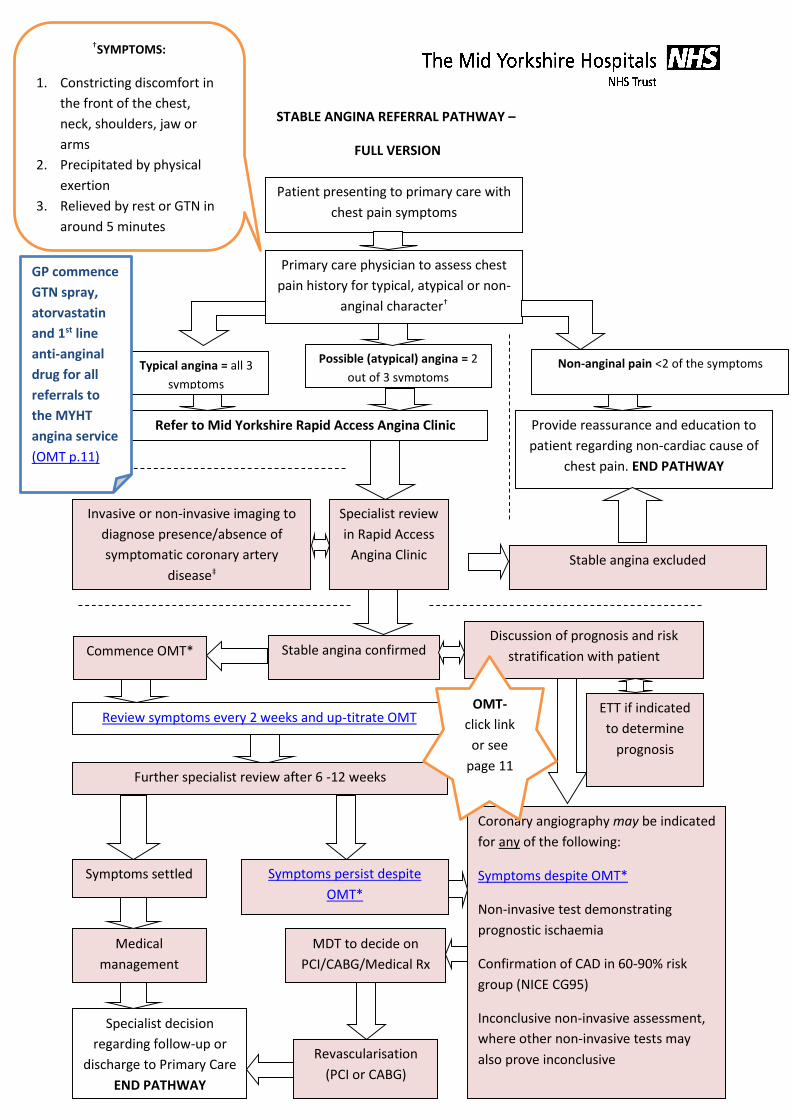
STABLE ANGINA REFERRAL PATHWAY –
FULL VERSION
Stable angina confirmed
Specialist review
in Rapid Access
Angina Clinic
Invasive or non-invasive imaging to
diagnose presence/absence of
symptomatic coronary artery
disease‡
Typical angina = all 3
symptoms
Possible (atypical) angina = 2
out of 3 symptoms Non-anginal pain <2 of the symptoms
Refer to Mid Yorkshire Rapid Access Angina Clinic Provide reassurance and education to
patient regarding non-cardiac cause of
chest pain. END PATHWAY
Stable angina excluded
Commence OMT*
Review symptoms every 2 weeks and up-titrate OMT
Coronary angiography may be indicated
for any of the following:
Symptoms despite OMT*
Non-invasive test demonstrating
prognostic ischaemia
Confirmation of CAD in 60-90% risk
group (NICE CG95)
Inconclusive non-invasive assessment,
where other non-invasive tests may
also prove inconclusive
Symptoms settled Symptoms persist despite
OMT*
Further specialist review after 6 -12 weeks
MDT to decide on
PCI/CABG/Medical Rx
Revascularisation
(PCI or CABG)
Medical
management
Specialist decision
regarding follow-up or
discharge to Primary Care
END PATHWAY
Discussion of prognosis and risk
stratification with patient
Patient presenting to primary care with
chest pain symptoms
Primary care physician to assess chest
pain history for typical, atypical or non-
anginal character†
ETT if indicated
to determine
prognosis
†SYMPTOMS:
1. Constricting discomfort in
the front of the chest,
neck, shoulders, jaw or
arms
2. Precipitated by physical
exertion
3. Relieved by rest or GTN in
around 5 minutes
OMT-
click link
or see
page 11
GP commence
GTN spray,
atorvastatin
and 1st line
anti-anginal
drug for all
referrals to
the MYHT
angina service
(OMT p.11)

7
NOTE :GP to commence GTN spray, atorvastatin and 1st
line anti-anginal drug for all referrals to the MYHT angina
service (OMT p.11) Refer to full guideline for further
information.
†SYMPTOMS:
1. Constricting
discomfort in the front
of the chest, neck,
shoulders, jaw or arms
2. Precipitated by
physical exertion
3. Relieved by rest or
GTN in around 5 min
Patient presenting to primary care with
chest pain symptoms
Primary care physician to assess
chest pain history for TYPICAL,
ATYPICAL or NON ANGINAL
character
Typical angina = all
3 symptoms
Possible (atypical) angina
= 2 out of 3 symptoms
Non-anginal pain <2 of the
symptoms
Refer to Mid Yorkshire Rapid Access Angina
Clinic Provide reassurance and education to
patient regarding non-cardiac cause
of chest pain.
Investigate other possible causes of
pain. Consider MSK pain, Lung
disease, radiculopathy from back
problems/joint pain etc
Likely non
cardiac
END
PATHWAY
IF doubt persists for
possible cardiovascular
cause for symptoms
Consider Cardiology e-consultation for further advice
OR refer to Mid Yorkshire General Cardiology Clinic
Advise patient to call 999 if usual chest pain is not
relieved by GTN used twice
Send patient to A&E if these red
flag symptoms are present
Suspected Acute MI or unstable angina
Or
Currently experiencing possible cardiac chest pain at rest,
Or
Suspected cardiac chest pain within the last 12 hours with
abnormal 12-lead ECG
STABLE ANGINA REFERRAL PATHWAY
PRIMARY CARE SUMMARY Prepared by: Wakefield and North Kirklees Cardiac Partnership Group, Published: October 2015, Review Due: October 2016
(unless clinical evidence base changes)

8
IDENTIFYING ANGINA FROM THE CHARACTER OF CHEST PAIN IN PRIMARY CARE
NICE highlighted evidence that a simple clinical history has the same diagnostic accuracy as exercise
ECG, and that angina should be diagnosed according to the following symptoms:
1. Constricting discomfort in the front of the chest, neck, shoulders, jaw or arms
2. Precipitated by physical exertion
3. Relieved by rest or GTN in around 5 minutes
All 3 of the above are present in ‘typical angina’, 2 out of 3 are present in ‘atypical angina’. If only
one or none of the above features are present, the symptoms are classified as ‘non-anginal’ and
should not be referred for angina investigation.
It is likely in clinical practice that primary care physicians will encounter some patients with
symptoms that are classified as ‘non-anginal’ but where the GP still suspects that cardiovascular
disease may be a cause. Such patients should not be referred via the Stable Angina pathway, but
may be referred to a general cardiology clinic if desired. However, it is important to recognise that
the majority of such patients will have non-cardiac causes of their symptoms, so it may be
appropriate to investigate the other possible causes before referral.
In cases where doubt still exists about referral, e-Consultation may provide an alternative route for
advice.

9
RAPID ACCESS ANGINA REFERRAL FORM
Patient Details GP Details
Name
Dr
Address
Surgery
Tel No (day) (evening) (mobile)
Telephone
Date of Birth Fax No
NHS number Gender Male Female
TO REFER YOUR PATIENT THIS REFERRAL FORM MUST BE FULLY COMPLETE AND THE FOLLOWING INVESTIGATIONS MUST BE REQUESTED AND REVIEWED BY GP
FBC/U&E/LFT/TFT/CHOLESTEROL/LIPID PROFILE/GLUCOSE/12-LEAD ECG
PLEASE TICK TO CONFIRM THAT ALL THE ABOVE RESULTS HAVE BEEN REVIEWED AND THAT YOU HAVE ATTACHED THE RESULTS TO THIS REFERRAL
ANGINAL SYMPTOMS YES NO
Precipitated by physical exertion
Constricting discomfort (front of chest/neck/shoulder/jaw/arm)
Relief in approximately 5 minutes after GTN or rest
Presenting History:
Please describe any abnormal findings on clinical examination:
Please advise if any investigations revealed abnormal results:
MEDICAL HISTORY
Yes No Yes No
Known IHD Previous PCI
Previous MI Previous CABG
Diabetes Heart murmur or known valve disease
Hypertension Has cholesterol level ever been >6.3 mmol/L?
Peripheral arterial disease Stroke or TIA
Current or ex-smoker Limited mobility
If yes to any of the above, please provide details (such as dates):
Height m Weight kg Systolic BP mmHg
Diastolic BP mmHg
Signed Print Date
Please attach details of current medications (including known allergies) and investigation results and FAX with completed referral form to outpatient appointment centre 01924 542702

10
Which patients should be referred to the Rapid Access Angina Assessment Clinic?
Only refer patients with 2-3 of the 3 characteristics of angina†
1 Symptoms precipitated by physical exertion 2 Constricting discomfort in the chest/neck/shoulders/jaw/arms 3 Relief in about 5 minutes after GTN or rest
Which patients should NOT be referred to the
Rapid Access Angina Assessment Clinic?
The following patients should be routed to hospital as an emergency via paramedic ambulance:
Suspected Acute MI or unstable angina, or
Currently experiencing possible cardiac chest pain at rest, or
Suspected cardiac chest pain within the last 12 hours with abnormal 12-lead ECG The following patients should be referred for same day hospital assessment in A&E or AAU/MAU:
Suspected cardiac chest pain in past 12-72 hours with abnormal 12-lead ECG The following patients should be referred to general cardiology clinic or via separate community pathways:
Suspected valve disease, hypertrophic cardiomyopathy, heart failure, arrhythmia Patients currently under cardiologist follow up:
Please liaise directly with usual cardiologist if need to expedite review for change in condition
Patients with non-anginal symptoms (<2 of the 3 angina characteristics):
If <2 ‘angina characteristics’ then symptoms are classified as non-anginal†. If concern at possible cardiac cause remains, patients may be referred by standard letter to general cardiology clinic or discussed via e-consultation. Alternatively, please consider investigating/treating for one of the many non-cardiac causes of chest pain symptoms
† NICE Clinical Guideline 95 Chest Pain of Recent Onset, March 2010

11
INVASIVE AND NON-INVASIVE TESTS FOR NEW PATIENTS WITH STABLE ANGINA
Which patients?
Clinical value Invasive? Available at MHYT?
Comments
12-lead ECG ALL PATIENTS prior to referral
Risk stratification
No Yes, but firstly in primary care
Essential for risk stratification
Exercise (treadmill) ECG
Not routine Risk stratification
No Yes Occasionally useful for patients with known CAD/MI
Cardiac CT CAD risk 10-30%
Identifies coronary artery disease
No (IV cannula only)
Yes High sensitivity, useful rule-out test
Stress ECHO CAD risk 30-60%
Identifies myocardial ischaemia
No (IV cannula only)
Yes Better diagnostic accuracy than treadmill ECG alone
Coronary Angiogram
CAD risk 60-90%
Identifies coronary artery disease
Yes (arterial puncture and catheterisation of coronaries)
Yes Also for patients with ischaemia confirmed by non-invasive tests
Perfusion MRI CAD risk 30-60%
Identifies myocardial ischaemia
No (IV canula only)
Yes Rarely needed, Stress ECHO provides similar information
Myocardial perfusion scintigraphy
CAD risk 30-60%
Identifies myocardial ischaemia
No (IV canula only)
Indirectly via Leeds
Rarely needed, Stress ECHO provides similar information
Intravascular Ultrasound of Coronaries
Not routine Additional quantification of coronary atheroma
Yes (arterial puncture and catheterisation of coronaries)
Yes May guide revascularisation decisions during PCI
Coronary pressure wire (fractional flow reserve)
Not routine Identifies myocardial ischaemia
Yes (arterial puncture and catheterisation of coronaries)
Yes May guide revascularisation decisions during catheterisation/PCI

12
OPTIMAL MEDICAL THERAPY (OMT)
Optimal drug treatment consists of 1 or 2 anti-anginal drugs, titrated to relieve symptoms, plus drugs for
secondary prevention of cardiovascular disease. Do not exclude patients from treatment for stable angina
based on age alone. Do not alter investigation or treatment based upon gender or ethnic group.
GTN spray/tablets Offer to all patients with suspected angina
Aspirin Consider 75mg daily in all patients with suspected angina, taking into account
bleeding risks and comorbidities. Most of the evidence for the benefits of aspirin is
among patients with established cardiovascular disease (stroke, MI, etc).
ACE inhibitors Consider in patients with stable angina and diabetes. Continue ACE inhibitors in
patients with a pre-existing ACEi indication (eg heart failure).
Statins Offer Atorvastatin 20-80mg to all patients with suspected angina (see NICE CG181
and Quality Standard 100).
Beta-blockers An option as first-line anti-anginal therapy. Start with a low dose and review every 2-
4 weeks with a view to uptitrating the dose to the maximum tolerable dosage or
until symptoms resolve. May be combined with a calcium antagonist if required
(watch for hypotension and bradycardia; use a dihydropyridine such as amlodipine
or felodipine in preference to diltiazem or verapamil in combination with
betablocker).
Calcium antagonists An option as first-line anti-anginal therapy. Start with a low dose and review every 2-
4 weeks with a view to uptitrating the dose to the maximum tolerable dosage or
until symptoms resolve. May be combined with a beta-blocker if required (watch for
hypotension and bradycardia; use a dihydropyridine such as amlodipine or
felodipine in preference to diltiazem or verapamil in combination with betablocker).
Do not routinely offer drugs other than beta-blocker or calcium antagonist as first-line. The following drugs
may sometimes be used as 3rd-line agents or in patients who fail to tolerate standard
first-line therapies:
Isosorbide Mononitrate
Ivabradine
Nicorandil
Ranolazine

13
PATIENT INFORMATION AND SUPPORT
Useful patient information about Angina can be found at:
http://patient.info/health/angina-leaflet