angle between the common and internal carotid arteries ... · sciences, 8-35-1 sakuragaoka,...
TRANSCRIPT

REGULAR ARTICLE
Angle between the common and internal carotid arteries detectedby ultrasound is related to intima-media thickness among thosewith atherosclerotic disease
Satoshi Daitoku • Toshinori Yuasa • Hiroshi Tsunenari • Shigeho Maenohara •
Kazuharu Mine • Yuichi Tamatsu • Kazuyuki Shimada •
Chihaya Koriyama • Mitsuru Ohishi • Masahisa Horiuchi
Received: 3 December 2014 / Accepted: 19 February 2015 / Published online: 13 March 2015
� The Japanese Society for Hygiene 2015
Abstract
Objectives Although carotid artery structural variations
have been detected by ultrasound, their clinical sig-
nificance is not fully understood. The objective of this
study was to determine whether the angle between the
common carotid artery (CCA) and the internal carotid
artery (ICA), designated angle a, an ultrasound-detectable
carotid artery structural variation, is related to carotid
artery intima-media thickness (IMT), a surrogate marker
for carotid atherosclerosis.
Methods As a cross-sectional study, we measured angle ain routine carotid artery ultrasounds from 176 subjects (130
men) with atherosclerotic disease/risk factors that attended
Kouseiren Hospital in Kagoshima City, Japan between
August 2007 and April 2009. We evaluated the correlation
between the angle a and CCA- or ICA-IMT.
Results Angle a was weakly correlated with age but
significantly correlated with ICA-IMT. The correlation was
stronger in subjects with an ICA-IMT C 0.5 mm than in
those with an ICA-IMT\ 0.5 mm (Right side r = 0.475
vs. 0.246, Left side r = 0.498 vs. 0.301, respectively).
Upon multivariate logistic regression analysis, angle a and
serum low-density lipoprotein cholesterol were indepen-
dent explanatory variables for ICA-IMT.
Conclusion Angle a is related to ICA-IMT in subjects
with atherosclerotic disease or risk factors in this study.
Keywords Atherosclerotic risk � Cross-sectional study �Intima-media thickness � Vessel structural variations
Introduction
Intima-media thickness (IMT) is an important surrogate
marker for atherosclerosis-based stroke and cardiovascular
disease [1, 2]. Many factors can increase IMT, including
conventional vascular risk factors such as hypercholes-
terolemia and hypertension [3, 4]. However, increased IMT
cannot be explained by conventional vascular risk factors
alone. Limited evidence suggests that local hemodynamic
factors associated with the structural variations are in-
volved in atheroma pathogenesis [5–7]. Thus, vessel
structural variations should be regarded as risk factors for
increased IMT [8]. In fact, Sitzer et al. [9] reported that
angle variations of internal carotid artery (ICA) origin,
Electronic supplementary material The online version of thisarticle (doi:10.1007/s12199-015-0453-7) contains supplementarymaterial, which is available to authorized users.
S. Daitoku � M. Horiuchi (&)
Department of Hygiene and Health Promotion Medicine,
Kagoshima University Graduate School of Medical and Dental
Sciences, 8-35-1 Sakuragaoka, Kagoshima 890-8544, Japan
e-mail: [email protected]
S. Daitoku � T. Yuasa � M. Ohishi (&)
Department of Cardiovascular Medicine and Hypertension,
Kagoshima University Graduate School of Medical and Dental
Sciences, 8-35-1 Sakuragaoka, Kagoshima 890-8544, Japan
e-mail: [email protected]
S. Daitoku � H. Tsunenari � S. Maenohara
Kagoshima Kouseiren Hospital, Kagoshima, Japan
K. Mine � Y. Tamatsu � K. Shimada
Department of Gross Anatomy Section,
Kagoshima University Graduate School of Medical
and Dental Sciences, Kagoshima, Japan
C. Koriyama
Department of Epidemiology and Preventive Medicine,
Kagoshima University Graduate School of Medical and Dental
Sciences, Kagoshima, Japan
123
Environ Health Prev Med (2015) 20:216–223
DOI 10.1007/s12199-015-0453-7

detected by ultrasound, may account for some of the un-
explained predisposition towards increased IMT and pla-
que formation in healthy subjects. However, the clinical
significance of these structural variations has not been fully
elucidated.
Diagnostic imaging, including magnetic resonance
imaging (MRI), computed tomography (CT), angiography,
and ultrasound, has shown that carotid vessels exhibit a
variety of anatomic configurations [10–12]. Although
structural variations have been identified by MRI, CT, and
angiography, carotid artery ultrasound detection may be
more clinically useful because it is portable and relatively
inexpensive [13, 14]. In the present study, we measured the
angle between the common carotid artery (CCA) and the
ICA in Japanese subjects with atherosclerotic risk factors
and/or atherosclerotic disease. We determined whether this
angle is related to carotid artery IMT, a surrogate marker
for carotid atherosclerosis.
Materials and methods
Study subjects
We retrieved data for 176 subjects (130 men) who under-
went successful routine carotid artery ultrasound ex-
aminations and blood tests at Kouseiren Hospital in
Kagoshima City, Japan from August 2007 to April 2009. A
total of 206 subjects underwent carotid artery ultrasound
during this time period; however, 30 subjects lacked blood
tests because they were less than 35 years. These subjects
were excluded from the study. We analyzed the previously
collected data including the ultrasound and blood tests.
This study, including the waiver of informed consent, was
approved by our institutional review board, the Ethics
Committee for Clinical Examination at Kouseiren Hospital.
The subjects’ clinical status and lifestyle were classified
as follows. Each subject was classified as a current smoker,
former smoker, or never smoker based on self-reporting.
Subjects with hypertension were defined as those with
[140/90 mmHg blood pressure on the day of the ultra-
sound examinations or those receiving antihypertensive
medications. Subjects with dyslipidemia were defined as
those receiving antidyslipidemic medications, including
statins, or those with the following serum values: low-
density lipoprotein cholesterol (LDL-C)[140 mg/dL,
high-density lipoprotein cholesterol \40 mg/dL, or
triglycerides[150 mg/dL. LDL-C was calculated using
the Friedewald equation in patients with triglycerides
\400 mg/dL [15]. Subjects with diabetes mellitus were
defined as those receiving antidiabetic medications or those
with a fasting blood glucose [126 mg/dL or an NGSP-
standardized glycated hemoglobin (HbA1c)[6.5 %.
Carotid artery ultrasound
The standard two-dimensional and Doppler ultrasound was
routinely performed using a commercially available ma-
chine (SSA-700A Aplio 50; Toshiba Co. Ltd., Tokyo, Ja-
pan) with a linear (8.5 MHz) or micro-convex (5.0 MHz)
probe [16]. The sonography specialist visualized the region
from the proximal CCA to the distal ICA into the carotid
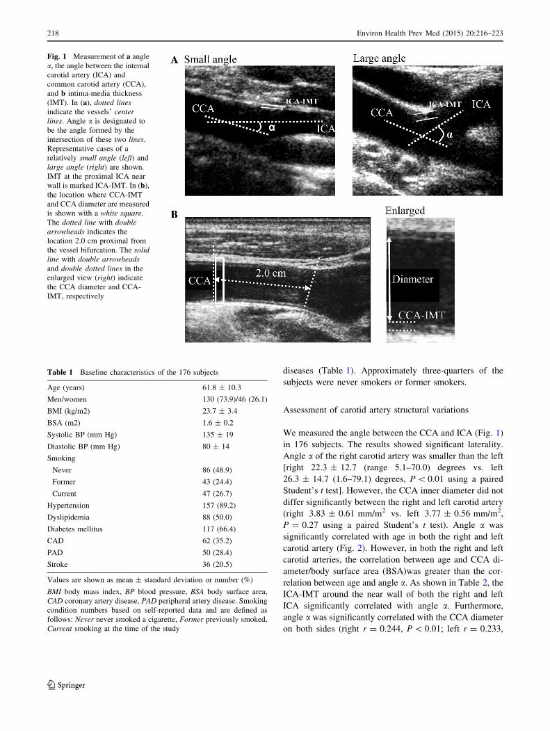
duct. We measured the angle between the CCA and ICA in
176 subjects from the previously collected data.
Specifically, we measured the angle according to the center
lines of the CCA and ICA using an image in plane with the
two arteries, obtained during the examination (Fig. 1a).
This angle was designated angle a. We measured the IMT
at the near wall of the proximal ICA (Fig. 1a) and at the
middle position of the CCA, meaning the CCA position
2.0 cm proximal to the bifurcation (Fig. 1b). These two
IMT determinations were designated the ICA-IMT and the
CCA-IMT, respectively. The CCA inner diameter, another
potential vessel structural variation, was measured 2.0 cm
proximal to the bifurcation during the contraction phase
using tomography (Fig. 1b) [17]. A blinded, Japanese
government-qualified sonographer evaluated the carotid
artery ultrasound images.
Statistical analysis
Values are shown as mean ± standard deviation. A two-
sided paired or unpaired Student’s t test was used, as ap-
propriate, to compare the values between groups, and the
Pearson’s product-moment correlation coefficient (r value)
was used to evaluate the correlation between values.
A Chi-square test was performed on the categorical vari-
ables. For the smoking status, analysis was performed us-
ing one-way analysis of variance (ANOVA). Multivariate
logistic regression analyses were conducted to evaluate
age, BMI, angle a, LDL-C, smoking status, hypertension,
and diabetes as explanatory variables for ICA-IMT. Stat-
Mate III (ATMS Co., Tokyo, Japan) or Stata 8.1 (Stata Co.,
College Station, TX) was used, as appropriate, for the
statistical analyses. P\ 0.05 was considered statistically
significant.
Results
Subject characteristics
Table 1 presents the characteristics of the subjects (130
men and 46 women) enrolled in the present study. The
mean age was 61.8 ± 10.3 years. All subjects were
C35 years old. Based on the clinical history, all subjects
had atherosclerotic risk factors and/or atherosclerotic
Environ Health Prev Med (2015) 20:216–223 217
123
diseases (Table 1). Approximately three-quarters of the
subjects were never smokers or former smokers.
Assessment of carotid artery structural variations
We measured the angle between the CCA and ICA (Fig. 1)
in 176 subjects. The results showed significant laterality.
Angle a of the right carotid artery was smaller than the left
[right 22.3 ± 12.7 (range 5.1–70.0) degrees vs. left
26.3 ± 14.7 (1.6–79.1) degrees, P\ 0.01 using a paired
Student’s t test]. However, the CCA inner diameter did not
differ significantly between the right and left carotid artery
(right 3.83 ± 0.61 mm/m2 vs. left 3.77 ± 0.56 mm/m2,
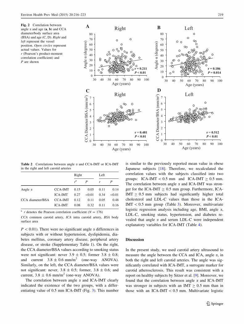
P = 0.27 using a paired Student’s t test). Angle a was
significantly correlated with age in both the right and left
carotid artery (Fig. 2). However, in both the right and left
carotid arteries, the correlation between age and CCA di-
ameter/body surface area (BSA)was greater than the cor-
relation between age and angle a. As shown in Table 2, the
ICA-IMT around the near wall of both the right and left
ICA significantly correlated with angle a. Furthermore,
angle a was significantly correlated with the CCA diameter
on both sides (right r = 0.244, P\ 0.01; left r = 0.233,
Fig. 1 Measurement of a angle
a, the angle between the internal
carotid artery (ICA) and
common carotid artery (CCA),
and b intima-media thickness
(IMT). In (a), dotted lines
indicate the vessels’ center
lines. Angle a is designated to
be the angle formed by the
intersection of these two lines.
Representative cases of a
relatively small angle (left) and
large angle (right) are shown.
IMT at the proximal ICA near
wall is marked ICA-IMT. In (b),the location where CCA-IMT
and CCA diameter are measured
is shown with a white square.
The dotted line with double
arrowheads indicates the
location 2.0 cm proximal from
the vessel bifurcation. The solid
line with double arrowheads
and double dotted lines in the
enlarged view (right) indicate
the CCA diameter and CCA-
IMT, respectively
Table 1 Baseline characteristics of the 176 subjects
Age (years) 61.8 ± 10.3
Men/women 130 (73.9)/46 (26.1)
BMI (kg/m2) 23.7 ± 3.4
BSA (m2) 1.6 ± 0.2
Systolic BP (mm Hg) 135 ± 19
Diastolic BP (mm Hg) 80 ± 14
Smoking
Never 86 (48.9)
Former 43 (24.4)
Current 47 (26.7)
Hypertension 157 (89.2)
Dyslipidemia 88 (50.0)
Diabetes mellitus 117 (66.4)
CAD 62 (35.2)
PAD 50 (28.4)
Stroke 36 (20.5)
Values are shown as mean ± standard deviation or number (%)
BMI body mass index, BP blood pressure, BSA body surface area,
CAD coronary artery disease, PAD peripheral artery disease. Smoking
condition numbers based on self-reported data and are defined as
follows: Never never smoked a cigarette, Former previously smoked,
Current smoking at the time of the study
218 Environ Health Prev Med (2015) 20:216–223
123
P\ 0.01). There were no significant angle a differences in
subjects with or without hypertension, dyslipidemia, dia-
betes mellitus, coronary artery disease, peripheral artery
disease, or stroke (Supplementary Table 1). On the right,
the CCA diameter/BSA values according to smoking status
were not significant: never 3.9 ± 0.5; former 3.8 ± 0.8;
and current 3.8 ± 0.6 mm/m2 (one-way ANOVA).
Similarly, on the left, the CCA diameter/BSA values were
not significant: never, 3.8 ± 0.5; former, 3.8 ± 0.6; and
current, 3.8 ± 0.6 mm/m2 (one-way ANOVA).
The correlation between angle a and ICA-IMT clearly
indicated the existence of the two groups, with a differ-
entiating value of 0.5 mm ICA-IMT (Fig. 3). This number
is similar to the previously reported mean value in obese
Japanese subjects [18]. Therefore, we recalculated the
correlation values with the subjects classified into two
groups: ICA-IMT\ 0.5 mm and ICA-IMT C 0.5 mm.
The correlation between angle a and ICA-IMT was stron-
ger for the ICA-IMT C 0.5 mm group. Furthermore, ICA-
IMT C 0.5 mm subjects had significantly higher total
cholesterol and LDL-C values than those in the ICA-
IMT\ 0.5 mm group (Table 3). Moreover, multivariate
logistic regression analysis including age, BMI, angle a,LDL-C, smoking status, hypertension, and diabetes re-
vealed that angle a and serum LDL-C were independent
explanatory variables for ICA-IMT (Table 4).
Discussion
In the present study, we used carotid artery ultrasound to
measure the angle between the CCA and ICA, angle a, inboth the right and left carotid arteries. The angle was sig-
nificantly correlated with ICA-IMT, a surrogate marker for
carotid atherosclerosis. This result was consistent with a
report on healthy subjects by Sitzer et al. [9]. Moreover, we
found that the correlation between angle a and ICA-IMT
was stronger in subjects with an IMT C 0.5 mm than in
those with an ICA-IMT\ 0.5 mm. Multivariate logistic
B A Left
r = 0.186 P = 0.014
Right C
Left D
r = 0.481 P < 0.01
r = 0.512 P < 0.01
Right
r = 0.211 P < 0.01
CC
A D
iam
eter
/BSA
(mm
/m2 )
Age (years)
0 10 20 30 40 50 60 70 80
30 40 50 60 70 80 90 100 Age (years)
0 10 20 30 40 50 60 70 80
30 40 50 60 70 80 90 100 C
CA
Dia
met
er/B
SA (m
m/m
2 )
Age (years)
0
1
2
3
4
5
6
7
30 40 50 60 70 80 90 100 Age (years)
0
1
2
3
4
5
6
7
30 40 50 60 70 80 90 100
Fig. 2 Correlation between
angle a and age (a, b) and CCA
diameter/body surface area
(BSA) and age (C, D). Right and
left represent the vessel
position. Open circles represent
actual values. Values for
r (Pearson’s product-moment
correlation coefficient) and
P are shown
Table 2 Correlations between angle a and CCA-IMT or ICA-IMT
in the right and left carotid arteries
Right Left
ra P r P
Angle a CCA-IMT 0.15 0.05 0.11 0.14
ICA-IMT 0.27 \0.01 0.34 \0.01
CCA diameter/BSA CCA-IMT 0.12 0.11 0.05 0.48
ICA-IMT 0.08 0.32 0.11 0.16
a r denotes the Pearson correlation coefficient (N = 176)
CCA common carotid artery, ICA intra carotid artery, BSA body
surface area
Environ Health Prev Med (2015) 20:216–223 219
123

regression analysis revealed that, in addition to serum
LDL-C, angle a was an independent explanatory variable
for ICA-IMT.
The angle a structural variation has been examined
previously with respect to its clinical significance. In a
study by Sitzer et al. [9], it was referred to as the angle of
ICA origin and correlated with ICA-IMT in healthy
subjects. However, in that study, the angle was measured
using a cross-sectional-axis view from the carotid artery
ultrasound. In this study, we used a longitudinal-axis view
to measure the angle from previously collected data. The
longitudinal-axis view is easier than the cross-sectional-
axis view, and the value could be measured from the pre-
viously collected data; therefore, we selected longitudinal-
B A Left Right
C Right D Left
r = 0.338 P < 0.01
r = 0.301 P < 0.01 r = 0.246
P < 0.01
r = 0.498 P < 0.01
r = 0.272 P < 0.01
r = 0.076 P = 0.319
r = 0.090 P = 0.306
r = -0.088 P = 0.572
r = 0.475 P < 0.01
ICA
-IM
T (m
m)
0
1
2
3
4
5
0 10 20 30 40 50 60 70 80
ICA
-IM
T (m
m)
0
1
2
3
4
5
0 10 20 30 40 50 60 70 80
r = 0.024 P = 0.789
ICA
-IM
T (m
m)
CCA diameter/BSA (mm/m2)
0
1
2
3
4
5
0 1 2 3 4 5 6 7
ICA
-IM
T (m
m)
CCA diameter/BSA (mm/m2)
r = 0.085 P = 0.583
r = 0.107 P = 0.156
0
1
2
3
4
5
0 1 2 3 4 5 6 7
Fig. 3 Correlation between
angle a and ICA-IMT (a, b) andCCA diameter/BSA and ICA-
IMT (c, d). Right and left
represent the vessel position.
Open and closed circles
represent values from the ICA-
IMT\ 0.5 mm group and ICA-
IMT C 0.5 mm group,
respectively. Solid, short-
dashed, and long-dashed lines
denote the correlation line for
the total subjects, the ICA-
IMT\ 0.5 mm group subjects,
and the ICA-IMT C 0.5 mm
group subjects, respectively. For
(A, B), the r and P values for
each group are shown to the
right of each correlation line.
For (c, d), the r and P values for
the ICA-IMT\ 0.5 mm group
and ICA-IMT C 0.5 mm group
are shown to the right of the
correlation line and total
subjects are shown to the left
Table 3 Differences in angle
a, patient characteristics, andblood test results between the
ICA-IMT\ 0.5 mm and ICA-
IMT C 0.5 mm groups
Values are shown as
mean ± standard deviation or
number (%). Statistical analyses
included unpaired Student’s
t tests (two-sided) or Chi-square
tests for categorical variables.
(N = 176)
TG triglyceride, TC total
cholesterol, LDL-C low-density
lipoprotein cholesterol, HDL-C
high-density lipoprotein
cholesterol
Right Left
\0.5 mm C0.5 mm P \0.5 mm C0.5 mm P
Angle a (degrees) 21.2 ± 12.2 25.5 ± 13.8 0.05 24.3 ± 14.6 33.2 ± 13.7 \0.01
Age (years) 61.5 ± 10.1 62.5 ± 11.1 0.57 61.7 ± 10.2 62.0 ± 10.6 0.88
Men/women 97/35 33/11 0.84 98/34 32/12 0.84
Smoking 0.06 0.06
Never 59 (44.7) 27 (62.4) 59 (44.7) 31 (70.5)
Former 35 (26.5) 8 (18.2) 35 (26.5) 9 (20.5)
Current 38 (28.8) 9 (20.5) 38 (28.8) 9 (20.5)
Hypertension 120 (90.9) 37 (84.1) 0.20 121 (91.7) 36 (81.8) 0.07
Dyslipidemia 75 (56.8) 13 (29.5) \0.01 76 (57.6) 12 (27.3) \0.01
Diabetes mellitus 91 (68.9) 26 (59.1) 0.23 92 (70.0) 25 (56.8) 0.11
CAD 49 (37.1) 13 (29.5) 0.36 48 (36.4) 14 (31.8) 0.58
PAD 38 (28.8) 12 (27.2) 0.84 39 (27.8) 11 (25) 0.56
Stroke 30 (22.7) 6 (13.6) 0.20 30 (22.7) 6 (13.6) 0.20
TG (mg/dL) 130.3 ± 83.5 120.8 ± 66.0 0.49 128.8 ± 81.7 125.2 ± 73.0 0.79
TC (mg/dL) 196.3 ± 36.6 211.1 ± 39.1 0.02 195.8 ± 36.9 212.8 ± 37.4 \0.01
LDL-C (mg/dL) 113.5 ± 33.5 130.8 ± 31.3 \0.01 113.2 ± 33.6 131.5 ± 30.3 \0.01
HDL-C (mg/dL) 56.8 ± 15.0 56.2 ± 16.1 0.82 56.8 ± 15.0 56.3 ± 16.0 0.84
220 Environ Health Prev Med (2015) 20:216–223
123

axis view measurements for this study. Furthermore, be-
cause of the advantage, the longitudinal-axis view may be
preferred when performing clinical examinations on a large
number of subjects.
The correlation between angle a and age was weaker
than the correlation between CCA diameter, another vessel
structural variation, and age (Fig. 2). Carotid vessel
structural variations are determined by both congenital and
lifestyle-related acquired factors [19–24]. In a twin study,
vessel configuration was more similar between monozy-
gotic than dizygotic twins [25], indicating that vessel
structural variation may be influenced by congenital fac-
tors. However, smoking, an acquired factor, can induce
vessel remodeling, and therefore, may increase CCA di-
ameter [24]. As angle a showed a significant but weak
correlation with age, angle a structural variations may
depend more on congenital factors than acquired ones.
Angle a exhibited a greater correlation with ICA-IMT
than CCA-IMT, suggesting that angle a’s influence is more
pronounced downstream of the blood flow. Vessel struc-
tural variations can affect blood flow hemodynamics in a
focal manner [5, 6]. Angle a may induce downstream
turbulent flow, depending on the size of the angle.
Theoretically, a greater angle may increase the risk of in-
creased IMT due to slower and possibly turbulent blood
flow. Although further experiments measuring actual blood
flow rates are needed to confirm this theory, the results of
this study may support it (Table 2; Fig. 3).
Our observation that there was a stronger correlation
between angle a and ICA-IMT in ICA-IMT C 0.5 mm
subjects suggests that IMT size may be influenced by other
factors such as risk factors for atherosclerosis (Fig. 3). We
did not observe this relationship with the CCA diameter.
When we examined clinical and serum biochemistry dif-
ferences between the subjects with those with \0.5 mm
ICA-IMT and those with C0.5 mm ICA-IMT, subjects with
C0.5 mm ICA-IMT had higher total cholesterol and LDL-C
(Table 3). Additionally, both LDL-C and angle a were in-
dependent explanatory factors based on multivariate logis-
tic regression analyses. This suggests that abnormal
cholesterol metabolism may be related to IMT, regardless
of angle a’s size. Interestingly, the percentage of subjects
with dyslipidemia was higher in those with an ICA-
IMT\ 0.5 mm than in those with an ICA-IMT C 0.5 mm.
This finding suggests that antidyslipidemic drugs were ef-
fective in lowering total cholesterol and LDL-C [26], as
well as limiting IMT. Abnormal cholesterol metabolism is a
conventional risk factor for increased IMT in many obser-
vational studies [27–29]. In addition, it should be noted that
the regression analysis also revealed that hypertension
combined with increased left ICA-IMT produced a sig-
nificant odds ratio value (odds ratio 0.27, P = 0.03).
However, the odds ratio is lower than 1.0, and the reason for
this result is not clear at present.
The present study had two main limitations. First,
although angle a is shown to be an IMT-related factor, our
study was cross-sectional. The correlations we noted should
be examined further in a longitudinal and observational
study over a prolonged period. An additional case–control
study involving, for example, individuals with a brain
Table 4 Results of multivariate logistic regression analysis of ICA-IMT for different values of angle a or LDL-cholesterol
Right ICA-IMT Left ICA-IMT
Group N Angle a (degrees) Odds ratio CI P Group N Angle a (degrees) Odds Ratio CI P
P for trend = 0.050 P for trend\0.010
0 60 10.5 ± 2.5 1 (reference) 0 59 12.0 ± 4.5 1 (reference)
1 59 19.8 ± 3.4 2.45 0.93–6.43 0.069 1 59 24.0 ± 3.4 1.64 0.56–4.81 0.368
2 57 37.3 ± 10.2 2.73 1.01–7.37 0.047 2 58 43.2 ± 11.3 6.07 2.11–17.41 \0.010
Group N LDL-C (mg/dL) Odds ratio CI P Group N LDL-C (mg/dL) Odds Ratio CI P
P for trend\0.010 P for trend = 0.012
0 59 80.5 ± 17.2 1 (reference) 0 59 80.5 ± 17.2 1 (reference)
1 59 118.4 ± 7.3 4.24 1.43–12.56 \0.010 1 59 118.4 ± 7.3 4.30 1.41–13.15 0.010
2 58 155.1 ± 16.5 5.17 1.74–15.41 \0.010 2 58 155.1 ± 16.5 4.44 1.46–13.48 \0.010
Values are shown as mean ± standard deviation. Multivariate logistic regression analyses including age, BMI, angle a, LDL-C, smoking status,
hypertension, and diabetes as explanatory variables were performed. N = 176 subjects. The subjects were divided into three groups containing
one-third of the subjects each: Group 0, (the reference group), Group 1, and Group 2, based on increasing angle a or LDL-C values. Odds ratios
were calculated by defining subjects with an ICA-IMT\ 0.5 mm as 0 and subjects with an ICA-IMT C 0.5 mm as 1. The trend of association
was assessed using a logistic regression model assigning consecutive integers to the angle a or LDL-C values. Additionally, hypertension in left
ICA-IMT showed a significant odds ratio value (odds ratio 0.27, CI 0.08–0.87, P = 0.028)
LDL-C low-density lipoprotein cholesterol, CI 95 % confidence interval
Environ Health Prev Med (2015) 20:216–223 221
123

infarction may also be appropriate. Second, the angle acorrelations were not confirmed for atherosclerosis surro-
gate markers other than ICA-IMT. Accordingly, future
studies examining the correlation between angle a and othervariables, such as pulse wave velocity (a measure of arterial
wall stiffness), when investigating the vessel properties
involved in atherosclerosis would be valuable [30, 31].
In conclusion, angle a was effectively measured using
longitudinal-axis view carotid artery ultrasound from the
data collected. We found that the vessel structure variation,
angle a, is related to ICA-IMT in Japanese subjects with
atherosclerotic risk factors or atherosclerotic disease,
especially those with higher serum cholesterol levels.
Acknowledgments We would like to thank the late Prof. Toru
Takeuchi for encouraging us to continue with this work and Mr.
Daniel Mrozek and Ms. Rinko Kawakami for copyediting. This re-
search was supported by a research fund from Kagoshima University.
Conflict of interest The authors declare that they have no conflict
of interest.
References
1. O’Leary DH, Polak JF, Kronmal RA, Manolio TA, Burke GL,
Wolfson SK. Carotid-artery intima and media thickness as a risk
factor for myocardial infarction and stroke in older adults. N Engl
J Med. 1999;340(1):14–22.
2. Lorenz MW, Markus HS, Bots ML, Rosvall M, Sitzer M. Pre-
diction of clinical cardiovascular events with carotid intima-me-
dia thickness: a systematic review and meta-analysis. Circulation.
2007;115(4):459–67.
3. Simons PCG, Algra A, Bots ML, Grobbee DE, van der Graaf Y,
The SMART study group. Common carotid intima-media thick-
ness and arterial stiffness: indicators of cardiovascular risk in
high-risk patients the SMART study (Second Manifestations of
ARTerial Disease). Circulation. 1999;100(9):951–7.
4. Salonen RM, Nyyssonen K, Kaikkonen J, Porkkala-Sarataho E,
Voutilainen S, Rissanen TH, et al. Six-year effect of combined
vitamin C and E supplementation on atherosclerotic progression:
the Antioxidant Supplementation in Atherosclerosis Prevention
(ASAP) study. Circulation. 2003;107(7):947–53.
5. Glagov S, Zarins C, Giddens DP, Ku DN. Hemodynamics and
atherosclerosis. Insights and perspectives gained from studies of
human arteries. Arch Pathol Lab Med. 1988;112(10):1018–31.
6. Perktold K, Resch M. Numerical flow studies in human carotid
artery bifurcations: basic discussion of the geometric factor in
atherogenesis. J Biomed Eng. 1990;12(2):111–23.
7. Schulz UGR, Rothwell PM. Major variation in carotid bifurcation
anatomy: a possible risk factor for plaque development? Stroke.
2001;32(11):2522–9.
8. Weibel J, Fields WS. Tortuosity, coiling, and kinking of the in-
ternal carotid artery. II. Relationship of morphological variation
to cerebrovascular insufficiency. Neurology. 1965;15:462–8.
9. Sitzer M, Puac D, Buehler A, Steckel DA, von Kegler S, Markus
HS, et al. Internal carotid artery angle of origin: a novel risk
factor for early carotid atherosclerosis. Stroke. 2003;34(4):950–5.
10. Anderson GB, Ashforth R, Steinke DE, Ferdinandy R, Findlay
JM. CT angiography for the detection and characterization of
carotid artery bifurcation disease. Stroke. 2000;31(9):2168–74.
11. Krishnaswamy A, Klein JP, Kapadia SR. Clinical cerebrovascular
anatomy. Catheter Cardiovasc Interv. 2010;75(4):530–9.
12. Aristokleous N, Seimenis I, Papaharilaou Y, Georgiou GC, Brott
BC, Eracleous E, et al. Effect of posture change on the geometric
features of the healthy carotid bifurcation. IEEE Trans Inf
Technol Biomed. 2011;15(1):148–54.
13. Lehmann ED, Hopkins KD, Rawesh A, Joseph RC, Kongola K,
Coppack SW, et al. Relation between number of cardiovascular
risk factors/events and noninvasive Doppler ultrasound assess-
ments of aortic compliance. Hypertension. 1998;32(3):565–9.
14. Ziegelbauer K, Schaefer C, Steinmetz H, Sitzer M, Lorenz MW.
Clinical usefulness of carotid ultrasound to improve stroke risk
assessment: ten-year results from the Carotid Atherosclerosis
Progression Study (CAPS). Eur J Prev Cardiol. 2013;20(5):
837–43.
15. Wilson PW, Zech LA, Gregg RE, Schaefer EJ, Hoeg JM, Spre-
cher DL, et al. Estimation of VLDL cholesterol in hyperlipi-
demia. Clin Chim Acta. 1985;151(3):285–91.
16. Payen T, Coron A, Lamuraglia M, Le Guillou-Buffello D, Gaud
E, Arditi M, et al. Echo-power estimation from log-compressed
video data in dynamic contrast-enhanced ultrasound imaging.
Ultrasound Med Biol. 2013;39(10):1826–37.
17. Sehirli US, Yalin A, Tulay CM, Cakmak YO, Gurdal E. The
diameters of common carotid artery and its branches in newborns.
Surg Radiol Anat. 2005;27(4):292–6.
18. Tokita A, Ishigaki Y, Okimoto H, Hasegawa H, Koiwa Y, Kato
M, et al. Carotid arterial elasticity is a sensitive atherosclerosis
value reflecting visceral fat accumulation in obese subjects.
Atherosclerosis. 2009;206(1):168–72.
19. Del Corso L, Moruzzo D, Conte B, Agelli M, Romanelli AM,
Pastine F, et al. Tortuosity, kinking, and coiling of the carotid
artery: expression of atherosclerosis or aging? Angiology.
1998;49(5):361–71.
20. Jartti L, Ronnemaa T, Kaprio J, Jarvisalo MJ, Toikka JO, Mar-
niemi J, et al. Population-based twin study of the effects of mi-
gration from Finland to Sweden on endothelial function and
intima-media thickness. Arterioscler Thromb Vasc Biol.
2002;33(5):832–7.
21. Krejza J, Arkuszewski M, Kasner SE, Weigele J, Ustymowicz A,
Hurst RW, et al. Carotid artery diameter in men and women and
the relation to body and neck size. Stroke. 2006;37(4):1103–5.
22. Zhao J, Cheema FA, Bremner JD, Goldberg J, Su S, Snieder H,
et al. Heritability of carotid intima-media thickness: a twin study.
Atherosclerosis. 2008;197(2):814–20.
23. Liang L-R, Wong ND, Shi P, Zhao L-C, Wu L-X, Xie G-Q, et al.
Cross-sectional and longitudinal association of cigarette smoking
with carotid atherosclerosis in Chinese adults. Prev Med.
2009;49(1):62–7.
24. Kweon SS, Lee YH, Shin MH, Choi JS, Rhee JA, Choi SW, et al.
Effects of cumulative smoking exposure and duration of smoking
cessation on carotid artery structure. Circ J. 2012;76(8):2041–7.
25. Le Bret E, Pineau E, Folliguet T, Garabedian EN, Brunelle F,
Vouhe P, et al. Congenital kinking of the internal carotid artery in
twin brothers. Circulation. 2000;102(22):E173–4.
26. Zhao S, Wang Y, Mu Y, Yu B, Ye P, Yan X, et al. Prevalence of
dyslipidaemia in patients treated with lipid-lowering agents in
China: results of the DYS lipidemia International Study (DYSIS).
Atherosclerosis. 2014;235(2):463–9.
27. Salonen R, Nyyssonen K, Porkkala E, Rummukainen J, Belder R,
Park JS, et al. Kuopio Atherosclerosis Prevention Study (KAPS).
A population-based primary preventive trial of the effect of LDL
lowering on atherosclerotic progression in carotid and femoral
arteries. Circulation. 1995;92(7):1758–64.
28. Okamura T, Kokubo Y, Watanabe M, Higashiyama A, Miyamoto
Y, Yoshimasa Y, et al. Low-density lipoprotein cholesterol and
non-high-density lipoprotein cholesterol and the incidence of
222 Environ Health Prev Med (2015) 20:216–223
123

cardiovascular disease in an urban Japanese cohort study: the
Suita study. Atherosclerosis. 2009;203(2):587–92.
29. Ikkruthi S, Rajappa M, Nandeesha H, Satheesh S, Sundar I,
Ananthanarayanan PH, et al. Hyperhomocysteinemia and hy-
perlipoproteinemia (a) in obese South Indian men: an indication
for increased cardiovascular risk. Acta Physiol Hung.
2014;101(1):13–20.
30. van Popele NM, Grobbee DE, Bots ML, Asmar R, Topouchian J,
Reneman RS, et al. Association between arterial stiffness and
atherosclerosis: the Rotterdam Study. Stroke. 2001;32(2):454–60.
31. Sawabe M, Takahashi R, Matsushita S, Ozawa T, Arai T, Ha-
mamatsu A, et al. Aortic pulse wave velocity and the degree of
atherosclerosis in the elderly: a pathological study based on 304
autopsy cases. Atherosclerosis. 2005;179(2):345–51.
Environ Health Prev Med (2015) 20:216–223 223
123