anillin regulates cell-cell junction integrity by organizing junctional accumulation of rho-gtp and...
TRANSCRIPT

Please cite this article in press as: Reyes et al., Anillin Regulates Cell-Cell Junction Integrity by Organizing Junctional Accumulation ofRho-GTP and Actomyosin, Current Biology (2014), http://dx.doi.org/10.1016/j.cub.2014.04.021
Anillin Regulates Cell-Cell Ju
Current Biology 24, 1–8, June 2, 2014 ª2014 Elsevier Ltd All rights reserved http://dx.doi.org/10.1016/j.cub.2014.04.021
Reportnction
Integrity by Organizing JunctionalAccumulation of Rho-GTP and Actomyosin
Ciara C. Reyes,1 Meiyan Jin,2 Elaina B. Breznau,1
Rhogelyn Espino,2 Ricard Delgado-Gonzalo,3
Andrew B. Goryachev,4 and Ann L. Miller1,2,*1The Cellular and Molecular Biology Program, University ofMichigan, Ann Arbor, MI 48109, USA2Department of Molecular, Cellular, and DevelopmentalBiology, University of Michigan, Ann Arbor, MI 48109, USA3Biomedical Imaging Group, Ecole Polytechnique Federale deLausanne, Lausanne 1015, Switzerland4Centre for Systems Biology, School of Biological Sciences,The University of Edinburgh, Mayfield Road, Edinburgh EH93JR, Scotland, UK
Summary
Anillin is a scaffolding protein that organizes and stabilizes
actomyosin contractile rings and was previously thoughtto function primarily in cytokinesis [1–10]. Using Xenopus
laevis embryos as a model system to examine Anillin’s rolein the intact vertebrate epithelium, we find that a population
of Anillin surprisingly localizes to epithelial cell-cell junc-tions throughout the cell cycle, whereas it was previously
thought to be nuclear during interphase [5, 11]. Furthermore,we show that Anillin plays a critical role in regulating cell-cell
junction integrity. Both tight junctions and adherens junc-tions are disrupted when Anillin is knocked down, leading
to altered cell shape and increased intercellular spaces. Anil-
lin interacts with Rho, F-actin, and myosin II [3, 8, 9], all ofwhich regulate cell-cell junction structure and function.
When Anillin is knocked down, active Rho (Rho-guanosinetriphosphate [GTP]), F-actin, and myosin II are misregulated
at junctions. Indeed, increased dynamic ‘‘flares’’ of Rho-GTPare observed at cell-cell junctions, whereas overall junc-
tional F-actin and myosin II accumulation is reduced whenAnillin is depleted. We propose that Anillin is required for
proper Rho-GTP distribution at cell-cell junctions and formaintenance of a robust apical actomyosin belt, which is
required for cell-cell junction integrity. These results reveala novel role for Anillin in regulating epithelial cell-cell
junctions.
Results and Discussion
Anillin Localizes to Cell-Cell Junctions in Epithelial CellsThe role of vertebrate Anillin has been characterized in isolatedcultured cells, where it promotes stable cleavage furrow posi-tioning during cytokinesis [3, 11–13]. Anillin is also enriched inthe actomyosin-rich structures required for modified forms ofcytokinesis, including cellularization and polar body emission[2, 4, 14]. However, almost nothing is known about Anillin’sfunction during cytokinesis in vertebrate organisms in vivo,and potential roles outside cytokinesis are completely unchar-acterized. Thus, we examined Anillin’s localization in gastrula-stage Xenopus laevis embryos in which a polarized epithelium
*Correspondence: [email protected]
with functional cell-cell junctions has formed (Figure S1A avail-able online) [15]. We first expressed tagged Anillin (Anillin-3XGFP) in embryos in which endogenous Anillin was depletedwith a morpholino oligonucleotide (MO) (Figures 1A andS1B–S1D). Consistent with work from isolated cultured cells[2, 3, 5, 11], Anillin-3XGFP was primarily nuclear during inter-phase and strongly accumulated at the contractile ring duringcytokinesis (Figures 1A, S1C, and S1D). Surprisingly, however,an additional population of Anillin-3XGFP was observed atcell-cell boundaries in both mitotic and interphase cells andwas focused toward the apical surface (Figures 1A, S1C, andS1D; Movies S1 and S2).Immunostaining with antibodies against Xenopus Anillin
confirmed that endogenous Anillin localized to cell-cell junc-tions in both interphase and mitotic cells and was clearlyfocused apically at cell-cell junctions (Figures 1B, S1E, andS1F). Upon Anillin MO injection, Anillin protein levels werereduced to 42% 6 8% of control levels (Figures S1H andS1I). Anillin knockdown (KD) also led to cytokinesis defects,consistent with previous reports (Figure S1G) [3]. Furthermore,endogenous Anillin signal was sharply reduced at cell-celljunctions and in the nucleus when Anillin was knocked down,confirming that the MO targets Anillin (Figures 1B–1D). Takentogether, these results demonstrate that a pool of endogenousAnillin is localized at cell-cell junctions in epithelial cells.
Anillin Is Required for Proper Adherens Junction and Tight
Junction StructureThe surprising observation that Anillin localizes at cell-celljunctions led us to examine whether Anillin functionally regu-lates the apical junctional complex (Figure S2A). Anillin KDproduced several striking junctional phenotypes. First,whereas the apical cell membranes were closely apposed incontrol cells, Anillin-depleted cells often exhibited intercellularspaces (Figure 2A). Second, control cells were polygonal andcame to a point at tricellular junctions (the sites where threecells come together), but Anillin KD cells exhibited a roundedshape (Figure 2A), suggesting that Anillin may be importantfor junctional tension. Third, b-catenin, an adherens junction(AJ) plaque protein, was apically enriched at the zonula adhe-rens in controls (Figures 2B and 2F). However, in Anillin KDembryos, basolateral localization of b-catenin was retained,but the increased apical concentration was lost (Figures 2Band 2F). Importantly, when Anillin mRNA was re-expressedin cells where endogenous Anillin was depleted, the effect onb-catenin was partially rescued (Figures S2B and S2C). Fourth,when Anillin was depleted, staining for E-Cadherin, an AJtransmembrane protein, showed strongly reduced signal andreduced apical concentration (Figure 2C).To determine whether Anillin likewise participates in tight
junction (TJ) structure, the TJ proteins ZO-1 and Claudinwere analyzed. In control cells, staining for the TJ plaqueprotein ZO-1 was sharp and linear at cell-cell junctions, pre-sent at the apical surface of each cell-cell junction, and en-riched at tricellular TJs relative to bicellular TJs (Figures 2Dand S2D). In contrast, in Anillin KD cells, ZO-1 accumulationwas discontinuous andwavy (Figures 2D andS2D), suggestingthat Anillin depletion may result in decreased apicolateral

Figure 1. Anillin Localizes at Cell-Cell Junctions in Interphase and Mitotic Epithelial Cells
(A) Live imaging of Anillin-3XGFP in gastrula-stage embryos in which endogenous Anillin was depleted. mChe-membrane labels the plasma membrane.
Images are brightest point projections of 17 apical z planes. Nuclear Anillin is not visible because only apical planes were captured. Right: z view shows
that Anillin-3XGFP is apically focused. Graph shows an average of five line scans drawn perpendicular to junctions, indicating that the peak intensities
of Anillin-3XGFP and mChe-membrane overlap.
(B) Embryos were injected with a GFP-membrane injection marker with or without the Anillin MO. Gastrula-stage embryos were fixed and stained with an
anti-Anillin antibody (pseudocolored green), anti-GFP to view themembrane (pseudocolored red), andDAPI (blue). z views show that apically focused Anillin
accumulation at cell-cell junctions is reduced when Anillin is depleted (yellow arrowheads).
(C) Quantification of the average intensity of endogenous Anillin at cell-cell junctions in control and Anillin KD cells (see Supplemental Experimental
Procedures for details). Data are from three separate experiments; n = 18 embryos for control and n = 19 embryos for Anillin KD, graphed as box-and-whisker
plot with the whiskers representing the 1st–99th percentile; p < 0.0001.
(D) Quantification of the average endogenous Anillin and DAPI intensity in the nucleus in control and Anillin KD cells (see Supplemental Experimental
Procedures for details). Data are from three independent experiments; n = 18 embryos for control and n = 19 embryos for Anillin KD, graphed as box-
and-whisker plot with the whiskers representing the 1st–99th percentile; p < 0.0001 for control versus Anillin KD for nuclear Anillin signal; p = 0.16 for control
versus Anillin KD for DAPI signal.
See also Figure S1 and Movies S1 and S2.
Current Biology Vol 24 No 112
Please cite this article in press as: Reyes et al., Anillin Regulates Cell-Cell Junction Integrity by Organizing Junctional Accumulation ofRho-GTP and Actomyosin, Current Biology (2014), http://dx.doi.org/10.1016/j.cub.2014.04.021
tension [16]. In Anillin KD cells, concentrated ZO-1 was notobserved at the apical surface of each cell-cell junction andwas sometimes buried basally (Figure 2D). Additionally, ZO-1was not strongly enriched at tricellular TJs in Anillin KD cells(Figure S2D). Staining for Claudin, a TJ transmembrane pro-tein, showed that the relative intensity of Claudin at TJs wassignificantly decreased in Anillin KD embryos (Figures 2E
and 2G). Taken together, these findings demonstrate that Anil-lin is required for proper organization of both AJ and TJ struc-ture in epithelial cells.
Anillin Is Required for Junctional Integrity
Because the apical junctional complex forms adhesive con-tacts between cells and limits the passage ofmolecules across

Figure 2. AJs and TJs Are Disrupted when Anillin Is Knocked Down
(A) Single intermediate plane views (top) and z views (bottom) of GFP-membrane in control and Anillin KD embryos reveal increased intercellular spaces in
Anillin KD embryos (yellow arrows and arrowheads).(legend continued on next page)
Anillin Regulates Cell-Cell Junctions3
Please cite this article in press as: Reyes et al., Anillin Regulates Cell-Cell Junction Integrity by Organizing Junctional Accumulation ofRho-GTP and Actomyosin, Current Biology (2014), http://dx.doi.org/10.1016/j.cub.2014.04.021

Figure 3. Junctional Integrity Is Compromised when Anillin Is Knocked Down
(A) Live control or Anillin KD embryos were mounted in 3,000 molecular weight (MW) Alexa 488-Dextran. Left: x-y views show that dextran can penetrate
between rounded cells in Anillin KD embryos. Right: z views generated along the indicated lines (top) and 3D views (bottom) show that, although dextran
remains at the surface in control embryos, it can penetrate between cells in Anillin KD embryos.
(B) Quantification of the average percentage of junctions where dextran penetrated into the intercellular space in control and Anillin KD embryos. Data are
from three independent experiments; n = 13 embryos for controls and n = 17 embryos for Anillin KD, graphed as mean + SEM; p < 0.0001.
(C) Quantification of the average depth of dextran penetration for control and Anillin KD embryos. Data are from three independent experiments; n = 13
embryos for controls and n = 17 embryos for Anillin KD, graphed as box-and-whisker plot with the whiskers representing the 1st–99th percentile; p < 0.0001.
See also Figure S2.
Current Biology Vol 24 No 114
Please cite this article in press as: Reyes et al., Anillin Regulates Cell-Cell Junction Integrity by Organizing Junctional Accumulation ofRho-GTP and Actomyosin, Current Biology (2014), http://dx.doi.org/10.1016/j.cub.2014.04.021
the epithelium, we tested how the defects in AJs and TJs inAnillin KD embryos affect passage of a low-molecular-weight(3 kDa) fluorescent dextran between cells (Figure S2E)[15, 17]. In control embryos, dextran was restricted abovethe surface of the epithelium; however, in Anillin KD embryos,dextran penetrated into intercellular spaces around the perim-eter of the cells, particularly at tricellular junctions (Figure 3A).A similar increase in dextran penetration was observed inembryos treated with EGTA to disrupt junctions by depletingcalcium (Figure S2F) [18, 19]. Both the average percentage ofjunctions breached by dextran and the average depth ofdextran penetration into the intercellular spaces were signifi-cantly increased in Anillin-depleted embryos (Figures 3B and3C). The increased dextran penetration in Anillin KD embryoslikely reflects both increased permeability, as we observedcases in which the 3 kDa fluorescent dextran penetrateddeeply between the cells as a thread-like protrusion
(B–E) Fixed staining of control and Anillin KD embryos for b-catenin (B), E-Cadh
used as a MO injection marker, and DAPI labels DNA. z views show the norma
disrupted localization in Anillin KD cells (see yellow arrowheads). The x-y TJ p
serial z sections. The red arrow in (D) highlights an intercellular space betwe
ZO-1 concentration that is buried basally.
(F) Quantification of b-catenin polarization in control and Anillin KD cells from lin
basal pointswas normalized to zero so that data frommultiple embryos could be
from two independent experiments; n = 26 embryos for control and n = 18 emb
0.001.
(G) Quantification of the relative intensity of Claudin at cell-cell junctions by g
mental Procedures for details). Data are from two independent experiments;
SEM; p < 0.0001.
See also Figure S2.
(Figure S2G), and the apically domed cell shape observed inAnillin KD embryos (see z views of Anillin KD cells in Figures1, 2, and 3). Taken together, these results suggest that junc-tional integrity is compromised when Anillin is depleted.
Anillin Is Necessary for Proper Distribution of Rho-GTP at
Cell-Cell JunctionsWe next examined the mechanism by which Anillin regulatescell-cell junctions. The interaction between Anillin and Rho in-volves a positive feedback loop: Anillin’s localization to thecleavage furrow is dependent on active Rho [3, 8, 20], andAnillin, in turn, promotes active Rho accumulation and stabilityat the cleavage furrow [8, 21]. Therefore, we reasoned thatjunctional Rho activity might be altered when Anillin is per-turbed. Using a fluorescent probe that binds specifically toRho-guanosine triphosphate (GTP) (GFP-Rho-binding domainof Rhotekin [rGBD]) [22], we observed that in control cells,
erin (C), ZO-1 (D), and Claudin (E). GFP-membrane or mChe-membrane was
l localization of the cell-cell junction proteins in control cells as well as the
rotein images on the left in (D) and (E) are maximal-intensity projections of
en a dividing cell and its neighbor, whereas the yellow arrow indicates a
e scans along the basolateral surface. The b-catenin signal at the ten most-
averaged (see Supplemental Experimental Procedures for details). Data are
ryos for Anillin KD, graphed as mean 6 SEM; *p % 0.05, **p % 0.01, ***p %
enerating line scans perpendicular to junctions (see Supplemental Experi-
n = 10 embryos for control and n = 12 for Anillin KD, graphed as mean 6
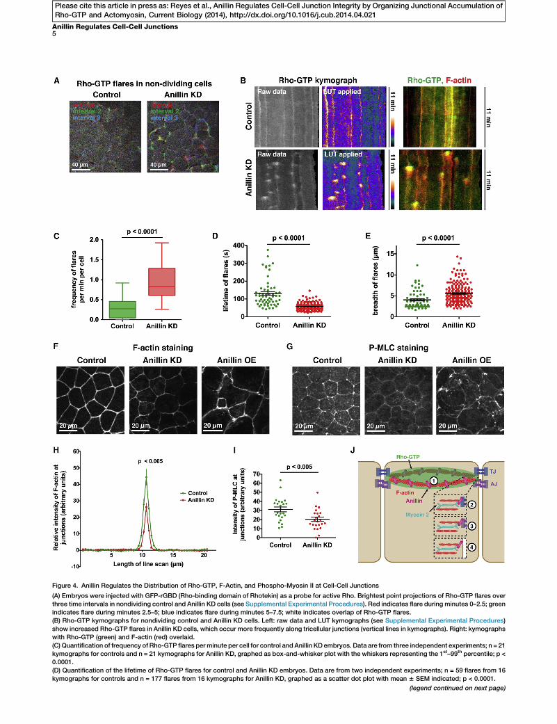
Figure 4. Anillin Regulates the Distribution of Rho-GTP, F-Actin, and Phospho-Myosin II at Cell-Cell Junctions
(A) Embryos were injected with GFP-rGBD (Rho-binding domain of Rhotekin) as a probe for active Rho. Brightest point projections of Rho-GTP flares over
three time intervals in nondividing control and Anillin KD cells (see Supplemental Experimental Procedures). Red indicates flare during minutes 0–2.5; green
indicates flare during minutes 2.5–5; blue indicates flare during minutes 5–7.5; white indicates overlap of Rho-GTP flares.
(B) Rho-GTP kymographs for nondividing control and Anillin KD cells. Left: raw data and LUT kymographs (see Supplemental Experimental Procedures)
show increased Rho-GTP flares in Anillin KD cells, which occur more frequently along tricellular junctions (vertical lines in kymographs). Right: kymographs
with Rho-GTP (green) and F-actin (red) overlaid.
(C) Quantification of frequency of Rho-GTP flares perminute per cell for control and Anillin KD embryos. Data are from three independent experiments; n = 21
kymographs for controls and n = 21 kymographs for Anillin KD, graphed as box-and-whisker plot with the whiskers representing the 1st–99th percentile; p <
0.0001.
(D) Quantification of the lifetime of Rho-GTP flares for control and Anillin KD embryos. Data are from two independent experiments; n = 59 flares from 16
kymographs for controls and n = 177 flares from 16 kymographs for Anillin KD, graphed as a scatter dot plot with mean 6 SEM indicated; p < 0.0001.
(legend continued on next page)
Anillin Regulates Cell-Cell Junctions5
Please cite this article in press as: Reyes et al., Anillin Regulates Cell-Cell Junction Integrity by Organizing Junctional Accumulation ofRho-GTP and Actomyosin, Current Biology (2014), http://dx.doi.org/10.1016/j.cub.2014.04.021
Current Biology Vol 24 No 116
Please cite this article in press as: Reyes et al., Anillin Regulates Cell-Cell Junction Integrity by Organizing Junctional Accumulation ofRho-GTP and Actomyosin, Current Biology (2014), http://dx.doi.org/10.1016/j.cub.2014.04.021
Rho-GTPwas present at cell-cell junctions and at the cleavagefurrow throughout cytokinesis (Figures S3A and S3C; MovieS3). Additionally, dividing cells pulled neighboring cells alongwith the constricting cleavage furrow (Figure S3B). In contrast,active Rho was not restricted to the cleavage furrow duringcytokinesis in Anillin KD embryos. Instead, intense ‘‘flares’’ ofactive Rho appeared at ectopic positions around the perimeterof the dividing cell, as well as in neighboring cells (Figures S3Aand S3C; Movie S3), indicating that tension asymmetries inAnillin KD cells may be mechanically integrated amongmultiple cells [23, 24]. Furthermore, junctions were often notproperly maintained during cell division in Anillin KD embryos,and the dividing cell separated from its neighboring cells(Figure S3B).
Because Anillin depletion disrupted cell-cell junctions inboth dividing and nondividing cells, we examined the effectof Anillin KD on active Rho localization at junctions in nondi-viding regions of the epithelium. In control cells, occasionalfluctuations in junctional Rho-GTP were observed (Figure 4Aand Movie S4); however, in Anillin KD cells, a pronouncedincrease in flares of Rho-GTP was observed around cell-celljunctions, particularly at tricellular junctions (Figure 4A andMovie S4). Kymographs generated from time-lapse moviesallowed us to quantify the frequency, lifetime, intensity, andbreadth of the Rho-GTP flares over time (Figures 4B andS3D), revealing a statistically significant increase in the fre-quency and a reduction in the lifetime of Rho-GTP flareswhen Anillin is knocked down (Figures 4C and 4D). Althougha significant change in Rho-GTP flare intensity was notobserved (data not shown), the breadth of flares wasincreased in Anillin KD embryos (Figure 4E). Notably, theRho-GTP flares were rapidly followed by strong F-actin accu-mulation (Figure 4B), indicating that Rho-GTP flares may besites of local mechanical perturbation in the epithelia.Together, these results suggest that Anillin is important forproper distribution of junctional Rho-GTP in both mitotic andinterphase cells.
Anillin Scaffolds the Apical Actomyosin Belt in Epithelial
CellsRho signaling can drive junction assembly and disassembly byregulating the tension in the apical actomyosin belt that con-nects to AJs and TJs (Figure S2A) [25, 26]. Because Anillin isrequired for proper accumulation of Rho-GTP at junctions (Fig-ures 4A–4E), and Anillin can bind directly to F-actin andmyosinII [2, 3], we hypothesized that loss of proper apical junctionalstructure and function in Anillin KD embryos could be due todisruption of the apical actomyosin belt. To test this idea, wefirst stained control, Anillin KD, and Anillin-overexpressing
(E) Quantification of breadth of Rho-GTP flares for control and Anillin KD em
kymographs for controls and n = 190 flares from 16 kymographs for Anillin KD
(F) Fixed staining for F-actin in control, Anillin KD, and Anillin OE embryos. In A
Anillin OE embryos, it is more intense at cell-cell junctions and the cell cortex,
(G) Fixed staining for P-MLC in control, Anillin KD, and Anillin OE embryos. In A
Anillin OE embryos, P-MLC is strongly accumulated at junctions and the cell c
(H) Quantification of the relative intensity of F-actin at cell-cell junctions. Line sc
Supplemental Experimental Procedures). Data are from three independent ex
graphed as mean 6 SEM; p < 0.005.
(I) Quantification of the intensity of P-MLC at cell-cell junctions. Data are from fo
for Anillin KD, graphed as a scatter dot plot with mean 6 SEM indicated; p < 0
(J)Model showing possiblemechanisms bywhich Anillinmay regulate cell-cell j
integrity by controlling the distribution of junctional Rho-GTP and stabilizing the
with a TJ component (2) or an AJ component (3) or link the apical actomyosin
See also Figure S3 and Movies S3, S4, and S5.
(OE) embryos for F-actin. F-actin accumulated in a strongapical band in controls, but Anillin depletion decreased theintensity and breadth of F-actin accumulation at cell-cell junc-tions (Figures 4F, 4H, and S3F). Moreover, Anillin OE increasedthe intensity of F-actin at cell-cell junctions and led to intense,spiky contractile rings in dividing cells (Figures 4F, S3E, andS3F; Movie S5), suggesting that Anillin is hyperactive in itsrole as a scaffolding protein when OE.Phosphorylation of the regulatory light chain of myosin II
(P-MLC) promotes the adenosine triphosphatase activity ofmyosin II, which is necessary for generating actomyosincontraction [27]. Therefore, increased P-MLC staining can beused as a readout for increased tension. In control embryos,P-MLC was localized along bicellular junctions and wasintensely localized at tricellular junctions (Figure 4G); however,P-MLC intensity was significantly reduced in Anillin KDembryos (Figures 4G and 4I). Furthermore, when Anillin wasOE, P-MLC accumulated strongly at junctions and at the apicalcell cortex, and cells appeared hypercontractile (Figures 4Gand S3G). These results support the idea that Anillin scaffoldsthe apical actomyosin belt. We propose that Anillin is neces-sary to stabilize and properly distribute tension in the apicalactomyosin belt (Figure 4J).
Conclusions
Our results demonstrate that Anillin, which was previouslythought to be nuclear during interphase and function solelyin cytokinesis [11, 12], plays a critical role in interphase anddividing epithelial cells, where it regulates cell-cell junctions.Whereas previous research on Anillin was generally conductedin isolated cells, our work in an intact vertebrate epitheliumrevealed this novel function for Anillin. Clues to Anillin’s local-ization at junctions were observed previously, including thecortical localization of Anillin in blastula-stage Xenopusembryos [28] and the apparent localization of Anillin to junc-tions in interphase epithelial cells of gastrulating Drosophilaembryos [2]. However, other studies in theDrosophila epitheliadid not reveal junctional localization for Anillin [29]. Weshow here that a pool of Anillin localizes to cell-cell junctionsin interphase and mitotic cells and regulates apical junctionalstructure and function in epithelial cells of the gastrulatingXenopus embryo. We predict that Anillin’s role in regulatingcell-cell junctions is likely conserved among higher verte-brates because Anillin and the other key players are highlyconserved.The defects reported in AJ and TJ structure in Anillin-
depleted cells were observed in both dividing and nondividingcells. Importantly, these defects were observed in mono-nucleate cells, demonstrating that the effects on cell-cell
bryos. Data are from two independent experiments; n = 62 flares from 16
, graphed as a scatter dot plot with mean 6 SEM indicated; p < 0.0001.
nillin KD embryos, junctional accumulation of F-actin is reduced, whereas in
and cell shapes are abnormal.
nillin KD embryos, junctional accumulation of P-MLC is reduced, whereas in
ortex.
ans from control and Anillin KD embryos were acquired and normalized (see
periments; n = 24 embryos for control and n = 23 embryos for Anillin KD,
ur independent experiments; n = 24 embryos for control and n = 24 embryos
.005.
unctions. Although our results suggest that Anillin regulates cell-cell junction
apical actomyosin belt (1), it is also possible that Anillin may directly interact
belt with the plasma membrane (4).
Anillin Regulates Cell-Cell Junctions7
Please cite this article in press as: Reyes et al., Anillin Regulates Cell-Cell Junction Integrity by Organizing Junctional Accumulation ofRho-GTP and Actomyosin, Current Biology (2014), http://dx.doi.org/10.1016/j.cub.2014.04.021
junctions are not secondary to the cytokinesis defect. We havenot yet examined how cell division failure elsewhere in theepithelium may perturb tension homeostasis or affect cell-cell junctions at a distance, but this will be an interesting ques-tion for future studies.
We propose that Anillin regulates cell-cell junction integrityby controlling the distribution of junctional Rho-GTP andstabilizing the apical actomyosin belt (Figure 4J). We showthat Anillin is required for proper distribution of Rho-GTP atapical junctions. Our live imaging of junctional Rho-GTP dy-namics extends previous fixed-imaging studies showing thata localized zone of Rho-GTP forms at cell-cell junctions [26,30–32]. We show that when Anillin is depleted, the sustainedjunctional Rho activation observed in controls is replaced byfrequent, dynamic flares of Rho-GTP followed rapidly byincreased F-actin accumulation. We propose that the pro-nounced Rho-GTP flares in Anillin KD embryos may representsites of junction disassembly or repair. Although the mecha-nisms that control localized formation and dynamics of thejunctional Rho-GTP zone are not well understood, emergingevidence implicates a number of proteins known to regulateRho activity during cytokinesis, including MgcRacGAP, Ect2,p190RhoGAP, andGEF-H1 [17, 30, 33, 34]. Interestingly, Anillinbinds MgcRacGAP [6, 7] and Ect2 [10] and could serve as ascaffold to recruit and/or retain them at cell-cell junctions.Thus, Anillin may be involved in regulating the distribution ofjunctional Rho-GTP directly through its ability to bind Rho orindirectly through its interactions with Ect2 and MgcRacGAP.Additionally, Ect2 can regulate function of the Par6/Par3/PKCzpolarity complex through Cdc42, thus playing a role in epithe-lial junction assembly and cell polarity [33]; therefore, it wouldbe interesting to test whether Anillin depletion also affectsCdc42 activation at cell-cell junctions.
Anillin is a strong candidate to scaffold and organize theapical actomyosin belt at cell-cell junctions, given its inter-actions with F-actin, myosin II, and the formin mDia2 [2, 3, 9,35]. We show here that Anillin regulates the proper accumula-tion of F-actin and P-MLC at cell-cell junctions. The cell round-ing and apical doming phenotypes observed when Anillin isperturbed likely result from changes in tension of the apicalactomyosin belt because apical doming has been observedin other situations in which apical tension is altered [36, 37].Our data suggest that Anillin is required for properly distrib-uting Rho-GTP and scaffolding the apical actomyosin belt(Figure 4J). However, Anillin could potentially make directconnections with a TJ and/or AJ component or use its pleck-strin homology domain to directly couple the apical actomy-osin belt to the plasma membrane (Figure 4J); these will beimportant avenues for future research. Finally, Anillin is OE 2-to 6-fold in diverse human tumors, and higher expressionlevels correlate with increased metastatic potential [38, 39].Therefore, misregulation of cell-cell junctions represents anovel mechanism by which Anillin may contribute to cancerprogression.
Supplemental Information
Supplemental Information includes Supplemental Experimental Proce-
dures, three figures, and fivemovies and can be foundwith this article online
at http://dx.doi.org/10.1016/j.cub.2014.04.021.
Acknowledgments
All studies with Xenopus laevis embryoswere conducted in compliancewith
the US Department of Health and Human Services Guide for the Care and
Use of Laboratory Animals and were approved by the University Committee
on Use and Care of Animals at the University of Michigan. We thank Aaron
Straight for the Xenopus Anillin construct and antibody; Zsuzsanna
Puspoki, Virginie Uhlmann, and Michael Unser for their respective contribu-
tions to the development of Kymographer and other members of the
Biomedical Imaging Group, EPFL, for helpful discussions; Sarah Woolner
and Jean-Pierre Tassan for staining advice; Megan Fekete for excellent
technical support; Billie Weber for making mChe-farnesyl; and members
of the A.L.M. laboratory for helpful input and critical reading of the manu-
script. We give special thanks to William Bement for advice, encourage-
ment, and useful feedback. This work was supported by a grant from the
NIH (R00 GM089765) to A.L.M. C.C.R. and E.B.B. were supported by the
NSF Predoctoral Fellowship and the NIH Cellular and Molecular Biology
Training Grant (T32-GM007315).
Received: August 20, 2013
Revised: February 24, 2014
Accepted: April 9, 2014
Published: May 15, 2014
References
1. Liu, J., Fairn, G.D., Ceccarelli, D.F., Sicheri, F., and Wilde, A. (2012).
Cleavage furrow organization requires PIP(2)-mediated recruitment of
anillin. Curr. Biol. 22, 64–69.
2. Field, C.M., and Alberts, B.M. (1995). Anillin, a contractile ring protein
that cycles from the nucleus to the cell cortex. J. Cell Biol. 131, 165–178.
3. Straight, A.F., Field, C.M., and Mitchison, T.J. (2005). Anillin binds
nonmuscle myosin II and regulates the contractile ring. Mol. Biol. Cell
16, 193–201.
4. Field, C.M., Coughlin, M., Doberstein, S., Marty, T., and Sullivan, W.
(2005). Characterization of anillin mutants reveals essential roles in
septin localization and plasma membrane integrity. Development 132,
2849–2860.
5. Oegema, K., Savoian, M.S., Mitchison, T.J., and Field, C.M. (2000).
Functional analysis of a human homologue of the Drosophila actin
binding protein anillin suggests a role in cytokinesis. J. Cell Biol. 150,
539–552.
6. D’Avino, P.P., Takeda, T., Capalbo, L., Zhang, W., Lilley, K.S., Laue,
E.D., and Glover, D.M. (2008). Interaction between Anillin and
RacGAP50C connects the actomyosin contractile ring with spindle
microtubules at the cell division site. J. Cell Sci. 121, 1151–1158.
7. Gregory, S.L., Ebrahimi, S., Milverton, J., Jones,W.M., Bejsovec, A., and
Saint, R. (2008). Cell division requires a direct link betweenmicrotubule-
bound RacGAP and Anillin in the contractile ring. Curr. Biol. 18, 25–29.
8. Piekny, A.J., and Glotzer, M. (2008). Anillin is a scaffold protein that links
RhoA, actin, and myosin during cytokinesis. Curr. Biol. 18, 30–36.
9. Miller, K.G., and Alberts, B.M. (1989). F-actin affinity chromatography:
technique for isolating previously unidentified actin-binding proteins.
Proc. Natl. Acad. Sci. USA 86, 4808–4812.
10. Frenette, P., Haines, E., Loloyan, M., Kinal, M., Pakarian, P., and Piekny,
A. (2012). An anillin-Ect2 complex stabilizes central spindle micro-
tubules at the cortex during cytokinesis. PLoS ONE 7, e34888.
11. D’Avino, P.P. (2009). How to scaffold the contractile ring for a safe
cytokinesis - lessons from Anillin-related proteins. J. Cell Sci. 122,
1071–1079.
12. Piekny, A.J., and Maddox, A.S. (2010). The myriad roles of Anillin during
cytokinesis. Semin. Cell Dev. Biol. 21, 881–891.
13. Goldbach, P., Wong, R., Beise, N., Sarpal, R., Trimble, W.S., and Brill,
J.A. (2010). Stabilization of the actomyosin ring enables spermatocyte
cytokinesis in Drosophila. Mol. Biol. Cell 21, 1482–1493.
14. Dorn, J.F., Zhang, L., Paradis, V., Edoh-Bedi, D., Jusu, S., Maddox, P.S.,
andMaddox, A.S. (2010). Actomyosin tube formation in polar body cyto-
kinesis requires Anillin in C. elegans. Curr. Biol. 20, 2046–2051.
15. Merzdorf, C.S., Chen, Y.H., and Goodenough, D.A. (1998). Formation
of functional tight junctions in Xenopus embryos. Dev. Biol. 195,
187–203.
16. Smutny, M., Cox, H.L., Leerberg, J.M., Kovacs, E.M., Conti, M.A.,
Ferguson, C., Hamilton, N.A., Parton, R.G., Adelstein, R.S., and Yap,
A.S. (2010). Myosin II isoforms identify distinct functional modules
that support integrity of the epithelial zonula adherens. Nat. Cell Biol.
12, 696–702.
17. Benais-Pont, G., Punn, A., Flores-Maldonado, C., Eckert, J., Raposo, G.,
Fleming, T.P., Cereijido, M., Balda, M.S., and Matter, K. (2003).

Current Biology Vol 24 No 118
Please cite this article in press as: Reyes et al., Anillin Regulates Cell-Cell Junction Integrity by Organizing Junctional Accumulation ofRho-GTP and Actomyosin, Current Biology (2014), http://dx.doi.org/10.1016/j.cub.2014.04.021
Identification of a tight junction-associated guanine nucleotide
exchange factor that activates Rho and regulates paracellular perme-
ability. J. Cell Biol. 160, 729–740.
18. Liu, K.C., and Cheney, R.E. (2012). Myosins in cell junctions.
BioArchitecture 2, 158–170.
19. Palmer, J.F., and Slack, C. (1970). Some bio-electric parameters of early
Xenopus embryos. J. Embryol. Exp. Morphol. 24, 535–553.
20. Hickson, G.R., and O’Farrell, P.H. (2008). Rho-dependent control of anil-
lin behavior during cytokinesis. J. Cell Biol. 180, 285–294.
21. Zhao, W.M., and Fang, G. (2005). Anillin is a substrate of anaphase-
promoting complex/cyclosome (APC/C) that controls spatial contrac-
tility of myosin during late cytokinesis. J. Biol. Chem. 280, 33516–33524.
22. Benink, H.A., and Bement, W.M. (2005). Concentric zones of active
RhoA and Cdc42 around single cell wounds. J. Cell Biol. 168, 429–439.
23. Fernandez-Gonzalez, R., Simoes, Sde.M., Roper, J.C., Eaton, S., and
Zallen, J.A. (2009). Myosin II dynamics are regulated by tension in inter-
calating cells. Dev. Cell 17, 736–743.
24. Clark, A.G., Miller, A.L., Vaughan, E., Yu, H.Y., Penkert, R., and Bement,
W.M. (2009). Integration of single and multicellular wound responses.
Curr. Biol. 19, 1389–1395.
25. Rodgers, L.S., and Fanning, A.S. (2011). Regulation of epithelial perme-
ability by the actin cytoskeleton. Cytoskeleton (Hoboken) 68, 653–660.
26. Terry, S., Nie, M., Matter, K., and Balda, M.S. (2010). Rho signaling and
tight junction functions. Physiology (Bethesda) 25, 16–26.
27. Vicente-Manzanares, M., Ma, X., Adelstein, R.S., and Horwitz, A.R.
(2009). Non-muscle myosin II takes centre stage in cell adhesion and
migration. Nat. Rev. Mol. Cell Biol. 10, 778–790.
28. Le Page, Y., Chartrain, I., Badouel, C., and Tassan, J.P. (2011). A func-
tional analysis of MELK in cell division reveals a transition in the mode
of cytokinesis during Xenopus development. J. Cell Sci. 124, 958–968.
29. Haglund, K., Nezis, I.P., Lemus, D., Grabbe, C., Wesche, J., Liestøl, K.,
Dikic, I., Palmer, R., and Stenmark, H. (2010). Cindr interacts with anillin
to control cytokinesis in Drosophila melanogaster. Curr. Biol. 20,
944–950.
30. Ratheesh, A., Gomez, G.A., Priya, R., Verma, S., Kovacs, E.M., Jiang, K.,
Brown, N.H., Akhmanova, A., Stehbens, S.J., and Yap, A.S. (2012).
Centralspindlin and a-catenin regulate Rho signalling at the epithelial
zonula adherens. Nat. Cell Biol. 14, 818–828.
31. Ratheesh, A., and Yap, A.S. (2012). A bigger picture: classical cadherins
and the dynamic actin cytoskeleton. Nat. Rev. Mol. Cell Biol. 13,
673–679.
32. Terry, S.J., Zihni, C., Elbediwy, A., Vitiello, E., Leefa Chong San, I.V.,
Balda, M.S., and Matter, K. (2011). Spatially restricted activation of
RhoA signalling at epithelial junctions by p114RhoGEF drives junction
formation and morphogenesis. Nat. Cell Biol. 13, 159–166.
33. Liu, X.F., Ishida, H., Raziuddin, R., and Miki, T. (2004). Nucleotide
exchange factor ECT2 interacts with the polarity protein complex
Par6/Par3/protein kinase Czeta (PKCzeta) and regulates PKCzeta activ-
ity. Mol. Cell. Biol. 24, 6665–6675.
34. Wildenberg, G.A., Dohn, M.R., Carnahan, R.H., Davis, M.A., Lobdell,
N.A., Settleman, J., and Reynolds, A.B. (2006). p120-catenin and
p190RhoGAP regulate cell-cell adhesion by coordinating antagonism
between Rac and Rho. Cell 127, 1027–1039.
35. Watanabe, S., Okawa, K., Miki, T., Sakamoto, S., Morinaga, T., Segawa,
K., Arakawa, T., Kinoshita, M., Ishizaki, T., and Narumiya, S. (2010). Rho
and anillin-dependent control of mDia2 localization and function in
cytokinesis. Mol. Biol. Cell 21, 3193–3204.
36. Fanning, A.S., Van Itallie, C.M., and Anderson, J.M. (2012). Zonula occlu-
dens-1 and -2 regulate apical cell structure and the zonula adherens
cytoskeleton in polarized epithelia. Mol. Biol. Cell 23, 577–590.
37. Yonemura, S., Wada, Y., Watanabe, T., Nagafuchi, A., and Shibata, M.
(2010). alpha-Catenin as a tension transducer that induces adherens
junction development. Nat. Cell Biol. 12, 533–542.
38. Hall, P.A., Todd, C.B., Hyland, P.L., McDade, S.S., Grabsch, H., Dattani,
M., Hillan, K.J., and Russell, S.E. (2005). The septin-binding protein anil-
lin is overexpressed in diverse human tumors. Clin. Cancer Res. 11,
6780–6786.
39. Suzuki, C., Daigo, Y., Ishikawa, N., Kato, T., Hayama, S., Ito, T.,
Tsuchiya, E., and Nakamura, Y. (2005). ANLN plays a critical role in
human lung carcinogenesis through the activation of RHOA and by
involvement in the phosphoinositide 3-kinase/AKT pathway. Cancer
Res. 65, 11314–11325.