animal models of diabetic retinopathy - springer · animal models of diabetic retinopathy ana maria...
TRANSCRIPT

MICROVASCULAR COMPLICATIONS—RETINOPATHY (JK SUN AND PS SILVA, SECTION EDITORS)
Animal Models of Diabetic Retinopathy
Ana Maria Olivares1 & Kristen Althoff1 & Gloria Fanghua Chen1& Siqi Wu1
&
Margaux A. Morrisson2& Margaret M. DeAngelis2 & Neena Haider1
Published online: 24 August 2017# The Author(s) 2017. This article is an open access publication
AbstractPurpose of Review Diabetic retinopathy (DR) is one of themost common complications associated with chronic hyper-glycemia seen in patients with diabetes mellitus. While manyfacets of DR are still not fully understood, animal studies havecontributed significantly to understanding the etiology andprogression of human DR. This review provides a compre-hensive discussion of the induced and genetic DR models indifferent species and the advantages and disadvantages ofeach model.
Recent Findings Rodents are the most commonly usedmodels, though dogs develop the most similar morphologicalretinal lesions as those seen in humans, and pigs and zebrafishhave similar vasculature and retinal structures to humans.Nonhuman primates can also develop diabetes mellitus spon-taneously or have focal lesions induced to simulate retinalneovascular disease observed in individuals with DR.Summary DR results in vascular changes and dysfunction ofthe neural, glial, and pancreatic β cells. Currently, no modelcompletely recapitulates the full pathophysiology of neuronaland vascular changes that occur at each stage of diabetic ret-inopathy; however, each model recapitulates many of the dis-ease phenotypes.
Keywords Animal models . Diabetic retinopathy . Diabetes .
Inducedmodels . Pancreatectomy . Alloxan . STZ . Geneticmodels . VEGF
AbbreviationsBB Biobreeding rat: widely used rat model of diabetesBRB Blood retinal barrier: wall of tightly joined cells
that prevent the movement of substances fromblood into the retina
DME Diabetic macular edema: leaking macular capil-laries that lead to loss of vision associated withdiabetes
DR Diabetic retinopathy: damage to the retina leadingto severe vision loss caused by diabetes
GCL Ganglion cell layer: inner most retina layer com-prised of ganglion cells and displaced amacrinecells
GLUT2 Glucose transporter 2: carrier protein that trans-ports glucose in the liver and blood
GK Goto-Kakizaki rat: rat model with hyperglycemia
This article is part of the Topical Collection on MicrovascularComplications—Retinopathy
* Neena [email protected]
Ana Maria [email protected]
Kristen [email protected]
Gloria Fanghua [email protected]
Siqi [email protected]
Margaux A. [email protected]
Margaret M. [email protected]
1 Schepens Eye Research Institute, Massachusetts Eye and EarInfirmary, Department of Ophthalmology, Harvard Medical School,20 Staniford Street, Boston, MA 02114, USA
2 Moran Eye Center, University of Utah, Salt Lake City, UT 84132,USA
Curr Diab Rep (2017) 17: 93DOI 10.1007/s11892-017-0913-0

GPR10 G-protein-coupled receptor: protein that binds toprolactin-releasing peptide
INL Inner nuclear layer: nuclear layer in the inner retinacomprised of bipolar, horizontal, and amacrinecells
IPL Inner plexiform layer: layer of neuronal synapsesconnecting the bipolar and amacrine cells to gan-glion cells
MESA Multi-Ethnic Study of Atherosclerosis: medicalresearch study involving 6000 men and womenfrom the six continents
NOD Nonobese diabetic mouse: mouse model of diabe-tes that is characterized by the absence of weightgain
NPDR Nonproliferative diabetic retinopathy: first stage ofdiabetic retinopathy characterized for the absenceof symptoms
ONL Outer nuclear layer: layer from the retina involve inthe detection of the light
OIR Oxygen-induced retinopathy: retinopathy causedby the exposure to oxygen concentrations in achamber
OLETF Otsuka Long-Evans Tokushima fatty rat: rat modelcharacterized by obesity and hyperglycemia
PSL Photoreceptor segment layer: retina layer com-posed of rod and cones.
P Postanatal: related to the time period after birthPDR Proliferative diabetic retinopathy: advance stage of
diabetic retinopathy characterized for the increaseof new blood vessels that eventually leak
QTL Quantitative trait locus: segment of DNA that cor-relates to a variation in phenotype
RGC Retinal ganglion cell: neuron present in the innersurface of the retina
SDT Spontaneously diabetic Torii rat: rat models ofnonobese type 2 diabetes
STZ Streptozotocin: chemical that is toxic to the insulinproducing β cells in the pancreas
VEGF Vascular endothelial growth factor: signal proteininvolved in angiogenesis
ZDF Zucker diabetic fatty rat: rat model with spontane-ous diabetes
Introduction
Diabetic retinopathy occurs in approximately one third of peo-ple with diabetes [1]. It is the leading cause of blindness inadults aged 24–70 [1, 2]; in 2010, an estimated 92.6 millionadults had diabetic retinopathy (DR), of which 28.4 millionindividuals experience vision impairment associated with DR[3]. The total prevalence of DR appears to be higher in patientswith type 1 than in those with type 2 diabetes [3, 4].
Approximately 25% of patients with type 1 diabetes start todevelop symptoms of DR within 5 years after diabetes onset,and the number increases to 80% by 15 years [5].Interestingly, while sex has not been found to impact suscep-tibility of type 1 or 2 [3], race [6–9] and socioeconomic status[8, 10] do influence susceptibility for DR. In 2006, the Multi-Ethnic Study of Atherosclerosis (MESA) reported disparitiesin DR prevalence between diabetic patients of different racialbackgrounds: 36.7% in African-Americans, 37.4% inHispanics, 24.8% in Caucasians, and 25.7% in Chinese-Americans [8, 9]. As a complex disease, it is clear that DRis strongly influenced by both genetics and environment [9,11••, 12–17]. Overall, the number of patients suffering fromDR is expected to rise due to increasing prevalence of diabetesand longer life expectancies for patients with diabetes [3].
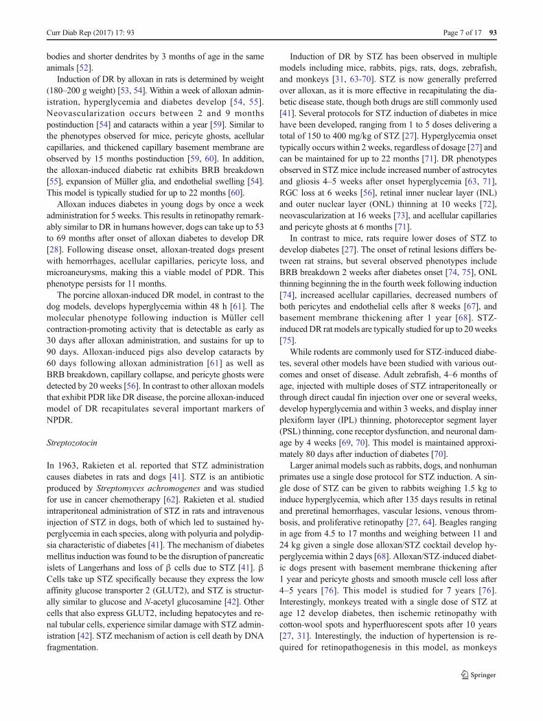
The onset and progression of DR is triggered by numerousfactors including extended duration of diabetes, poor controlof blood glucose, and elevated blood pressure [9].Hyperglycemia leads to the development of microangiopathy,including microaneurysms, hemorrhages, and basement mem-brane thickening [18, 19]. This results in increased vascularpermeability of the blood-retinal barrier (BRB) causing leak-age and diabetic macular edema (DME) [1, 18, 19]. Vascularpermeability also causes increased capillary occlusion thatleads to retinal ischemia, triggering an increase in the levelsof vascular endothelial growth factor (VEGF) [1, 18]. Retinalischemia and elevated VEGF levels then promote neovascu-larization [1, 18]. A schematic summary of various factors thatcontribute to disease progression is depicted in Fig. 1.
DR is classified as either nonproliferative (NPDR) or pro-liferative (PDR) based on the presence of neovascularizationthat typifies the proliferative form [1, 20]. NPDR, which canprogress to preproliferative DR, exhibits microaneurysms, dotand blot hemorrhages, cotton-wool spots, and capillarynonperfusion due to microvascular damage and pericyte loss.Microglial changes and DME can also occur in NPDR. InPDR, neovascularization results in retinal and vitreous hem-orrhages and can lead to retinal detachment. DME may alsooccur in PDR [1]. Disease models have contributed greatly tothe understanding of mechanisms that lead to DR disease.
Several animal models have been developed to investigatethe etiology and pathogenesis of DR and to develop and testtherapies to treat the disease. As DR is a complex disease withboth genetic and environmental influences, animal models aresimilarly developed by induction or genetic mutation. Inducedmodels are generated through surgery, drugs, diet, and laser orchemical damage. Genetic models are created using selectivebreeding and gene editing. While a large selection of specieshave been used to generate DR models, including mice, rats,cats, dogs, pigs, and nonhuman primates, mouse and ratmodels are most often studied, as their small size, short lifespan, and fast breeding rates allow for the most efficient stud-ies. Rodents have also been the focus of most genetic studies,
93 Page 2 of 17 Curr Diab Rep (2017) 17: 93

with the discovery of inherited hyperglycemia or obesity par-ticular to certain strains [21, 22, 23•, 24–26]. DR phenotypesin dog models, however, appear to be most similar to humanDR [27–30]. Surprisingly, nonhuman primates have provenrelatively resistant to induced DR [31]. Cats generally do notdevelop cataracts [32]. Pigs and zebrafish, in contrast, arepreferred for the similarity of their eye structure to humans,easily visualized vascular structures, short life spans, and largebreeding sizes (zebrafish) [27, 33, 34]. Though no single an-imal model to date represents the complete range of vascularand neural complications of human DR in both early and latestages, the models described in this review have been instru-mental in determining the mechanisms behind DR in thehopes of developing novel therapies.
Induced Models
Induced models have been created through five methods: sur-gical removal of the pancreas, administration of the drug al-loxan, administration of the drug streptozotocin (STZ), high-galactose diets, and laser or chemical damage to the eye [27,35–46]. While all methods of induction are still studied today,the most common is STZ administration, as it results in thefastest rate of disease development [41]. Alloxan is consideredto be less efficient in diabetic induction, and dietary methodsrequire the most time for disease progression [43]. Surgery-and damage-induced models are the most technically chal-lenging, limiting their use historically. The most frequentlyused models for inducing DR are mice and rats, but dogs, cats,pigs, rabbits, monkeys, and zebrafish are also used.
Presentation of induced DR pathology is generally slower inlarger animals, making rodents and, recently, zebrafish morefavored models. A comparison of the available inducedmodels can be found in Table 1.
Pancreatectomy
One of the oldest methods used to induce diabetes in animalmodels is pancreatectomy, the removal of the pancreas orremoval of β cells from the pancreas. Pancreatectomy wasobserved as early as 1922 to increase blood sugar levels indogs [35], and by 1968–1971, a technique of complete pan-createctomy in adult dogs had been developed to induce dia-betes [36, 37]. This technically difficult method is usuallyapplied to large animals such as cats and monkeys [27]. Inadult cats, hyperglycemia develops within 3 weekspostsurgery; this time can be reduced to within 1 week bycombining pancreatectomy with administration of alloxan[47] (described in the “Alloxan” section). Thickening of thebasement capillary membrane can occur from 3 months afterthe onset of hyperglycemia [48]. Other DR symptoms, includ-ing microaneurysm, intraretinal hemorrhages, capillarynonperfusion, and neovascularization, may take 5–9 years todevelop [49]. Maintenance of this model thus requires an ex-tended period of time.
In monkeys, pancreatectomy at various ages between 6 and15 years resulted in insulin dependency and hyperglycemia,which was then deliberately uncontrolled [50]. This was ob-served to lead to BRB leakage within 1 year of hyperglycemiaonset. However, 10 years postinduction, monkeys still did not
Fig. 1 Schematic representationof diabetic retinopathy (DR)disease progression. DR initiateswith hyperglycemia, whichinduces microangiopathy. Thisleads to vascular permeability,followed by diabetic macularedema and capillary occlusion.Capillary occlusion leads toretinal ischemia, followed byelevated levels of VEGF,ultimately resulting inneovascularization. Boxesrepresent the point in the pathwaytargeted by animal models.Italicized text corresponds to theinduced models and bold textcorresponds to the genetic models
Curr Diab Rep (2017) 17: 93 Page 3 of 17 93
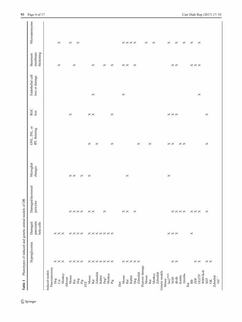
Tab
le1
Phenotypes
ofinducedandgenetic
anim
almodelsof
DR
Hyperglycem
iaDam
aged
pancreatic
beta-cells
Dam
aged/decreased
pericytes
Microglial
changes
ONL,INL,or
IPLthinning
RGC
loss
Endothelialcell
loss
ordamage
Basem
ent
mem
brane
thickening
Microaneurysm
s
Inducedmodels
Pancreatectomy
Dog
XX
Cat
XX
XX
Monkey
XX
Allo
xan
Mouse
XX
XX
XX
Rat
XX
XX
XDog
XX
XX
Pig
XX
XX
STZ Mouse
XX
XX
XX
Rat
XX
XX
XX
Zebrafish
XX
XRabbit
XX
Dog*
XX
XX
Monkey
XX
Pig
XX
XX
X
Diet Mouse
XX
XX
XRat
XX
XX
XRabbit
XX
Dog
XX
XX
Zebrafish
XX
Hypoxicdamage
Mouse
XRat
XMonkey
XZebrafish
Geneticmodels
Mouse
Ins2
Akita
XX
XX
XNOD
XX
XX
XX
db/db
XX
XX
XX
Kim
baX
XAkimba
XX
XRat B
BX
XX
XZDF
XX
XOLETF
XX
XX
XWBN/Kob
SDT
XX
XX
GK
XZebrafish
Vhl
−/−
Hem
orrhages
Increasedvascular
perm
eability
BRBleakageor
breakdow
nMacular
edem
aIncreasedacellular
capillaries
Neovascularization
Retinal
detachment
Com
mentsandreferences
93 Page 4 of 17 Curr Diab Rep (2017) 17: 93

Tab
le1
(contin
ued)
Hem
orrhages
Increasedvascular
perm
eability
BRBleakageor
breakdow
nMacular
edem
aIncreasedacellular
capillaries
Neovascularization
Retinal
detachment
Com
mentsandreferences
Inducedmodels
Pancreatectomy
Dog
X[35–37]
Cat
XCapillarynonperfusion
[47–49]
Monkey
X[50]
Allo
xan
Mouse
XFu
nctio
nald
efects[51,52]
Rat
XX
XEndothelialswellin
g[53–55,59,60]
Dog
XX
[28]
Pig
Capillarycollapse[61]
STZ Mouse
XX
Increasedastrocytes
[27,56,63
71–73]
Rat
XX
X[67,68,74,75]
Zebrafish
[69,70]
Rabbit
XVascularlesions[27,64]
Dog*
*Allo
xan/ST
Z-induced
[68,76]
Monkey
XX
Ischem
icretin
opathy
[27,31]
Pig
X[65,66,77]
Diet Mouse
X[27,44,78,79]
Rat
X[20,60,80]
Rabbit
X[27,81]
Dog
XX
X[43]
Zebrafish
[82]
Hypoxicdamage
Mouse
XNonperfusion[86–88]
Rat
XAbnormalvascular
tufts[89–93]
Monkey
XX
XNonperfusion[96]
Zebrafish
X[94,95]
Geneticmodels
Mouse
Ins2
Akita
XX
XInsulin
2[99–104]
NOD
Disorderfocalp
roliferation
vessels[105–113]
db/db
XX
Lepr
[24,25,114–120]
Kim
baVegf
[121–124]
Akimba
XX
XX
Insulin
2,Vegf
[125,126]
Rat B
BX
Ian4
[135–144]
ZDF
Lepr;increased
capillary
density
[128,129]
OLETF
GPR10;leukocyteentrapment
[131–134]
WBN/Kob
XX
QTLPd
wk1
[20,145–148]
SDT
QTLGisdt1,2,3[149–155]
Curr Diab Rep (2017) 17: 93 Page 5 of 17 93

develop proliferative retinopathy [50]. Monkeys thus appearto be surprisingly resilient to induction of DR despite removalof the pancreas, long-term diabetes, and poor control of bloodsugar levels. This is a similar phenomenon to humans whereonly 30% of individuals with diabetes develop DR, suggestingprimates may have additional biological mechanisms to re-regulate homeostatic state in the presence of chronic insult.Understanding the regulatory factors that contribute to thephysiological differences in species is important for develop-ing appropriate disease models.
Alloxan
The first drug found to induce diabetes, alloxan, was discov-ered by Dunn and McLetchie in 1942 [38]. Alloxan is a de-rivative of uric acid and directly targets β cells found in thepancreas [39], and was first produced by Wöhler and Liebigthrough a reaction of uric acid with nitric acid. Whileconducting rabbit studies focused on kidney disorders, Dunnand McLetchie found that intravenous injection of alloxanresulted in hypoglycemia due to necrosis in the islets ofLangerhans in the pancreas. Death of β cells led to the releaseof insulin stores in these cells, causing the observed hypogly-cemia followed by onset of diabetes within 24 h. Dunn andMcLetchie also created the diabetic rat model induced by al-loxan via intraperitoneal administration. While the diabeticrabbits appeared listless and lost weight, rats that were givenalloxan ate voraciously and presented with polydipsia, poly-uria, glycosuria, and hyperglycemia, characteristic of diabetes[38].
Alloxan-directed cell death is mediated by inhibition ofglucokinase, an enzyme involved in the glucose-insulin regu-latory pathway and expressed in the liver and pancreas. Thedrug can be toxic to liver and kidney cells, but with properdosing, toxicity can be avoided. The action of alloxan in thepancreas is specific toβ cells, with no toxic effect on aα, δ, orpancreatic exocrine cells. The compound is also unstable inwater at room and body temperature, making it difficult toadminister [38, 40]. In recent times, alloxan has fallen in pop-ularity in favor of STZ, described in the “Streptozotocin” sec-tion, due to the latter’s greater ease of use and efficacy.
Alloxan has been used to induce DR in a large variety ofanimals including mice, rats, dogs, and pigs, as well as therabbits and rats [28, 51•, 52–58]. All models experience dam-age to pancreatic β cells. Mice aged 8–10 weeks can be givena single dose of alloxan to induce hyperglycemia leading todiabetes [51•]. It was previously believed that the alloxan-induced diabetic mouse did not develop cellular or vascularlesions, but a recent study found that alloxan does inducepericyte ghosts and loss of retinal ganglion cells (RGCs) with-in 7 days and microaneurysms with increased acellular capil-laries by 21 days in mice from the FOT_FB strain [51•].Alloxan also induced microglial changes, with thicker cellT
able1
(contin
ued)
Hem
orrhages
Increasedvascular
perm
eability
BRBleakageor
breakdow
nMacular
edem
aIncreasedacellular
capillaries
Neovascularization
Retinal
detachment
Com
mentsandreferences
GK
Increasedendothelialcells
[156–161]
Zebrafish
Vhl−/−
XX
XVhl;increased
hyaloids
[162]
Modelsshow
nto
presentw
itheach
phenotypearemarkedwith
anX
DRdiabeticretin
opathy,O
NLouternuclearlayer,INLinnernuclearlayer,IPLinnerplexiform
layer,RGCretin
alganglio
ncell
93 Page 6 of 17 Curr Diab Rep (2017) 17: 93
bodies and shorter dendrites by 3 months of age in the sameanimals [52].
Induction of DR by alloxan in rats is determined by weight(180–200 g weight) [53, 54]. Within a week of alloxan admin-istration, hyperglycemia and diabetes develop [54, 55].Neovascularization occurs between 2 and 9 monthspostinduction [54] and cataracts within a year [59]. Similar tothe phenotypes observed for mice, pericyte ghosts, acellularcapillaries, and thickened capillary basement membrane areobserved by 15 months postinduction [59, 60]. In addition,the alloxan-induced diabetic rat exhibits BRB breakdown[55], expansion of Müller glia, and endothelial swelling [54].This model is typically studied for up to 22 months [60].
Alloxan induces diabetes in young dogs by once a weekadministration for 5 weeks. This results in retinopathy remark-ably similar to DR in humans however, dogs can take up to 53to 69 months after onset of alloxan diabetes to develop DR[28]. Following disease onset, alloxan-treated dogs presentwith hemorrhages, acellular capillaries, pericyte loss, andmicroaneurysms, making this a viable model of PDR. Thisphenotype persists for 11 months.
The porcine alloxan-induced DR model, in contrast to thedog models, develops hyperglycemia within 48 h [61]. Themolecular phenotype following induction is Müller cellcontraction-promoting activity that is detectable as early as30 days after alloxan administration, and sustains for up to90 days. Alloxan-induced pigs also develop cataracts by60 days following alloxan administration [61] as well asBRB breakdown, capillary collapse, and pericyte ghosts weredetected by 20 weeks [56]. In contrast to other alloxan modelsthat exhibit PDR like DR disease, the porcine alloxan-inducedmodel of DR recapitulates several important markers ofNPDR.
Streptozotocin
In 1963, Rakieten et al. reported that STZ administrationcauses diabetes in rats and dogs [41]. STZ is an antibioticproduced by Streptomyces achromogenes and was studiedfor use in cancer chemotherapy [62]. Rakieten et al. studiedintraperitoneal administration of STZ in rats and intravenousinjection of STZ in dogs, both of which led to sustained hy-perglycemia in each species, along with polyuria and polydip-sia characteristic of diabetes [41]. The mechanism of diabetesmellitus induction was found to be the disruption of pancreaticislets of Langerhans and loss of β cells due to STZ [41]. βCells take up STZ specifically because they express the lowaffinity glucose transporter 2 (GLUT2), and STZ is structur-ally similar to glucose and N-acetyl glucosamine [42]. Othercells that also express GLUT2, including hepatocytes and re-nal tubular cells, experience similar damage with STZ admin-istration [42]. STZ mechanism of action is cell death by DNAfragmentation.
Induction of DR by STZ has been observed in multiplemodels including mice, rabbits, pigs, rats, dogs, zebrafish,and monkeys [31, 63-70]. STZ is now generally preferredover alloxan, as it is more effective in recapitulating the dia-betic disease state, though both drugs are still commonly used[41]. Several protocols for STZ induction of diabetes in micehave been developed, ranging from 1 to 5 doses delivering atotal of 150 to 400 mg/kg of STZ [27]. Hyperglycemia onsettypically occurs within 2 weeks, regardless of dosage [27] andcan be maintained for up to 22 months [71]. DR phenotypesobserved in STZ mice include increased number of astrocytesand gliosis 4–5 weeks after onset hyperglycemia [63, 71],RGC loss at 6 weeks [56], retinal inner nuclear layer (INL)and outer nuclear layer (ONL) thinning at 10 weeks [72],neovascularization at 16 weeks [73], and acellular capillariesand pericyte ghosts at 6 months [71].
In contrast to mice, rats require lower doses of STZ todevelop diabetes [27]. The onset of retinal lesions differs be-tween rat strains, but several observed phenotypes includeBRB breakdown 2 weeks after diabetes onset [74, 75], ONLthinning beginning the in the fourth week following induction[74], increased acellular capillaries, decreased numbers ofboth pericytes and endothelial cells after 8 weeks [67], andbasement membrane thickening after 1 year [68]. STZ-inducedDR rat models are typically studied for up to 20weeks[75].
While rodents are commonly used for STZ-induced diabe-tes, several other models have been studied with various out-comes and onset of disease. Adult zebrafish, 4–6 months ofage, injected with multiple doses of STZ intraperitoneally orthrough direct caudal fin injection over one or several weeks,develop hyperglycemia and within 3 weeks, and display innerplexiform layer (IPL) thinning, photoreceptor segment layer(PSL) thinning, cone receptor dysfunction, and neuronal dam-age by 4 weeks [69, 70]. This model is maintained approxi-mately 80 days after induction of diabetes [70].
Larger animal models such as rabbits, dogs, and nonhumanprimates use a single dose protocol for STZ induction. A sin-gle dose of STZ can be given to rabbits weighing 1.5 kg toinduce hyperglycemia, which after 135 days results in retinaland preretinal hemorrhages, vascular lesions, venous throm-bosis, and proliferative retinopathy [27, 64]. Beagles rangingin age from 4.5 to 17 months and weighing between 11 and24 kg given a single dose alloxan/STZ cocktail develop hy-perglycemia within 2 days [68]. Alloxan/STZ-induced diabet-ic dogs present with basement membrane thickening after1 year and pericyte ghosts and smooth muscle cell loss after4–5 years [76]. This model is studied for 7 years [76].Interestingly, monkeys treated with a single dose of STZ atage 12 develop diabetes, then ischemic retinopathy withcotton-wool spots and hyperfluorescent spots after 10 years[27, 31]. Interestingly, the induction of hypertension is re-quired for retinopathogenesis in this model, as monkeys
Curr Diab Rep (2017) 17: 93 Page 7 of 17 93

without hypertension fail to develop retinopathy [31]. Theporcine model of STZ is induced, at 20 kg, with STZ admin-istration for three consecutive days [65]. Induced pigs develophyperglycemia within 1 week and are studied for up to32 weeks. Diabetes lasting 4–8 months after STZ inductionresults in increased BRB permeability, thinning of the INLand ganglion cell layer (GCL), and thickening of the capillarybasement membrane [65, 77]. When STZ-induced pigs aresubject to hyperlipidemic diets, they acquire dyslipidemiasimilar to that experienced by patients with type 2 diabetes.Diabetic pigs also experience increased BRB permeability, aswell as compromised tight junctions in the retina. The pig’slarge size and hierarchal vascular structures make its metabol-ic and circulatory functions highly similar to humans [66],thus making it a common model for DR.
High-Sugar Diets
Kern and Engerman first reported an animal model of DRinduced by galactose-heavy diet [43]. Several high-sugar dietmodels have been developed including mice, rats, rabbits,dogs, and zebrafish [33, 43, 44, 60, 78–83] that were persis-tently exposed to galactose developing retinopathy similar tothat observed in human diabetes. Maintenance of galactoseexposure results in continued disease progression. However,galactose-fed animals lack some metabolic abnormalities ex-perienced in diabetes [20]. Mice developed hyperglycemia by6 weeks of age following high-galactose diet [44]. After15 months of hyperglycemia, endothelial cell loss and in-creased acellular capillaries were observed [44, 78]. After21 months, lesions including pericyte ghosts, microaneurysms,and retinal thickening are observed [27, 44, 79].While retinop-athy takes longer to develop in these mice, they do live longerthan other models, allowing them to be observed over a longerperiod of time, up to 26 months [78]. Similarly, rats have beenkept on high-galactose diets for over 2 years. Phenotypes ob-served in rodents on a continuous high-sugar diet includepericyte ghosts, acellular capillaries, and capillary basementmembrane thickening by 18 months of hyperglycemia [60,80], as well as gliosis and microaneurysm by 28 months[20]. While rodents can develop diet-induced DR, drug-induced and genetic models are more commonly studied insmall animals due to their faster onset of disease.
In contrast, larger animals generally take longer to developDR whether by drug induction or diet. Rabbits fed a high-sucrose diet for 24 weeks develop hyperfluorescent dots andmicroaneurysms appeared by the 12th week of the diet [27,81]. Dogs fed a diet with 30% increased galactose develop amore complex disease phenotype including DR and cataractswithin 1 year; pericyte ghosts, microaneurysms, dot and blothemorrhages, and acellular capillaries by 32 months; andbasement membrane thickening by 60 months [43]. As ob-served with all dog models of DR, disease can take many
years to develop, but phenotypes in the dog are most similarto those in humans [27, 29, 30, 80, 84].
Most recently, hyperglycemic zebrafish have been devel-oped as a model for DR. Zebrafish are housed in freshwaterwith alternating concentration 0 and 2% glucose every otherday and develop hyperglycemia after 28 days and IPL thin-ning [82]. As this model has only been maintained for 28 daysto date, several attributes including similar retinal topography,ease of vascular structures visualization with fluorescent ex-pression [33, 83], short life span, and large breeding size re-duce experimental time and make zebrafish a strong model tostudy DR [27].
Hypoxic Damage-Induced Retinopathy
Models of retinal neovascularization and vasculature leakagelacking hyperglycemia have been used in recent years to studyDR. These models simulate advanced-stage PDR observed inhuman patients. In a 1969 study, Dollery, Bulpitt, and Kohnerexposed newborn kittens to hyperoxic conditions and foundthat returning the kittens to normal air made them experiencehypoxia, leading to neovascularization [85]. It was later dis-covered that retinal damage induced the release of angiogen-esis factors [45]. This discovery led to a number of differentdamage models for retinal neovascularization using mouse,rats, primates, and zebrafish [27, 46]. Hyperoxic mousemodels are generated by exposing juvenile mice, typicallypostnatal days 7–12, to hyperoxic conditions, which resultsin hypoxic conditions of the retina once they return to normalair and the growth of blood vessels in the retina [86, 87].These models of oxygen-induced retinopathy (OIR) exhibitneovascularization and nonperfusion, accompanied by the ap-pearance of microaneurysms, which typically occur within5 days postexposure [88].
Similar to mice, OIR in nondiabetic rats results in neovas-cularization. Rat pups were exposed to the hyperoxic condi-tions between 11 and 14 days [89, 90]. Neovascularization isapparent immediately once rats are returned to normoxic con-ditions followed by astrocyte degeneration [91] and subse-quent reduction in INL and IPL thickness, with disorganizedouter segments [92, 93]. A distinct feature of this model is theincomplete development of the vascular plexus and the pres-ence of abnormal endothelial tufts [91]. Two nonrodent OIRmodels, monkey and zebrafish, also develop neovascular dis-ease. OIR-induced neovascularization in zebrafish requires forthe animal to be placed in normoxic water followed by thegradual reduction of O2 tension over a period of 48–72 h untilreaching 10% of air saturation (820 ppb) [94]. Zebrafish canbemaintained in this environment for up to 15 days [95]. Afterexposure, neovascularization is evident as well as reduction inintercapillary distance [94]. The primate model of OIR-induced neovascularization is distinct in that induction is lo-calized by laser vein occlusion. Thus, focal regions of hypoxia
93 Page 8 of 17 Curr Diab Rep (2017) 17: 93

are created rather than a whole organism exposure to hypoxicconditions. Retinal neovascularization in OIR primates typi-cally occurs 4–7 days postexposure. Hypoxic monkeys showvascular leakage, venous occlusion, capillary nonperfusion,venous dilation, and dot and blot hemorrhages, which resultfrom microaneurysm ruptures [96]. Interestingly, a primatemodel was used to develop anti-VEGF treatment [96].
Cytokine Induction
The alkali burn model results in increased cytokine activity toproduce DR like neovascularization. This model is less com-monly used and involves a more painful method to induceretinal neovascularization in mice. This technique has beenused in inbred mouse strains such as BALB/c and involvesplacing 2-mm filter disks soaked in 1 N NaOH on the ocularsurface adult mice [97]. Neovascularization is typically ob-served within 2 weeks. Not surprisingly, treated mice alsoexhibit increased levels of inflammatory cytokines inneovascularized retinas [96]. While cytokines may be second-ary to the neovascular disease phenotype, they likely play asignificant role in maintaining the disease state.
Genetic Models
There are several genetic modes of DR in mouse, rat, andzebrafish. These models include spontaneous, strain-specific,and genetically edited mutations. Several inbred mouse strainsfor example, including the nonobese diabetic (NOD) and db/db (Leprdb), exhibit hyperglycemia, one of the main charac-teristics of diabetes, and are thus maintained and studied asdiabetic models. Rodents are frequently used as geneticmodels of DR as they are easy to maintain, have well-characterized genetic backgrounds, and are easily manipulat-ed to generate knockout or transgenic models. Genetic modelsexist for both type 1 and type 2 diabetes, and models for type 2can be either obese or nonobese [98]. A comparison of thecurrently available genetic models can be found in Table 1.
Mouse Genetic Models of DR
There are five known genetic mouse models of DR: Ins2Akita,nonobese diabetic (NOD), db/db (Leprdb), Kimba, andAkimba. These models vary in mode of inheritance, diseaseetiology, pathology, and progression of disease. The Ins2Akita
mouse is a model for type 1 diabetes that harbors a missensemutation in the Insulin 2 gene. The missense mutation leads toa conformational change in the insulin protein, causing theprotein to accumulate in pancreatic β cells, leading to β-celldeath [99, 100]. Disease onset in this model is at 8 weeks, withan increase in retinal vascular permeability and reactivegliosis. Disease progression continues up to 8 months ofage, with a reduction in axons and dendrites of RGCs by
12 weeks, an increase in acellular capillaries at 36 weeks,and an increase in leukocytes in the vascular wall, as a resultof inflammation [101, 102]. Additionally, a decreased numberof cholinergic and dopaminergic amacrine cells [103] is alsoobserved leading to a reduction in the thickness of the IPL andINL [101, 104]. The Ins2Akitamouse is useful for studying theearly progression of DR and the neuroprotective effects oftreatments, as loss of RGCs can be detected in a short spanof time [20].
A second commonly used model for type 1 diabetes is theNOD mouse, which exhibits an autoimmune response inwhich the CD4+ and CD8+ cells attack pancreatic β cells[21, 105, 106]. Diabetes in NOD mice has been well docu-mented and, similar to humans, is a polygenic model withseveral loci associated with the disease phenotype[107–109]. Similar to what is observed in humans with type1 diabetes, NODmice suffer from infiltration of dendritic cellsand macrophages in pancreatic islets leading to inflammation,hyperglycemia, and apoptosis of insulin-producing β cells[21, 22, 23•]. Disease onset begins when spontaneous hyper-glycemia occurs in these mice by 12 weeks of age. NODmiceshow apoptosis of pericytes, endothelial cells, and RGCs, aswell as retinal capillary basement membrane thickeningstarting at 4 weeks [110]. Vasoconstriction and degenerationof major vessels with abnormal microvessels can be detectedapproximately 4 months after hyperglycemia [111].Additionally, focal proliferation of vessels was also detected[112]. In contrast to the human disease, NOD mice exhibit agender bias where by 30 weeks, 80% of females and 20% ofmales become diabetic [21]. Due to the variation of diabetesseen in females and males, constant monitoring of glucoselevels is important for appropriate study design. Despite thegender bias, the pathophysiology of type 1 diabetes is verysimilar between the NOD mouse model and humans, makingit a desirable model for DR research [113].
The db/db (Leprdb) mice were developed to study type 2diabetes. db/db mice harbor a mutation in the leptin receptorand develop hyperglycemia and obesity after 4–8 weeks [24,25], and disease progression continues for 10 months.Homozygous animals exhibit chronic hyperglycemia, morbidobesity, atrophy of pancreatic β cells, and eventually becomehypoinsulinemic [25, 114, 115]. After 6 weeks, a reduction inthe number of RGCs and increased thickness in the centralretina are observed [116]. By 18 weeks, reactive gliosis as wellas pericyte loss is detected [117]. This model is used to studylate stages of the disease as db/db mice present late reactivegliosis along with vessel leakages [118]. db/db mice have con-tinued increases in blood sugar levels, severe depletion of pan-creatic islets, and myocardial diseases, eventually leading todeath at approximately 10 months of age [119, 120].
Another method to develop a more physiologically relevantmouse model for DR is breeding two mutant mouse strains.The Akimba mouse was generated by crossing two mouse
Curr Diab Rep (2017) 17: 93 Page 9 of 17 93

strains. The Kimba mice, a nondiabetic model of proliferativeretinopathy resulting from overexpression of Vegf driven bythe rhodopsin promoter [121, 122], were crossed with the dia-betic Ins2Akita mice to create the Akimba model. Kimba miceshow reduction in the INL and ONL by postnatal day 7 (P7)[123]. By P28, microvascular abnormalities and capillarydropout are observed and continue until 9 weeks of age, atwhich time pericyte loss is detected [123, 124]. The Akimbamice are hyperglycemic and appear to have additive effectsfrom both parental strains [125, 126]. Akimba mice are char-acterized by pericyte and vessel loss and retinal neovasculari-zation with diffuse vascular leakage that is observed in late-stage DR [125, 127]. Additionally, the Akimba mouse exhibitsleaky capillaries, tortuous vessels, and microaneurysm by8 weeks [125]. Enhanced photoreceptor loss, reduction of ret-ina thickness, increased persistence of edema, and retinal de-tachment are observed as the animal ages and disease pro-gresses [125].
Rat Genetic Models of DR
There are six genetic rat models of DR: Zucker diabetic fatty(ZDF), Otsuka Long-Evans Tokushima fatty (OLETF),biobreeding (BB), WBN/Kob, spontaneously diabetic Torii(SDT), and Goto-Kakizaki (GK). The ZDF, OLETF, and BBare monogenic models of DRwith independent mutations thatperturb different nodes of the DR disease pathway (Fig. 1).The SDT, WBN/Kob, and GK models, in contrast, are poly-genic models. These models demonstrate the genetic com-plexity of DR. Quantitative trait locus (QTL) analysis identi-fying novel DR loci may provide insight into the multiplegenes and networks that are impacted and lead to DR patho-genesis in humans.
There are three monogenic rat models of DR. The ZDF ratis a monogenic model for severe spontaneous type 2 diabetes.These animals have a missense mutation in the leptin receptorgene, Lepr, that results in insulin tolerance along with exces-sive body weight gain [128, 129]. ZDF rats are characterizedby hyperglycemia at 6 weeks that continues throughout theirlives. As a result, thickening of the capillary basement mem-brane can be detected along with increased capillary cell nu-clear density 5 months after hyperglycemia [130].
The OLETF rat is a monogenic DR model that was createdby the selective breeding of Long-Evans rats that is character-ized by obesity, hyperglycemia, and glycosuria [26]. OLETFis a model for spontaneous type 2 diabetes and obesity andharbors a mutation in the initiation codon of the G-protein-coupled receptor GPR10 that leads to obesity [131]. Diseaseonset is characterized by increased blood glucose that is ob-served by 5–6 months [132]. Six weeks following the onset ofhyperglycemia, microvessel-related symptoms are observed,including leukocyte entrapment in retinal microcirculation[26]. At 3 months posthyperglycemia, a reduction in the
number of pericytes is detected and damage of endothelialcells is observed [133]. In addition, increased thickness ofthe capillary basement membrane, microaneurysms, capillaryformation in loops, and tortuosity are also detected [26, 134].This model is not commonly used to study DR as it lacksacellular capillaries and has a late onset of diabetes.
The BB monogenic model is widely used to study type 1diabetes. This model presents with retinal lesions, pericyteloss, capillary degeneration, and microaneuryms by 8–11 months, as well as apoptosis of pancreatic β cells due toan autoimmune response [135, 136]. The BB model harbors aframeshift mutation in the immune-associated nucleotide-binding protein gene Ian4, also known as Ian5, Iddm1, andGimap5. The mutation generates the lymphopenia phenotypeassociated with diabetes [137-139]. Alterations in lymphope-nia are associated with both type 1 and type 2 diabetes [140,141]. Lymphophenia is also commonly observed in cancerand autoimmune disease [142–144].
The WBN/Kob rat model presents acellular capillaries andis a spontaneous model of type 2 diabetes whose causativegene remains unknown [20, 145]. This model is characterizedby intraretinal angiopathy with new vessel formation andhyalinization of intraretinal vessels, making it an ideal modelfor understanding the progression of DR [146]. To determinethe gene or genes involved in generating the described pheno-type in this model, QTL analysis identified two significantregions in chromosome 7 and X hinting that several genesmight be involved [147]. Later studies were able to narrowdown the search to chromosome 7, a region designated asPdwk1 (pancreatitis and diabetes mellitus in WBN/Kob locus1) that harbors 14 genes [148].
There are two rat models of nonobese type 2 diabetes: theSDTand the GK rats. The SDT male rats develop glycosuria atapproximately 20 weeks compared to females at 45 weeks[149, 150]. Similarly diabetes develops at different rates: by40 weeks, 100% of males have diabetes, compared to 33% offemales at 65 weeks when the study ended [150]. SDT rats arecharacterized by retinal dysfunction, which includes retinal de-tachment with fibrous proliferation, absence of retinal ischemiain the presence of neovascularization, leukostasis, increasednumber of apoptotic cells in the GCL and INL, vascular lesions,and pericyte loss [151–154]. A distinct feature of this model isthe appearance of large retinal folds with extensive leakagearound the optic disc, similar to retinal detachment observedin humans [151–154]. The SDT rat is the rat model that mostclosely resembles the pathophysiology seen in humans; how-ever, the absence of microaneurysm makes them a more suit-able model for studying NPDR. The characteristic phenotypeassociated with SDT rats has been linked to three QTL assignedas Gisdt1, Gisdt2, and Gisdt 3 for glucose intolerance found inchromosomes 1, 2, and X, respectively [155].
GK rats, in contrast to SDT rats, develop hyperglycemiaearlier at 4–6 weeks and have an increased number of retinal
93 Page 10 of 17 Curr Diab Rep (2017) 17: 93
endothelial cells compared to the number of pericytes present[156, 157]. GK rats are characterized by having a reducedretinal blood flow with no changes in the diameter of veinsand arteries in the early stages of diabetes, making it an excel-lent model to study microcirculatory changes in the retina [20,158]. Increased BRB permeability is observed at 3 monthsfollowed by increased endothelial/perycite ratio at 7 months[158, 159]. GK rats were generated by multiple inbreedingcrosses of the glucose-intolerant Wistar rats. Consequently,the exact genetic background as well as the causative genesremains unknown [160]. Whole genome sequencing and QTLanalysis revealed 192 potential genes [161].
Zebrafish Genetic Model of DR
Zebrafish are valuable genetic models for human diseases asgenetic manipulation is easily performed and they often reca-pitulate human retinal vascular disease accurately. The Vhlzebrafish have a mutation in the von Hippel-Lindau tumorsuppressor gene and are characterized by increased blood ves-sel formation, along with upregulation of the hypoxia-inducible factor, which triggers expression of Vegf [162].This model is characterized by an increased number of hyaloid
vasculature with concomitant vascular leakage, macular ede-ma, retinal detachment, and severe neovascularization [151].
Conclusions and Future Directions
Animal models play a crucial role in understanding the etiol-ogy and pathophysiology of DR and in the development ofviable therapeutics to prevent and attenuate disease. DR is acomplex disease that involves multiple genetic and environ-mental inputs and, therefore, a challenging disease to model.Most models focus on a genetic or environmental insult to oneof the major DR phenotypes. Combining genetic and/or ge-netic and induced models may provide more accurate DRmodels. An example is the generation of the Akimba mousethat results from breeding Kimba mice, which overexpressVegf, with the Akita mice, which have spontaneous type 1diabetes designed to generate a model that has the key char-acteristic of the early and late phases of the disease and asmany traits of the phenotype as possible. As of today, theavailable models, both induced and genetic, are mostly char-acteristic of NPDR, such as microaneurysm and retinal degen-eration, and key characterist ics of PDR, such as
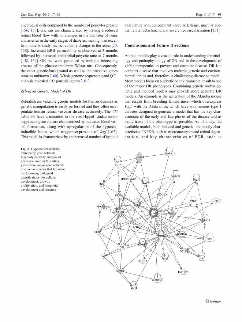
Fig. 2 Hypothetical diabeticretinopathy gene network.Ingenuity pathway analysis ofgenes reviewed in this articleyielded one major gene networkthat contains genes that fall underthe following biologicalclassifications: for cellulardevelopment, growth,proliferation, and lymphoiddevelopment and structure
Curr Diab Rep (2017) 17: 93 Page 11 of 17 93
neovascularization, are less likely to be seen in the models.Similarly, the majority of models imitate the aspects of earlyDR, and only few of the high-order animals recapitulate theretinopathies of the later stages of the disease.
The majority of the available models better recapitulate theearly stages of the diseases, limiting the availability of modelsto evaluate comprehensive therapies for DR. Treatments aregenerally restricted to targeting the early progression of thedisease due to the available models. Additionally, animalmodels of retinal neovascularization, without hyperglycemia,have been developed. These models may provide valuabletools to understand pathogenesis and develop appropriatetreatment options for late-stage DR disease. It is critical toproperly understand the pathophysiology and limitations ofeach available model to determine the best model for a study.Based on current research, we generated a hypothetical DRgene network (Fig. 2) [163]. Data were analyzed through theuse of IPA (QIAGEN Inc., https://www.qiagenbioinformatics.com/products/ingenuity-pathway-analysis). The review andhypothetical network will provide bases for improvedtherapeutic study design to develop viable treatments for thiscomplex and common disease.
Compliance with Ethical Standards
Conflict of Interest Ana Maria Olivares Kristen Althoff, GloriaFanghua Chen, Siqi Wu, Margaux A. Morrisson, Margaret M.DeAngelis, and Neena Haider declare that they have no conflict ofinterest.
Human and Animal Rights and Informed Consent This article doesnot contain any studies with human or animal subjects performed by anyof the authors.
Open Access This article is distributed under the terms of the CreativeCommons At t r ibut ion 4 .0 In te rna t ional License (h t tp : / /creativecommons.org/licenses/by/4.0/), which permits unrestricted use,distribution, and reproduction in any medium, provided you give appro-priate credit to the original author(s) and the source, provide a link to theCreative Commons license, and indicate if changes were made.
References
Papers of particular interest, published recently, have beenhighlighted as:• Of importance•• Of major importance
1. Cheung N, Mitchell P, Wong TY. Diabetic retinopathy. Lancet[Internet]. 2010;376(9735):124–36. Available from: http://www.sciencedirect.com/science/article/pii/S0140673609621243.
2. Lee R, Wong TY, Sabanayagam C. Epidemiology of diabetic ret-inopathy, diabetic macular edema and related vision loss. Eye Vis[Internet] London BioMed Central. 2015;2:17. Available from:http://www.ncbi.nlm.nih.gov/pmc/articles/PMC4657234/.
3. Yau JWY, Rogers SL, Kawasaki R, Lamoureux EL, Kowalski JW,Bek T, et al. Global prevalence and major risk factors of diabeticretinopathy. Diabetes Care [Internet]. Am Diabetes Assoc.2012;35(3):556–64. Available from: http://www.ncbi.nlm.nih.gov/pmc/articles/PMC3322721/.
4. Rabb MF, Gagliano DA, Sweeney HE. Diabetic retinopathy inBlacks. Diabetes Care [Internet]. 1990;13(11):1202 LP–1206.Available from: http://care.diabetesjournals.org/content/13/11/1202.abstract.
5. Aiello LP, Gardner TW, King GL, Blankenship G, CavalleranoJD, Ferris FL, et al. Diabetic retinopathy. Diabetes Care [Internet].1998;21(1):143 LP–156. Available from: http://care.diabetesjournals.org/content/21/1/143.abstract.
6. Haffner SM, Fong D, Stern MP, Pugh JA, Hazuda HP, PattersonJK, et al. Diabetic retinopathy in Mexican Americans and non-Hispanic Whites. Diabetes [Internet]. 1988;37(7):878 LP–884.Available from: http://diabetes.diabetesjournals.org/content/37/7/878.abstract.
7. Haffner SM, Mitchell BD, Moss SE, Stern MP, Hazuda HP,Patterson J, et al. Is there an ethnic difference in the effect of riskfactors for diabetic retinopathy? Ann Epidemiol [Internet].1993;3(1):2–8. Available from: http://www.sciencedirect.com/science/article/pii/104727979390003M.
8. Wong T, Klein R, Islam F, Cotch M, Folsom A, Klein B, et al.Diabetic retinopathy in a multi-ethnic cohort in the United States.Am J Ophthalmol [Internet]. 2006;141(3):446–55. Availablefrom: http://www.ncbi.nlm.nih.gov/pmc/articles/PMC2246042/.
9. Liew G, Klein R, Wong TY. The role of genetics in susceptibilityto diabetic retinopathy. Int Ophthalmol Clin [Internet].2009;49(2):35–52. Available from: http://www.ncbi.nlm.nih.gov/pmc/articles/PMC2746819/.
10. Haffner SM, Hazuda HP, Stern MP, Patterson JK, WAJ VH, FongD. Effect of socioeconomic status on hyperglycemia and retinop-athy levels in Mexican Americans with NIDDM. Diabetes Care[Internet]. 1989;12(2):128 LP–134. Available from: http://care.diabetesjournals.org/content/12/2/128.abstract.
11.•• Cho H, Sobrin L. Genetics of diabetic retinopathy. Curr Diab Rep[Internet]. 2014;14(8):515. doi:10.1007/s11892-014-0515-z. Thisis an important reference because it provides insight on howgenetics plays a role in diabetic retinopathy. Understandingthis will give more information on future development oftherapies for treatment of this area.
12. Karalliedde J, Gnudi L. Diabetes mellitus, a complex and hetero-geneous disease, and the role of insulin resistance as a determinantof diabetic kidney disease. Nephrol Dial Transplant [Internet].2016;31(2):206–13. doi:10.1093/ndt/gfu405.
13. Kowluru RA, Santos JM,MishraM. Epigenetic modifications anddiabetic retinopathy. Biomed Res Int Hindawi PublishingCorporation; 2013;2013.
14. Torres JM, Cox NJ, Philipson LH. Genome wide association stud-ies for diabetes: perspective on results and challenges. PediatrDiabetes Wiley Online Library. 2013;14(2):90–6.
15. Madsen-Bouterse SA, Kowluru RA. Oxidative stress and diabeticretinopathy: pathophysiological mechanisms and treatment per-spectives. Rev Endocr Metab Disord Springer. 2008;9(4):315–27.
16. Santos JM, Tewari S, Goldberg AFX, Kowluru RA.Mitochondrial biogenesis and the development of diabetic reti-nopathy. Free Radic Biol Med Elsevier. 2011;51(10):1849–60.
17. Madsen-Bouterse SA, Mohammad G, Kanwar M, Kowluru RA.Role of mitochondrial DNA damage in the development of dia-betic retinopathy, and the metabolic memory phenomenon associ-ated with its progression. Antioxid Redox Signal [Internet]. 140Huguenot Street, 3rd FloorNew Rochelle, NY 10801USA: MaryAnn Liebert, Inc.; 2010 15;13(6):797–805 Available from: http://www.ncbi.nlm.nih.gov/pmc/articles/PMC2935337/.
93 Page 12 of 17 Curr Diab Rep (2017) 17: 93

18. Nentwich MM, Ulbig MW. Diabetic retinopathy—ocular compli-cations of diabetes mellitus. World J Diabetes [Internet]Baishideng Publishing Group Inc. 2015;6(3):489–99. Availablefrom: http://www.ncbi.nlm.nih.gov/pmc/articles/PMC4398904/.
19. Cunha-Vaz J. Characterization and relevance of different diabeticretinopathy phenotypes. Dev Ophthalmol. 2007;39:13–30.
20. Robinson R, Barathi VA, Chaurasia SS, Wong TY, Kern TS.Update on animal models of diabetic retinopathy: from molecularapproaches to mice and higher mammals. Dis Model Mech[Internet] Company Biologists Limited. 2012;5(4):444–56.Available from: http://www.ncbi.nlm.nih.gov/pmc/articles/PMC3380708/.
21. Makino S, Kunimoto K, Muraoka Y, Mizushima Y, Katagiri K,Tochino Y. Breeding of a non-obese, diabetic strain of mice.Jikken Dobutsu. 1980;29:1–13.
22. Thayer TC, Wilson BS, Mathews CE. Use of NOD mice to un-derstand human type 1 diabetes. EndocrinolMetab Clin North Am[Internet]. 2010;39(3):541–61. Available from: http://www.ncbi.nlm.nih.gov/pmc/articles/PMC2925291/.
23.• Pearson JA, Wong FS, Wen L. The importance of the non obesediabetic (NOD) mouse model in autoimmune diabetes. JAutoimmun [Internet]. 2016;66:76–88. Available from: http://www.ncbi.nlm.nih.gov/pmc/articles/PMC4765310/. The articlediscusses the use of a nonobese diabetic animal model. Theuse of this model has helped uncover the significance ofenvironmental factors in the DR disease. It is also animportant model to understanding autoimmune diseases indiabetes and identify the role of the immune system in diseaseprogression.
24. Hummel KP, Dickie MM, Coleman DL. Diabetes, a newmutationin the mouse. Science (80- ) [Internet]. 1966;153(3740):1127 LP–1128. Available from: http://science.sciencemag.org/content/153/3740/1127.abstract.
25. Chen H, Charlat O, Tartaglia LA, Woolf EA, Weng X, Ellis SJ,et al. Evidence that the diabetes gene encodes the leptin receptor:identification of a mutation in the leptin receptor gene in db/dbmice. Cell [Internet]. 1996;84(3):491–5. Available from: http://www.sciencedirect.com/science/article/pii/S0092867400812945.
26. Miyamoto K, Hiroshiba N, Tsujikawa A, Ogura Y. In vivo dem-onstration of increased leukocyte entrapment in retinal microcir-culation of diabetic rats. Invest Ophthalmol Vis Sci Assoc Res VisOphthalmol. 1998;39(11):2190–4.
27. Lai AKW, Lo ACY. Animal models of diabetic retinopathy: sum-mary and comparison. J Diabetes Res. Hindawi PublishingCorporation; 2013;2013.
28. Engerman RL, Bloodworth JMB. Experimental diabetic retinopa-thy in dogs. Arch Ophthalmol Am Med Assoc. 1965;73(2):205–10.
29. Kador PF, Takahashi Y, Wyman M, Ferris F. Diabeteslike prolif-erative retinal changes in galactose-fed dogs. Arch OphthalmolAm Med Assoc. 1995;113(3):352–4.
30. Kobayashi T, Kubo E, Takahashi Y, Kasahara T, Yonezawa H,Akagi Y. Retinal vessel changes in galactose-fed dogs. ArchOphthalmol Am Med Assoc. 1998;116(6):785–9.
31. Tso MO, Kurosawa A, Benhamou E, Bauman A, Jeffrey J,JonassonO.Microangiopathic retinopathy in experimental diabet-ic monkeys. Trans Am Ophthalmol Soc Am Ophthalmol Soc.1988;86:389.
32. Linsenmeier RA, Braun RD, McRipley MA, Padnick LB, AhmedJ, Hatchell DL, et al. Retinal hypoxia in long-term diabetic cats.Invest Ophthalmol Vis Sci Assoc Res Vis Ophthalmol.1998;39(9):1647–57.
33. Jo DH, Cho CS, Kim JH, Jun HO, Kim JH. Animal models ofdiabetic retinopathy: doors to investigate pathogenesis and poten-tial therapeutics. J Biomed Sci BioMed Central. 2013;20(1):38.
34. Goldsmith JR, Jobin C. Think small: zebrafish as a model systemof human pathology. Biomed Res Int. Hindawi PublishingCorporation; 2012;2012.
35. Banting FG, Best CH. The internal secretion of the pancreas.Indian J Med Res Indian Counc Med Res. 2007;125(3):L251.
36. Sirek A. Handbook of diabetes mellitus. Pathophysiology andclinical considerations. Pfeiffer E, Verlag L, editors. Annals ofinternal medicine. München; 1968. 727–743 p.
37. Kumar S, Singh R, Vasudeva N, Sharma S. Acute and chronicanimal models for the evaluation of anti-diabetic agents.Cardiovasc Diabetol BioMed Central. 2012;11(1):9.
38. McLetchie NG. Alloxan diabetes: a discovery, albeit a minor one.2001.
39. Dixon KC, King AJ, Malinin T. Protein in dying β-cells of thepancreatic islets. Q J Exp Physiol Cogn Med Sci [Internet].1960;45(2):202–12. doi:10.1113/expphysiol.1960.sp001458.
40. Cooperstein S, Watkins D. Alloxan. In The islets of Langerhans.Press A, editor. New York; 1981. 388–411 p.
41. Rakieten N, Rakieten ML, Nadkarni MV. Studies on the diabeto-genic action of streptozotocin (NSC-37917). Cancer ChemotherRep. 1963;29:91–8.
42. Eleazu CO, Eleazu KC, Chukwuma S, Essien UN. Review of themechanism of cell death resulting from streptozotocin challenge inexperimental animals, its practical use and potential risk tohumans. J Diabetes Metab Disord BioMed Central. 2013;12(1):60.
43. Engerman RL, Kern TS. Experimental galactosemia producesdiabetic-like retinopathy. Diabetes Am Diabetes Assoc.1984;33(1):97–100.
44. Joussen AM, Doehmen S, Le ML, Koizumi K, Radetzky S,Krohne TU, et al. TNF-α mediated apoptosis plays an importantrole in the development of early diabetic retinopathy and long-term histopathological alterations. Molecular Vision; 2009.
45. Stone J, Itin A, Alon T, Pe’Er J, Gnessin H, Chan-Ling T, et al.Development of retinal vasculature is mediated by hypoxia-induced vascular endothelial growth factor (VEGF) expressionby neuroglia. J Neurosci Soc Neurosci. 1995;15(7):4738–47.
46. Grossniklaus HE, Kang SJ, Berglin L. Animal models of choroi-dal and retinal neovascularization. Prog Retin Eye Res Elsevier.2010;29(6):500–19.
47. Reiser HJ, Whitworth UG Jr, Hatchell DL, Sutherland FS, NandaS, McAdoo T, et al. Experimental diabetes in cats induced bypartial pancreatectomy alone or combined with local injection ofalloxan. Lab Anim Sci. 1987;37(4):449–52.
48. Mansour SZ, Hatchell DL, Chandler D, Saloupis P, Hatchell MC.Reduction of basement membrane thickening in diabetic cat retinaby sulindac. Invest Ophthalmol Vis Sci Assoc Res VisOphthalmol. 1990;31(3):457–63.
49. Hatchell DL, Toth CA, Barden CA, Saloupis P. Diabetic retinop-athy in a cat. Exp Eye Res Academic Press. 1995;60(5):591–3.
50. JonassonO, Jones CW, BaumanA, John E,Manaligod J, TsoMO,et al. Ann Surg Lippincott, Williams, and Wilkins. 1985;201(1):27.
51.• Weerasekera LY, Balmer LA, RamR,MorahanG. Characterizationof retinal vascular and neural damage in a novel model of diabeticretinopathy. A novel mouse model of DR. Invest Ophthalmol VisSci Assoc Res Vis Ophthalmol. 2015;56(6):3721–30. Using therecently developed “Collaborative Cross” mouse resource,this study showed for the first time that alloxan-induced DRin mice could manifest in cellular lesions. The creation ofmouse strains with high genetic diversity allows for develop-ment of new DR models that recapitulate more phenotypes ofhuman disease than were previously possible.
52. Gaucher D, Chiappore J-A, Pâques M, Simonutti M, Boitard C,Sahel JA, et al. Microglial changes occur without neural cell deathin diabetic retinopathy. Vis Res Elsevier. 2007;47(5):612–23.
Curr Diab Rep (2017) 17: 93 Page 13 of 17 93
53. Kowluru RA, Tang J, Kern TS. Abnormalities of retinal metabo-lism in diabetes and experimental galactosemia. Diabetes AmDiabetes Assoc. 2001;50(8):1938–42.
54. Schröder S, Palinski W, Schmid-Schönbein GW. Activated mono-cytes and granulocytes, capillary nonperfusion, and neovasculari-zation in diabetic retinopathy. Am J Pathol Am Soc InvestigPathol. 1991;139(1):81.
55. Doczi-Keresztesi Z, Jung J, Kiss I, Mezei T, Szabo L, Ember I.Retinal and renal vascular permeability changes caused by stemcell stimulation in alloxan-induced diabetic rats, measured by ex-travasation of fluorescein. In Vivo (Brooklyn) Int Inst AnticancerRes. 2012;26(3):427–35.
56. Yang Y, Hayden MR, Sowers S, Bagree SV, Sowers JR. Retinalredox stress and remodeling in cardiometabolic syndrome anddiabetes. Oxid Med Cell Longev Hindawi PublishingCorporation. 2010;3(6):392–403.
57. DiPietro DL, Weinhouse S. Hepatic glucokinase in the fed, fasted,and alloxan-diabetic rat. J Biol Chem ASBMB. 1960;235(9):2542–5.
58. Crane RK. An effect of alloxan-diabetes on the active transport ofsugars by rat small intestine, in vitro. Biochem Biophys ResCommun Elsevier. 1961;4(6):436–40.
59. Spadella CT, Machado JLM, Lerco MM, Ortolan EVP, SchelliniSA, Gregório EA. Temporal relationship between successful pan-creas transplantation and control of ocular complications inalloxan-induced diabetic rats. In: Transplantation proceedings.Elsevier; 2008. p. 518–23.
60. Kern TS, Engerman RL. Comparison of retinal lesions in alloxan-diabetic rats and galactose-fed rats. Curr Eye Res Taylor &Francis. 1994;13(12):863–7.
61. King JL, Mason JO, Cartner SC, Guidry C. The influence ofalloxan-induced diabetes on Müller cell contraction-promotingactivities in vitreous. Invest Ophthalmol Vis Sci Assoc Res VisOphthalmol. 2011;52(10):7485–91.
62. White FR. Streptozotocin. Cancer Chemother Rep. 1963;30:49–53.
63. Kumar S, Zhuo L. Longitudinal in vivo imaging of retinal gliosisin a diabetic mouse model. Exp Eye Res Elsevier. 2010;91(4):530–6.
64. Drago F, La Manna C, Emmi I, Marino A. Effects of sulfinpyra-zone on retinal damage induced by experimental diabetes mellitusin rabbits. Pharmacol Res Elsevier. 1998;38(2):97–100.
65. Lee SE, Ma W, Rattigan EM, Aleshin A, Chen L, Johnson LL,et al. Ultrastructural features of retinal capillary basement mem-brane thickening in diabetic swine. Ultrastruct Pathol Taylor &Francis. 2010;34(1):35–41.
66. Acharya NK, Qi X, Goldwaser EL, Godsey GA, Wu H, KosciukMC, et al.. Retinal pathology is associated with increased blood-retina barrier permeability in a diabetic and hypercholesterolaemicpig model: beneficial effects of the LpPLA2 inhibitor darapladib.Diabetes Vasc Dis Res [Internet]. 2017;1479164116683149.Available from: http://journals.sagepub.com/doi/10.1177/1479164116683149%0A http://www.ncbi.nlm.nih.gov/pubmed/28301218
67. Li L, Li Y, Zhou Y, Ge Z, Wang L, Li Z, et al. Jiangtang Xiaozhirecipe (降糖消脂方) prevents diabetic retinopathy in streptozotocin-induced diabetic rats. Chin J Integr Med Springer. 2016:1–8.
68. Anderson HR, Stitt AW, Gardiner TA, Lloyd SJ, Archer DB.Induction of alloxan/streptozotocin diabetes in dogs: a revisedexperimental technique. Lab Anim SAGE Publications SageUK: London, England. 1993;27(3):281–5.
69. Olsen AS, Sarras MP, Intine RV. Limb regeneration is impaired inan adult zebrafish model of diabetes mellitus.Wound repair RegenWiley Online Library. 2010;18(5):532–42.
70. Intine RV, Olsen AS, Sarras Jr MP. A zebrafish model of diabetesmellitus and metabolic memory. JoVE J Vis Exp. 2013;(72):e50232–e50232.
71. Feit-Leichman RA, Kinouchi R, Takeda M, Fan Z, Mohr S, KernTS, et al. Vascular damage in a mouse model of diabetic retinop-athy: relation to neuronal and glial changes. Invest OphthalmolVis Sci Assoc Res Vis Ophthalmol. 2005;46(11):4281–7.
72. Martin PM, Roon P, Van Ells TK, Ganapathy V, Smith SB. Deathof retinal neurons in streptozotocin-induced diabetic mice. InvestOphthalmol Vis Sci Assoc Res Vis Ophthalmol. 2004;45(9):3330–6.
73. Su L, Ji J, Bian J, Fu Y, Ge Y, Yuan Z. Tacrolimus (FK506)prevents early retinal neovascularization in streptozotocin-induced diabetic mice. Int Immunopharmacol Elsevier.2012;14(4):606–12.
74. Zhang J, Wu Y, Jin Y, Ji F, Sinclair SH, Luo Y, et al. Intravitrealinjection of erythropoietin protects both retinal vascular and neu-ronal cells in early diabetes. Invest Ophthalmol Vis Sci Assoc ResVis Ophthalmol. 2008;49(2):732–42.
75. Rungger-Brändle E, Dosso AA, Leuenberger PM. Glial reactivity,an early feature of diabetic retinopathy. Invest Ophthalmol Vis SciAssoc Res Vis Ophthalmol. 2000;41(7):1971–80.
76. Gardiner TA, Stitt AW, Anderson HR, Archer DB. Selective lossof vascular smooth muscle cells in the retinal microcirculation ofdiabetic dogs. Br J Ophthalmol BMJ Publishing Group Ltd.1994;78(1):54–60.
77. Hainsworth DP, Katz ML, Sanders DA, Sanders DN, Wright EJ,Sturek M. Retinal capillary basement membrane thickening in aporcine model of diabetes mellitus. Comp Med Am Assoc LabAnim Sci. 2002;52(6):523–9.
78. Kern TS, Engerman RL. A mouse model of diabetic retinopathy.Arch Ophthalmol Am Med Assoc. 1996;114(8):986–90.
79. Joussen AM, Poulaki V, Le ML, Koizumi K, Esser C, Janicki H,et al. A central role for inflammation in the pathogenesis of dia-betic retinopathy. FASEB J FASEB. 2004;18(12):1450–2.
80. Engerman RL, Kern TS. Retinopathy in animal models of diabe-tes. Diabetes Metab Res Rev Wiley Online Library. 1995;11(2):109–20.
81. Helfenstein T, Fonseca FA, Ihara SS, Bottos JM, Moreira FT, PottH Jr, et al. Impaired glucose tolerance plus hyperlipidaemia in-duced by diet promotes retina microaneurysms in New Zealandrabbits. Int J Exp Pathol Wiley Online Library. 2011;92(1):40–9.
82. Gleeson M, Connaughton V, Arneson LS. Induction ofhyperglycaemia in zebrafish (Danio rerio) leads to morphologicalchanges in the retina. Acta Diabetol Springer. 2007;44(3):157–63.
83. Hyoung Kim J, Suk YuY, KimK, HunKJ. Investigation of barriercharacteristics in the hyaloid-retinal vessel of zebrafish. J NeurosciRes Wiley Online Library. 2011;89(6):921–8.
84. Cusick M, Chew EY, Ferris F, Cox TA, Chan C-C, Kador PF.Effects of aldose reductase inhibitors and galactose withdrawalon fluorescein angiographic lesions in galactose-fed dogs. ArchOphthalmol Am Med Assoc. 2003;121(12):1745–51.
85. Dollery CT, Bulpitt CJ, Kohner EM. Oxygen supply to the retinafrom the retinal and choroidal circulations at normal and increasedarterial oxygen tensions. Invest Ophthalmol Vis Sci Assoc Res VisOphthalmol. 1969;8(6):588–94.
86. Aiello LP, Pierce EA, Foley ED, Takagi H, Chen H, Riddle L,et al. Suppression of retinal neovascularization in vivo by inhibi-tion of vascular endothelial growth factor (VEGF) using solubleVEGF-receptor chimeric proteins. Proc Natl Acad Sci U S A[Internet]. 1995;92(23):10457–61. Available from: http://www.pubmedcentral.nih.gov/articlerender.fcgi?artid=40630&tool=pmcentrez&rendertype=abstract.
87. Chen X, Ouyang L, Yin Z, Xia Y, Chen X, Shi H, et al. Effects ofmicroRNA-29a on retinopathy of prematurity by targeting AGT ina mouse model. Am J Transl Res. 2017;9(2):791–801.
93 Page 14 of 17 Curr Diab Rep (2017) 17: 93
88. Zhang P, Wang H, Cao H, Xu X, Sun T. Insulin-like growth factorbinding protein-related protein 1 inhibit retinal neovascularizationin the mouse model of oxygen-induced retinopathy. J OculPharmacol Ther. Mary Ann Liebert, Inc. 140 Huguenot Street,3rd Floor New Rochelle, NY 10801 USA; 2017.
89. van Wijngaarden P, Coster DJ, Brereton HM, Gibbins IL,Williams KA. Strain-dependent differences in oxygen-inducedretinopathy in the inbred rat. Invest Ophthalmol Vis Sci AssocRes Vis Ophthalmol. 2005;46(4):1445–52.
90. Penn JS, HenryMM, Tolman BL. Exposure to alternating hypoxiaand hyperoxia causes severe proliferative retinopathy in the new-born rat. Pediatr Res Nature Publishing Group. 1994;36(6):724–31.
91. Downie LE, PiantaMJ, Vingrys AJ,Wilkinson-Berka JL, FletcherEL. AT1 receptor inhibition prevents astrocyte degeneration andrestores vascular growth in oxygen-induced retinopathy. GliaWiley Online Library. 2008;56(10):1076–90.
92. Downie LE, PiantaMJ, Vingrys AJ,Wilkinson-Berka JL, FletcherEL. Neuronal and glial cell changes are determined by retinalvascularization in retinopathy of prematurity. J Comp NeurolWiley Online Library. 2007;504(4):404–17.
93. Fulton AB, Reynaud X, Hansen RM, Lemere CA, Parker C,Williams TP. Rod photoreceptors in infant rats with a history ofoxygen exposure. Invest Ophthalmol Vis Sci Assoc Res VisOphthalmol. 1999;40(1):168–74.
94. Cao Z, Jensen LD, Rouhi P, Hosaka K, Länne T, Steffensen JF,et al. Hypoxia-induced retinopathy model in adult zebrafish. NatProtoc Nature Publishing Group. 2010;5(12):1903–10.
95. Cao R, Jensen LDE, Söll I, Hauptmann G, Cao Y. Hypoxia-induced retinal angiogenesis in zebrafish as a model to study ret-inopathy. PLoS One Public Library of Science. 2008;3(7):e2748.
96. Miller JW, Adamis AP, Shima DT, D’Amore PA, Moulton RS,O’Reilly MS, et al. Vascular endothelial growth factor/vascularpermeability factor is temporally and spatially correlated with oc-ular angiogenesis in a primate model. Am J Pathol. 1994;145(3):574–84.
97. Liu G, Zhang W, Xiao Y, Lu P. Critical role of IP-10 on reducingexperimental corneal neovascularization. CurrEye Res.2014;3683(1460–2202 (Electronic)):1–11.
98. Weir GC, Marselli L, Marchetti P, Katsuta H, Jung MH, Bonner-Weir S. Towards better understanding of the contributions of over-work and glucotoxicity to the β-cell inadequacy of type 2 diabe-tes. Diabetes Obes Metab Wiley Online Library. 2009;11(s4):82–90.
99. Wang J, Takeuchi T, Tanaka S, Kubo S-K, Kayo T, Lu D, et al. Amutation in the insulin 2 gene induces diabetes with severe pan-creatic β-cell dysfunction in the Mody mouse. J Clin Invest[Internet] Am Soc Clin Investig. 1999;103(1):27–37. Availablefrom: http://www.ncbi.nlm.nih.gov/pmc/articles/PMC407861/.
100. Izumi T, Yokota-Hashimoto H, Zhao S, Wang J, Halban PA,Takeuchi T. Dominant negative pathogenesis bymutant proinsulinin the Akita diabetic mouse. Diabetes [Internet]. 2003;52(2):409LP–416. Available from: http://diabetes.diabetesjournals.org/content/52/2/409.abstract.
101. Barber AJ, Antonetti DA, Kern TS, Reiter CEN, Soans RS, KradyJK, et al. The Ins2Akita mouse as a model of early retinal compli-cations in diabetes. Invest Ophthalmol Vis Sci [Internet].2005;46(6):2210–8. doi:10.1167/iovs.04-1340.
102. Han Z, Guo J, Conley SM, Naash MI. Retinal angiogenesis in theIns2(Akita) mouse model of diabetic retinopathy. InvestOphthalmol Vis Sci [Internet] Assoc Res Vis Ophthalmol.2013;54(1):574–84. Available from: http://www.ncbi.nlm.nih.gov/pmc/articles/PMC3558298/.
103. Gastinger MJ, Singh RSJ, Barber AJ. Loss of cholinergic anddopaminergic amacrine cells in streptozotocin-diabetic rat and
Ins2Akita-diabetic mouse retinas. Invest Ophthalmol Vis Sci[Internet]. 2006;47(7):3143–50. doi:10.1167/iovs.05-1376.
104. Gastinger MJ, Kunselman AR, Conboy EE, Bronson SK, BarberAJ. Dendrite remodeling and other abnormalities in the retinalganglion cells of Ins2Akita diabetic mice. Invest Ophthalmol VisSci [Internet]. 2008;49(6):2635–42. doi:10.1167/iovs.07-0683.
105. Leiter EH. The NOD mouse: a model for analyzing the interplaybetween heredity and environment in development of autoimmunedisease. ILAR J [Internet]. 1993 Jan 1;35(1):4–14. doi:10.1093/ilar.35.1.4.
106. Serreze DV, Chapman HD, Varnum DS, Gerling I, Leiter EH,Shultz LD. Initiation of autoimmune diabetes in NOD/Lt mice isMHC class I-dependent. J Immunol [Internet]. 1997;158(8):3978LP–3986. http://www.jimmunol.org/content/158/8/3978.abstract.
107. Baxter AG, Cooke A. The genetics of the NOD mouse. DiabetesMetab Rev [Internet] Wiley. 1995;11(4):315–35. doi:10.1002/dmr.5610110403.
108. Leiter EH, Prochazka M, Coleman DL. The non-obese diabetic(NOD) mouse. Am J Pathol AmSoc Investig Pathol. 1987;128(2):380.
109. Simpson PB, Mistry MS, Maki RA, Yang W, Schwarz DA,Johnson EB, et al. Cutting edge: diabetes-associated quantitativetrait locus, Idd4, is responsible for the IL-12p40 overexpressiondefect in nonobese diabetic (NOD) mice. J Immunol Am AssocImmnol. 2003;171(7):3333–7.
110. Li C-R, Sun S-G. VEGF expression and cell apoptosis in NODmouse retina. Int J Ophthalmol [Internet] Int J Ophthalmol Press.2010;3(3):224–7. Available from: http://www.ncbi.nlm.nih.gov/pmc/articles/PMC3340636/.
111. Shaw SG, Boden JP, Biecker E, Reichen J, Rothen B. Endothelinantagonism prevents diabetic retinopathy in NODmice: a potentialrole of the angiogenic factor adrenomedullin. Exp Biol MedSAGE Publications. 2006;231(6):1101–5.
112. Lee S, Harris NR. Losartan and ozagrel reverse retinal arteriolarconstriction in non-obese diabetic mice. Microcirculation[Internet]. 2008;15(5):379–87. Available from: http://www.ncbi.nlm.nih.gov/pmc/articles/PMC2527598/.
113. Faulds MH, Zhao C, Dahlman-Wright K, Gustafsson J-Å. Thediversity of sex steroid action: regulation of metabolism by estro-gen signaling. J Endocrinol [Internet]. 2012;212(1):3–12.Available from: http://joe.endocrinology-journals.org/content/212/1/3.abstract.
114. Yoon J-W, Leiter EH, Coleman DL, KimMK, Pak CY, McArthurRG, et al. Genetic control of organ-reactive autoantibody produc-tion in mice by obesity (ob) diabetes (db) genes. Diabetes[Internet]. 1988;37(9):1287 LP–1293. Available from: http://diabetes.diabetesjournals.org/content/37/9/1287.abstract.
115. Leiter EH, Coleman DL, Eppig JJ. Endocrine pancreatic cells ofpostatal “diabetes” (DB) mice in cell culture. In Vitro Springer.1979;15(7):507–21.
116. Tang L, Zhang Y, Jiang Y, Willard L, Ortiz E, Wark L, et al.Dietary wolfberry ameliorates retinal structure abnormalities indb/db mice at the early stage of diabetes. Exp Biol Med SAGEPublications Sage UK: London, England. 2011;236(9):1051–63.
117. Clements RS, Robison WG, Cohen MP. Anti-glycated albumintherapy ameliorates early retinal microvascular pathology in db/db mice. J Diabetes Complicat Elsevier. 1998;12(1):28–33.
118. Cheung AKH, Fung MKL, Lo ACY, Lam TTL, So KF, ChungSSM, et al. Aldose reductase deficiency prevents diabetes-inducedblood-retinal barrier breakdown, apoptosis, and glial reactivationin the retina of db/db mice. Diabetes Am Diabetes Assoc.2005;54(11):3119–25.
119. Belke DD, Larsen TS, Gibbs EM, Severson DL. Altered metabo-lism causes cardiac dysfunction in perfused hearts from diabetic(db/db) mice. Am J Physiol Metab Am Physiol Soc. 2000;279(5):E1104–13.
Curr Diab Rep (2017) 17: 93 Page 15 of 17 93
120. Aasum E, Hafstad AD, Severson DL, Larsen TS. Age-dependentchanges in metabolism, contractile function, and ischemic sensi-tivity in hearts from db/db mice. Diabetes Am Diabetes Assoc.2003;52(2):434–41.
121. Okamoto N, Tobe T, Hackett SF, Ozaki H, Vinores MA,LaRochelle W, et al. Transgenic mice with increased expressionof vascular endothelial growth factor in the retina: a newmodel ofintraretinal and subretinal neovascularization. Am J Pathol AmSoc Investig Pathol. 1997;151(1):281.
122. Tee LBG, Penrose MA, O’Shea JE, Lai CM, Rakoczy EP, DunlopSA. VEGF-induced choroidal damage in a murinemodel of retinalneovascularisation. Br J Ophthalmol BMJ Publishing Group Ltd.2008;92(6):832–8.
123. van Eeden PE, LBG T, Lukehurst S, Lai C-M, Rakoczy EP,Beazley LD, et al. Early vascular and neuronal changes in aVEGF transgenic mouse model of retinal neovascularization.Invest Ophthalmol Vis Sci Assoc Res Vis Ophthalmol.2006;47(10):4638–45.
124. Shen W-Y, Lai CM, Graham CE, Binz N, Lai YKY, Eade J, et al.Long-term global retinal microvascular changes in a transgenicvascular endothelial growth factor mouse model. DiabetologiaSpringer. 2006;49(7):1690–701.
125. Rakoczy EP, Rahman ISA, Binz N, Li C-R, Vagaja NN, de PinhoM, et al. Characterization of a mouse model of hyperglycemia andretinal neovascularization. Am J Pathol Elsevier. 2010;177(5):2659–70.
126. McLenachan S, Magno AL, Ramos D, Catita J, McMenamin PG,Chen FK, et al. Angiography reveals novel features of the retinalvasculature in healthy and diabetic mice. Exp Eye Res Elsevier.2015;138:6–21.
127. Wisniewska-Kruk J, Klaassen I, Vogels IMC, Magno AL, Lai C-M, Van Noorden CJF, et al. Molecular analysis of blood–retinalbarrier loss in the Akimba mouse, a model of advanced diabeticretinopathy. Exp Eye Res Elsevier. 2014;122:123–31.
128. Schmidt RE, Dorsey DA, Beaudet LN, Peterson RG. Analysis ofthe Zucker diabetic fatty (ZDF) type 2 diabetic rat model suggestsa neurotrophic role for insulin/IGF-I in diabetic autonomic neu-ropathy. Am J Pathol Elsevier. 2003;163(1):21–8.
129. Seino S. A novel rat model of type 2 diabetes: the Zucker fattydiabetes mellitus ZFDM rat. Exp Diabetes Res HindawiPublishing Corporation. 2013;2013
130. Danis RP, Yang Y. Microvascular retinopathy in the Zucker dia-betic fatty rat. Invest Ophthalmol Vis Sci Assoc Res VisOphthalmol. 1993;34(7):2367–71.
131. Watanabe TK, Suzuki M, Yamasaki Y, Okuno S, Hishigaki H,Ono T, et al. Mutated G-protein-coupled receptor GPR10 is re-sponsible for the hyperphagia/dyslipidaemia/obesity locus ofDmo1 in the OLETF Rat. Clin Exp Pharmacol Physiol WileyOnline Library. 2005;32(5–6):355–66.
132. Lu Z-Y, Bhutto IA, Amemiya T. Retinal changes in Otsuka Long-Evans Tokushima fatty rats (spontaneously diabetic rat)—possi-bility of a new experimental model for diabetic retinopathy. Jpn JOphthalmol Elsevier. 2003;47(1):28–35.
133. Miyamura N, Bhutto IA, Amemiya T. Retinal capillary changes inOtsuka Long-Evans Tokushima fatty rats (spontaneously diabeticstrain). Ophthalmic Res Karger Publishers. 1999;31(5):358–66.
134. Bhutto IA, Lu Z-Y, Takami Y, Amemiya T. Retinal and choroidalvasculature in rats with spontaneous diabetes type 2 treated withthe angiotensin-converting enzyme inhibitor cilazapril: corrosioncast and electron-microscopic study. Ophthalmic Res KargerPublishers. 2002;34(4):220–31.
135. Sima AAF, Chakrabarti S, Garcia-Salinas R, Basu PK. The BB-rat—an authentic model of human diabetic retinopathy. Curr EyeRes Taylor & Francis. 1985;4(10):1087–92.
136. Wallis RH, Wang K, Marandi L, Hsieh E, Ning T, Chao GYC,et al. Type 1 diabetes in the BB rat: a polygenic disease. DiabetesAm Diabetes Assoc. 2009;58(4):1007–17.
137. MacMurray AJ, Moralejo DH, Kwitek AE, Rutledge EA, VanYserloo B, Gohlke P, et al. Lymphopenia in the BB rat model oftype 1 diabetes is due to a mutation in a novel immune-associatednucleotide (Ian)-related gene. Genome Res Cold Spring HarborLab. 2002;12(7):1029–39.
138. Hornum L, Rømer J, Markholst H. The diabetes-prone BB ratcarries a frameshift mutation in Ian4, a positional candidate ofIddm1. Diabetes Am Diabetes Assoc. 2002;51(6):1972–9.
139. Rutledge EA, Fuller JM, Van Yserloo B, Moralejo DH, EttingerRA, Gaur P, et al. Sequence variation and expression of the Gimapgene family in the BB rat. Exp Diabetes Res Hindawi PublishingCorporation. 2009;2009
140. Ash S, Yarkoni S, Askenasy N. Lymphopenia is detrimental totherapeutic approaches to type 1 diabetes using regulatory T cells.Immunol Res Springer. 2014;58(1):101–5.
141. von Känel R, Mills PJ, Dimsdale JE. Short-term hyperglycemiainduces lymphopenia and lymphocyte subset redistribution. LifeSci Elsevier. 2001;69(3):255–62.
142. Ray-Coquard I, Cropet C, Van Glabbeke M, Sebban C, Le CesneA, Judson I, et al. Lymphopenia as a prognostic factor for overallsurvival in advanced carcinomas, sarcomas, and lymphomas.Cancer Res AACR. 2009;69(13):5383–91.
143. Grossman SA, Ellsworth S, Campian J, Wild AT, Herman JM,Laheru D, et al. Survival in patients with severe lymphopeniafollowing treatment with radiation and chemotherapy for newlydiagnosed solid tumors. J Natl Compr Cancer Netw. Harborsidepress, LLC; 2015;13(10):1225–1231.
144. Schulze-Koops H. Lymphopenia and autoimmune diseases.Arthritis Res Ther BioMed Central. 2004;6(4):178.
145. Miyamura N, Amemiya T. Lens and retinal changes in the WBN/Kob rat (spontaneously diabetic strain). Ophthalmic Res KargerPublishers. 1998;30(4):221–32.
146. Tsuji N, Matsuura T, Ozaki K, Tomoya S, Narama I. Diabeticretinopathy and choroidal angiopathy in diabetic rats (WBN/Kob). Exp Anim. Japanese Association for Laboratory AnimalScience; 2009;58(5):481–7.
147. Tsuji A, Nishikawa T, Mori M, Suda K, Nishimori I, NishimuraM. Quantitative trait locus analysis for chronic pancreatitis anddiabetes mellitus in the WBN/Kob rat. Genomics Elsevier.2001;74(3):365–9.
148. Xiaoying FU, Lei C, Zhang G, Higuchi K. Hereditary pancreatitismodel WBN/Kob rat strain has a unique haplotype in the Pdwk1region on chromosome 7. Exp Anim Japanese Assoc Lab AnimSci. 2009;58(4):409–13.
149. Yamada H, Yamada E, Higuchi A, Matsumura M. Retinal neovas-cularisation without ischaemia in the spontaneously diabetic Toriirat. Diabetologia. Springer. 2005;48(8):1663–8.
150. Shinohara M, Masuyama T, Shoda T, Takahashi T, Katsuda Y,Komeda K, et al. A new spontaneously diabetic non-obese Toriirat strain with severe ocular complications. J Diabetes ResHindawi Publishing Corporation. 2000;1(2):89–100.
151. Sasase T, Morinaga H, Abe T, Miyajima K, Ohta T, Shinohara M,et al. Protein kinase C beta inhibitor prevents diabetic peripheralneuropathy, but not histopathological abnormalities of retina inspontaneously diabetic Torii rat. Diabetes Obes Metab WileyOnline Library. 2009;(11):11, 1084–1017.
152. Matsuoka M, Ogata N, Minamino K, Matsumura M. Leukostasisand pigment epithelium-derived factor in rat models of diabeticretinopathy. Mol Vis Citeseer. 2007;13:1058–65.
153. Fukuda M, Nakanishi Y, Fuse M, Yokoi N, Hamada Y, FukagawaM, et al. Altered expression of aquaporins 1 and 4 coincides withneurodegenerative events in retinas of spontaneously diabeticTorii rats. Exp Eye Res Elsevier. 2010;90(1):17–25.
93 Page 16 of 17 Curr Diab Rep (2017) 17: 93

154. Kakehashi A, Saito Y, Mori K, Sugi N, Ono R, Yamagami H, et al.Characteristics of diabetic retinopathy in SDT rats. DiabetesMetab Res Rev Wiley Online Library. 2006;22(6):455–61.
155. Masuyama T, FuseM, Yokoi N, ShinoharaM, Tsujii H, KanazawaM, et al. Genetic analysis for diabetes in a new rat model ofnonobese type 2 diabetes, spontaneously diabetic Torii rat.Biochem Biophys Res Commun Elsevier. 2003;304(1):196–206.
156. Goto Y, Suzuki K, Ono T, Sasaki M, Toyota T. Development ofdiabetes in the non-obese NIDDM rat (GK rat). In: Prediabetes.Springer; 1988. p. 29–31.
157. Agardh C-D, Agardh E, Zhang H, Östenson C-G. Alteredendothelial/pericyte ratio in Goto-Kakizaki rat retina. J DiabetesComplications Elsevier. 1997;11(3):158–62.
158. Miyamoto K, Ogura Y, Nishiwaki H, Matsuda N, Honda Y, KatoS, et al. Evaluation of retinal microcirculatory alterations in theGoto-Kakizaki rat. A spontaneous model of non-insulin-dependent diabetes. Invest Ophthalmol Vis Sci Assoc Res VisOphthalmol. 1996;37(5):898–905.
159. Carmo A, Cunha-Vaz JG, Carvalho AP, Lopes MC. Nitric oxidesynthase activity in retinas from non-insulin-dependent diabetic
Goto-Kakizaki rats: correlation with blood–retinal barrier perme-ability. Nitric Oxide Elsevier. 2000;4(6):590–6.
160. Kakizaki M, Masaki N. Spontaneous diabetes produced by selec-tive breeding of normal Wistar rats. Proc Jpn Acad The JapanAcademy. 1975;51(1):80–5.
161. Liu T, Li H, Ding G, Wang Z, Chen Y, Liu L, et al. Comparativegenome of GK and Wistar rats reveals genetic basis of type 2diabetes. PLoS One Public Library of Science. 2015;10(11):e0141859.
162. van Rooijen E, Voest EE, Logister I, Bussmann J, Korving J, vanEeden FJ, et al. von Hippel-Lindau tumor suppressor mutantsfaithfully model pathological hypoxia-driven angiogenesis andvascular retinopathies in zebrafish. Dis Model Mech [Internet].2010;3(5–6):343 LP–353. Available from: http://dmm.biologists.org/content/3/5-6/343.abstract.
163. Krämer A, Green J, Pollard J, Tugendreich S. Causal analysisapproaches in ingenuity pathway analysis. Bioinformatics[Internet] Oxford University Press. 2014;30(4):523–30.Available from: http://www.ncbi.nlm.nih.gov/pmc/articles/PMC3928520/.
Curr Diab Rep (2017) 17: 93 Page 17 of 17 93