anion receptors based on ion pairing and hydrogen bonding
TRANSCRIPT

Facultad de Ciencias Departamento de Química Orgánica
Anion Receptors Based on Ion Pairing and Hydrogen Bonding:
from Nitrate to Naproxen Director: Dr. Javier de Mendoza Sans Co-directora: Dra. Pilar Prados Hernando
Memoria que presenta Pascal Blondeau
Para optar el grado de Doctor en Ciencias Químicas
Madrid, Mayo de 2007
“[…] Every night and every morn Some to misery are born,
Every morn and every night Some are born to sweet delight.
Some are born to sweet delight,
Some are born to endless night.”
William Blake, Auguries of Innocence
Agradecimientos
Ahora hace más de cuatro años que llegué a España para empezar este trabajo en el grupo. En primer lugar me gustaría agradecer a toda la gente del grupo así como a mi amigo Santiago por haberme acogido y ayudado puesto que yo no entendía mucho lo que me intentaban decir en español (¡Recordaré siempre aquel primer momento en el que bajamos al laboratorio y Javier empezó a hablar en español!). Hubo muchos momentos de desilusión con el trabajo pero siempre se recuerdan los mejores (cuando cristalizan los productillos…). Aprendí mucho durante estos cuatros años y quiero aprovechar la ocasión para agradecer particularmente a algunas personas.
En primer lugar al profesor Javier de Mendoza por darme la oportunidad de trabajar en su grupo de investigación y contagiarme su entusiasmo por la química supramolecular, por poder hablar tanto de ciencia como de muchos otros temas y sobre todo por dejar mucha libertad e independencia a sus alumnos para que el aprendizaje sea más interesante.
Aunque sólo coincidí un año y medio con Pilar Prados, quiero agradecerle muy atentamente su apoyo constante, paciencia y su gran saber científico. Pilar sabe establecer una excelente relación de confianza aconsejándote siempre lo mejor para ti. Por estas cualidades y por mucho más, se te echa mucho de menos aquí, en Tarragona.
Se me ha brindado la oportunidad de estar en la red europea “Chiral recognition of carboxylic acid derivatives” y aprovecho para agradecer todas esas personas por los consejos, discusiones científicas y buenos momentos compartidos. En primer lugar, a Joe Hayes y Martin Smiesko por los cálculos así como por las respuestas a mis preguntas con respeto a un campo de la química que me queda muy lejano. En segundo lugar Prof. Johannes G. de Vries y a Dr. Andy Hallet en DSM de Holanda y Prof. Christian Roussel, Federico Andreoli y Nicolas Vanthuyne de la Universidad de Marsella por las prácticas realizadas en sus respectivos centros.
En el Institut Catala de Investigació Química, tenemos una infraestructura muy bien desarrollada. Quiero primero agradecer a Jordi Benet-Buchholz y Eduardo Escudero por el análisis por difracción de radio X. A Susana Delgado y Enrique Cequier por el análisis por HPLC y su disponibilidad para contestar a las preguntas analíticas. A Jonathan Barr y Joan Salles por el LC-MS y por la identificación de los “beads” por masas. Al Dr. Pau Ballester por haberme enseñado el manejo de la calorimetría y por las discusiones sobre termodinámica. Además, quiero agradecer a la Dra. Sandrine Perroche por toda la parte de química combinatoria, el desarrollo de un
Naproxeno fluorescente y por compartir mucho tiempo de trabajo juntos “dans la joie et la bonne humeur”. A Alex Fragoso por haberme permitido utilizar el microscopio en la Universidad Rovira y Virgili.
Quería también dar las gracias a todos los del grupo de investigación allí en Madrid y aquí en Tarragona. En particular a Ruth con quien aprendí mucho en el laboratorio, por su gran saber, su humildad, su amistad y por ser “mi coach” en muchas ocasiones. A Eric “le comique-troupier du groupe” pour son training sur la cristalisation, sa générosité et son amitié très importante surtout ces derniers mois. Quiero también agradecer a toda la gente del laboratorio por haber hecho la vida en el laboratorio menos rutinaria, con un buen intercambio cultural y químico: a “Monsieur Cinema” Jesus, “Reggae” Fred, “el artista” Julian, “Pilar y Vera”, “Mass Master” Gerald, Enrique, Aritz, Elisa, Hitos, Jose, “los sabios” Curra y Gunther y Roger. Además a Margot por los drafts, settings, correcciones y bibliografía.
Muy especialmente quisiera agradecer a mi familia su continuo apoyo, a mis amigos por dejarme mantener una vida social normal y seguir aceptándome sin tener en cuenta mi bruixismo: Dr. Chi, Yaya, Boris, “Niko, Nono y Batiste”, Fabrice, Santiago, Go-Go Oliver, TTC, Azrotator, Guns of Brixton, M. Manhattan, Antoine Doisnel, Henri Husson, “Elvis & John” y sobre todo a Cristina por mantenerme los pies en la tierra en los momentos menos entretenidos y apoyarme en los más entretenidos.
Esta tesis ha sido realizada gracias a la financiación del proyecto europeo “Enantioselective separation” (TMR) así como al ICIQ.
List of Abbreviations Ar: aromatic Bn: benzyl Boc: tert-butoxycarbonyl BzO: benzoate t-Bu: tert-butyl CDI: N,N´-carbonyldiimidazole COX-2: cyclooxygenase-2 DCL: Dynamic Combinatorial Library DIPEA: diisopropylethylamine DMF: N,N´-dimethylformamide DMSO: dimethylsulfoxide EtOAc: ethyl acetate FDA: food and drug administration Fmoc: 9H-fluoren-9-ylmethoxycarbonyl Guan: guanidinium HOBt: 1-hydroxybenzotriazole MD: molecular modeling M.p.: melting point Ms: methanesulfonyl or mesyl NMM: N-methylmorpholine Nap: naproxenate Naph: naphthoyl NSAID: Non-Steroidal Anti-inflammatory Drug ORTEP: Oak Ridge Thermal Ellipsoid Plot Program Ph: phenyl PyBOP: 1-H-benzotriazol-1-yloxy-tris(pyrrolidino)phosphonium hexafluorophosphate SLM: supported liquid membrane TBDMS: tert-butyldimethylsilyl TBDPS: tert-butyldiphenylsilyl TFA: trifluoroacetic acid THF: tetrahydrofuran Techniques ESI-MS: electrospray ionisation mass spectrometry

FAB/LSIMS: fast atom bombardment/liquid secondary ion mass spectroscopy HPLC: high performance liquid chromatography ITC: isothermal calorimetry LC-MS: liquid chromatography-mass spectroscopy NMR: nuclear magnetic resonance Amino Acids Ala (A): alanine Leu (L): Leucine Arg (R): arginine Lys (K): lysine Asn (N): asparagine Met (M): methionine Asp (D): aspartate Phe (F): phenylalanine Cys (C): cysteine Pro (P): praline Glu (E): glutamate Ser (S): serine Gln (Q): glutamine Thr (T): threonine Gly (G): glycine Trp (W): tryptophan His (H): hystidine Tyr (Y): tyrosine Ile (I): isoleucine Val (V): valine
Contents
Chapter 1 General Introduction 1.1 Molecular Recognition of Oxoanions Based on Guanidinium Receptors 1 1.1.1 Introduction 1 1.1.2 Guanidinium-based Artificial Receptors for Oxoanions 2 1.1.3 Chiral Guanidines for the Enantioselective Recognition of Carboxylate 8 1.1.4 Phosphate and Sulfate Recognition 13 1.1.4.1 Phosphate Recognition 13 1.1.4.2 Sulfate Recognition 16 1.2 Objectives 19 1.2.1 Nitrate Recognition 19 1.2.2 Naproxen Enantiorecognition 20 Chapter 2 Nitrate Recognition 2.1 Introduction 25 2.1.1 Abiotic Receptors for Small Anions 25 2.1.2 Design of Guanidinium-based Receptors for Nitrate Recognition 29 2.2 Guanidinium Receptors Synthesis 32 2.3 Binding Study 35 2.3.1 1H-NMR and ITC Titrations with Nitrate 35 2.3.2 I.T.C. Binding Titrations and Selectivity 39 2.3.3 I.T.C. Titrations Binding with TBA Fluoride 44 2.3.4 Influence of Stereochemistry of Guanidinium Scaffold on Anion Association 47 2.3.5 Liquid-liquid Extractions: CH-π Interactions 49 2.4 X-ray Solid State Structures 52 2.5 Conclusions 63 2.6 Experimental Section 64 2.6.1 General Procedures 64 2.6.2. Synthesis 65 2.6.3 X-ray Data 74
Chapter 3 Naproxen Enantiorecognition 3.1 Introduction 79 3.1.1 Naproxen Overview 79 3.1.2 Naproxen Synthesis 80 3.1.3 Racemates Resolution 83 3.1.4 Current Strategies 86 3.1.5 Synthetic Receptors for Naproxen Recognition 86 3.2 Rational Design Approach 90 3.2.1 Additional Steric Hindrance 91 3.2.2 Pincer-like Receptor 94 3.2.2.1 Spacer Influence 95 3.2.2.2 π-Interactions Evaluation 100 3.2.2.3 Additional Chirality 103 3.2.3 Pre-Organized Receptors 104 3.2.3.1 Introduction 104 3.2.3.1 Lysine Derivative Macrocycle 106 3.2.3.2 Nobin Macrocycles 110 3.3 Combinatorial Approach 114 3.3.1 Introduction 114 3.3.1.1 General Principles 114 3.3.1.2 Combinatorial Approach based on Guanidinium Receptors 117 3.3.1.3 Combinatorial Approach for Enantioselective Substrate 120 3.3.2 Combinatorial Approach to (S)-Naproxen Enantiorecognition 123 3.3.2.1 Previous Work 123 3.3.2.2 New Approach 124 3.3.2.3 Synthesis of Guanidinium Receptors Libraries 126 3.3.2.4 Synthesis of a Fluorescent Naproxen 129 3.3.2.5 Screening Experiments 134 3.4 Conclusions 137 3.5 Experimental Section 138 3.5.1 General Procedures 138 3.5.2 Synthesis 138 3.5.3 Libraries Synthesis and Screening 167

Contents
Summary 173 Introducción General y Conclusiones 175
Explanation notes: Literature references as well as compounds number have been treated independently in each chapter (repetitions indeed occur in some necessary case).
1.1 Molecular Recognition of Oxoanions Based on Guanidinium Receptors _____________________________________________________________________
1
1.1 Molecular Recognition of Oxoanions Based on Guanidinium Receptors 1.1.1 Introduction Nature frequently uses the guanidinium group to coordinate anions. Present in the side chain of the amino acid arginine, the guanidinium group forms strong ion-pairs with oxoanions such as carboxylates or phosphates of enzymes and antibodies, and also contributes to the stabilization of protein tertiary structures via internal salt bridges, mainly carboxylates.1 Not surprisingly, guanidinium-based compounds are found in many drugs and have been extensively used in molecular recognition studies, leading to the design and synthesis of various receptors for anions.2
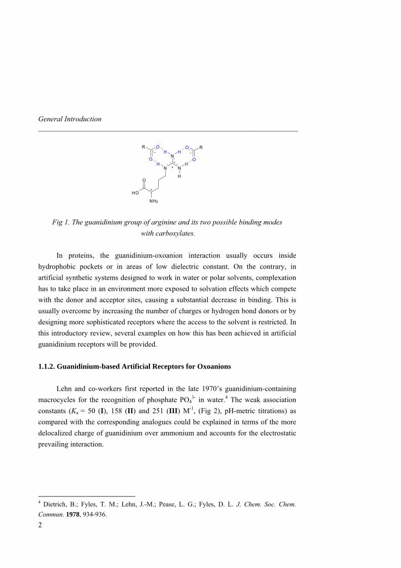
The capacity of the guanidinium group to bind oxoanions is due to its geometrical Y-shaped, planar orientation, which directs the hydrogen bonding, and to its high pKa value (around 12-13),3 which ensures protonation over a wide pH range. The positive charge is delocalized over the three nitrogen atoms, and four out of the five hydrogen bond donors present in the guanidinium group of arginine can complement bidentate oxoanion acceptors, along the two edges available (Fig 1). This accounts for the geometrical versatility of the binding modes. From the energy point of view, binding to oxoanions results from both ion-pairing and hydrogen bonding, and this turns out to be a difficult challenge in highly polar solvents or in water. In fact, the binding energy arises from the difference of the energy released by the host-guest interactions and the energy penalty necessary to remove the solvation shell around the host, which is quite high in water.
1 Schug, K. A.; Lindner. W. Chem. Rev. 2005, 105, 67-113. 2 Best, M. D.; Tobey, S. L.; Anslyn, E. V. Coord. Chem. Rev. 2003, 240, 3-15. 3 The pKa value of the guanidinium moiety may vary depending on adjacent group effects; in general it is lowered by acyl ≥ phenyl ≥ alkyl direct substitution (see ref. 1).
General Introduction ______________________________________________________________________
N
H
N
N
HH
H H
NH2
HO
O
*
O
O
O
O RR
Fig 1. The guanidinium group of arginine and its two possible binding modes with carboxylates.
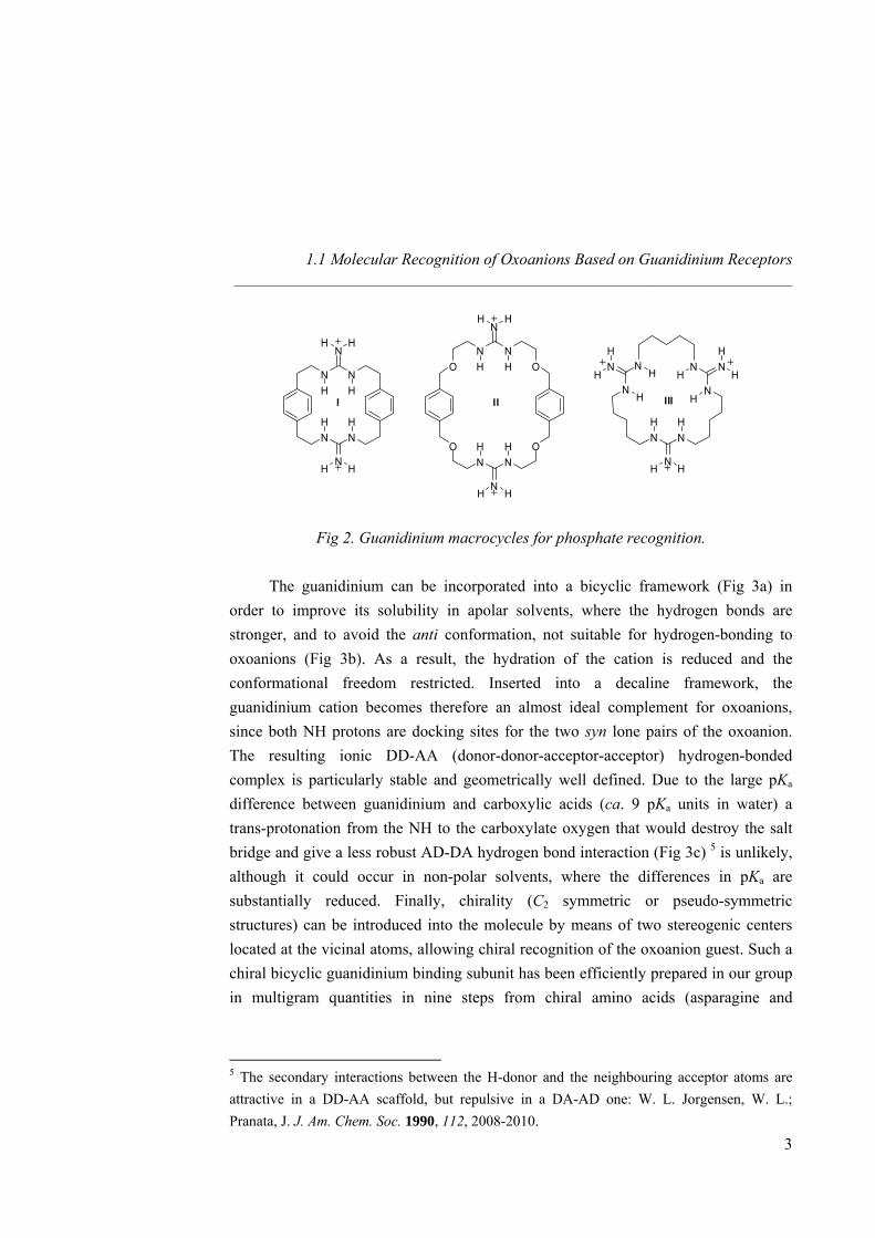
In proteins, the guanidinium-oxoanion interaction usually occurs inside hydrophobic pockets or in areas of low dielectric constant. On the contrary, in artificial synthetic systems designed to work in water or polar solvents, complexation has to take place in an environment more exposed to solvation effects which compete with the donor and acceptor sites, causing a substantial decrease in binding. This is usually overcome by increasing the number of charges or hydrogen bond donors or by designing more sophisticated receptors where the access to the solvent is restricted. In this introductory review, several examples on how this has been achieved in artificial guanidinium receptors will be provided. 1.1.2. Guanidinium-based Artificial Receptors for Oxoanions Lehn and co-workers first reported in the late 1970’s guanidinium-containing macrocycles for the recognition of phosphate PO4
3- in water.4 The weak association constants (Ka = 50 (I), 158 (II) and 251 (III) M-1, (Fig 2), pH-metric titrations) as compared with the corresponding analogues could be explained in terms of the more delocalized charge of guanidinium over ammonium and accounts for the electrostatic prevailing interaction.
4 Dietrich, B.; Fyles, T. M.; Lehn, J.-M.; Pease, L. G.; Fyles, D. L. J. Chem. Soc. Chem. Commun. 1978, 934-936.
2
1.1 Molecular Recognition of Oxoanions Based on Guanidinium Receptors _____________________________________________________________________
N NH H
N
N NH H
N
N NH H
N
O O
N NH H
N
O ON NH H
N
HN
HN
NNN NH H
HH
H H
H
H
H
H
HH
HH
H H
I II III
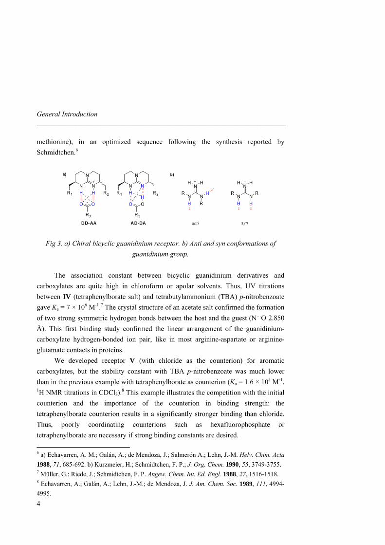
Fig 2. Guanidinium macrocycles for phosphate recognition. The guanidinium can be incorporated into a bicyclic framework (Fig 3a) in order to improve its solubility in apolar solvents, where the hydrogen bonds are stronger, and to avoid the anti conformation, not suitable for hydrogen-bonding to oxoanions (Fig 3b). As a result, the hydration of the cation is reduced and the conformational freedom restricted. Inserted into a decaline framework, the guanidinium cation becomes therefore an almost ideal complement for oxoanions, since both NH protons are docking sites for the two syn lone pairs of the oxoanion. The resulting ionic DD-AA (donor-donor-acceptor-acceptor) hydrogen-bonded complex is particularly stable and geometrically well defined. Due to the large pKa difference between guanidinium and carboxylic acids (ca. 9 pKa units in water) a trans-protonation from the NH to the carboxylate oxygen that would destroy the salt bridge and give a less robust AD-DA hydrogen bond interaction (Fig 3c) 5 is unlikely, although it could occur in non-polar solvents, where the differences in pKa are substantially reduced. Finally, chirality (C2 symmetric or pseudo-symmetric structures) can be introduced into the molecule by means of two stereogenic centers located at the vicinal atoms, allowing chiral recognition of the oxoanion guest. Such a chiral bicyclic guanidinium binding subunit has been efficiently prepared in our group in multigram quantities in nine steps from chiral amino acids (asparagine and
5 The secondary interactions between the H-donor and the neighbouring acceptor atoms are attractive in a DD-AA scaffold, but repulsive in a DA-AD one: W. L. Jorgensen, W. L.; Pranata, J. J. Am. Chem. Soc. 1990, 112, 2008-2010.
3
General Introduction ______________________________________________________________________
methionine), in an optimized sequence following the synthesis reported by Schmidtchen.6
N
N
NH HR1 R2
OO
R3
N NRR
H H
N
N NHR
H R
NH H H H
b)a)
N
N
NHR1 R2
OO
R3
DD-AA
H
AD-DA ant i syn
Fig 3. a) Chiral bicyclic guanidinium receptor. b) Anti and syn conformations of guanidinium group.
The association constant between bicyclic guanidinium derivatives and carboxylates are quite high in chloroform or apolar solvents. Thus, UV titrations between IV (tetraphenylborate salt) and tetrabutylammonium (TBA) p-nitrobenzoate gave Ka = 7 × 10⁶ M-1.7 The crystal structure of an acetate salt confirmed the formation of two strong symmetric hydrogen bonds between the host and the guest (N…O 2.850 Å). This first binding study confirmed the linear arrangement of the guanidinium-carboxylate hydrogen-bonded ion pair, like in most arginine-aspartate or arginine-glutamate contacts in proteins. We developed receptor V (with chloride as the counterion) for aromatic carboxylates, but the stability constant with TBA p-nitrobenzoate was much lower than in the previous example with tetraphenylborate as counterion (Ka = 1.6 × 10³ M-1, 1H NMR titrations in CDCl3).8 This example illustrates the competition with the initial counterion and the importance of the counterion in binding strength: the tetraphenylborate counterion results in a significantly stronger binding than chloride. Thus, poorly coordinating counterions such as hexafluorophosphate or tetraphenylborate are necessary if strong binding constants are desired.
6 a) Echavarren, A. M.; Galán, A.; de Mendoza, J.; Salmerón A.; Lehn, J.-M. Helv. Chim. Acta 1988, 71, 685-692. b) Kurzmeier, H.; Schmidtchen, F. P.; J. Org. Chem. 1990, 55, 3749-3755. 7 Müller, G.; Riede, J.; Schmidtchen, F. P. Angew. Chem. Int. Ed. Engl. 1988, 27, 1516-1518. 8 Echavarren, A.; Galán, A.; Lehn, J.-M.; de Mendoza, J. J. Am. Chem. Soc. 1989, 111, 4994-4995.
4
1.1 Molecular Recognition of Oxoanions Based on Guanidinium Receptors _____________________________________________________________________
N
N
NH H
N
N
NH H
ClO OO O
BPh4
O
N
N
N
HH
H H
HO
NH
N
NHH
H
H
OP
O OO
PhPh
N
N
NH H
BrOTBDPS
IV V
TBDMSO
VII
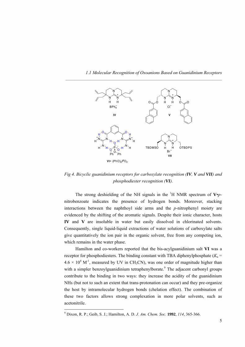
VI (PhO)2PO2 Fig 4. Bicyclic guanidinium receptors for carboxylate recognition (IV, V and VII) and
phosphodiester recognition (VI).
The strong deshielding of the NH signals in the 1H NMR spectrum of V•p-nitrobenzoate indicates the presence of hydrogen bonds. Moreover, stacking interactions between the naphthoyl side arms and the p-nitrophenyl moiety are evidenced by the shifting of the aromatic signals. Despite their ionic character, hosts IV and V are insoluble in water but easily dissolved in chlorinated solvents. Consequently, single liquid-liquid extractions of water solutions of carboxylate salts give quantitatively the ion pair in the organic solvent, free from any competing ion, which remains in the water phase. Hamilton and co-workers reported that the bis-acylguanidinium salt VI was a receptor for phosphodiesters. The binding constant with TBA diphenylphosphate (Ka = 4.6 × 10⁴ M-1, measured by UV in CH3CN), was one order of magnitude higher than with a simpler benzoylguanidinium tetraphenylborate.9 The adjacent carbonyl groups contribute to the binding in two ways: they increase the acidity of the guanidinium NHs (but not to such an extent that trans-protonation can occur) and they pre-organize the host by intramolecular hydrogen bonds (chelation effect). The combination of these two factors allows strong complexation in more polar solvents, such as acetonitrile. 9 Dixon, R. P.; Geib, S. J.; Hamilton, A. D. J. Am. Chem. Soc. 1992, 114, 365-366.
5
General Introduction ______________________________________________________________________
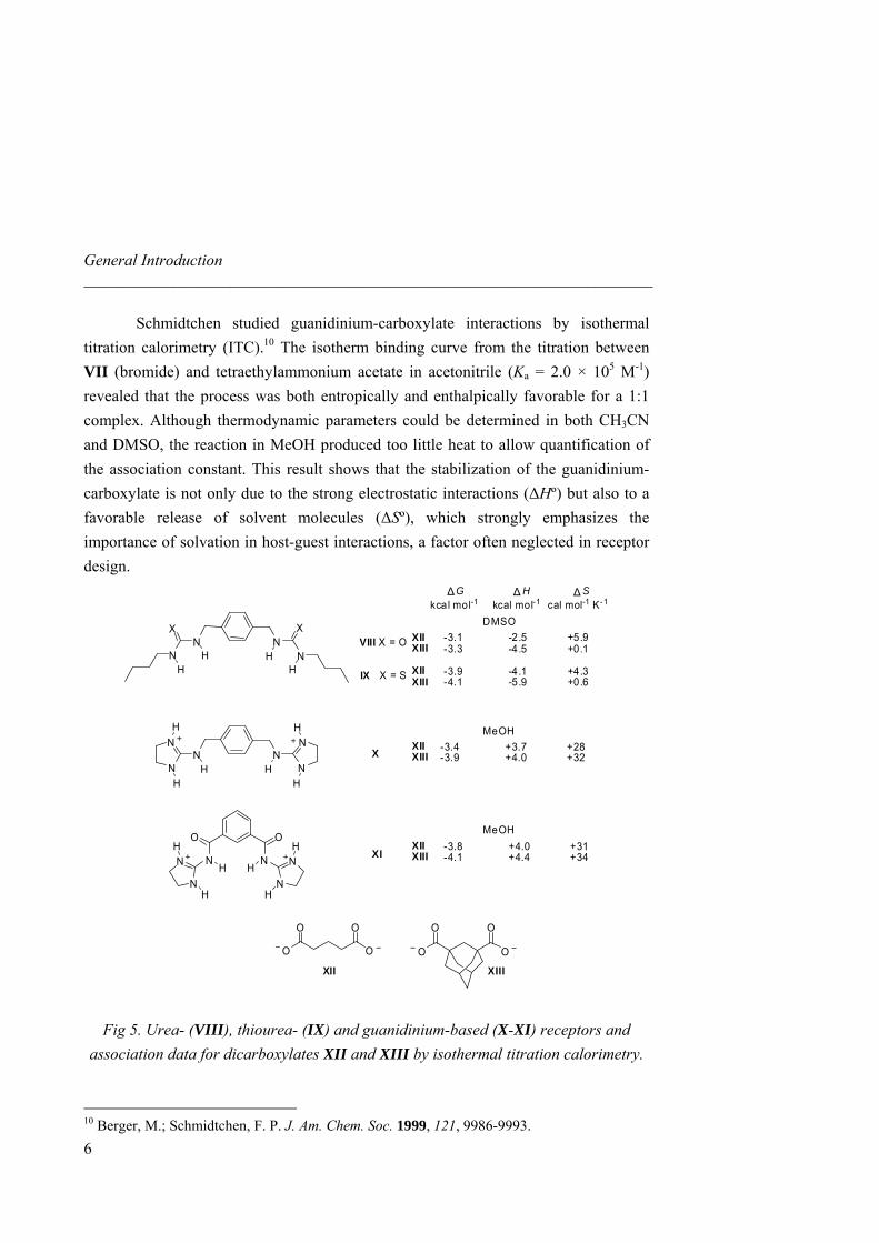
Schmidtchen studied guanidinium-carboxylate interactions by isothermal titration calorimetry (ITC).10 The isotherm binding curve from the titration between VII (bromide) and tetraethylammonium acetate in acetonitrile (Ka = 2.0 × 10⁵ M-1) revealed that the process was both entropically and enthalpically favorable for a 1:1 complex. Although thermodynamic parameters could be determined in both CH3CN and DMSO, the reaction in MeOH produced too little heat to allow quantification of the association constant. This result shows that the stabilization of the guanidinium-carboxylate is not only due to the strong electrostatic interactions (∆Hº) but also to a favorable release of solvent molecules (∆Sº), which strongly emphasizes the importance of solvation in host-guest interactions, a factor often neglected in receptor design.
∆ ∆ ∆
O
NH
O
NH
VIII X = O
N
N N
N
H H
H H
IX X = S
NN
NH
X
N
X
HH
H
N NHH
N
NH
H
N
N
H
H
O O
OO
O O
OO
G H
DMSOXII -3.1 -2.5 +5.9XIII -3.3 -4.5 +0.1
XII -3.9 -4.1 +4.3XIII -4.1 -5.9 +0.6
XII -3.4 +3.7 +28XIII -3.9 +4.0 +32
MeOH
XII -3.8 +4.0 +31XIII -4.1 +4.4 +34
MeOH
kcal mol-1 kcal mol-1 cal mol-1 K-1
X
XI
XII XIII
S
Fig 5. Urea- (VIII), thiourea- (IX) and guanidinium-based (X-XI) receptors and association data for dicarboxylates XII and XIII by isothermal titration calorimetry.
10 Berger, M.; Schmidtchen, F. P. J. Am. Chem. Soc. 1999, 121, 9986-9993.
6
1.1 Molecular Recognition of Oxoanions Based on Guanidinium Receptors _____________________________________________________________________
The thermodynamic aspects of dicarboxylate recognition by artificial receptors with increasingly acidic hydrogen bond donor groups such as two ureas (VIII), thioureas (IX), or guanidiniums (X and XI) in polar solvents (from DMSO to water) were studied by Hamilton (Fig 5).11
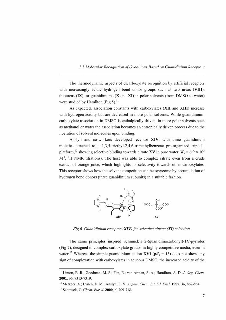
As expected, association constants with carboxylates (XII and XIII) increase with hydrogen acidity but are decreased in more polar solvents. While guanidinium-carboxylate association in DMSO is enthalpically driven, in more polar solvents such as methanol or water the association becomes an entropically driven process due to the liberation of solvent molecules upon binding. Anslyn and co-workers developed receptor XIV, with three guanidinium moieties attached to a 1,3,5-triethyl-2,4,6-trimethylbenzene pre-organized tripodal platform,12 showing selective binding towards citrate XV in pure water (Ka = 6.9 × 10³ M-1, 1H NMR titrations). The host was able to complex citrate even from a crude extract of orange juice, which highlights its selectivity towards other carboxylates. This receptor shows how the solvent competition can be overcome by accumulation of hydrogen bond donors (three guanidinium subunits) in a suitable fashion.
N
NHN
N
H
H
H N
NH
H
NN
N
H
HH COOOOC
OH
COO
XIV XV
Fig 6. Guanidinium receptor (XIV) for selective citrate (XI) selection.
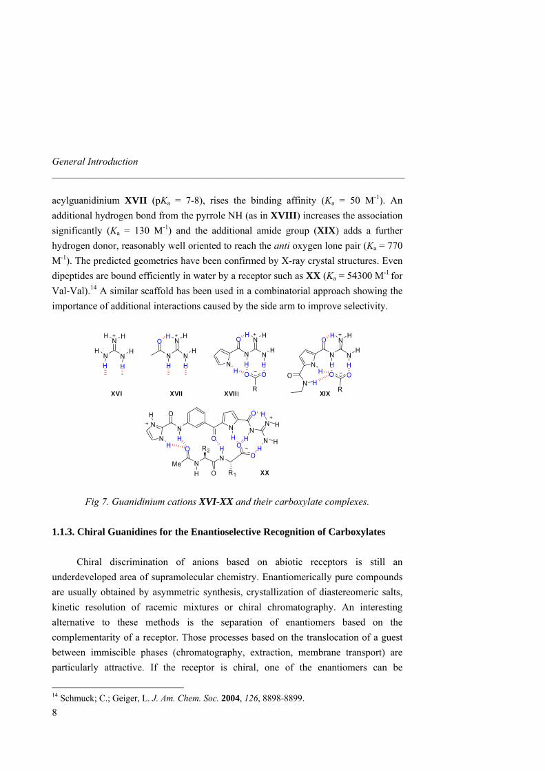
The same principles inspired Schmuck’s 2-(guanidiniocarbonyl)-1H-pyrroles (Fig 7), designed to complex carboxylate groups in highly competitive media, even in water.13 Whereas the simple guanidinium cation XVI (pKa = 13) does not show any sign of complexation with carboxylates in aqueous DMSO, the increased acidity of the
11 Linton, B. R.; Goodman, M. S.; Fan, E.; van Arman, S. A.; Hamilton, A. D. J. Org. Chem. 2001, 66, 7313-7319. 12 Metzger, A.; Lynch, V. M.; Anslyn, E. V. Angew. Chem. Int. Ed. Engl. 1997, 36, 862-864. 13 Schmuck, C. Chem. Eur. J. 2000, 6, 709-718..
7
General Introduction ______________________________________________________________________
acylguanidinium XVII (pKa = 7-8), rises the binding affinity (Ka = 50 M-1). An additional hydrogen bond from the pyrrole NH (as in XVIII) increases the association significantly (Ka = 130 M-1) and the additional amide group (XIX) adds a further hydrogen donor, reasonably well oriented to reach the anti oxygen lone pair (Ka = 770 M-1). The predicted geometries have been confirmed by X-ray crystal structures. Even dipeptides are bound efficiently in water by a receptor such as XX (Ka = 54300 M-1 for Val-Val).14 A similar scaffold has been used in a combinatorial approach showing the importance of additional interactions caused by the side arm to improve selectivity.
NN
O
N
N
N
O
N
N
N N
N
NN
O
N
N
NO
XVI XVII XVIII XIX
H H HH H H H H
HH
H
H
H
H
H
H H
H
HO O
R
H
HHH
HO O
R
ONH
ON
NN N
O
H N
N
H
H
H HH
HH
OON
H
R1O
R2
NH
O
MeXX
Fig 7. Guanidinium cations XVI-XX and their carboxylate complexes.
1.1.3. Chiral Guanidines for the Enantioselective Recognition of Carboxylates Chiral discrimination of anions based on abiotic receptors is still an underdeveloped area of supramolecular chemistry. Enantiomerically pure compounds are usually obtained by asymmetric synthesis, crystallization of diastereomeric salts, kinetic resolution of racemic mixtures or chiral chromatography. An interesting alternative to these methods is the separation of enantiomers based on the complementarity of a receptor. Those processes based on the translocation of a guest between immiscible phases (chromatography, extraction, membrane transport) are particularly attractive. If the receptor is chiral, one of the enantiomers can be
14 Schmuck; C.; Geiger, L. J. Am. Chem. Soc. 2004, 126, 8898-8899.
8
1.1 Molecular Recognition of Oxoanions Based on Guanidinium Receptors _____________________________________________________________________
complexed preferentially and a kinetic resolution could be achieved. Moreover, the process needs only a catalytic amount of receptor since it can transfer several substrate molecules across the phases, without being removed from its own (stationary or liquid) phase. In this context, a useful concept, developed for chiral chromatography, is the three-point binding rule, which states that a minimum of three simultaneous interactions between the chiral stationary phase and for instance one of the enantiomers are necessary to achieve enantioselection, with at least one of these interactions being stereochemically dependent.15 For anions, receptors containing ammonium groups, amides, ureas, thioureas and guanidinium subunits, as well as porphyrins, saphyrins, or metal-containing ligands have been employed. Only chiral guanidines aimed at the discrimination of the enantiomers of amino acids will be reviewed here.
N
N
NH H OX
OO
OO
N
O
OXXI X = O-2-naphthoyl
XXII X = OSitBuPh2
XXIII X = OSiiPr3
XXIV X = OH
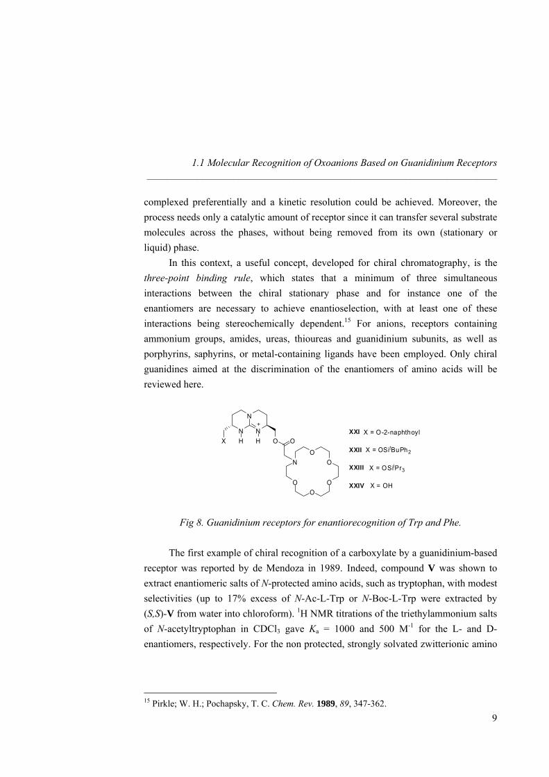
Fig 8. Guanidinium receptors for enantiorecognition of Trp and Phe.
The first example of chiral recognition of a carboxylate by a guanidinium-based receptor was reported by de Mendoza in 1989. Indeed, compound V was shown to extract enantiomeric salts of N-protected amino acids, such as tryptophan, with modest selectivities (up to 17% excess of N-Ac-L-Trp or N-Boc-L-Trp were extracted by (S,S)-V from water into chloroform). 1H NMR titrations of the triethylammonium salts of N-acetyltryptophan in CDCl3 gave Ka = 1000 and 500 M-1 for the L- and D-enantiomers, respectively. For the non protected, strongly solvated zwitterionic amino
15 Pirkle; W. H.; Pochapsky, T. C. Chem. Rev. 1989, 89, 347-362.
9
General Introduction ______________________________________________________________________
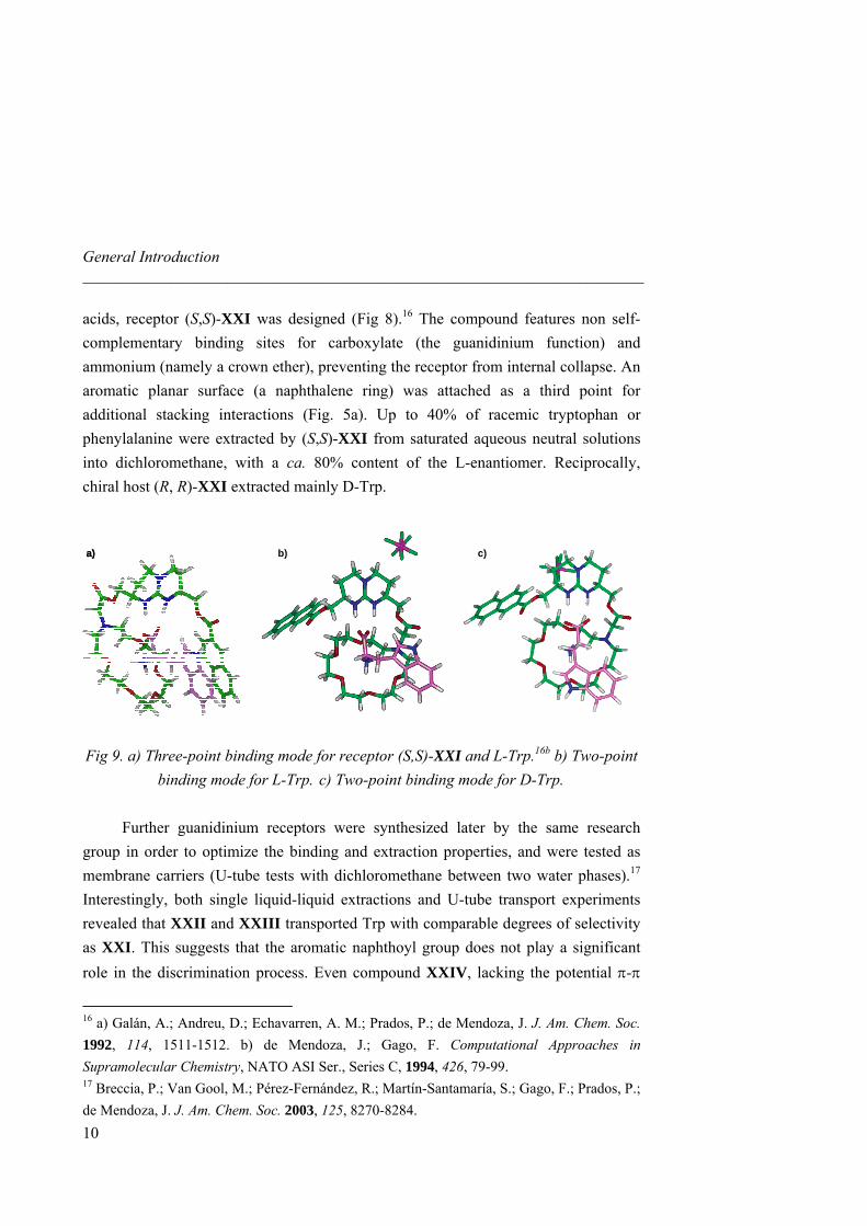
acids, receptor (S,S)-XXI was designed (Fig 8).16 The compound features non self-complementary binding sites for carboxylate (the guanidinium function) and ammonium (namely a crown ether), preventing the receptor from internal collapse. An aromatic planar surface (a naphthalene ring) was attached as a third point for additional stacking interactions (Fig. 5a). Up to 40% of racemic tryptophan or phenylalanine were extracted by (S,S)-XXI from saturated aqueous neutral solutions into dichloromethane, with a ca. 80% content of the L-enantiomer. Reciprocally, chiral host (R, R)-XXI extracted mainly D-Trp.
a) b) c)a) b) c)
Fig 9. a) Three-point binding mode for receptor (S,S)-XXI and L-Trp.16b b) Two-point
binding mode for L-Trp. c) Two-point binding mode for D-Trp. Further guanidinium receptors were synthesized later by the same research group in order to optimize the binding and extraction properties, and were tested as membrane carriers (U-tube tests with dichloromethane between two water phases).17 Interestingly, both single liquid-liquid extractions and U-tube transport experiments revealed that XXII and XXIII transported Trp with comparable degrees of selectivity as XXI. This suggests that the aromatic naphthoyl group does not play a significant role in the discrimination process. Even compound XXIV, lacking the potential π-π
16 a) Galán, A.; Andreu, D.; Echavarren, A. M.; Prados, P.; de Mendoza, J. J. Am. Chem. Soc. 1992, 114, 1511-1512. b) de Mendoza, J.; Gago, F. Computational Approaches in Supramolecular Chemistry, NATO ASI Ser., Series C, 1994, 426, 79-99. 17 Breccia, P.; Van Gool, M.; Pérez-Fernández, R.; Martín-Santamaría, S.; Gago, F.; Prados, P.; de Mendoza, J. J. Am. Chem. Soc. 2003, 125, 8270-8284.
10
1.1 Molecular Recognition of Oxoanions Based on Guanidinium Receptors _____________________________________________________________________
interaction, was enantioselective, although to a lesser extent. Another binding mode was then proposed, without participation of the naphthoyl arm, as the outcome of molecular dynamics calculations with explicit solvent molecules. In this model, binding of D-Trp exposes a highly polar area of the receptor (around the crown ether nitrogen) to the apolar solvent, causing the overall energy to increase (Fig 9b,c).17
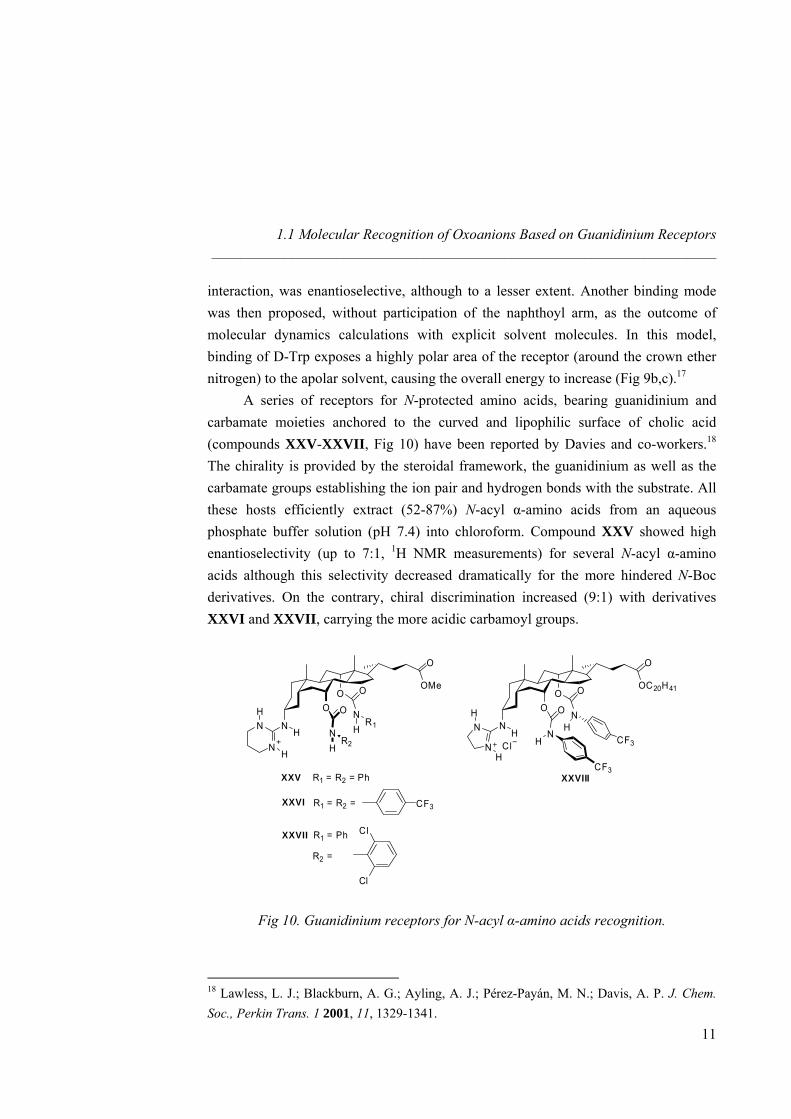
A series of receptors for N-protected amino acids, bearing guanidinium and carbamate moieties anchored to the curved and lipophilic surface of cholic acid (compounds XXV-XXVII, Fig 10) have been reported by Davies and co-workers.18 The chirality is provided by the steroidal framework, the guanidinium as well as the carbamate groups establishing the ion pair and hydrogen bonds with the substrate. All these hosts efficiently extract (52-87%) N-acyl α-amino acids from an aqueous phosphate buffer solution (pH 7.4) into chloroform. Compound XXV showed high enantioselectivity (up to 7:1, 1H NMR measurements) for several N-acyl α-amino acids although this selectivity decreased dramatically for the more hindered N-Boc derivatives. On the contrary, chiral discrimination increased (9:1) with derivatives XXVI and XXVII, carrying the more acidic carbamoyl groups.
OMe
OO
NH
O
N
N
O
XXV R1 = R2 = Ph
N
N
H
H
H
H
XXVI R1 = R2 = CF3
XXVII R1 = Ph
R2 =
Cl
Cl
O
R1
R2
OC20H41
OO
NHN
N
H
H
O
NH
CF3
NH
O
CF3Cl
O
XXVIII
Fig 10. Guanidinium receptors for N-acyl α-amino acids recognition.
18 Lawless, L. J.; Blackburn, A. G.; Ayling, A. J.; Pérez-Payán, M. N.; Davis, A. P. J. Chem. Soc., Perkin Trans. 1 2001, 11, 1329-1341.
11
General Introduction ______________________________________________________________________
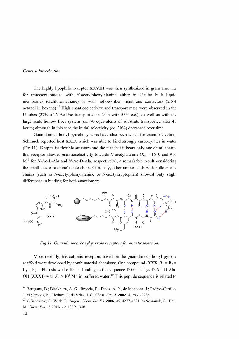
The highly lipophilic receptor XXVIII was then synthesized in gram amounts for transport studies with N-acetylphenylalanine either in U-tube bulk liquid membranes (dichloromethane) or with hollow-fiber membrane contactors (2.5% octanol in hexane).19 High enantioselectivity and transport rates were observed in the U-tubes (27% of N-Ac-Phe transported in 24 h with 56% e.e.), as well as with the large scale hollow fiber system (ca. 70 equivalents of substrate transported after 48 hours) although in this case the initial selectivity (ca. 30%) decreased over time. Guanidiniocarbonyl pyrrole systems have also been tested for enantioselection. Schmuck reported host XXIX which was able to bind strongly carboxylates in water (Fig 11). Despite its flexible structure and the fact that it bears only one chiral centre, this receptor showed enantioselectivity towards N-acetylalanine (Ka = 1610 and 910 M-1 for N-Ac-L-Ala and N-Ac-D-Ala, respectively), a remarkable result considering the small size of alanine’s side chain. Curiously, other amino acids with bulkier side chains (such as N-acetylphenylalanine or N-acetyltryptophan) showed only slight differences in binding for both enantiomers.
NR3 O
NN
R1 O
NN
O
HN
O
N
HN
O
NO
R2 O O
NH
N
NH
H
H
HH
H
H
dansyl
OOH HNH
NH
O
NH2
N
NHO
HN2OC
XXIX
Pr i
H H
H3N
O2C
H
XXX
XXXI
Fig 11. Guanidiniocarbonyl pyrrole receptors for enantioselection.
More recently, tris-cationic receptors based on the guanidiniocarbonyl pyrrole
scaffold were developed by combinatorial chemistry. One compound (XXX, R1 = R2 = Lys; R3 = Phe) showed efficient binding to the sequence D-Glu-L-Lys-D-Ala-D-Ala-OH (XXXI) with Ka > 104 M-1 in buffered water.20 This peptide sequence is related to
19 Baragana, B.; Blackburn, A. G.; Breccia, P.; Davis, A. P.; de Mendoza, J.; Padrón-Carrillo, J. M.; Prados, P.; Riedner, J.; de Vries, J. G. Chem. Eur. J. 2002, 8, 2931-2936. 20 a) Schmuck; C.; Wich, P. Angew. Chem. Int. Ed. 2006, 45, 4277-4281. b) Schmuck, C.; Heil, M. Chem. Eur. J. 2006, 12, 1339-1348.
12
1.1 Molecular Recognition of Oxoanions Based on Guanidinium Receptors _____________________________________________________________________
13
1.1.4. Phosphate and Sulfate Recognition
In addition to carboxylates or phosphodiesters, other oxoanions such as
.1.4.1. Phosphates
Anslyn and co-workers showed that metallo-receptor XXXII displayed a
the bacterial peptidoglycan that is recognized by the vancomycin family of antibiotics, preventing formation of the cell wall.
phosphate, sulfate and nitrate are biologically relevant21 and chemically challenging to recognize, due to their weak basicity. At neutral pH, phosphate (as HPO4
2-) and sulfate present a tetrahedral binding mode with two negative charges, although nitrate has a trigonal planar binding motif with just one negative charge. Thus, for a neutral complex, two guanidines are required for phosphate and sulfate whereas only one is needed for nitrate, in addition to other hydrogen bond donor atoms. The design of suitable linkers between the hydrogen donors with optimal orientation and maximum participation of host’s lone pairs represents a major issue in the field. 1 selective binding for monoprotonated phosphate (HPO4
2-) and arsenate (HAsO42-) over
other anions (such as AcO-, NO3-, HCO3
- or Cl-) at biological pH (Ka = 104 M-1 in 98:2 water/methanol, UV/vis and ITC titrations).22 Hosts bearing only the Cu(II) centre were less effective (Ka = 102 M-1), highlighting the role of the cavity and the presence of the guanidinium groups. Thermodynamic data showed that association of HPO4
2- with guanidinium derivative XXXII was both enthalpically and entropically driven, whereas complexation with an ammonium analogue was mainly governed by entropy. The different mode of binding was rationalized in terms of the different solvation energies of both binding groups.
21 a) Jacobson, B. L.; Quiocho, F. A. J. Mol. Biol. 1988, 204, 783-787. b) Luecke, H.; Quiocho, F. A. Nature 1990, 347, 402-406. 22 Tobey; S. L.; Anslyn, E. V. J. Am. Chem. Soc. 2003, 125, 14807-14815.
General Introduction ______________________________________________________________________
NN
N
N
NN
Cu2+
NN HH NH NN H
NNH
H
H
FeN N
N
NH2N
H2N
H H
HH
HH
H
XXXII XXXIII XXXIV
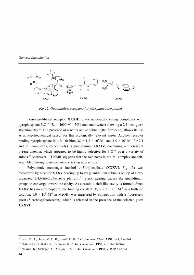
Fig 12. Guanidinium receptors for phosphate recognition.
Ferrocenyl-based receptor XXXIII ves moderately strong complexes with
te (XXXVI, Fig 13) was
gi
pyrophosphate P2O74- (Ka = 4600 M-2, 50% methanol-water) showing a 2:1 host-guest
stoichiometry.23 The presence of a redox active subunit (the ferrocene) allows its use as an electrochemical sensor for this biologically relevant anion. Another receptor binding pyrophosphate in a 2:1 fashion (Ka = 1.2 × 10⁸ M-2 and 1.0 × 10⁴ M-1 for 2:1 and 1:1 complexes, respectively) is guanidinium XXXIV, containing a fluorescent pyrene antenna, which appeared to be highly selective for P2O7
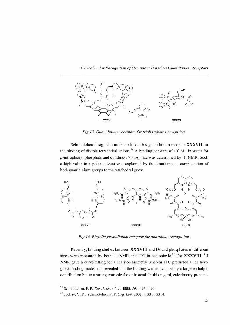
4- over a variety of anions.24 Moreover, 1H NMR suggests that the two hosts in the 2:1 complex are self-assembled through pyrene-pyrene stacking interactions. Polyanionic messenger inositol-1,4,5-triphospharecognized by receptor XXXV bearing up to six guanidinium subunits on top of a pre-organized 2,4,6-triethylbenzene platform.25 Steric gearing causes the guanidinium groups to converge toward the cavity. As a result, a cleft-like cavity is formed. Since XXXV has no chromophore, the binding constant (Ka = 2.2 × 10⁴ M-1 in a buffered solution, 1.0 × 10⁸ M-1 in MeOH) was measured by competition with a fluorescent guest (5-carboxyfluorescein), which is released in the presence of the selected guest XXXVI.
23 Beer, P. D.; Drew, M. G. B.; Smith, D. K. J. Organomet. Chem. 1997, 543, 259-261. 24 Nishizawa, S.; Kato, Y.; Teramae, N. J. Am. Chem. Soc. 1999, 121, 9463-9464. 25 Niikura, K.; Metzger, A.; Anslyn, E. V. J. Am. Chem. Soc. 1998, 120, 8533-8534.
14
1.1 Molecular Recognition of Oxoanions Based on Guanidinium Receptors _____________________________________________________________________
N NNH
RRRR
H H
RR
R =NN
NH
HH
OH
HO
O
OH
OO
P
POO
OO
P
OO
O
O
O
XXXV XXXVI
Fig 13. Guanidinium receptors for triphosphate recognition.
Schmidtchen designed a urethane-linked bis-guanidinium receptor XXXVII for the binding of ditopic tetrahedral anions.26 A binding constant of 106 M-1 in water for p-nitrophenyl phosphate and cytidine-5’-phosphate was determined by 1H NMR. Such a high value in a polar solvent was explained by the simultaneous complexation of both guanidinium groups to the tetrahedral guest.
N
NH
NH
NH
C3H7
O
O
NH
OC3H7
O NH
NH I
NN
N
H
H
HO
O
O
HN
NN
N
H
H
OH
O
O
HN
C3H7 C3H7
N
N
NH HO
NH
O N
O
H
O
tBu tBu
NH
O
N
O O
HMe M e
Me Me
HH X
XXXVII XXXVIII XXXIX
Fig 14. Bicyclic guanidinium receptor for phosphate recognition. Recently, binding studies between XXXVIII and IV and phosphates of different sizes were measured by both 1H NMR and ITC in acetonitrile.27 For XXXVIII, 1H NMR gave a curve fitting for a 1:1 stoichiometry whereas ITC predicted a 1:2 host-guest binding model and revealed that the binding was not caused by a large enthalpic contribution but to a strong entropic factor instead. In this regard, calorimetry prevents
26 Schmidtchen, F. P. Tetrahedron Lett. 1989, 30, 4493-4496. 27 Jadhav, V. D.; Schmidtchen, F. P. Org. Lett. 2005, 7, 3311-3314.
15
General Introduction ______________________________________________________________________
16
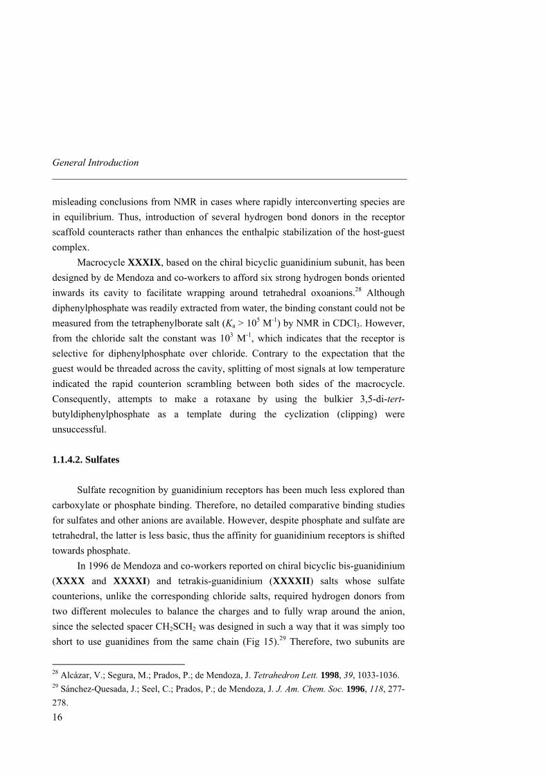
misleading conclusions from NMR in cases where rapidly interconverting species are in equilibrium. Thus, introduction of several hydrogen bond donors in the receptor scaffold counteracts rather than enhances the enthalpic stabilization of the host-guest complex. Macrocycle XXXIX, based on the chiral bicyclic guanidinium subunit, has been designed by de Mendoza and co-workers to afford six strong hydrogen bonds oriented inwards its cavity to facilitate wrapping around tetrahedral oxoanions.28 Although diphenylphosphate was readily extracted from water, the binding constant could not be measured from the tetraphenylborate salt (Ka > 105 M-1) by NMR in CDCl3. However, from the chloride salt the constant was 103 M-1, which indicates that the receptor is selective for diphenylphosphate over chloride. Contrary to the expectation that the guest would be threaded across the cavity, splitting of most signals at low temperature indicated the rapid counterion scrambling between both sides of the macrocycle. Consequently, attempts to make a rotaxane by using the bulkier 3,5-di-tert-butyldiphenylphosphate as a template during the cyclization (clipping) were unsuccessful. 1.1.4.2. Sulfates Sulfate recognition by guanidinium receptors has been much less explored than carboxylate or phosphate binding. Therefore, no detailed comparative binding studies for sulfates and other anions are available. However, despite phosphate and sulfate are tetrahedral, the latter is less basic, thus the affinity for guanidinium receptors is shifted towards phosphate. In 1996 de Mendoza and co-workers reported on chiral bicyclic bis-guanidinium (XXXX and XXXXI) and tetrakis-guanidinium (XXXXII) salts whose sulfate counterions, unlike the corresponding chloride salts, required hydrogen donors from two different molecules to balance the charges and to fully wrap around the anion, since the selected spacer CH2SCH2 was designed in such a way that it was simply too short to use guanidines from the same chain (Fig 15).29 Therefore, two subunits are
28 Alcázar, V.; Segura, M.; Prados, P.; de Mendoza, J. Tetrahedron Lett. 1998, 39, 1033-1036. 29 Sánchez-Quesada, J.; Seel, C.; Prados, P.; de Mendoza, J. J. Am. Chem. Soc. 1996, 118, 277-278.
1.1 Molecular Recognition of Oxoanions Based on Guanidinium Receptors _____________________________________________________________________
forced to self-assemble orthogonally around the tetrahedral anion in a double-helical structure (sulfate helicates). 1H NMR spectra showed large downfield shifts of guanidinium NH’s as dimers or tetramers complexed sulfate anion. Moreover, ROESY spectra confirmed intermolecular contacts due to the folded conformation.
a)
N
N
NH H
SPh2BuSitON
N
NH H 2
RN
N
NH H 2
XXXX R = OSitBuPh2
XXXXI R = CH2C6H13O
O
S
S
XXXXII
b)
Fig 15. a) Chiral bicyclic bis-guanidinium salts XXXX and XXXXI and tetrakis-guanidinium salt XXXXII. b) Optimized model of a sulfate helicate from
a strand of (S,S)-guanidines.
A recent computational study concludes that for simple sulphate-guanidinium interactions several minima of similar energies could be found.30 For more complex guanidines, such as the closely related ligands XXXXIII and XXXXIV, the crystal structure shows 1:1 sulfate complexes, with a good docking of the anion into the cavity of XXXXIV, but this was not the case for XXXXIII (Fig 16).31 Interestingly, the pyridine nitrogen of XXXXIII induces pre-organization by intramolecular hydrogen bonding but causes repulsion of the anion due to the increased charge density in the pocket around the heteroatoms.
17
30 Rozas, I.; Kruger, P. K. J. Chem. Theory Comput. 2005, 1, 1055-1062. 31 Grossel, M. C.; Merckel, D. A. S.; Hutchings, M. G. Cryst. Eng. Comm. 2003, 5, 77-81.
General Introduction ______________________________________________________________________
NN
HN
HN
N
N
N
a)
H
H
H
H
HH HH
XXXXIII
NH
NH
N
N
N
N
OOHH
b)
HH
HHHH
XXXXIV
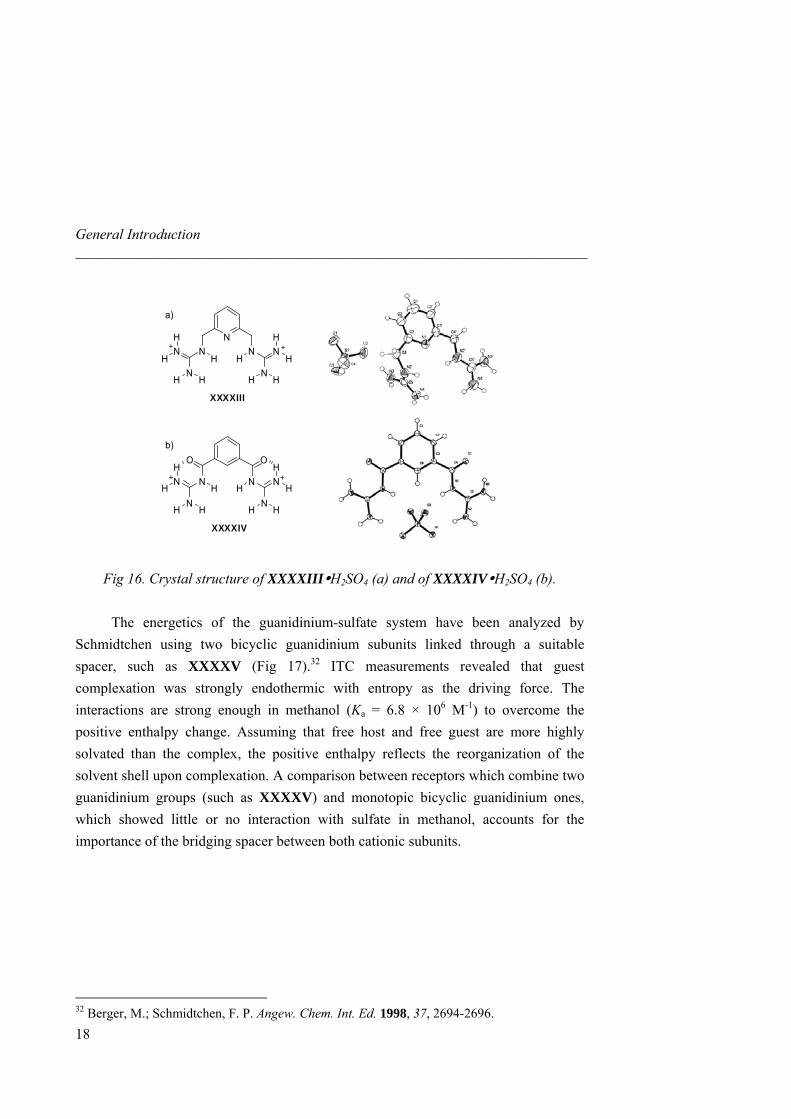
Fig 16. Crystal structure of XXXXIII H2SO4 (a) and of XXXXIV H2SO4 (b).
The energetics of the guanidinium-sulfate system have been analyzed by Schmidtchen using two bicyclic guanidinium subunits linked through a suitable spacer, such as XXXXV (Fig 17).32 ITC measurements revealed that guest complexation was strongly endothermic with entropy as the driving force. The interactions are strong enough in methanol (Ka = 6.8 × 10⁶ M-1) to overcome the positive enthalpy change. Assuming that free host and free guest are more highly solvated than the complex, the positive enthalpy reflects the reorganization of the solvent shell upon complexation. A comparison between receptors which combine two guanidinium groups (such as XXXXV) and monotopic bicyclic guanidinium ones, which showed little or no interaction with sulfate in methanol, accounts for the importance of the bridging spacer between both cationic subunits.
18
32 Berger, M.; Schmidtchen, F. P. Angew. Chem. Int. Ed. 1998, 37, 2694-2696.
1.1 Molecular Recognition of Oxoanions Based on Guanidinium Receptors _____________________________________________________________________
N N
NH
H O
N
OCH2C6H5
Cl-
NN
NH
HO
N
Cl-
HH
OSitBuPh2Ph2BuSitO
XXXXV
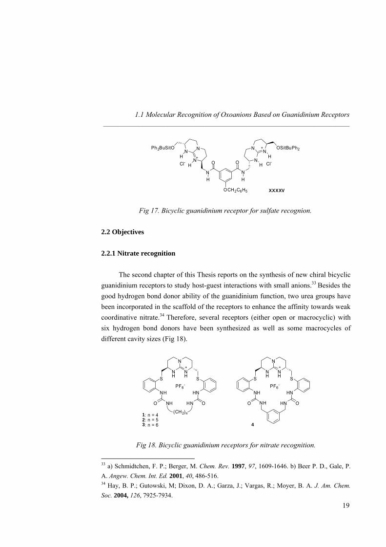
Fig 17. Bicyclic guanidinium receptor for sulfate recognion.
2.2 Objectives 2.2.1 Nitrate recognition The second chapter of this Thesis reports on the synthesis of new chiral bicyclic guanidinium receptors to study host-guest interactions with small anions.33 Besides the good hydrogen bond donor ability of the guanidinium function, two urea groups have been incorporated in the scaffold of the receptors to enhance the affinity towards weak coordinative nitrate.34 Therefore, several receptors (either open or macrocyclic) with six hydrogen bond donors have been synthesized as well as some macrocycles of different cavity sizes (Fig 18).
N
NH
NH SS
PF6-
HNNH
OHNO NH
N
NH
NH SS
PF6-
HNNH
OHNO NH
(CH2)n1: n = 42: n = 53: n = 6 4
Fig 18. Bicyclic guanidinium receptors for nitrate recognition.
33 a) Schmidtchen, F. P.; Berger, M. Chem. Rev. 1997, 97, 1609-1646. b) Beer P. D., Gale, P. A. Angew. Chem. Int. Ed. 2001, 40, 486-516. 34 Hay, B. P.; Gutowski, M; Dixon, D. A.; Garza, J.; Vargas, R.; Moyer, B. A. J. Am. Chem. Soc. 2004, 126, 7925-7934.
19
General Introduction _____________________________________________________________________
20
In summary, 1H-NMR and isothermal calorimetric (ITC) techniques were used to quantify the association between hosts and guests and to determine thermodynamic parameters. Small anions such as nitrate, acetate, chloride and bromide were investigated as well as larger one such as benzoate. Selectivity was established for the different anions under study. Solid state X-ray crystal structures have been resolved for several complexes, showing the inclusion of nitrate, acetate or chloride anions inside the cavity of the macrocyclic guanidinium receptors, and shedding light on the structural parameters governing the binding. 2.2.2 Naproxen Enantiorecognition The third chapter deals with the challenging problem of the chiral recognition of the NSAID (Non-Steroidal Anti-inflammatory Drug) Naproxen.35 To do so, two different strategies have been employed: on the first hand, the rational design for a step to step host-guest interactions assay, on the second hand, the combinatorial approach with the synthesis of amino acid libraries to be screened with a fluorescent Naproxen derivative. Since multiple hydrogen bond donor hosts synthesized in the second chapter revealed as being good receptors for carboxylate, we first checked open-chain, symmetric guanidinium receptors for our target Naproxen. Therefore, chiral receptors with six hydrogen bond donors and bearing bulky group as adamantyl, 2-tert-butyl-6-methylphenyl or diphenylmethyl were synthesized in order to discriminate one enantiomer from the other (Fig 19).
35 Giordano, C.; Villa, M.; Panossian, S. Naproxen: Chirality in Industry, Wiley (New York), 1992.

1.2 Objectives _____________________________________________________________________
N
NH
NH SS
PF6-
HNNH
OHNO NHRR
5 : R =
6 : R =
7 : R =
Fig 19. Symmetric chiral guanidinium receptors bearing bulky.
Moreover, several symmetric hosts bearing butyl or phenyl as well as benzyl or phenylethyl groups have been prepared as a preliminary step in the synthesis of pincer-like receptors. Additional π-π stacking interactions were then investigated with Naproxen (bearing an electronically enriched ring). We also introduced additional chirality in both arms of the host in a view of leading the enantioselective recognition of (S)-Naproxen. Besides, this study was run for both chiral (R, R) and achiral meso (R, S) guanidinium scaffolds in an attempt to select the best pre-organized pocket.
N
NH
NH
SSPF6
-
HNNH
OHNO NHRR
8: R =
9: R =
10: R =
11: R =
12: R =
13: R =
NO2
NO2
Fig 20. Symmetric receptors for pincer-like binding assays. A more rigid and pre-organized macrocycle 14, incorporating a lysine derivative with both additional hydrogen bond and an electronically poor aromatic ring, as a chiral linker between the two urea groups, was also designed and synthesized.
21
General Introduction _____________________________________________________________________
N
NH
NH SS
PF6-
HNNH
OHNO NHO
HN
NO2
14
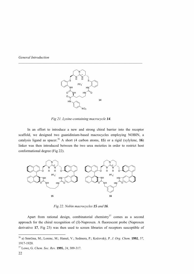
Fig 21. Lysine-containing macrocycle 14.
In an effort to introduce a new and strong chiral barrier into the receptor scaffold, we designed two guanidinium-based macrocycles employing NOBIN, a catalysis ligand as spacer.36 A short (4 carbon atoms, 15) or a rigid (xylylene, 16) linker was then introduced between the two urea moieties in order to restrict host conformational degree (Fig 22).
15 16
N
N
NH H
+
O O
NHPF 6
-
HN
O NH OHN
N
N
NH H
+
O O
NHPF 6
-
HN
O NH OHN
Fig 22. Nobin macrocycles 15 and 16.
Apart from rational design, combinatorial chemistry37 comes as a second approach for the chiral recognition of (S)-Naproxen. A fluorescent probe (Naproxen derivative 17, Fig 23) was then used to screen libraries of receptors susceptible of
36 a) Smrčina, M.; Lorenc, M.; Hanuš, V.; Sedmera, P.; Kočovskỳ, P. J. Org. Chem. 1992, 57, 1917-1920. 37 Lowe, G. Chem. Soc. Rev. 1995, 24, 309-317.
22
1.2 Objectives _____________________________________________________________________
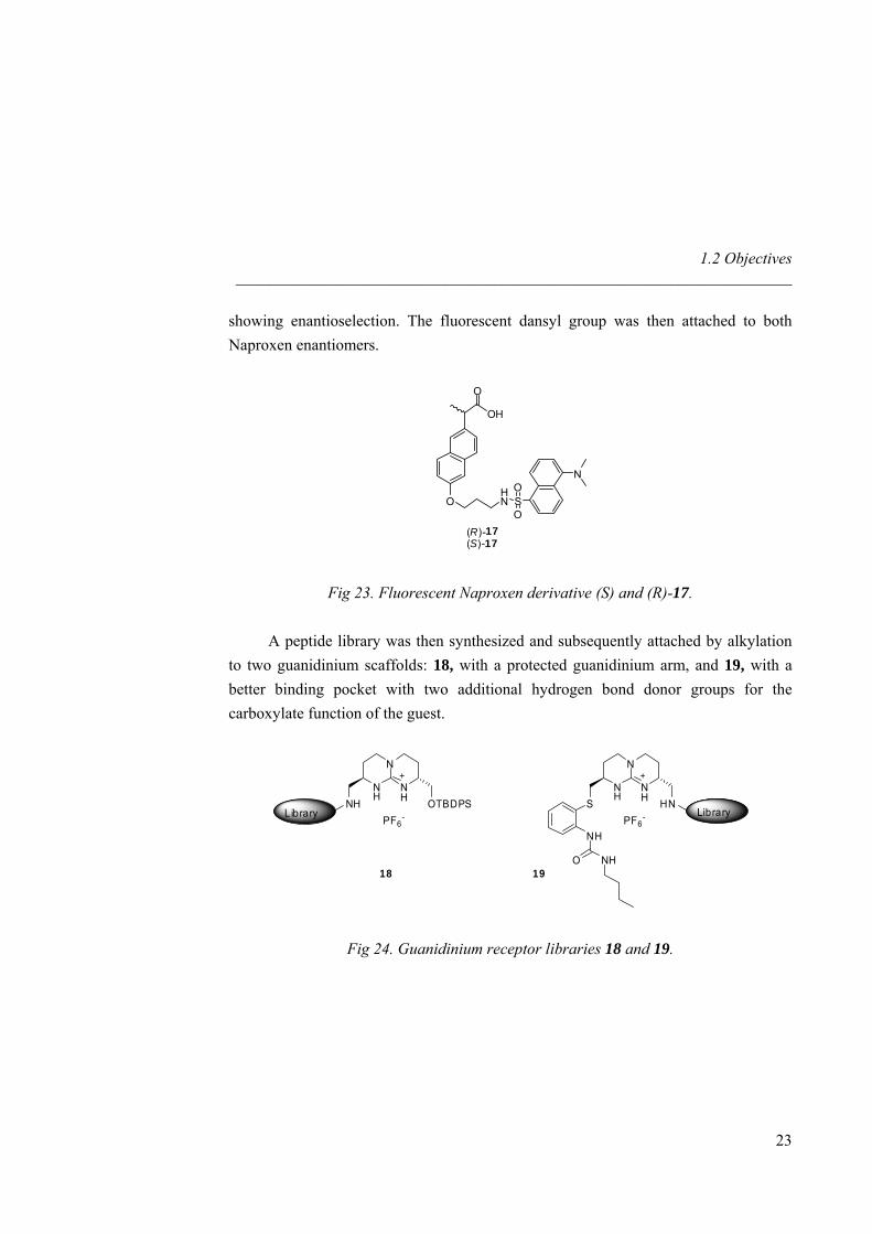
showing enantioselection. The fluorescent dansyl group was then attached to both Naproxen enantiomers.
O
OH
OHN S
O
ON
(R)-17(S)-17
Fig 23. Fluorescent Naproxen derivative (S) and (R)-17.
A peptide library was then synthesized and subsequently attached by alkylation to two guanidinium scaffolds: 18, with a protected guanidinium arm, and 19, with a better binding pocket with two additional hydrogen bond donor groups for the carboxylate function of the guest.
N
NH
NH
OTBDPSPF6
-
18
NH
N
NH
NH
HNPF6
-S
19
NH
O NH
Library Library
Fig 24. Guanidinium receptor libraries 18 and 19.
23
2.1 Introduction _____________________________________________________________________
2 Nitrate Recognition 2.1 Introduction 2.1.1 Abiotic Receptors for Small Anions
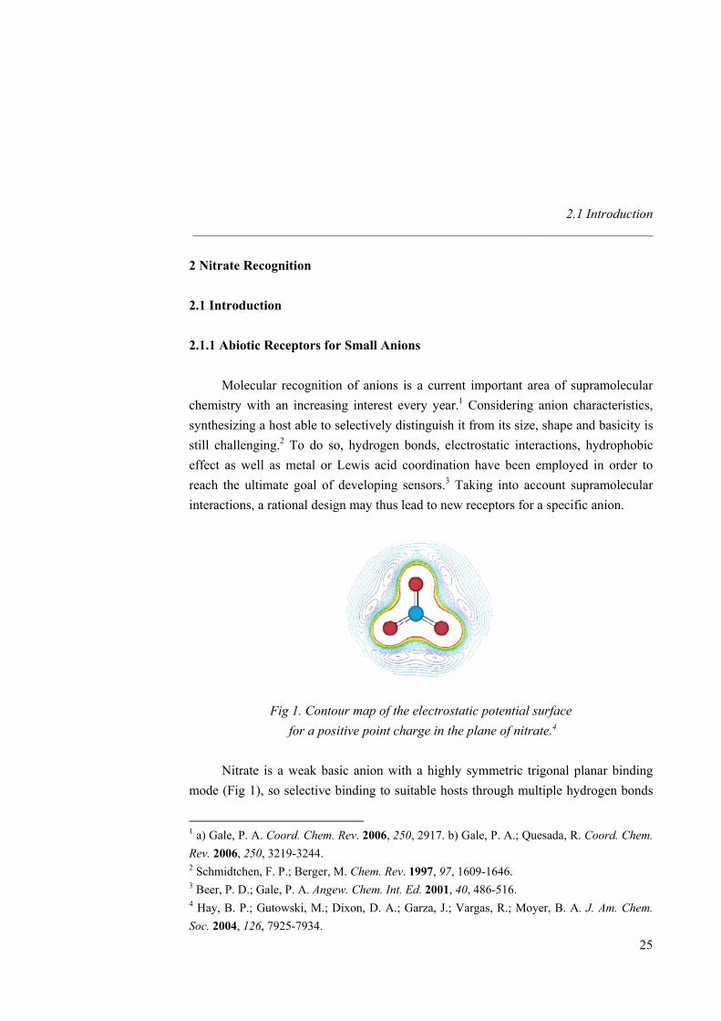
Molecular recognition of anions is a current important area of supramolecular chemistry with an increasing interest every year.1 Considering anion characteristics, synthesizing a host able to selectively distinguish it from its size, shape and basicity is still challenging.2 To do so, hydrogen bonds, electrostatic interactions, hydrophobic effect as well as metal or Lewis acid coordination have been employed in order to reach the ultimate goal of developing sensors.3 Taking into account supramolecular interactions, a rational design may thus lead to new receptors for a specific anion.
Fig 1. Contour map of the electrostatic potential surface for a positive point charge in the plane of nitrate.4
Nitrate is a weak basic anion with a highly symmetric trigonal planar binding
mode (Fig 1), so selective binding to suitable hosts through multiple hydrogen bonds
1 a) Gale, P. A. Coord. Chem. Rev. 2006, 250, 2917. b) Gale, P. A.; Quesada, R. Coord. Chem. Rev. 2006, 250, 3219-3244. 2 Schmidtchen, F. P.; Berger, M. Chem. Rev. 1997, 97, 1609-1646. 3 Beer, P. D.; Gale, P. A. Angew. Chem. Int. Ed. 2001, 40, 486-516. 4 Hay, B. P.; Gutowski, M.; Dixon, D. A.; Garza, J.; Vargas, R.; Moyer, B. A. J. Am. Chem. Soc. 2004, 126, 7925-7934.
25
Nitrate Recognition _____________________________________________________________________
26
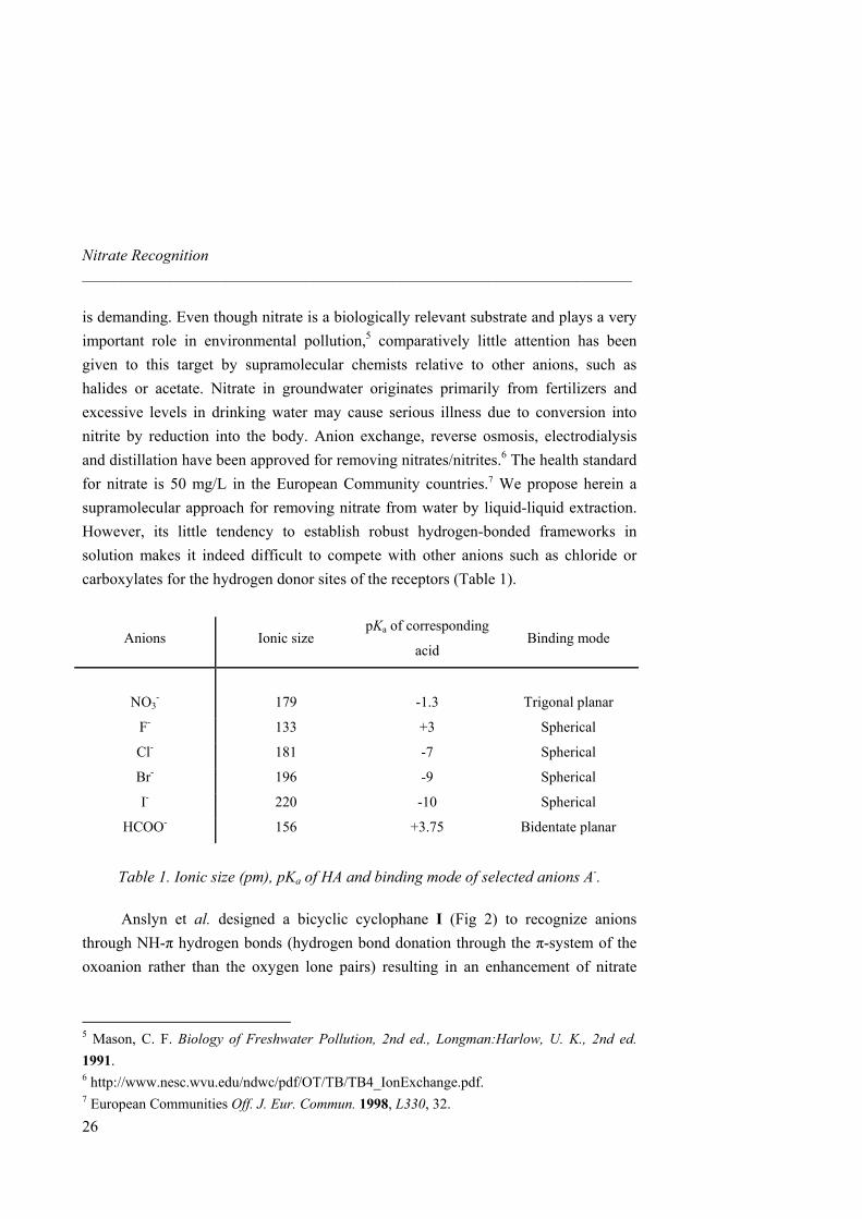
is demanding. Even though nitrate is a biologically relevant substrate and plays a very important role in environmental pollution,5 comparatively little attention has been given to this target by supramolecular chemists relative to other anions, such as halides or acetate. Nitrate in groundwater originates primarily from fertilizers and excessive levels in drinking water may cause serious illness due to conversion into nitrite by reduction into the body. Anion exchange, reverse osmosis, electrodialysis and distillation have been approved for removing nitrates/nitrites.6 The health standard for nitrate is 50 mg/L in the European Community countries.7 We propose herein a supramolecular approach for removing nitrate from water by liquid-liquid extraction. However, its little tendency to establish robust hydrogen-bonded frameworks in solution makes it indeed difficult to compete with other anions such as chloride or carboxylates for the hydrogen donor sites of the receptors (Table 1).
Anions Ionic size pKa of corresponding
acid Binding mode
NO3-
179
-1.3
Trigonal planar
F- 133 +3 Spherical
Cl- 181 -7 Spherical
Br- 196 -9 Spherical
I- 220 -10 Spherical
HCOO- 156 +3.75 Bidentate planar
Table 1. Ionic size (pm), pKa of HA and binding mode of selected anions A-.
Anslyn et al. designed a bicyclic cyclophane I (Fig 2) to recognize anions
through NH-π hydrogen bonds (hydrogen bond donation through the π-system of the oxoanion rather than the oxygen lone pairs) resulting in an enhancement of nitrate
5 Mason, C. F. Biology of Freshwater Pollution, 2nd ed., Longman:Harlow, U. K., 2nd ed. 1991. 6 http://www.nesc.wvu.edu/ndwc/pdf/OT/TB/TB4_IonExchange.pdf. 7 European Communities Off. J. Eur. Commun. 1998, L330, 32.
2.1 Introduction _____________________________________________________________________
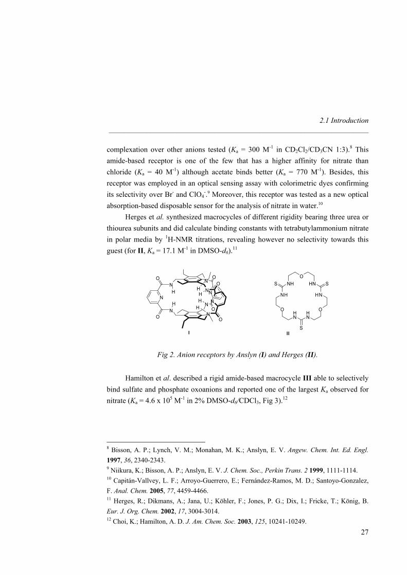
complexation over other anions tested (Ka = 300 M-1 in CD2Cl2/CD3CN 1:3).8 This amide-based receptor is one of the few that has a higher affinity for nitrate than chloride (Ka = 40 M-1) although acetate binds better (Ka = 770 M-1). Besides, this receptor was employed in an optical sensing assay with colorimetric dyes confirming its selectivity over Br- and ClO4
-.9 Moreover, this receptor was tested as a new optical absorption-based disposable sensor for the analysis of nitrate in water.10
Herges et al. synthesized macrocycles of different rigidity bearing three urea or thiourea subunits and did calculate binding constants with tetrabutylammonium nitrate in polar media by 1H-NMR titrations, revealing however no selectivity towards this guest (for II, Ka = 17.1 M-1 in DMSO-d6).11
NN
NN
HN
O
O
H
H H
NH
O
N
ON
O
HN
O
I
OHN
HN
OHN
HN
O
NH
NH SS
SII
Fig 2. Anion receptors by Anslyn (I) and Herges (II).
Hamilton et al. described a rigid amide-based macrocycle III able to selectively bind sulfate and phosphate oxoanions and reported one of the largest Ka observed for nitrate (Ka = 4.6 x 10⁵ M-1 in 2% DMSO-d6/CDCl3, Fig 3).12
8 Bisson, A. P.; Lynch, V. M.; Monahan, M. K.; Anslyn, E. V. Angew. Chem. Int. Ed. Engl. 1997, 36, 2340-2343. 9 Niikura, K.; Bisson, A. P.; Anslyn, E. V. J. Chem. Soc., Perkin Trans. 2 1999, 1111-1114. 10 Capitán-Vallvey, L. F.; Arroyo-Guerrero, E.; Fernández-Ramos, M. D.; Santoyo-Gonzalez, F. Anal. Chem. 2005, 77, 4459-4466. 11 Herges, R.; Dikmans, A.; Jana, U.; Köhler, F.; Jones, P. G.; Dix, I.; Fricke, T.; König, B. Eur. J. Org. Chem. 2002, 17, 3004-3014. 12 Choi, K.; Hamilton, A. D. J. Am. Chem. Soc. 2003, 125, 10241-10249.
27
Nitrate Recognition _____________________________________________________________________
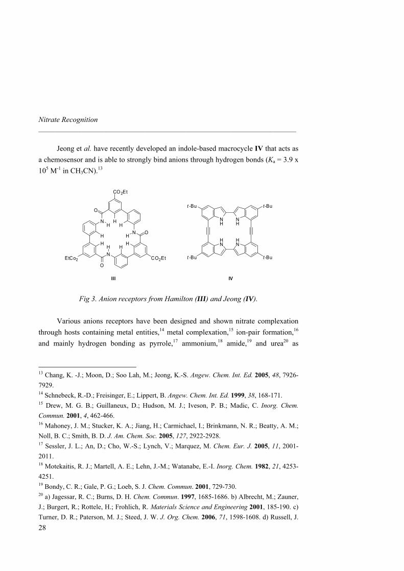
Jeong et al. have recently developed an indole-based macrocycle IV that acts as a chemosensor and is able to strongly bind anions through hydrogen bonds (Ka = 3.9 x 10⁵ M-1 in CH3CN).13
CO 2Et
HNH
N
H
CO2Et
HN
H
EtCo2
H
H
H
O
O
OH
III
NH
HN
HN
NH
t -Bu
t -Bu
t-Bu
t-Bu
IV
Fig 3. Anion receptors from Hamilton (III) and Jeong (IV).
Various anions receptors have been designed and shown nitrate complexation through hosts containing metal entities,14 metal complexation,15 ion-pair formation,16 and mainly hydrogen bonding as pyrrole,17 ammonium,18 amide,19 and urea20 as
13 Chang, K. -J.; Moon, D.; Soo Lah, M.; Jeong, K.-S. Angew. Chem. Int. Ed. 2005, 48, 7926-7929. 14 Schnebeck, R.-D.; Freisinger, E.; Lippert, B. Angew. Chem. Int. Ed. 1999, 38, 168-171. 15 Drew, M. G. B.; Guillaneux, D.; Hudson, M. J.; Iveson, P. B.; Madic, C. Inorg. Chem. Commun. 2001, 4, 462-466. 16 Mahoney, J. M.; Stucker, K. A.; Jiang, H.; Carmichael, I.; Brinkmann, N. R.; Beatty, A. M.; Noll, B. C.; Smith, B. D. J. Am. Chem. Soc. 2005, 127, 2922-2928.17 Sessler, J. L.; An, D.; Cho, W.-S.; Lynch, V.; Marquez, M. Chem. Eur. J. 2005, 11, 2001-2011. 18 Motekaitis, R. J.; Martell, A. E.; Lehn, J.-M.; Watanabe, E.-I. Inorg. Chem. 1982, 21, 4253-4251. 19 Bondy, C. R.; Gale, P. G.; Loeb, S. J. Chem. Commun. 2001, 729-730. 20 a) Jagessar, R. C.; Burns, D. H. Chem. Commun. 1997, 1685-1686. b) Albrecht, M.; Zauner, J.; Burgert, R.; Rottele, H.; Frohlich, R. Materials Science and Engineering 2001, 185-190. c) Turner, D. R.; Paterson, M. J.; Steed, J. W. J. Org. Chem. 2006, 71, 1598-1608. d) Russell, J.
28
2.1 Introduction _____________________________________________________________________
29
hydrogen bond donors. These synthetic anion receptors revealed an increase of its association even in polar media, although selectivity for nitrate remained difficult to achieve. For instance, similar sized Cl- often gives a better association with the host because of its spherical shape mode and higher basicity. Most remarkably, Beer and co-workers succeeded in inverting the chloride vs nitrate selectivity by using a solvation effect. In a 9:1 (CH2Cl2/MeOH) mixture, the ruthenium(II) tris(5,5´-diamide-2,2´-bipyridine) receptor is indeed selective towards chloride whereas in 1:1 (CH2Cl2/MeOH) mixture, this order is inversed.21
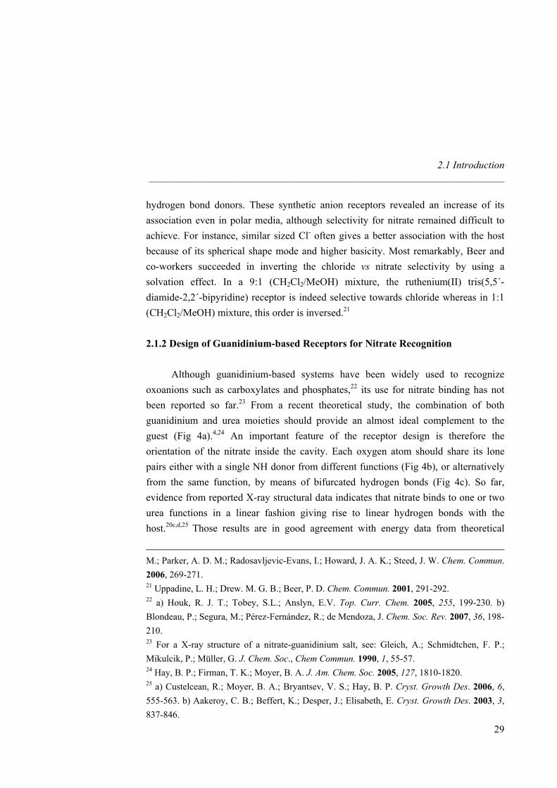
2.1.2 Design of Guanidinium-based Receptors for Nitrate Recognition Although guanidinium-based systems have been widely used to recognize oxoanions such as carboxylates and phosphates,22 its use for nitrate binding has not been reported so far.23 From a recent theoretical study, the combination of both guanidinium and urea moieties should provide an almost ideal complement to the guest (Fig 4a).4,24 An important feature of the receptor design is therefore the orientation of the nitrate inside the cavity. Each oxygen atom should share its lone pairs either with a single NH donor from different functions (Fig 4b), or alternatively from the same function, by means of bifurcated hydrogen bonds (Fig 4c). So far, evidence from reported X-ray structural data indicates that nitrate binds to one or two urea functions in a linear fashion giving rise to linear hydrogen bonds with the host.20c,d, 25 Those results are in good agreement with energy data from theoretical
M.; Parker, A. D. M.; Radosavljevic-Evans, I.; Howard, J. A. K.; Steed, J. W. Chem. Commun. 2006, 269-271. 21 Uppadine, L. H.; Drew. M. G. B.; Beer, P. D. Chem. Commun. 2001, 291-292. 22 a) Houk, R. J. T.; Tobey, S.L.; Anslyn, E.V. Top. Curr. Chem. 2005, 255, 199-230. b) Blondeau, P.; Segura, M.; Pérez-Fernández, R.; de Mendoza, J. Chem. Soc. Rev. 2007, 36, 198-210. 23 For a X-ray structure of a nitrate-guanidinium salt, see: Gleich, A.; Schmidtchen, F. P.; Mikulcik, P.; Müller, G. J. Chem. Soc., Chem Commun. 1990, 1, 55-57. 24 Hay, B. P.; Firman, T. K.; Moyer, B. A. J. Am. Chem. Soc. 2005, 127, 1810-1820. 25 a) Custelcean, R.; Moyer, B. A.; Bryantsev, V. S.; Hay, B. P. Cryst. Growth Des. 2006, 6, 555-563. b) Aakeroy, C. B.; Beffert, K.; Desper, J.; Elisabeth, E. Cryst. Growth Des. 2003, 3, 837-846.
Nitrate Recognition _____________________________________________________________________
calculations, which revealed that forcing urea to bind a single oxygen atom in a bifurcated fashion yields complexes that are less stable (more than a 10% difference) than those involving hydrogen bonds with two oxygen atoms.24 Considering the hydrogen bond donor strength26 and the electrostatic interactions, the hydrogen donor atoms should be along the edge of the polyhedron defined by the oxygen atoms (Fig 4b). Higher stability of linear hydrogen bonds should indeed prevent formation of a second complex based on significant geometrically unfavorable bonds (Fig 4c).
+N
N
N
N N
N NO O
NOO
O
H H
H
H H
H
+N
N
N
N N
N NO O
NO
O O
H H
H
H H
H
a) b) c)
Fig 4. Top and side views of optimized geometries (B3LYP) for representative examples of (a) 3:1 urea: NO3
- complexes24 and (b) & (c) binding mode for nitrate inclusion.
The nitrate anion offers six optimal sites for proton location according to the number and orientation of the lone pair orbitals, but so far no structural evidence on an example fulfilling this feature has been reported. This represents in fact a key point in host design because spacing between hydrogen bond donors is different for linear and non-linear host-guest bonds. We present herein a nitrate recognition example for the first binding mode.
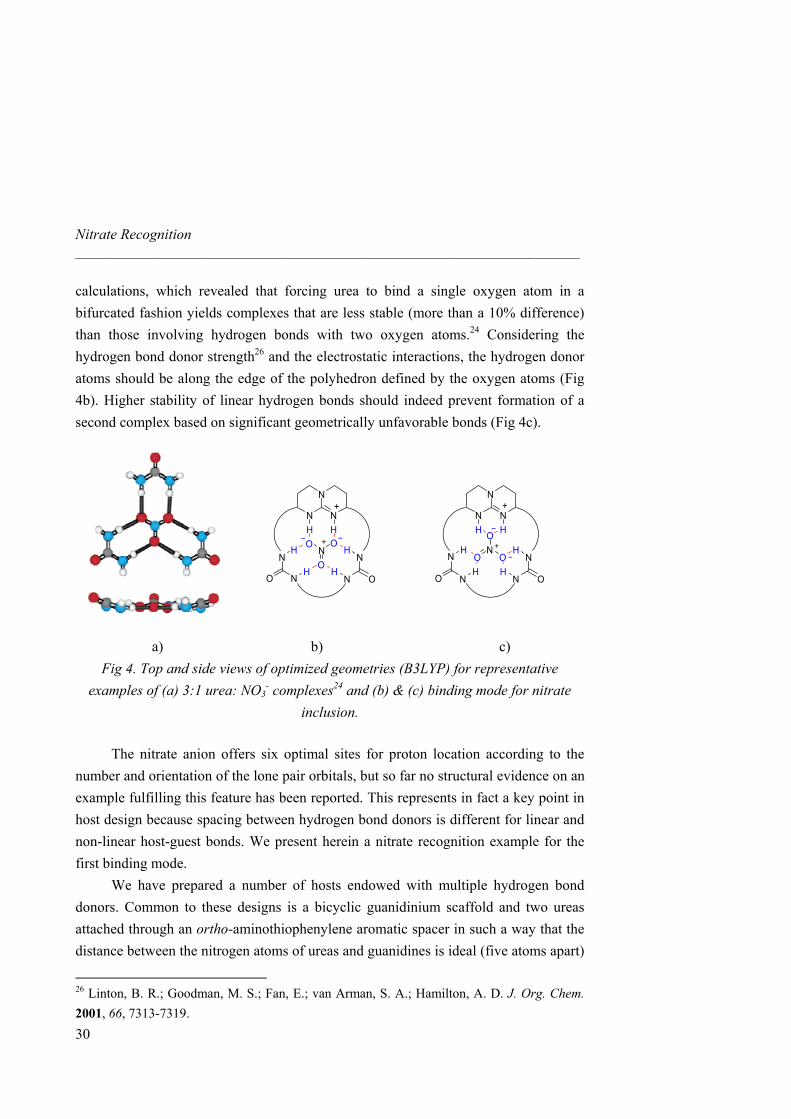
We have prepared a number of hosts endowed with multiple hydrogen bond donors. Common to these designs is a bicyclic guanidinium scaffold and two ureas attached through an ortho-aminothiophenylene aromatic spacer in such a way that the distance between the nitrogen atoms of ureas and guanidines is ideal (five atoms apart)
26 Linton, B. R.; Goodman, M. S.; Fan, E.; van Arman, S. A.; Hamilton, A. D. J. Org. Chem. 2001, 66, 7313-7319.
30
2.1 Introduction _____________________________________________________________________
for the hydrogen donors to complement both the syn and anti lone pairs of each oxygen.27 Moreover, the aromatic ring increases the acidity of the urea and brings pre-organization to the overall structure (Fig 5). Also, the sulfur atoms should contribute to shape the host by chelation while not introducing a too electronegative atom that would cause electronic repulsion with the guest.28 The second urea NH’s, located seven atoms away from guanidinium NH, would interact with each lone pair of the third oxygen atom guest. Besides, among the receptors reported so far, use of macrocycles take the advantage of affording a pre-organized scaffold with convergent binding groups to include the guest by size and shape complementarities.29 Macrocycles of different cavity sizes (four, five and six carbon atoms spacers) and rigidities (xylylene based host) have been synthesized for nitrate inclusion. Regarding the linker between the ureas in the macrocycles, a five carbon chain should be again the best choice for nitrate encapsulation.
+N
N
NS S
N N
N NO O
NOO
O
H H
H
H H
H
Rigid bicyclic guanidinium scaffold
Rigid five-atom spacer
Additional hydrogen bonds
Ion pairing & H-bonds donor
Diff erent cavity size and rig id ity
Fig 5. Design of guanidinium receptors for nitrate recognition.
27 González, S.; Peláez, R.; Sanz, F.; Jiménez, M. B.; Morán, J. R.; Caballero, M. C. Org. Lett. 2006, 21, 4679-4683. 28 Kavallieratos, K.; Bertao, C. M.; Crabtree, R. H. J. Org. Chem. 1999, 64, 1675-1683. 29 Choi, K.; Hamilton, A. D. Coord. Chem. Rev. 2003, 240, 101-110.
31
Nitrate Recognition _____________________________________________________________________
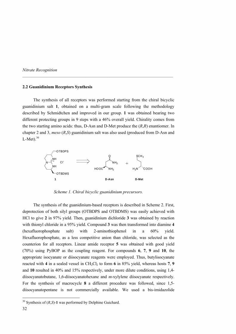
2.2 Guanidinium Receptors Synthesis The synthesis of all receptors was performed starting from the chiral bicyclic guanidinium salt 1, obtained on a multi-gram scale following the methodology described by Schmidtchen and improved in our group. 1 was obtained bearing two different protecting groups in 9 steps with a 46% overall yield. Chirality comes from the two starting amino acids: thus, D-Asn and D-Met produce the (R,R) enantiomer. In chapter 2 and 3, meso (R,S) guanidinium salt was also used (produced from D-Asn and L-Met).30
NNH
NH
OTBDPS
OTBDMS
Cl-
1
NH2
O
NH2HOOC
SCH3
COOHH2N
D-Asn D-Met
Scheme 1. Chiral bicyclic guanidinium precursors.
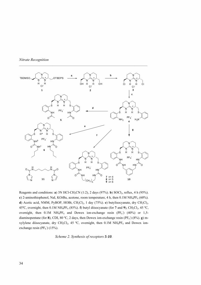
The synthesis of the guanidinium-based receptors is described in Scheme 2. First, deprotection of both silyl groups (OTBDPS and OTBDMS) was easily achieved with HCl to give 2 in 97% yield. Then, guanidinium dichloride 3 was obtained by reaction with thionyl chloride in a 95% yield. Compound 3 was then transformed into diamine 4 (hexafluorophosphate salt) with 2-aminothiophenol in a 60% yield. Hexafluorophosphate, as a less competitive anion than chloride, was selected as the counterion for all receptors. Linear amide receptor 5 was obtained with good yield (70%) using PyBOP as the coupling reagent. For compounds 6, 7, 9 and 10, the appropriate isocyanate or diisocyanate reagents were employed. Thus, butylisocyanate reacted with 4 in a sealed vessel in CH2Cl2 to form 6 in 85% yield, whereas hosts 7, 9 and 10 resulted in 40% and 15% respectively, under more dilute conditions, using 1,4-diisocyanatobutane, 1,6-diisocyanatohexane and m-xylylene diisocyanate respectively. For the synthesis of macrocycle 8 a different procedure was followed, since 1,5-diisocyanatopentane is not commercially available. We used a bis-imidazolide
30 Synthesis of (R,S)-1 was performed by Delphine Guichard.
32
2.2 Guanidinium Receptors Synthesis _____________________________________________________________________
33
intermediate (11, 95% yield), prepared from 1,5-diaminopentane and 1,1’-carbonyldiimidazole (CDI) which was reacted afterwards with 4 in dry MeOH to obtain 8 though in a modest 8% yield. Aromatic receptor 10 was obtained also in a rather modest yield (15%) as compared to alkyl analogues 7 and 9 using the same procedure. We then studied the cyclization of receptor 10 from 4 with PF6
-, Cl- or AcO- as counterions. In situ macrocyclization was followed with analytical HPLC during 24 hours but did not show any templating effect. Since HPLC monitoring did not show substantial amounts of impurities, the rather low yield was attributed to the purification process (column chromatography). Receptors 5-10 were soluble in solvents like CH2Cl2, CH3CN or MeOH. For NMR and ITC determinations, aprotic CH3CN of medium polarity seemed thus appropriate to avoid solvent interferences.
Nitrate Recognition _____________________________________________________________________
N
N
NH H
+
Cl ClCl-
N
N
NH H
+
OH OHCl-
a b
c
d N
N
NH H
+
S S
PF 6-
NH2 H2N
N
N
NH H
+
S S
PF6-
NH HN
N
N
NH H
+
Cl-
OTBDPSTBDMSO
O O
N
N
NHa H
+
S S
PF6-
NHb HN
O ONHc HNN
N
NH H
+
S S
PF 6-
NH HN
O ONH HNS S
NH HN
NH HNO O
CH2 n
7 n= 48 n= 59 n= 6
10
6
5 4
321
e
f
g
PF6-
N
N
NH H
+
HN
HNO O
NN
N N11
Reagents and conditions: a) 3N HCl-CH3CN (1:2), 2 days (97%). b) SOCl2, reflux, 4 h (95%). c) 2-aminothiophenol, NaI, KOtBu, acetone, room temperature, 4 h, then 0.1M NH4PF6 (60%). d) Acetic acid, NMM, PyBOP, HOBt, CH2Cl2, 1 day (75%). e) butylisocyanate, dry CH2Cl2, 45ºC, overnight, then 0.1M NH4PF6 (85%). f) butyl diisocyanate (for 7 and 9), CH2Cl2, 45 ºC, overnight, then 0.1M NH4PF6 and Dowex ion-exchange resin (PF6
-) (40%) or 1,5-diaminopentane (for 8), CDI, 80 ºC, 2 days, then Dowex ion-exchange resin (PF6
-) (8%). g) m-xylylene diisocyanate, dry CH2Cl2, 45 ºC, overnight, then 0.1M NH4PF6 and Dowex ion-exchange resin (PF6
-) (15%).
Scheme 2. Synthesis of receptors 5-10.
34
2.3 Binding Study _____________________________________________________________________
2.3 Binding Study 2.3.1 1H-NMR and ITC Titrations with Nitrate
TBA nitrate equivalents
∆δ
(ppm
)
0 1 2 3 4 50,0
0,2
0,4
0,6
0,8
1,0
1,2
1,4
1,6
1,8
2,0
TBA nitrate equivalents
∆δ
(ppm
)
0 1 2 3 4 50,0
0,2
0,4
0,6
0,8
1,0
1,2
1,4
1,6
1,8
2,0
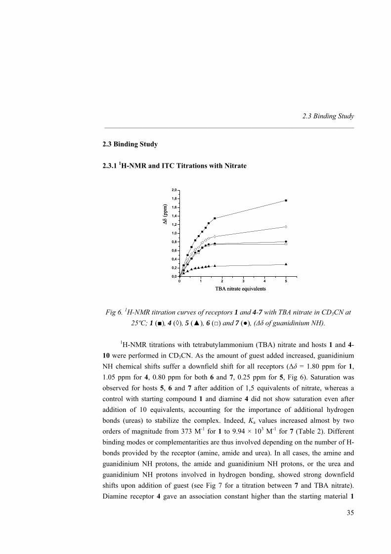
Fig 6. 1H-NMR titration curves of receptors 1 and 4-7 with TBA nitrate in CD3CN at 25ºC; 1 (■), 4 (◊), 5 (▲), 6 (□) and 7 (●), (∆δ of guanidinium NH).
1H-NMR titrations with tetrabutylammonium (TBA) nitrate and hosts 1 and 4-10 were performed in CD3CN. As the amount of guest added increased, guanidinium NH chemical shifts suffer a downfield shift for all receptors (∆δ = 1.80 ppm for 1, 1.05 ppm for 4, 0.80 ppm for both 6 and 7, 0.25 ppm for 5, Fig 6). Saturation was observed for hosts 5, 6 and 7 after addition of 1,5 equivalents of nitrate, whereas a control with starting compound 1 and diamine 4 did not show saturation even after addition of 10 equivalents, accounting for the importance of additional hydrogen bonds (ureas) to stabilize the complex. Indeed, Ka values increased almost by two orders of magnitude from 373 M-1 for 1 to 9.94 × 10³ M-1 for 7 (Table 2). Different binding modes or complementarities are thus involved depending on the number of H-bonds provided by the receptor (amine, amide and urea). In all cases, the amine and guanidinium NH protons, the amide and guanidinium NH protons, or the urea and guanidinium NH protons involved in hydrogen bonding, showed strong downfield shifts upon addition of guest (see Fig 7 for a titration between 7 and TBA nitrate). Diamine receptor 4 gave an association constant higher than the starting material 1
35
Nitrate Recognition _____________________________________________________________________
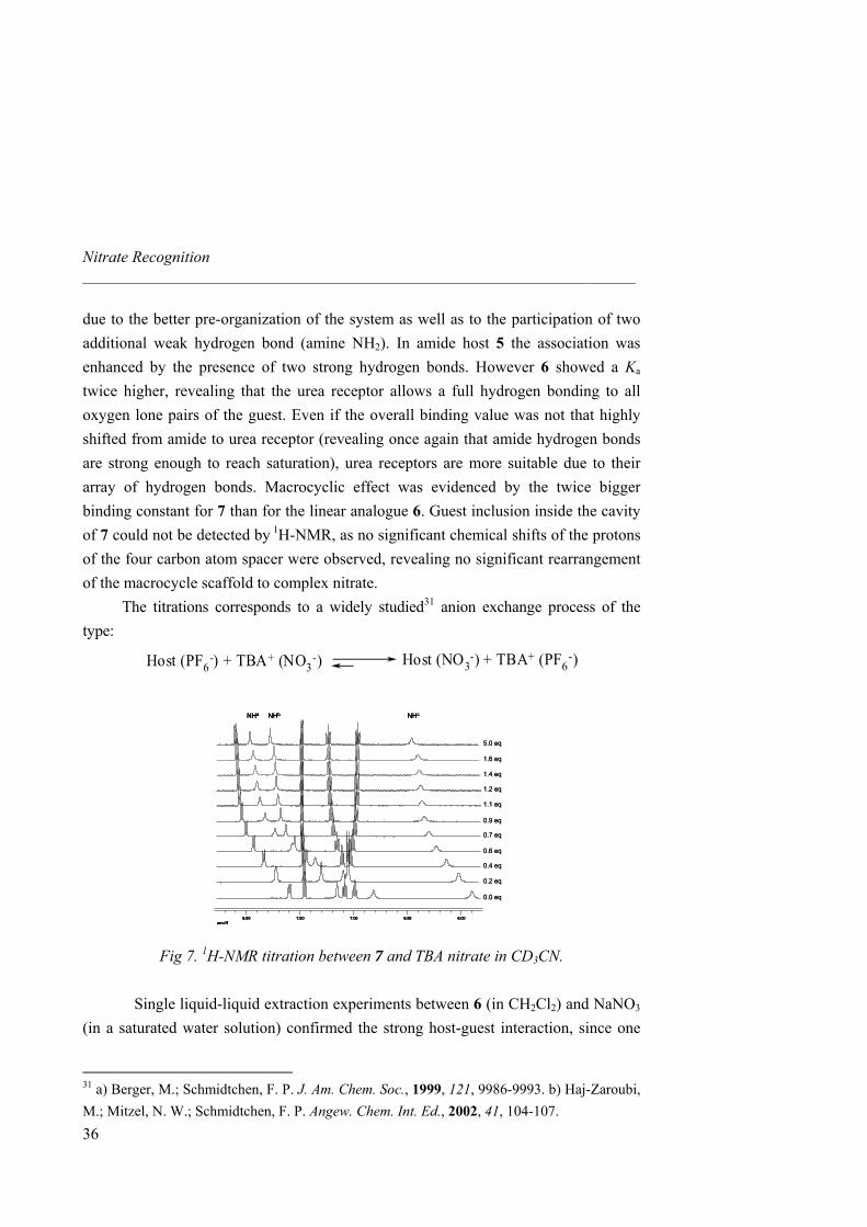
due to the better pre-organization of the system as well as to the participation of two additional weak hydrogen bond (amine NH2). In amide host 5 the association was enhanced by the presence of two strong hydrogen bonds. However 6 showed a Ka twice higher, revealing that the urea receptor allows a full hydrogen bonding to all oxygen lone pairs of the guest. Even if the overall binding value was not that highly shifted from amide to urea receptor (revealing once again that amide hydrogen bonds are strong enough to reach saturation), urea receptors are more suitable due to their array of hydrogen bonds. Macrocyclic effect was evidenced by the twice bigger binding constant for 7 than for the linear analogue 6. Guest inclusion inside the cavity of 7 could not be detected by 1H-NMR, as no significant chemical shifts of the protons of the four carbon atom spacer were observed, revealing no significant rearrangement of the macrocycle scaffold to complex nitrate. The titrations corresponds to a widely studied31 anion exchange process of the type:
Host (PF6-) + TBA+ (NO3
-) Host (NO3-) + TBA+ (PF6
-)
NHa NHb NHc
ppm (f1)6.006.507.007.508.00
0.0 eq
0.2 eq
0.4 eq
0.6 eq
0.7 eq
0.9 eq
1.1 eq
1.2 eq
1.4 eq
1.6 eq
5.0 eq
NHa NHb NHc
ppm (f1)6.006.507.007.508.00
NHa NHb NHc
ppm (f1)6.006.507.007.508.00
ppm (f1)6.006.507.007.508.00
0.0 eq
0.2 eq
0.4 eq
0.6 eq
0.7 eq
0.9 eq
1.1 eq
1.2 eq
1.4 eq
1.6 eq
5.0 eq
Fig 7. 1H-NMR titration between 7 and TBA nitrate in CD3CN.
Single liquid-liquid extraction experiments between 6 (in CH2Cl2) and NaNO3 (in a saturated water solution) confirmed the strong host-guest interaction, since one
31 a) Berger, M.; Schmidtchen, F. P. J. Am. Chem. Soc., 1999, 121, 9986-9993. b) Haj-Zaroubi, M.; Mitzel, N. W.; Schmidtchen, F. P. Angew. Chem. Int. Ed., 2002, 41, 104-107.
36
2.3 Binding Study _____________________________________________________________________
37
full equivalent of nitrate was extracted and a 1:1 stoichiometry was observed in the organic layer.32 An important characteristic of lipophilic bicyclic guanidinium receptors is thus the property of extracting anions from aqueous media to organic phases through single liquid-liquid extraction or membrane transport such anions from a feeding to a receiving phase.33
No accurate NMR binding data could be obtained for 8, 9 and 10 due to in situ crystallization at the concentrations required by the titrations. To overcome this problem, we used isothermal titration calorimetry (ITC) as an alternative tool for measuring binding constants and thermodynamic parameters at much lower concentrations.34 The binding isotherms are characteristic of exothermic 1:1 complexes and values for Ka are in good agreement with those previously obtained by 1H-NMR (Table 2). Increase of cavity size results in a binding affinity enhancement between the macrcocycle and nitrate. Hence, macrocycle 9 showed an increase of two orders of magnitude respect to 4 and presents one of the highest Ka values reported so far for nitrate. Increase of cavity rigidity afforded however a weaker association between the host and the guest. Indeed, macrocycle 10 afforded the lowest Ka among all macrocycles synthezised and even lower than linear receptor 6. Likely, the aromatic xylylene proton pointing inward the cavity prevents the guest to get inside and difficults the correct orientation of the urea NHs towards the nitrate oxygen atoms.
32 Single liquid-liquid extractions were performed by shaking for 5 min a 1 mg ml-1 host solution in CH2Cl2 and a saturated NaNO3 solution in water. The organic phase was isolated after centrifugation, the solvent removed (without addition of any drying agent) and the 1H-NMR spectrum was recorded. As for other related guanidinium receptors and carriers,33 spectra of both aqueous and organic phases confirmed the complete partition of the host into the organic phase in a 1:1 host-guest stoichiometry. 33 Breccia, P.; Van Gool, M.; Pérez-Fernández, R.; Martín-Santamaría, S.; Gago, F.; Prados, P.; de Mendoza, J. J. Am. Chem. Soc. 2003, 125, 8270-8284. 34 a) Ladbury, J. E.; Chowdhry, B. Z. Chem. Biol. 1996, 3, 791-801. b) Wadsö, I. Chem. Soc. Rev. 1997, 79-86.
Nitrate Recognition _____________________________________________________________________
38
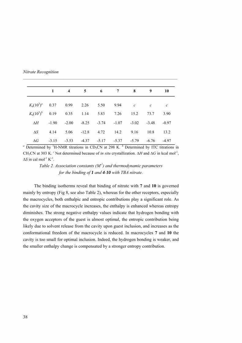
1 4 5 6 7 8 9 10
Ka(103)a
0.37
0.99
2.26
5.50
9.94
c
c
c
Ka(103)b 0.19 0.35 1.14 5.83 7.26 15.2 73.7 3.90
∆H -1.90 -2.00 -8.25 -3.74 -1.07 -3.02 -3.48 -0.97
∆S 4.14 5.06 -12.8 4.72 14.2 9.16 10.8 13.2
∆G -3.15 -3.53 -4.37 -5.17 -5.37 -5.79 -6.76 -4.97 a Determined by 1H-NMR titrations in CD3CN at 298 K. b Determined by ITC titrations in CH3CN at 303 K. c Not determined because of in situ crystallization. ∆H and ∆G in kcal mol-1, ∆S in cal mol-1 K-1.
Table 2. Association constants (M-1) and thermodynamic parameters for the binding of 1 and 4-10 with TBA nitrate.
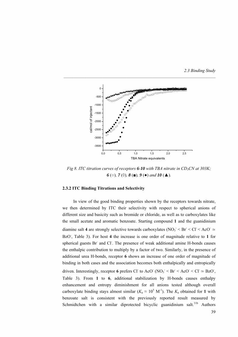
The binding isotherms reveal that binding of nitrate with 7 and 10 is governed
mainly by entropy (Fig 8, see also Table 2), whereas for the other receptors, especially the macrocycles, both enthalpic and entropic contributions play a significant role. As the cavity size of the macrocycle increases, the enthalpy is enhanced whereas entropy diminishes. The strong negative enthalpy values indicate that hydrogen bonding with the oxygen acceptors of the guest is almost optimal, the entropic contribution being likely due to solvent release from the cavity upon guest inclusion, and increases as the conformational freedom of the macrocycle is reduced. In macrocycles 7 and 10 the cavity is too small for optimal inclusion. Indeed, the hydrogen bonding is weaker, and the smaller enthalpy change is compensated by a stronger entropy contribution.
2.3 Binding Study _____________________________________________________________________
0,0 0,5 1,0 1,5 2,0 2,5
-3500
-3000
-2500
-2000
-1500
-1000
-500
0ca
l/mol
of i
njec
tant
TBA Nitrate equivalents
Fig 8. ITC titration curves of receptors 6-10 with TBA nitrate in CD3CN at 303K; 6 (○), 7 (◊), 8 (■), 9 (●) and 10 (▲).
2.3.2 ITC Binding Titrations and Selectivity
In view of the good binding properties shown by the receptors towards nitrate, we then determined by ITC their selectivity with respect to spherical anions of different size and basicity such as bromide or chloride, as well as to carboxylates like the small acetate and aromatic benzoate. Starting compound 1 and the guanidinium
diamine salt 4 are strongly selective towards carboxylates (NO3- < Br- < Cl- < AcO- ≃
BzO-, Table 3). For host 4 the increase is one order of magnitude relative to 1 for spherical guests Br- and Cl-. The presence of weak additional amine H-bonds causes the enthalpic contribution to multiply by a factor of two. Similarly, in the presence of additional urea H-bonds, receptor 6 shows an increase of one order of magnitude of binding in both cases and the association becomes both enthalpically and entropically
driven. Interestingly, receptor 6 prefers Cl- to AcO- (NO3- < Br- < AcO- < Cl- ≃ BzO-,
Table 3). From 1 to 6, additional stabilization by H-bonds causes enthalpy enhancement and entropy diminishment for all anions tested although overall carboxylate binding stays almost similar (Ka ≈ 105 M-1). The Ka obtained for 1 with benzoate salt is consistent with the previously reported result measured by Schmidtchen with a similar diprotected bicyclic guanidinium salt.31b Authors
39
Nitrate Recognition _____________________________________________________________________
40
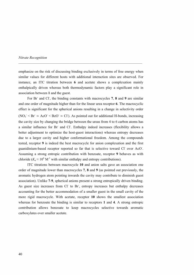
emphasize on the risk of discussing binding exclusively in terms of free energy when similar values for different hosts with additional interaction sites are observed. For instance, an ITC titration between 6 and acetate shows a complexation mainly enthalpically driven whereas both thermodynamic factors play a significant role in association between 1 and the guest.
For Br- and Cl-, the binding constants with macrocycles 7, 8 and 9 are similar and one order of magnitude higher than for the linear urea receptor 6. The macrocyclic effect is significant for the spherical anions resulting in a change in selectivity order
(NO3- < Br- ≃ AcO- < BzO- ≃ Cl-). As pointed out for additional H-bonds, increasing
the cavity size by changing the bridge between the ureas from 4 to 6 carbon atoms has a similar influence for Br- and Cl-. Enthalpy indeed increases (flexibility allows a better adjustment to optimize the host-guest interactions) whereas entropy decreases due to a larger cavity and higher conformational freedom. Among the compounds tested, receptor 9 is indeed the best macrocycle for anion complexation and the first guanidinium-based receptor reported so far that is selective toward Cl- over AcO-. Assuming a strong entropic contribution with benzoate, receptor 9 behaves as with chloride (Ka ≈ 106 M-1 with similar enthalpy and entropy contributions).
ITC titrations between macrocycle 10 and anion salts gave an association one order of magnitude lower than macrocycles 7, 8 and 9 (as pointed out previously, the aromatic hydrogen atom pointing inwards the cavity may contribute to diminish guest association). Unlike 7-9, spherical anions present a strong entropically driven binding. As guest size increases from Cl- to Br-, entropy increases but enthalpy decreases accounting for the better accommodation of a smaller guest in the small cavity of the more rigid macrocycle. With acetate, receptor 10 shows the smallest association whereas for benzoate the binding is similar to receptors 1 and 4. A strong entropic contribution allows benzoate to keep macrocycles selective towards aromatic carboxylates over smaller acetate.
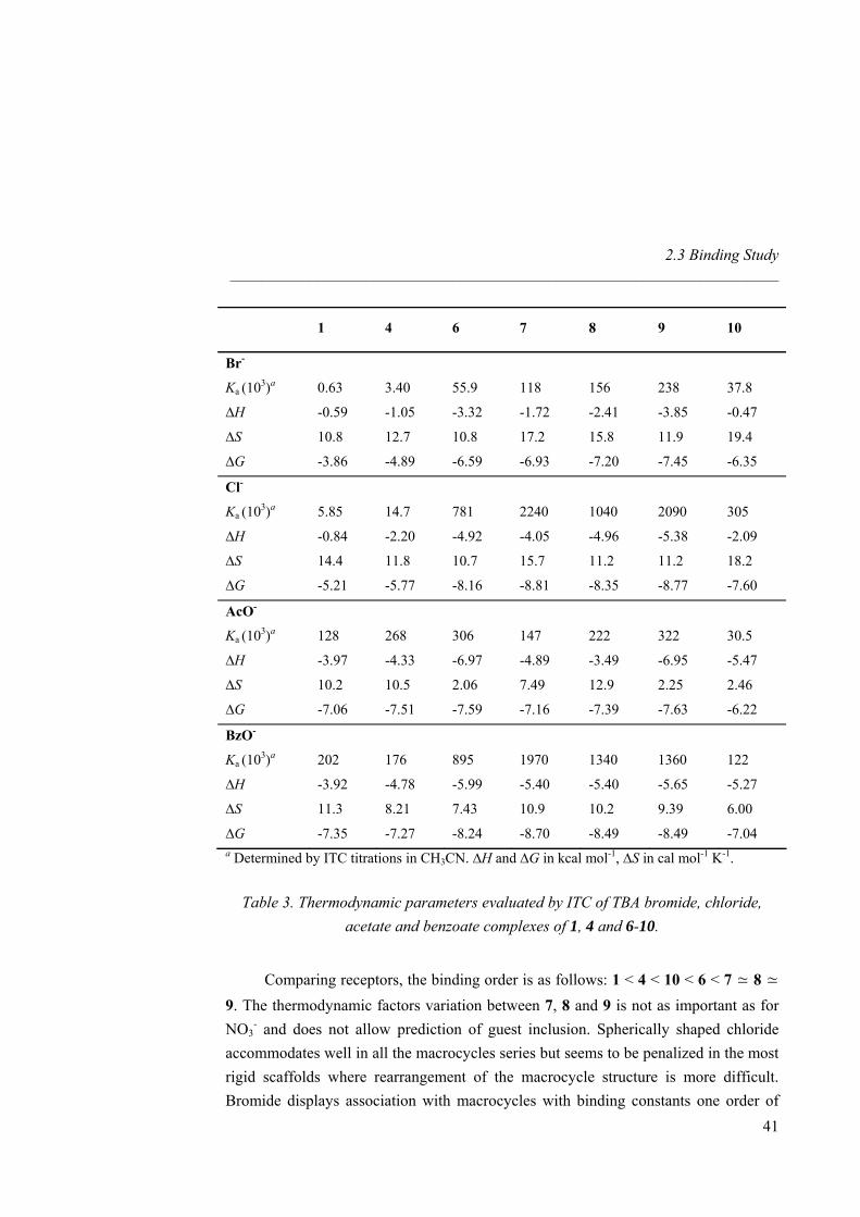
2.3 Binding Study _____________________________________________________________________
41
1 4 6 7 8 9 10
Br-
Ka (103)a 0.63 3.40 55.9 118 156 238 37.8
∆H -0.59 -1.05 -3.32 -1.72 -2.41 -3.85 -0.47
∆S 10.8 12.7 10.8 17.2 15.8 11.9 19.4
∆G -3.86 -4.89 -6.59 -6.93 -7.20 -7.45 -6.35
Cl-
Ka (103)a 5.85 14.7 781 2240 1040 2090 305
∆H -0.84 -2.20 -4.92 -4.05 -4.96 -5.38 -2.09
∆S 14.4 11.8 10.7 15.7 11.2 11.2 18.2
∆G -5.21 -5.77 -8.16 -8.81 -8.35 -8.77 -7.60
AcO-
Ka (103)a 128 268 306 147 222 322 30.5
∆H -3.97 -4.33 -6.97 -4.89 -3.49 -6.95 -5.47
∆S 10.2 10.5 2.06 7.49 12.9 2.25 2.46
∆G -7.06 -7.51 -7.59 -7.16 -7.39 -7.63 -6.22
BzO-
Ka (103)a 202 176 895 1970 1340 1360 122
∆H -3.92 -4.78 -5.99 -5.40 -5.40 -5.65 -5.27
∆S 11.3 8.21 7.43 10.9 10.2 9.39 6.00
∆G -7.35 -7.27 -8.24 -8.70 -8.49 -8.49 -7.04 a Determined by ITC titrations in CH3CN. ∆H and ∆G in kcal mol-1, ∆S in cal mol-1 K-1.
Table 3. Thermodynamic parameters evaluated by ITC of TBA bromide, chloride, acetate and benzoate complexes of 1, 4 and 6-10.
Comparing receptors, the binding order is as follows: 1 < 4 < 10 < 6 < 7 ≃ 8 ≃ 9. The thermodynamic factors variation between 7, 8 and 9 is not as important as for NO3
- and does not allow prediction of guest inclusion. Spherically shaped chloride accommodates well in all the macrocycles series but seems to be penalized in the most rigid scaffolds where rearrangement of the macrocycle structure is more difficult. Bromide displays association with macrocycles with binding constants one order of
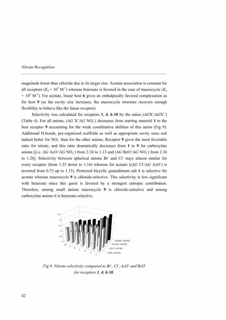
Nitrate Recognition _____________________________________________________________________
magnitude lower than chloride due to its larger size. Acetate association is constant for all receptors (Ka = 105 M-1) whereas benzoate is favored in the case of macrocycle (Ka
= 106 M-1). For acetate, linear host 6 gives an enthalpically favored complexation as for host 9 (as the cavity size increases, the macrocycle structure recovers enough flexibility to behave like the linear receptor).
Selectivity was calculated for receptors 1, 4, 6-10 by the ratios (∆GX-/∆GX’-) (Table 4). For all anions, (∆G X-/∆G NO3
-) decreases from starting material 1 to the best receptor 9 accounting for the weak coordinative abilities of this anion (Fig 9). Additional H-bonds, pre-organized scaffolds as well as appropriate cavity sizes suit indeed better for NO3
- than for the other anions. Receptor 9 gives the most favorable ratio for nitrate, and this ratio dramatically decreases from 1 to 9 for carboxylate anions [(i.e. ∆G AcO-/∆G NO3
-) from 2.24 to 1.13 and (∆G BzO-/∆G NO3-) from 2.36
to 1.26]. Selectivity between spherical anions Br- and Cl- stays almost similar for every receptor (from 1.35 down to 1.16) whereas for acetate ((∆G Cl-/∆G AcO-) is inverted from 0.73 up to 1.15). Protected bicyclic guanidinium salt 1 is selective for acetate whereas macrocycle 9 is chloride-selective. This selectivity is less significant with benzoate since this guest is favored by a strongest entropic contribution. Therefore, among small anions macrocycle 9 is chloride-selective and among carboxylate anions it is benzoate-selective.
14
67
89
10
∆G BzO- / ∆G NO3-
∆G AcO- /∆G NO3-
∆G Cl- / ∆G NO3-
∆G Br- / ∆G NO3-
0
0,5
1
1,5
2
2,5
∆G BzO- / ∆G NO3-
∆G AcO- / ∆G NO3-
∆G Cl- / ∆G NO3-
∆G Br- / ∆G NO3-
109
87
64
114
67
89
10
∆G BzO- / ∆G NO3-
∆G AcO- /∆G NO3-
∆G Cl- / ∆G NO3-
∆G Br- / ∆G NO3-
0
0,5
1
1,5
2
2,5
∆G BzO- / ∆G NO3-
∆G AcO- / ∆G NO3-
∆G Cl- / ∆G NO3-
∆G Br- / ∆G NO3-
109
87
64
1
Fig 9. Nitrate selectivity compared to Br-, Cl-, AcO- and BzO-
for receptors 1, 4, 6-10.
42
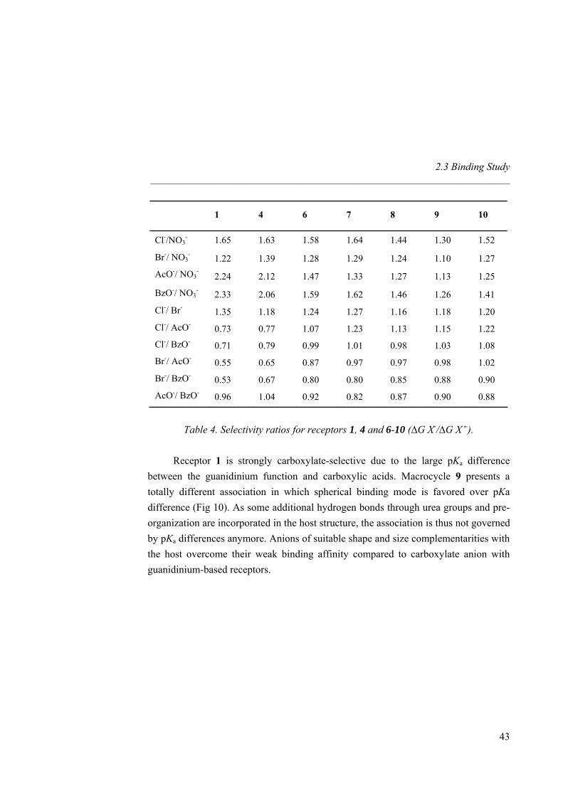
2.3 Binding Study _____________________________________________________________________
43
1 4 6 7 8 9 10
Cl-/NO3- 1.65 1.63 1.58 1.64 1.44 1.30 1.52
Br-/ NO3- 1.22 1.39 1.28 1.29 1.24 1.10 1.27
AcO-/ NO3-
2.24 2.12 1.47 1.33 1.27 1.13 1.25
BzO-/ NO3- 2.33 2.06 1.59 1.62 1.46 1.26 1.41
Cl-/ Br- 1.35 1.18 1.24 1.27 1.16 1.18 1.20
Cl-/ AcO- 0.73 0.77 1.07 1.23 1.13 1.15 1.22
Cl-/ BzO- 0.71 0.79 0.99 1.01 0.98 1.03 1.08 Br-/ AcO- 0.55 0.65 0.87 0.97 0.97 0.98 1.02
Br-/ BzO- 0.53 0.67 0.80 0.80 0.85 0.88 0.90
AcO-/ BzO- 0.96 1.04 0.92 0.82 0.87 0.90 0.88
Table 4. Selectivity ratios for receptors 1, 4 and 6-10 (∆G X-/∆G X’-).
Receptor 1 is strongly carboxylate-selective due to the large pKa difference
between the guanidinium function and carboxylic acids. Macrocycle 9 presents a totally different association in which spherical binding mode is favored over pKa difference (Fig 10). As some additional hydrogen bonds through urea groups and pre-organization are incorporated in the host structure, the association is thus not governed by pKa differences anymore. Anions of suitable shape and size complementarities with the host overcome their weak binding affinity compared to carboxylate anion with guanidinium-based receptors.
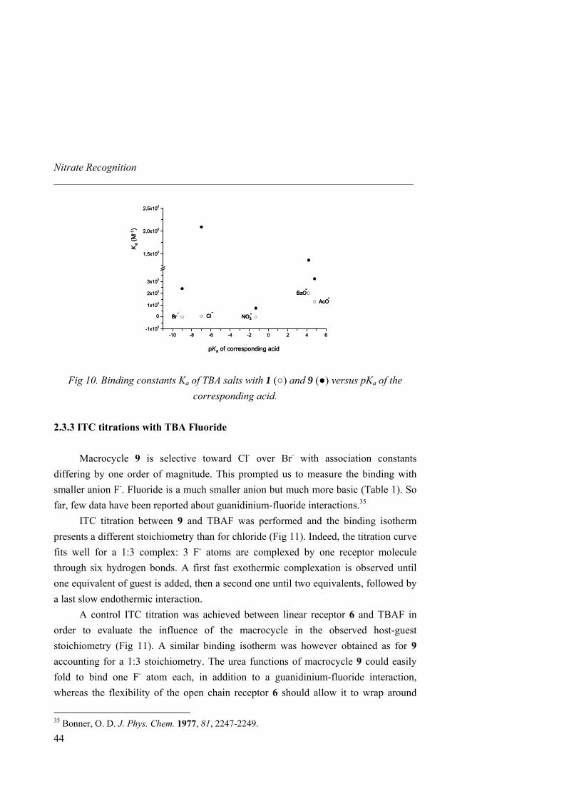
Nitrate Recognition _____________________________________________________________________
-10 -8 -6 -4 -2 0 2 4 6-1x10⁵
0
1x10⁵
2x10⁵
3x10⁵
1,5x10⁶
2,0x10⁶
2,5x10⁶
Br- Cl
-NO3
-
AcO-
BzO-
pKa of corresponding acid
Ka
(M-1
)
-10 -8 -6 -4 -2 0 2 4 6-1x10⁵
0
1x10⁵
2x10⁵
3x10⁵
1,5x10⁶
2,0x10⁶
2,5x10⁶
Br-
Br- Cl
-Cl
-NO3
-NO3-
AcO-
AcO-
BzO-
BzO-
pKa of corresponding acid
Ka
(M-1
)
Fig 10. Binding constants Ka of TBA salts with 1 (○) and 9 (●) versus pKa of the corresponding acid.
2.3.3 ITC titrations with TBA Fluoride
Macrocycle 9 is selective toward Cl- over Br- with association constants differing by one order of magnitude. This prompted us to measure the binding with smaller anion F-. Fluoride is a much smaller anion but much more basic (Table 1). So far, few data have been reported about guanidinium-fluoride interactions.35
ITC titration between 9 and TBAF was performed and the binding isotherm presents a different stoichiometry than for chloride (Fig 11). Indeed, the titration curve fits well for a 1:3 complex: 3 F- atoms are complexed by one receptor molecule through six hydrogen bonds. A first fast exothermic complexation is observed until one equivalent of guest is added, then a second one until two equivalents, followed by a last slow endothermic interaction.
A control ITC titration was achieved between linear receptor 6 and TBAF in order to evaluate the influence of the macrocycle in the observed host-guest stoichiometry (Fig 11). A similar binding isotherm was however obtained as for 9 accounting for a 1:3 stoichiometry. The urea functions of macrocycle 9 could easily fold to bind one F- atom each, in addition to a guanidinium-fluoride interaction, whereas the flexibility of the open chain receptor 6 should allow it to wrap around
35 Bonner, O. D. J. Phys. Chem. 1977, 81, 2247-2249.
44
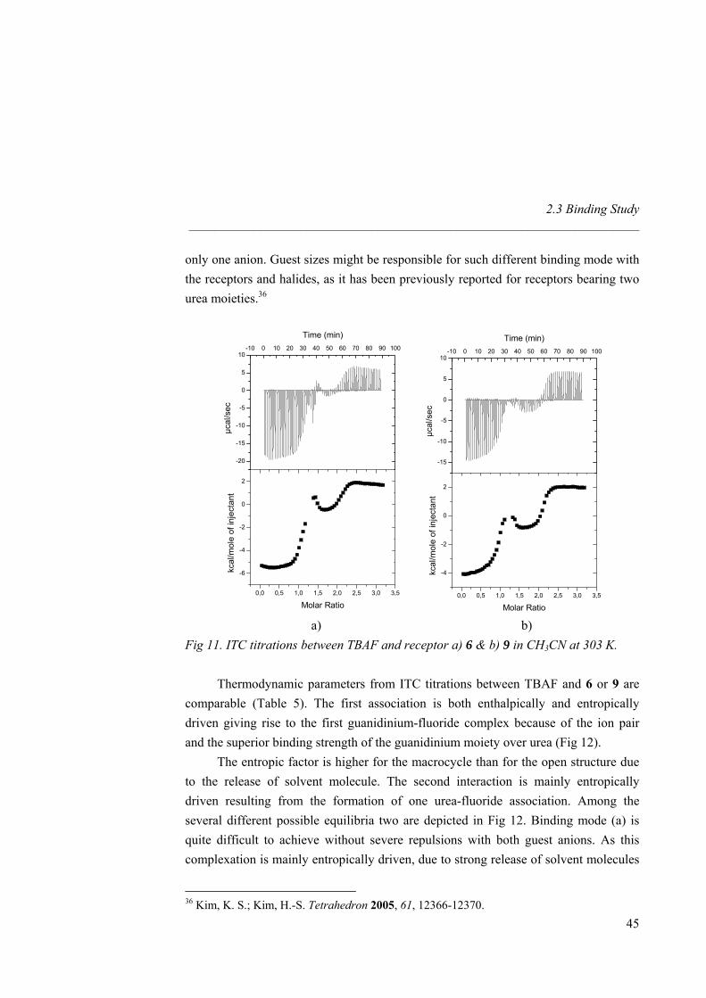
2.3 Binding Study _____________________________________________________________________
only one anion. Guest sizes might be responsible for such different binding mode with the receptors and halides, as it has been previously reported for receptors bearing two urea moieties.36
0,0 0,5 1,0 1,5 2,0 2,5 3,0 3,5
-6
-4
-2
0
2
-20
-15
-10
-5
0
5
10-10 0 10 20 30 40 50 60 70 80 90 100
Time (min)
µcal
/sec
Molar Ratio
kcal
/mol
e of
inje
ctan
t
0,0 0,5 1,0 1,5 2,0 2,5 3,0 3,5
-4
-2
0
2
-15
-10
-5
0
5
10-10 0 10 20 30 40 50 60 70 80 90 100
Time (min)
µcal
/sec
Molar Ratio
kcal
/mol
e of
inje
ctan
t
a) b) Fig 11. ITC titrations between TBAF and receptor a) 6 & b) 9 in CH3CN at 303 K.
Thermodynamic parameters from ITC titrations between TBAF and 6 or 9 are comparable (Table 5). The first association is both enthalpically and entropically driven giving rise to the first guanidinium-fluoride complex because of the ion pair and the superior binding strength of the guanidinium moiety over urea (Fig 12).
The entropic factor is higher for the macrocycle than for the open structure due to the release of solvent molecule. The second interaction is mainly entropically driven resulting from the formation of one urea-fluoride association. Among the several different possible equilibria two are depicted in Fig 12. Binding mode (a) is quite difficult to achieve without severe repulsions with both guest anions. As this complexation is mainly entropically driven, due to strong release of solvent molecules
36 Kim, K. S.; Kim, H.-S. Tetrahedron 2005, 61, 12366-12370.
45
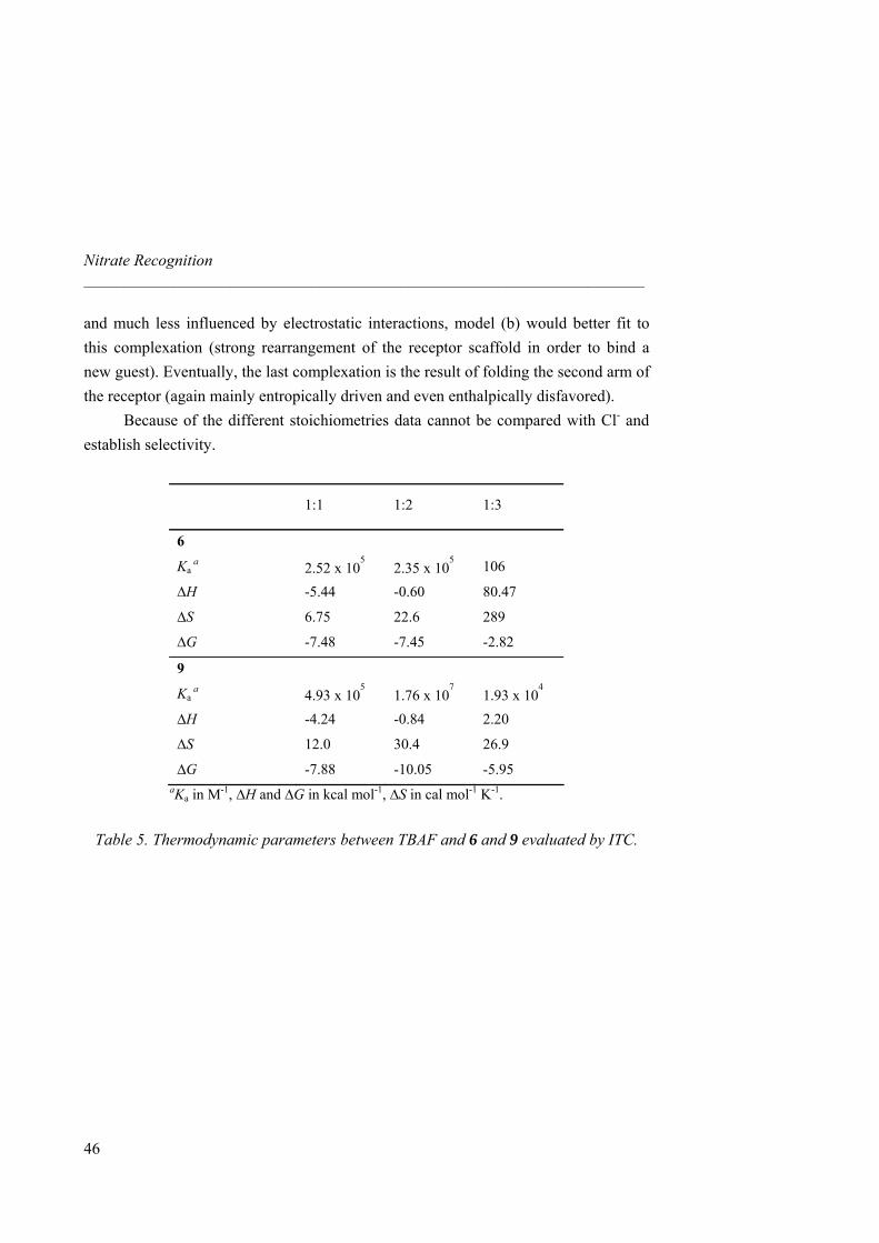
Nitrate Recognition _____________________________________________________________________
46
and much less influenced by electrostatic interactions, model (b) would better fit to this complexation (strong rearrangement of the receptor scaffold in order to bind a new guest). Eventually, the last complexation is the result of folding the second arm of the receptor (again mainly entropically driven and even enthalpically disfavored).
Because of the different stoichiometries data cannot be compared with Cl- and establish selectivity.
1:1 1:2 1:3
6
Ka a 2.52 x 10
52.35 x 10
5 106
∆H -5.44 -0.60 80.47
∆S 6.75 22.6 289
∆G -7.48 -7.45 -2.82
9
Ka a 4.93 x 10
51.76 x 10
71.93 x 10
4
∆H -4.24 -0.84 2.20
∆S 12.0 30.4 26.9
∆G -7.88 -10.05 -5.95 aKa in M-1, ∆H and ∆G in kcal mol-1, ∆S in cal mol-1 K-1.
Table 5. Thermodynamic parameters between TBAF and 6 and 9 evaluated by ITC.
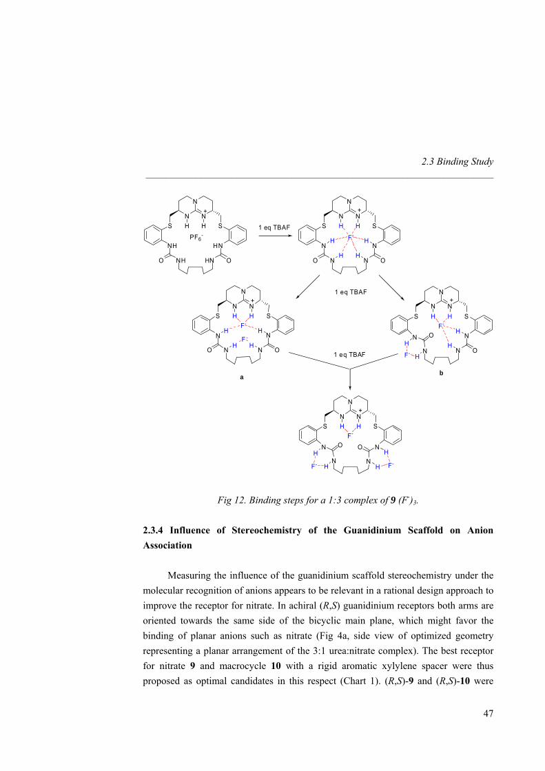
2.3 Binding Study _____________________________________________________________________
N
N
NH H
+
S S
F-
N N
N N OO
N
N
NH H
+
S S
PF6-
NH HN
NH HN OO
1 eq TBAF
N
N
NH H
+
S SF-
N N
N N OOF -
H
H
1 eq TBAF
N
N
NH H
+
S SF-
N
NN
N O
O
F- H
H
1 eq TBAF
N
N
NH H
+
S SF-
N N
N NOO
F- H
H
F-
H
H
a b
H
H H
H
H
H
H
H
Fig 12. Binding steps for a 1:3 complex of 9 (F-)3.
2.3.4 Influence of Stereochemistry of the Guanidinium Scaffold on Anion Association
Measuring the influence of the guanidinium scaffold stereochemistry under the molecular recognition of anions appears to be relevant in a rational design approach to improve the receptor for nitrate. In achiral (R,S) guanidinium receptors both arms are oriented towards the same side of the bicyclic main plane, which might favor the binding of planar anions such as nitrate (Fig 4a, side view of optimized geometry representing a planar arrangement of the 3:1 urea:nitrate complex). The best receptor for nitrate 9 and macrocycle 10 with a rigid aromatic xylylene spacer were thus proposed as optimal candidates in this respect (Chart 1). (R,S)-9 and (R,S)-10 were
47
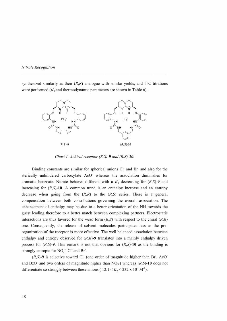
Nitrate Recognition _____________________________________________________________________
synthesized similarly as their (R,R) analogue with similar yields, and ITC titrations were performed (Ka and thermodynamic parameters are shown in Table 6).
N
N
NH H
+
S S
PF6-
NH HN
O ONH HN
N
N
NH H
+
S S
PF6-
NH HN
O ONH HN
(R,S)-9 (R,S)-10
Chart 1. Achiral receptor (R,S)-9 and (R,S)-10.
Binding constants are similar for spherical anions Cl- and Br- and also for the sterically unhindered carboxylate AcO- whereas the association diminishes for aromatic benzoate. Nitrate behaves different with a Ka decreasing for (R,S)-9 and increasing for (R,S)-10. A common trend is an enthalpy increase and an entropy decrease when going from the (R,R) to the (R,S) series. There is a general compensation between both contributions governing the overall association. The enhancement of enthalpy may be due to a better orientation of the NH towards the guest leading therefore to a better match between complexing partners. Electrostatic interactions are thus favored for the meso form (R,S) with respect to the chiral (R,R) one. Consequently, the release of solvent molecules participates less as the pre-organization of the receptor is more effective. The well balanced association between enthalpy and entropy observed for (R,R)-9 translates into a mainly enthalpy driven process for (R,S)-9. This remark is not that obvious for (R,S)-10 as the binding is strongly entropic for NO3
-, Cl- and Br-. (R,S)-9 is selective toward Cl- (one order of magnitude higher than Br-, AcO- and BzO- and two orders of magnitude higher than NO3
-) whereas (R,S)-10 does not differentiate so strongly between these anions ( 12.1 < Ka < 232 x 103 M-1).
48
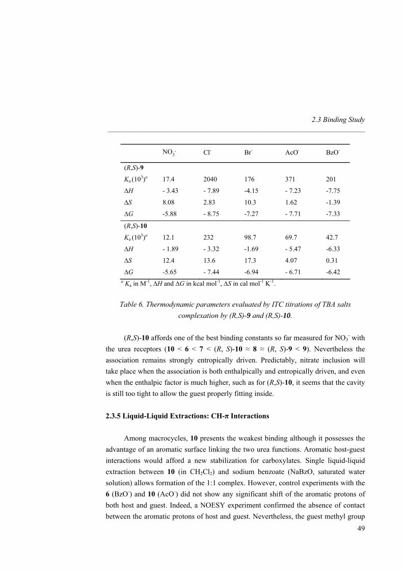
2.3 Binding Study _____________________________________________________________________
49
NO3- Cl- Br- AcO- BzO-
(R,S)-9
Ka (103)a 17.4 2040 176 371 201
∆H - 3.43 - 7.89 -4.15 - 7.23 -7.75
∆S 8.08 2.83 10.3 1.62 -1.39
∆G -5.88 - 8.75 -7.27 - 7.71 -7.33
(R,S)-10
Ka (103)a 12.1 232 98.7 69.7 42.7
∆H - 1.89 - 3.32 -1.69 - 5.47 -6.33
∆S 12.4 13.6 17.3 4.07 0.31
∆G -5.65 - 7.44 -6.94 - 6.71 -6.42 a Ka in M-1, ∆H and ∆G in kcal mol-1, ∆S in cal mol-1 K-1.
Table 6. Thermodynamic parameters evaluated by ITC titrations of TBA salts complexation by (R,S)-9 and (R,S)-10.
(R,S)-10 affords one of the best binding constants so far measured for NO3
- with the urea receptors (10 < 6 < 7 < (R, S)-10 ≈ 8 ≈ (R, S)-9 < 9). Nevertheless the association remains strongly entropically driven. Predictably, nitrate inclusion will take place when the association is both enthalpically and entropically driven, and even when the enthalpic factor is much higher, such as for (R,S)-10, it seems that the cavity is still too tight to allow the guest properly fitting inside. 2.3.5 Liquid-Liquid Extractions: CH-π Interactions
Among macrocycles, 10 presents the weakest binding although it possesses the advantage of an aromatic surface linking the two urea functions. Aromatic host-guest interactions would afford a new stabilization for carboxylates. Single liquid-liquid extraction between 10 (in CH2Cl2) and sodium benzoate (NaBzO, saturated water solution) allows formation of the 1:1 complex. However, control experiments with the 6 (BzO-) and 10 (AcO-) did not show any significant shift of the aromatic protons of both host and guest. Indeed, a NOESY experiment confirmed the absence of contact between the aromatic protons of host and guest. Nevertheless, the guest methyl group
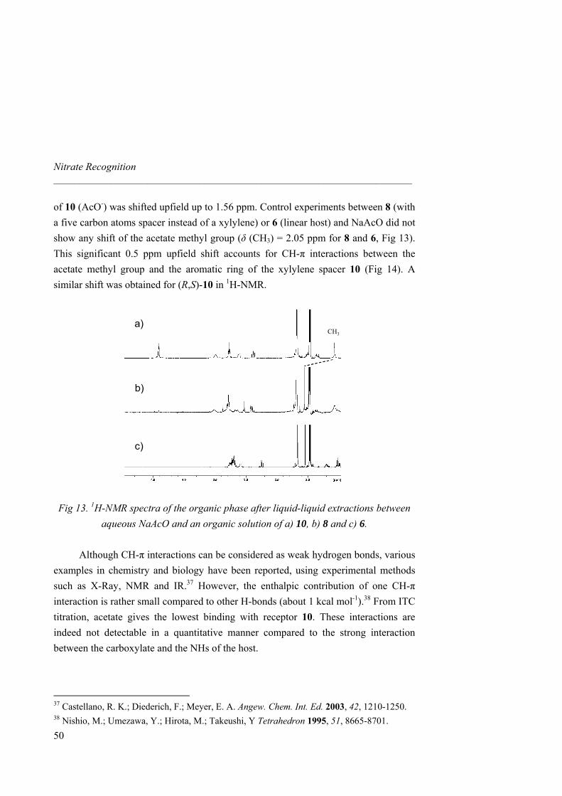
Nitrate Recognition _____________________________________________________________________
of 10 (AcO-) was shifted upfield up to 1.56 ppm. Control experiments between 8 (with a five carbon atoms spacer instead of a xylylene) or 6 (linear host) and NaAcO did not show any shift of the acetate methyl group (δ (CH3) = 2.05 ppm for 8 and 6, Fig 13). This significant 0.5 ppm upfield shift accounts for CH-π interactions between the acetate methyl group and the aromatic ring of the xylylene spacer 10 (Fig 14). A similar shift was obtained for (R,S)-10 in 1H-NMR.
CH3
a)
b)
c)
CH3
a)
b)
c)
Fig 13. 1H-NMR spectra of the organic phase after liquid-liquid extractions between aqueous NaAcO and an organic solution of a) 10, b) 8 and c) 6.
Although CH-π interactions can be considered as weak hydrogen bonds, various
examples in chemistry and biology have been reported, using experimental methods such as X-Ray, NMR and IR.37 However, the enthalpic contribution of one CH-π interaction is rather small compared to other H-bonds (about 1 kcal mol-1).38 From ITC titration, acetate gives the lowest binding with receptor 10. These interactions are indeed not detectable in a quantitative manner compared to the strong interaction between the carboxylate and the NHs of the host.
37 Castellano, R. K.; Diederich, F.; Meyer, E. A. Angew. Chem. Int. Ed. 2003, 42, 1210-1250. 38 Nishio, M.; Umezawa, Y.; Hirota, M.; Takeushi, Y Tetrahedron 1995, 51, 8665-8701.
50
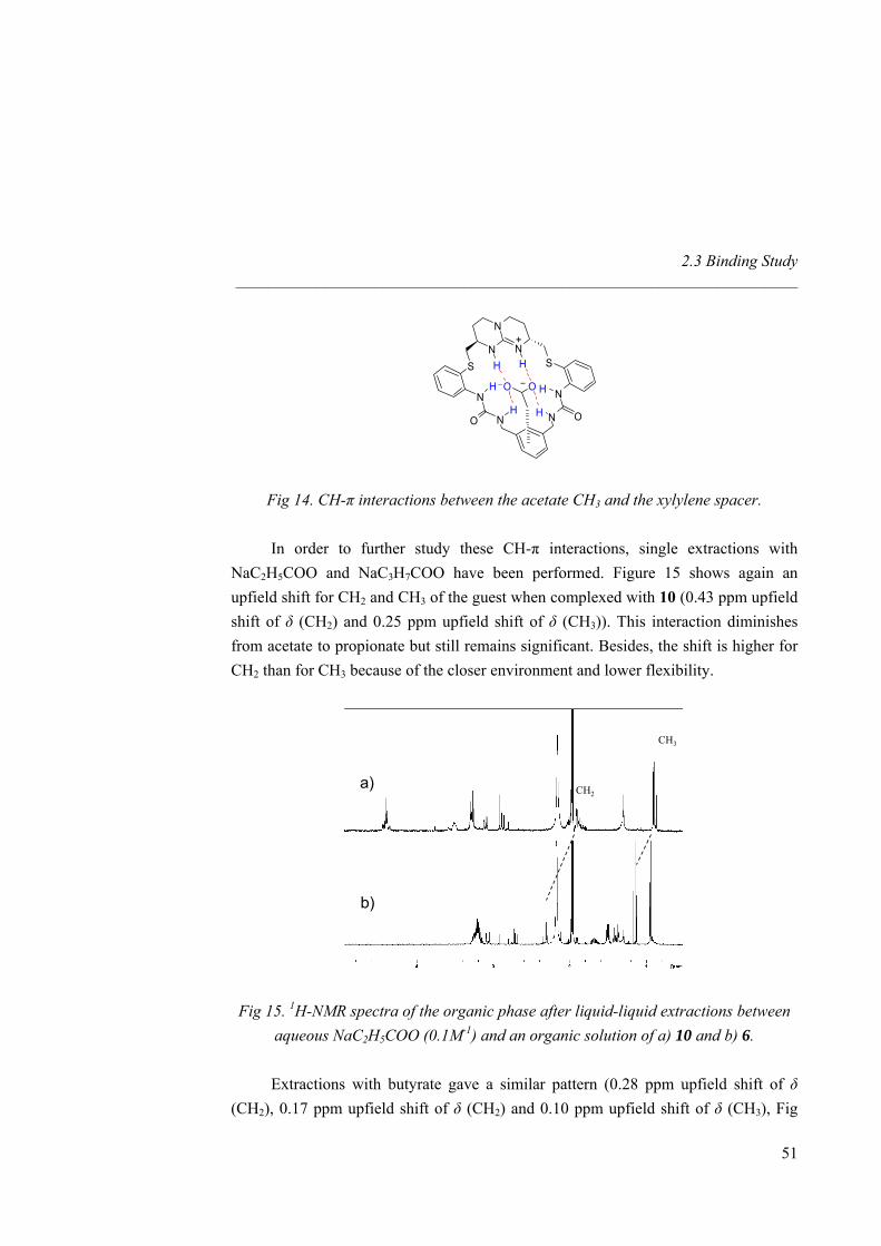
2.3 Binding Study _____________________________________________________________________
N
N
NH H
+
S S
N N
O ON N
OOH
H
H
H
Fig 14. CH-π interactions between the acetate CH3 and the xylylene spacer.
In order to further study these CH-π interactions, single extractions with NaC2H5COO and NaC3H7COO have been performed. Figure 15 shows again an upfield shift for CH2 and CH3 of the guest when complexed with 10 (0.43 ppm upfield shift of δ (CH2) and 0.25 ppm upfield shift of δ (CH3)). This interaction diminishes from acetate to propionate but still remains significant. Besides, the shift is higher for CH2 than for CH3 because of the closer environment and lower flexibility.
CH3
a)
b)
CH2
CH3
a)
b)
CH2
Fig 15. 1H-NMR spectra of the organic phase after liquid-liquid extractions between aqueous NaC2H5COO (0.1M-1) and an organic solution of a) 10 and b) 6.
Extractions with butyrate gave a similar pattern (0.28 ppm upfield shift of δ
(CH2), 0.17 ppm upfield shift of δ (CH2) and 0.10 ppm upfield shift of δ (CH3), Fig
51
Nitrate Recognition _____________________________________________________________________
16). Therefore, as the carbon chain of the carboxylate increases, the magnitude of the shift decreases (accounting for the higher flexibility of the guest).
CH3
a)
b)
CH2CH2
CH3
a)
b)
CH2CH2
Fig 16. 1H-NMR spectra of the organic phase after liquid-liquid extractions between aqueous NaC3H7COO (0.1M-1) and an organic solution of a) 10 and b) 6.
2.4 X-Ray Solid State Structures
7 (NO3-) was able to crystallize in CH3CN and its X-ray solid state structure was
resolved (an ORTEP plot is shown in Fig 17). The asymmetric unit contains three independent molecules partially disordered (molecules A/B/C). The NO3
- anion and the NHs of the hydrogen bond donor functions are not affected by this disorder (atoms C2, C3, C4 and C25, C26, C27 involved). The three refined independent molecules differ broadly in their conformations, but have a similar arrangement of the NO3
- in the center of the molecule. In all cases, NO3
- is bound to both NHguan and to one of the NHurea of only one of the urea functions (distances in Table 8). The second urea is pointing away from the macrocycle cavity forming intermolecular hydrogen bonds with another urea (NH···O=C hydrogen bonding network). As a result, the macrocycle is strongly folded with the guest pointing to the centre of the molecule with only two of the oxygen atoms located in this central area. The third oxygen is pointing to neighboring molecules achieving additional intermolecular contacts. Besides, for
52
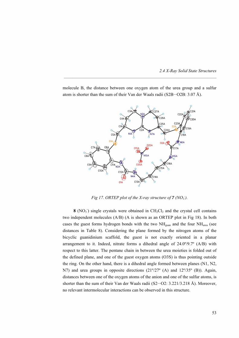
2.4 X-Ray Solid State Structures _____________________________________________________________________
molecule B, the distance between one oxygen atom of the urea group and a sulfur atom is shorter than the sum of their Van der Waals radii (S2B···O2B: 3.07 Å).
Fig 17. ORTEP plot of the X-ray structure of 7 (NO3-).
8 (NO3
-) single crystals were obtained in CH2Cl2 and the crystal cell contains two independent molecules (A/B) (A is shown as an ORTEP plot in Fig 18). In both cases the guest forms hydrogen bonds with the two NHguan and the four NHurea (see distances in Table 8). Considering the plane formed by the nitrogen atoms of the bicyclic guanidinium scaffold, the guest is not exactly oriented in a planar arrangement to it. Indeed, nitrate forms a dihedral angle of 24.0º/9.7º (A/B) with respect to this latter. The pentane chain in between the urea moieties is folded out of the defined plane, and one of the guest oxygen atoms (O3S) is thus pointing outside the ring. On the other hand, there is a dihedral angle formed between planes (N1, N2, N7) and urea groups in opposite directions (21º/27º (A) and 12º/35º (B)). Again, distances between one of the oxygen atoms of the anion and one of the sulfur atoms, is shorter than the sum of their Van der Waals radii (S2···O2: 3.221/3.218 Å). Moreover, no relevant intermolecular interactions can be observed in this structure.
53
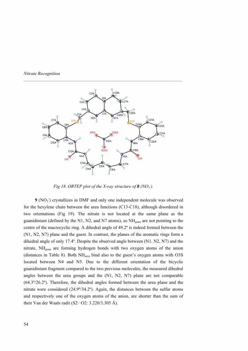
Nitrate Recognition _____________________________________________________________________
Fig 18. ORTEP plot of the X-ray structure of 8 (NO3-).
9 (NO3
-) crystallizes in DMF and only one independent molecule was observed for the hexylene chain between the urea functions (C13-C18), although disordered in two orientations (Fig 19). The nitrate is not located at the same plane as the guanidinium (defined by the N1, N2, and N7 atoms), so NHguan are not pointing to the centre of the macrocyclic ring. A dihedral angle of 49.2º is indeed formed between the (N1, N2, N7) plane and the guest. In contrast, the planes of the aromatic rings form a dihedral angle of only 17.4º. Despite the observed angle between (N1, N2, N7) and the nitrate, NHguan are forming hydrogen bonds with two oxygen atoms of the anion (distances in Table 8). Both NHurea bind also to the guest’s oxygen atoms with O3S located between N4 and N5. Due to the different orientation of the bicyclic guanidinium fragment compared to the two previous molecules, the measured dihedral angles between the urea groups and the (N1, N2, N7) plane are not comparable (64.3º/26.2º). Therefore, the dihedral angles formed between the urea plane and the nitrate were considered (24.9º/34.2º). Again, the distances between the sulfur atoms and respectively one of the oxygen atoms of the anion, are shorter than the sum of their Van der Waals radii (S2···O2: 3.220/3.305 Å).
54
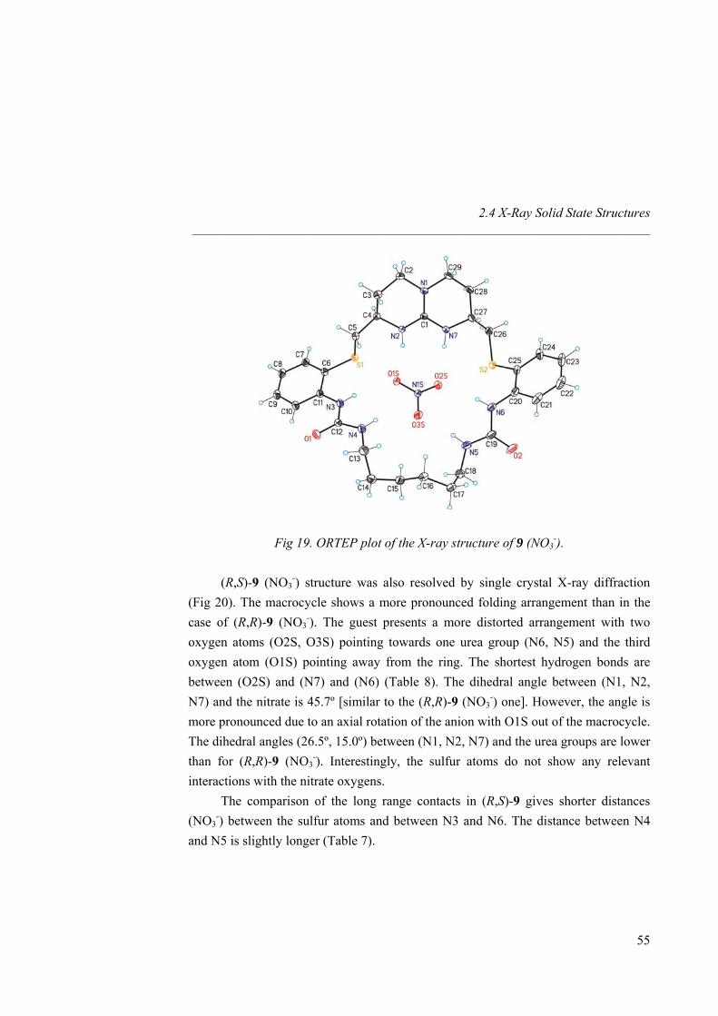
2.4 X-Ray Solid State Structures _____________________________________________________________________
Fig 19. ORTEP plot of the X-ray structure of 9 (NO3-).
(R,S)-9 (NO3
-) structure was also resolved by single crystal X-ray diffraction (Fig 20). The macrocycle shows a more pronounced folding arrangement than in the case of (R,R)-9 (NO3
-). The guest presents a more distorted arrangement with two oxygen atoms (O2S, O3S) pointing towards one urea group (N6, N5) and the third oxygen atom (O1S) pointing away from the ring. The shortest hydrogen bonds are between (O2S) and (N7) and (N6) (Table 8). The dihedral angle between (N1, N2, N7) and the nitrate is 45.7º [similar to the (R,R)-9 (NO3
-) one]. However, the angle is more pronounced due to an axial rotation of the anion with O1S out of the macrocycle. The dihedral angles (26.5º, 15.0º) between (N1, N2, N7) and the urea groups are lower than for (R,R)-9 (NO3
-). Interestingly, the sulfur atoms do not show any relevant interactions with the nitrate oxygens. The comparison of the long range contacts in (R,S)-9 gives shorter distances (NO3
-) between the sulfur atoms and between N3 and N6. The distance between N4 and N5 is slightly longer (Table 7).
55
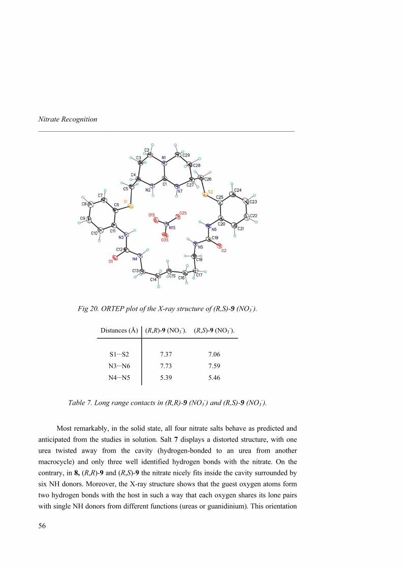
Nitrate Recognition _____________________________________________________________________
Fig 20. ORTEP plot of the X-ray structure of (R,S)-9 (NO3-).
Distances (Å) (R,R)-9 (NO3
-). (R,S)-9 (NO3-).
S1···S2
7.37
7.06
N3···N6 7.73 7.59
N4···N5 5.39 5.46
Table 7. Long range contacts in (R,R)-9 (NO3-) and (R,S)-9 (NO3
-).
Most remarkably, in the solid state, all four nitrate salts behave as predicted and anticipated from the studies in solution. Salt 7 displays a distorted structure, with one urea twisted away from the cavity (hydrogen-bonded to an urea from another macrocycle) and only three well identified hydrogen bonds with the nitrate. On the contrary, in 8, (R,R)-9 and (R,S)-9 the nitrate nicely fits inside the cavity surrounded by six NH donors. Moreover, the X-ray structure shows that the guest oxygen atoms form two hydrogen bonds with the host in such a way that each oxygen shares its lone pairs with single NH donors from different functions (ureas or guanidinium). This orientation
56
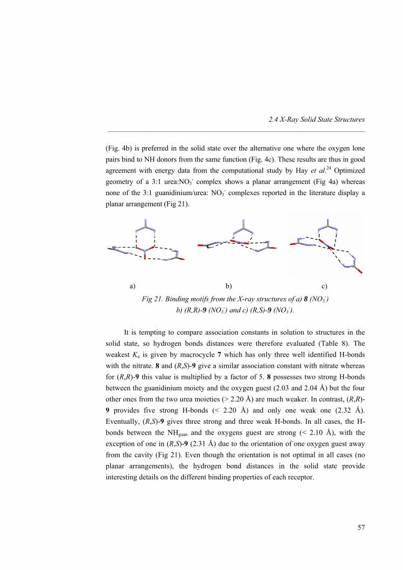
2.4 X-Ray Solid State Structures _____________________________________________________________________
(Fig. 4b) is preferred in the solid state over the alternative one where the oxygen lone pairs bind to NH donors from the same function (Fig. 4c). These results are thus in good agreement with energy data from the computational study by Hay et al.24 Optimized geometry of a 3:1 urea:NO3
- complex shows a planar arrangement (Fig 4a) whereas none of the 3:1 guanidinium/urea: NO3
- complexes reported in the literature display a planar arrangement (Fig 21).
a) b) c)
Fig 21. Binding motifs from the X-ray structures of a) 8 (NO3-)
b) (R,R)-9 (NO3-) and c) (R,S)-9 (NO3
-).
It is tempting to compare association constants in solution to structures in the solid state, so hydrogen bonds distances were therefore evaluated (Table 8). The weakest Ka is given by macrocycle 7 which has only three well identified H-bonds with the nitrate. 8 and (R,S)-9 give a similar association constant with nitrate whereas for (R,R)-9 this value is multiplied by a factor of 5. 8 possesses two strong H-bonds between the guanidinium moiety and the oxygen guest (2.03 and 2.04 Å) but the four other ones from the two urea moieties (> 2.20 Å) are much weaker. In contrast, (R,R)-9 provides five strong H-bonds (< 2.20 Å) and only one weak one (2.32 Å). Eventually, (R,S)-9 gives three strong and three weak H-bonds. In all cases, the H-bonds between the NHguan and the oxygens guest are strong (< 2.10 Å), with the exception of one in (R,S)-9 (2.31 Å) due to the orientation of one oxygen guest away from the cavity (Fig 21). Even though the orientation is not optimal in all cases (no planar arrangements), the hydrogen bond distances in the solid state provide interesting details on the different binding properties of each receptor.
57

Nitrate Recognition _____________________________________________________________________
58
Distance in Åa 7 8 9 (R,S)-9
O1S···N2 (O1S···H2)
2.887 (2.04)
2.864 (2.04)
2.842 (2.02)
3.135 (2.31)
O2S···N7 (O2S···H7) 2.884 (2.03) 2.883 (2.03) 2.951 (2.10) 2.917 (2.07)
O1S···N3 (O1S···H3) 2.904 (2.05) 3.205 (2.39) 3.114 (2.32) 3.089 (2.35)
O3S···N4 (O3S···H4) 3.082 (2.30) 3.036 (2.28) 2.932 (2.18) 3.047 (2.17)
O3S···N5 (O3S···H5) 3.067 (2.23) 2.903 (2.07) 3.095 (2.26)
O2S···N6 (O2S···H6) 3.074 (2.23) 3.048 (2.19) 2.961 (2.10) a The observed distances to hydrogen atoms are uncorrected.
Table 8. Intramolecular contacts of 7, 8, 9 and (R,S)-9 with NO3-.
4 (Cl-) crystallizes in an ideal C2 symmetry with two independent half molecules
(A/B) in the crystal packing (Fig 22). The independent molecules differ in the orientation of the aromatic rings and the conformation of the central bicyclic guanidinium. The guest is located on the C2-axis centered between the hydrogen atoms of N2 forming hydrogen bonds (Table 9). On the other hand, the guest is stabilized by one of the hydrogen atoms of each N3. Moreover, chloride has intermolecular contacts to different hydrogen atoms of neighboring molecules: the shortest one is with the N3-atoms (Cl1A···N3B2: 3.705, Cl1A···H3BE 2.83 Å, uncorrected). Considering the plane (C1, N1, N2) and the plane formed by N3 and the hydrogen atoms linked to it a dihedral angle of 41.8º/49.1º (A/B) results. This dihedral angle indicates that one hydrogen atom of N3 is pointing to the central Cl- whereas the second one is pointing to the guest of a neighboring molecule.
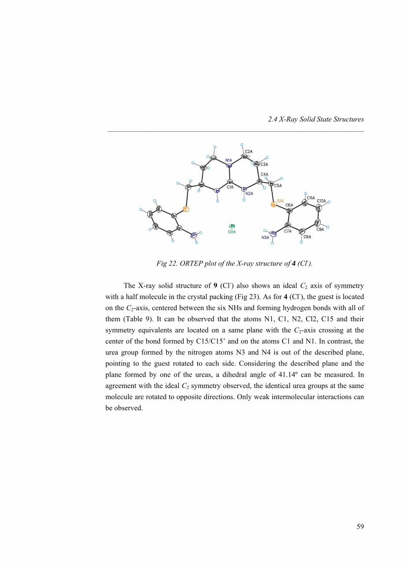
2.4 X-Ray Solid State Structures _____________________________________________________________________
Fig 22. ORTEP plot of the X-ray structure of 4 (Cl-).
The X-ray solid structure of 9 (Cl-) also shows an ideal C2 axis of symmetry with a half molecule in the crystal packing (Fig 23). As for 4 (Cl-), the guest is located on the C2-axis, centered between the six NHs and forming hydrogen bonds with all of them (Table 9). It can be observed that the atoms N1, C1, N2, Cl2, C15 and their symmetry equivalents are located on a same plane with the C2-axis crossing at the center of the bond formed by C15/C15’ and on the atoms C1 and N1. In contrast, the urea group formed by the nitrogen atoms N3 and N4 is out of the described plane, pointing to the guest rotated to each side. Considering the described plane and the plane formed by one of the ureas, a dihedral angle of 41.14º can be measured. In agreement with the ideal C2 symmetry observed, the identical urea groups at the same molecule are rotated to opposite directions. Only weak intermolecular interactions can be observed.
59
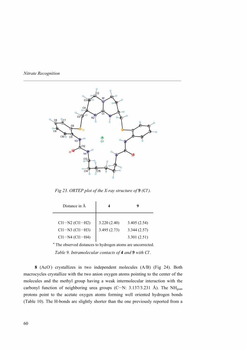
Nitrate Recognition _____________________________________________________________________
Fig 23. ORTEP plot of the X-ray structure of 9 (Cl-).
Distance in Å 4 9
Cl1···N2 (Cl1···H2)
3.220 (2.40)
3.405 (2.54)
Cl1···N3 (Cl1···H3) 3.495 (2.73) 3.344 (2.57)
Cl1···N4 (Cl1···H4) 3.301 (2.51) a The observed distances to hydrogen atoms are uncorrected.
Table 9. Intramolecular contacts of 4 and 9 with Cl-.
8 (AcO-) crystallizes in two independent molecules (A/B) (Fig 24). Both
macrocycles crystallize with the two anion oxygen atoms pointing to the center of the molecules and the methyl group having a weak intermolecular interaction with the carbonyl function of neighboring urea groups (C···N: 3.137/3.231 Å). The NHguan protons point to the acetate oxygen atoms forming well oriented hydrogen bonds (Table 10). The H-bonds are slightly shorter than the one previously reported from a
60
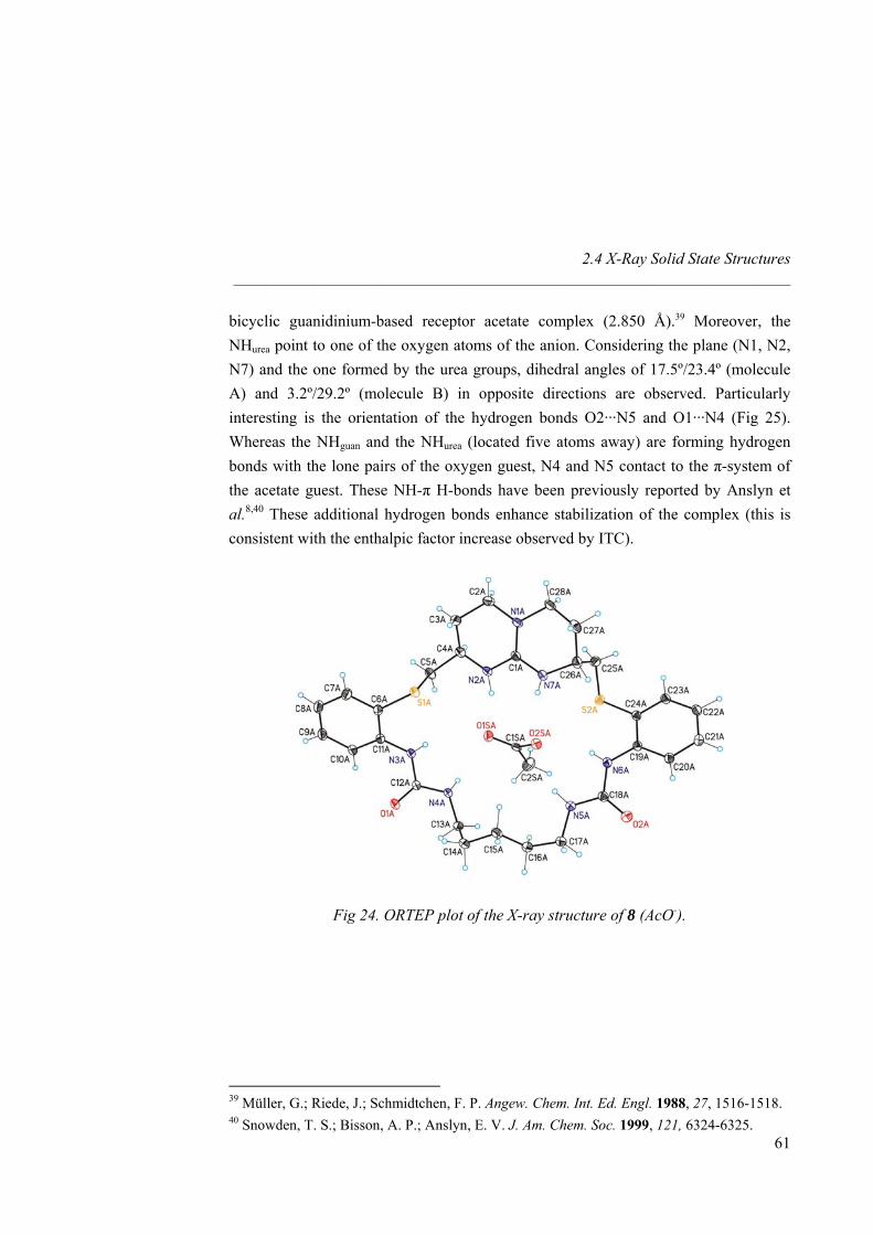
2.4 X-Ray Solid State Structures _____________________________________________________________________
bicyclic guanidinium-based receptor acetate complex (2.850 Å).39 Moreover, the NHurea point to one of the oxygen atoms of the anion. Considering the plane (N1, N2, N7) and the one formed by the urea groups, dihedral angles of 17.5º/23.4º (molecule A) and 3.2º/29.2º (molecule B) in opposite directions are observed. Particularly interesting is the orientation of the hydrogen bonds O2···N5 and O1···N4 (Fig 25). Whereas the NHguan and the NHurea (located five atoms away) are forming hydrogen bonds with the lone pairs of the oxygen guest, N4 and N5 contact to the π-system of the acetate guest. These NH-π H-bonds have been previously reported by Anslyn et al.8,40 These additional hydrogen bonds enhance stabilization of the complex (this is consistent with the enthalpic factor increase observed by ITC).
Fig 24. ORTEP plot of the X-ray structure of 8 (AcO-).
39 Müller, G.; Riede, J.; Schmidtchen, F. P. Angew. Chem. Int. Ed. Engl. 1988, 27, 1516-1518. 40 Snowden, T. S.; Bisson, A. P.; Anslyn, E. V. J. Am. Chem. Soc. 1999, 121, 6324-6325.
61

Nitrate Recognition _____________________________________________________________________
Distance in Åa 8
O1S···N2 (O1S···H2)
2.812 (1.93)
O2S···N7 (O2S···H7) 2.806 (2.10)
O1S···N3 (O1S···H3) 3.273 (2.65)
O1S···N4 (O1S···H4) 2.928 (2.24)
O2S···N5 (O2S···H5) 2.858 (2.06)
O2S···N6 (O2S···H6) 2.947 (2.28) a The observed distances to hydrogen atoms are uncorrected.
Table 10. Intramolecular contacts of 8 with AcO-.
Fig 25. Binding motif from the X-ray structure of 8 (AcO-)
(only three H-bonds are shown for clarity).
Comparison of these X-ray structures revealed that the guanidinium-based receptors are flexible enough to adapt themselves to the guest structures. Spherical anions of suitable size (such as Cl-) fit nicely inside the macrocycle cavity of 9 with six strong hydrogen bonds wrapping around the anion. The structural results in the solid state are in good agreement with those obtained in solution (ITC titrations) and by the theoretical model studies by Hay et al. (Fig 26).
62
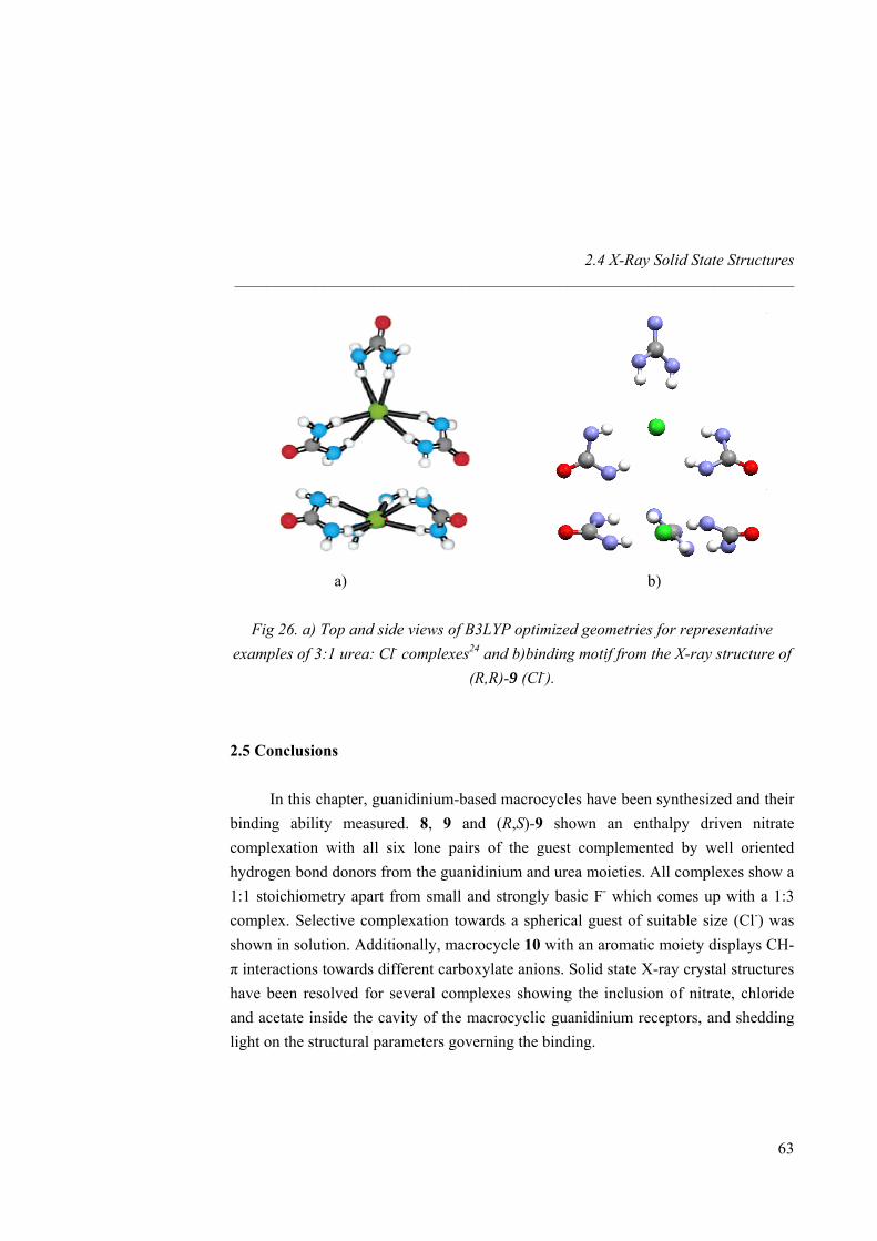
2.4 X-Ray Solid State Structures _____________________________________________________________________
a) b)
Fig 26. a) Top and side views of B3LYP optimized geometries for representative examples of 3:1 urea: Cl- complexes24 and b)binding motif from the X-ray structure of
(R,R)-9 (Cl-). 2.5 Conclusions
In this chapter, guanidinium-based macrocycles have been synthesized and their binding ability measured. 8, 9 and (R,S)-9 shown an enthalpy driven nitrate complexation with all six lone pairs of the guest complemented by well oriented hydrogen bond donors from the guanidinium and urea moieties. All complexes show a 1:1 stoichiometry apart from small and strongly basic F- which comes up with a 1:3 complex. Selective complexation towards a spherical guest of suitable size (Cl-) was shown in solution. Additionally, macrocycle 10 with an aromatic moiety displays CH-π interactions towards different carboxylate anions. Solid state X-ray crystal structures have been resolved for several complexes showing the inclusion of nitrate, chloride and acetate inside the cavity of the macrocyclic guanidinium receptors, and shedding light on the structural parameters governing the binding.
63
Nitrate Recognition _____________________________________________________________________
64
2.6 Experimental section 2.6.1 General Procedures
Synthesis. All commercially available reagents (Aldrich, Fluka, Acros, NovaBiochem, Panreac) were used without any further purification. The solvents were dried and distilled as described in the literature.41 Unless specified, all reactions were performed under nitrogen.
Chromatography. Thin layer chromatography (TLC) was performed on aluminum supported Alugram Sil G/UV254 (Macherey-Nagel). Column chromatography was done using silica gel by SDS (chromagel 60 ACC, 40-60 mm) following the procedure described by W. C. Still.42 HPLC chromatograms were recorded on an Agilent Technologies Serie 1100, with UV-diode array, fluorescence and light scattering detectors. The template synthesis experiments was followed by analytical HPLC (column: Synergi Max-RP 4 µm, 4.6 x 250 mm, UV-detector with λ = 254 nm). The mobile phase consisted of (ACN (0.1% TFA) / H2O (0.1% TFA), gradient: 35-100% ACN (0.1%TFA) in 15 minutes. The solvents were provided by Scharlau (HPLC gradient grade).
Analysis. Melting points were measured with a Büchi B-540 apparatus. The optical rotations [α]25
D were determined on a Perkin-Elmer 241 MC polarimeter using a cell (1 dm) at 20°C (NaD 589 nm). 1H-NMR and 13C-NMR spectra were recorded on a Bruker Avance 500 Ultrashield NMR spectrometer (1H: 500 MHz; 13C: 125 MHz) using the residual solvent peak as internal standard. Mass spectra were recorded on a Waters LCT Premier spectrometer using ESI technique or on a Bruker Autoflex MALDI-TOF instrument. Elemental analyses were performed on Perkin-Elmer 2400 CHN and 2400 CHNS apparatus.
ITC titrations. ITC titrations were performed using an isothermal titration Microcal VP-ITC microcalorimeter. All measurements were performed at 303K. The host solution was filled into the cell of the ITC instrument and guest solutions were added with the syringe. In each case control experiments with dilution of guest in the
41 Perrin, D. D.; Perrin, D. R.; Amarego, W. L. F. Purification of Laboratory Chemicals, 2nd Edition, Pergamon Press Ltd, Oxford, 1980. 42 Still, W. C.; Kahn, M.; Mitra, A. J. Org. Chem. 1978, 43, 2923-2925.
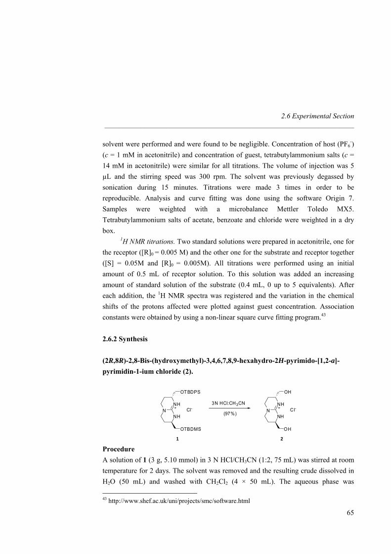
2.6 Experimental Section _____________________________________________________________________
solvent were performed and were found to be negligible. Concentration of host (PF6-)
(c = 1 mM in acetonitrile) and concentration of guest, tetrabutylammonium salts (c = 14 mM in acetonitrile) were similar for all titrations. The volume of injection was 5 µL and the stirring speed was 300 rpm. The solvent was previously degassed by sonication during 15 minutes. Titrations were made 3 times in order to be reproducible. Analysis and curve fitting was done using the software Origin 7. Samples were weighted with a microbalance Mettler Toledo MX5. Tetrabutylammonium salts of acetate, benzoate and chloride were weighted in a dry box.
1H NMR titrations. Two standard solutions were prepared in acetonitrile, one for the receptor ([R]0 = 0.005 M) and the other one for the substrate and receptor together ([S] = 0.05M and [R]0 = 0.005M). All titrations were performed using an initial amount of 0.5 mL of receptor solution. To this solution was added an increasing amount of standard solution of the substrate (0.4 mL, 0 up to 5 equivalents). After each addition, the 1H NMR spectra was registered and the variation in the chemical shifts of the protons affected were plotted against guest concentration. Association constants were obtained by using a non-linear square curve fitting program.43
2.6.2 Synthesis (2R,8R)-2,8-Bis-(hydroxymethyl)-3,4,6,7,8,9-hexahydro-2H-pyrimido-[1,2-a]-pyrimidin-1-ium chloride (2).
NNH
NH
OTBDPS
OTBDMS
Cl- NNH
NH
OH
OH
Cl-3N HCl:CH3CN
(97%)
1 2 Procedure A solution of 1 (3 g, 5.10 mmol) in 3 N HCl/CH3CN (1:2, 75 mL) was stirred at room temperature for 2 days. The solvent was removed and the resulting crude dissolved in H2O (50 mL) and washed with CH2Cl2 (4 × 50 mL). The aqueous phase was 43 http://www.shef.ac.uk/uni/projects/smc/software.html
65
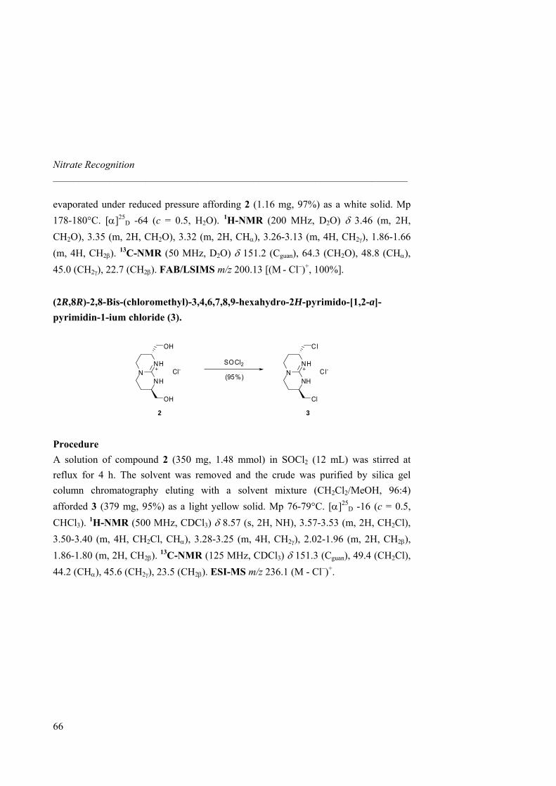
Nitrate Recognition _____________________________________________________________________
evaporated under reduced pressure affording 2 (1.16 mg, 97%) as a white solid. Mp 178-180°C. [α]25
D -64 (c = 0.5, H2O). 1H-NMR (200 MHz, D2O) δ 3.46 (m, 2H, CH2O), 3.35 (m, 2H, CH2O), 3.32 (m, 2H, CHα), 3.26-3.13 (m, 4H, CH2γ), 1.86-1.66 (m, 4H, CH2β). 13C-NMR (50 MHz, D2O) δ 151.2 (Cguan), 64.3 (CH2O), 48.8 (CHα), 45.0 (CH2γ), 22.7 (CH2β). FAB/LSIMS m/z 200.13 [(M - Cl–)+, 100%]. (2R,8R)-2,8-Bis-(chloromethyl)-3,4,6,7,8,9-hexahydro-2H-pyrimido-[1,2-a]-pyrimidin-1-ium chloride (3).
NNH
NH
OH
OH
Cl- NNH
NH
Cl
Cl
Cl-(95%)
2 3
SOCl2
Procedure A solution of compound 2 (350 mg, 1.48 mmol) in SOCl2 (12 mL) was stirred at reflux for 4 h. The solvent was removed and the crude was purified by silica gel column chromatography eluting with a solvent mixture (CH2Cl2/MeOH, 96:4) afforded 3 (379 mg, 95%) as a light yellow solid. Mp 76-79°C. [α]25
D -16 (c = 0.5, CHCl3). 1H-NMR (500 MHz, CDCl3) δ 8.57 (s, 2H, NH), 3.57-3.53 (m, 2H, CH2Cl), 3.50-3.40 (m, 4H, CH2Cl, CHα), 3.28-3.25 (m, 4H, CH2γ), 2.02-1.96 (m, 2H, CH2β), 1.86-1.80 (m, 2H, CH2β). 13C-NMR (125 MHz, CDCl3) δ 151.3 (Cguan), 49.4 (CH2Cl), 44.2 (CHα), 45.6 (CH2γ), 23.5 (CH2β). ESI-MS m/z 236.1 (M - Cl–)+.
66
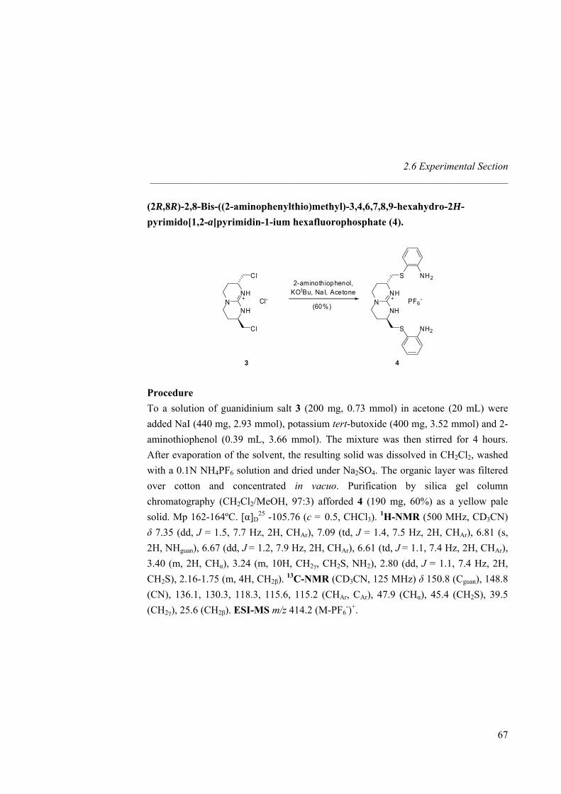
2.6 Experimental Section _____________________________________________________________________
(2R,8R)-2,8-Bis-((2-aminophenylthio)methyl)-3,4,6,7,8,9-hexahydro-2H-pyrimido[1,2-a]pyrimidin-1-ium hexafluorophosphate (4).
NNH
NH
Cl
Cl
Cl- NNH
NH
S
S
PF6-
(60%)
3 4
NH2
NH2
2-aminothiophenol,KOtBu, NaI, Acetone
Procedure To a solution of guanidinium salt 3 (200 mg, 0.73 mmol) in acetone (20 mL) were added NaI (440 mg, 2.93 mmol), potassium tert-butoxide (400 mg, 3.52 mmol) and 2-aminothiophenol (0.39 mL, 3.66 mmol). The mixture was then stirred for 4 hours. After evaporation of the solvent, the resulting solid was dissolved in CH2Cl2, washed with a 0.1N NH4PF6 solution and dried under Na2SO4. The organic layer was filtered over cotton and concentrated in vacuo. Purification by silica gel column chromatography (CH2Cl2/MeOH, 97:3) afforded 4 (190 mg, 60%) as a yellow pale solid. Mp 162-164ºC. [α]D
25 -105.76 (c = 0.5, CHCl3). 1H-NMR (500 MHz, CD3CN) δ 7.35 (dd, J = 1.5, 7.7 Hz, 2H, CHAr), 7.09 (td, J = 1.4, 7.5 Hz, 2H, CHAr), 6.81 (s, 2H, NHguan), 6.67 (dd, J = 1.2, 7.9 Hz, 2H, CHAr), 6.61 (td, J = 1.1, 7.4 Hz, 2H, CHAr), 3.40 (m, 2H, CHα), 3.24 (m, 10H, CH2γ, CH2S, NH2), 2.80 (dd, J = 1.1, 7.4 Hz, 2H, CH2S), 2.16-1.75 (m, 4H, CH2β). 13C-NMR (CD3CN, 125 MHz) δ 150.8 (Cguan), 148.8 (CN), 136.1, 130.3, 118.3, 115.6, 115.2 (CHAr, CAr), 47.9 (CHα), 45.4 (CH2S), 39.5 (CH2γ), 25.6 (CH2β). ESI-MS m/z 414.2 (M-PF6
-)+.
67
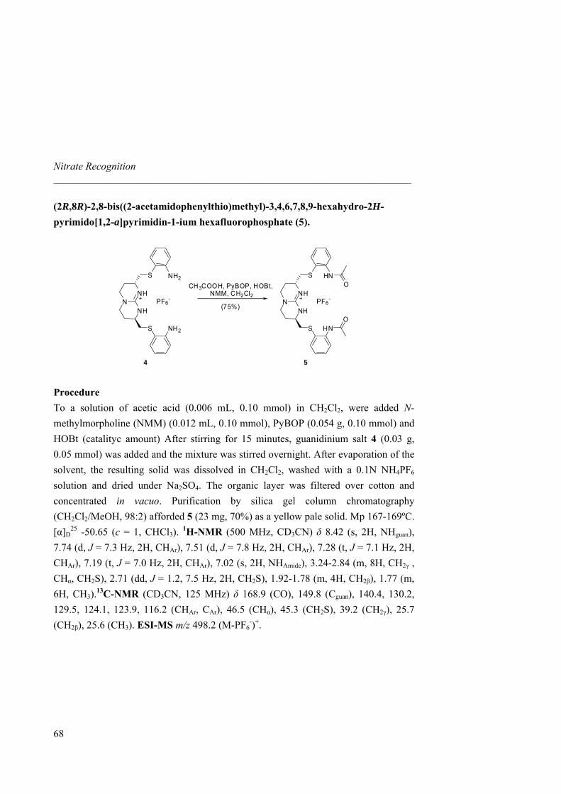
Nitrate Recognition _____________________________________________________________________
(2R,8R)-2,8-bis((2-acetamidophenylthio)methyl)-3,4,6,7,8,9-hexahydro-2H-pyrimido[1,2-a]pyrimidin-1-ium hexafluorophosphate (5).
NNH
NH
S
S
PF6-
(75%)
4 5
HN
HN
NNH
NH
S
S
PF6-
NH2
NH2
O
O
CH3COOH, PyBOP, HOBt,NMM, CH2Cl2
Procedure To a solution of acetic acid (0.006 mL, 0.10 mmol) in CH2Cl2, were added N-methylmorpholine (NMM) (0.012 mL, 0.10 mmol), PyBOP (0.054 g, 0.10 mmol) and HOBt (catalityc amount) After stirring for 15 minutes, guanidinium salt 4 (0.03 g, 0.05 mmol) was added and the mixture was stirred overnight. After evaporation of the solvent, the resulting solid was dissolved in CH2Cl2, washed with a 0.1N NH4PF6 solution and dried under Na2SO4. The organic layer was filtered over cotton and concentrated in vacuo. Purification by silica gel column chromatography (CH2Cl2/MeOH, 98:2) afforded 5 (23 mg, 70%) as a yellow pale solid. Mp 167-169ºC. [α]D
25 -50.65 (c = 1, CHCl3). 1H-NMR (500 MHz, CD3CN) δ 8.42 (s, 2H, NHguan), 7.74 (d, J = 7.3 Hz, 2H, CHAr), 7.51 (d, J = 7.8 Hz, 2H, CHAr), 7.28 (t, J = 7.1 Hz, 2H, CHAr), 7.19 (t, J = 7.0 Hz, 2H, CHAr), 7.02 (s, 2H, NHAmide), 3.24-2.84 (m, 8H, CH2γ , CHα, CH2S), 2.71 (dd, J = 1.2, 7.5 Hz, 2H, CH2S), 1.92-1.78 (m, 4H, CH2β), 1.77 (m, 6H, CH3).13C-NMR (CD3CN, 125 MHz) δ 168.9 (CO), 149.8 (Cguan), 140.4, 130.2, 129.5, 124.1, 123.9, 116.2 (CHAr, CAr), 46.5 (CHα), 45.3 (CH2S), 39.2 (CH2γ), 25.7 (CH2β), 25.6 (CH3). ESI-MS m/z 498.2 (M-PF6
-)+.
68
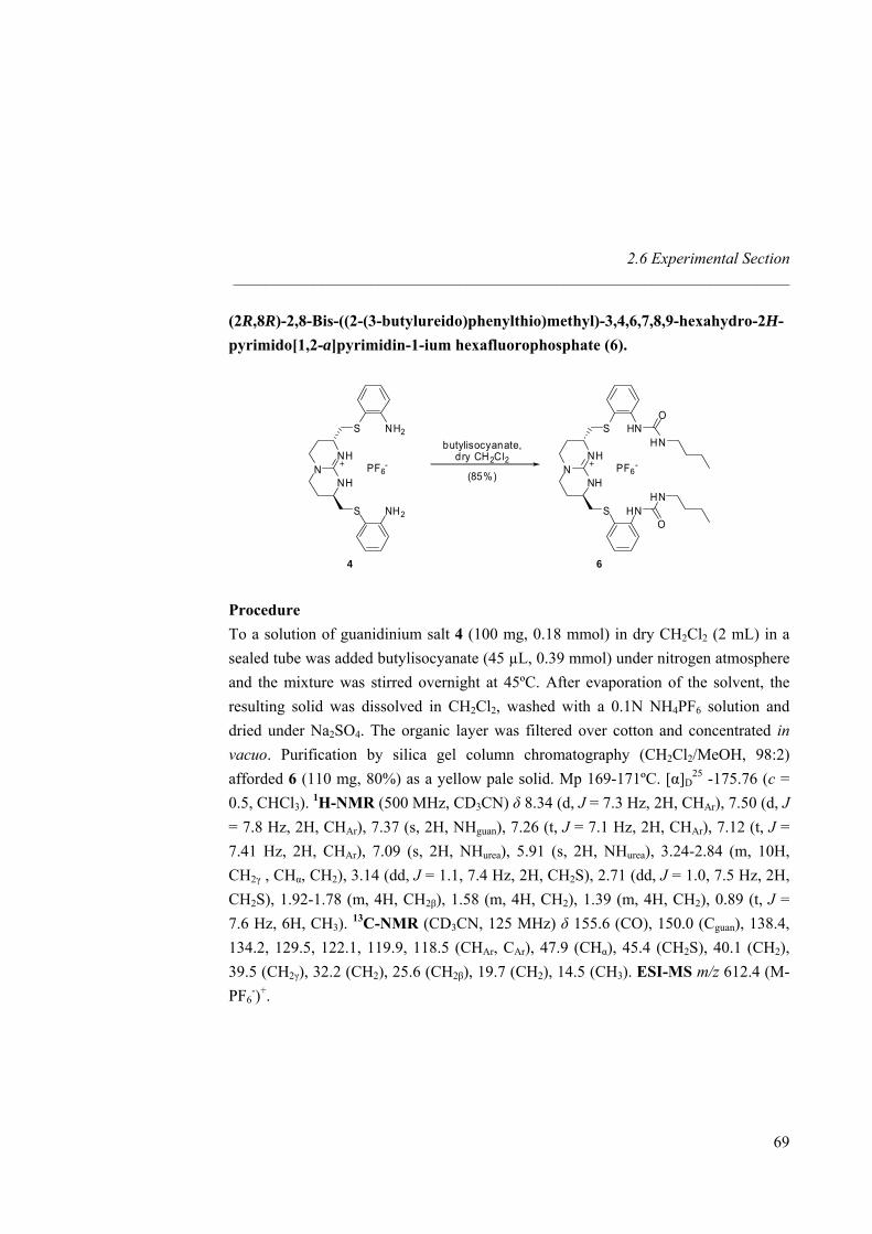
2.6 Experimental Section _____________________________________________________________________
(2R,8R)-2,8-Bis-((2-(3-butylureido)phenylthio)methyl)-3,4,6,7,8,9-hexahydro-2H-pyrimido[1,2-a]pyrimidin-1-ium hexafluorophosphate (6).
NNH
NH
S
S
PF6-
(85%)
4 6
HN
HN
NNH
NH
S
S
PF6-
NH2
NH2
O
HN
O
HN
butylisocyanate,dry CH2Cl2
Procedure To a solution of guanidinium salt 4 (100 mg, 0.18 mmol) in dry CH2Cl2 (2 mL) in a sealed tube was added butylisocyanate (45 µL, 0.39 mmol) under nitrogen atmosphere and the mixture was stirred overnight at 45ºC. After evaporation of the solvent, the resulting solid was dissolved in CH2Cl2, washed with a 0.1N NH4PF6 solution and dried under Na2SO4. The organic layer was filtered over cotton and concentrated in vacuo. Purification by silica gel column chromatography (CH2Cl2/MeOH, 98:2) afforded 6 (110 mg, 80%) as a yellow pale solid. Mp 169-171ºC. [α]D
25 -175.76 (c = 0.5, CHCl3). 1H-NMR (500 MHz, CD3CN) δ 8.34 (d, J = 7.3 Hz, 2H, CHAr), 7.50 (d, J = 7.8 Hz, 2H, CHAr), 7.37 (s, 2H, NHguan), 7.26 (t, J = 7.1 Hz, 2H, CHAr), 7.12 (t, J = 7.41 Hz, 2H, CHAr), 7.09 (s, 2H, NHurea), 5.91 (s, 2H, NHurea), 3.24-2.84 (m, 10H, CH2γ , CHα, CH2), 3.14 (dd, J = 1.1, 7.4 Hz, 2H, CH2S), 2.71 (dd, J = 1.0, 7.5 Hz, 2H, CH2S), 1.92-1.78 (m, 4H, CH2β), 1.58 (m, 4H, CH2), 1.39 (m, 4H, CH2), 0.89 (t, J = 7.6 Hz, 6H, CH3). 13C-NMR (CD3CN, 125 MHz) δ 155.6 (CO), 150.0 (Cguan), 138.4, 134.2, 129.5, 122.1, 119.9, 118.5 (CHAr, CAr), 47.9 (CHα), 45.4 (CH2S), 40.1 (CH2), 39.5 (CH2γ), 32.2 (CH2), 25.6 (CH2β), 19.7 (CH2), 14.5 (CH3). ESI-MS m/z 612.4 (M-PF6
-)+.
69

Nitrate Recognition _____________________________________________________________________
(2R,8R) bicyclic guanidinium macrocycle (7).
NNH
NH
S
S
PF6-
(40%)
4 7
HN
HN
NNH
NH
S
S
PF6-
NH2
NH2
O
HN
O
HN
1,6-diisocyanatobutane,dry CH2Cl2
Procedure To a solution of guanidinium salt 4 (30 mg, 0.06 mmol) in dry CH2Cl2 (2 mL), in a sealed tube was added 1,4-diisocyanatobutane (0.01 mL, 0.06 mmol) under nitrogen atmosphere and the mixture was stirred overnight at 45ºC. After evaporation of the solvent, the resulting solid was dissolved in CH2Cl2, washed with a 0.1N NH4PF6 solution and dried under Na2SO4. The organic layer was filtered over cotton and concentrated in vacuo. Purification by silica gel column chromatography (MeOH/CH2Cl2 100:0 to 98:2) afforded 7 as a pure yellow pale solid. The solid was then dissolved in acetonitrile and eluted in a Dowex resin (PF6
-) column to obtained 7 (PF6
-) (15 mg, 40 %). Mp 123-125ºC. [α]D25 -76.24 (c = 0.25, CHCl3). 1H-NMR (500
MHz, CD3CN) δ 7.61 (d, J = 8.3 Hz, 2H, CHAr), 7.48 (d, J = 7.8 Hz, 2H, CHAr), 7.15 (s, 2H, NHguan), 7.09 (t, J = 7.6 Hz, 2H, CHAr), 6.98 (t, J = 11.0 Hz, 2H, CHAr), 6.82 (s, 2H, NHurea), 5.92 (s, 2H, NHurea), 3.24-3.06 ( m, 10H, CH2γ, CHα, CH2S), 2.89 (m, 4H, CH2), 2.01-1.71 (m, 4H, CH2β), 1.51 (m, 4H, CH2). 13C-NMR (CD3CN, 125 MHz) δ 165.5 (Curea), 150.6 (Cguan), 140.9, 133.5, 128.5, 121.8, 120.1, 119.6 (CHAr, CAr), 46.9 (CHα), 44.4 (CH2S), 41.9 (CH2γ), 28.7 (CH2β), 25.9 (CH2), 25.0 (CH2). ESI-MS m/z 554.2493 (M-PF6
-)+.
70
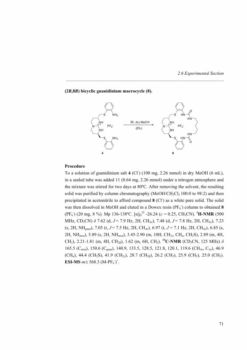
2.6 Experimental Section _____________________________________________________________________
(2R,8R) bicyclic guanidinium macrocycle (8).
NNH
NH
S
S
PF6-
(8%)
4 8
HN
HN
NNH
NH
S
S
PF6-
NH2
NH2
O
HN
O
HN
11, dry MeOH
Procedure To a solution of guanidinium salt 4 (Cl-) (100 mg, 2.26 mmol) in dry MeOH (6 mL), in a sealed tube was added 11 (0.64 mg, 2.26 mmol) under a nitrogen atmosphere and the mixture was stirred for two days at 80ºC. After removing the solvent, the resulting solid was purified by column chromatography (MeOH/CH2Cl2 100:0 to 98:2) and then precipitated in acetonitrile to afford compound 8 (Cl-) as a white pure solid. The solid was then dissolved in MeOH and eluted in a Dowex resin (PF6
-) column to obtained 8 (PF6
-) (20 mg, 8 %). Mp 136-138ºC. [α]D25 -26.24 (c = 0.25, CH3CN). 1H-NMR (500
MHz, CD3CN) δ 7.62 (d, J = 7.9 Hz, 2H, CHAr), 7.48 (d, J = 7.8 Hz, 2H, CHAr), 7.23 (s, 2H, NHguan), 7.05 (t, J = 7.5 Hz, 2H, CHAr), 6.97 (t, J = 7.1 Hz, 2H, CHAr), 6.85 (s, 2H, NHurea), 5.89 (s, 2H, NHurea), 3.45-2.90 (m, 10H, CH2γ, CHα, CH2S), 2.89 (m, 4H, CH2), 2.21-1.81 (m, 4H, CH2β), 1.62 (m, 6H, CH2). 13C-NMR (CD3CN, 125 MHz) δ 165.5 (Curea), 150.6 (Cguan), 140.9, 133.5, 128.5, 121.8, 120.1, 119.6 (CHAr, CAr), 46.9 (CHα), 44.4 (CH2S), 41.9 (CH2γ), 28.7 (CH2β), 26.2 (CH2), 25.9 (CH2), 25.0 (CH2). ESI-MS m/z 568.3 (M-PF6
-)+.
71
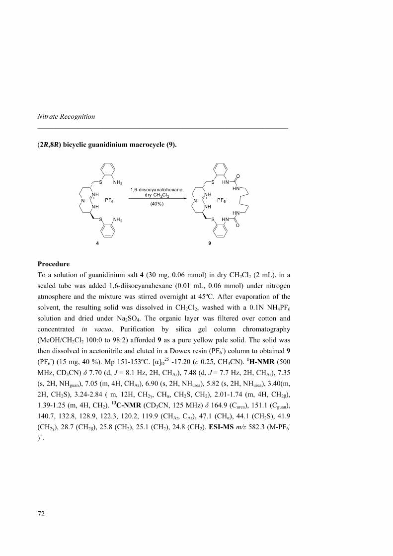
Nitrate Recognition _____________________________________________________________________
(2R,8R) bicyclic guanidinium macrocycle (9).
NNH
NH
S
S
PF6-
(40%)
4 9
HN
HN
NNH
NH
S
S
PF6-
NH2
NH2
O
HN
O
HN
1,6-diisocyanatohexane,dry CH2Cl2
Procedure To a solution of guanidinium salt 4 (30 mg, 0.06 mmol) in dry CH2Cl2 (2 mL), in a sealed tube was added 1,6-diisocyanahexane (0.01 mL, 0.06 mmol) under nitrogen atmosphere and the mixture was stirred overnight at 45ºC. After evaporation of the solvent, the resulting solid was dissolved in CH2Cl2, washed with a 0.1N NH4PF6 solution and dried under Na2SO4. The organic layer was filtered over cotton and concentrated in vacuo. Purification by silica gel column chromatography (MeOH/CH2Cl2 100:0 to 98:2) afforded 9 as a pure yellow pale solid. The solid was then dissolved in acetonitrile and eluted in a Dowex resin (PF6
-) column to obtained 9 (PF6
-) (15 mg, 40 %). Mp 151-153ºC. [α]D25 -17.20 (c 0.25, CH3CN). 1H-NMR (500
MHz, CD3CN) δ 7.70 (d, J = 8.1 Hz, 2H, CHAr), 7.48 (d, J = 7.7 Hz, 2H, CHAr), 7.35 (s, 2H, NHguan), 7.05 (m, 4H, CHAr), 6.90 (s, 2H, NHurea), 5.82 (s, 2H, NHurea), 3.40(m, 2H, CH2S), 3.24-2.84 ( m, 12H, CH2γ, CHα, CH2S, CH2), 2.01-1.74 (m, 4H, CH2β), 1.39-1.25 (m, 4H, CH2). 13C-NMR (CD3CN, 125 MHz) δ 164.9 (Curea), 151.1 (Cguan), 140.7, 132.8, 128.9, 122.3, 120.2, 119.9 (CHAr, CAr), 47.1 (CHα), 44.1 (CH2S), 41.9 (CH2γ), 28.7 (CH2β), 25.8 (CH2), 25.1 (CH2), 24.8 (CH2). ESI-MS m/z 582.3 (M-PF6
-
)+.
72
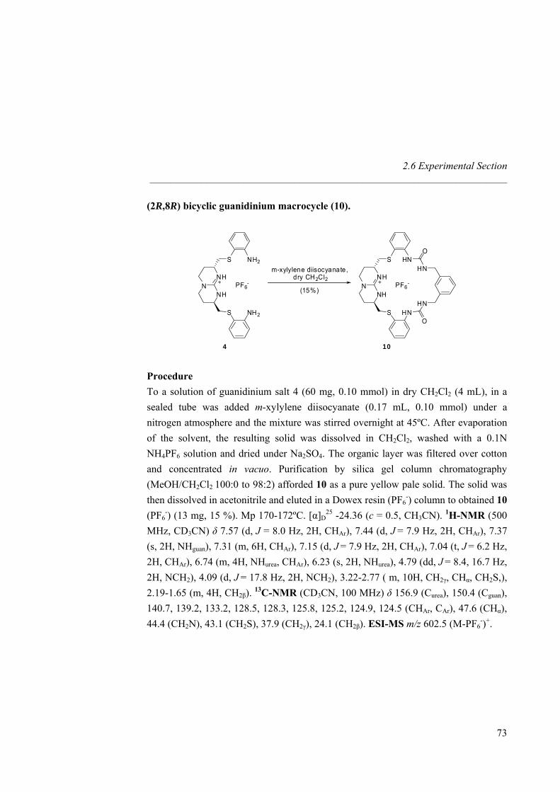
2.6 Experimental Section _____________________________________________________________________
(2R,8R) bicyclic guanidinium macrocycle (10).
NNH
NH
S
S
PF6-
(15%)
4 10
HN
HN
NNH
NH
S
S
PF6-
NH2
NH2
O
HN
O
HN
m-xylylene diisocyanate,dry CH2Cl2
Procedure To a solution of guanidinium salt 4 (60 mg, 0.10 mmol) in dry CH2Cl2 (4 mL), in a sealed tube was added m-xylylene diisocyanate (0.17 mL, 0.10 mmol) under a nitrogen atmosphere and the mixture was stirred overnight at 45ºC. After evaporation of the solvent, the resulting solid was dissolved in CH2Cl2, washed with a 0.1N NH4PF6 solution and dried under Na2SO4. The organic layer was filtered over cotton and concentrated in vacuo. Purification by silica gel column chromatography (MeOH/CH2Cl2 100:0 to 98:2) afforded 10 as a pure yellow pale solid. The solid was then dissolved in acetonitrile and eluted in a Dowex resin (PF6
-) column to obtained 10 (PF6
-) (13 mg, 15 %). Mp 170-172ºC. [α]D25 -24.36 (c = 0.5, CH3CN). 1H-NMR (500
MHz, CD3CN) δ 7.57 (d, J = 8.0 Hz, 2H, CHAr), 7.44 (d, J = 7.9 Hz, 2H, CHAr), 7.37 (s, 2H, NHguan), 7.31 (m, 6H, CHAr), 7.15 (d, J = 7.9 Hz, 2H, CHAr), 7.04 (t, J = 6.2 Hz, 2H, CHAr), 6.74 (m, 4H, NHurea, CHAr), 6.23 (s, 2H, NHurea), 4.79 (dd, J = 8.4, 16.7 Hz, 2H, NCH2), 4.09 (d, J = 17.8 Hz, 2H, NCH2), 3.22-2.77 ( m, 10H, CH2γ, CHα, CH2S,), 2.19-1.65 (m, 4H, CH2β). 13C-NMR (CD3CN, 100 MHz) δ 156.9 (Curea), 150.4 (Cguan), 140.7, 139.2, 133.2, 128.5, 128.3, 125.8, 125.2, 124.9, 124.5 (CHAr, CAr), 47.6 (CHα), 44.4 (CH2N), 43.1 (CH2S), 37.9 (CH2γ), 24.1 (CH2β). ESI-MS m/z 602.5 (M-PF6
-)+.
73
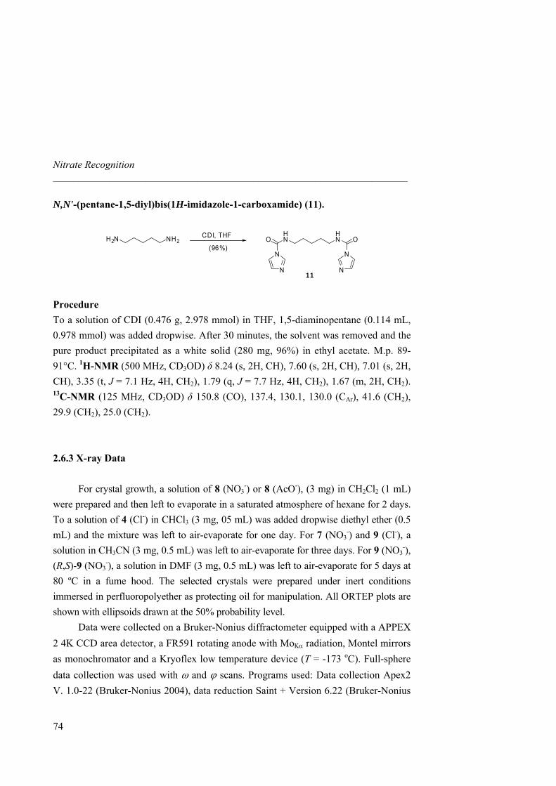
Nitrate Recognition _____________________________________________________________________
N,N'-(pentane-1,5-diyl)bis(1H-imidazole-1-carboxamide) (11).
NH2H2NHN
HNO
N
O
N
N N11
CDI, THF
(96%)
Procedure To a solution of CDI (0.476 g, 2.978 mmol) in THF, 1,5-diaminopentane (0.114 mL, 0.978 mmol) was added dropwise. After 30 minutes, the solvent was removed and the pure product precipitated as a white solid (280 mg, 96%) in ethyl acetate. M.p. 89-91°C. 1H-NMR (500 MHz, CD3OD) δ 8.24 (s, 2H, CH), 7.60 (s, 2H, CH), 7.01 (s, 2H, CH), 3.35 (t, J = 7.1 Hz, 4H, CH2), 1.79 (q, J = 7.7 Hz, 4H, CH2), 1.67 (m, 2H, CH2). 13C-NMR (125 MHz, CD3OD) δ 150.8 (CO), 137.4, 130.1, 130.0 (CAr), 41.6 (CH2), 29.9 (CH2), 25.0 (CH2). 2.6.3 X-ray Data For crystal growth, a solution of 8 (NO3
-) or 8 (AcO-), (3 mg) in CH2Cl2 (1 mL) were prepared and then left to evaporate in a saturated atmosphere of hexane for 2 days. To a solution of 4 (Cl-) in CHCl3 (3 mg, 05 mL) was added dropwise diethyl ether (0.5 mL) and the mixture was left to air-evaporate for one day. For 7 (NO3
-) and 9 (Cl-), a solution in CH3CN (3 mg, 0.5 mL) was left to air-evaporate for three days. For 9 (NO3
-), (R,S)-9 (NO3
-), a solution in DMF (3 mg, 0.5 mL) was left to air-evaporate for 5 days at 80 ºC in a fume hood. The selected crystals were prepared under inert conditions immersed in perfluoropolyether as protecting oil for manipulation. All ORTEP plots are shown with ellipsoids drawn at the 50% probability level.
Data were collected on a Bruker-Nonius diffractometer equipped with a APPEX 2 4K CCD area detector, a FR591 rotating anode with MoKα radiation, Montel mirrors as monochromator and a Kryoflex low temperature device (T = -173 oC). Full-sphere data collection was used with ω and ϕ scans. Programs used: Data collection Apex2 V. 1.0-22 (Bruker-Nonius 2004), data reduction Saint + Version 6.22 (Bruker-Nonius
74
2.6 Experimental Section _____________________________________________________________________
75
2001) and absorption correction SADABS V. 2.10 (2003). Structure Solution and Refinement: SHELXTL Version 6.10 (Sheldrick, 2000) were used.44
The structure of 7 (NO3-) can be solved and refined in the higher symmetric
orthorhombic chiral space group P212121 or in the lower symmetric monoclinic chiral space group P21 as a monoclinic twin which emulates a higher symmetric orthorhombic crystal with a β angle close to 90º. In the higher symmetric space group the three independent refined structures are partly disordered and the obtained R-values are higher [R1 = 0.0752 (Fo > 4σFo)]. In the lower symmetric space group which refines with better R-values applying a twin matrix (1 0 0 0 -1 0 0 0 -1, basf 0.43), six different independent molecules without disorder can be refined [R1 = 0.0654 (Fo > 4σFo)]. In this case the six independent molecules are showing correlation effects. In order to discuss the crystal packing and the arrangement of the NO3
- anions the higher symmetrical and partly disordered structure solution was selected.
The different data obtained for compound 8 (NO3-) corresponded always to the
overlap of the reflections of more than one crystal. The best R1-value (Fo > 4σFo) reached was not lower than 11%. Using the program Gemini45 the overlapped data of two crystals could be first separated for integration and later refined as one data set taking in account the overlapping reflections. If only the reflections with not overlap were considered the R1-value (Fo > 4σFo) reached for 6182 independent reflections was 6.86%. If a factor was included for reflections overlapping more than 50% a R1-value (Fo > 4σFo) 8.12% (20146 reflections) was reached. If different factors were applied in correlation with the different degrees of overlapping of the reflections a R1-value (Fo > 4σFo) 9.31% (35238 reflections) could be reached. For the structure description the refinement model with one overlapping factor was used. Additionally to the described overlapping of crystals pseudosymmetry from P1 to P-1 was detected, being, for a chiral structure, the correct space group P1.
44 G. M. Sheldrick (1998) SHELXTL Crystallographic System Ver. 5.10, Bruker AXS, Inc.: Madison, Wisconsin. 45 Gemini, Autoindexing Program for Twinned Crystals, Vers. 10.02 Release 5/2000; Bruker AXS Inc., Madison, Wisconsin (USA).
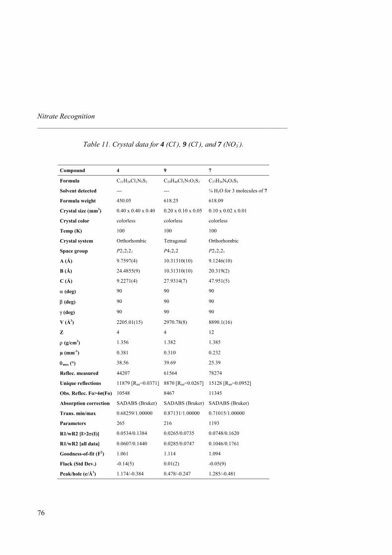
Nitrate Recognition _____________________________________________________________________
76
Table 11. Crystal data for 4 (Cl-), 9 (Cl-), and 7 (NO3-).
Compound 4 9 7
Formula C21H28Cl1N5S2 C29H40Cl1N7O2S2 C27H36N8O5S2
Solvent detected --- --- ¼ H2O for 3 molecules of 7
Formula weight 450.05 618.25 618.09
Crystal size (mm3) 0.40 x 0.40 x 0.40 0.20 x 0.10 x 0.05 0.10 x 0.02 x 0.01
Crystal color colorless colorless colorless
Temp (K) 100 100 100
Crystal system Orthorhombic Tetragonal Orthorhombic
Space group P212121 P41212 P212121
A (Å) 9.7597(4) 10.31310(10) 9.1246(10)
B (Å) 24.4855(9) 10.31310(10) 20.319(2)
C (Å) 9.2271(4) 27.9314(7) 47.951(5)
α (deg) 90 90 90
β (deg) 90 90 90
γ (deg) 90 90 90
V (Å3) 2205.01(15) 2970.78(8) 8890.1(16)
Z 4 4 12
ρ (g/cm3) 1.356 1.382 1.385
µ (mm-1) 0.381 0.310 0.232
θmax (°) 38.56 39.69 25.39
Reflec. measured 44207 61564 78274
Unique reflections 11879 [Rint=0.0371] 8870 [Rint=0.0267] 15128 [Rint=0.0952]
Obs. Reflec. Fo>4σ(Fo) 10548 8467 11345
Absorption correction SADABS (Bruker) SADABS (Bruker) SADABS (Bruker)
Trans. min/max 0.68259/1.00000 0.87131/1.00000 0.71015/1.00000
Parameters 265 216 1193
R1/wR2 [I>2σ(I)] 0.0534/0.1384 0.0265/0.0735 0.0748/0.1620
R1/wR2 [all data] 0.0607/0.1440 0.0285/0.0747 0.1046/0.1761
Goodness-of-fit (F2) 1.061 1.114 1.094
Flack (Std Dev.) -0.14(5) 0.01(2) -0.05(9)
Peak/hole (e/Å3) 1.174/-0.384 0.478/-0.247 1.285/-0.481
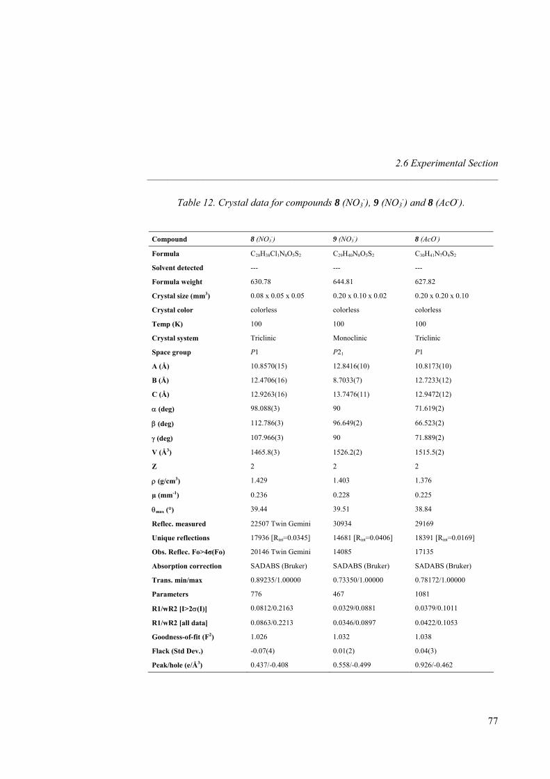
2.6 Experimental Section _____________________________________________________________________
77
Table 12. Crystal data for compounds 8 (NO3-), 9 (NO3
-) and 8 (AcO-).
Compound 8 (NO3-) 9 (NO3
-) 8 (AcO-)
Formula C28H38Cl1N8O5S2 C29H40N8O5S2 C30H41N7O4S2
Solvent detected --- --- ---
Formula weight 630.78 644.81 627.82
Crystal size (mm3) 0.08 x 0.05 x 0.05 0.20 x 0.10 x 0.02 0.20 x 0.20 x 0.10
Crystal color colorless colorless colorless
Temp (K) 100 100 100
Crystal system Triclinic Monoclinic Triclinic
Space group P1 P21 P1
A (Å) 10.8570(15) 12.8416(10) 10.8173(10)
B (Å) 12.4706(16) 8.7033(7) 12.7233(12)
C (Å) 12.9263(16) 13.7476(11) 12.9472(12)
α (deg) 98.088(3) 90 71.619(2)
β (deg) 112.786(3) 96.649(2) 66.523(2)
γ (deg) 107.966(3) 90 71.889(2)
V (Å3) 1465.8(3) 1526.2(2) 1515.5(2)
Z 2 2 2
ρ (g/cm3) 1.429 1.403 1.376
µ (mm-1) 0.236 0.228 0.225
θmax (°) 39.44 39.51 38.84
Reflec. measured 22507 Twin Gemini 30934 29169
Unique reflections 17936 [Rint=0.0345] 14681 [Rint=0.0406] 18391 [Rint=0.0169]
Obs. Reflec. Fo>4σ(Fo) 20146 Twin Gemini 14085 17135
Absorption correction SADABS (Bruker) SADABS (Bruker) SADABS (Bruker)
Trans. min/max 0.89235/1.00000 0.73350/1.00000 0.78172/1.00000
Parameters 776 467 1081
R1/wR2 [I>2σ(I)] 0.0812/0.2163 0.0329/0.0881 0.0379/0.1011
R1/wR2 [all data] 0.0863/0.2213 0.0346/0.0897 0.0422/0.1053
Goodness-of-fit (F2) 1.026 1.032 1.038
Flack (Std Dev.) -0.07(4) 0.01(2) 0.04(3)
Peak/hole (e/Å3) 0.437/-0.408 0.558/-0.499 0.926/-0.462
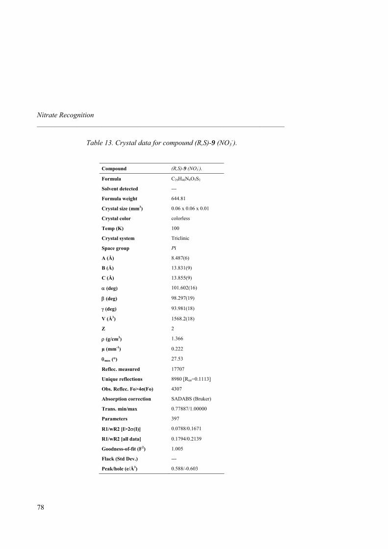
Nitrate Recognition _____________________________________________________________________
78
Table 13. Crystal data for compound (R,S)-9 (NO3-).
Compound (R,S)-9 (NO3-).
Formula C29H40N8O5S2
Solvent detected ---
Formula weight 644.81
Crystal size (mm3) 0.06 x 0.06 x 0.01
Crystal color colorless
Temp (K) 100
Crystal system Triclinic
Space group Pī
A (Å) 8.487(6)
B (Å) 13.831(9)
C (Å) 13.855(9)
α (deg) 101.602(16)
β (deg) 98.297(19)
γ (deg) 93.981(18)
V (Å3) 1568.2(18)
Z 2
ρ (g/cm3) 1.366
µ (mm-1) 0.222
θmax (°) 27.53
Reflec. measured 17707
Unique reflections 8980 [Rint=0.1113]
Obs. Reflec. Fo>4σ(Fo) 4307
Absorption correction SADABS (Bruker)
Trans. min/max 0.77887/1.00000
Parameters 397
R1/wR2 [I>2σ(I)] 0.0788/0.1671
R1/wR2 [all data] 0.1794/0.2139
Goodness-of-fit (F2) 1.005
Flack (Std Dev.) ---
Peak/hole (e/Å3) 0.588/-0.603
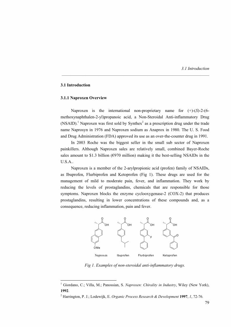
3.1 Introduction _____________________________________________________________________
3.1 Introduction 3.1.1 Naproxen Overview Naproxen is the international non-proprietary name for (+)-(S)-2-(6-methoxynaphthalen-2-yl)propanoic acid, a Non-Steroidal Anti-inflammatory Drug (NSAID).1 Naproxen was first sold by Synthex2 as a prescription drug under the trade name Naprosyn in 1976 and Naproxen sodium as Anaprox in 1980. The U. S. Food and Drug Administration (FDA) approved its use as an over-the-counter drug in 1991. In 2003 Roche was the biggest seller in the small sub sector of Naproxen painkillers. Although Naproxen sales are relatively small, combined Bayer-Roche sales amount to $1.3 billion (€970 million) making it the best-selling NSAIDs in the U.S.A.. Naproxen is a member of the 2-arylpropionic acid (profen) family of NSAIDs, as Ibuprofen, Flurbiprofen and Ketoprofen (Fig 1). These drugs are used for the management of mild to moderate pain, fever, and inflammation. They work by reducing the levels of prostaglandins, chemicals that are responsible for those symptoms. Naproxen blocks the enzyme cyclooxygenase-2 (COX-2) that produces prostaglandins, resulting in lower concentrations of these compounds and, as a consequence, reducing inflammation, pain and fever.
OH
O
OMe
OH
O
OH
O
OH
O
Naproxen Ibuprofen Flurbiprofen Ketoprofen
OF
Fig 1. Examples of non-steroidal anti-inflammatory drugs.
1 Giordano, C.; Villa, M.; Panossian, S. Naproxen: Chirality in Industry, Wiley (New York), 1992. 2 Harrington, P. J.; Lodewijk, E. Organic Process Research & Development 1997, 1, 72-76.
79
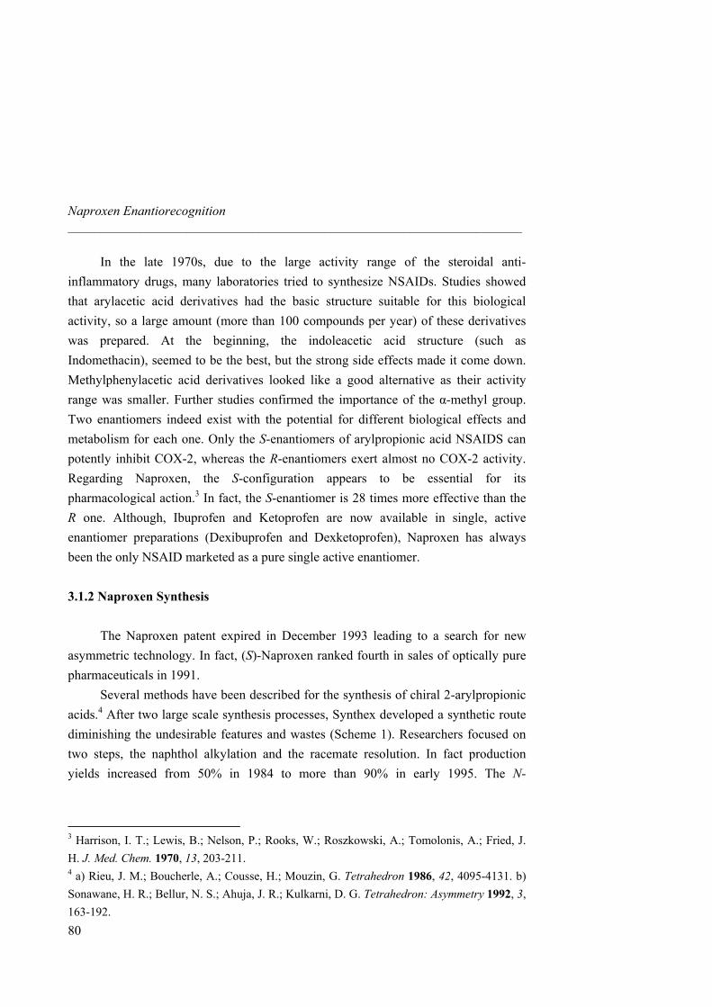
Naproxen Enantiorecognition _____________________________________________________________________
80
In the late 1970s, due to the large activity range of the steroidal anti-inflammatory drugs, many laboratories tried to synthesize NSAIDs. Studies showed that arylacetic acid derivatives had the basic structure suitable for this biological activity, so a large amount (more than 100 compounds per year) of these derivatives was prepared. At the beginning, the indoleacetic acid structure (such as Indomethacin), seemed to be the best, but the strong side effects made it come down. Methylphenylacetic acid derivatives looked like a good alternative as their activity range was smaller. Further studies confirmed the importance of the α-methyl group. Two enantiomers indeed exist with the potential for different biological effects and metabolism for each one. Only the S-enantiomers of arylpropionic acid NSAIDS can potently inhibit COX-2, whereas the R-enantiomers exert almost no COX-2 activity. Regarding Naproxen, the S-configuration appears to be essential for its pharmacological action.3 In fact, the S-enantiomer is 28 times more effective than the R one. Although, Ibuprofen and Ketoprofen are now available in single, active enantiomer preparations (Dexibuprofen and Dexketoprofen), Naproxen has always been the only NSAID marketed as a pure single active enantiomer. 3.1.2 Naproxen Synthesis The Naproxen patent expired in December 1993 leading to a search for new asymmetric technology. In fact, (S)-Naproxen ranked fourth in sales of optically pure pharmaceuticals in 1991. Several methods have been described for the synthesis of chiral 2-arylpropionic acids.4 After two large scale synthesis processes, Synthex developed a synthetic route diminishing the undesirable features and wastes (Scheme 1). Researchers focused on two steps, the naphthol alkylation and the racemate resolution. In fact production yields increased from 50% in 1984 to more than 90% in early 1995. The N-
3 Harrison, I. T.; Lewis, B.; Nelson, P.; Rooks, W.; Roszkowski, A.; Tomolonis, A.; Fried, J. H. J. Med. Chem. 1970, 13, 203-211. 4 a) Rieu, J. M.; Boucherle, A.; Cousse, H.; Mouzin, G. Tetrahedron 1986, 42, 4095-4131. b) Sonawane, H. R.; Bellur, N. S.; Ahuja, J. R.; Kulkarni, D. G. Tetrahedron: Asymmetry 1992, 3, 163-192.
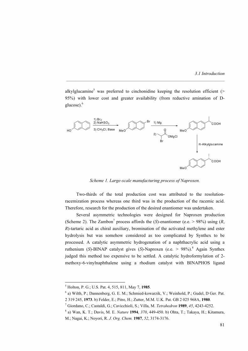
3.1 Introduction _____________________________________________________________________
alkylglucamine5 was preferred to cinchonidine keeping the resolution efficient (> 95%) with lower cost and greater availability (from reductive amination of D-glucose).6
HO MeO
Br1) Br22) NaHSO3
3) CH3Cl, Base MeO
COOH1) Mg
2)
BrOMgCl
O
N -Alkylglucamine
MeO
COOH
Scheme 1. Large-scale manufacturing process of Naproxen. Two-thirds of the total production cost was attributed to the resolution-racemization process whereas one third was in the production of the racemic acid. Therefore, research for the production of the desired enantiomer was undertaken. Several asymmetric technologies were designed for Naproxen production (Scheme 2). The Zambon7 process affords the (S)-enantiomer (e.e. > 98%) using (R, R)-tartaric acid as chiral auxiliary, bromination of the activated methylene and ester hydrolysis but was somehow considered as too complicated by Synthex to be processed. A catalytic asymmetric hydrogenation of a naphthacrylic acid using a ruthenium (S)-BINAP catalyst gives (S)-Naproxen (e.e. > 98%).8 Again Synthex judged this method too expensive to be settled. A catalytic hydroformylation of 2-methoxy-6-vinylnaphthalene using a rhodium catalyst with BINAPHOS ligand
5 Holton, P. G.; U.S. Pat. 4, 515, 811, May 7, 1985. 6 a) Wilth, P.; Dannenberg, G. E. M.; Schmied-kowarzik, V.; Weinhold, P.; Gudel, D Ger. Pat. 2 319 245, 1973. b) Felder, E.; Pitre, H.; Zutter, M.M. U.K. Pat. GB 2 025 968A, 1980. 7 Giordano, C.; Castaldi, G.; Cavicchioli, S.; Villa, M. Tetrahedron 1989, 45, 4243-4252. 8 a) Wan, K. T.; Davis, M. E. Nature 1994, 370, 449-450. b) Ohta, T.; Takaya, H.; Kitamura, M.; Nagai, K.; Noyori, R. J. Org. Chem. 1987, 52, 3174-3176.
81
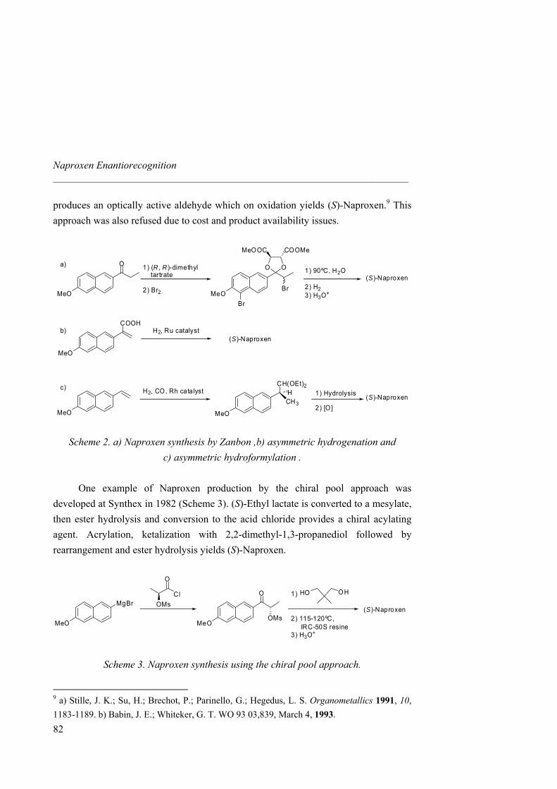
Naproxen Enantiorecognition _____________________________________________________________________
produces an optically active aldehyde which on oxidation yields (S)-Naproxen.9 This approach was also refused due to cost and product availability issues.
MeO
O
MeO
OO
MeOOC COOMe
Br(S)-Naproxen
1) (R , R )-dimethyltartrate
2) Br2
1) 90ºC, H2O
2) H23) H3O+
Br
a)
b)
c)
MeO
COOHH2, Ru catalyst
(S)-Naproxen
MeO
H2, CO, Rh catalyst
MeO
CH3
CH(OEt)2H 1) Hydrolysis
2) [O](S)-Naproxen
Scheme 2. a) Naproxen synthesis by Zanbon ,b) asymmetric hydrogenation and c) asymmetric hydroformylation .
One example of Naproxen production by the chiral pool approach was developed at Synthex in 1982 (Scheme 3). (S)-Ethyl lactate is converted to a mesylate, then ester hydrolysis and conversion to the acid chloride provides a chiral acylating agent. Acrylation, ketalization with 2,2-dimethyl-1,3-propanediol followed by rearrangement and ester hydrolysis yields (S)-Naproxen.
MeO
MgBr
MeO
O
(S)-NaproxenOMs
Cl
O
OMs
HO OH1)
2) 115-120ºC,IRC-50S resine
3) H3O+
Scheme 3. Naproxen synthesis using the chiral pool approach.
9 a) Stille, J. K.; Su, H.; Brechot, P.; Parinello, G.; Hegedus, L. S. Organometallics 1991, 10, 1183-1189. b) Babin, J. E.; Whiteker, G. T. WO 93 03,839, March 4, 1993.
82
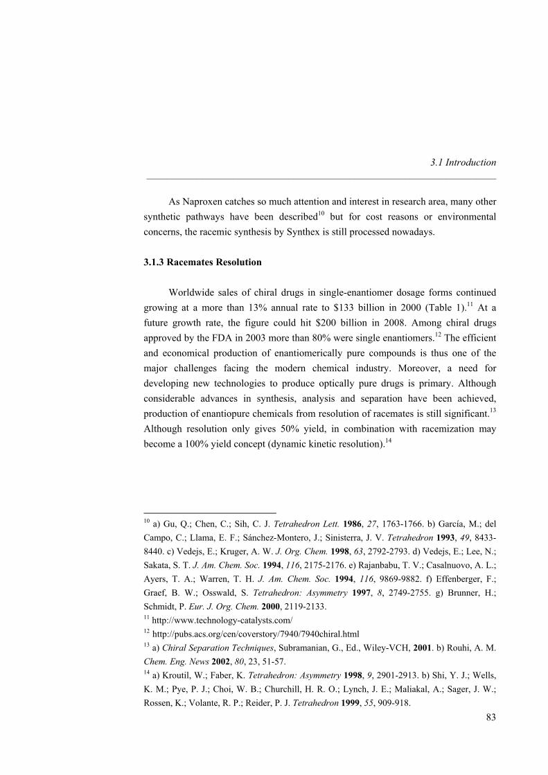
3.1 Introduction _____________________________________________________________________
83
As Naproxen catches so much attention and interest in research area, many other synthetic pathways have been described10 but for cost reasons or environmental concerns, the racemic synthesis by Synthex is still processed nowadays. 3.1.3 Racemates Resolution
Worldwide sales of chiral drugs in single-enantiomer dosage forms continued growing at a more than 13% annual rate to $133 billion in 2000 (Table 1).11 At a future growth rate, the figure could hit $200 billion in 2008. Among chiral drugs approved by the FDA in 2003 more than 80% were single enantiomers.12 The efficient and economical production of enantiomerically pure compounds is thus one of the major challenges facing the modern chemical industry. Moreover, a need for developing new technologies to produce optically pure drugs is primary. Although considerable advances in synthesis, analysis and separation have been achieved, production of enantiopure chemicals from resolution of racemates is still significant.13 Although resolution only gives 50% yield, in combination with racemization may become a 100% yield concept (dynamic kinetic resolution).14
10 a) Gu, Q.; Chen, C.; Sih, C. J. Tetrahedron Lett. 1986, 27, 1763-1766. b) García, M.; del Campo, C.; Llama, E. F.; Sánchez-Montero, J.; Sinisterra, J. V. Tetrahedron 1993, 49, 8433-8440. c) Vedejs, E.; Kruger, A. W. J. Org. Chem. 1998, 63, 2792-2793. d) Vedejs, E.; Lee, N.; Sakata, S. T. J. Am. Chem. Soc. 1994, 116, 2175-2176. e) Rajanbabu, T. V.; Casalnuovo, A. L.; Ayers, T. A.; Warren, T. H. J. Am. Chem. Soc. 1994, 116, 9869-9882. f) Effenberger, F.; Graef, B. W.; Osswald, S. Tetrahedron: Asymmetry 1997, 8, 2749-2755. g) Brunner, H.; Schmidt, P. Eur. J. Org. Chem. 2000, 2119-2133. 11 http://www.technology-catalysts.com/ 12 http://pubs.acs.org/cen/coverstory/7940/7940chiral.html 13 a) Chiral Separation Techniques, Subramanian, G., Ed., Wiley-VCH, 2001. b) Rouhi, A. M. Chem. Eng. News 2002, 80, 23, 51-57. 14 a) Kroutil, W.; Faber, K. Tetrahedron: Asymmetry 1998, 9, 2901-2913. b) Shi, Y. J.; Wells, K. M.; Pye, P. J.; Choi, W. B.; Churchill, H. R. O.; Lynch, J. E.; Maliakal, A.; Sager, J. W.; Rossen, K.; Volante, R. P.; Reider, P. J. Tetrahedron 1999, 55, 909-918.
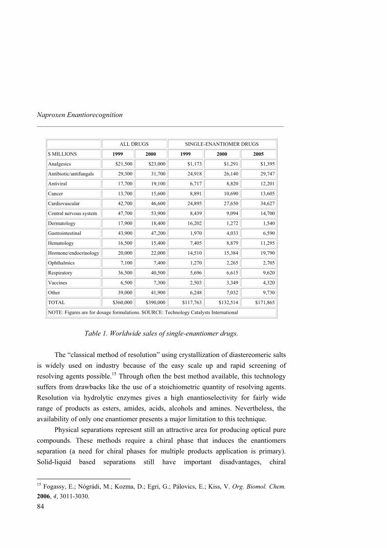
Naproxen Enantiorecognition _____________________________________________________________________
84
ALL DRUGS SINGLE-ENANTIOMER DRUGS
$ MILLIONS 1999 2000 1999 2000 2005
Analgesics $21,500 $23,000 $1,173 $1,291 $1,395
Antibiotic/antifungals 29,300 31,700 24,918 26,140 29,747
Antiviral 17,700 19,100 6,717 8,820 12,201
Cancer 13,700 15,600 8,891 10,690 13,605
Cardiovascular 42,700 46,600 24,895 27,650 34,627
Central nervous system 47,700 53,900 8,439 9,094 14,700
Dermatology 17,900 18,400 16,202 1,272 1,540
Gastrointestinal 43,900 47,200 1,970 4,033 6,590
Hematology 16,500 15,400 7,405 8,879 11,295
Hormone/endocrinology 20,000 22,000 14,510 15,384 19,790
Ophthalmics 7,100 7,400 1,270 2,265 2,705
Respiratory 36,500 40,500 5,696 6,615 9,620
Vaccines 6,500 7,300 2,503 3,349 4,320
Other 39,000 41,900 6,248 7,032 9,730
TOTAL $360,000 $390,000 $117,763 $132,514 $171,865
NOTE: Figures are for dosage formulations. SOURCE: Technology Catalysts International
Table 1. Worldwide sales of single-enantiomer drugs.
The “classical method of resolution” using crystallization of diastereomeric salts is widely used on industry because of the easy scale up and rapid screening of resolving agents possible.15 Through often the best method available, this technology suffers from drawbacks like the use of a stoichiometric quantity of resolving agents. Resolution via hydrolytic enzymes gives a high enantioselectivity for fairly wide range of products as esters, amides, acids, alcohols and amines. Nevertheless, the availability of only one enantiomer presents a major limitation to this technique. Physical separations represent still an attractive area for producing optical pure compounds. These methods require a chiral phase that induces the enantiomers separation (a need for chiral phases for multiple products application is primary). Solid-liquid based separations still have important disadvantages, chiral
15 Fogassy, E.; Nógrádi, M.; Kozma, D.; Egri, G.; Pálovics, E.; Kiss, V. Org. Biomol. Chem. 2006, 4, 3011-3030.
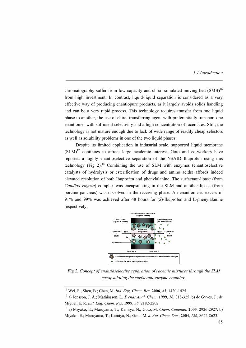
3.1 Introduction _____________________________________________________________________
chromatography suffer from low capacity and chiral simulated moving bed (SMB)16 from high investment. In contrast, liquid-liquid separation is considered as a very effective way of producing enantiopure products, as it largely avoids solids handling and can be a very rapid process. This technology requires transfer from one liquid phase to another, the use of chiral transferring agent with preferentially transport one enantiomer with sufficient selectivity and a high concentration of racemates. Still, the technology is not mature enough due to lack of wide range of readily cheap selectors as well as solubility problems in one of the two liquid phases.
Despite its limited application in industrial scale, supported liquid membrane (SLM)17 continues to attract large academic interest. Goto and co-workers have reported a highly enantioselective separation of the NSAID Ibuprofen using this technology (Fig 2).18 Combining the use of SLM with enzymes (enantioselective catalysts of hydrolysis or esterification of drugs and amino acids) affords indeed elevated resolution of both Ibuprofen and phenylalanine. The surfactant-lipase (from Candida rugosa) complex was encapsulating in the SLM and another lipase (from porcine pancreas) was dissolved in the receiving phase. An enantiomeric excess of 91% and 99% was achieved after 48 hours for (S)-Ibuprofen and L-phenylalanine respectively.
Fig 2. Concept of enantioselective separation of racemic mixtures through the SLM
encapsulating the surfactant-enzyme complex.
16 Wei, F.; Shen, B.; Chen, M. Ind. Eng. Chem. Res. 2006, 45, 1420-1425. 17 a) Jönsson, J. Å.; Mathiasson, L. Trends Anal. Chem. 1999, 18, 318-325. b) de Gyves, J.; de Miguel, E. R. Ind. Eng. Chem. Res. 1999, 38, 2182-2202. 18 a) Miyako, E.; Maruyama, T.; Kamiya, N.; Goto, M. Chem. Commun. 2003, 2926-2927. b) Miyako, E.; Maruyama, T.; Kamiya, N.; Goto, M. J. Am. Chem. Soc., 2004, 126, 8622-8623.
85
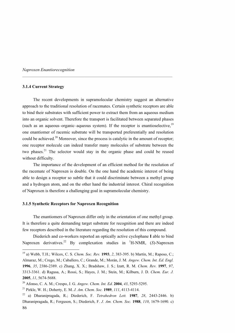
Naproxen Enantiorecognition _____________________________________________________________________
86
3.1.4 Current Strategy
The recent developments in supramolecular chemistry suggest an alternative approach to the traditional resolution of racemates. Certain synthetic receptors are able to bind their substrates with sufficient power to extract them from an aqueous medium into an organic solvent. Therefore the transport is facilitated between separated phases (such as an aqueous–organic–aqueous system). If the receptor is enantioselective,19 one enantiomer of racemic substrate will be transported preferentially and resolution could be achieved.20 Moreover, since the process is catalytic in the amount of receptor; one receptor molecule can indeed transfer many molecules of substrate between the two phases.21 The selector would stay in the organic phase and could be reused without difficulty.
The importance of the development of an efficient method for the resolution of the racemate of Naproxen is double. On the one hand the academic interest of being able to design a receptor so subtle that it could discriminate between a methyl group and a hydrogen atom, and on the other hand the industrial interest. Chiral recognition of Naproxen is therefore a challenging goal in supramolecular chemistry.
3.1.5 Synthetic Receptors for Naproxen Recognition
The enantiomers of Naproxen differ only in the orientation of one methyl group. It is therefore a quite demanding target substrate for recognition and there are indeed few receptors described in the literature regarding the resolution of this compound.
Diederich and co-workers reported an optically active cyclophane I able to bind Naproxen derivatives.22 By complexation studies in 1H-NMR, (S)-Naproxen
19 a) Webb, T.H.; Wilcox, C. S. Chem. Soc. Rev. 1993, 2, 383-395. b) Martín, M.; Raposo, C.; Almaraz, M.; Crego, M.; Caballero, C.; Grande, M.; Morán, J. M. Angew. Chem. Int. Ed. Engl. 1996, 35, 2386-2389. c) Zhang, X. X.; Bradshaw, J. S.; Izatt, R. M. Chem. Rev. 1997, 97, 3313-3361. d) Ragusa, A.; Rossi, S.; Hayes, J. M.; Stein, M.; Kilburn, J. D. Chem. Eur. J. 2005, 11, 5674-5688. 20 Afonso, C. A. M.; Crespo, J. G. Angew. Chem. Int. Ed. 2004, 43, 5293-5295. 21 Pirkle, W. H.; Doherty, E. M. J. Am. Chem. Soc. 1989, 111, 4113-4114. 22 a) Dharanipragada, R.; Diederich, F. Tetrahedron Lett. 1987, 28, 2443-2446. b) Dharanipragada, R.; Ferguson, S.; Diederich, F. J. Am. Chem. Soc. 1988, 110, 1679-1690. c)
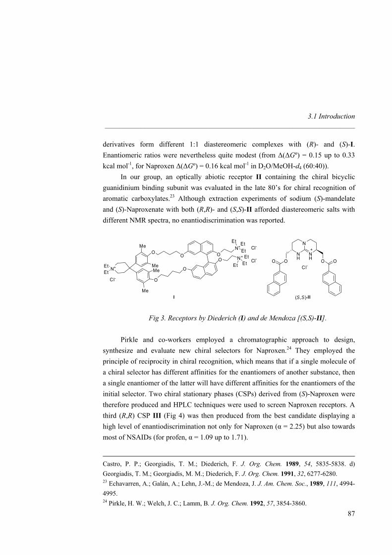
3.1 Introduction _____________________________________________________________________
derivatives form different 1:1 diastereomeric complexes with (R)- and (S)-I. Enantiomeric ratios were nevertheless quite modest (from ∆(∆Gº) = 0.15 up to 0.33 kcal mol-1, for Naproxen ∆(∆Gº) = 0.16 kcal mol-1 in D2O/MeOH-d4 (60:40)).
In our group, an optically abiotic receptor II containing the chiral bicyclic guanidinium binding subunit was evaluated in the late 80’s for chiral recognition of aromatic carboxylates.23 Although extraction experiments of sodium (S)-mandelate and (S)-Naproxenate with both (R,R)- and (S,S)-II afforded diastereomeric salts with different NMR spectra, no enantiodiscrimination was reported.
O N+ON+
Et
Et Et
Et
EtO
O
O
O
Me
Me
MeMe
N+EtEt
Cl-
Et
Cl-
Cl-
I
NH
N
NH OOO O
Cl-
(S,S)-II
Fig 3. Receptors by Diederich (I) and de Mendoza [(S,S)-II]. Pirkle and co-workers employed a chromatographic approach to design,
synthesize and evaluate new chiral selectors for Naproxen.24 They employed the principle of reciprocity in chiral recognition, which means that if a single molecule of a chiral selector has different affinities for the enantiomers of another substance, then a single enantiomer of the latter will have different affinities for the enantiomers of the initial selector. Two chiral stationary phases (CSPs) derived from (S)-Naproxen were therefore produced and HPLC techniques were used to screen Naproxen receptors. A third (R,R) CSP III (Fig 4) was then produced from the best candidate displaying a high level of enantiodiscrimination not only for Naproxen (α = 2.25) but also towards most of NSAIDs (for profen, α = 1.09 up to 1.71).
Castro, P. P.; Georgiadis, T. M.; Diederich, F. J. Org. Chem. 1989, 54, 5835-5838. d) Georgiadis, T. M.; Georgiadis, M. M.; Diederich, F. J. Org. Chem. 1991, 32, 6277-6280. 23 Echavarren, A.; Galán, A.; Lehn, J.-M.; de Mendoza, J. J. Am. Chem. Soc., 1989, 111, 4994-4995. 24 Pirkle, H. W.; Welch, J. C.; Lamm, B. J. Org. Chem. 1992, 57, 3854-3860.
87
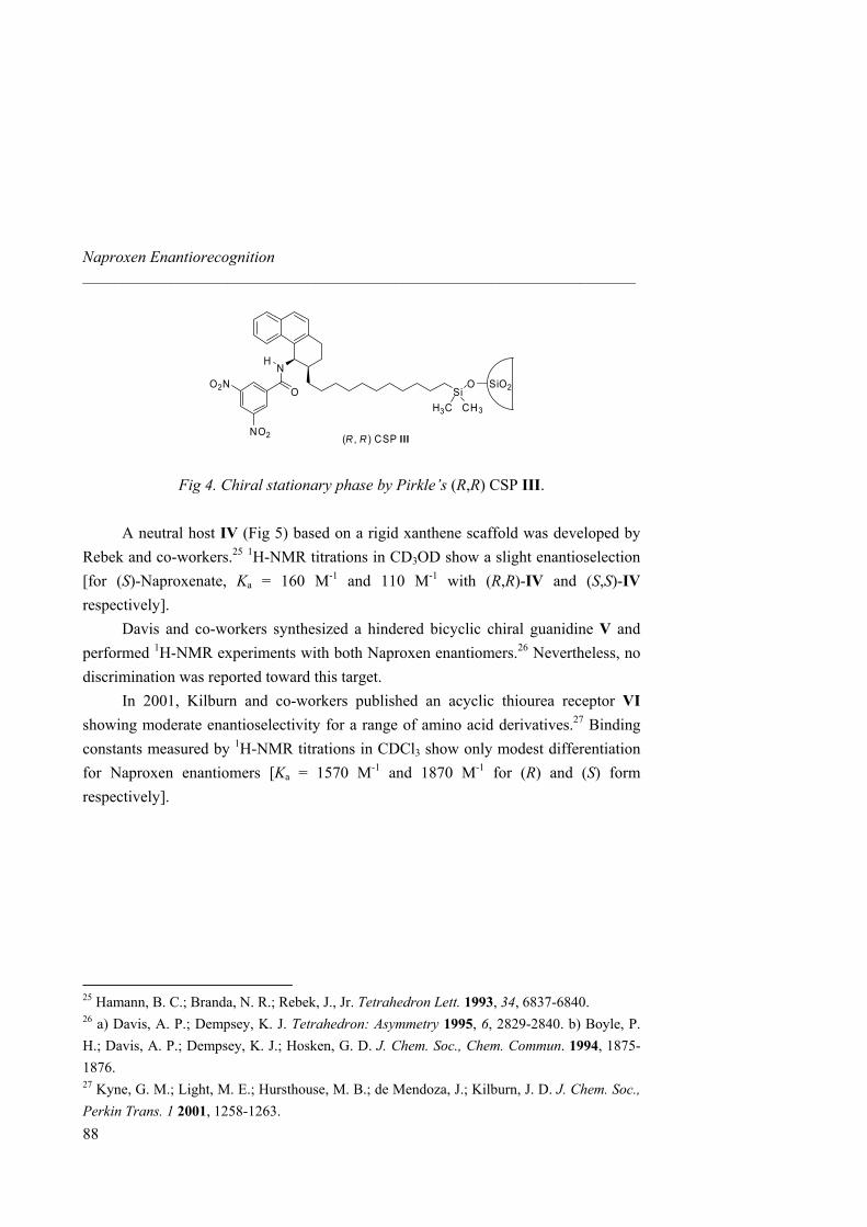
Naproxen Enantiorecognition _____________________________________________________________________
NH
OO2N
NO2
SiO
H3C CH3
SiO2
(R , R ) CSP III
Fig 4. Chiral stationary phase by Pirkle’s (R,R) CSP III. A neutral host IV (Fig 5) based on a rigid xanthene scaffold was developed by
Rebek and co-workers.25 1H-NMR titrations in CD3OD show a slight enantioselection [for (S)-Naproxenate, Ka = 160 M-1 and 110 M-1 with (R,R)-IV and (S,S)-IV respectively].
Davis and co-workers synthesized a hindered bicyclic chiral guanidine V and performed 1H-NMR experiments with both Naproxen enantiomers.26 Nevertheless, no discrimination was reported toward this target.
In 2001, Kilburn and co-workers published an acyclic thiourea receptor VI showing moderate enantioselectivity for a range of amino acid derivatives.27 Binding constants measured by 1H-NMR titrations in CDCl3 show only modest differentiation for Naproxen enantiomers [Ka = 1570 M-1 and 1870 M-1 for (R) and (S) form respectively].
25 Hamann, B. C.; Branda, N. R.; Rebek, J., Jr. Tetrahedron Lett. 1993, 34, 6837-6840. 26 a) Davis, A. P.; Dempsey, K. J. Tetrahedron: Asymmetry 1995, 6, 2829-2840. b) Boyle, P. H.; Davis, A. P.; Dempsey, K. J.; Hosken, G. D. J. Chem. Soc., Chem. Commun. 1994, 1875-1876. 27 Kyne, G. M.; Light, M. E.; Hursthouse, M. B.; de Mendoza, J.; Kilburn, J. D. J. Chem. Soc., Perkin Trans. 1 2001, 1258-1263.
88
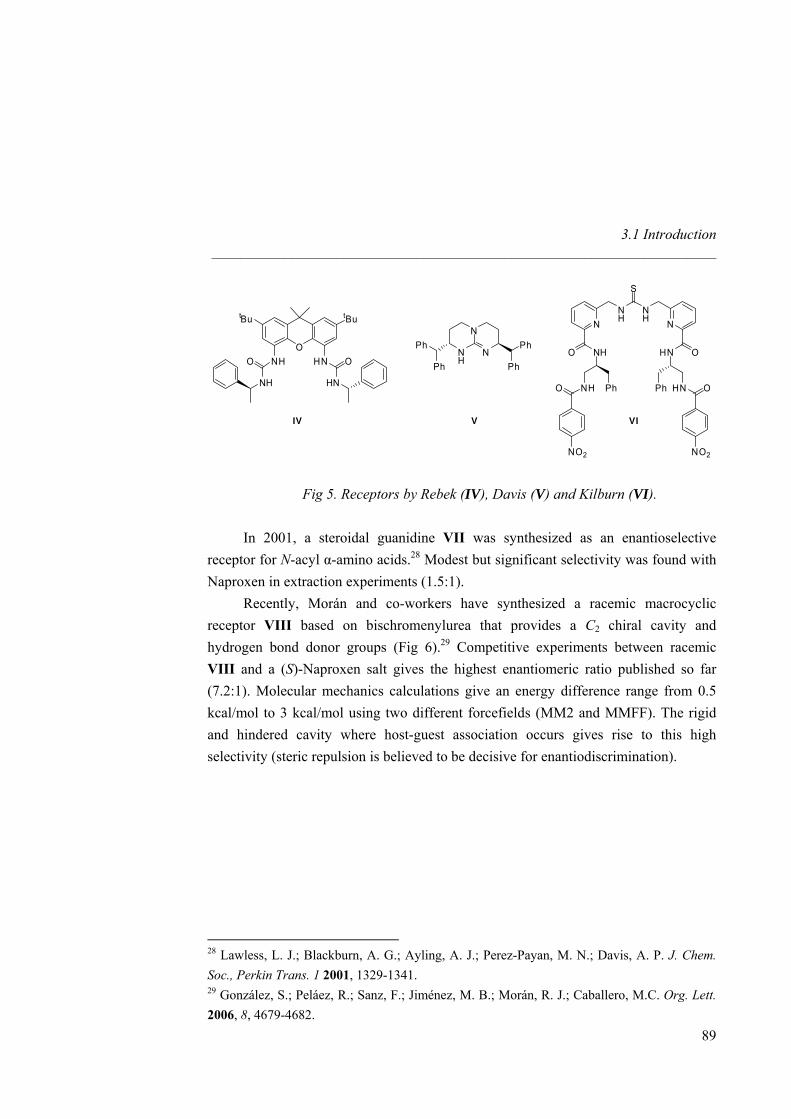
3.1 Introduction _____________________________________________________________________
O
tBu tBu
NH HN
HN
O
NH
O
IV
NH
N
NPh
Ph
Ph
Ph
V
NH
NH
S
N N
NHO
NHO
NO2
Ph
OHN
HN O
NO2
Ph
VI
Fig 5. Receptors by Rebek (IV), Davis (V) and Kilburn (VI). In 2001, a steroidal guanidine VII was synthesized as an enantioselective
receptor for N-acyl α-amino acids.28 Modest but significant selectivity was found with Naproxen in extraction experiments (1.5:1).
Recently, Morán and co-workers have synthesized a racemic macrocyclic receptor VIII based on bischromenylurea that provides a C2 chiral cavity and hydrogen bond donor groups (Fig 6).29 Competitive experiments between racemic VIII and a (S)-Naproxen salt gives the highest enantiomeric ratio published so far (7.2:1). Molecular mechanics calculations give an energy difference range from 0.5 kcal/mol to 3 kcal/mol using two different forcefields (MM2 and MMFF). The rigid and hindered cavity where host-guest association occurs gives rise to this high selectivity (steric repulsion is believed to be decisive for enantiodiscrimination).
28 Lawless, L. J.; Blackburn, A. G.; Ayling, A. J.; Perez-Payan, M. N.; Davis, A. P. J. Chem. Soc., Perkin Trans. 1 2001, 1329-1341. 29 González, S.; Peláez, R.; Sanz, F.; Jiménez, M. B.; Morán, R. J.; Caballero, M.C. Org. Lett. 2006, 8, 4679-4682.
89
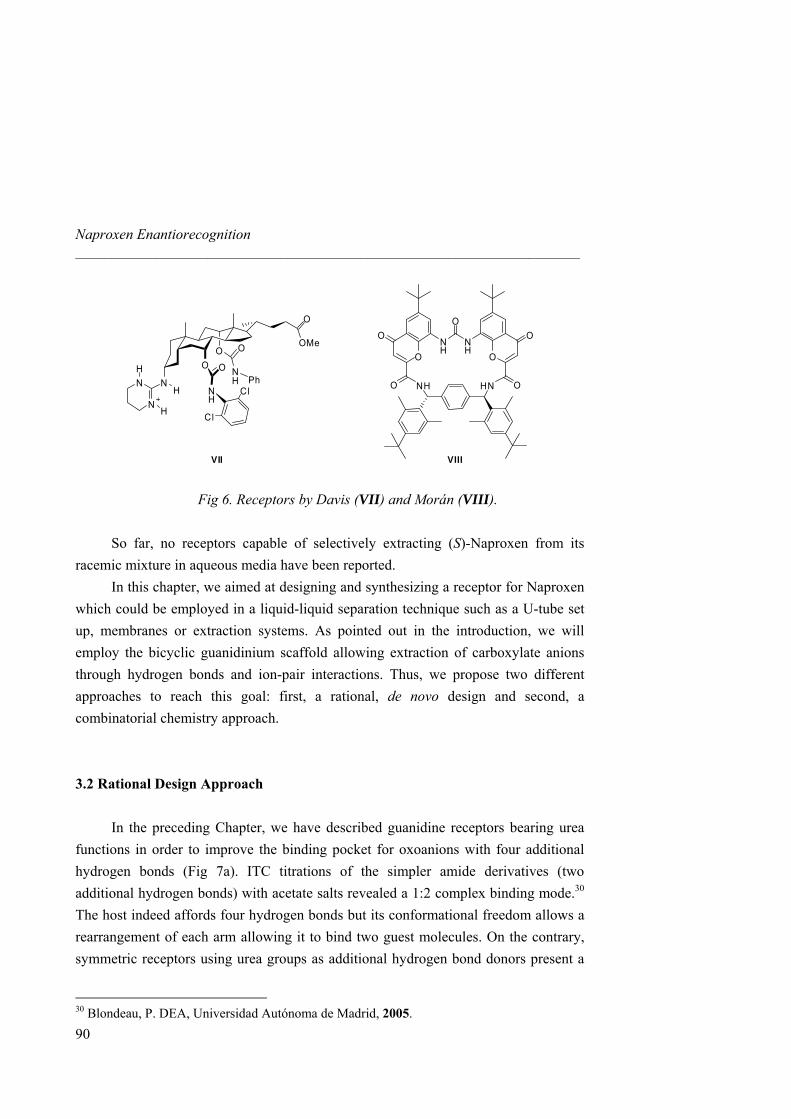
Naproxen Enantiorecognition _____________________________________________________________________
O
OMe
OO
NH
O
N
N
O
N
N
H
H
H Ph
H
Cl
Cl
VII
NH
O
NH
O O
O O
O ONH HN
VIII
Fig 6. Receptors by Davis (VII) and Morán (VIII). So far, no receptors capable of selectively extracting (S)-Naproxen from its
racemic mixture in aqueous media have been reported. In this chapter, we aimed at designing and synthesizing a receptor for Naproxen
which could be employed in a liquid-liquid separation technique such as a U-tube set up, membranes or extraction systems. As pointed out in the introduction, we will employ the bicyclic guanidinium scaffold allowing extraction of carboxylate anions through hydrogen bonds and ion-pair interactions. Thus, we propose two different approaches to reach this goal: first, a rational, de novo design and second, a combinatorial chemistry approach.
3.2 Rational Design Approach
In the preceding Chapter, we have described guanidine receptors bearing urea
functions in order to improve the binding pocket for oxoanions with four additional hydrogen bonds (Fig 7a). ITC titrations of the simpler amide derivatives (two additional hydrogen bonds) with acetate salts revealed a 1:2 complex binding mode.30 The host indeed affords four hydrogen bonds but its conformational freedom allows a rearrangement of each arm allowing it to bind two guest molecules. On the contrary, symmetric receptors using urea groups as additional hydrogen bond donors present a
30 Blondeau, P. DEA, Universidad Autónoma de Madrid, 2005.
90
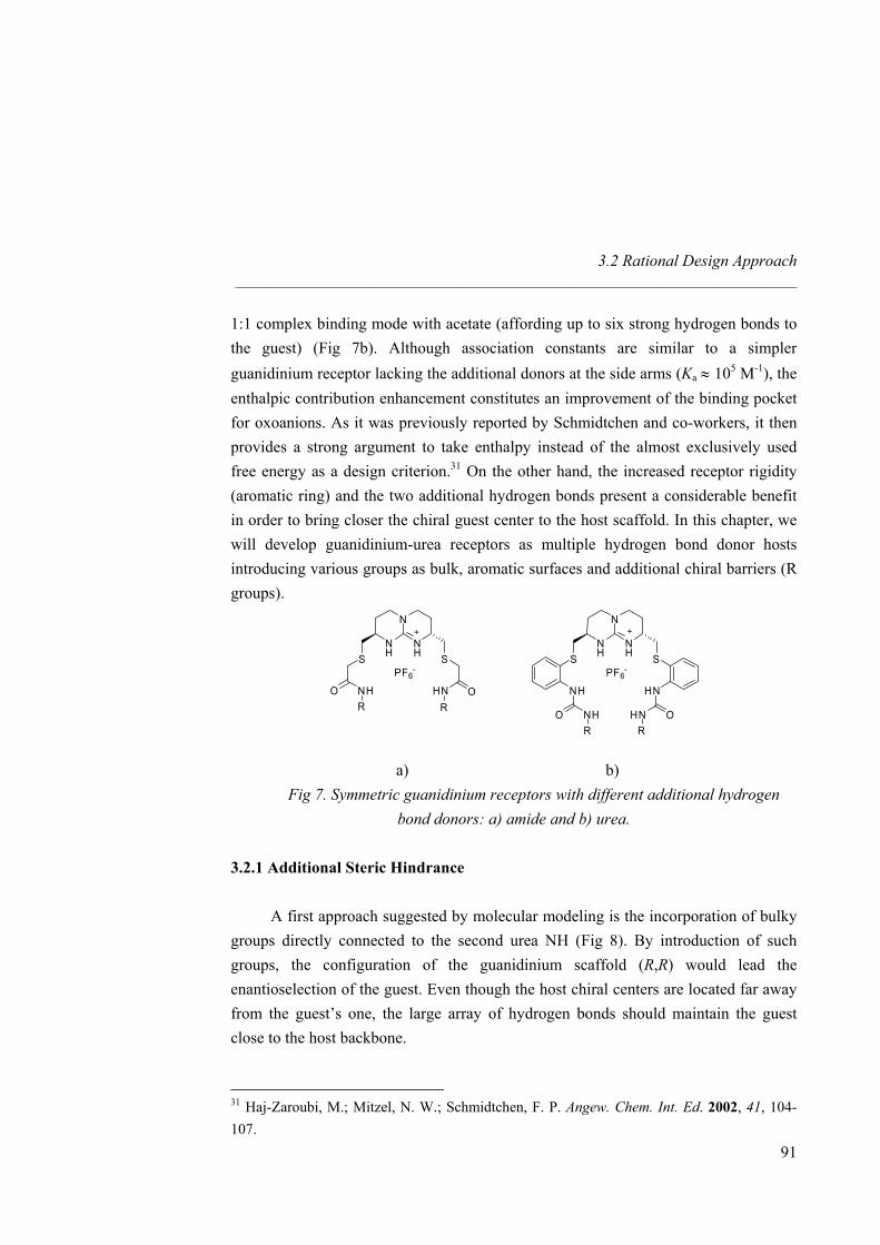
3.2 Rational Design Approach _____________________________________________________________________
1:1 complex binding mode with acetate (affording up to six strong hydrogen bonds to the guest) (Fig 7b). Although association constants are similar to a simpler guanidinium receptor lacking the additional donors at the side arms (Ka ≈ 105 M-1), the enthalpic contribution enhancement constitutes an improvement of the binding pocket for oxoanions. As it was previously reported by Schmidtchen and co-workers, it then provides a strong argument to take enthalpy instead of the almost exclusively used free energy as a design criterion.31 On the other hand, the increased receptor rigidity (aromatic ring) and the two additional hydrogen bonds present a considerable benefit in order to bring closer the chiral guest center to the host scaffold. In this chapter, we will develop guanidinium-urea receptors as multiple hydrogen bond donor hosts introducing various groups as bulk, aromatic surfaces and additional chiral barriers (R groups).
N
NH
NH SS
PF6-
HNNH
OHNO NHRR
N
NH
NH SS
PF6-
OHNO NHR R
a) b)
Fig 7. Symmetric guanidinium receptors with different additional hydrogen bond donors: a) amide and b) urea.
3.2.1 Additional Steric Hindrance
A first approach suggested by molecular modeling is the incorporation of bulky
groups directly connected to the second urea NH (Fig 8). By introduction of such groups, the configuration of the guanidinium scaffold (R,R) would lead the enantioselection of the guest. Even though the host chiral centers are located far away from the guest’s one, the large array of hydrogen bonds should maintain the guest close to the host backbone.
31 Haj-Zaroubi, M.; Mitzel, N. W.; Schmidtchen, F. P. Angew. Chem. Int. Ed. 2002, 41, 104-107.
91
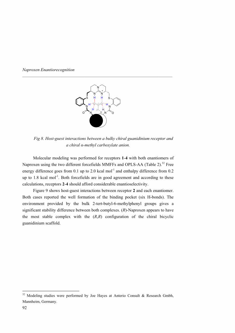
Naproxen Enantiorecognition _____________________________________________________________________
N
NNSS
NN
ONO N∗
OO
H H
H
H H
H
R
Fig 8. Host-guest interactions between a bulky chiral guanidinium receptor and a chiral α-methyl carboxylate anion.
Molecular modeling was performed for receptors 1-4 with both enantiomers of
Naproxen using the two different forcefields MMFFs and OPLS-AA (Table 2).32 Free energy difference goes from 0.1 up to 2.0 kcal mol-1 and enthalpy difference from 0.2 up to 1.8 kcal mol-1. Both forcefields are in good agreement and according to these calculations, receptors 2-4 should afford considerable enantioselectivity.
Figure 9 shows host-guest interactions between receptor 2 and each enantiomer. Both cases reported the well formation of the binding pocket (six H-bonds). The environment provided by the bulk 2-tert-butyl-6-methylphenyl groups gives a significant stability difference between both complexes. (R)-Naproxen appears to have the most stable complex with the (R,R) configuration of the chiral bicyclic guanidinium scaffold.
32 Modeling studies were performed by Joe Hayes at Anterio Consult & Research Gmbh, Mannheim, Germany.
92
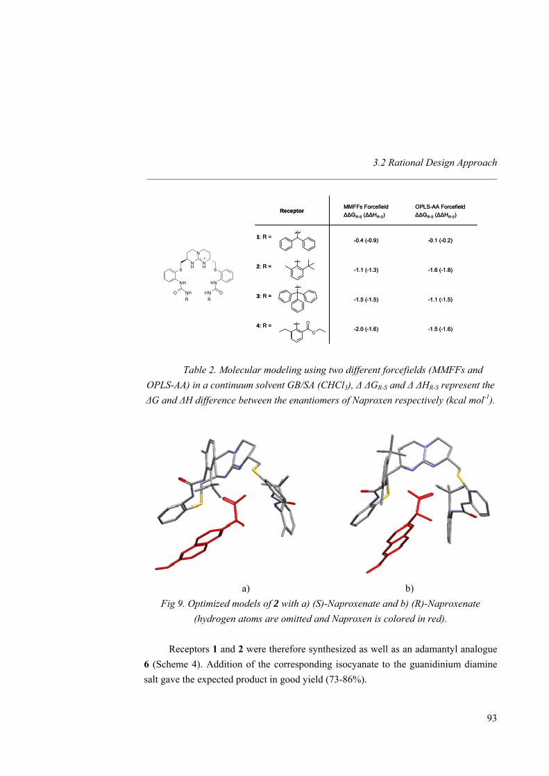
3.2 Rational Design Approach _____________________________________________________________________
-1.5 (-1.6)-2.0 (-1.6)4: R =
-1.1 (-1.5)-1.5 (-1.5)3: R =
-1.6 (-1.8)-1.1 (-1.3)2: R =
-0.1 (-0.2)-0.4 (-0.9)1: R =
OPLS-AA Forcefield∆∆GR-S (∆∆HR-S)
MMFFs Forcefield∆∆GR-S (∆∆HR-S)
Receptor
-1.5 (-1.6)-2.0 (-1.6)4: R =
-1.1 (-1.5)-1.5 (-1.5)3: R =
-1.6 (-1.8)-1.1 (-1.3)2: R =
-0.1 (-0.2)-0.4 (-0.9)1: R =
OPLS-AA Forcefield∆∆GR-S (∆∆HR-S)
MMFFs Forcefield∆∆GR-S (∆∆HR-S)
Receptor
N
NH
NH SS
HNNH
OHNO NHRR
O
O
Table 2. Molecular modeling using two different forcefields (MMFFs and OPLS-AA) in a continuum solvent GB/SA (CHCl3), ∆ ∆GR-S and ∆ ∆HR-S represent the ∆G and ∆H difference between the enantiomers of Naproxen respectively (kcal mol-1).
a) b)
Fig 9. Optimized models of 2 with a) (S)-Naproxenate and b) (R)-Naproxenate (hydrogen atoms are omitted and Naproxen is colored in red).
Receptors 1 and 2 were therefore synthesized as well as an adamantyl analogue
6 (Scheme 4). Addition of the corresponding isocyanate to the guanidinium diamine salt gave the expected product in good yield (73-86%).
93
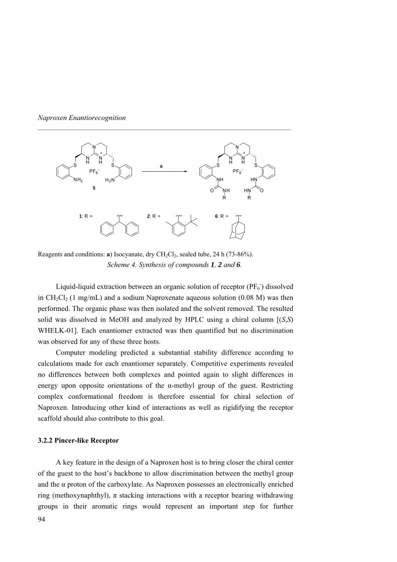
Naproxen Enantiorecognition _____________________________________________________________________
N
NH
NH SS
PF6-
HNNH
N
NH
NH SS
PF6-
H2NNH2
OHNO NHRR
1: R = 2: R = 6: R =
5
a
Reagents and conditions: a) Isocyanate, dry CH2Cl2, sealed tube, 24 h (73-86%).
Scheme 4. Synthesis of compounds 1, 2 and 6. Liquid-liquid extraction between an organic solution of receptor (PF6
-) dissolved in CH2Cl2 (1 mg/mL) and a sodium Naproxenate aqueous solution (0.08 M) was then performed. The organic phase was then isolated and the solvent removed. The resulted solid was dissolved in MeOH and analyzed by HPLC using a chiral column [(S,S) WHELK-01]. Each enantiomer extracted was then quantified but no discrimination was observed for any of these three hosts.
Computer modeling predicted a substantial stability difference according to calculations made for each enantiomer separately. Competitive experiments revealed no differences between both complexes and pointed again to slight differences in energy upon opposite orientations of the α-methyl group of the guest. Restricting complex conformational freedom is therefore essential for chiral selection of Naproxen. Introducing other kind of interactions as well as rigidifying the receptor scaffold should also contribute to this goal.
3.2.2 Pincer-like Receptor
A key feature in the design of a Naproxen host is to bring closer the chiral center
of the guest to the host’s backbone to allow discrimination between the methyl group and the α proton of the carboxylate. As Naproxen possesses an electronically enriched ring (methoxynaphthyl), π stacking interactions with a receptor bearing withdrawing groups in their aromatic rings would represent an important step for further
94
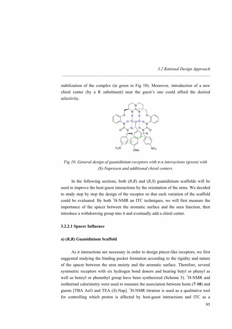
3.2 Rational Design Approach _____________________________________________________________________
stabilization of the complex (in green in Fig 10). Moreover, introduction of a new chiral center (by a R substituent) near the guest’s one could afford the desired selectivity.
N
N
NS S
N N
N NO O∗
OO
H H
H
H H
H
OMe
∗
O2N NO 2
R R
Fig 10. General design of guanidinium receptors with π-π interactions (green) with (S)-Naproxen and additional chiral centers.
In the following sections, both (R,R) and (R,S) guanidinium scaffolds will be used to improve the host-guest interactions by the orientation of the arms. We decided to study step by step the design of the receptor so that each variation of the scaffold could be evaluated. By both 1H-NMR an ITC techniques, we will first measure the importance of the spacer between the aromatic surface and the urea function, then introduce a withdrawing group into it and eventually add a chiral center. 3.2.2.1 Spacer Influence
a) (R,R) Guanidinium Scaffold
As π interactions are necessary in order to design pincer-like receptors, we first
suggested studying the binding pocket formation according to the rigidity and nature of the spacer between the urea moiety and the aromatic surface. Therefore, several symmetric receptors with six hydrogen bond donors and bearing butyl or phenyl as well as benzyl or phenethyl group have been synthesized (Scheme 5). 1H-NMR and isothermal calorimetry were used to measure the association between hosts (7-10) and guests [TBA AcO and TEA (S)-Nap]. 1H-NMR titration is used as a qualitative tool for controlling which proton is affected by host-guest interactions and ITC as a
95
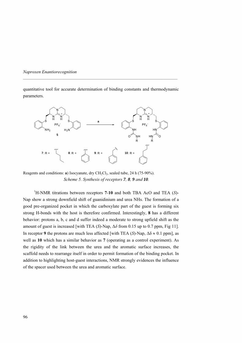
Naproxen Enantiorecognition _____________________________________________________________________
quantitative tool for accurate determination of binding constants and thermodynamic parameters.
N
NH
NH SS
PF6-
HNNH
N
NH
NH SS
PF6-
H2NNH2
OHNO NHRR
8: R = 9: R = 10: R =
5
a
7 : R =
Reagents and conditions: a) Isocyanate, dry CH2Cl2, sealed tube, 24 h (75-90%).
Scheme 5. Synthesis of receptors 7, 8, 9 and 10. 1H-NMR titrations between receptors 7-10 and both TBA AcO and TEA (S)-
Nap show a strong downfield shift of guanidinium and urea NHs. The formation of a good pre-organized pocket in which the carboxylate part of the guest is forming six strong H-bonds with the host is therefore confirmed. Interestingly, 8 has a different behavior: protons a, b, c and d suffer indeed a moderate to strong upfield shift as the amount of guest is increased [with TEA (S)-Nap, ∆δ from 0.15 up to 0.7 ppm, Fig 11]. In receptor 9 the protons are much less affected [with TEA (S)-Nap, ∆δ ≈ 0.1 ppm], as well as 10 which has a similar behavior as 7 (operating as a control experiment). As the rigidity of the link between the urea and the aromatic surface increases, the scaffold needs to rearrange itself in order to permit formation of the binding pocket. In addition to highlighting host-guest interactions, NMR strongly evidences the influence of the spacer used between the urea and aromatic surface.
96

3.2 Rational Design Approach _____________________________________________________________________
d
1.21.41.61.82.02.22.42.62.83.03.23.4 ppm
N
NH
NH SS
X-
HNNH
OHNO NH
bc
a
97
Fig 11. 1H-NMR titration between (R,R)-8 and TEA (S)-Nap in CD3CN at 298K [from bottom to top: 0 to 1.1 equivalents TEA (S)-Nap].
Ka’s between 8 and both carboxylate salts are one order of magnitude lower than
for 7, 9 and 10 (Ka from 104 up to 105 M-1, Table 3). For TBA AcO and TEA (S)-Nap, the more pronounced negative entropic contribution is the important difference between these four hosts. The association for 8 is indeed entropically disfavored and contributes to a lower free energy ∆G. Receptors 7, 9 and 10 display a similar binding behavior mainly enthalpically driven (Ka from 3.06 x 10⁵ up to 5.41 x 10⁵ M-1). This significant enthalpic factor is due to electrostatic interactions whereas the entropic one comes from the release of solvent molecules. 8 suffers from an unfavorable spatial arrangement to complete complex formation.
Therefore, both 1H-NMR and ITC techniques are in good agreement to evaluate the spacer influence on binding. 8 is thus too rigid to achieve optimal association but as the host scaffold is recovering flexibility (by addition of CH2 groups to the linker); the stability of the complex is again higher.
ab ca c
ad b d c c a
d
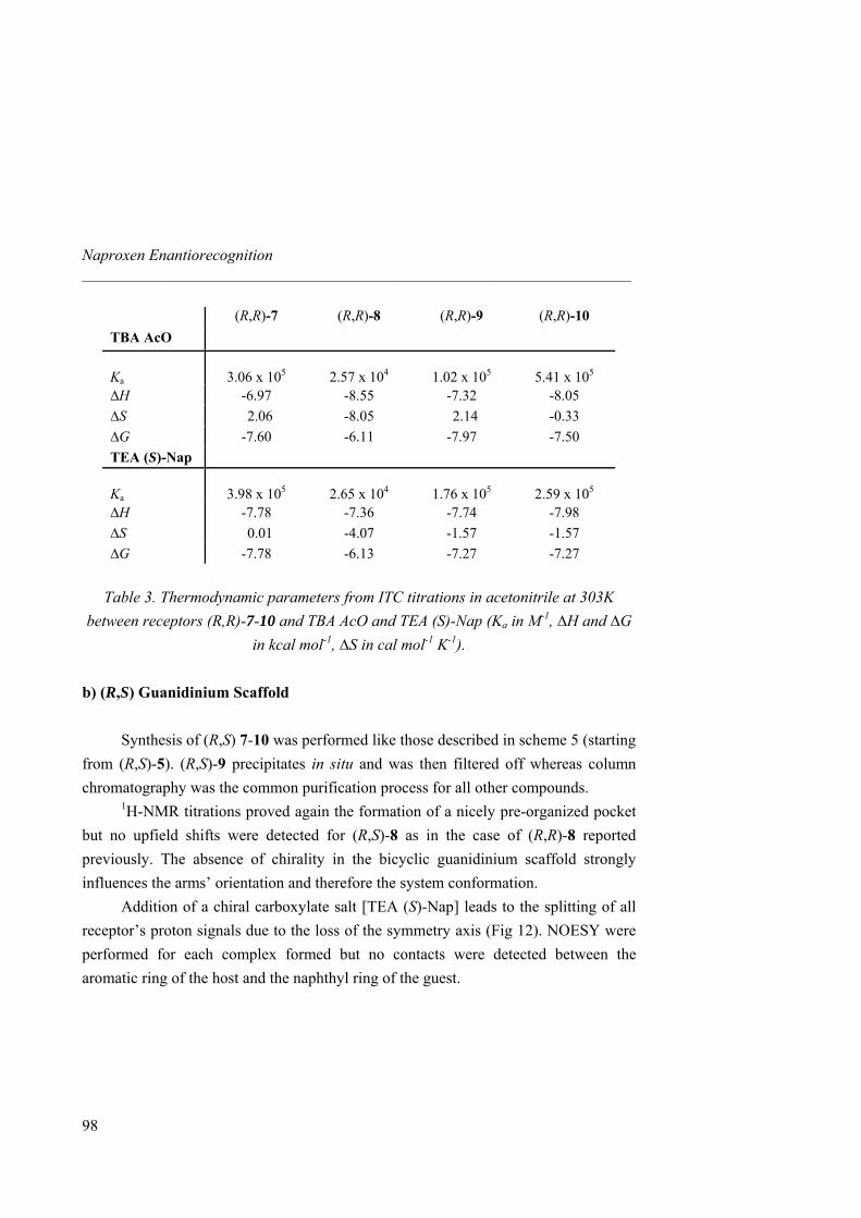
Naproxen Enantiorecognition _____________________________________________________________________
98
(R,R)-7 (R,R)-8 (R,R)-9 (R,R)-10 TBA AcO Ka
3.06 x 10⁵
2.57 x 10⁴
1.02 x 10⁵
5.41 x 10⁵
∆H -6.97 -8.55 -7.32 -8.05 ∆S 2.06 -8.05 2.14 -0.33 ∆G -7.60 -6.11 -7.97 -7.50 TEA (S)-Nap Ka
3.98 x 10⁵
2.65 x 10⁴
1.76 x 10⁵
2.59 x 10⁵
∆H -7.78 -7.36 -7.74 -7.98 ∆S 0.01 -4.07 -1.57 -1.57 ∆G -7.78 -6.13 -7.27 -7.27
Table 3. Thermodynamic parameters from ITC titrations in acetonitrile at 303K
between receptors (R,R)-7-10 and TBA AcO and TEA (S)-Nap (Ka in M-1, ∆H and ∆G in kcal mol-1, ∆S in cal mol-1 K-1).
b) (R,S) Guanidinium Scaffold
Synthesis of (R,S) 7-10 was performed like those described in scheme 5 (starting
from (R,S)-5). (R,S)-9 precipitates in situ and was then filtered off whereas column chromatography was the common purification process for all other compounds.
1H-NMR titrations proved again the formation of a nicely pre-organized pocket but no upfield shifts were detected for (R,S)-8 as in the case of (R,R)-8 reported previously. The absence of chirality in the bicyclic guanidinium scaffold strongly influences the arms’ orientation and therefore the system conformation.
Addition of a chiral carboxylate salt [TEA (S)-Nap] leads to the splitting of all receptor’s proton signals due to the loss of the symmetry axis (Fig 12). NOESY were performed for each complex formed but no contacts were detected between the aromatic ring of the host and the naphthyl ring of the guest.
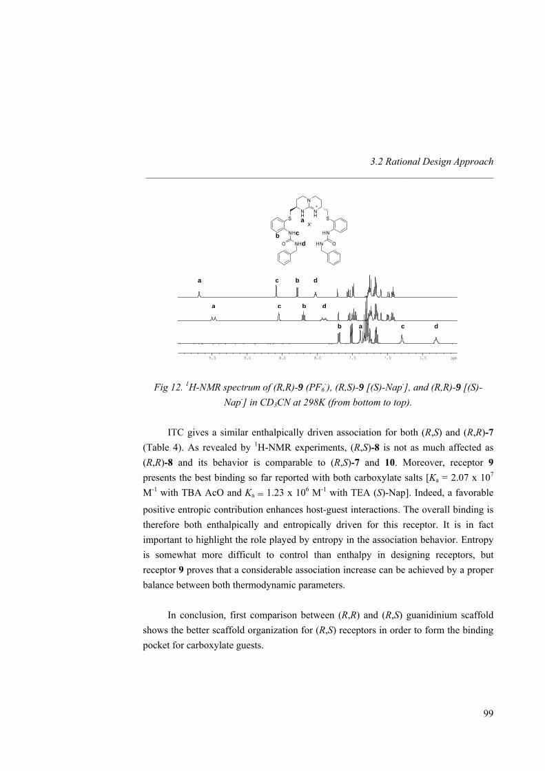
3.2 Rational Design Approach _____________________________________________________________________
N
NH
NHa SS
X-
HNNH
OHNO NH
cbd
6.57.07.58.08.59.09.5 ppm
a c b d
a c b d
ab c d
Fig 12. 1H-NMR spectrum of (R,R)-9 (PF6-), (R,S)-9 [(S)-Nap-], and (R,R)-9 [(S)-
Nap-] in CD3CN at 298K (from bottom to top). ITC gives a similar enthalpically driven association for both (R,S) and (R,R)-7 (Table 4). As revealed by 1H-NMR experiments, (R,S)-8 is not as much affected as (R,R)-8 and its behavior is comparable to (R,S)-7 and 10. Moreover, receptor 9 presents the best binding so far reported with both carboxylate salts [Ka = 2.07 x 10⁷ M-1 with TBA AcO and Ka = 1.23 x 10⁶ M-1 with TEA (S)-Nap]. Indeed, a favorable
positive entropic contribution enhances host-guest interactions. The overall binding is therefore both enthalpically and entropically driven for this receptor. It is in fact important to highlight the role played by entropy in the association behavior. Entropy is somewhat more difficult to control than enthalpy in designing receptors, but receptor 9 proves that a considerable association increase can be achieved by a proper balance between both thermodynamic parameters.
In conclusion, first comparison between (R,R) and (R,S) guanidinium scaffold shows the better scaffold organization for (R,S) receptors in order to form the binding pocket for carboxylate guests.
99
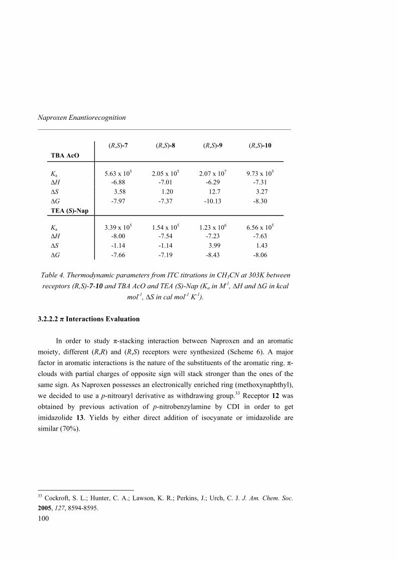
Naproxen Enantiorecognition _____________________________________________________________________
100
(R,S)-7 (R,S)-8 (R,S)-9 (R,S)-10 TBA AcO Ka
5.63 x 10⁵
2.05 x 10⁵
2.07 x 10⁷
9.73 x 10⁵
∆H -6.88 -7.01 -6.29 -7.31 ∆S 3.58 1.20 12.7 3.27 ∆G -7.97 -7.37 -10.13 -8.30 TEA (S)-Nap Ka
3.39 x 10⁵
1.54 x 10⁵
1.23 x 10⁶
6.56 x 10⁵
∆H -8.00 -7.54 -7.23 -7.63 ∆S -1.14 -1.14 3.99 1.43 ∆G -7.66 -7.19 -8.43 -8.06
Table 4. Thermodynamic parameters from ITC titrations in CH3CN at 303K between receptors (R,S)-7-10 and TBA AcO and TEA (S)-Nap (Ka in M-1, ∆H and ∆G in kcal
mol-1, ∆S in cal mol-1 K-1).
3.2.2.2 π Interactions Evaluation In order to study π-stacking interaction between Naproxen and an aromatic
moiety, different (R,R) and (R,S) receptors were synthesized (Scheme 6). A major factor in aromatic interactions is the nature of the substituents of the aromatic ring. π-clouds with partial charges of opposite sign will stack stronger than the ones of the same sign. As Naproxen possesses an electronically enriched ring (methoxynaphthyl), we decided to use a p-nitroaryl derivative as withdrawing group.33 Receptor 12 was obtained by previous activation of p-nitrobenzylamine by CDI in order to get imidazolide 13. Yields by either direct addition of isocyanate or imidazolide are similar (70%).
33 Cockroft, S. L.; Hunter, C. A.; Lawson, K. R.; Perkins, J.; Urch, C. J. J. Am. Chem. Soc. 2005, 127, 8594-8595.
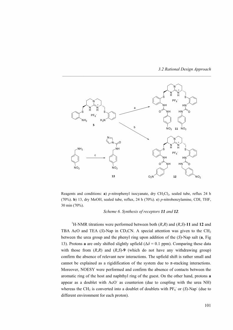
3.2 Rational Design Approach _____________________________________________________________________
N
NH
NH
SSPF6
-
HNNHN
NH
NH SS
PF6-
H2NNH2
OHNO NH
5
N
NH
NH
SSPF6
-
HNNH
OHNO NH
O2N NO2
NO2 NO2
a
b
NO2NO2
NH2 NH
ON
N
c
11
1213
Reagents and conditions: a) p-nitrophenyl isocyanate, dry CH2Cl2, sealed tube, reflux 24 h (70%). b) 13, dry MeOH, sealed tube, reflux, 24 h (70%). c) p-nitrobenzylamine, CDI, THF, 30 min (70%).
Scheme 6. Synthesis of receptors 11 and 12. 1H-NMR titrations were performed between both (R,R) and (R,S)-11 and 12 and
TBA AcO and TEA (S)-Nap in CD3CN. A special attention was given to the CH2 between the urea group and the phenyl ring upon addition of the (S)-Nap salt (a, Fig 13). Protons a are only shifted slightly upfield (∆δ = 0.1 ppm). Comparing these data with those from (R,R) and (R,S)-9 (which do not have any withdrawing group) confirm the absence of relevant new interactions. The upfield shift is rather small and cannot be explained as a rigidification of the system due to π-stacking interactions. Moreover, NOESY were performed and confirm the absence of contacts between the aromatic ring of the host and naphthyl ring of the guest. On the other hand, protons a appear as a doublet with AcO- as counterion (due to coupling with the urea NH) whereas the CH2 is converted into a doublet of doublets with PF6
- or (S)-Nap- (due to different environment for each proton).
101
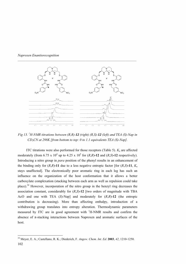
Naproxen Enantiorecognition _____________________________________________________________________
N
NH
NH SS
X-
HNNH
OHNO NH
O2N NO 2
N
NH
NH SS
X-
HNNH
OHNO NH
O2N NO 2
4.24.34.44.54.64.7 ppm 4.24.34.44.54.64.7 ppm
a a
Fig 13. 1H-NMR titrations between (R,R)-12 (right) (R,S)-12 (left) and TEA (S)-Nap in CD3CN at 298K [from bottom to top: 0 to 1.1 equivalents TEA (S)-Nap].
ITC titrations were also performed for those receptors (Table 5). Ka are affected
moderately (from 6.75 x 10⁴ up to 4.25 x 10⁵ for (R,R)-12 and (R,S)-12 respectively). Introducing a nitro group in para position of the phenyl results in an enhancement of the binding only for (R,R)-11 due to a less negative entropic factor [for (R,S)-11, Ka stays unaffected]. The electronically poor aromatic ring in each leg has such an influence on the organization of the host conformation that it allows a better carboxylate complexation (stacking between each arm as well as repulsion could take place).34 However, incorporation of the nitro group in the benzyl ring decreases the association constant, considerably for (R,S)-12 [two orders of magnitude with TBA AcO and one with TEA (S)-Nap] and moderately for (R,R)-12 (the entropic contribution is decreasing). More than affecting enthalpy, introduction of a withdrawing group translates into entropy alteration. Thermodynamic parameters measured by ITC are in good agreement with 1H-NMR results and confirm the absence of π-stacking interactions between Naproxen and aromatic surfaces of the host.
102
34 Meyer, E. A.; Castellano, R. K.; Diederich, F. Angew. Chem. Int. Ed. 2003, 42, 1210-1250.
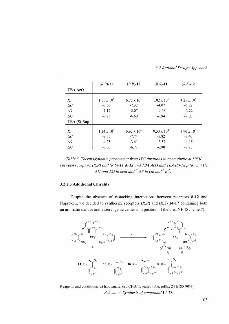
3.2 Rational Design Approach _____________________________________________________________________
(R,R)-11 (R,R)-12 (R,S)-11 (R,S)-12 TBA AcO Ka
1.65 x 10⁵
6.75 x 10⁴
1.02 x 10⁵
4.25 x 10⁵
∆H -7.66 -7.32 -4.07 -6.82 ∆S -1.17 -2.07 9.46 3.22 ∆G -7.25 -6.69 -6.94 -7.80 TEA (S)-Nap Ka
1.24 x 10⁵
6.92 x 10⁴
9.53 x 10⁴
3.90 x 10⁵
∆H -8.35 -7.74 -5.82 -7.40 ∆S -4.25 -3.41 3.57 1.15 ∆G -7.06 -6.71 -6.90 -7.75
Table 5. Thermodynamic parameters from ITC titrations in acetonitrile at 303K
between receptors (R,R) and (R,S)-11 & 12 and TBA AcO and TEA (S)-Nap (Ka in M-1, ∆H and ∆G in kcal mol-1, ∆S in cal mol-1 K-1).
3.2.2.3 Additional Chirality
Despite the absence of π-stacking interactions between receptors 8-12 and
Naproxen, we decided to synthesize receptors (R,R) and (R,S) 14-17 containing both an aromatic surface and a stereogenic center in α position of the urea NH (Scheme 7).
N
NH
NH SS
PF6-
HNNH
N
NH
NH SS
PF6-
H2NNH2
OHNO NHRR
15: R = 16: R = 17: R =
5
a
14: R =
Reagents and conditions: a) Isocyanate, dry CH2Cl2, sealed tube, reflux 24 h (85-90%).
Scheme 7. Synthesis of compound 14-17.
103
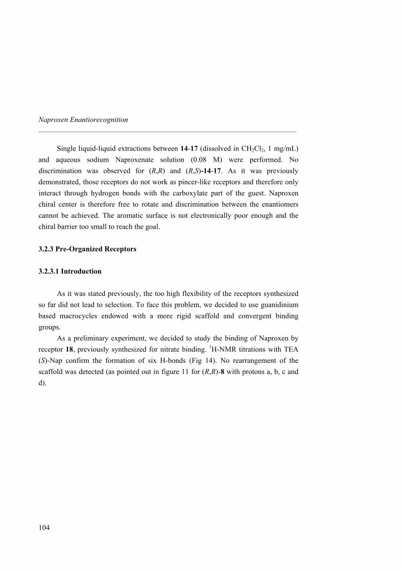
Naproxen Enantiorecognition _____________________________________________________________________
104
Single liquid-liquid extractions between 14-17 (dissolved in CH2Cl2, 1 mg/mL) and aqueous sodium Naproxenate solution (0.08 M) were performed. No discrimination was observed for (R,R) and (R,S)-14-17. As it was previously demonstrated, those receptors do not work as pincer-like receptors and therefore only interact through hydrogen bonds with the carboxylate part of the guest. Naproxen chiral center is therefore free to rotate and discrimination between the enantiomers cannot be achieved. The aromatic surface is not electronically poor enough and the chiral barrier too small to reach the goal.
3.2.3 Pre-Organized Receptors 3.2.3.1 Introduction
As it was stated previously, the too high flexibility of the receptors synthesized
so far did not lead to selection. To face this problem, we decided to use guanidinium based macrocycles endowed with a more rigid scaffold and convergent binding groups.
As a preliminary experiment, we decided to study the binding of Naproxen by receptor 18, previously synthesized for nitrate binding. 1H-NMR titrations with TEA (S)-Nap confirm the formation of six H-bonds (Fig 14). No rearrangement of the scaffold was detected (as pointed out in figure 11 for (R,R)-8 with protons a, b, c and d).
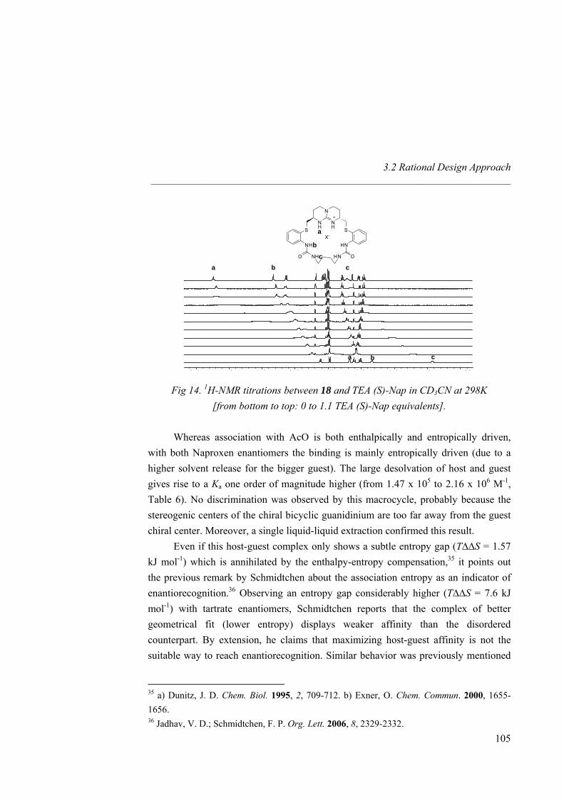
3.2 Rational Design Approach _____________________________________________________________________
. 06 . 57 . 07 . 58 . 08 . 59 . 09 . 5 p m
N
NH
NHa SS
X-
HNNH
OHNO NH
bc
a b c
a c b
Fig 14. 1H-NMR titrations between 18 and TEA (S)-Nap in CD3CN at 298K
[from bottom to top: 0 to 1.1 TEA (S)-Nap equivalents].
Whereas association with AcO is both enthalpically and entropically driven, with both Naproxen enantiomers the binding is mainly entropically driven (due to a higher solvent release for the bigger guest). The large desolvation of host and guest gives rise to a Ka one order of magnitude higher (from 1.47 x 10⁵ to 2.16 x 10⁶ M-1, Table 6). No discrimination was observed by this macrocycle, probably because the stereogenic centers of the chiral bicyclic guanidinium are too far away from the guest chiral center. Moreover, a single liquid-liquid extraction confirmed this result.
Even if this host-guest complex only shows a subtle entropy gap (T∆∆S = 1.57 kJ mol-1) which is annihilated by the enthalpy-entropy compensation,35 it points out the previous remark by Schmidtchen about the association entropy as an indicator of enantiorecognition.36 Observing an entropy gap considerably higher (T∆∆S = 7.6 kJ mol-1) with tartrate enantiomers, Schmidtchen reports that the complex of better geometrical fit (lower entropy) displays weaker affinity than the disordered counterpart. By extension, he claims that maximizing host-guest affinity is not the suitable way to reach enantiorecognition. Similar behavior was previously mentioned
105
35 a) Dunitz, J. D. Chem. Biol. 1995, 2, 709-712. b) Exner, O. Chem. Commun. 2000, 1655-1656. 36 Jadhav, V. D.; Schmidtchen, F. P. Org. Lett. 2006, 8, 2329-2332.
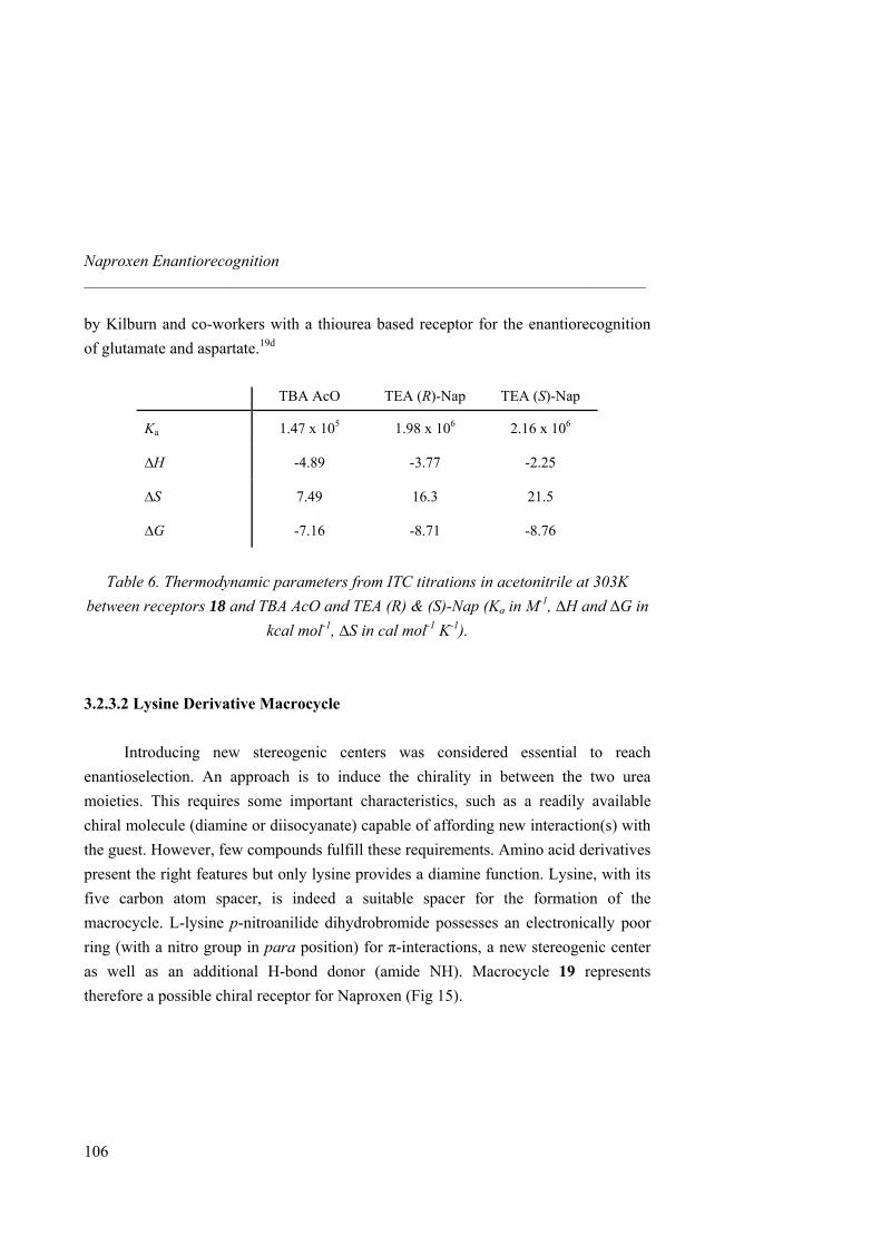
Naproxen Enantiorecognition _____________________________________________________________________
106
by Kilburn and co-workers with a thiourea based receptor for the enantiorecognition of glutamate and aspartate.19d
TBA AcO TEA (R)-Nap TEA (S)-Nap
Ka 1.47 x 10⁵ 1.98 x 10⁶ 2.16 x 10⁶
∆H -4.89 -3.77 -2.25
∆S 7.49 16.3 21.5
∆G -7.16 -8.71 -8.76
Table 6. Thermodynamic parameters from ITC titrations in acetonitrile at 303K
between receptors 18 and TBA AcO and TEA (R) & (S)-Nap (Ka in M-1, ∆H and ∆G in kcal mol-1, ∆S in cal mol-1 K-1).
3.2.3.2 Lysine Derivative Macrocycle
Introducing new stereogenic centers was considered essential to reach
enantioselection. An approach is to induce the chirality in between the two urea moieties. This requires some important characteristics, such as a readily available chiral molecule (diamine or diisocyanate) capable of affording new interaction(s) with the guest. However, few compounds fulfill these requirements. Amino acid derivatives present the right features but only lysine provides a diamine function. Lysine, with its five carbon atom spacer, is indeed a suitable spacer for the formation of the macrocycle. L-lysine p-nitroanilide dihydrobromide possesses an electronically poor ring (with a nitro group in para position) for π-interactions, a new stereogenic center as well as an additional H-bond donor (amide NH). Macrocycle 19 represents therefore a possible chiral receptor for Naproxen (Fig 15).
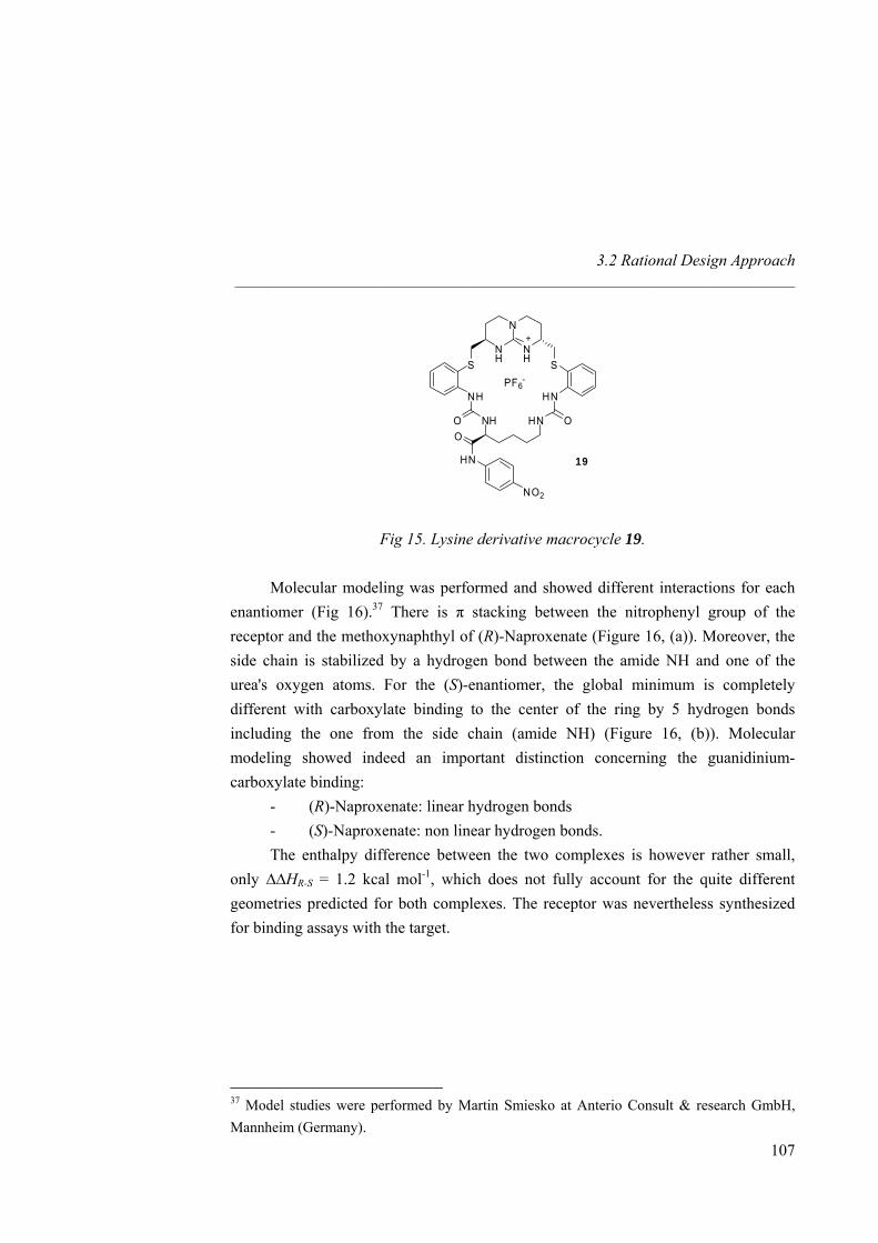
3.2 Rational Design Approach _____________________________________________________________________
N
NH
NH SS
PF6-
HNNH
OHNO NHO
HN
NO2
19
Fig 15. Lysine derivative macrocycle 19.
Molecular modeling was performed and showed different interactions for each enantiomer (Fig 16).37 There is π stacking between the nitrophenyl group of the receptor and the methoxynaphthyl of (R)-Naproxenate (Figure 16, (a)). Moreover, the side chain is stabilized by a hydrogen bond between the amide NH and one of the urea's oxygen atoms. For the (S)-enantiomer, the global minimum is completely different with carboxylate binding to the center of the ring by 5 hydrogen bonds including the one from the side chain (amide NH) (Figure 16, (b)). Molecular modeling showed indeed an important distinction concerning the guanidinium-carboxylate binding:
- (R)-Naproxenate: linear hydrogen bonds - (S)-Naproxenate: non linear hydrogen bonds.
The enthalpy difference between the two complexes is however rather small, only ∆∆HR-S = 1.2 kcal mol-1, which does not fully account for the quite different geometries predicted for both complexes. The receptor was nevertheless synthesized for binding assays with the target.
37 Model studies were performed by Martin Smiesko at Anterio Consult & research GmbH, Mannheim (Germany).
107
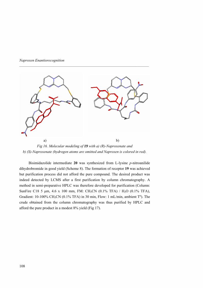
Naproxen Enantiorecognition _____________________________________________________________________
a) b)
Fig 16. Molecular modeling of 19 with a) (R)-Naproxenate and b) (S)-Naproxenate (hydrogen atoms are omitted and Naproxen is colored in red).
Bisimidazolide intermediate 20 was synthesized from L-lysine p-nitroanilide dihydrobromide in good yield (Scheme 8). The formation of receptor 19 was achieved but purification process did not afford the pure compound. The desired product was indeed detected by LCMS after a first purification by column chromatography. A method in semi-preparative HPLC was therefore developed for purification (Column: SunFire C18 5 µm, 4.6 x 100 mm, FM: CH3CN (0.1% TFA) / H2O (0.1% TFA), Gradient: 10-100% CH3CN (0.1% TFA) in 30 min, Flow: 1 mL/min, ambient Tº). The crude obtained from the column chromatography was thus purified by HPLC and afford the pure product in a modest 8% yield (Fig 17).
108
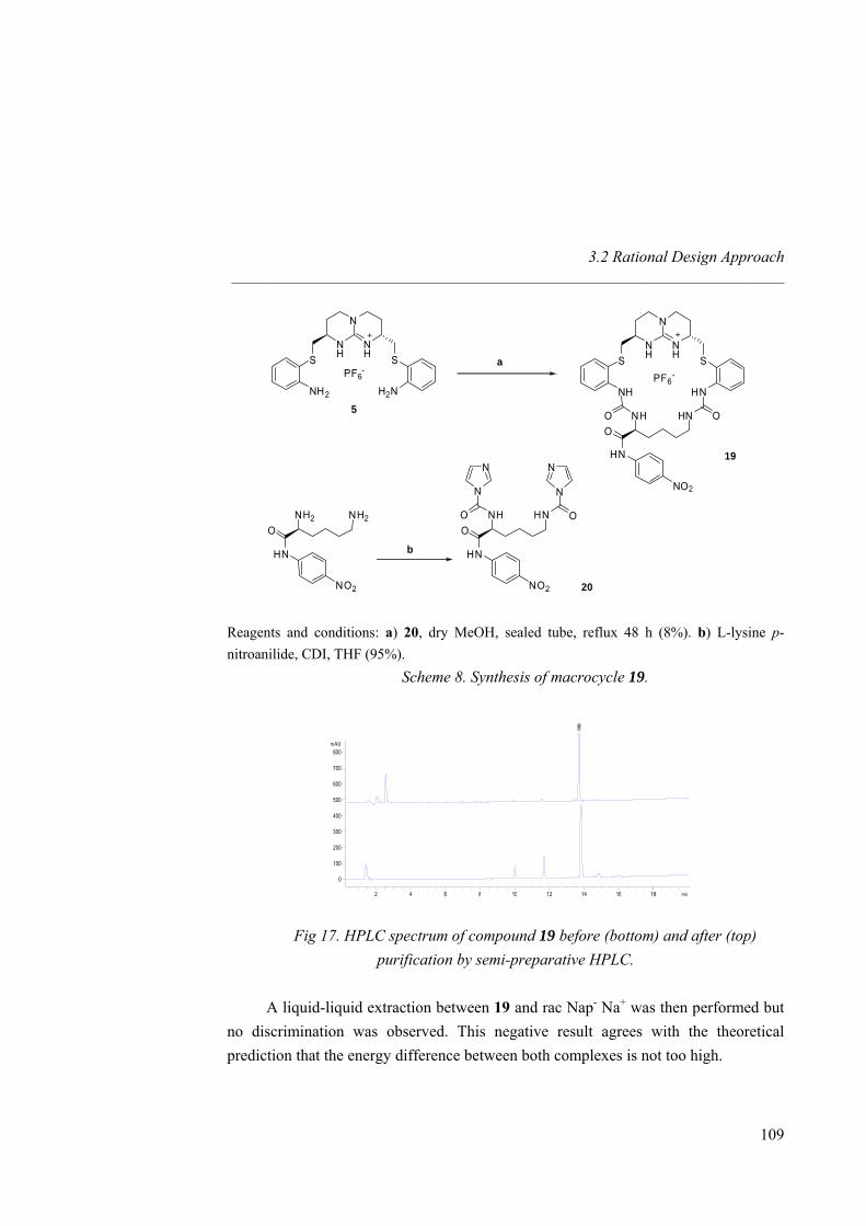
3.2 Rational Design Approach _____________________________________________________________________
N
NH
NH SS
PF6-
HNNH
OHNO NHO
HN
NO2
19
N
NH
NH SS
PF6-
H2NNH2
5
a
NN
OHNO NHO
HN
NO2
N N
NH2NH2O
HN
NO2
b
20
Reagents and conditions: a) 20, dry MeOH, sealed tube, reflux 48 h (8%). b) L-lysine p-nitroanilide, CDI, THF (95%).
Scheme 8. Synthesis of macrocycle 19.
min2 4 6 8 10 12 14 16 18
mAU
0
100
200
300
400
500
600
700
800
13.59
0
min2 4 6 8 10 12 14 16 18
mAU
0
100
200
300
400
500
600
700
800
13.59
0
Fig 17. HPLC spectrum of compound 19 before (bottom) and after (top) purification by semi-preparative HPLC.
A liquid-liquid extraction between 19 and rac Nap- Na+ was then performed but
no discrimination was observed. This negative result agrees with the theoretical prediction that the energy difference between both complexes is not too high.
109
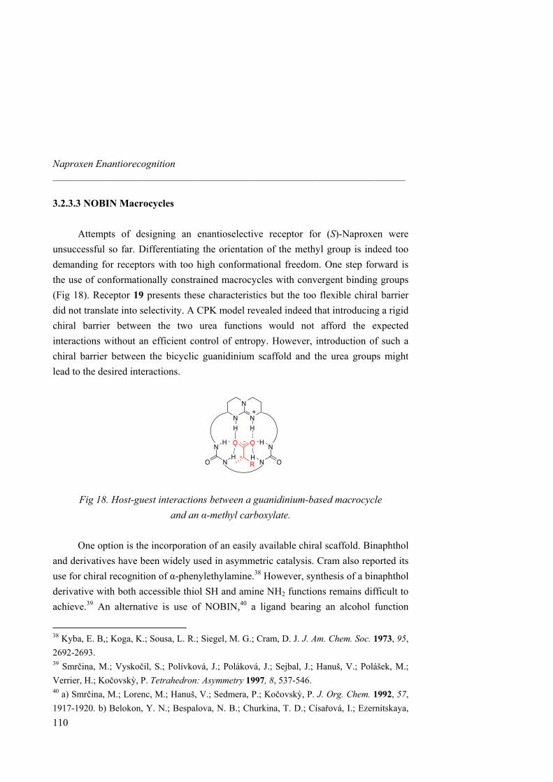
Naproxen Enantiorecognition _____________________________________________________________________
3.2.3.3 NOBIN Macrocycles Attempts of designing an enantioselective receptor for (S)-Naproxen were
unsuccessful so far. Differentiating the orientation of the methyl group is indeed too demanding for receptors with too high conformational freedom. One step forward is the use of conformationally constrained macrocycles with convergent binding groups (Fig 18). Receptor 19 presents these characteristics but the too flexible chiral barrier did not translate into selectivity. A CPK model revealed indeed that introducing a rigid chiral barrier between the two urea functions would not afford the expected interactions without an efficient control of entropy. However, introduction of such a chiral barrier between the bicyclic guanidinium scaffold and the urea groups might lead to the desired interactions.
+N
N
N
N N
N NO O
OO
R∗
H H
H
H H
H
Fig 18. Host-guest interactions between a guanidinium-based macrocycle
and an α-methyl carboxylate.
One option is the incorporation of an easily available chiral scaffold. Binaphthol and derivatives have been widely used in asymmetric catalysis. Cram also reported its use for chiral recognition of α-phenylethylamine.38 However, synthesis of a binaphthol derivative with both accessible thiol SH and amine NH2 functions remains difficult to achieve.39 An alternative is use of NOBIN,40 a ligand bearing an alcohol function
38 Kyba, E. B,; Koga, K.; Sousa, L. R.; Siegel, M. G.; Cram, D. J. J. Am. Chem. Soc. 1973, 95, 2692-2693. 39 Smrčina, M.; Vyskočil, S.; Polívková, J.; Poláková, J.; Sejbal, J.; Hanuš, V.; Polášek, M.; Verrier, H.; Kočovskỳ, P. Tetrahedron: Asymmetry 1997, 8, 537-546. 40 a) Smrčina, M.; Lorenc, M.; Hanuš, V.; Sedmera, P.; Kočovskỳ, P. J. Org. Chem. 1992, 57, 1917-1920. b) Belokon, Y. N.; Bespalova, N. B.; Churkina, T. D.; Císařová, I.; Ezernitskaya,
110
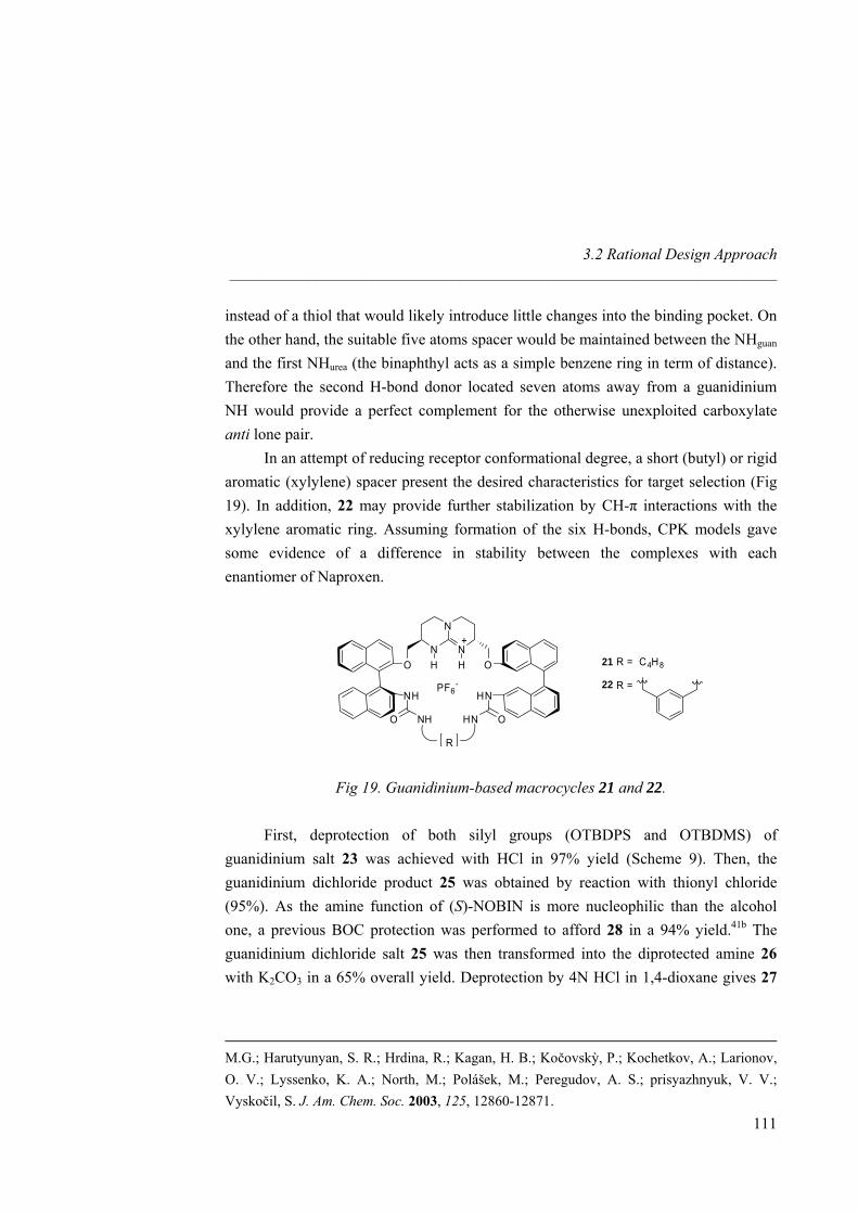
3.2 Rational Design Approach _____________________________________________________________________
instead of a thiol that would likely introduce little changes into the binding pocket. On the other hand, the suitable five atoms spacer would be maintained between the NHguan and the first NHurea (the binaphthyl acts as a simple benzene ring in term of distance). Therefore the second H-bond donor located seven atoms away from a guanidinium NH would provide a perfect complement for the otherwise unexploited carboxylate anti lone pair.
In an attempt of reducing receptor conformational degree, a short (butyl) or rigid aromatic (xylylene) spacer present the desired characteristics for target selection (Fig 19). In addition, 22 may provide further stabilization by CH-π interactions with the xylylene aromatic ring. Assuming formation of the six H-bonds, CPK models gave some evidence of a difference in stability between the complexes with each enantiomer of Naproxen.
N
N
NH H
+
O O
NH HN
O ONH HN
PF6-
21 R = C4H8
22 R =
R
Fig 19. Guanidinium-based macrocycles 21 and 22.
First, deprotection of both silyl groups (OTBDPS and OTBDMS) of guanidinium salt 23 was achieved with HCl in 97% yield (Scheme 9). Then, the guanidinium dichloride product 25 was obtained by reaction with thionyl chloride (95%). As the amine function of (S)-NOBIN is more nucleophilic than the alcohol one, a previous BOC protection was performed to afford 28 in a 94% yield.41b The guanidinium dichloride salt 25 was then transformed into the diprotected amine 26 with K2CO3 in a 65% overall yield. Deprotection by 4N HCl in 1,4-dioxane gives 27
M.G.; Harutyunyan, S. R.; Hrdina, R.; Kagan, H. B.; Kočovskỳ, P.; Kochetkov, A.; Larionov, O. V.; Lyssenko, K. A.; North, M.; Polášek, M.; Peregudov, A. S.; prisyazhnyuk, V. V.; Vyskočil, S. J. Am. Chem. Soc. 2003, 125, 12860-12871.
111
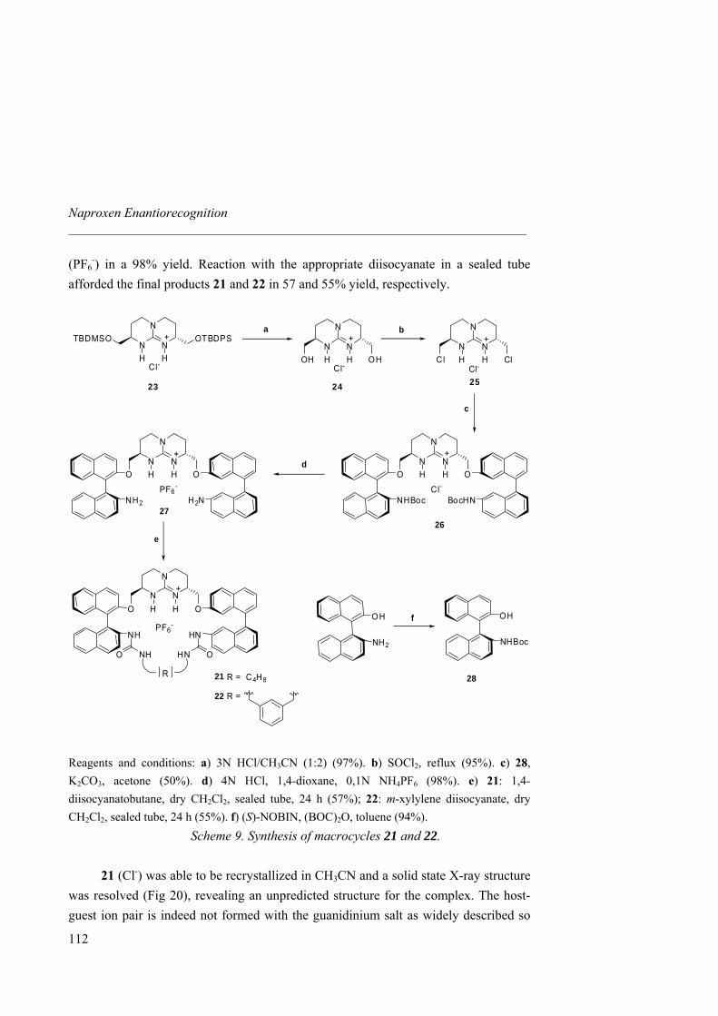
Naproxen Enantiorecognition _____________________________________________________________________
(PF6-) in a 98% yield. Reaction with the appropriate diisocyanate in a sealed tube
afforded the final products 21 and 22 in 57 and 55% yield, respectively.
N
N
NH H
+
O O
NH HN
O ONH HN
PF6-
R
N
N
NH H
+
O O
NH2
PF6-
H2N
N
N
NH H
+
Cl ClCl-
N
N
NH H
+
OH OHCl-
a b
N
N
NH H
+
Cl-
OTBDPSTBDMSO
252423
d
e26
27
c
OH
NHBoc
OH
NH2
f
2821 R = C4H8
22 R =
N
N
NH H
+
O O
NHBocCl-
BocHN
Reagents and conditions: a) 3N HCl/CH3CN (1:2) (97%). b) SOCl2, reflux (95%). c) 28, K2CO3, acetone (50%). d) 4N HCl, 1,4-dioxane, 0,1N NH4PF6 (98%). e) 21: 1,4-diisocyanatobutane, dry CH2Cl2, sealed tube, 24 h (57%); 22: m-xylylene diisocyanate, dry CH2Cl2, sealed tube, 24 h (55%). f) (S)-NOBIN, (BOC)2O, toluene (94%).
Scheme 9. Synthesis of macrocycles 21 and 22. 21 (Cl-) was able to be recrystallized in CH3CN and a solid state X-ray structure was resolved (Fig 20), revealing an unpredicted structure for the complex. The host-guest ion pair is indeed not formed with the guanidinium salt as widely described so
112
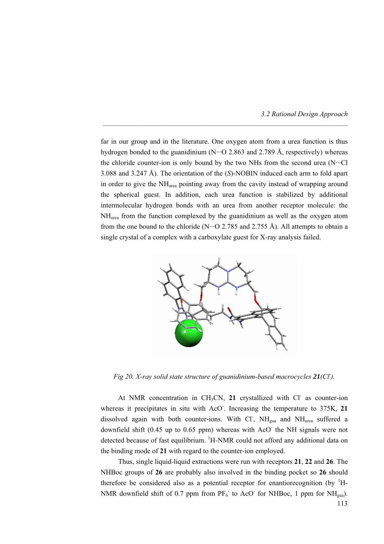
3.2 Rational Design Approach _____________________________________________________________________
far in our group and in the literature. One oxygen atom from a urea function is thus hydrogen bonded to the guanidinium (N···O 2.863 and 2.789 Å, respectively) whereas the chloride counter-ion is only bound by the two NHs from the second urea (N···Cl 3.088 and 3.247 Å). The orientation of the (S)-NOBIN induced each arm to fold apart in order to give the NHurea pointing away from the cavity instead of wrapping around the spherical guest. In addition, each urea function is stabilized by additional intermolecular hydrogen bonds with an urea from another receptor molecule: the NHurea from the function complexed by the guanidinium as well as the oxygen atom from the one bound to the chloride (N···O 2.785 and 2.755 Å). All attempts to obtain a single crystal of a complex with a carboxylate guest for X-ray analysis failed.
Fig 20. X-ray solid state structure of guanidinium-based macrocycles 21(Cl-). At NMR concentration in CH3CN, 21 crystallized with Cl- as counter-ion
whereas it precipitates in situ with AcO-. Increasing the temperature to 375K, 21 dissolved again with both counter-ions. With Cl-, NHgua and NHurea suffered a downfield shift (0.45 up to 0.65 ppm) whereas with AcO- the NH signals were not detected because of fast equilibrium. 1H-NMR could not afford any additional data on the binding mode of 21 with regard to the counter-ion employed.
Thus, single liquid-liquid extractions were run with receptors 21, 22 and 26. The NHBoc groups of 26 are probably also involved in the binding pocket so 26 should therefore be considered also as a potential receptor for enantiorecognition (by 1H-NMR downfield shift of 0.7 ppm from PF6
- to AcO- for NHBoc, 1 ppm for NHgua). 113
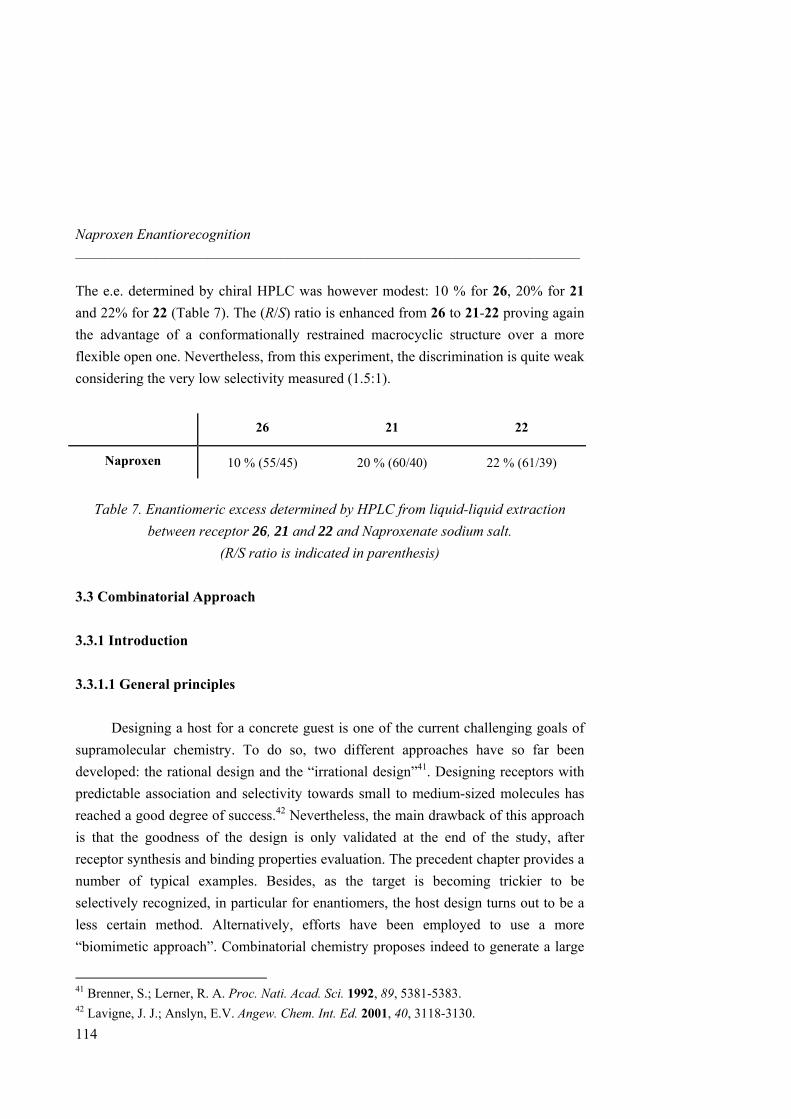
Naproxen Enantiorecognition _____________________________________________________________________
114
The e.e. determined by chiral HPLC was however modest: 10 % for 26, 20% for 21 and 22% for 22 (Table 7). The (R/S) ratio is enhanced from 26 to 21-22 proving again the advantage of a conformationally restrained macrocyclic structure over a more flexible open one. Nevertheless, from this experiment, the discrimination is quite weak considering the very low selectivity measured (1.5:1).
26 21 22
Naproxen 10 % (55/45) 20 % (60/40) 22 % (61/39)
Table 7. Enantiomeric excess determined by HPLC from liquid-liquid extraction
between receptor 26, 21 and 22 and Naproxenate sodium salt. (R/S ratio is indicated in parenthesis)
3.3 Combinatorial Approach 3.3.1 Introduction
3.3.1.1 General principles
Designing a host for a concrete guest is one of the current challenging goals of
supramolecular chemistry. To do so, two different approaches have so far been developed: the rational design and the “irrational design”41. Designing receptors with predictable association and selectivity towards small to medium-sized molecules has reached a good degree of success.42 Nevertheless, the main drawback of this approach is that the goodness of the design is only validated at the end of the study, after receptor synthesis and binding properties evaluation. The precedent chapter provides a number of typical examples. Besides, as the target is becoming trickier to be selectively recognized, in particular for enantiomers, the host design turns out to be a less certain method. Alternatively, efforts have been employed to use a more “biomimetic approach”. Combinatorial chemistry proposes indeed to generate a large
41 Brenner, S.; Lerner, R. A. Proc. Nati. Acad. Sci. 1992, 89, 5381-5383. 42 Lavigne, J. J.; Anslyn, E.V. Angew. Chem. Int. Ed. 2001, 40, 3118-3130.
3.3 Combinatorial Approach _____________________________________________________________________
115
number of compounds capable of binding a target. Selection of the most suitable one for its desired property is though achieved by rapid screening of the library. Synthetic expenditure is reduced while the possibilities of generating an effective receptor are enhanced highlighting this approach as very attracting.
The combinatorial chemistry concept was first reported in 198843 and published in 1991.44 Since then, this approach has been widely employed and successfully reported. One reason of this fast growth is the rapid generation of compounds and screening of biological activity.45 Combinatorial chemistry first played a significant role in chemical biology and currently extends to almost all fields of chemistry such as host-guest chemistry.46 The combinatorial approach to the identification of selective peptide receptors was pioneered by Still in the 1990’s.47 Afterwards, another approach for receptor discovery appears by the use of dynamic combinatorial libraries (DCL) pioneered by the research group of Sanders and Lehn.48
Considering the field of supramolecular chemistry, combinatorial receptor design is the first step toward the generation of a library. “Tweezers” or “two-armed” receptors have been then widely developed.49 Such receptors possess a scaffold or “head group” which pre-organizes the substrate binding arms as well as containing the binding functionality. Attachment of this scaffold to a solid support is necessary for solid phase synthesis of the library. Parallel synthesis of the arms by means of Split
43 Furka, A.; Sebestyen, F.; Asgedom, M.; Dibo, G. Abstr. 14th Int. Congre. Biochem., Prague, Czechoslovakia, 1988, Budapest, Hungary, 288. 44 a) Furka, A.; Sebestyen, F.; Asgedom, M.; Dibo, G. Int. J. Pept. Protein Res. 1991, 37, 487-493. b) Houghten, R. A.; Pinilla, C.; Blondelle, S. E.; Appel, J. R.; Dooley, C. T.; Cuervo, J. H. Nature 1991, 354, 84-86. c) Lam, K.; Salmon, S.; Herch, E.; Hruby, V.; Kazmierski, W.; Knapp, R. Nature, 1991, 354, 82-84. 45 Lowe, G. Chem. Soc. Rev. 1995, 309-317. 46 Linton, B.; Hamilton, A. D. Curr. Opin. Chem. Biol. 1999, 3, 307-312. 47 Still, W. C. Acc. Chem. Res. 1996, 29, 155-163. 48 a) Huc, I.; Lehn, J. M. Proc. Natl. Acad. Sci. U.S.A. 1997, 94, 2106-2110. b) Otto, S.; Burlan, R. L. E.; Sanders, J. K. M. Curr. Opin. Chem. Biol. 2002, 6, 321-327. c) Rowan, S. J.; Cantrill, S. J.; Cousins, G. R. L.; Sanders, J. K. M.; Stoddart, J. F. Angew. Chem. Int. Ed. 2002, 41, 898-952. 49 Srinivasan, N.; Kilburn, J. D. Curr. Opin. Chem. Biol. 2004, 8, 305-310.
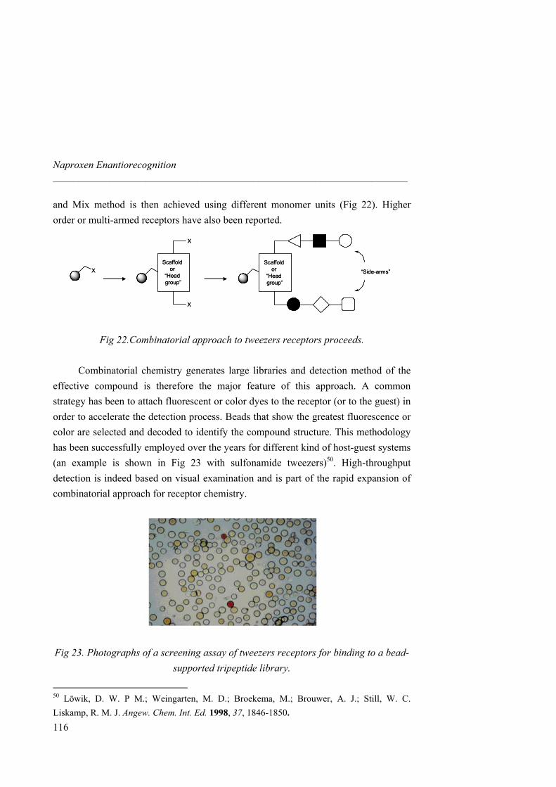
Naproxen Enantiorecognition _____________________________________________________________________
and Mix method is then achieved using different monomer units (Fig 22). Higher order or multi-armed receptors have also been reported.
XScaffold
or “Head group”
X
X
Scaffold or
“Head group”
“Side-arms”XScaffold
or “Head group”
X
X
Scaffold or
“Head group”
“Side-arms”
Fig 22.Combinatorial approach to tweezers receptors proceeds. Combinatorial chemistry generates large libraries and detection method of the
effective compound is therefore the major feature of this approach. A common strategy has been to attach fluorescent or color dyes to the receptor (or to the guest) in order to accelerate the detection process. Beads that show the greatest fluorescence or color are selected and decoded to identify the compound structure. This methodology has been successfully employed over the years for different kind of host-guest systems (an example is shown in Fig 23 with sulfonamide tweezers)50. High-throughput detection is indeed based on visual examination and is part of the rapid expansion of combinatorial approach for receptor chemistry.
Fig 23. Photographs of a screening assay of tweezers receptors for binding to a bead-supported tripeptide library.
50 Löwik, D. W. P M.; Weingarten, M. D.; Broekema, M.; Brouwer, A. J.; Still, W. C. Liskamp, R. M. J. Angew. Chem. Int. Ed. 1998, 37, 1846-1850.
116
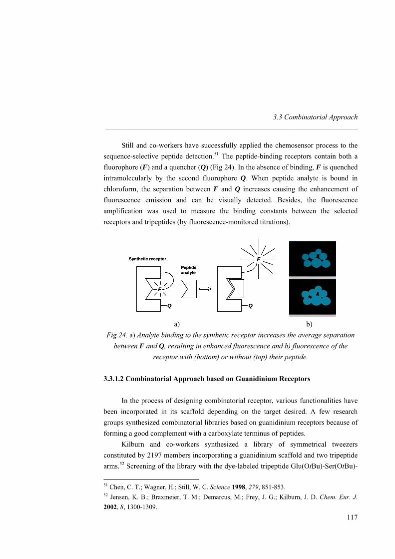
3.3 Combinatorial Approach _____________________________________________________________________
Still and co-workers have successfully applied the chemosensor process to the sequence-selective peptide detection.51 The peptide-binding receptors contain both a fluorophore (F) and a quencher (Q) (Fig 24). In the absence of binding, F is quenched intramolecularly by the second fluorophore Q. When peptide analyte is bound in chloroform, the separation between F and Q increases causing the enhancement of fluorescence emission and can be visually detected. Besides, the fluorescence amplification was used to measure the binding constants between the selected receptors and tripeptides (by fluorescence-monitored titrations).
F
Q
F
Q
Synthetic receptor
Peptideanalyte
F
Q
F
Q
Synthetic receptor
Peptideanalyte
a) b) Fig 24. a) Analyte binding to the synthetic receptor increases the average separation
between F and Q, resulting in enhanced fluorescence and b) fluorescence of the receptor with (bottom) or without (top) their peptide.
3.3.1.2 Combinatorial Approach based on Guanidinium Receptors
In the process of designing combinatorial receptor, various functionalities have
been incorporated in its scaffold depending on the target desired. A few research groups synthesized combinatorial libraries based on guanidinium receptors because of forming a good complement with a carboxylate terminus of peptides.
Kilburn and co-workers synthesized a library of symmetrical tweezers constituted by 2197 members incorporating a guanidinium scaffold and two tripeptide arms.52 Screening of the library with the dye-labeled tripeptide Glu(OtBu)-Ser(OtBu)-
51 Chen, C. T.; Wagner, H.; Still, W. C. Science 1998, 279, 851-853. 52 Jensen, K. B.; Braxmeier, T. M.; Demarcus, M.; Frey, J. G.; Kilburn, J. D. Chem. Eur. J. 2002, 8, 1300-1309.
117
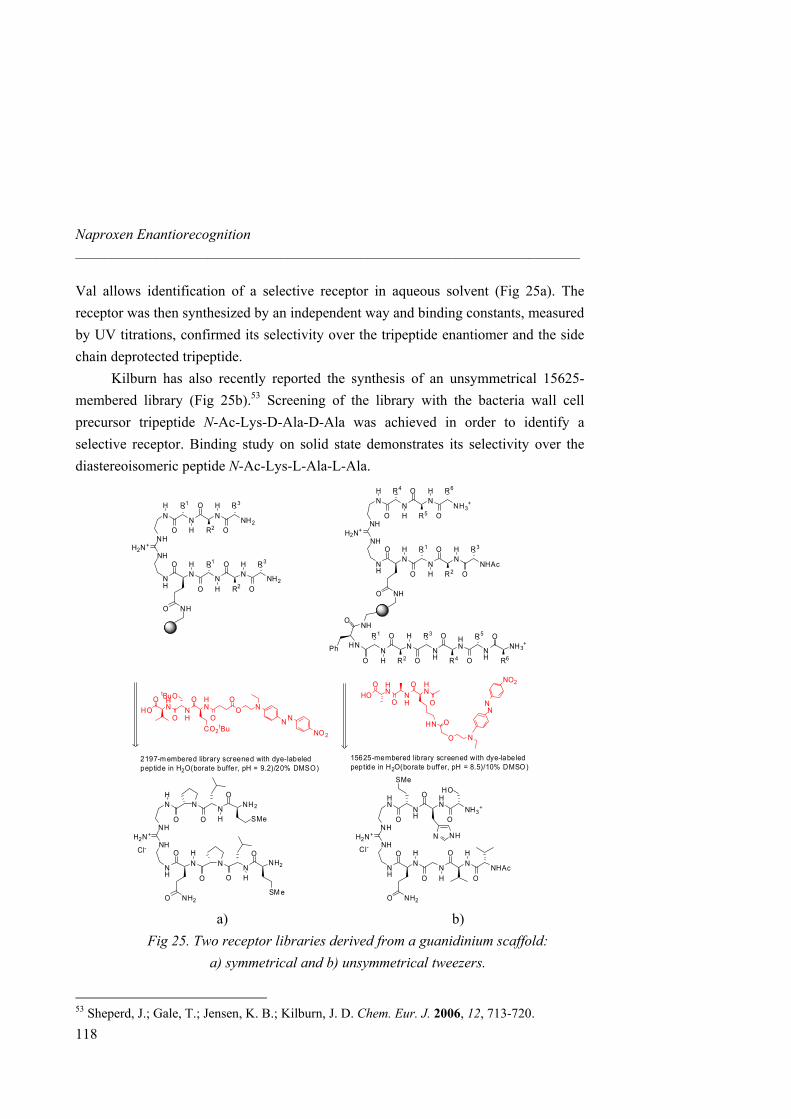
Naproxen Enantiorecognition _____________________________________________________________________
Val allows identification of a selective receptor in aqueous solvent (Fig 25a). The receptor was then synthesized by an independent way and binding constants, measured by UV titrations, confirmed its selectivity over the tripeptide enantiomer and the side chain deprotected tripeptide.
Kilburn has also recently reported the synthesis of an unsymmetrical 15625-membered library (Fig 25b).53 Screening of the library with the bacteria wall cell precursor tripeptide N-Ac-Lys-D-Ala-D-Ala was achieved in order to identify a selective receptor. Binding study on solid state demonstrates its selectivity over the diastereoisomeric peptide N-Ac-Lys-L-Ala-L-Ala.
N N N NH2
H
H
HR1 R3
R2O
O
ONH
NHH2N+
N N N NH2
H
H
HR1 R3
R2O
O
ONH
O
O NH
NO 2
NN
NOO
NO
HN
OHNHO
CO2tBu
HO
OtBuO N
O
HN
OHNHO
HO
O
OHN
NO
NO2
NN
NH
ONH
NHH2N+
NH
ONH
O
O NH2
N
ONH
ONH2
SMe
N
ONH
ONH2
SM e
2197-m embered library screened with dye-labeledpeptide in H2O(borate buffer, pH = 9.2)/20% DMSO)
Cl-
HN
NH
HN
NH3+
O
O
ONH
NHH2N+
N N N NHAc
H
H
H
O
O
ONH
O
O NH2
SMe
N NH
HO
15625-membered library screened with dye-labeledpeptide in H2O(borate buffer, pH = 8.5)/10% DMSO)
Cl-
NN
NNH3
+
H
H
HR4 R6
R5O
O
ONH
NHH2N+
NN
NNHAc
H
H
HR1 R3
R2O
O
ONH
O
O NH
N N NHH
HR1 R3
R2O
O
O
HN N
H
O
O
ONH3
+
R4 R6
R5NH
O
HNPh
a) b) Fig 25. Two receptor libraries derived from a guanidinium scaffold:
a) symmetrical and b) unsymmetrical tweezers.
53 Sheperd, J.; Gale, T.; Jensen, K. B.; Kilburn, J. D. Chem. Eur. J. 2006, 12, 713-720.
118
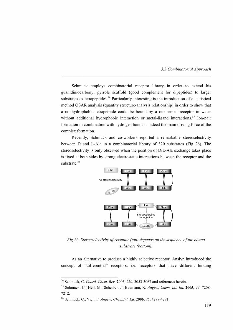
3.3 Combinatorial Approach _____________________________________________________________________
Schmuck employs combinatorial receptor library in order to extend his guanidiniocarbonyl pyrrole scaffold (good complement for dipeptides) to larger substrates as tetrapeptides.54 Particularly interesting is the introduction of a statistical method QSAR analysis (quantity structure-analysis relationship) in order to show that a nonhydrophobic tetrapetpide could be bound by a one-armed receptor in water without additional hydrophobic interaction or metal-ligand interactions.55 Ion-pair formation in combination with hydrogen bonds is indeed the main driving force of the complex formation.
Recently, Schmuck and co-workers reported a remarkable stereoselectivity between D and L-Ala in a combinatorial library of 320 substrates (Fig 26). The stereoselectivity is only observed when the position of D/L-Ala exchange takes place is fixed at both sides by strong electrostatic interactions between the receptor and the substrate.56
Fig 26. Stereoselectivity of receptor (top) depends on the sequence of the bound substrate (bottom).
As an alternative to produce a highly selective receptor, Anslyn introduced the concept of “differential” receptors, i.e. receptors that have different binding
54 Schmuck, C. Coord. Chem. Rev. 2006, 250, 3053-3067 and references herein. 55 Schmuck, C.; Heil, M.; Scheiber, J.; Baumann, K. Angew. Chem. Int. Ed. 2005, 44, 7208-7212. 56 Schmuck, C.; Vich, P. Angew. Chem.Int. Ed. 2006, 45, 4277-4281.
119
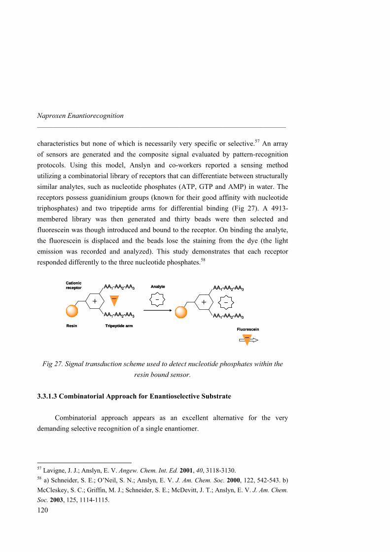
Naproxen Enantiorecognition _____________________________________________________________________
characteristics but none of which is necessarily very specific or selective.57 An array of sensors are generated and the composite signal evaluated by pattern-recognition protocols. Using this model, Anslyn and co-workers reported a sensing method utilizing a combinatorial library of receptors that can differentiate between structurally similar analytes, such as nucleotide phosphates (ATP, GTP and AMP) in water. The receptors possess guanidinium groups (known for their good affinity with nucleotide triphosphates) and two tripeptide arms for differential binding (Fig 27). A 4913-membered library was then generated and thirty beads were then selected and fluorescein was though introduced and bound to the receptor. On binding the analyte, the fluorescein is displaced and the beads lose the staining from the dye (the light emission was recorded and analyzed). This study demonstrates that each receptor responded differently to the three nucleotide phosphates.58
AA1-AA2-AA3
AA1-AA2-AA3
AA1-AA2-AA3
AA1-AA2-AA3
AnalyteCationicreceptor
Resin Tripeptide armFluorescein
AA1-AA2-AA3
AA1-AA2-AA3
AA1-AA2-AA3
AA1-AA2-AA3
AnalyteCationicreceptor
Resin Tripeptide armFluorescein
Fig 27. Signal transduction scheme used to detect nucleotide phosphates within the resin bound sensor.
3.3.1.3 Combinatorial Approach for Enantioselective Substrate
Combinatorial approach appears as an excellent alternative for the very
demanding selective recognition of a single enantiomer.
57 Lavigne, J. J.; Anslyn, E. V. Angew. Chem. Int. Ed. 2001, 40, 3118-3130. 58 a) Schneider, S. E.; O’Neil, S. N.; Anslyn, E. V. J. Am. Chem. Soc. 2000, 122, 542-543. b) McCleskey, S. C.; Griffin, M. J.; Schneider, S. E.; McDevitt, J. T.; Anslyn, E. V. J. Am. Chem. Soc. 2003, 125, 1114-1115.
120
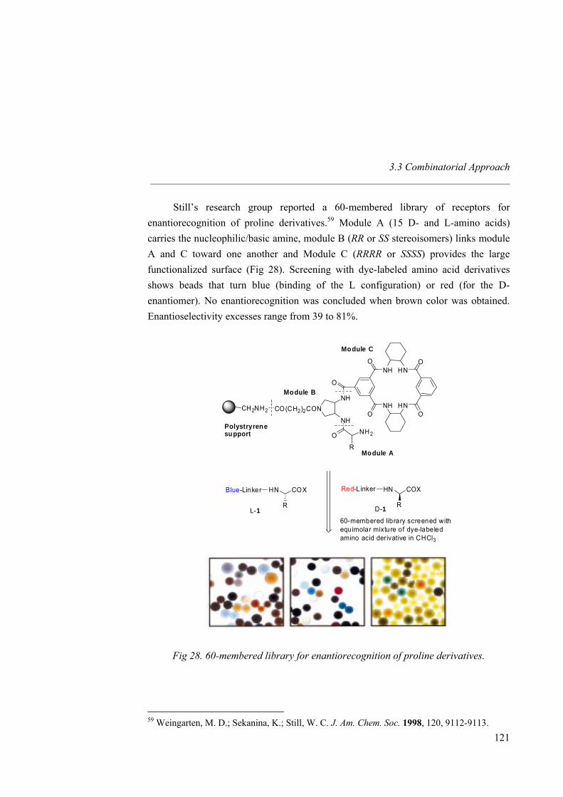
3.3 Combinatorial Approach _____________________________________________________________________
Still’s research group reported a 60-membered library of receptors for enantiorecognition of proline derivatives.59 Module A (15 D- and L-amino acids) carries the nucleophilic/basic amine, module B (RR or SS stereoisomers) links module A and C toward one another and Module C (RRRR or SSSS) provides the large functionalized surface (Fig 28). Screening with dye-labeled amino acid derivatives shows beads that turn blue (binding of the L configuration) or red (for the D-enantiomer). No enantiorecognition was concluded when brown color was obtained. Enantioselectivity excesses range from 39 to 81%.
CH2NH2 CO(CH2)2CONNH
NH
O
O
NHO
NHO
HN
HN
O
O
R
NH2Polystryrenesupport
Module B
Module A
Module C
COX
R
HN COX
R
HNBlue-Linker
Red-Linker
L-1 D-160-membered library screened withequimolar mixture of dye-labeledamino acid derivative in CHCl3
Fig 28. 60-membered library for enantiorecognition of proline derivatives.
59 Weingarten, M. D.; Sekanina, K.; Still, W. C. J. Am. Chem. Soc. 1998, 120, 9112-9113.
121
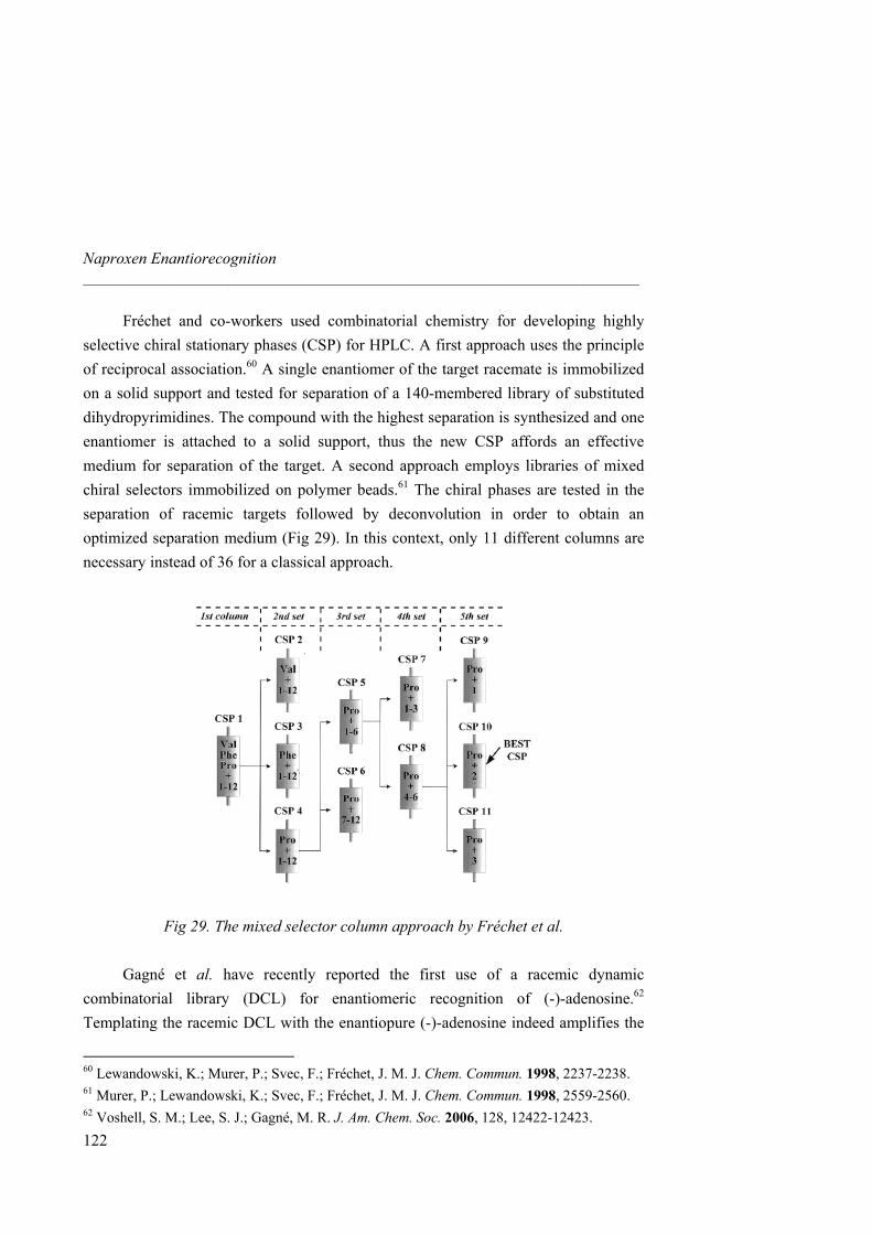
Naproxen Enantiorecognition _____________________________________________________________________
Fréchet and co-workers used combinatorial chemistry for developing highly selective chiral stationary phases (CSP) for HPLC. A first approach uses the principle of reciprocal association.60 A single enantiomer of the target racemate is immobilized on a solid support and tested for separation of a 140-membered library of substituted dihydropyrimidines. The compound with the highest separation is synthesized and one enantiomer is attached to a solid support, thus the new CSP affords an effective medium for separation of the target. A second approach employs libraries of mixed chiral selectors immobilized on polymer beads.61 The chiral phases are tested in the separation of racemic targets followed by deconvolution in order to obtain an optimized separation medium (Fig 29). In this context, only 11 different columns are necessary instead of 36 for a classical approach.
Fig 29. The mixed selector column approach by Fréchet et al.
Gagné et al. have recently reported the first use of a racemic dynamic combinatorial library (DCL) for enantiomeric recognition of (-)-adenosine.62 Templating the racemic DCL with the enantiopure (-)-adenosine indeed amplifies the
60 Lewandowski, K.; Murer, P.; Svec, F.; Fréchet, J. M. J. Chem. Commun. 1998, 2237-2238. 61 Murer, P.; Lewandowski, K.; Svec, F.; Fréchet, J. M. J. Chem. Commun. 1998, 2559-2560. 62 Voshell, S. M.; Lee, S. J.; Gagné, M. R. J. Am. Chem. Soc. 2006, 128, 12422-12423.
122
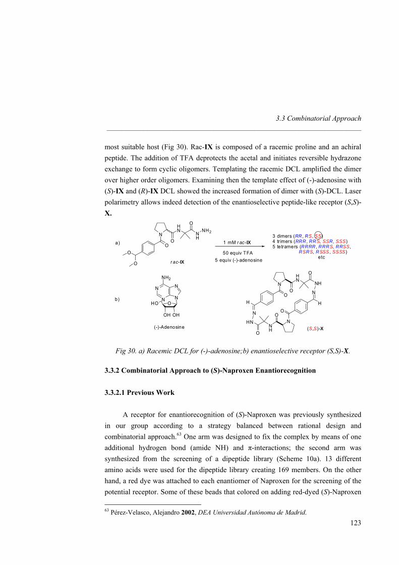
3.3 Combinatorial Approach _____________________________________________________________________
most suitable host (Fig 30). Rac-IX is composed of a racemic proline and an achiral peptide. The addition of TFA deprotects the acetal and initiates reversible hydrazone exchange to form cyclic oligomers. Templating the racemic DCL amplified the dimer over higher order oligomers. Examining then the template effect of (-)-adenosine with (S)-IX and (R)-IX DCL showed the increased formation of dimer with (S)-DCL. Laser polarimetry allows indeed detection of the enantioselective peptide-like receptor (S,S)-X.
NH
NH2HN
O
O
N
OO
O
1 mM r ac-IX
r ac-IX
50 equiv TFA5 equiv (-)-adenosine
3 dimers (RR , R
S, SS)S, SS SSS)
S, SS,S S, SSS, SSSS)
(S,S)-
4 trimers (RRR , RR R,5 tetramers (RRRR , RRR RR
R R Retc
O
OH OH
NHO
NN
N
NH2
(-)-Adenosine
NHHN
O
O
N
OH
N
HNNH
O
O
N
OH
N
X
a)
b)
Fig 30. a) Racemic DCL for (-)-adenosine;b) enantioselective receptor (S,S)-X.
3.3.2 Combinatorial Approach to (S)-Naproxen Enantiorecognition
3.3.2.1 Previous Work A receptor for enantiorecognition of (S)-Naproxen was previously synthesized
in our group according to a strategy balanced between rational design and combinatorial approach.63 One arm was designed to fix the complex by means of one additional hydrogen bond (amide NH) and π-interactions; the second arm was synthesized from the screening of a dipeptide library (Scheme 10a). 13 different amino acids were used for the dipeptide library creating 169 members. On the other hand, a red dye was attached to each enantiomer of Naproxen for the screening of the potential receptor. Some of these beads that colored on adding red-dyed (S)-Naproxen
63 Pérez-Velasco, Alejandro 2002, DEA Universidad Autónoma de Madrid.
123

Naproxen Enantiorecognition _____________________________________________________________________
but not with (R)-Naproxen were identified. The sequence Trp-Ser appeared three times and therefore a receptor containing this dipeptide was synthesized (Scheme 10b). Extraction was performed between racemic Naproxenate and the final receptor. However, 1H-NMR as well as chiral HPLC pointed out the lack of selectivity of this receptor. Besides, two equivalents of guest were extracted whereas guanidinium receptors form only 1:1 complexes with the guest. Likely, a too low number of dyed beds were selected for screening, so the found consensus was simply an artifact. This was due to the deconvolution method employed, based on quite expensive Edman degradation sequences, that prevented a larger number of beads to be screened.
N
N
NS SHH
Cl
O NHOO
N
N
NS SHH
O NH
O2N
O
N
NN
NO2
HN O
AA2
AA1 O2N
HN O
O
NH
HN
O
HN O
a) b) Scheme 10. a) Previous combinatorial approach to enantiorecognition of
(S)-Naproxen and b) receptor synthesized.
3.3.2.2 New Approach It appears essential to generate a peptide library because of the presence of the
amino acid chiral center as well as the additional peptidic hydrogen bonds. Here, we aim at employing a larger peptide library using 8 different amino acids with 4 coupling steps: a library of 4096 members is therefore available.
In addition to the creation of a larger library, the combinatorial receptor was redesigned in order to get closer the peptide to the chiral center of the guest. Attaching the tetrapeptide directly to the receptor scaffold through an amine function located
124
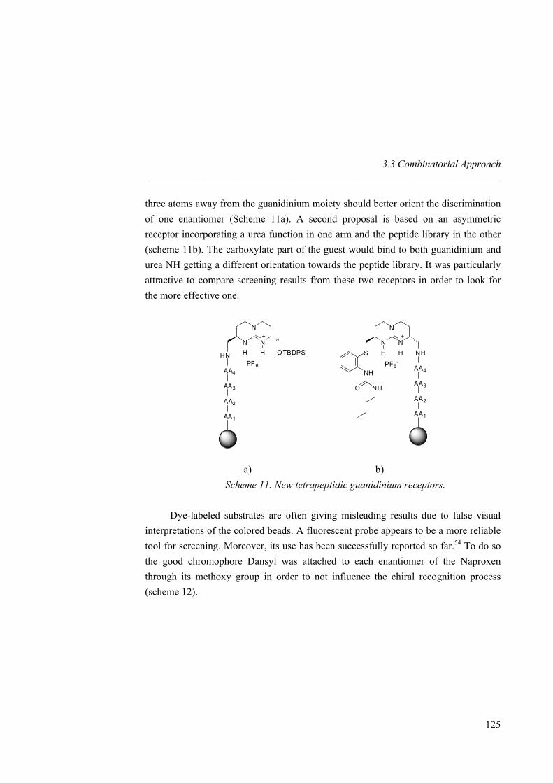
3.3 Combinatorial Approach _____________________________________________________________________
three atoms away from the guanidinium moiety should better orient the discrimination of one enantiomer (Scheme 11a). A second proposal is based on an asymmetric receptor incorporating a urea function in one arm and the peptide library in the other (scheme 11b). The carboxylate part of the guest would bind to both guanidinium and urea NH getting a different orientation towards the peptide library. It was particularly attractive to compare screening results from these two receptors in order to look for the more effective one.
N
N
NS NHHH
NH
AA2
AA1
O NH
AA4
AA3
PF6-
N
N
NOTBDPSHH
PF 6-
HN
AA2
AA1
AA4
AA3
a) b)
Scheme 11. New tetrapeptidic guanidinium receptors. Dye-labeled substrates are often giving misleading results due to false visual
interpretations of the colored beads. A fluorescent probe appears to be a more reliable tool for screening. Moreover, its use has been successfully reported so far.54 To do so the good chromophore Dansyl was attached to each enantiomer of the Naproxen through its methoxy group in order to not influence the chiral recognition process (scheme 12).
125
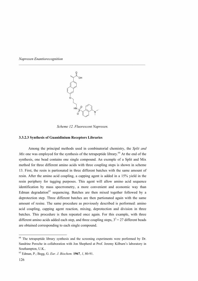
Naproxen Enantiorecognition _____________________________________________________________________
OH
O
O
HNS
O
O
N
Scheme 12. Fluorescent Naproxen.
3.3.2.3 Synthesis of Guanidinium Receptors Libraries
Among the principal methods used in combinatorial chemistry, the Split and Mix one was employed for the synthesis of the tetrapeptide library.64 At the end of the synthesis, one bead contains one single compound. An exemple of a Split and Mix method for three different amino acids with three coupling steps is shown in scheme 13. First, the resin is partionated in three different batches with the same amount of resin. After the amino acid coupling, a capping agent is added in a 15% yield in the resin periphery for tagging purposes. This agent will allow amino acid sequence identification by mass specrrometry, a more convenient and economic way than Edman degradation65 sequencing. Batches are then mixed together followed by a deprotection step. Three different batches are then partionated again with the same amount of resine. The same procedure as previously described is performed: amino acid coupling, capping agent reaction, mixing, deprotection and division in three batches. This procedure is then repeated once again. For this example, with three different amino acids added each step, and three coupling steps, 33 = 27 different beads are obtained corresponding to each single compound.
64 The tetrapeptide library synthesis and the screening experiments were performed by Dr. Sandrine Peroche in collaboration with Jon Shepherd at Prof. Jeremy Kilburn’s laboratory in Southampton, U.K.. 65 Edman, P.; Begg, G. Eur. J. Biochem. 1967, 1, 80-91.
126
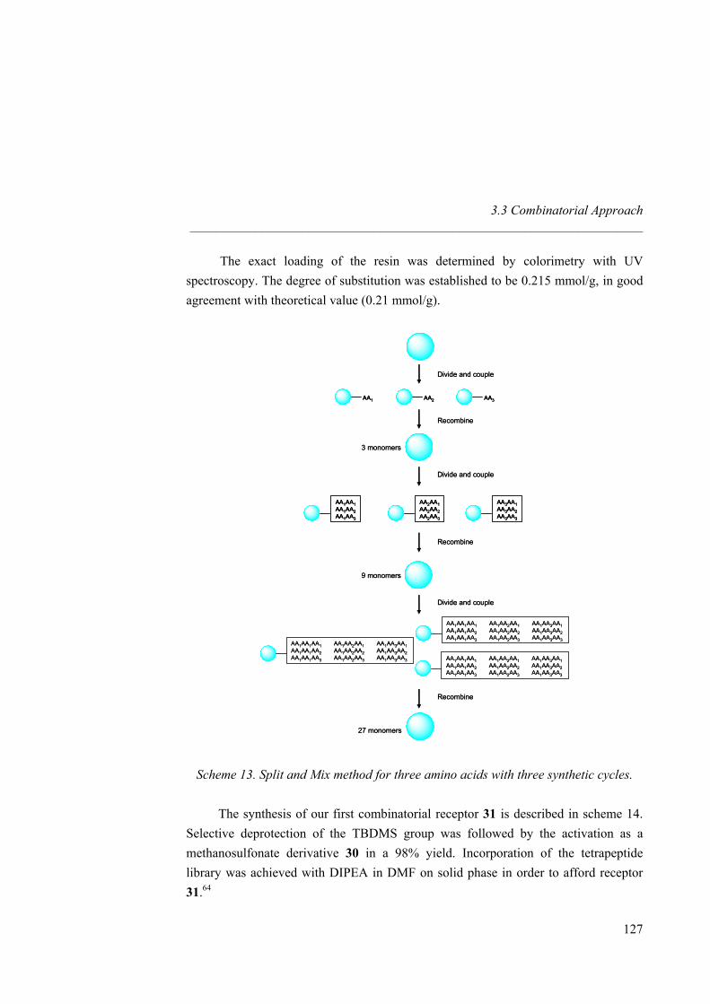
3.3 Combinatorial Approach _____________________________________________________________________
The exact loading of the resin was determined by colorimetry with UV spectroscopy. The degree of substitution was established to be 0.215 mmol/g, in good agreement with theoretical value (0.21 mmol/g).
AA2 AA3AA1
AA1AA1AA1AA2AA1AA3
AA2AA1AA2AA2AA2AA3
AA3AA1AA3AA2AA3AA3
AA1AA1AA1 AA1AA2AA1 AA1AA3AA1 AA1AA1AA2 AA1AA2AA2 AA1AA3AA2AA1AA1AA3 AA1AA2AA3 AA1AA3AA3
AA1AA1AA1 AA1AA2AA1 AA1AA3AA1 AA1AA1AA2 AA1AA2AA2 AA1AA3AA2AA1AA1AA3 AA1AA2AA3 AA1AA3AA3
AA1AA1AA1 AA1AA2AA1 AA1AA3AA1 AA1AA1AA2 AA1AA2AA2 AA1AA3AA2AA1AA1AA3 AA1AA2AA3 AA1AA3AA3
Divide and couple
Divide and couple
Divide and couple
Recombine
Recombine
Recombine
3 monomers
9 monomers
27 monomers
AA2AA2 AA3AA3AA1AA1
AA1AA1AA1AA2AA1AA3
AA1AA1AA1AA2AA1AA3
AA2AA1AA2AA2AA2AA3
AA2AA1AA2AA2AA2AA3
AA3AA1AA3AA2AA3AA3
AA3AA1AA3AA2AA3AA3
AA1AA1AA1 AA1AA2AA1 AA1AA3AA1 AA1AA1AA2 AA1AA2AA2 AA1AA3AA2AA1AA1AA3 AA1AA2AA3 AA1AA3AA3
AA1AA1AA1 AA1AA2AA1 AA1AA3AA1 AA1AA1AA2 AA1AA2AA2 AA1AA3AA2AA1AA1AA3 AA1AA2AA3 AA1AA3AA3
AA1AA1AA1 AA1AA2AA1 AA1AA3AA1 AA1AA1AA2 AA1AA2AA2 AA1AA3AA2AA1AA1AA3 AA1AA2AA3 AA1AA3AA3
Divide and couple
Divide and couple
Divide and couple
Recombine
Recombine
Recombine
3 monomers
9 monomers
27 monomers
Scheme 13. Split and Mix method for three amino acids with three synthetic cycles. The synthesis of our first combinatorial receptor 31 is described in scheme 14. Selective deprotection of the TBDMS group was followed by the activation as a methanosulfonate derivative 30 in a 98% yield. Incorporation of the tetrapeptide library was achieved with DIPEA in DMF on solid phase in order to afford receptor 31.64
127
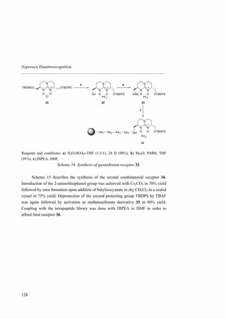
Naproxen Enantiorecognition _____________________________________________________________________
N
N
NH H
+
OH OTBDPSPF6
-
a b
N
N
NH H
+
Cl-
OTBDPSTBDMSO
302923
c
N
N
NH H
+
OMs OTBDPSPF6
-
N
N
NOTBDPSHH
PF6-
NHAA2AA1 AA4AA3
31
Reagents and conditions: a) H2O-HOAc-THF (1:3:1), 24 H (98%). b) Ms2O, NMM, THF (95%). c) DIPEA, DMF.
Scheme 14. Synthesis of guanidinium receptor 31. Scheme 15 describes the synthesis of the second combinatorial receptor 36.
Introduction of the 2-aminothiophenol group was achieved with Cs2CO3 in 70% yield followed by urea formation upon addition of butylisocyanate in dry CH2Cl2 in a sealed vessel in 75% yield. Deprotection of the second protecting group TBDPS by TBAF was again followed by activation as methansulfonate derivative 35 in 90% yield. Coupling with the tetrapeptide library was done with DIPEA in DMF in order to afford final receptor 36.
128
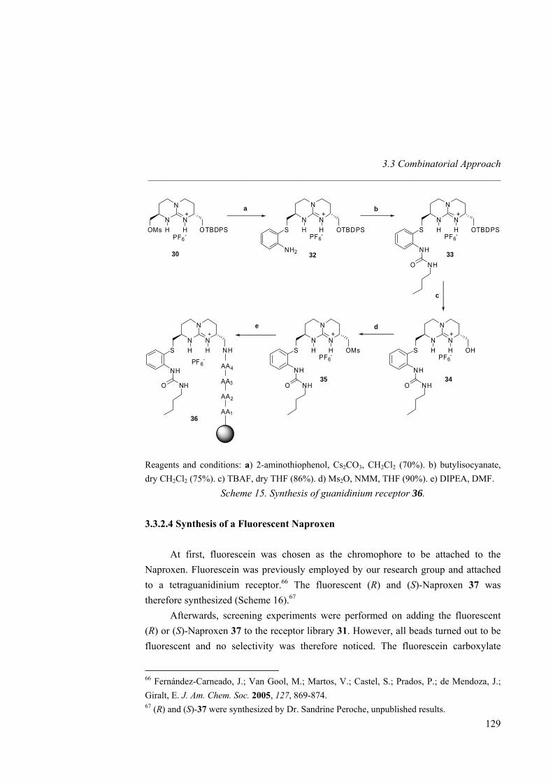
3.3 Combinatorial Approach _____________________________________________________________________
a b
30
c
N
N
NH H
+
OMs OTBDPSPF6
-
N
N
NH H
+
S OTBDPSPF6
-
NH2
N
N
NH H
+
S OTBDPSPF6
-
NH
NHO
N
N
NH H
+
S OHPF6
-
NH
NHO
N
N
NH H
+
S OMsPF6
-
NH
NHO
N
N
NS NHHH
NH
AA2
AA1
O NH
AA4
AA3
PF6-
de
32 33
3435
36
Reagents and conditions: a) 2-aminothiophenol, Cs2CO3, CH2Cl2 (70%). b) butylisocyanate, dry CH2Cl2 (75%). c) TBAF, dry THF (86%). d) Ms2O, NMM, THF (90%). e) DIPEA, DMF.
Scheme 15. Synthesis of guanidinium receptor 36.
3.3.2.4 Synthesis of a Fluorescent Naproxen At first, fluorescein was chosen as the chromophore to be attached to the
Naproxen. Fluorescein was previously employed by our research group and attached to a tetraguanidinium receptor.66 The fluorescent (R) and (S)-Naproxen 37 was therefore synthesized (Scheme 16).67
Afterwards, screening experiments were performed on adding the fluorescent (R) or (S)-Naproxen 37 to the receptor library 31. However, all beads turned out to be fluorescent and no selectivity was therefore noticed. The fluorescein carboxylate
66 Fernández-Carneado, J.; Van Gool, M.; Martos, V.; Castel, S.; Prados, P.; de Mendoza, J.; Giralt, E. J. Am. Chem. Soc. 2005, 127, 869-874. 67 (R) and (S)-37 were synthesized by Dr. Sandrine Peroche, unpublished results.
129
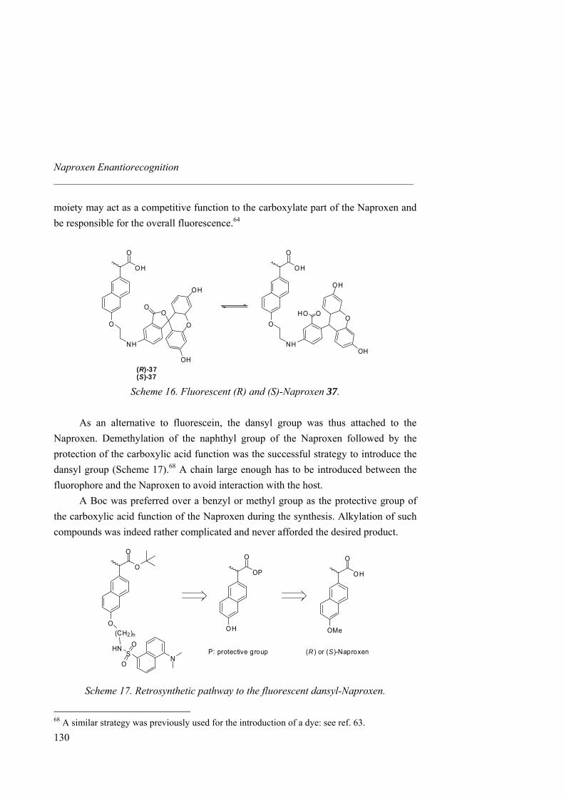
Naproxen Enantiorecognition _____________________________________________________________________
moiety may act as a competitive function to the carboxylate part of the Naproxen and be responsible for the overall fluorescence.64
OH
O
O
NH
O
OH
OH
OO
(R)-37(S)-37
OH
O
O
NH
O
OH
OH
OHO
Scheme 16. Fluorescent (R) and (S)-Naproxen 37.
As an alternative to fluorescein, the dansyl group was thus attached to the Naproxen. Demethylation of the naphthyl group of the Naproxen followed by the protection of the carboxylic acid function was the successful strategy to introduce the dansyl group (Scheme 17).68 A chain large enough has to be introduced between the fluorophore and the Naproxen to avoid interaction with the host. A Boc was preferred over a benzyl or methyl group as the protective group of the carboxylic acid function of the Naproxen during the synthesis. Alkylation of such compounds was indeed rather complicated and never afforded the desired product.
OH
O
OMe
OP
O
OH
O
O
O(CH2)n
HNS
O
O
NP: protective group (R ) or (S)-Naproxen
Scheme 17. Retrosynthetic pathway to the fluorescent dansyl-Naproxen.
68 A similar strategy was previously used for the introduction of a dye: see ref. 63.
130
3.3 Combinatorial Approach _____________________________________________________________________
131
Demethylation of the naphthyl group was first achieved with BBr3 in CH2Cl2 at 0ºC in quantitative yield. However, characterization of purified 38 by chiral HPLC (Whelk-01(S,S)) revealed a partial racemization of the compound. Almost 40% of (R)-38 was formed starting from (S)-Naproxen. The procedure using BBr3 was then replaced by HBr and eventually HCl at reflux giving the desired compound 38 without racemization (according to results reported by Dickinson et al.)69. The too harsh conditions of BBr3 procedure may induce a proton transfer in α position of the carboxylic acid moiety through formation of the corresponding, easily enolizable acyl bromide. Protection of the carboxylic acid function was then performed with trifluoroacetic anhydride and tert-butanol in dry CH3CN in 87% yield.70
Starting from dansyl chloride, the derivatives 41 and 42 with respectively two different leaving groups MsO and Br were obtained in good yields. However, coupling the chromophore derivatives 41 or 42 with the protected Naproxen 39 never produced the desired compound. It seems that 41 or 42 degrades during the reaction prior to reacting. 39 was indeed recovered unchanged (Scheme 18).
69 Lo, A.; Addison, R. S.; Hooper, W. D.; Dickinson, R. G. Xenobiotica, 2001, 31, 309-319. 70 Optimization synthesis of 39 was performed by Dr. Sandrine Peroche.

Naproxen Enantiorecognition _____________________________________________________________________
OH
O
OMe
OH
O
OH
O
O
OH(R ) (R )-38 (R )-39(S) (S)-38 (S)-39
N
S OOCl
N
S OOHN OH
N
S OOHN X
40 41 X=OMs42 X=Br
O
O
O
HNS
O
O
N
a b
c d
c
Reagents and conditions: a) HCl reflux (88%). b) (CF3CO)2O, tBuOH, dry THF (87%). c) Et3N, THF (99%). d) for X = OMs : Ms2O, NMM, THF (88%); for X = Br : CBr4, PH3P, CH2Cl2 (70%). e) K2CO3, THF.
Scheme 18. Synthesis attempt of fluorescent Dansyl-Naproxen. A reverse strategy was then employed: alkylation of the naphthyl group followed by introduction of the dansyl part (Scheme 19). Alkylation of 39 by 1,3-dibromopropane was achieved in THF with K2CO3. Yield increased from 50 up to 75% as the number of 1,3-dibromopropane equivalents is increased from one to five (to prevent disubstitution). Besides, dansyl amine 44 was obtained from dansyl chloride in 50 % yield.71 Reaction between protected Naproxen 43 and dansyl amine 44 gives the desired compound 45 in a 90% yield. Deprotection of the Boc group in the presence of 30% TFA in dichloromethane and a catalytic amount of triethylsilane gives the final compound 46 in 95% yield.
71 Jersey, J. A.; Choshen, E.; Jensen, J. N.; Johnson, J. D. Environ. Sci. Technol. 1990, 24, 1536-1541.
132
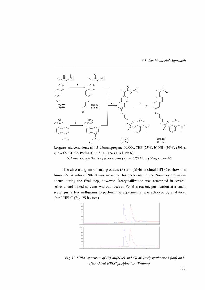
3.3 Combinatorial Approach _____________________________________________________________________
O
O
OH(R)-39(S)-39
N
S OOCl
N
S OONH2
44
a
c
b
O
O
O(R )-43(S)-43
O
O
O
HNS
O
O
N
OH
O
O
HNS
O
O
N
(R)-45 (R )-46(S)-45 (S)-46
Br
d
Reagents and conditions: a) 1,3-dibromopropane, K2CO3, THF (75%). b) NH3 (30%), (50%). c) K2CO3, CH3CN (90%). d) Et3SiH, TFA, CH2Cl2 (95%).
Scheme 19. Synthesis of fluorescent (R) and (S) Dansyl-Naproxen 46.
The chromatogram of final products (R) and (S)-46 in chiral HPLC is shown in figure 29. A ratio of 90/10 was measured for each enantiomer. Some racemization occurs during the final step, however. Recrystallization was attempted in several solvents and mixed solvents without success. For this reason, purification at a small scale (just a few milligrams to perform the experiments) was achieved by analytical chiral HPLC (Fig. 29 bottom).
m in0 5 1 0 1 5 20 2 5
N o 1 rm .
0
1 00
2 00
3 00
4 00
5 00
6 00
16.00
8
18.33
6
m in0 5 10 1 5 20 2 5
N o 1 rm .
0
1 00
2 00
3 00
4 00
5 00 15.89
3
18.57
3
m in0 5 1 0 1 5 20 2 5
N o 1 rm .
0
1 00
2 00
3 00
4 00
5 00
6 00
16.00
8
18.33
6
m in0 5 10 1 5 20 2 5
N o 1 rm .
0
1 00
2 00
3 00
4 00
5 00 15.89
3
18.57
3
133
Fig 31. HPLC spectrum of (R)-46(blue) and (S)-46 (red) synthesized (top) and after chiral HPLC purification (Bottom).
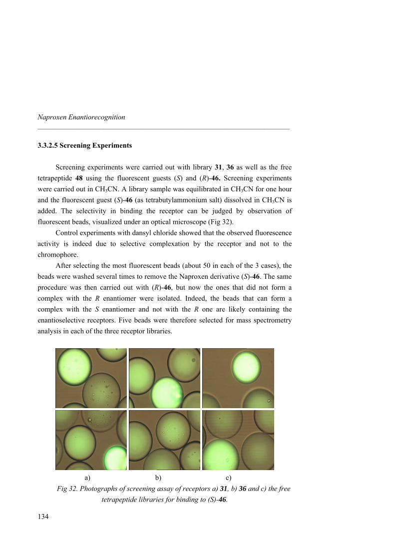
Naproxen Enantiorecognition _____________________________________________________________________
3.3.2.5 Screening Experiments
Screening experiments were carried out with library 31, 36 as well as the free tetrapeptide 48 using the fluorescent guests (S) and (R)-46. Screening experiments were carried out in CH3CN. A library sample was equilibrated in CH3CN for one hour and the fluorescent guest (S)-46 (as tetrabutylammonium salt) dissolved in CH3CN is added. The selectivity in binding the receptor can be judged by observation of fluorescent beads, visualized under an optical microscope (Fig 32). Control experiments with dansyl chloride showed that the observed fluorescence activity is indeed due to selective complexation by the receptor and not to the chromophore. After selecting the most fluorescent beads (about 50 in each of the 3 cases), the beads were washed several times to remove the Naproxen derivative (S)-46. The same procedure was then carried out with (R)-46, but now the ones that did not form a complex with the R enantiomer were isolated. Indeed, the beads that can form a complex with the S enantiomer and not with the R one are likely containing the enantioselective receptors. Five beads were therefore selected for mass spectrometry analysis in each of the three receptor libraries.
a) b) c)
Fig 32. Photographs of screening assay of receptors a) 31, b) 36 and c) the free tetrapeptide libraries for binding to (S)-46.
134
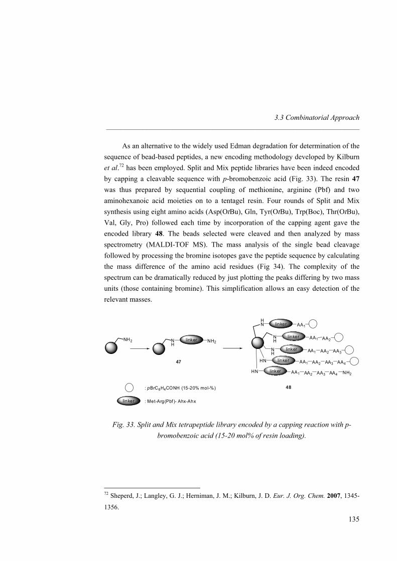
3.3 Combinatorial Approach _____________________________________________________________________
As an alternative to the widely used Edman degradation for determination of the sequence of bead-based peptides, a new encoding methodology developed by Kilburn et al.72 has been employed. Split and Mix peptide libraries have been indeed encoded by capping a cleavable sequence with p-bromobenzoic acid (Fig. 33). The resin 47 was thus prepared by sequential coupling of methionine, arginine (Pbf) and two aminohexanoic acid moieties on to a tentagel resin. Four rounds of Split and Mix synthesis using eight amino acids (Asp(OtBu), Gln, Tyr(OtBu), Trp(Boc), Thr(OtBu), Val, Gly, Pro) followed each time by incorporation of the capping agent gave the encoded library 48. The beads selected were cleaved and then analyzed by mass spectrometry (MALDI-TOF MS). The mass analysis of the single bead cleavage followed by processing the bromine isotopes gave the peptide sequence by calculating the mass difference of the amino acid residues (Fig 34). The complexity of the spectrum can be dramatically reduced by just plotting the peaks differing by two mass units (those containing bromine). This simplification allows an easy detection of the relevant masses.
NH2 NH
linker NH2
HN linker AA1
NH
linker AA1 AA2
NH
linker AA1 AA2 AA3
HN linker AA1 AA2 AA3 AA4
HN linker AA1 AA2 AA3 AA4 NH2
: pBrC6H4CONH (15-20% mol-%)
linker : Met-Arg(Pbf )- Ahx-Ahx
47
48
Fig. 33. Split and Mix tetrapeptide library encoded by a capping reaction with p-bromobenzoic acid (15-20 mol% of resin loading).
72 Sheperd, J.; Langley, G. J.; Herniman, J. M.; Kilburn, J. D. Eur. J. Org. Chem. 2007, 1345-
1356.
135
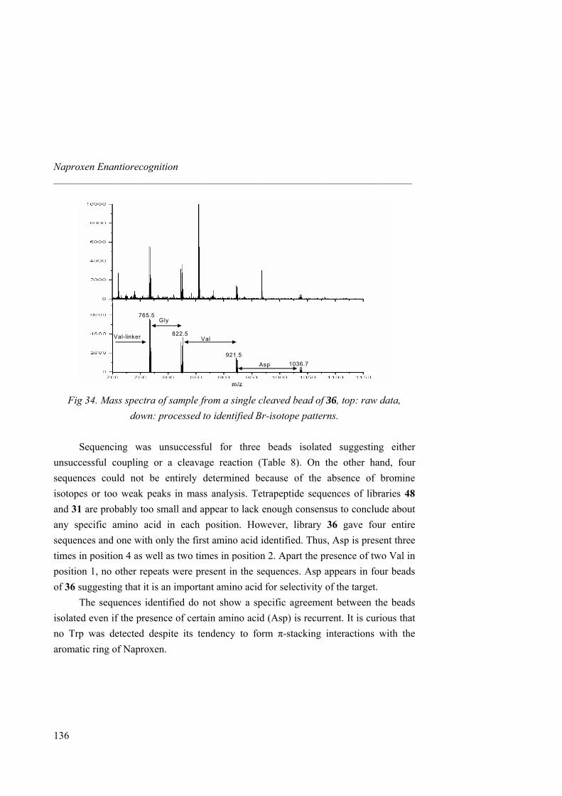
Naproxen Enantiorecognition _____________________________________________________________________
765.5
822.5
921.51036.7
Val-linker
Gly
Val
Asp
m/z
765.5
822.5
921.51036.7
Val-linker
Gly
Val
Asp
m/z Fig 34. Mass spectra of sample from a single cleaved bead of 36, top: raw data,
down: processed to identified Br-isotope patterns. Sequencing was unsuccessful for three beads isolated suggesting either unsuccessful coupling or a cleavage reaction (Table 8). On the other hand, four sequences could not be entirely determined because of the absence of bromine isotopes or too weak peaks in mass analysis. Tetrapeptide sequences of libraries 48 and 31 are probably too small and appear to lack enough consensus to conclude about any specific amino acid in each position. However, library 36 gave four entire sequences and one with only the first amino acid identified. Thus, Asp is present three times in position 4 as well as two times in position 2. Apart the presence of two Val in position 1, no other repeats were present in the sequences. Asp appears in four beads of 36 suggesting that it is an important amino acid for selectivity of the target. The sequences identified do not show a specific agreement between the beads isolated even if the presence of certain amino acid (Asp) is recurrent. It is curious that no Trp was detected despite its tendency to form π-stacking interactions with the aromatic ring of Naproxen.
136
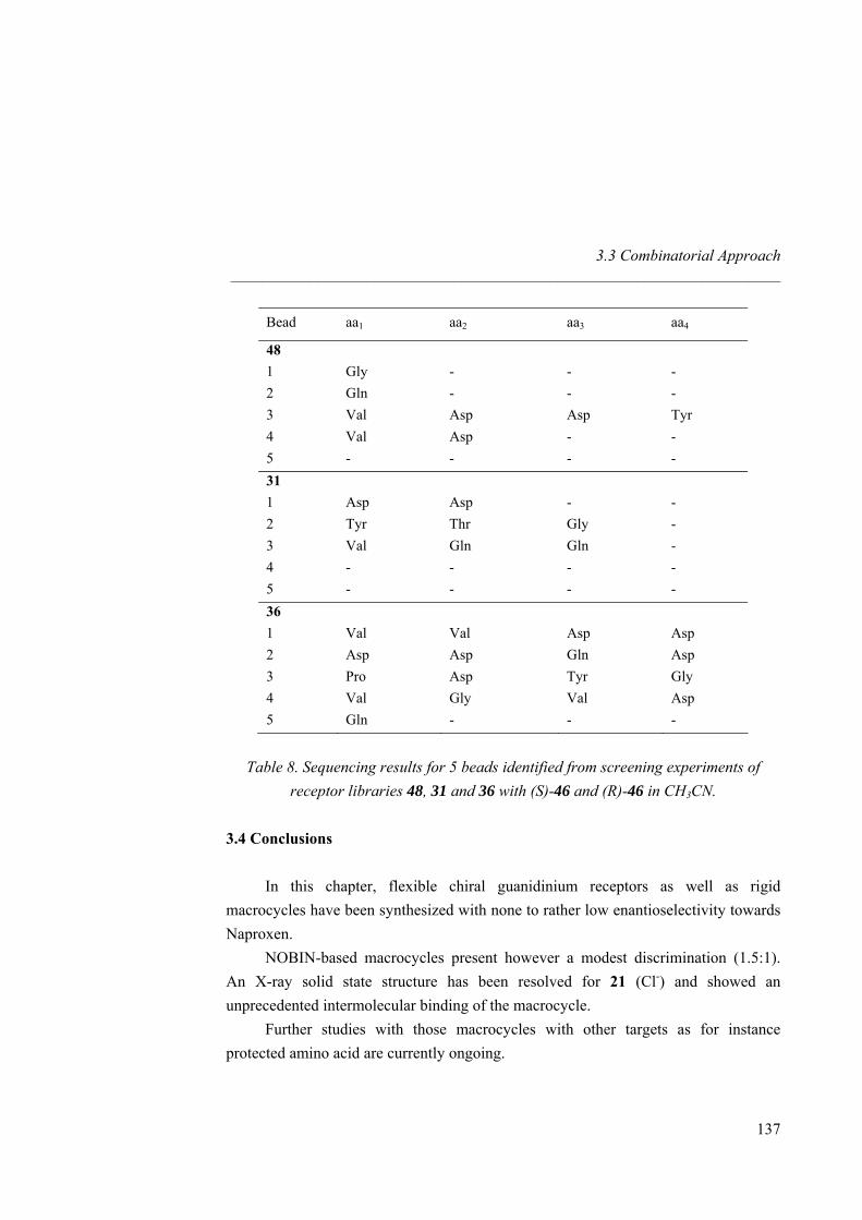
3.3 Combinatorial Approach _____________________________________________________________________
137
Bead aa1 aa2 aa3 aa4
48 1 Gly - - - 2 Gln - - - 3 Val Asp Asp Tyr 4 Val Asp - - 5 - - - - 31 1 Asp Asp - - 2 Tyr Thr Gly - 3 Val Gln Gln - 4 - - - - 5 - - - - 36 1 Val Val Asp Asp 2 Asp Asp Gln Asp 3 Pro Asp Tyr Gly 4 Val Gly Val Asp 5 Gln - - -
Table 8. Sequencing results for 5 beads identified from screening experiments of
receptor libraries 48, 31 and 36 with (S)-46 and (R)-46 in CH3CN. 3.4 Conclusions
In this chapter, flexible chiral guanidinium receptors as well as rigid macrocycles have been synthesized with none to rather low enantioselectivity towards Naproxen.
NOBIN-based macrocycles present however a modest discrimination (1.5:1). An X-ray solid state structure has been resolved for 21 (Cl-) and showed an unprecedented intermolecular binding of the macrocycle.
Further studies with those macrocycles with other targets as for instance protected amino acid are currently ongoing.
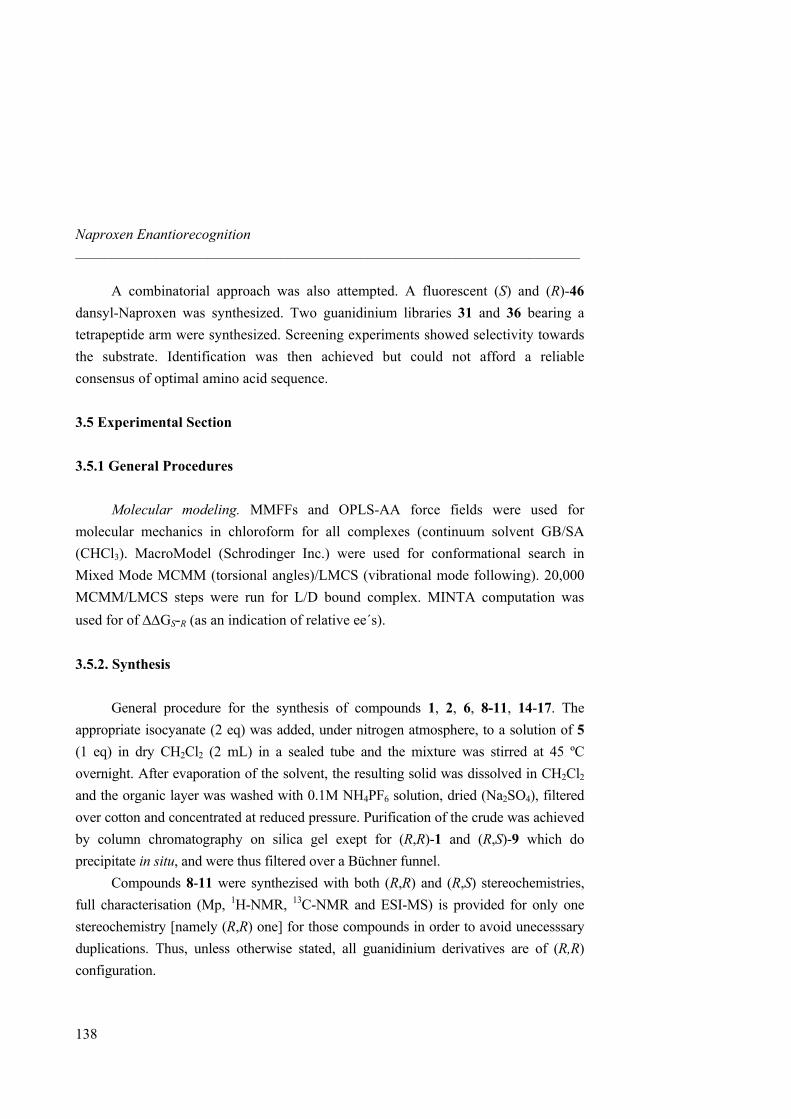
Naproxen Enantiorecognition _____________________________________________________________________
138
A combinatorial approach was also attempted. A fluorescent (S) and (R)-46 dansyl-Naproxen was synthesized. Two guanidinium libraries 31 and 36 bearing a tetrapeptide arm were synthesized. Screening experiments showed selectivity towards the substrate. Identification was then achieved but could not afford a reliable consensus of optimal amino acid sequence.
3.5 Experimental Section
3.5.1 General Procedures
Molecular modeling. MMFFs and OPLS-AA force fields were used for molecular mechanics in chloroform for all complexes (continuum solvent GB/SA (CHCl3). MacroModel (Schrodinger Inc.) were used for conformational search in Mixed Mode MCMM (torsional angles)/LMCS (vibrational mode following). 20,000 MCMM/LMCS steps were run for L/D bound complex. MINTA computation was used for of ∆∆GS-R (as an indication of relative ee´s). 3.5.2. Synthesis
General procedure for the synthesis of compounds 1, 2, 6, 8-11, 14-17. The appropriate isocyanate (2 eq) was added, under nitrogen atmosphere, to a solution of 5 (1 eq) in dry CH2Cl2 (2 mL) in a sealed tube and the mixture was stirred at 45 ºC overnight. After evaporation of the solvent, the resulting solid was dissolved in CH2Cl2 and the organic layer was washed with 0.1M NH4PF6 solution, dried (Na2SO4), filtered over cotton and concentrated at reduced pressure. Purification of the crude was achieved by column chromatography on silica gel exept for (R,R)-1 and (R,S)-9 which do precipitate in situ, and were thus filtered over a Büchner funnel. Compounds 8-11 were synthezised with both (R,R) and (R,S) stereochemistries, full characterisation (Mp, 1H-NMR, 13C-NMR and ESI-MS) is provided for only one stereochemistry [namely (R,R) one] for those compounds in order to avoid unecesssary duplications. Thus, unless otherwise stated, all guanidinium derivatives are of (R,R) configuration.
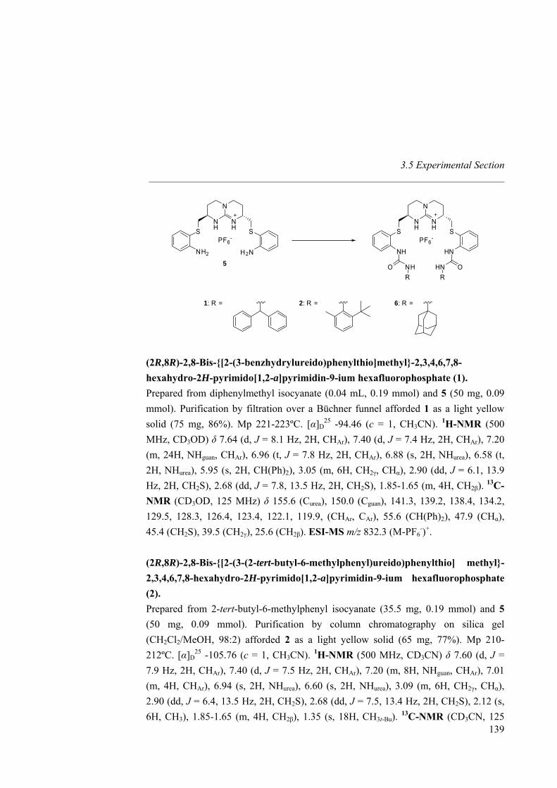
3.5 Experimental Section _____________________________________________________________________
N
NH
NH SS
PF6-
HNNH
N
NH
NH SS
PF6-
H2NNH2
OHNO NHRR
1: R = 2: R = 6: R =
5
(2R,8R)-2,8-Bis-{[2-(3-benzhydrylureido)phenylthio]methyl}-2,3,4,6,7,8-hexahydro-2H-pyrimido[1,2-a]pyrimidin-9-ium hexafluorophosphate (1). Prepared from diphenylmethyl isocyanate (0.04 mL, 0.19 mmol) and 5 (50 mg, 0.09 mmol). Purification by filtration over a Büchner funnel afforded 1 as a light yellow solid (75 mg, 86%). Mp 221-223ºC. [α]D
25 -94.46 (c = 1, CH3CN). 1H-NMR (500 MHz, CD3OD) δ 7.64 (d, J = 8.1 Hz, 2H, CHAr), 7.40 (d, J = 7.4 Hz, 2H, CHAr), 7.20 (m, 24H, NHguan, CHAr), 6.96 (t, J = 7.8 Hz, 2H, CHAr), 6.88 (s, 2H, NHurea), 6.58 (t, 2H, NHurea), 5.95 (s, 2H, CH(Ph)2), 3.05 (m, 6H, CH2γ, CHα), 2.90 (dd, J = 6.1, 13.9 Hz, 2H, CH2S), 2.68 (dd, J = 7.8, 13.5 Hz, 2H, CH2S), 1.85-1.65 (m, 4H, CH2β). 13C-NMR (CD3OD, 125 MHz) δ 155.6 (Curea), 150.0 (Cguan), 141.3, 139.2, 138.4, 134.2, 129.5, 128.3, 126.4, 123.4, 122.1, 119.9, (CHAr, CAr), 55.6 (CH(Ph)2), 47.9 (CHα), 45.4 (CH2S), 39.5 (CH2γ), 25.6 (CH2β). ESI-MS m/z 832.3 (M-PF6
-)+. (2R,8R)-2,8-Bis-{[2-(3-(2-tert-butyl-6-methylphenyl)ureido)phenylthio] methyl}-2,3,4,6,7,8-hexahydro-2H-pyrimido[1,2-a]pyrimidin-9-ium hexafluorophosphate (2).
139
Prepared from 2-tert-butyl-6-methylphenyl isocyanate (35.5 mg, 0.19 mmol) and 5 (50 mg, 0.09 mmol). Purification by column chromatography on silica gel (CH2Cl2/MeOH, 98:2) afforded 2 as a light yellow solid (65 mg, 77%). Mp 210-212ºC. [α]D
25 -105.76 (c = 1, CH3CN). 1H-NMR (500 MHz, CD3CN) δ 7.60 (d, J = 7.9 Hz, 2H, CHAr), 7.40 (d, J = 7.5 Hz, 2H, CHAr), 7.20 (m, 8H, NHguan, CHAr), 7.01 (m, 4H, CHAr), 6.94 (s, 2H, NHurea), 6.60 (s, 2H, NHurea), 3.09 (m, 6H, CH2γ, CHα), 2.90 (dd, J = 6.4, 13.5 Hz, 2H, CH2S), 2.68 (dd, J = 7.5, 13.4 Hz, 2H, CH2S), 2.12 (s, 6H, CH3), 1.85-1.65 (m, 4H, CH2β), 1.35 (s, 18H, CH3t-Bu). 13C-NMR (CD3CN, 125
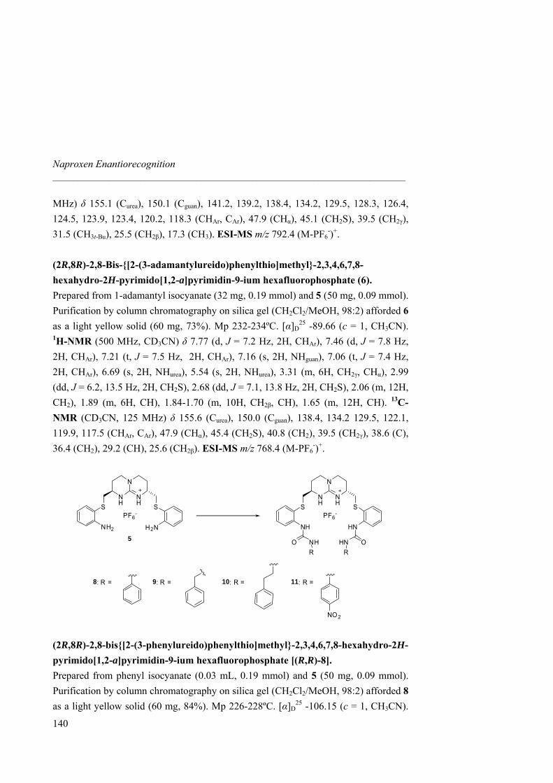
Naproxen Enantiorecognition _____________________________________________________________________
MHz) δ 155.1 (Curea), 150.1 (Cguan), 141.2, 139.2, 138.4, 134.2, 129.5, 128.3, 126.4, 124.5, 123.9, 123.4, 120.2, 118.3 (CHAr, CAr), 47.9 (CHα), 45.1 (CH2S), 39.5 (CH2γ), 31.5 (CH3t-Bu), 25.5 (CH2β), 17.3 (CH3). ESI-MS m/z 792.4 (M-PF6
-)+. (2R,8R)-2,8-Bis-{[2-(3-adamantylureido)phenylthio]methyl}-2,3,4,6,7,8-hexahydro-2H-pyrimido[1,2-a]pyrimidin-9-ium hexafluorophosphate (6). Prepared from 1-adamantyl isocyanate (32 mg, 0.19 mmol) and 5 (50 mg, 0.09 mmol). Purification by column chromatography on silica gel (CH2Cl2/MeOH, 98:2) afforded 6 as a light yellow solid (60 mg, 73%). Mp 232-234ºC. [α]D
25 -89.66 (c = 1, CH3CN). 1H-NMR (500 MHz, CD3CN) δ 7.77 (d, J = 7.2 Hz, 2H, CHAr), 7.46 (d, J = 7.8 Hz, 2H, CHAr), 7.21 (t, J = 7.5 Hz, 2H, CHAr), 7.16 (s, 2H, NHguan), 7.06 (t, J = 7.4 Hz, 2H, CHAr), 6.69 (s, 2H, NHurea), 5.54 (s, 2H, NHurea), 3.31 (m, 6H, CH2γ, CHα), 2.99 (dd, J = 6.2, 13.5 Hz, 2H, CH2S), 2.68 (dd, J = 7.1, 13.8 Hz, 2H, CH2S), 2.06 (m, 12H, CH2), 1.89 (m, 6H, CH), 1.84-1.70 (m, 10H, CH2β, CH), 1.65 (m, 12H, CH). 13C-NMR (CD3CN, 125 MHz) δ 155.6 (Curea), 150.0 (Cguan), 138.4, 134.2 129.5, 122.1, 119.9, 117.5 (CHAr, CAr), 47.9 (CHα), 45.4 (CH2S), 40.8 (CH2), 39.5 (CH2γ), 38.6 (C), 36.4 (CH2), 29.2 (CH), 25.6 (CH2β). ESI-MS m/z 768.4 (M-PF6
-)+.
N
NH
NH SS
PF6-
HNNH
N
NH
NH SS
PF6-
H2NNH2
OHNO NHRR
8: R = 9: R = 10: R =
5
11: R =
NO 2 (2R,8R)-2,8-bis{[2-(3-phenylureido)phenylthio]methyl}-2,3,4,6,7,8-hexahydro-2H-pyrimido[1,2-a]pyrimidin-9-ium hexafluorophosphate [(R,R)-8]. Prepared from phenyl isocyanate (0.03 mL, 0.19 mmol) and 5 (50 mg, 0.09 mmol). Purification by column chromatography on silica gel (CH2Cl2/MeOH, 98:2) afforded 8 as a light yellow solid (60 mg, 84%). Mp 226-228ºC. [α]D
25 -106.15 (c = 1, CH3CN).
140
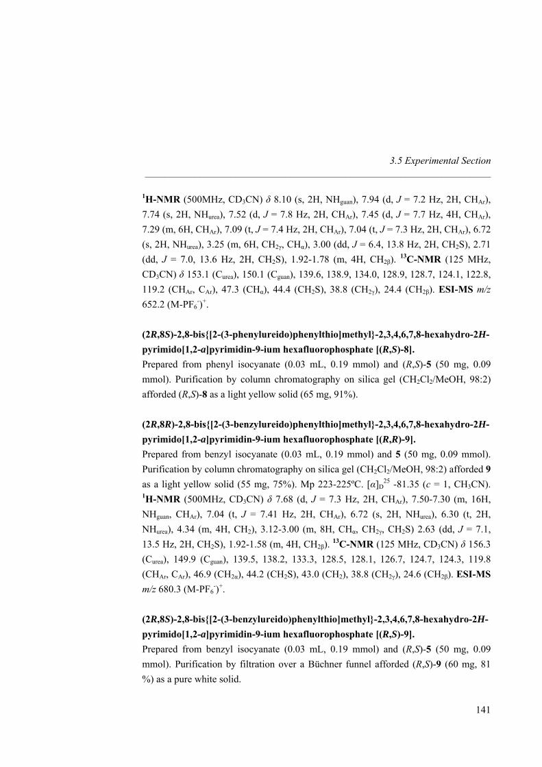
3.5 Experimental Section _____________________________________________________________________
141
1H-NMR (500MHz, CD3CN) δ 8.10 (s, 2H, NHguan), 7.94 (d, J = 7.2 Hz, 2H, CHAr), 7.74 (s, 2H, NHurea), 7.52 (d, J = 7.8 Hz, 2H, CHAr), 7.45 (d, J = 7.7 Hz, 4H, CHAr), 7.29 (m, 6H, CHAr), 7.09 (t, J = 7.4 Hz, 2H, CHAr), 7.04 (t, J = 7.3 Hz, 2H, CHAr), 6.72 (s, 2H, NHurea), 3.25 (m, 6H, CH2γ, CHα), 3.00 (dd, J = 6.4, 13.8 Hz, 2H, CH2S), 2.71 (dd, J = 7.0, 13.6 Hz, 2H, CH2S), 1.92-1.78 (m, 4H, CH2β). 13C-NMR (125 MHz, CD3CN) δ 153.1 (Curea), 150.1 (Cguan), 139.6, 138.9, 134.0, 128.9, 128.7, 124.1, 122.8, 119.2 (CHAr, CAr), 47.3 (CHα), 44.4 (CH2S), 38.8 (CH2γ), 24.4 (CH2β). ESI-MS m/z 652.2 (M-PF6
-)+. (2R,8S)-2,8-bis{[2-(3-phenylureido)phenylthio]methyl}-2,3,4,6,7,8-hexahydro-2H-pyrimido[1,2-a]pyrimidin-9-ium hexafluorophosphate [(R,S)-8]. Prepared from phenyl isocyanate (0.03 mL, 0.19 mmol) and (R,S)-5 (50 mg, 0.09 mmol). Purification by column chromatography on silica gel (CH2Cl2/MeOH, 98:2) afforded (R,S)-8 as a light yellow solid (65 mg, 91%). (2R,8R)-2,8-bis{[2-(3-benzylureido)phenylthio]methyl}-2,3,4,6,7,8-hexahydro-2H-pyrimido[1,2-a]pyrimidin-9-ium hexafluorophosphate [(R,R)-9]. Prepared from benzyl isocyanate (0.03 mL, 0.19 mmol) and 5 (50 mg, 0.09 mmol). Purification by column chromatography on silica gel (CH2Cl2/MeOH, 98:2) afforded 9 as a light yellow solid (55 mg, 75%). Mp 223-225ºC. [α]D
25 -81.35 (c = 1, CH3CN). 1H-NMR (500MHz, CD3CN) δ 7.68 (d, J = 7.3 Hz, 2H, CHAr), 7.50-7.30 (m, 16H, NHguan, CHAr), 7.04 (t, J = 7.41 Hz, 2H, CHAr), 6.72 (s, 2H, NHurea), 6.30 (t, 2H, NHurea), 4.34 (m, 4H, CH2), 3.12-3.00 (m, 8H, CHα, CH2γ, CH2S) 2.63 (dd, J = 7.1, 13.5 Hz, 2H, CH2S), 1.92-1.58 (m, 4H, CH2β). 13C-NMR (125 MHz, CD3CN) δ 156.3 (Curea), 149.9 (Cguan), 139.5, 138.2, 133.3, 128.5, 128.1, 126.7, 124.7, 124.3, 119.8 (CHAr, CAr), 46.9 (CH2α), 44.2 (CH2S), 43.0 (CH2), 38.8 (CH2γ), 24.6 (CH2β). ESI-MS m/z 680.3 (M-PF6
-)+. (2R,8S)-2,8-bis{[2-(3-benzylureido)phenylthio]methyl}-2,3,4,6,7,8-hexahydro-2H-pyrimido[1,2-a]pyrimidin-9-ium hexafluorophosphate [(R,S)-9]. Prepared from benzyl isocyanate (0.03 mL, 0.19 mmol) and (R,S)-5 (50 mg, 0.09 mmol). Purification by filtration over a Büchner funnel afforded (R,S)-9 (60 mg, 81 %) as a pure white solid.
Naproxen Enantiorecognition _____________________________________________________________________
142
(2R,8R)-2,8-bis{[2-(3-phenethylureido)phenylthio]methyl}-2,3,4,6,7,8-hexahydro-2H-pyrimido[1,2-a]pyrimidin-9-ium hexafluorophosphate [(R,R)-10]. Prepared from phenethyl isocyanate (0.03 mL, 0.19 mmol) and 5 (50 mg, 0.09 mmol). Purification by column chromatography on silica gel (CH2Cl2/MeOH, 98:2) afforded 10 as a light yellow solid (58 mg, 76%). Mp 218-220ºC. [α]D
25 -85.54 (c = 1, CH3CN). 1H-NMR (500MHz, CD3CN) δ 7.70 (d, J = 7.3 Hz, 2H, CHAr), 7.47 (d, J = 7.4 Hz, 2H, CHAr), 7.50-7.30 (m, 12H, NHguan, CHAr), 7.09 (t, J = 7.2 Hz, 2H, CHAr), 6.90 (s, 2H, NHurea), 5.84 (t, 2H, NHurea), 3.43 (q, J = 7.6 Hz, 4H, CH2), 3.28 (m, 6H, CHα, CH2γ), 3.13 (dd, J = 4.4, 14.5 Hz, 2H, CH2S), (t, J = 6.8 Hz, 4H, CH2), 2.71 (dd, J = 10.1, 12.4 Hz, 2H, CH2S), 2.04-1.72 (m, 4H, CH2β). 13C-NMR (125 MHz, CD3CN) δ 157.6 (Curea), 150.1 (Cguan), 139.5, 138.2, 134.4, 130.0, 129.6, 128.1, 126.6, 125.8, 120.5 (CHAr, CAr), 48.4 (CH2α), 45.8 (CH2S), 44.6 (CH2), 40.4 (CH2γ), 26.7 (CH2β). ESI-MS m/z 708.3 (M-PF6
-)+. (2R,8S)-2,8-bis{[2-(3-phenethylureido)phenylthio]methyl}-2,3,4,6,7,8-hexahydro-2H-pyrimido[1,2-a]pyrimidin-9-ium hexafluorophosphate [(R,S)-10]. Prepared from phenethyl isocyanate (0.03 mL, 0.19 mmol) and (R,S)-5 (50 mg, 0.09 mmol). Purification by column chromatography on silica gel (CH2Cl2/MeOH, 98:2) afforded (R, S)-10 as a light yellow solid (65 mg, 85%).
(2R,8R)-2,8-Bis{[2-(3-(4-nitrophenyl)ureido)phenylthio]methyl}-2,3,4,6,7,8-hexahydro-2H-pyrimido[1,2-a]pyrimidin-9-ium hexafluorophosphate [(R,R)-11]. Prepared from p-nitrophenyl isocyanate (30 mg, 0.19 mmol) and 5 (50 mg, 0.09 mmol). Purification by column chromatography on silica gel (CH2Cl2/MeOH, 98:2) afforded 6 as a light yellow solid (57 mg, 72%). Mp 215-217ºC. [α]D
25 -109.58 (c = 1, CH3CN).1H-NMR (500MHz, CD3CN) δ 8.62 (s, 2H, NHguan), 8.12 (d, J = 7.1 Hz, 4H, CHAr), 8.00 (d, J = 7.2 Hz, 2H, CHAr), 7.87 (s, 2H, NHurea),7.69 (d, 4H, J = 7.7 Hz, CHAr), 7.57 (d, J = 7.3 Hz, 2H, CHAr), 7.31 (t, J = 7.4 Hz, 2H, CHAr), 7.14 (t, J = 7.3 Hz, 2H, CHAr), 6.25 (s, 2H, NHurea), 3.24-3.18 (m, 6H, CH2γ, CHα), 3.03 (dd, J = 5.7, 13.8 Hz, 2H, CH2S), 2.72 (dd, J = 9, 14.9 Hz, 2H, CH2S), 1.90-1.70 (m, 4H, CH2β). 13C-NMR (125 MHz, CD3CN) δ 153.1 (Curea), 150.1 (Cguan), 139.6, 138.9 134.0, 128.9, 128.7, 124.1, 122.8, 119.2 (CHAr, CAr), 47.3 (CH2α), 44.4 (CH2S), 38.8 (CH2γ), 24.4 (CH2β). ESI-MS m/z 742.2 (M-PF6
-)+.
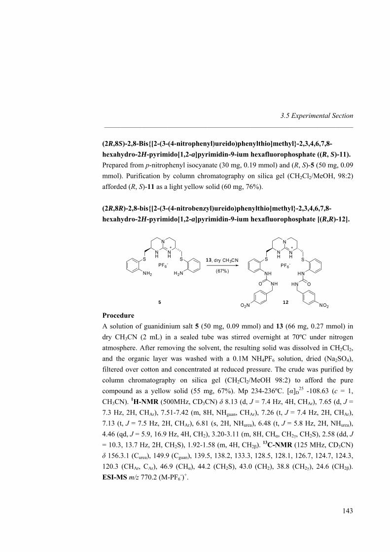
3.5 Experimental Section _____________________________________________________________________
(2R,8S)-2,8-Bis{[2-(3-(4-nitrophenyl)ureido)phenylthio]methyl}-2,3,4,6,7,8-hexahydro-2H-pyrimido[1,2-a]pyrimidin-9-ium hexafluorophosphate ((R, S)-11). Prepared from p-nitrophenyl isocyanate (30 mg, 0.19 mmol) and (R, S)-5 (50 mg, 0.09 mmol). Purification by column chromatography on silica gel (CH2Cl2/MeOH, 98:2) afforded (R, S)-11 as a light yellow solid (60 mg, 76%). (2R,8R)-2,8-bis{[2-(3-(4-nitrobenzyl)ureido)phenylthio]methyl}-2,3,4,6,7,8-hexahydro-2H-pyrimido[1,2-a]pyrimidin-9-ium hexafluorophosphate [(R,R)-12].
N
NH
NH SS
PF6-
H2NNH2
N
NH
NH SS
PF6-
HNNH
OHNO NH
O2N NO2
13, dry CH3CN
(67%)
5 12 Procedure A solution of guanidinium salt 5 (50 mg, 0.09 mmol) and 13 (66 mg, 0.27 mmol) in dry CH3CN (2 mL) in a sealed tube was stirred overnight at 70ºC under nitrogen atmosphere. After removing the solvent, the resulting solid was dissolved in CH2Cl2, and the organic layer was washed with a 0.1M NH4PF6 solution, dried (Na2SO4), filtered over cotton and concentrated at reduced pressure. The crude was purified by column chromatography on silica gel (CH2Cl2/MeOH 98:2) to afford the pure compound as a yellow solid (55 mg, 67%). Mp 234-236ºC. [α]D
25 -108.63 (c = 1, CH3CN). 1H-NMR (500MHz, CD3CN) δ 8.13 (d, J = 7.4 Hz, 4H, CHAr), 7.65 (d, J = 7.3 Hz, 2H, CHAr), 7.51-7.42 (m, 8H, NHguan, CHAr), 7.26 (t, J = 7.4 Hz, 2H, CHAr), 7.13 (t, J = 7.5 Hz, 2H, CHAr), 6.81 (s, 2H, NHurea), 6.48 (t, J = 5.8 Hz, 2H, NHurea), 4.46 (qd, J = 5.9, 16.9 Hz, 4H, CH2), 3.20-3.11 (m, 8H, CHα, CH2γ, CH2S), 2.58 (dd, J = 10.3, 13.7 Hz, 2H, CH2S), 1.92-1.58 (m, 4H, CH2β). 13C-NMR (125 MHz, CD3CN) δ 156.3.1 (Curea), 149.9 (Cguan), 139.5, 138.2, 133.3, 128.5, 128.1, 126.7, 124.7, 124.3, 120.3 (CHAr, CAr), 46.9 (CHα), 44.2 (CH2S), 43.0 (CH2), 38.8 (CH2γ), 24.6 (CH2β). ESI-MS m/z 770.2 (M-PF6
-)+.
143
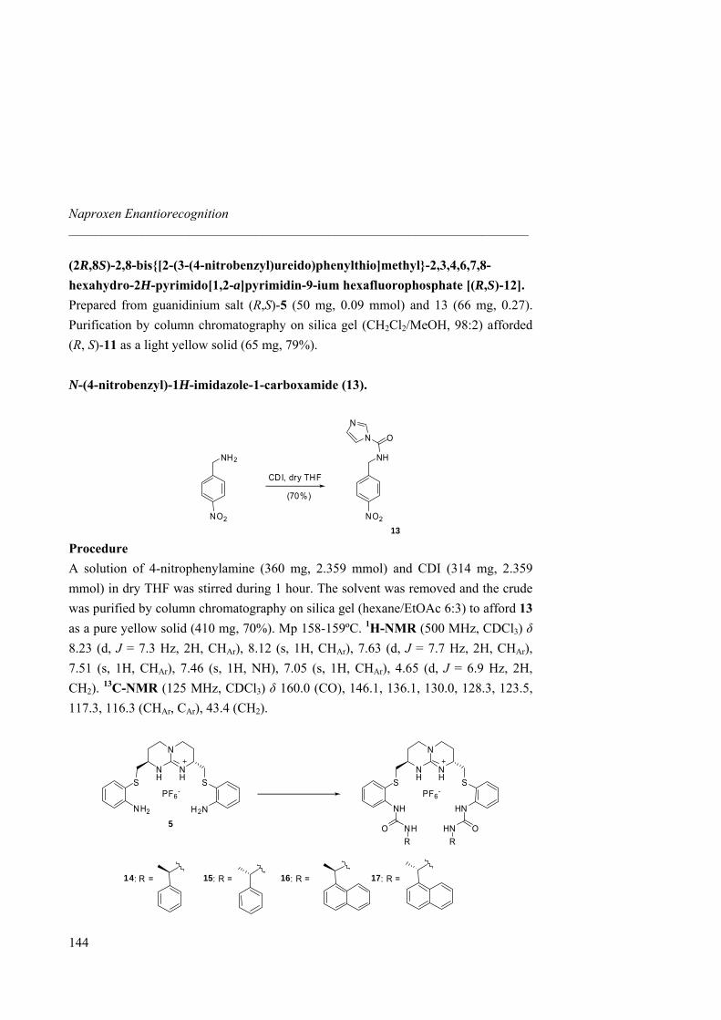
Naproxen Enantiorecognition _____________________________________________________________________
(2R,8S)-2,8-bis{[2-(3-(4-nitrobenzyl)ureido)phenylthio]methyl}-2,3,4,6,7,8-hexahydro-2H-pyrimido[1,2-a]pyrimidin-9-ium hexafluorophosphate [(R,S)-12]. Prepared from guanidinium salt (R,S)-5 (50 mg, 0.09 mmol) and 13 (66 mg, 0.27). Purification by column chromatography on silica gel (CH2Cl2/MeOH, 98:2) afforded (R, S)-11 as a light yellow solid (65 mg, 79%). N-(4-nitrobenzyl)-1H-imidazole-1-carboxamide (13).
NO2NO2
NH2 NH
ON
N
CDI, dry THF
(70%)
13 Procedure A solution of 4-nitrophenylamine (360 mg, 2.359 mmol) and CDI (314 mg, 2.359 mmol) in dry THF was stirred during 1 hour. The solvent was removed and the crude was purified by column chromatography on silica gel (hexane/EtOAc 6:3) to afford 13 as a pure yellow solid (410 mg, 70%). Mp 158-159ºC. 1H-NMR (500 MHz, CDCl3) δ 8.23 (d, J = 7.3 Hz, 2H, CHAr), 8.12 (s, 1H, CHAr), 7.63 (d, J = 7.7 Hz, 2H, CHAr), 7.51 (s, 1H, CHAr), 7.46 (s, 1H, NH), 7.05 (s, 1H, CHAr), 4.65 (d, J = 6.9 Hz, 2H, CH2). 13C-NMR (125 MHz, CDCl3) δ 160.0 (CO), 146.1, 136.1, 130.0, 128.3, 123.5, 117.3, 116.3 (CHAr, CAr), 43.4 (CH2).
N
NH
NH SS
PF6-
HNNH
N
NH
NH SS
PF6-
H2NNH2
OHNO NHRR
15: R = 16: R = 17: R =
5
14: R =
144
3.5 Experimental Section _____________________________________________________________________
145
(2R,8R)-2,8-bis{[2-(3-((S)-1-phenylethyl)ureido)phenylthio]methyl}-2,3,4,6,7,8-hexahydro-2H-pyrimido[1,2-a]pyrimidin-9-ium hexafluorophosphate [(R,R)-14]. Prepared from (R)-(−)-α-methylbenzyl isocyanate (0.03 mL, 0.19 mmol) and 5 (50 mg, 0.09 mmol). Purification by column chromatography on silica gel (CH2Cl2/MeOH, 98:2) afforded 14 as a light yellow solid (65 mg, 85%). Mp 195-197ºC. [α]D
25 -70.84 (c = 1, CH3CN). 1H-NMR (500 MHz, CD3CN) δ 7.79 (d, J = 6.8 Hz, 2H, CHAr), 7.68 (d, J = 7.6 Hz, 2H, CHAr), 7.40 (s, 2H, NHguan),7.35-7.20 (m, 12H, CHAr), 7.10 (t, J = 7.4 Hz, 2H, CHAr), 6.55 (s, 2H, NHurea), 6.26 (s, 2H, NHurea), 4.84 (m, 2H, CH), 3.18 (m, 6H, CHα, CH2γ), 3.05 (dd, J = 5.1, 13.2 Hz, 2H, CH2S), 2.67 (dd, J = 9.3, 13.0 Hz, 2H, CH2S), 1.92-1.58 (m, 4H, CH2β), 1.44 (d, J = 7.0 Hz, 6H, CH3). 13C-NMR (125 MHz, CD3CN) δ 156.3.1 (Curea), 149.9 (Cguan), 139.5, 138.2, 133.3, 128.5, 128.1, 126.7, 124.7, 124.3, 119.8 (CHAr, CAr), 50.8 (CH), 46.9 (CHα), 44.2 (CH2S), 38.8 (CH2γ), 24.6 (CH2β), 21.2 (CH3). ESI-MS m/z 708.3 (M-PF6
-)+. (2R,8S)-2,8-bis{[2-(3-((S)-1-phenylethyl)ureido)phenylthio]methyl}-2,3,4,6,7,8-hexahydro-2H-pyrimido[1,2-a]pyrimidin-9-ium hexafluorophosphate [(R,S)-14]. Prepared from (R)-(−)-α-methylbenzyl isocyanate (0.03 mL, 0.19 mmol) and (R, S)-5 (50 mg, 0.09 mmol). Purification by column chromatography on silica gel (CH2Cl2/MeOH, 98:2) afforded (R, S)-14 as a light yellow solid (68 mg, 89%). Mp 195-197ºC. [α]D
25 -18.31 (c = 1, CH3CN). 1H-NMR (500 MHz, CD3CN) δ 7.89 (d, J = 9.1 Hz, 1H, CHAr), 7.63 (d, J = 8.1 Hz, 1H, CHAr), 7.52 (d, J = 7.6 Hz, 1H, CHAr), 7.48 (d, J = 7.6 Hz, 1H, CHAr), 7.32-7.19 (m, 14H, CHAr, NHguan), 7.16 (t, J = 7.3 Hz, 1H, CHAr), 7.10 (t, J = 7.3 Hz, 1H, CHAr), 6.73 (s, 1H, NHurea), 6.64 (s, 1H, NHurea), 6.28 (m, 2H, NHurea), 4.89 (m, 1H, CH), 4.81 (m, 1H, CH), 3.16 (m, 6H, CHα, CH2γ), 3.01-2.85 (m, 4H, CH2S), 2.01-1.57 (m, 4H, CH2β), 1.46 (d, J = 7.0 Hz, 3H, CH3), 1.41 (d, J = 7.0 Hz, 3H, CH3). 13C-NMR (125 MHz, CD3CN) δ 155.2, 154.9 (Curea), 150.3 (Cguan), 139.3, 138.2, 133.9, 133.6, 129.1, 128.7, 128.5, 128.3, 126.9, 126.8, 125.8, 125.6, 125.5, 125.0. 124.0, 122.9 (CHAr, CAr), 50.1, 49.8 (CH), 47.9, 47.4 (CHα), 45.2, 44.9 (CH2S), 39.7, 38.6 (CH2γ), 25.1, 24.8 (CH2β), 22.4, 22.3 (CH3). ESI-MS m/z 708.3 (M-PF6
-)+. (2R,8R)-2,8-bis{[2-(3-((R)-1-phenylethyl)ureido)phenylthio]methyl}-2,3,4,6,7,8-hexahydro-2H-pyrimido[1,2-a]pyrimidin-9-ium hexafluorophosphate [(R,R)-15].
Naproxen Enantiorecognition _____________________________________________________________________
146
Prepared from (S)-(+)-α-methylbenzyl isocyanate (0.03 mL, 0.19 mmol) and 5 (50 mg, 0.09 mmol). Purification by column chromatography on silica gel (CH2Cl2/MeOH, 98:2) afforded 15 as a light yellow solid (65 mg, 85%). [α]D
25 -96.30 (c = 1, CH3CN). (2R,8S)-2,8-bis{[2-(3-((R)-1-phenylethyl)ureido)phenylthio]methyl}-2,3,4,6,7,8-hexahydro-2H-pyrimido[1,2-a]pyrimidin-9-ium hexafluorophosphate [(R,S)-15]. Prepared from (S)-(+)-α-methylbenzyl isocyanate (0.03 mL, 0.19 mmol) and (R, S)-5 (50 mg, 0.09 mmol). Purification by column chromatography on silica gel (CH2Cl2/MeOH, 98:2) afforded (R, S)-15 as a light yellow solid (68 mg, 89%). [α]D
25 41.52 (c = 1, CH3CN). (2R,8R)-2,8-bis{[2-(3-((S)-1-(naphthalen-1-yl)ethyl)ureido)phenylthio]methyl}-2,3,4,6,7,8-hexahydro-2H-pyrimido[1,2-a]pyrimidin-9-ium hexafluorophosphate [(R,R)-16]. Prepared from (R)-(−)-α-methylnaphthyl isocyanate (0.03 mL, 0.19 mmol) and 5 (50 mg, 0.09 mmol). Purification by column chromatography on silica gel (CH2Cl2/MeOH, 98:2) afforded 16 as a light yellow solid (75 mg, 88%). Mp 209-211ºC. [α]D
25 -61.04 (c = 1, CH3CN). 1H-NMR (500 MHz, CD3CN) δ 8.15 (d, J = 7.8 Hz, 2H, CHAr), 7.94 (d, J = 7.9 Hz, 2H, CHAr), 7.83 (d, J = 7.8 Hz, 2H, CHAr), 7.74 (d, J = 7.6 Hz, 2H, CHAr), 7.66 (d, J = 8.0 Hz, 2H, CHAr), 7.53 (m, 6H, CHAr), 7.42 (d, J = 7.8 Hz, 2H, CHAr), 7.36 (s, 2H, NHguan), 7.24 (t, J = 7.5 Hz, 2H, CHAr), 7.08 (t, J = 7.4 Hz, 2H, CHAr), 6.44 (s, 2H, NHurea), 6.36 (s, 2H, NHurea), 5.64 (m, 2H, CH), 2.97 (m, 8H, CHα, CH2γ, CH2S), 2.46 (m, 2H, CH2S), 1.74 (m, 2H, CH2β), 1.59 (d, J = 6.1 Hz, 6H, CH3), 1.30 (m, 2H, CH2β). 13C-NMR (CD3CN, 125 MHz) δ 156.1 (Curea), 150.2 (Cguan), 139.3, 138.9, 133.9, 129.1, 128.7, 128.5, 127.2, 126.8, 125.6, 124.0, 122.8 (CHAr, CAr), 47.7 (CHα), 45.1 (CH2S), 38.9 (CH2γ), 25.6 (CH2β), 21.8 (CH3). ESI-MS m/z 808.3 (M-PF6
-)+. (2R,8S)-2,8-bis{[2-(3-((S)-1-(naphthalen-1-yl)ethyl)ureido)phenylthio]methyl}-2,3,4,6,7,8-hexahydro-2H-pyrimido[1,2-a]pyrimidin-9-ium hexafluorophosphate [(R,S)-16]. Prepared from (R)-(−)-α-methylnaphthyl isocyanate (0.03 mL, 0.19 mmol) and (R,S)-5 (50 mg, 0.09 mmol). Purification by column chromatography on silica gel (CH2Cl2/MeOH, 98:2) afforded (R,S)-16 as a light yellow solid (75 mg, 88%). Mp
3.5 Experimental Section _____________________________________________________________________
147
209-211ºC. [α]D25 -20.32 (c = 1, CH3CN). 1H-NMR (500 MHz, CD3CN) δ 8.15 (d, J =
7.0 Hz, 1H, CHAr), 7.93 (m, 4H, CHAr), 7.83 (d, J = 7.5 Hz, 1H, CHAr), 7.80 (d, J = 7.3 Hz, 1H, CHAr), 7.69 (d, J = 6.9 Hz, 1H, CHAr), 7.66 (d, J = 7.0 Hz, 1H, CHAr), 7.46 (m, 9H, NHguan, CHAr), 7.34 (s, 1H, NHguan), 7.29 (t, J = 7.0 Hz, 1H, CHAr), 7.22 (t, J = 7.4 Hz, 1H, CHAr), 7.09 (m, 3H, CHAr), 6.68 (s, 1H, NHurea), 6.44 (m, 2H, NHurea), 6.29 (s, 1H, NHurea), 5.68 (m, 1H, CH), 5.61 (m, 1H, CH), 3.13 (m, 1H, CH2S), 2.89 (m, 6H, CHα, CH2γ, CH2S), 2.66 (m, 1H, CHα), 2.45 (m, 2H, CHα, CH2S), 1.79 (m, 1H, CH2β), 1.64 (m, 1H, CH2β), 1.58 (d, J = 6.0 Hz, 3H, CH3), 1.55 (d, J = 6.0 Hz, 3H, CH3), 1.43 (m, 1H, CH2β). 13C-NMR (CD3CN, 125 MHz) δ 156.1 (Curea), 150.2 (Cguan), 139.3, 138.9, 133.9, 133.6, 129.1, 128.9, 128.7, 128.5, 128.3, 127.2, 127.0, 126.8, 125.8, 125.6, 125.5, 125.0, 124.3, 124.0, 122.9, 122.8 (CHAr, CAr), 48.1, 48.0 (CHα), 45.3, 45.1 (CH2S), 38.9, 38.6 (CH2γ), 25.7, 25.6 (CH2β), 21.8, 21.5 (CH3).ESI-MS m/z 808.3 (M-PF6
-)+. (2R,8R)-2,8-bis{[2-(3-((R)-1-(naphthalen-1-yl)ethyl)ureido)phenylthio]methyl}-2,3,4,6,7,8-hexahydro-2H-pyrimido[1,2-a]pyrimidin-9-ium hexafluorophosphate [(R,R)-17]. Prepared from (S)-(+)-α-methylnaphthyl isocyanate (0.03 mL, 0.19 mmol) and 5 (50 mg, 0.09 mmol). Purification by column chromatography on silica gel (CH2Cl2/MeOH, 98:2) afforded 17 as a light yellow solid (75 mg, 88%). [α]D
25 -134.06 (c = 1, CH3CN). (2R,8S)-2,8-bis{[2-(3-((R)-1-(naphthalen-1-yl)ethyl)ureido)phenylthio]methyl}-2,3,4,6,7,8-hexahydro-2H-pyrimido[1,2-a]pyrimidin-9-ium hexafluorophosphate [(R,S)-17]. Prepared from (S)-(+)-α-methylnaphthyl isocyanate (0.03 mL, 0.19 mmol) and (R,S)-5 (50 mg, 0.09 mmol). Purification by column chromatography on silica gel (CH2Cl2/MeOH, 98:2) afforded (R,S)-17 as a light yellow solid (75 mg, 88%). [α]D
25 10.05 (c = 1, CH3CN).
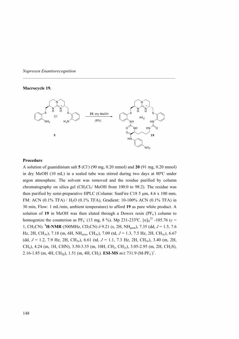
Naproxen Enantiorecognition _____________________________________________________________________
Macrocycle 19.
N
NH
NH SS
PF6-
HNNH
OHNO NHO
HN
NO2
N
NH
NH SS
Cl-
H2NNH2
5 19
20, dry MeOH
(8%)
Procedure A solution of guanidinium salt 5 (Cl-) (90 mg, 0.20 mmol) and 20 (91 mg, 0.20 mmol) in dry MeOH (10 mL) in a sealed tube was stirred during two days at 80ºC under argon atmosphere. The solvent was removed and the residue purified by column chromatography on silica gel (CH2Cl2/ MeOH from 100:0 to 98:2). The residue was then purified by semi-preparative HPLC (Column: SunFire C18 5 µm, 4.6 x 100 mm, FM: ACN (0.1% TFA) / H2O (0.1% TFA), Gradient: 10-100% ACN (0.1% TFA) in 30 min, Flow: 1 mL/min, ambient temperature) to afford 19 as pure white product. A solution of 19 in MeOH was then eluted through a Dowex resin (PF6
-) column to homogenize the counterion as PF6
- (13 mg, 8 %). Mp 231-233ºC. [α]D25 -105.76 (c =
1, CH3CN). 1H-NMR (500MHz, CD3CN) δ 9.21 (s, 2H, NHguan), 7.35 (dd, J = 1.5, 7.6 Hz, 2H, CHAr), 7.18 (m, 6H, NHurea, CHAr), 7.09 (td, J = 1.3, 7.5 Hz, 2H, CHAr), 6.67 (dd, J = 1.2, 7.9 Hz, 2H, CHAr), 6.61 (td, J = 1.1, 7.3 Hz, 2H, CHAr), 3.40 (m, 2H, CHα), 4.24 (m, 1H, CHN), 3.50-3.35 (m, 10H, CH2, CH2γ), 3.05-2.95 (m, 2H, CH2S), 2.16-1.85 (m, 4H, CH2β), 1.51 (m, 4H, CH2). ESI-MS m/z 731.9 (M-PF6
-)+.
148
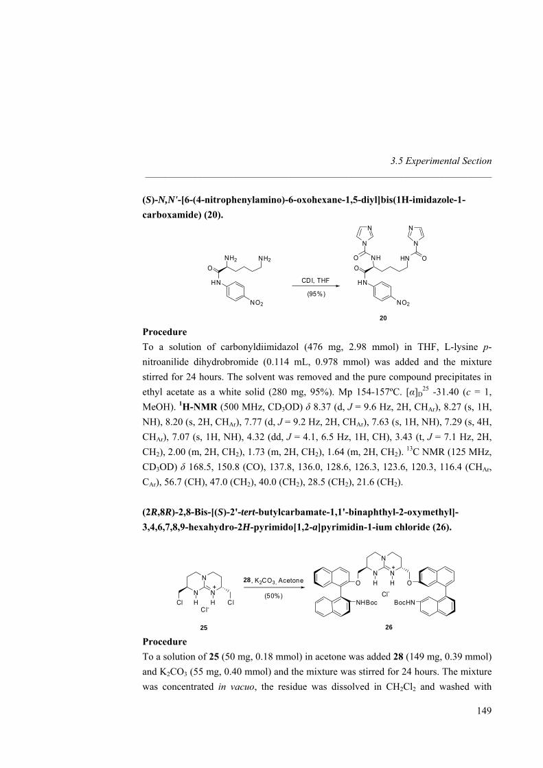
3.5 Experimental Section _____________________________________________________________________
(S)-N,N'-[6-(4-nitrophenylamino)-6-oxohexane-1,5-diyl]bis(1H-imidazole-1-carboxamide) (20).
NN
OHNO NHO
HN
NO2
N N
NH2NH2
O
HN
NO2
CDI, THF
(95%)
20 Procedure To a solution of carbonyldiimidazol (476 mg, 2.98 mmol) in THF, L-lysine p-nitroanilide dihydrobromide (0.114 mL, 0.978 mmol) was added and the mixture stirred for 24 hours. The solvent was removed and the pure compound precipitates in ethyl acetate as a white solid (280 mg, 95%). Mp 154-157ºC. [α]D
25 -31.40 (c = 1, MeOH). 1H-NMR (500 MHz, CD3OD) δ 8.37 (d, J = 9.6 Hz, 2H, CHAr), 8.27 (s, 1H, NH), 8.20 (s, 2H, CHAr), 7.77 (d, J = 9.2 Hz, 2H, CHAr), 7.63 (s, 1H, NH), 7.29 (s, 4H, CHAr), 7.07 (s, 1H, NH), 4.32 (dd, J = 4.1, 6.5 Hz, 1H, CH), 3.43 (t, J = 7.1 Hz, 2H, CH2), 2.00 (m, 2H, CH2), 1.73 (m, 2H, CH2), 1.64 (m, 2H, CH2). 13C NMR (125 MHz, CD3OD) δ 168.5, 150.8 (CO), 137.8, 136.0, 128.6, 126.3, 123.6, 120.3, 116.4 (CHAr, CAr), 56.7 (CH), 47.0 (CH2), 40.0 (CH2), 28.5 (CH2), 21.6 (CH2). (2R,8R)-2,8-Bis-[(S)-2'-tert-butylcarbamate-1,1'-binaphthyl-2-oxymethyl]-3,4,6,7,8,9-hexahydro-2H-pyrimido[1,2-a]pyrimidin-1-ium chloride (26).
N
N
NH H
+
Cl ClCl-
N
N
NH H
+
O O
NHBocCl-
BocHN
25 26
28 , K2CO3, Acetone
(50%)
Procedure To a solution of 25 (50 mg, 0.18 mmol) in acetone was added 28 (149 mg, 0.39 mmol) and K2CO3 (55 mg, 0.40 mmol) and the mixture was stirred for 24 hours. The mixture was concentrated in vacuo, the residue was dissolved in CH2Cl2 and washed with
149
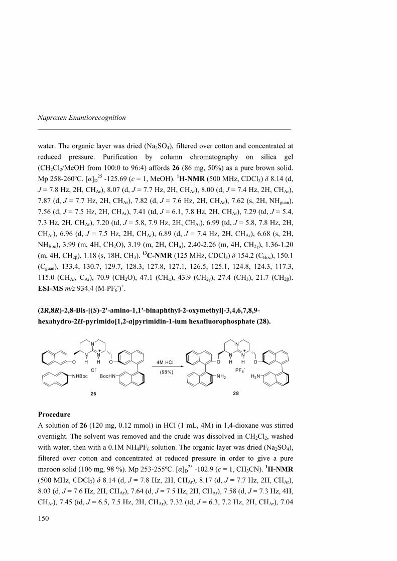
Naproxen Enantiorecognition _____________________________________________________________________
water. The organic layer was dried (Na2SO4), filtered over cotton and concentrated at reduced pressure. Purification by column chromatography on silica gel (CH2Cl2/MeOH from 100:0 to 96:4) affords 26 (86 mg, 50%) as a pure brown solid. Mp 258-260ºC. [α]D
25 -125.69 (c = 1, MeOH). 1H-NMR (500 MHz, CDCl3) δ 8.14 (d, J = 7.8 Hz, 2H, CHAr), 8.07 (d, J = 7.7 Hz, 2H, CHAr), 8.00 (d, J = 7.4 Hz, 2H, CHAr), 7.87 (d, J = 7.7 Hz, 2H, CHAr), 7.82 (d, J = 7.6 Hz, 2H, CHAr), 7.62 (s, 2H, NHguan), 7.56 (d, J = 7.5 Hz, 2H, CHAr), 7.41 (td, J = 6.1, 7.8 Hz, 2H, CHAr), 7.29 (td, J = 5.4, 7.3 Hz, 2H, CHAr), 7.20 (td, J = 5.8, 7.9 Hz, 2H, CHAr), 6.99 (td, J = 5.8, 7.8 Hz, 2H, CHAr), 6.96 (d, J = 7.5 Hz, 2H, CHAr), 6.89 (d, J = 7.4 Hz, 2H, CHAr), 6.68 (s, 2H, NHBoc), 3.99 (m, 4H, CH2O), 3.19 (m, 2H, CHα), 2.40-2.26 (m, 4H, CH2γ), 1.36-1.20 (m, 4H, CH2β), 1.18 (s, 18H, CH3). 13C-NMR (125 MHz, CDCl3) δ 154.2 (CBoc), 150.1 (Cguan), 133.4, 130.7, 129.7, 128.3, 127.8, 127.1, 126.5, 125.1, 124.8, 124.3, 117.3, 115.0 (CHAr, CAr), 70.9 (CH2O), 47.1 (CHα), 43.9 (CH2γ), 27.4 (CH3), 21.7 (CH2β). ESI-MS m/z 934.4 (M-PF6
-)+.
(2R,8R)-2,8-Bis-[(S)-2'-amino-1,1'-binaphthyl-2-oxymethyl]-3,4,6,7,8,9-hexahydro-2H-pyrimido[1,2-a]pyrimidin-1-ium hexafluorophosphate (28).
26 28
N
N
NH H
+
O O
NHBocCl-
BocHN
N
N
NH H
+
O O
NH2
PF6-
H2N
4M HCl
(98%)
Procedure A solution of 26 (120 mg, 0.12 mmol) in HCl (1 mL, 4M) in 1,4-dioxane was stirred overnight. The solvent was removed and the crude was dissolved in CH2Cl2, washed with water, then with a 0.1M NH4PF6 solution. The organic layer was dried (Na2SO4), filtered over cotton and concentrated at reduced pressure in order to give a pure maroon solid (106 mg, 98 %). Mp 253-255ºC. [α]D
25 -102.9 (c = 1, CH3CN). 1H-NMR (500 MHz, CDCl3) δ 8.14 (d, J = 7.8 Hz, 2H, CHAr), 8.17 (d, J = 7.7 Hz, 2H, CHAr), 8.03 (d, J = 7.6 Hz, 2H, CHAr), 7.64 (d, J = 7.5 Hz, 2H, CHAr), 7.58 (d, J = 7.3 Hz, 4H, CHAr), 7.45 (td, J = 6.5, 7.5 Hz, 2H, CHAr), 7.32 (td, J = 6.3, 7.2 Hz, 2H, CHAr), 7.04
150
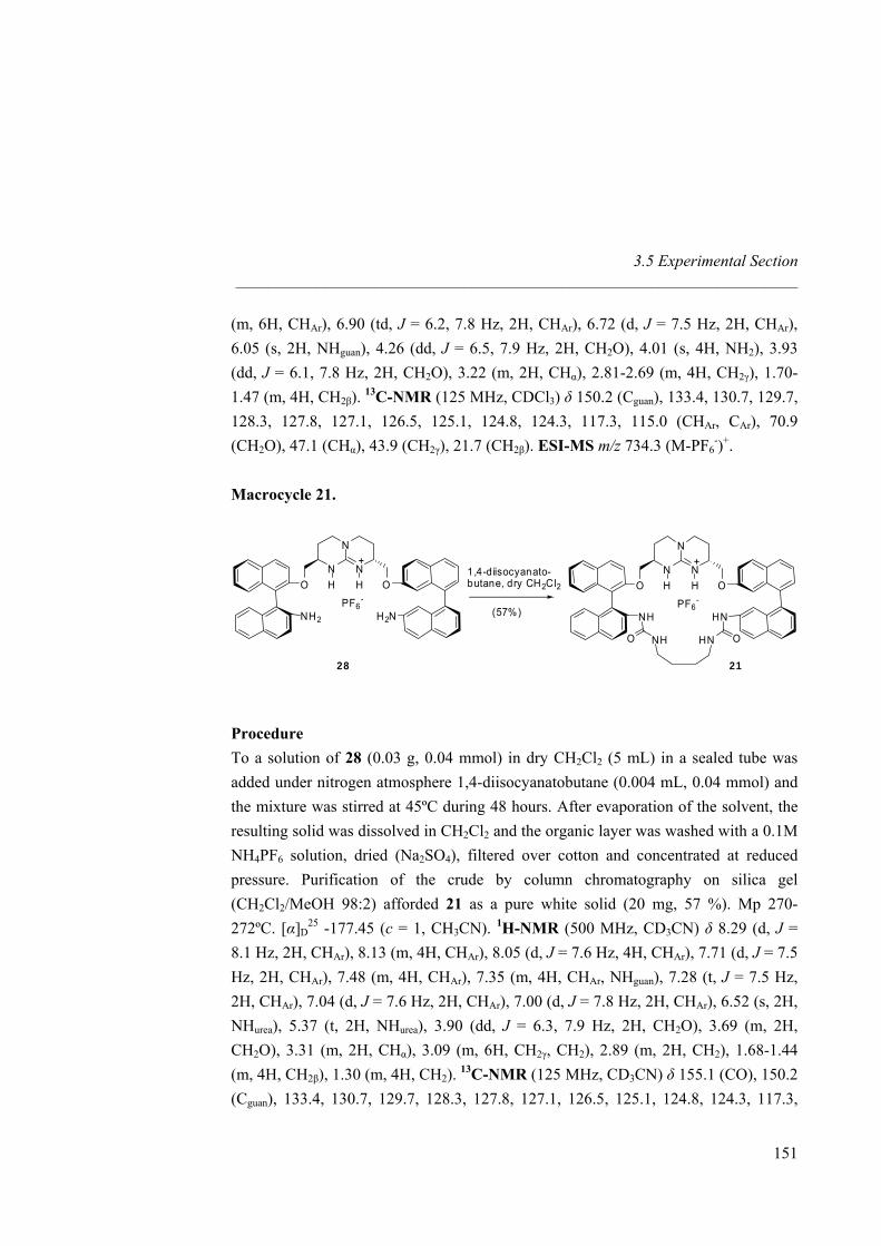
3.5 Experimental Section _____________________________________________________________________
(m, 6H, CHAr), 6.90 (td, J = 6.2, 7.8 Hz, 2H, CHAr), 6.72 (d, J = 7.5 Hz, 2H, CHAr), 6.05 (s, 2H, NHguan), 4.26 (dd, J = 6.5, 7.9 Hz, 2H, CH2O), 4.01 (s, 4H, NH2), 3.93 (dd, J = 6.1, 7.8 Hz, 2H, CH2O), 3.22 (m, 2H, CHα), 2.81-2.69 (m, 4H, CH2γ), 1.70-1.47 (m, 4H, CH2β). 13C-NMR (125 MHz, CDCl3) δ 150.2 (Cguan), 133.4, 130.7, 129.7, 128.3, 127.8, 127.1, 126.5, 125.1, 124.8, 124.3, 117.3, 115.0 (CHAr, CAr), 70.9 (CH2O), 47.1 (CHα), 43.9 (CH2γ), 21.7 (CH2β). ESI-MS m/z 734.3 (M-PF6
-)+. Macrocycle 21.
N
N
NH H
+
O O
NH2
PF6-
H2N
N
N
NH H
+
O O
NHPF6
-
HN
O NH OHN
28 21
1,4-diisocyanato-butane, dry CH2Cl2
(57%)
Procedure To a solution of 28 (0.03 g, 0.04 mmol) in dry CH2Cl2 (5 mL) in a sealed tube was added under nitrogen atmosphere 1,4-diisocyanatobutane (0.004 mL, 0.04 mmol) and the mixture was stirred at 45ºC during 48 hours. After evaporation of the solvent, the resulting solid was dissolved in CH2Cl2 and the organic layer was washed with a 0.1M NH4PF6 solution, dried (Na2SO4), filtered over cotton and concentrated at reduced pressure. Purification of the crude by column chromatography on silica gel (CH2Cl2/MeOH 98:2) afforded 21 as a pure white solid (20 mg, 57 %). Mp 270-272ºC. [α]D
25 -177.45 (c = 1, CH3CN). 1H-NMR (500 MHz, CD3CN) δ 8.29 (d, J = 8.1 Hz, 2H, CHAr), 8.13 (m, 4H, CHAr), 8.05 (d, J = 7.6 Hz, 4H, CHAr), 7.71 (d, J = 7.5 Hz, 2H, CHAr), 7.48 (m, 4H, CHAr), 7.35 (m, 4H, CHAr, NHguan), 7.28 (t, J = 7.5 Hz, 2H, CHAr), 7.04 (d, J = 7.6 Hz, 2H, CHAr), 7.00 (d, J = 7.8 Hz, 2H, CHAr), 6.52 (s, 2H, NHurea), 5.37 (t, 2H, NHurea), 3.90 (dd, J = 6.3, 7.9 Hz, 2H, CH2O), 3.69 (m, 2H, CH2O), 3.31 (m, 2H, CHα), 3.09 (m, 6H, CH2γ, CH2), 2.89 (m, 2H, CH2), 1.68-1.44 (m, 4H, CH2β), 1.30 (m, 4H, CH2). 13C-NMR (125 MHz, CD3CN) δ 155.1 (CO), 150.2 (Cguan), 133.4, 130.7, 129.7, 128.3, 127.8, 127.1, 126.5, 125.1, 124.8, 124.3, 117.3,
151
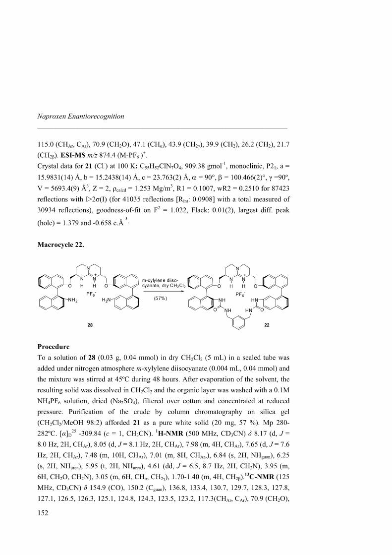
Naproxen Enantiorecognition _____________________________________________________________________
115.0 (CHAr, CAr), 70.9 (CH2O), 47.1 (CHα), 43.9 (CH2γ), 39.9 (CH2), 26.2 (CH2), 21.7 (CH2β). ESI-MS m/z 874.4 (M-PF6
-)+. Crystal data for 21 (Cl-) at 100 K: C55H52ClN7O4, 909.38 gmol-1, monoclinic, P21, a = 15.9831(14) Å, b = 15.2438(14) Å, c = 23.763(2) Å, α = 90°, β = 100.466(2)°, γ =90º, V = 5693.4(9) Å3, Z = 2, ρcalcd = 1.253 Mg/m3, R1 = 0.1007, wR2 = 0.2510 for 87423 reflections with I>2σ(I) (for 41035 reflections [Rint: 0.0908] with a total measured of 30934 reflections), goodness-of-fit on F2 = 1.022, Flack: 0.01(2), largest diff. peak
(hole) = 1.379 and -0.658 e.Å-3.
Macrocycle 22.
28 22
m-xylylene diiso-cyanate, dry CH2Cl2
(57%)
N
N
NH H
+
O O
NH2
PF6-
H2N
N
N
NH H
+
O O
NHPF6
-
HN
O NH OHN
Procedure To a solution of 28 (0.03 g, 0.04 mmol) in dry CH2Cl2 (5 mL) in a sealed tube was added under nitrogen atmosphere m-xylylene diisocyanate (0.004 mL, 0.04 mmol) and the mixture was stirred at 45ºC during 48 hours. After evaporation of the solvent, the resulting solid was dissolved in CH2Cl2 and the organic layer was washed with a 0.1M NH4PF6 solution, dried (Na2SO4), filtered over cotton and concentrated at reduced pressure. Purification of the crude by column chromatography on silica gel (CH2Cl2/MeOH 98:2) afforded 21 as a pure white solid (20 mg, 57 %). Mp 280-282ºC. [α]D
25 -309.84 (c = 1, CH3CN). 1H-NMR (500 MHz, CD3CN) δ 8.17 (d, J = 8.0 Hz, 2H, CHAr), 8.05 (d, J = 8.1 Hz, 2H, CHAr), 7.98 (m, 4H, CHAr), 7.65 (d, J = 7.6 Hz, 2H, CHAr), 7.48 (m, 10H, CHAr), 7.01 (m, 8H, CHAr,), 6.84 (s, 2H, NHguan), 6.25 (s, 2H, NHurea), 5.95 (t, 2H, NHurea), 4.61 (dd, J = 6.5, 8.7 Hz, 2H, CH2N), 3.95 (m, 6H, CH2O, CH2N), 3.05 (m, 6H, CHα, CH2γ), 1.70-1.40 (m, 4H, CH2β).13C-NMR (125 MHz, CD3CN) δ 154.9 (CO), 150.2 (Cguan), 136.8, 133.4, 130.7, 129.7, 128.3, 127.8, 127.1, 126.5, 126.3, 125.1, 124.8, 124.3, 123.5, 123.2, 117.3(CHAr, CAr), 70.9 (CH2O),
152
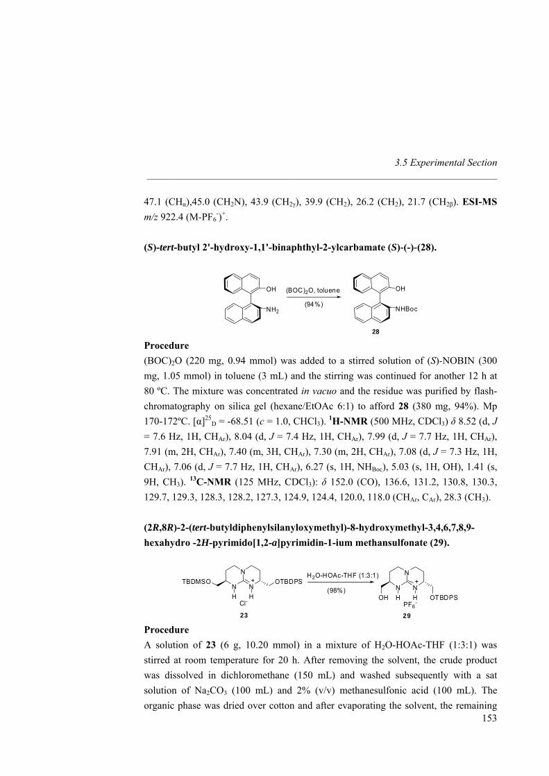
3.5 Experimental Section _____________________________________________________________________
47.1 (CHα),45.0 (CH2N), 43.9 (CH2γ), 39.9 (CH2), 26.2 (CH2), 21.7 (CH2β). ESI-MS m/z 922.4 (M-PF6
-)+. (S)-tert-butyl 2'-hydroxy-1,1'-binaphthyl-2-ylcarbamate (S)-(-)-(28).
OH
NHBoc
OH
NH2
28
(BOC)2O, toluene
(94%)
Procedure (BOC)2O (220 mg, 0.94 mmol) was added to a stirred solution of (S)-NOBIN (300 mg, 1.05 mmol) in toluene (3 mL) and the stirring was continued for another 12 h at 80 ºC. The mixture was concentrated in vacuo and the residue was purified by flash-chromatography on silica gel (hexane/EtOAc 6:1) to afford 28 (380 mg, 94%). Mp 170-172ºC. [α]25
D = -68.51 (c = 1.0, CHCl3). 1H-NMR (500 MHz, CDCl3) δ 8.52 (d, J = 7.6 Hz, 1H, CHAr), 8.04 (d, J = 7.4 Hz, 1H, CHAr), 7.99 (d, J = 7.7 Hz, 1H, CHAr), 7.91 (m, 2H, CHAr), 7.40 (m, 3H, CHAr), 7.30 (m, 2H, CHAr), 7.08 (d, J = 7.3 Hz, 1H, CHAr), 7.06 (d, J = 7.7 Hz, 1H, CHAr), 6.27 (s, 1H, NHBoc), 5.03 (s, 1H, OH), 1.41 (s, 9H, CH3). 13C-NMR (125 MHz, CDCl3): δ 152.0 (CO), 136.6, 131.2, 130.8, 130.3, 129.7, 129.3, 128.3, 128.2, 127.3, 124.9, 124.4, 120.0, 118.0 (CHAr, CAr), 28.3 (CH3). (2R,8R)-2-(tert-butyldiphenylsilanyloxymethyl)-8-hydroxymethyl-3,4,6,7,8,9-hexahydro -2H-pyrimido[1,2-a]pyrimidin-1-ium methansulfonate (29).
N
N
NH H
+
OH OTBDPSPF6
-
N
N
NH H
+
Cl-
OTBDPSTBDMSO
23 29
H2O-HOAc-THF (1:3:1)
(98%)
Procedure
153
A solution of 23 (6 g, 10.20 mmol) in a mixture of H2O-HOAc-THF (1:3:1) was stirred at room temperature for 20 h. After removing the solvent, the crude product was dissolved in dichloromethane (150 mL) and washed subsequently with a sat solution of Na2CO3 (100 mL) and 2% (v/v) methanesulfonic acid (100 mL). The organic phase was dried over cotton and after evaporating the solvent, the remaining
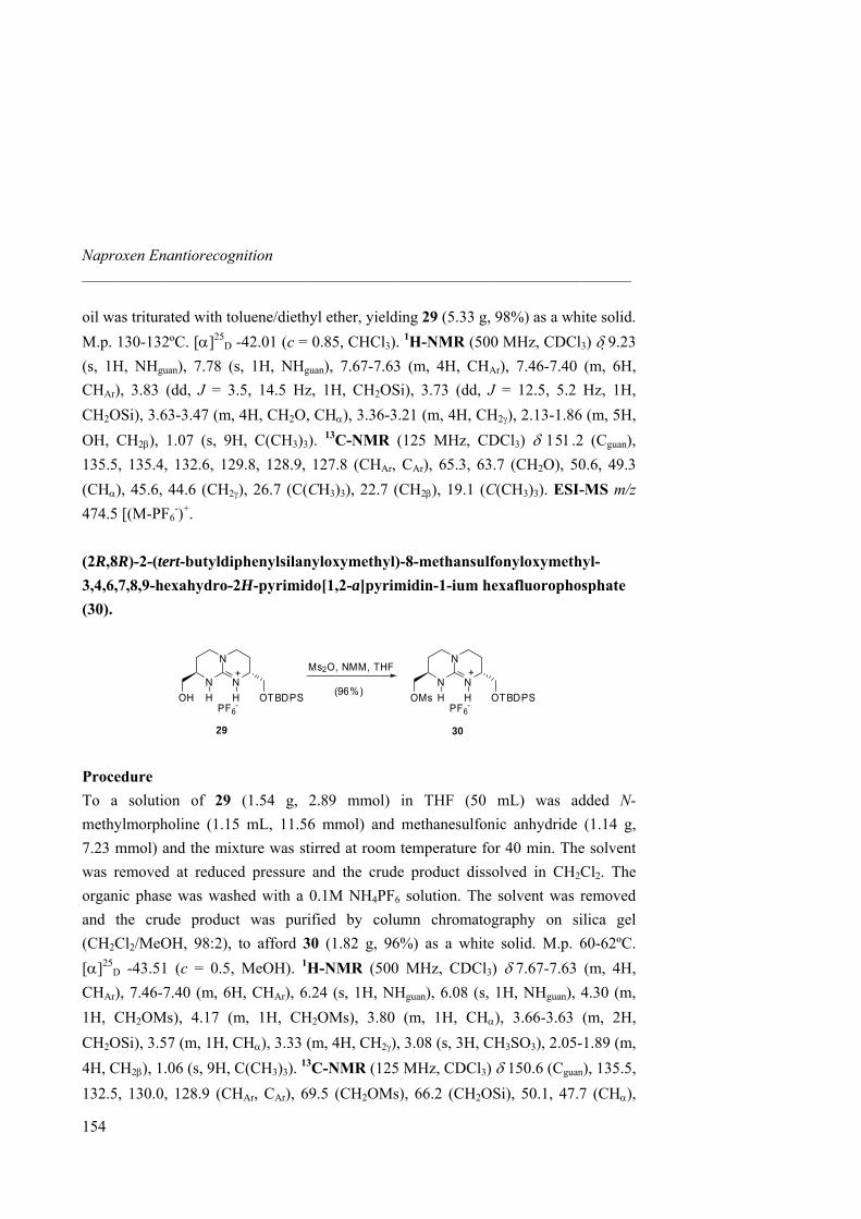
Naproxen Enantiorecognition _____________________________________________________________________
oil was triturated with toluene/diethyl ether, yielding 29 (5.33 g, 98%) as a white solid. M.p. 130-132ºC. [α]25
D -42.01 (c = 0.85, CHCl3). 1H-NMR (500 MHz, CDCl3) δ 9.23 (s, 1H, NHguan), 7.78 (s, 1H, NHguan), 7.67-7.63 (m, 4H, CHAr), 7.46-7.40 (m, 6H, CHAr), 3.83 (dd, J = 3.5, 14.5 Hz, 1H, CH2OSi), 3.73 (dd, J = 12.5, 5.2 Hz, 1H, CH2OSi), 3.63-3.47 (m, 4H, CH2O, CHα), 3.36-3.21 (m, 4H, CH2γ), 2.13-1.86 (m, 5H, OH, CH2β), 1.07 (s, 9H, C(CH3)3). 13C-NMR (125 MHz, CDCl3) δ 151.2 (Cguan), 135.5, 135.4, 132.6, 129.8, 128.9, 127.8 (CHAr, CAr), 65.3, 63.7 (CH2O), 50.6, 49.3 (CHα), 45.6, 44.6 (CH2γ), 26.7 (C(CH3)3), 22.7 (CH2β), 19.1 (C(CH3)3). ESI-MS m/z 474.5 [(M-PF6
-)+. (2R,8R)-2-(tert-butyldiphenylsilanyloxymethyl)-8-methansulfonyloxymethyl-3,4,6,7,8,9-hexahydro-2H-pyrimido[1,2-a]pyrimidin-1-ium hexafluorophosphate (30).
29 30
N
N
NH H
+
OH OTBDPSPF6
-
N
N
NH H
+
OMs OTBDPSPF6
-
Ms2O, NMM, THF
(96%)
Procedure To a solution of 29 (1.54 g, 2.89 mmol) in THF (50 mL) was added N-methylmorpholine (1.15 mL, 11.56 mmol) and methanesulfonic anhydride (1.14 g, 7.23 mmol) and the mixture was stirred at room temperature for 40 min. The solvent was removed at reduced pressure and the crude product dissolved in CH2Cl2. The organic phase was washed with a 0.1M NH4PF6 solution. The solvent was removed and the crude product was purified by column chromatography on silica gel (CH2Cl2/MeOH, 98:2), to afford 30 (1.82 g, 96%) as a white solid. M.p. 60-62ºC. [α]25
D -43.51 (c = 0.5, MeOH). 1H-NMR (500 MHz, CDCl3) δ 7.67-7.63 (m, 4H, CHAr), 7.46-7.40 (m, 6H, CHAr), 6.24 (s, 1H, NHguan), 6.08 (s, 1H, NHguan), 4.30 (m, 1H, CH2OMs), 4.17 (m, 1H, CH2OMs), 3.80 (m, 1H, CHα), 3.66-3.63 (m, 2H, CH2OSi), 3.57 (m, 1H, CHα), 3.33 (m, 4H, CH2γ), 3.08 (s, 3H, CH3SO3), 2.05-1.89 (m, 4H, CH2β), 1.06 (s, 9H, C(CH3)3). 13C-NMR (125 MHz, CDCl3) δ 150.6 (Cguan), 135.5, 132.5, 130.0, 128.9 (CHAr, CAr), 69.5 (CH2OMs), 66.2 (CH2OSi), 50.1, 47.7 (CHα),
154
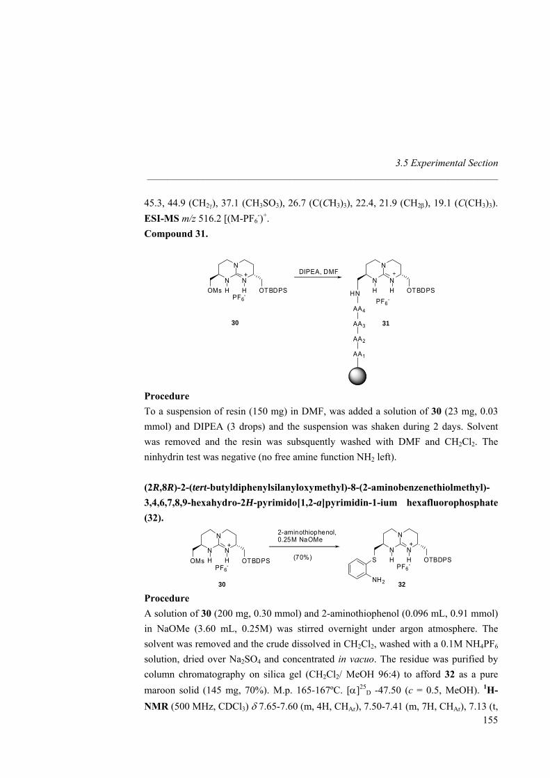
3.5 Experimental Section _____________________________________________________________________
45.3, 44.9 (CH2γ), 37.1 (CH3SO3), 26.7 (C(CH3)3), 22.4, 21.9 (CH2β), 19.1 (C(CH3)3). ESI-MS m/z 516.2 [(M-PF6
-)+. Compound 31.
3130
N
N
NH H
+
OMs OTBDPSPF6
-
N
N
NOTBDPSHH
PF6-
HN
AA2
AA1
AA4
AA3
DIPEA, DMF
Procedure To a suspension of resin (150 mg) in DMF, was added a solution of 30 (23 mg, 0.03 mmol) and DIPEA (3 drops) and the suspension was shaken during 2 days. Solvent was removed and the resin was subsquently washed with DMF and CH2Cl2. The ninhydrin test was negative (no free amine function NH2 left). (2R,8R)-2-(tert-butyldiphenylsilanyloxymethyl)-8-(2-aminobenzenethiolmethyl)-3,4,6,7,8,9-hexahydro-2H-pyrimido[1,2-a]pyrimidin-1-ium hexafluorophosphate (32).
30 32
N
N
NH H
+
OMs OTBDPSPF6
-
N
N
NH H
+
S OTBDPSPF6
-
NH2
2-aminothiophenol,0.25M NaOMe
(70%)
Procedure
155
A solution of 30 (200 mg, 0.30 mmol) and 2-aminothiophenol (0.096 mL, 0.91 mmol) in NaOMe (3.60 mL, 0.25M) was stirred overnight under argon atmosphere. The solvent was removed and the crude dissolved in CH2Cl2, washed with a 0.1M NH4PF6 solution, dried over Na2SO4 and concentrated in vacuo. The residue was purified by column chromatography on silica gel (CH2Cl2/ MeOH 96:4) to afford 32 as a pure maroon solid (145 mg, 70%). M.p. 165-167ºC. [α]25
D -47.50 (c = 0.5, MeOH). 1H-NMR (500 MHz, CDCl3) δ 7.65-7.60 (m, 4H, CHAr), 7.50-7.41 (m, 7H, CHAr), 7.13 (t,
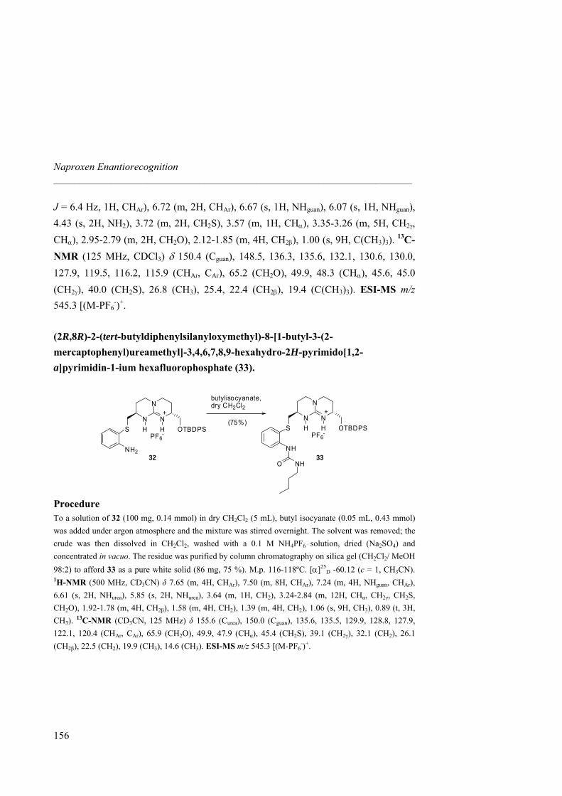
Naproxen Enantiorecognition _____________________________________________________________________
J = 6.4 Hz, 1H, CHAr), 6.72 (m, 2H, CHAr), 6.67 (s, 1H, NHguan), 6.07 (s, 1H, NHguan), 4.43 (s, 2H, NH2), 3.72 (m, 2H, CH2S), 3.57 (m, 1H, CHα), 3.35-3.26 (m, 5H, CH2γ, CHα), 2.95-2.79 (m, 2H, CH2O), 2.12-1.85 (m, 4H, CH2β), 1.00 (s, 9H, C(CH3)3). 13C-NMR (125 MHz, CDCl3) δ 150.4 (Cguan), 148.5, 136.3, 135.6, 132.1, 130.6, 130.0, 127.9, 119.5, 116.2, 115.9 (CHAr, CAr), 65.2 (CH2O), 49.9, 48.3 (CHα), 45.6, 45.0 (CH2γ), 40.0 (CH2S), 26.8 (CH3), 25.4, 22.4 (CH2β), 19.4 (C(CH3)3). ESI-MS m/z 545.3 [(M-PF6
-)+. (2R,8R)-2-(tert-butyldiphenylsilanyloxymethyl)-8-[1-butyl-3-(2-mercaptophenyl)ureamethyl]-3,4,6,7,8,9-hexahydro-2H-pyrimido[1,2-a]pyrimidin-1-ium hexafluorophosphate (33).
32 33
N
N
NH H
+
S OTBDPSPF6
-
NH2
N
N
NH H
+
S OTBDPSPF6
-
NH
NHO
butylisocyanate,dry CH2Cl2
(75%)
Procedure To a solution of 32 (100 mg, 0.14 mmol) in dry CH2Cl2 (5 mL), butyl isocyanate (0.05 mL, 0.43 mmol) was added under argon atmosphere and the mixture was stirred overnight. The solvent was removed; the crude was then dissolved in CH2Cl2, washed with a 0.1 M NH4PF6 solution, dried (Na2SO4) and concentrated in vacuo. The residue was purified by column chromatography on silica gel (CH2Cl2/ MeOH 98:2) to afford 33 as a pure white solid (86 mg, 75 %). M.p. 116-118ºC. [α]25
D -60.12 (c = 1, CH3CN). 1H-NMR (500 MHz, CD3CN) δ 7.65 (m, 4H, CHAr), 7.50 (m, 8H, CHAr), 7.24 (m, 4H, NHguan, CHAr), 6.61 (s, 2H, NHurea), 5.85 (s, 2H, NHurea), 3.64 (m, 1H, CH2), 3.24-2.84 (m, 12H, CHα, CH2γ, CH2S, CH2O), 1.92-1.78 (m, 4H, CH2β), 1.58 (m, 4H, CH2), 1.39 (m, 4H, CH2), 1.06 (s, 9H, CH3), 0.89 (t, 3H, CH3). 13C-NMR (CD3CN, 125 MHz) δ 155.6 (Curea), 150.0 (Cguan), 135.6, 135.5, 129.9, 128.8, 127.9, 122.1, 120.4 (CHAr, CAr), 65.9 (CH2O), 49.9, 47.9 (CHα), 45.4 (CH2S), 39.1 (CH2γ), 32.1 (CH2), 26.1 (CH2β), 22.5 (CH2), 19.9 (CH3), 14.6 (CH3). ESI-MS m/z 545.3 [(M-PF6
-)+.
156
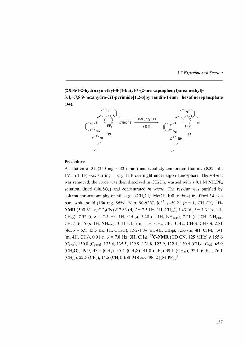
3.5 Experimental Section _____________________________________________________________________
(2R,8R)-2-hydroxymethyl-8-[1-butyl-3-(2-mercaptophenyl)ureamethyl]-3,4,6,7,8,9-hexahydro-2H-pyrimido[1,2-a]pyrimidin-1-ium hexafluorophosphate (34).
33 34
N
N
NH H
+
S OTBDPSPF6
-
NH
NHO
N
N
NH H
+
S OHPF6
-
NH
NHO
TBAF, dry THF
(86%)
Procedure A solution of 33 (250 mg, 0.32 mmol) and tetrabutylammonium fluoride (0.32 mL, 1M in THF) was stirring in dry THF overnight under argon atmosphere. The solvent was removed; the crude was then dissolved in CH2Cl2, washed with a 0.1 M NH4PF6 solution, dried (Na2SO4) and concentrated in vacuo. The residue was purified by column chromatography on silica gel (CH2Cl2/ MeOH 100 to 96:4) to afford 34 as a pure white solid (150 mg, 86%). M.p. 90-92ºC. [α]25
D -50.21 (c = 1, CH3CN). 1H-NMR (500 MHz, CD3CN) δ 7.63 (d, J = 7.5 Hz, 1H, CHAr), 7.43 (d, J = 7.3 Hz, 1H, CHAr), 7.32 (t, J = 7.5 Hz, 1H, CHAr), 7.28 (s, 1H, NHguan), 7.21 (m, 2H, NHguan, CHAr), 6.55 (s, 1H, NHurea), 3.44-3.15 (m, 11H, CH2, CHα, CH2γ, CH2S, CH2O), 2.81 (dd, J = 6.9, 13.5 Hz, 1H, CH2O), 1.92-1.84 (m, 4H, CH2β), 1.56 (m, 4H, CH2), 1.41 (m, 4H, CH2), 0.91 (t, J = 7.8 Hz, 3H, CH3). 13C-NMR (CD3CN, 125 MHz) δ 155.6 (Curea), 150.0 (Cguan), 135.6, 135.5, 129.9, 128.8, 127.9, 122.1, 120.4 (CHAr, CAr), 65.9 (CH2O), 49.9, 47.9 (CHα), 45.4 (CH2S), 41.0 (CH2) 39.1 (CH2γ), 32.1 (CH2), 26.1 (CH2β), 22.5 (CH2), 14.5 (CH3). ESI-MS m/z 406.2 [(M-PF6
-)+.
157
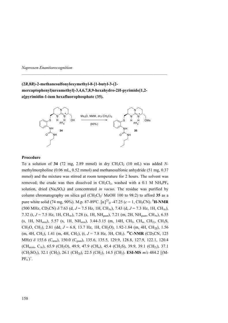
Naproxen Enantiorecognition _____________________________________________________________________
(2R,8R)-2-methanesulfonyloxymethyl-8-[1-butyl-3-(2-mercaptophenyl)ureamethyl]-3,4,6,7,8,9-hexahydro-2H-pyrimido[1,2-a]pyrimidin-1-ium hexafluorophosphate (35).
34 35
N
N
NH H
+
S OHPF6
-
NH
NHO
N
N
NH H
+
S OMsPF6
-
NH
NHO
Ms2O, NMM , dry CH2Cl2
(90%)
Procedure To a solution of 34 (72 mg, 2.89 mmol) in dry CH2Cl2 (10 mL) was added N-methylmorpholine (0.06 mL, 0.52 mmol) and methanesulfonic anhydride (51 mg, 0.37 mmol) and the mixture was stirred at room temperature for 2 hours. The solvent was removed; the crude was then dissolved in CH2Cl2, washed with a 0.1 M NH4PF6 solution, dried (Na2SO4) and concentrated in vacuo. The residue was purified by column chromatography on silica gel (CH2Cl2/ MeOH 100 to 98:2) to afford 35 as a pure white solid (74 mg, 90%). M.p. 87-89ºC. [α]25
D -47.25 (c = 1, CH3CN). 1H-NMR (500 MHz, CD3CN) δ 7.63 (d, J = 7.5 Hz, 1H, CHAr), 7.43 (d, J = 7.3 Hz, 1H, CHAr), 7.32 (t, J = 7.5 Hz, 1H, CHAr), 7.28 (s, 1H, NHguan), 7.21 (m, 2H, NHguan, CHAr), 6.55 (s, 1H, NHurea), 5.57 (s, 1H, NHurea), 3.44-3.15 (m, 14H, CH2, CHα, CH2γ, CH2S, CH2O, CH3), 2.81 (dd, J = 6.8, 13.7 Hz, 1H, CH2O), 1.92-1.84 (m, 4H, CH2β), 1.56 (m, 4H, CH2), 1.41 (m, 4H, CH2), (t, J = 7.8 Hz, 3H, CH3). 13C-NMR (CD3CN, 125 MHz) δ 155.6 (Curea), 150.0 (Cguan), 135.6, 135.5, 129.9, 128.8, 127.9, 122.1, 120.4 (CHarom, CAr), 65.9 (CH2O), 49.9, 47.9 (CHα), 45.4 (CH2S), 39.9, 39.1 (CH2γ), 37.1 (CH3SO3), 32.1 (CH2), 26.1 (CH2β), 22.5 (CH2), 14.5 (CH3). ESI-MS m/z 484.2 [(M-PF6
-)+.
158
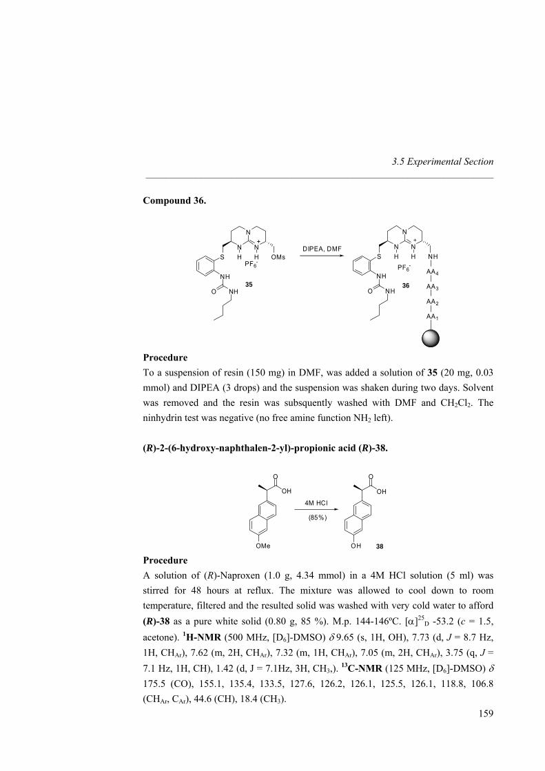
3.5 Experimental Section _____________________________________________________________________
Compound 36.
35 36
N
N
NH H
+
S OMsPF6
-
NH
NHO
N
N
NS NHHH
NH
AA2
AA1
O NH
AA4
AA3
PF6-
DIPEA, DMF
Procedure To a suspension of resin (150 mg) in DMF, was added a solution of 35 (20 mg, 0.03 mmol) and DIPEA (3 drops) and the suspension was shaken during two days. Solvent was removed and the resin was subsquently washed with DMF and CH2Cl2. The ninhydrin test was negative (no free amine function NH2 left).
(R)-2-(6-hydroxy-naphthalen-2-yl)-propionic acid (R)-38.
OH
O
OMe
OH
O
OH 38
4M HCl
(85%)
Procedure A solution of (R)-Naproxen (1.0 g, 4.34 mmol) in a 4M HCl solution (5 ml) was stirred for 48 hours at reflux. The mixture was allowed to cool down to room temperature, filtered and the resulted solid was washed with very cold water to afford (R)-38 as a pure white solid (0.80 g, 85 %). M.p. 144-146ºC. [α]25
D -53.2 (c = 1.5, acetone). 1H-NMR (500 MHz, [D6]-DMSO) δ 9.65 (s, 1H, OH), 7.73 (d, J = 8.7 Hz, 1H, CHAr), 7.62 (m, 2H, CHAr), 7.32 (m, 1H, CHAr), 7.05 (m, 2H, CHAr), 3.75 (q, J = 7.1 Hz, 1H, CH), 1.42 (d, J = 7.1Hz, 3H, CH3,). 13C-NMR (125 MHz, [D6]-DMSO) δ 175.5 (CO), 155.1, 135.4, 133.5, 127.6, 126.2, 126.1, 125.5, 126.1, 118.8, 106.8 (CHAr, CAr), 44.6 (CH), 18.4 (CH3).
159
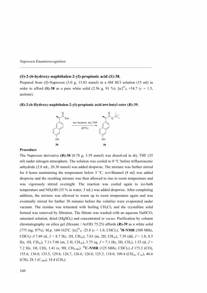
Naproxen Enantiorecognition _____________________________________________________________________
(S)-2-(6-hydroxy-naphthalen-2-yl)-propionic acid (S)-38. Prepared from (S)-Naproxen (3.0 g, 13.03 mmol) in a 4M HCl solution (15 ml) in order to afford (S)-38 as a pure white solid (2.56 g, 91 %). [α]25
D +54.7 (c = 1.5, acetone).
(R)-2-(6-Hydroxy-naphthalen-2-yl)-propionic acid tert-butyl ester (R)-39.
38
OH
O
OH
O
O
OH
39
(87%)
ter t-butanol, dry THF
Procedure
The Naproxen derivative (R)-38 (0.78 g, 3.39 mmol) was dissolved in dry THF (35 ml) under nitrogen atmosphere. The solution was cooled to 0 °C before trifluoroacetic anhydride (2.8 mL, 20.30 mmol) was added dropwise. The mixture was further stirred for 4 hours maintaining the temperature below 5 °C. tert-Butanol (8 ml) was added dropwise and the resulting mixture was then allowed to rise to room temperature and was vigorously stirred overnight. The reaction was cooled again to ice-bath temperature and NH4OH (35 % in water, 3 mL) was added dropwise. After completing addition, the mixture was allowed to warm up to room temperature again and was eventually stirred for further 30 minutes before the volatiles were evaporated under vacuum. The residue was triturated with boiling CH2Cl2 and the crystalline solid formed was removed by filtration. The filtrate was washed with an aqueous NaHCO3 saturated solution, dried (MgSO4) and concentrated in vacuo. Purification by column chromatography on silica gel (Hexane / AcOEt 75:25) affords (R)-39 as a white solid (775 mg, 87%). M.p. 160-162ºC. [α]25
D -25.8 (c = 1.0, CHCl3). 1H-NMR (500 MHz, CDCl3) δ 7.69 (d, J = 8.7 Hz, 1H, CHAr), 7.63 (m, 2H, CHAr), 7.39 (dd, J = 1.8, 8.5 Hz, 1H, CHAr), 7.11-7.06 (m, 2 H, CHAr), 3.75 (q, J = 7.1 Hz, 3H, CH3), 1.53 (d, J = 7.2 Hz, 1H, CH), 1.41 (s, 9H, CH3t-Bu). 13C-NMR (125 MHz, CDCl3) δ 175.2 (CO), 155.6, 136.0, 133.5, 129.8, 126.7, 126.6, 126.0, 125.3, 118.0, 109.4 (CHAr, CAr), 46.6 (CH), 28.1 (Ct-Bu), 18.4 (CH3).
160
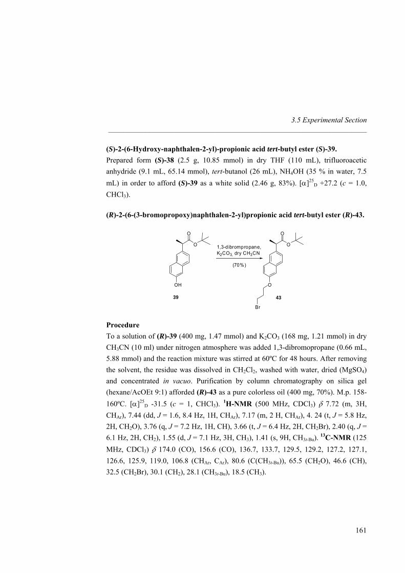
3.5 Experimental Section _____________________________________________________________________
(S)-2-(6-Hydroxy-naphthalen-2-yl)-propionic acid tert-butyl ester (S)-39. Prepared form (S)-38 (2.5 g, 10.85 mmol) in dry THF (110 mL), trifluoroacetic anhydride (9.1 mL, 65.14 mmol), tert-butanol (26 mL), NH4OH (35 % in water, 7.5 mL) in order to afford (S)-39 as a white solid (2.46 g, 83%). [α]25
D +27.2 (c = 1.0, CHCl3).
(R)-2-(6-(3-bromopropoxy)naphthalen-2-yl)propionic acid tert-butyl ester (R)-43.
O
O
OH
O
O
O
Br
1,3-dibrompropane,K2CO3, dry CH3CN
(70%)
39 43
Procedure To a solution of (R)-39 (400 mg, 1.47 mmol) and K2CO3 (168 mg, 1.21 mmol) in dry CH3CN (10 ml) under nitrogen atmosphere was added 1,3-dibromopropane (0.66 mL, 5.88 mmol) and the reaction mixture was stirred at 60ºC for 48 hours. After removing the solvent, the residue was dissolved in CH2Cl2, washed with water, dried (MgSO4) and concentrated in vacuo. Purification by column chromatography on silica gel (hexane/AcOEt 9:1) afforded (R)-43 as a pure colorless oil (400 mg, 70%). M.p. 158-160ºC. [α]25
D -31.5 (c = 1, CHCl3). 1H-NMR (500 MHz, CDCl3) δ 7.72 (m, 3H, CHAr), 7.44 (dd, J = 1.6, 8.4 Hz, 1H, CHAr), 7.17 (m, 2 H, CHAr), 4. 24 (t, J = 5.8 Hz, 2H, CH2O), 3.76 (q, J = 7.2 Hz, 1H, CH), 3.66 (t, J = 6.4 Hz, 2H, CH2Br), 2.40 (q, J = 6.1 Hz, 2H, CH2), 1.55 (d, J = 7.1 Hz, 3H, CH3), 1.41 (s, 9H, CH3t-Bu). 13C-NMR (125 MHz, CDCl3) δ 174.0 (CO), 156.6 (CO), 136.7, 133.7, 129.5, 129.2, 127.2, 127.1, 126.6, 125.9, 119.0, 106.8 (CHAr, CAr), 80.6 (C(CH3t-Bu)), 65.5 (CH2O), 46.6 (CH), 32.5 (CH2Br), 30.1 (CH2), 28.1 (CH3t-Bu), 18.5 (CH3).
161
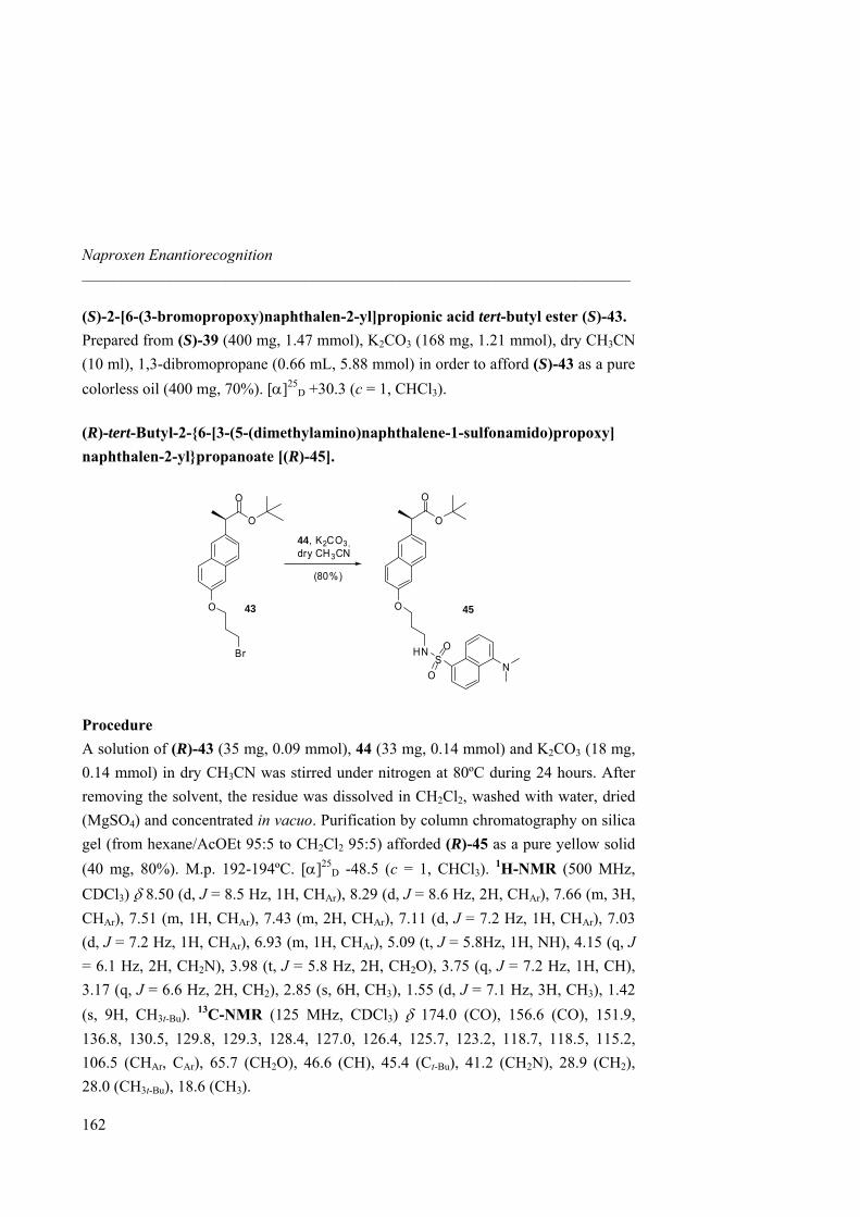
Naproxen Enantiorecognition _____________________________________________________________________
(S)-2-[6-(3-bromopropoxy)naphthalen-2-yl]propionic acid tert-butyl ester (S)-43. Prepared from (S)-39 (400 mg, 1.47 mmol), K2CO3 (168 mg, 1.21 mmol), dry CH3CN (10 ml), 1,3-dibromopropane (0.66 mL, 5.88 mmol) in order to afford (S)-43 as a pure colorless oil (400 mg, 70%). [α]25
D +30.3 (c = 1, CHCl3).
(R)-tert-Butyl-2-{6-[3-(5-(dimethylamino)naphthalene-1-sulfonamido)propoxy] naphthalen-2-yl}propanoate [(R)-45].
43 45
O
O
O
O
O
O
HNS
O
O
NBr
44, K2CO3,dry CH3CN
(80%)
Procedure A solution of (R)-43 (35 mg, 0.09 mmol), 44 (33 mg, 0.14 mmol) and K2CO3 (18 mg, 0.14 mmol) in dry CH3CN was stirred under nitrogen at 80ºC during 24 hours. After removing the solvent, the residue was dissolved in CH2Cl2, washed with water, dried (MgSO4) and concentrated in vacuo. Purification by column chromatography on silica gel (from hexane/AcOEt 95:5 to CH2Cl2 95:5) afforded (R)-45 as a pure yellow solid (40 mg, 80%). M.p. 192-194ºC. [α]25
D -48.5 (c = 1, CHCl3). 1H-NMR (500 MHz, CDCl3) δ 8.50 (d, J = 8.5 Hz, 1H, CHAr), 8.29 (d, J = 8.6 Hz, 2H, CHAr), 7.66 (m, 3H, CHAr), 7.51 (m, 1H, CHAr), 7.43 (m, 2H, CHAr), 7.11 (d, J = 7.2 Hz, 1H, CHAr), 7.03 (d, J = 7.2 Hz, 1H, CHAr), 6.93 (m, 1H, CHAr), 5.09 (t, J = 5.8Hz, 1H, NH), 4.15 (q, J = 6.1 Hz, 2H, CH2N), 3.98 (t, J = 5.8 Hz, 2H, CH2O), 3.75 (q, J = 7.2 Hz, 1H, CH), 3.17 (q, J = 6.6 Hz, 2H, CH2), 2.85 (s, 6H, CH3), 1.55 (d, J = 7.1 Hz, 3H, CH3), 1.42 (s, 9H, CH3t-Bu). 13C-NMR (125 MHz, CDCl3) δ 174.0 (CO), 156.6 (CO), 151.9, 136.8, 130.5, 129.8, 129.3, 128.4, 127.0, 126.4, 125.7, 123.2, 118.7, 118.5, 115.2, 106.5 (CHAr, CAr), 65.7 (CH2O), 46.6 (CH), 45.4 (Ct-Bu), 41.2 (CH2N), 28.9 (CH2), 28.0 (CH3t-Bu), 18.6 (CH3).
162
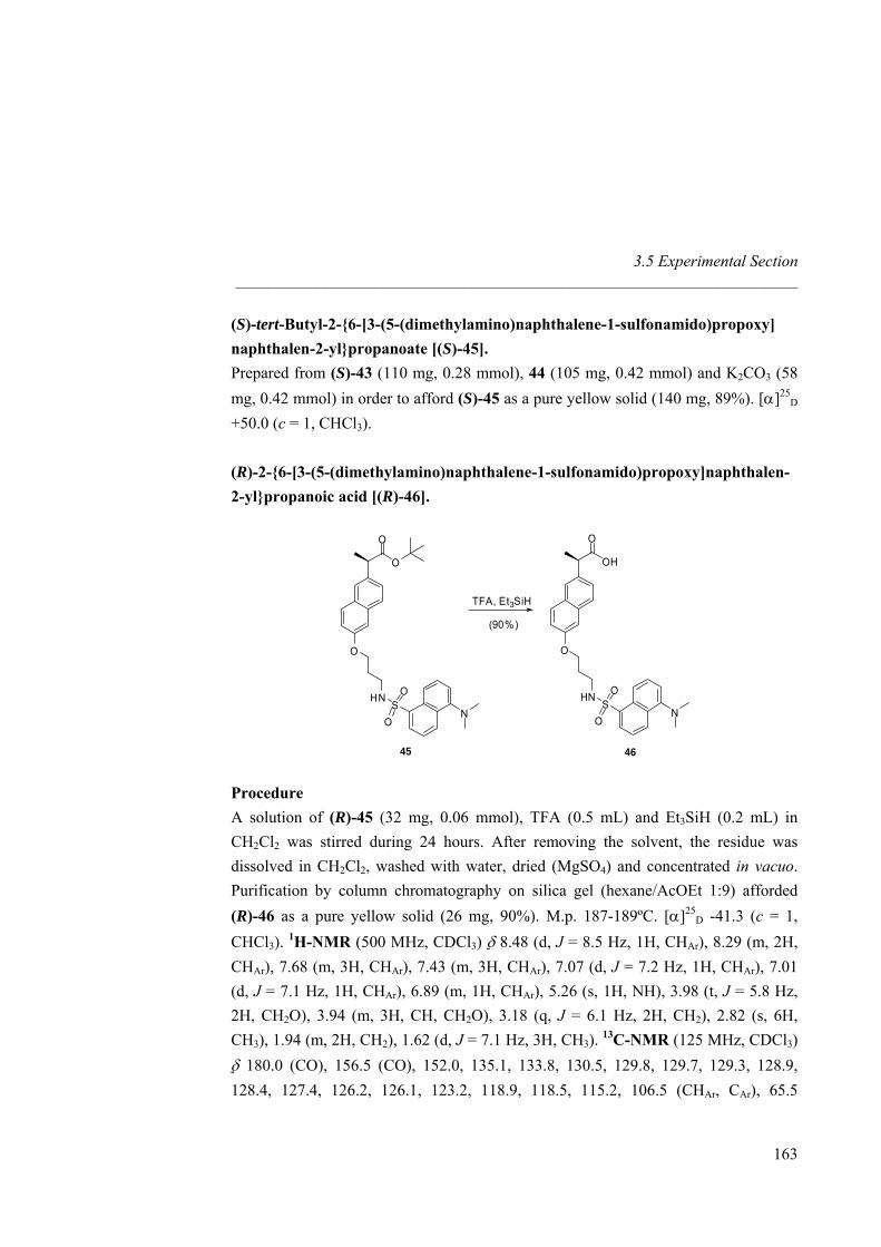
3.5 Experimental Section _____________________________________________________________________
(S)-tert-Butyl-2-{6-[3-(5-(dimethylamino)naphthalene-1-sulfonamido)propoxy] naphthalen-2-yl}propanoate [(S)-45]. Prepared from (S)-43 (110 mg, 0.28 mmol), 44 (105 mg, 0.42 mmol) and K2CO3 (58 mg, 0.42 mmol) in order to afford (S)-45 as a pure yellow solid (140 mg, 89%). [α]25
D +50.0 (c = 1, CHCl3). (R)-2-{6-[3-(5-(dimethylamino)naphthalene-1-sulfonamido)propoxy]naphthalen-2-yl}propanoic acid [(R)-46].
O
O
O
HNS
O
O
N
OH
O
O
HNS
O
O
N
45 46
TFA, Et3SiH
(90%)
Procedure A solution of (R)-45 (32 mg, 0.06 mmol), TFA (0.5 mL) and Et3SiH (0.2 mL) in CH2Cl2 was stirred during 24 hours. After removing the solvent, the residue was dissolved in CH2Cl2, washed with water, dried (MgSO4) and concentrated in vacuo. Purification by column chromatography on silica gel (hexane/AcOEt 1:9) afforded (R)-46 as a pure yellow solid (26 mg, 90%). M.p. 187-189ºC. [α]25
D -41.3 (c = 1, CHCl3). 1H-NMR (500 MHz, CDCl3) δ 8.48 (d, J = 8.5 Hz, 1H, CHAr), 8.29 (m, 2H, CHAr), 7.68 (m, 3H, CHAr), 7.43 (m, 3H, CHAr), 7.07 (d, J = 7.2 Hz, 1H, CHAr), 7.01 (d, J = 7.1 Hz, 1H, CHAr), 6.89 (m, 1H, CHAr), 5.26 (s, 1H, NH), 3.98 (t, J = 5.8 Hz, 2H, CH2O), 3.94 (m, 3H, CH, CH2O), 3.18 (q, J = 6.1 Hz, 2H, CH2), 2.82 (s, 6H, CH3), 1.94 (m, 2H, CH2), 1.62 (d, J = 7.1 Hz, 3H, CH3). 13C-NMR (125 MHz, CDCl3) δ 180.0 (CO), 156.5 (CO), 152.0, 135.1, 133.8, 130.5, 129.8, 129.7, 129.3, 128.9, 128.4, 127.4, 126.2, 126.1, 123.2, 118.9, 118.5, 115.2, 106.5 (CHAr, CAr), 65.5
163
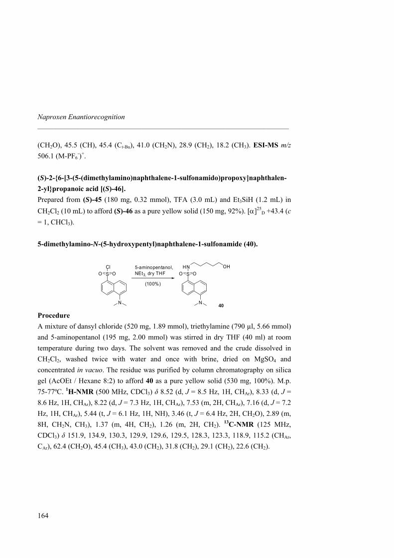
Naproxen Enantiorecognition _____________________________________________________________________
(CH2O), 45.5 (CH), 45.4 (Ct-Bu), 41.0 (CH2N), 28.9 (CH2), 18.2 (CH3). ESI-MS m/z 506.1 (M-PF6
-)+. (S)-2-{6-[3-(5-(dimethylamino)naphthalene-1-sulfonamido)propoxy]naphthalen-2-yl}propanoic acid [(S)-46]. Prepared from (S)-45 (180 mg, 0.32 mmol), TFA (3.0 mL) and Et3SiH (1.2 mL) in CH2Cl2 (10 mL) to afford (S)-46 as a pure yellow solid (150 mg, 92%). [α]25
D +43.4 (c = 1, CHCl3). 5-dimethylamino-N-(5-hydroxypentyl)naphthalene-1-sulfonamide (40).
N
S OOCl
N
S OOHN OH5-aminopentanol,
NEt3, dry THF
(100%)
40 Procedure A mixture of dansyl chloride (520 mg, 1.89 mmol), triethylamine (790 µl, 5.66 mmol) and 5-aminopentanol (195 mg, 2.00 mmol) was stirred in dry THF (40 ml) at room temperature during two days. The solvent was removed and the crude dissolved in CH2Cl2, washed twice with water and once with brine, dried on MgSO4 and concentrated in vacuo. The residue was purified by column chromatography on silica gel (AcOEt / Hexane 8:2) to afford 40 as a pure yellow solid (530 mg, 100%). M.p. 75-77ºC. 1H-NMR (500 MHz, CDCl3) δ 8.52 (d, J = 8.5 Hz, 1H, CHAr), 8.33 (d, J = 8.6 Hz, 1H, CHAr), 8.22 (d, J = 7.3 Hz, 1H, CHAr), 7.53 (m, 2H, CHAr), 7.16 (d, J = 7.2 Hz, 1H, CHAr), 5.44 (t, J = 6.1 Hz, 1H, NH), 3.46 (t, J = 6.4 Hz, 2H, CH2O), 2.89 (m, 8H, CH2N, CH3), 1.37 (m, 4H, CH2), 1.26 (m, 2H, CH2). 13C-NMR (125 MHz, CDCl3) δ 151.9, 134.9, 130.3, 129.9, 129.6, 129.5, 128.3, 123.3, 118.9, 115.2 (CHAr, CAr), 62.4 (CH2O), 45.4 (CH3), 43.0 (CH2), 31.8 (CH2), 29.1 (CH2), 22.6 (CH2).
164
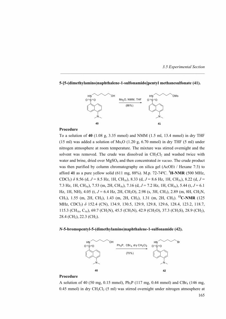
3.5 Experimental Section _____________________________________________________________________
5-[5-(dimethylamino)naphthalene-1-sulfonamido]pentyl methanesulfonate (41).
40
N
S OOHN OH
N
S OOHN OMs
Ms2O, NMM, THF
(88%)
41 Procedure To a solution of 40 (1.08 g, 3.35 mmol) and NMM (1.5 ml, 13.4 mmol) in dry THF (15 ml) was added a solution of Ms2O (1.20 g, 6.70 mmol) in dry THF (5 ml) under nitrogen atmosphere at room temperature. The mixture was stirred overnight and the solvent was removed. The crude was dissolved in CH2Cl2 and washed twice with water and brine, dried over MgSO4 and then concentrated in vacuo. The crude product was then purified by column chromatography on silica gel (AcOEt / Hexane 7:3) to afford 41 as a pure yellow solid (611 mg, 88%). M.p. 72-74ºC. 1H-NMR (500 MHz, CDCl3) δ 8.56 (d, J = 8.5 Hz, 1H, CHAr), 8.33 (d, J = 8.6 Hz, 1H, CHAr), 8.22 (d, J = 7.3 Hz, 1H, CHAr), 7.53 (m, 2H, CHAr), 7.16 (d, J = 7.2 Hz, 1H, CHAr), 5.44 (t, J = 6.1 Hz, 1H, NH), 4.05 (t, J = 6.4 Hz, 2H, CH2O), 2.98 (s, 3H, CH3), 2.89 (m, 8H, CH2N, CH3), 1.55 (m, 2H, CH2), 1.43 (m, 2H, CH2), 1.31 (m, 2H, CH2). 13C-NMR (125 MHz, CDCl3) δ 152.4 (CN), 134.9, 130.5, 129.9, 129.8, 129.6, 128.4, 123.2, 118.7, 115.3 (CHAr, CAr), 69.7 (CH2N), 45.5 (CH3N), 42.9 (CH2O), 37.3 (CH3S), 28.9 (CH2), 28.4 (CH2), 22.3 (CH2).
N-5-bromopentyl-5-(dimethylamino)naphthalene-1-sulfonamide (42).
40 42
N
S OOHN OH
N
S OOHN Br
Ph3P, CBr4, dry CH2Cl2
(70%)
Procedure A solution of 40 (50 mg, 0.15 mmol), Ph3P (117 mg, 0.44 mmol) and CBr4 (146 mg, 0.45 mmol) in dry CH2Cl2 (5 ml) was stirred overnight under nitrogen atmosphere at
165
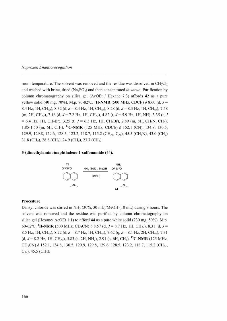
Naproxen Enantiorecognition _____________________________________________________________________
room temperature. The solvent was removed and the residue was dissolved in CH2Cl2 and washed with brine, dried (Na2SO4) and then concentrated in vacuo. Purification by column chromatography on silica gel (AcOEt / Hexane 7:3) affords 42 as a pure yellow solid (40 mg, 70%). M.p. 80-82ºC. 1H-NMR (500 MHz, CDCl3) δ 8.60 (d, J = 8.4 Hz, 1H, CHAr), 8.32 (d, J = 8.4 Hz, 1H, CHAr), 8.28 (d, J = 8.3 Hz, 1H, CHAr), 7.58 (m, 2H, CHAr), 7.16 (d, J = 7.2 Hz, 1H, CHAr), 4.82 (t, J = 5.9 Hz, 1H, NH), 3.35 (t, J = 6.4 Hz, 1H, CH2Br), 3.25 (t, J = 6.3 Hz, 1H, CH2Br), 2.89 (m, 8H, CH2N, CH3), 1.85-1.50 (m, 6H, CH2). 13C-NMR (125 MHz, CDCl3) δ 152.1 (CN), 134.8, 130.5, 129.9, 129.8, 129.6, 128.5, 123.2, 118.7, 115.2 (CHAr, CAr), 45.5 (CH3N), 43.0 (CH2) 31.8 (CH2), 28.8 (CH2), 24.9 (CH2), 23.7 (CH2). 5-(dimethylamino)naphthalene-1-sulfonamide (44).
N
S OOCl
N
S OONH2
NH3 (30%), MeOH
(50%)
44
Procedure Dansyl chloride was stirred in NH3 (30%, 30 mL)/MeOH (10 mL) during 8 hours. The solvent was removed and the residue was purified by column chromatography on silica gel (Hexane/ AcOEt 1:1) to afford 44 as a pure white solid (230 mg, 50%). M.p. 60-62ºC. 1H-NMR (500 MHz, CD3CN) δ 8.57 (d, J = 8.7 Hz, 1H, CHAr), 8.31 (d, J = 8.5 Hz, 1H, CHAr), 8.22 (d, J = 8.7 Hz, 1H, CHAr), 7.62 (q, J = 8.1 Hz, 2H, CHAr), 7.31 (d, J = 8.2 Hz, 1H, CHAr), 5.83 (s, 2H, NH2), 2.91 (s, 6H, CH3). 13C-NMR (125 MHz, CD3CN) δ 152.1, 134.8, 130.5, 129.9, 129.8, 129.6, 128.5, 123.2, 118.7, 115.2 (CHAr, CAr), 45.5 (CH3).
166
3.5 Experimental Section _____________________________________________________________________
167
3.5.3 Library synthesis and Screening Experiments Chloranil Test.73
To a small sample were added two drops of 2% acetaldehyde/DMF and two drops of 2% p-chloranil/DMF. The sample was allowed to stand for 5 minutes. Blue stained beads indicate the presence of secondary amines on the resin (“positive” result). Resin Synthesis
The Tentagel NH2 resin (1.0902, loading 0.51 mmol) was swelled in CH2Cl2. It was then washed several times with DMF. Met(Fmoc)OH (0.381 mg, 1.02 mmol), DIC (240 µL, 1.53 mmol) and HOBT (208 mg, 1.53 mmol) were dissolved in DMF (2ml) and added to the resin. A solution of DIPEA (262 µL, 1.53 mmol) in DMF was added. The funnel was degazed and DMF was added about for ¾ of the tube. The solution was stirred during two hours. The solvent was removed by filtration. DMF was added and filtered. The resin was washed with CH2Cl2 (three times), DMF (once). A qualitative ninhydrin test was negative. The Fmoc group was removed using treatment by 20% piperidine in DMF during 20 min and the resin washed with DMF and CH2Cl2. A qualitative ninhydrin test was positive.
A solution of FmocArg(Pbf)OH (0.663 mg, 1.02 mmol), DIC (240 µL, 1.53 mmol) and HOBT (208 mg, 1.53 mmol) in DMF (2 mL) was added to the resin, followed by DIPEA (262 µL, 1.53 mmol) and the suspension was shaken overnight. Solvent was removed and the resin was subsquently washed with DMF, CH2Cl2, DMF and CH2Cl2. The ninhydrin test was negative. Fmoc deprotection was achieved by 20% piperidine-DMF solution. A qualitative ninhydrin test was positive.
73 Vojkovsky, T. Pept. Res., 1995, 8, 236-237.
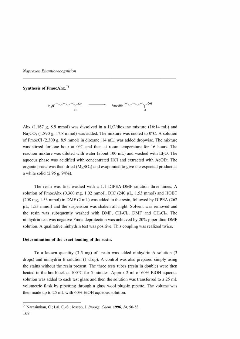
Naproxen Enantiorecognition _____________________________________________________________________
Synthesis of FmocAhx.74
OH
OFmocHNOH
OH2N
Ahx (1.167 g, 8.9 mmol) was dissolved in a H2O/dioxane mixture (16:14 mL) and Na2CO3 (1.890 g, 17.8 mmol) was added. The mixture was cooled to 0°C. A solution of FmocCl (2.300 g, 8.9 mmol) in dioxane (14 mL) was added dropwise. The mixture was stirred for one hour at 0°C and then at room temperature for 16 hours. The reaction mixture was diluted with water (about 100 mL) and washed with Et2O. The aqueous phase was acidified with concentrated HCl and extracted with AcOEt. The organic phase was then dried (MgSO4) and evaporated to give the expected product as a white solid (2.95 g, 94%).
The resin was first washed with a 1:1 DIPEA-DMF solution three times. A
solution of FmocAhx (0.360 mg, 1.02 mmol), DIC (240 µL, 1.53 mmol) and HOBT (208 mg, 1.53 mmol) in DMF (2 mL) was added to the resin, followed by DIPEA (262 µL, 1.53 mmol) and the suspension was shaken all night. Solvent was removed and the resin was subsquently washed with DMF, CH2Cl2, DMF and CH2Cl2. The ninhydrin test was negative Fmoc deprotection was achieved by 20% piperidine-DMF solution. A qualitative ninhydrin test was positive. This coupling was realized twice. Determination of the exact loading of the resin.
To a known quantity (3-5 mg) of resin was added ninhydrin A solution (3 drops) and ninhydrin B solution (1 drop). A control was also prepared simply using the stains without the resin present. The three tests tubes (resin in double) were then heated in the hot block at 100°C for 5 minutes. Approx 2 ml of 60% EtOH aqueous solution was added to each test glass and then the solution was transferred to a 25 mL volumetric flask by pipetting through a glass wool plug-in pipette. The volume was then made up to 25 mL with 60% EtOH aqueous solution.
74 Narasimhan, C.; Lai, C.-S.; Joseph, J. Bioorg. Chem. 1996, 24, 50-58.
168
3.5 Experimental Section _____________________________________________________________________
169
UV absorbance is measured at 570 nm. Use the control as a blank, i.e. the background with it, then measure the absorbance of the resin sample. Substitution (mmol/g) = (A570. V.103) / E.m Where m: mass (mg) V: volume (mL) E = 1.5.104 (M/cm) Done four times: Substitution: 0.215 mmol/g: theoretical: 0.210 mmol/g “Split and Mix” synthesis.
The peptide library was then prepared, using four cycles of “split and mix method”. The resulting resin was divided in eight equal portions. To each resin portion one of the following Fmoc amino acids was added: L-Asp (OtBu), L-Gln, L-Tyr(tBu), L-Trp(BOC), L-Thr(tBu), L-Val, L-Gly, L-Pro (0.72 mmol amino acid per resin portion), along with HOBt (16 mg, 1.1 mmol), EDCI (18 µL, 1.1 mmol), followed by DIPEA addition (2 drops) in DMF. The reaction mixture was shaken for 2 hours. Qualitative ninhydrin test was carried out to check that all the couplings were complete. The terminal Fmoc-groups were removed with 20% piperidine in DMF during 20 minutes.
Ninhydrin test was different for Pro (secondary amine): to a sample of resin were added 2 drops of 2% acetaldehyde in DMF and 2 drops of 2% p-chloranil/DMF. The sample was allowed to stand for 5 mn. Blue stained beads indicate the presence of the secondary amines of the resin (positive deprotection).
Capping agent coupling.
In order to know the amino acid sequence of a selected bead, a capping agent is coupled in 15% on the resin periphery. It is introduced by a classical coupling reaction. As the loading is proportional to the amount of resin (about 0.170 g of resin and 0.215 mmol/g), a stock solution of bomophenylacetic acid is prepared and added to the resin.
To be sure that the distribution will be in all the beads, the solution was stirred during 1 hour with the capping agent and then coupling reagents (HOBT, EDCI and
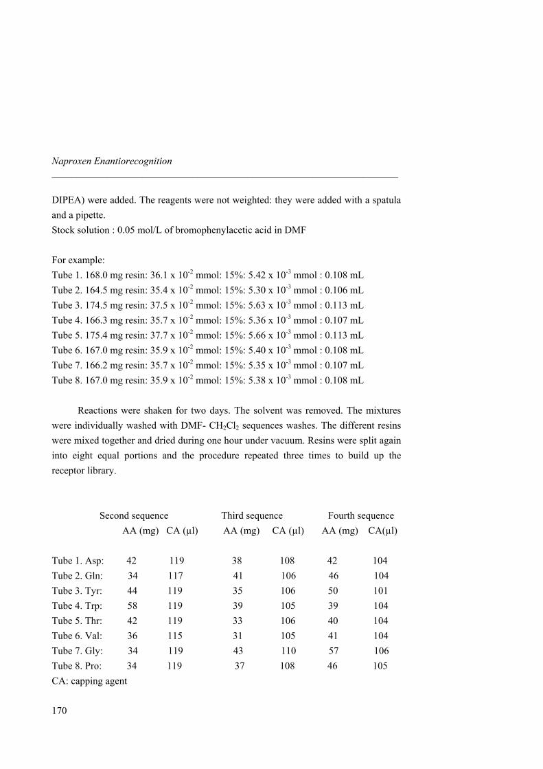
Naproxen Enantiorecognition _____________________________________________________________________
170
DIPEA) were added. The reagents were not weighted: they were added with a spatula and a pipette. Stock solution : 0.05 mol/L of bromophenylacetic acid in DMF For example: Tube 1. 168.0 mg resin: 36.1 x 10⁻² mmol: 15%: 5.42 x 10⁻³ mmol : 0.108 mL Tube 2. 164.5 mg resin: 35.4 x 10⁻² mmol: 15%: 5.30 x 10⁻³ mmol : 0.106 mL Tube 3. 174.5 mg resin: 37.5 x 10⁻² mmol: 15%: 5.63 x 10⁻³ mmol : 0.113 mL Tube 4. 166.3 mg resin: 35.7 x 10⁻² mmol: 15%: 5.36 x 10⁻³ mmol : 0.107 mL Tube 5. 175.4 mg resin: 37.7 x 10⁻² mmol: 15%: 5.66 x 10⁻³ mmol : 0.113 mL Tube 6. 167.0 mg resin: 35.9 x 10⁻² mmol: 15%: 5.40 x 10⁻³ mmol : 0.108 mL Tube 7. 166.2 mg resin: 35.7 x 10⁻² mmol: 15%: 5.35 x 10⁻³ mmol : 0.107 mL Tube 8. 167.0 mg resin: 35.9 x 10⁻² mmol: 15%: 5.38 x 10⁻³ mmol : 0.108 mL
Reactions were shaken for two days. The solvent was removed. The mixtures were individually washed with DMF- CH2Cl2 sequences washes. The different resins were mixed together and dried during one hour under vacuum. Resins were split again into eight equal portions and the procedure repeated three times to build up the receptor library. Second sequence Third sequence Fourth sequence
AA (mg) CA (µl) AA (mg) CA (µl) AA (mg) CA(µl) Tube 1. Asp: 42 119 38 108 42 104 Tube 2. Gln: 34 117 41 106 46 104 Tube 3. Tyr: 44 119 35 106 50 101 Tube 4. Trp: 58 119 39 105 39 104 Tube 5. Thr: 42 119 33 106 40 104 Tube 6. Val: 36 115 31 105 41 104 Tube 7. Gly: 34 119 43 110 57 106 Tube 8. Pro: 34 119 37 108 46 105 CA: capping agent
3.5 Experimental Section _____________________________________________________________________
171
Screening Experiments A sample of library 31 (25.0 mg) was equilibrated in CH3CN (300 µL) for 24 h. A solution of guest (S)-46 (20 µM, 500 µL) in CH3CN was added to the library sample to give a 12.5 µM concentration in guest. Equilibration was continued for 24 h. Beads were analyzed in flat-bottomed glass pots under a Leica inverted DML microscope and fifty highly fluorescent beads were selected with tweezers and washed several times to remove the Naproxen derivative (S)-46. The same procedure was then carried out with (R)-46, but now the ones that did not form a complex with the R enantiomer were isolated. In addition, screening experiments were achieved for library 36 as well as for the free tetrapeptide library 48 with both (R)-46 and (S)-46. As control experiments, a sample of the library 31 was equilibrated in CH3CN for 24 h and a solution of Dansyl chloride was added to the equilibrated solution. The concentrations and conditions were the same as described above for the screening experiments. After 48 h (to allow for equilibration time) no selective staining was observed. Deprotection and cleavage of material from the single resin beads A single resin bead to be analyzed was placed in a glass insert using tweezers. For Pbf deprotection: the bead was treated with 50 µL of 50% TFA/CH2Cl2 for 1 hour. The solvent was then removed under vacuum. Cleavage was performed by treatment of the bead with 20 µL of a solution of CNBr in 1:1 TFA/H2O (concentration 50-80mg/mL) and incubation in the dark for 18 hours, sealed. The cleavage solution was evaporated from the sample tubes in vacuo. 20 µL of CH3CN was then added to the tube. The samples were sonicated for 5 minutes. 1 µL of each sample was spotted onto MALDI-platen. 1 µL of a solution of matrix was then applied on top and the sample mixed on the platen surface. The spot was allowed to dry under a stream of air before analysis. The α-cyanahydroxycinnamic acid matrix was prepared as a saturated solution in acetone for application as above.
173
Summary
This thesis reports the synthesis and design of guanidinium-based receptors as multiple hydrogen bond hosts for molecular recognition of anions with a special emphasis on small anions such as nitrate and the anti-inflammatory drug Naproxen.
The first chapter reviews the guanidinium-based artificial receptors for
molecular recognition of oxoanions such as carboxylates as well as biologically relevant phosphates and sulfates in order to introduce the objectives of this work. Chiral recognition is also reported as catching a large interest in supramolecular chemistry focusing on the enantiorecognition of amino acids.
Despite its significant involvement in environmental pollution, little attention
has been given to nitrate by supramolecular chemists. Chapter two reports the design of several macrocycles combining a guanidinium and two urea moieties for an optimal complement of size, charge and binding sites to nitrate. Synthesis of hosts of different sizes, rigidity and stereochemistry as well as quantification of their binding affinity towards small anions is reported. Although association constants evaluated for nitrate with one of the receptors described herein is one the highests reported so far for this anion, spherical chloride is the most suitable guest for stabilization of the complex. Both results in solution and in solid state are in good agreement to show encapsulation of nitrate, chloride and acetate guest inside the host cavities.
Chapter tree presents two different approaches for Naproxen enantiorecognition,
such as a rational design and a combinatorial chemistry approach. Naproxen is the only Non-Steroidal Anti-Inflammatory drug (NSAID) marketed as a pure enantiomer. The industrial preparation of the drug involves the synthesis of the racemic acid followed by a resolution process to afford the desired compound (resolution accounts for two thirds of the production costs). Here, we propose as an alternative the use of a physical separation method (liquid-liquid extraction) employing a chiral receptor.
Various guanidinium receptors have been synthesized based on concepts of steric hindrance, tweezers and pre-organization. None to small enantioselectivity was observed by this approach.
174
A tetrapeptide library was synthesized by solid phase synthesis and attached to two guanidinium receptors. On the other hand, a fluorescent derivative of Naproxen was prepared for the screening of the libraries highlighting the selectivity of the receptors by enhancement of fluorescence of several beads. Identification of receptors was then achieved by mass spectrometry analysis but could not afford a reliable consensus of optimal amino acid sequence.
175
Introducción general y resultados
Más allá de la química de los enlaces covalentes, que unen los átomos para formar moléculas, la química supramolecular1 es la química de las interacciones débiles: una disciplina que agrupa el concepto de reconocimiento de las moléculas entre sí mismas hacia una mayor estabilidad. En esta memoria se trata una parte específica de dicho campo: el reconocimiento de aniones cuyo desarrollo, en comparación al de los cationes, está mucho menos avanzado. Los receptores descritos en este trabajo llevan una unidad de guanidinio como esqueleto principal. De hecho, la naturaleza utiliza frecuentemente grupos guanidinio, como por ejemplo en la cadena lateral del aminoácido arginina, que permite establecer pares iónicos fuertes con grupos carboxilatos o fosfatos de sistemas biológicos.
En el primer Capítulo se revisa la importancia de la interacción guanidinio-
oxoaniones a través de la bibliografía, con ejemplos característicos de reconocimiento de carboxilatos así como de fosfatos y sulfatos.2 Se pone especial énfasis en el reconocimiento quiral de moléculas de relevancia biológica, como los aminoácidos. Tras la introducción, los objetivos de la tesis se presentan de forma resumida: el reconocimiento del anión nitrato y el reconocimiento quiral del fármaco Naproxeno.
Uno de los problemas más importantes del nitrato es su presencia en el agua
procedente de la filtración de pesticidas utilizados por los agricultores. Una vez ingerido, el nitrato es reducido por el organismo a nitrito, y éste es responsable de diversas enfermedades infantiles. Las técnicas utilizadas para eliminar los nitratos del agua son esencialmente el intercambio de aniones, la osmosis inversa y la electrodiálisis. Una alternativa sería el uso de un receptor capaz de extraer el anión de una fase acuosa a otra mediante extracciones en paralelo o transporte a través de membranas liquidas. Este concepto ha sido ya descrito por nuestro grupo de investigación en relación con los aminoácidos.3
1 Lehn, J.-M. Science 1985, 227, 849-851. 2 Blondeau, P.; Segura, M.; Pérez-Fernández, R.; de Mendoza, J. Chem. Soc. Rev. 2007, 36, 198-210. 3 Breccia, P.; Van Gool, M.; Pérez-Fernández, R.; Martin-Santamaria, S.; Gago, F.; Prados, P.;
176
A continuación, en el Capítulo 2, se describe el diseño de macrociclos con una subunidad de guanidinio y dos de urea para complementar el nitrato de forma ideal en cuanto a tamaño, carga y centros de unión. Se describe la síntesis de macrociclos de diferente tamaño, rigidez y estereoquímica para alcanzar la mejor estabilización del complejo con el anión. Por calorimetría isotérmica, las constantes de asociación han sido medidas, extrayéndose conclusiones sobre la selectividad de cada compuesto. Se ha conseguido así una de las asociaciones con el nitrato más altas descritas hasta ahora.4 Sin embargo, los compuestos presentan mejores constantes de complejación con el esférico anión cloruro. Estructuras resueltas por difracción de rayos X muestran el tipo de interacciones presentes entre el receptor y el anión. Tanto los resultados en disolución como los obtenidos en estado sólido ponen en evidencia la inclusión de nitrato, acetato y cloruro en el interior de la cavidad del macrociclo.
Cada año, el porcentaje de fármacos enantioméricamente puros que salen al
mercado aumenta, hasta superar un 30% en el año 2000. El Naproxeno es el único fármaco anti-inflamatorio no esteroídico vendido como un único enantiómero. La forma (S) es de hecho 28 veces más efectiva que la (R). Tras la síntesis de la mezcla racémica se lleva a cabo la resolución del acido para obtener el enantiómero deseado (más de 66% de los costes de la producción provienen de esta última etapa de resolución). Así pues, nos proponemos emplear métodos de separación física (como extracción líquido-líquido) mediante un receptor quiral para reconocer un enantiómero del Naproxeno. Ninguna molécula ha sido descrita con la capacidad de extraer enantioselectivamente este fármaco hasta ahora. El Capítulo 3 muestra dos estrategias complementarias utilizadas para alcanzar dicho objetivo: el diseño de novo y la química combinatoria.
Además de incorporar una unidad de guanidinio y dos de urea, los receptores requieren de otras interacciones, además de centros de quiralidad adicionales, para alcanzar la selectividad entre ambos enantiómeros. Unos grupos voluminosos o superficies aromáticas se incorporan en primer lugar sobre receptores abiertos, sin que se produzca ninguna discriminación. Receptores con más pre-organizados han sido
de Mendoza, J. J. Am. Chem. Soc. 2003, 125, 8270-8284. 4 Blondeau, P.; Benet-Buchholz, J.; de Mendoza, J. New J. Chem. 2007, in press, DOI: 10.1039/b616409a.
177
sintetizados a continuación, mediante la introducción de un derivado del aminoácido lisina o del ligando de catálisis NOBIN, con poca selectividad en este último caso.
El secundo método implica el uso de química combinatoria como medio para sintetizar un gran conjunto de receptores para luego identificar el más selectivo. Una librería de tetrapéptidos ha sido sintetizada en fase sólida y luego incorporada a la unidad de guanidinio. Al Naproxeno se le ha incorporado un grupo fluorescente para realizar los experimentos de selectividad. Las partículas poliméricas se tornan fluorescentes cuando la interacción con el Naproxeno es fuerte y se pueden identificar por espectrometría de masas. Las secuencias identificadas no presentan suficiente consenso como para llevar a una conclusión definida respeto a una secuencia optima de aminoácidos.