annual review 2014 - 国立大学法人 神戸大学 (kobe ... · is a major pathways. in order to...
TRANSCRIPT

1
Annual Review
2014
Molecular Photoscience
Research Center
Kobe University
2
Preface
This annual review provides a summary of the research activity of Molecular Photoscience
Research Center for the 2014 fiscal year. We are further promoting advanced research and
international collaboration on molecular photoscience and related topics. Any constructive
comments and questions, and any suggestion for collaboration research are welcome.
MPRC belongs to Cluster of Centers of Organization of Advanced Science and Technology
Kobe University. In this year OAST selected 19 Core Research Teams from five Graduate
Schools in the fields of natural science (Science, Engineering, System Information, Agricultural
Science, and Maritime Sciences), and each team has started its research activity. Some
researchers of MPRC also participate in these Core Research Teams to further advanced and
interdisciplinary science by collaborating with members in the Teams.
March, 2015
Keisuke Tominaga
Director of Molecular Photoscience Research Center,
Kobe University

3
Contents
Members 4
Research Activities
Laser Molecular Photoscience Laboratory 5
Ultrafast Photoscience Laboratory 16
Coherent Photoscience Laboratory 31
Original Papers 39
Invited Talks 42
Presentation at Conferences (International and domestic) 44
Presentation by Graduate Students and Postdocs 46
Books 51
Other Publications 52
Lectures to Public 53
Awards 54
Conference Organization 55
Molecular Photoscience Seminar 56

4
Members
Keisuke Tominaga Director
Hitoshi Ohta Vice-Director
Yuma Kato Assistant
Keiko Katayama Assistant (~ June 2014)
Megumi Soma Assistant
Takako Miyazaki Assistant (August 2014 ~)
Laser Molecular Photoscience Laboratory
Akihide Wada Professor
Shunji Kasahara Associate Professor
Neeraj Kumar Joshi Kobe University Visiting Research Fellow (~ January 2015)
Ultrafast Photoscience Laboratory
Keisuke Tominaga Professor
Seiji Akimoto Associate Professor
Kaoru Ohta Research Associate Professor
Naoki Yamamoto Postdoctoral Fellow (~ June 2014)
Minako Kondo JSPS Postdoctoral Fellow (~ September 2014)
Feng Zhang Postdoctoral Fellow
Jessica Afalla Kobe University Visiting Research Fellow (March 2015 ~)
Coherent Photoscience Laboratory
Hitoshi Ohta Professor
Susumu Okubo Assistant Professor
Keigo Hijii Postdoctoral Fellow
Alexey Alfonsov Kobe University Visiting Research Fellow
5
Research Activity
I. Laser Molecular Photoscience Laboratory
I-A. OPTICAL CONTROL OF PHOTO-REACTION NETWORK
Chemical reaction induced by photo-irradiation consists of several reaction pathways such as
multiphoton/multistep reaction paths subsequent to photo-excitation to S1 state, even if one-photon reaction
is a major pathways. In order to understand and control such branched reaction pathways called
photo-reaction network, in addition to the knowledge of each reaction oaths, knowledge of correlation and
balance between paths is necessary in addition to the information about each reaction path. The goal of
this study is to understand and control the whole photo-reaction network. The initial step for understanding
a natural phenomenon is to observe it carefully. Recent developments in laser technology have made it
possible to observe the structure and the dynamics of atoms and molecules at high energy resolutions
and/or high time resolutions. When the observation has been done as much as possible, scientific research
should then progress to the next stage, namely interpretation of the reaction mechanism and construction of
the model of reaction mechanism.
One of the method to confirm the validity of constructed model is to examine the response of the
photo-reaction network to shaped pulse on the basis of the model. In addition, pulse-shaping techniques
have great potential to contribute to the progress of theoretical and experimental studies concerned with
optically controlling the dynamics of molecular systems. These techniques are particularly pertinent for
research into polyatomic molecules or molecules in condensed phases. However, the pulse-shaping method
usually needs a huge number of parameters such as amplitude and phase on each frequency included in the
shaped pulse. Then reduction of the number of parameters is an important issue. Two-pulse correlation
technique is a kind of the simplest pulse-shaping techniques, in which a variable parameter is pulse interval.
This technique is an effective tool to investigate and control the photo-reaction network. In this study, the
reaction mechanism and control method of photo-reaction network was investigated by time-resolved
measurements and pulse-shaping technique such as two-pulse correlation.
On the Branching Ratio of Photoisomerization
in 4-Aminoazobenzene
Neeraj K. Joshi, Masanori Fuyuki, and Akihide
Wada
(Annual Meeting of Japan Society for Molecular
Science, 2014)
Study of the reaction path ways and the relaxation
processes of photoexcited molecules are
fundamental part of investigation for any
photoinduced reaction and such information yields
an opportunity to control the photochemical
reactions. For that purpose, photoisomerization
reaction in azobenezne (AB) and its derivatives has
been well studied in which trans-to-cis
isomerization reaction is initiated by the irradiation
of UV-visible light. On the other hand, cis-to-trans
thermal isomerization spontaneously takes place in
the dark owing to the thermodynamic stability of
the trans isomer1, and our recent study on
4-aminoazobenzene (AAB) has revealed that the

6
mechanism and the kinetics of thermal
isomerization of AAB strongly depends on the
polarity of the medium and is independent of the
viscosity of the medium.
Scheme 1
In this study, as a part of our investigation on
the photoreaction network of AAB, we focused on
the branching ratio of photoisomerization, namely
the production ratio between cis-isomer and
trans-isomer from photoexcited state of
trans-isomer. The most significant aspect of the
study is that the method for estimating the
branching ratio does not require the ultrafast time
resolved measurement but a simplistic approach
has been employed. Briefly, the population at
photostationary state (cis isomer) is measured as
function of pumping rate, and the observed results
are discussed quantitatively based on the
three-level model (described below) which provide
some insight on the mechanism of
photoisomerization in viscous medium.
Scheme 2
In general, for understanding the
photoisomerization in azobenzene family
schematically, a five level energy diagram (scheme
1) is considered. In our case, photoexcitation of
cis-AAB is negligible due to very low absorption
cross-section for pump 405 nm. Therefore, the
effect of excited state of cis-isomer can be
neglected and the five level system can be
modified to four level system as indicated by red
broken line in the scheme 1. Furthermore, under
the definition of branching ratio as described above,
the four level system can be simplified to three
level system as shown in scheme 2. In scheme 2,
n0 corresponds to the population of trans-form in
the ground state under photostationary state, n1
represents the sum of the population of trans-form
in the excited state and population in the
intermediate state. The population of cis produced
by photoisomerization of trans is denoted by n2.
The rate constants, 0k and 1k , represent the
decay rate of those molecules which do not
undergo for photoisomerization and the decay rate
of those molecules which relax after
photoisomerization, respectively. Rate of thermal

7
cis-to-trans isomerization is shown by 2k . In our
recent study the order of the 2k in MeOH and
EG has been determined2 as in the order of one.
However the rate constant for 0k and 1k has
also been determined which is much higher3 (in the
order of 1012 to1014 s-1) than 2k . Thus the total
population (N) of the three level system which is
sum of n0, n1, and n2, i.e., N = n0 + n1 + n2, can be
expressed as N ~ n0 + n2.
Using three level system and above
approximation, the branching ratio (r) defined as
above is equivalent to ratio of 0k to 1k . Namely,
the branching ratio can be expressed as
0 1/r k k . For estimation of branching ratio, the
population under photostationary state as function
of pumping rate (IB) is calculated by monitoring
the bleaching at absorption peak for methanol
(MeOH) and ethylene glycol (EG). In the analysis,
under steady state condition, it is assumed that
number of bleached molecules in level-0 are equal
to the number of molecules present at level 2
(scheme 2). The ratio of N/n0 versus IB is plotted
and fitted to the equation as follows;
0
1N m IBn
(1)
where 21/ 1m r k and 0 1/r k k .
Equation (1) is derived from the solution of
differential equation for scheme 2 under steady
state condition. For calculating IB, at first,
decrease in pump power before and after the
sample is noticed and divided by per photon
energy (i.e., 5 x 10-19 J for 405 nm) which is the
measure of bleached /excited molecules or
absorbed photon per second. Further, by dividing
to number of photon absorbed per second by active
sample volume, the value of IBno can be obtained.
Once the IBno is known and by measuring the no,
IB can be estimated.
The plot of N/no versus IB is fitted to Eq. 1
gives a slope (m) as shown in Fig. 2 and hence
branching ratio (r) can be estimated based on the
value of 2k reported in our previous report2. It is
observed that branching ratio is significant
different between MeOH and EG. Calculated value
of the branching ratio ( 0 1/k k ) for MeOH and EG
is 0.9 ( ±0.06) and 0.3 (±0.02), respectively. Here it
should be mentioned that MeOH and EG have
similar polarity but quite different viscosity.
Therefore, it seems that in contrast to thermal
isomerization, mechanism for photoisomerization
for 4-AAB is sensitive to viscosity and
photoisomerization mechanism may be different in
EG than MeOH. Alternatively, this situation can be
better probed by ultrafast transient experiments
which are under course of investigation.
I-B. NEW TECHNIQUE FOR ANALYZING PHOTO-REACTION NETWORK
Photochemical reactions initiated by multiphoton/multistep absorption are essentially different from
conventional one-photon photochemical reactions because the character of the populated state depends on
the optical order of the excitation process and new photochemical reaction channels are opened through the
excitation of reaction intermediates. In this regard, knowledge of the contribution of the

8
multiphoton/multistep process to photochemical reactions is necessary to exploit new photochemical
reaction pathways, and the multiphoton process is expected to play a vital role in the optical control of
chemical reactions. In this study, Fourier transformed two-dimensional excitation spectrometer was
developed on the basis of the Fabry-Pérot interferometer.
Fourier Transform Two-dimensional
Fluorescence Excitation Spectrometer
(FT-2DFES) by Using Tandem Fabry-Pérot
Interferometer
Hirosi Anzai, Neeraj Kumar Joshi, Masanori
Fuyuki, Akihide Wada
(Rev. Sci. Inst., 2015)
Inter- and intramolecular interaction processes play
an important role in photochemical reactions, and
knowledge of those processes is necessary to
understand the reaction mechanism and the reaction
path. One of the methods to investigate inter- and
intramolecular interactions is two-dimensional (2D)
spectroscopy and the 2D observation has several
advantages: simplification of complex spectra
consisting of many overlapping peaks, clarification
of correlation between the observed spectral peaks,
and establishment of unambiguous assignments
based on correlation bands.
In this study, a Fourier transform 2D
fluorescence excitation spectrometer (FT-2DFES)
shown in Figure 1 was developed. The system has
a wide excitation spectral range of 380 nm to 800
nm and an intense excitation power achieved by
using a high-power Xe lamp. In order to realize
intense excitation and high signal-to-noise ratio on
a 2D spectrum, a multiplex Fourier transform
technique was adopted. For the excitation, a tandem
Fabry-Pérot interferometer (tandem FPI) was used
to modulate the excitation light instead of
grating-based devices for tuning the wavelength of
excitation light, because the throughput of the
interferometer is larger than that of a grating or a
prism by approximately one order of magnitude.
In addition to the advantage of the multiplex
technique using the interferometer, the main
advantage of the tandem FPI is applicable to the
modulation of transition with a large absorption
bandwidth (larger than 100 THz), and is thus
applicable to the modulation of the excitation of
molecules in the condensed phase.
Figure 1. Schematic setup of FT-2DFES. FP-1
and FP-2: Fabry-Pérot interferometer, Xe-L: Xe
lamp. Mirror gap of FP-2 is controlled by a PZT
stage.

9
Figure 2. (a) Fluorescence spectrum of mixed
solution. (b) 2D fluorescence excitation spectrum
of mixed solution. Solid curves (1) and (2) are
cross sections indicated by dashed lines (1) and (2),
respectively.
In order to demonstrate the effectiveness of
the FT-2DFES based on the tandem FPI, the
2D-FES was compared with a one-dimensional
fluorescence spectrum of the mixed methanol
solution of coumarin 480, rhodamine 6G, DCM,
and LDS750, as described in Experimental.
Figure 2(a) shows a one-dimensional fluorescence
spectrum of the mixed methanol solution excited
by white light from the Xe lamp. All of the
observed peaks overlapped with each other.
Figure 2(b) shows the 2D-FES of the mixed
methanol solution and the cross sections at the
excitation frequencies indicated by dashed lines
(1) and (2). The spectra are represented by solid
lines (1) and (2), respectively. All the peaks are
well separated. In particular, the overlapping
peaks around 650 nm in the one-dimensional
fluorescence spectrum were separately observed
at approximately 630 nm and 700 nm, as shown
by solid lines (1) and (2), respectively.
Figure 3. 2D fluorescence excitation spectrum of
(a) LDS750 and (b) mixed solution of
Rhodamin6G and LDS750.
Figure 3 shows the 2D-FES of (a) LDS750
and (b) a mixed solution of rhodamine 6G and
LDS750. By comparing Figure 6(a) with Figure
6(b), it is clear that the 2D peak profile of
LDS750 observed at approximately 700 nm is
different between the two figures in the high

10
excitation frequency region. The results suggest
that the difference would be caused by the energy
transfer from rhodamine 6G to LDS750, because
it is well known that energy transfer occurs from
rhodamine 6G to malachite green, whose
absorption peak overlaps with the fluorescence
peak of rhodamine 6G.
11
I-C. HIGH-RESOLUTION SPECTOSCOPY OF POLIATOMIC MOLECULES
Doppler-free high-resolution spectroscopic techniques are powerful tools for studying the structure and
dynamics of excited polyatomic molecules in detail and unambiguously. Single-mode auto-scan laser
systems in UV-Visible region, the absolute wavenumber measurement system, and several Doppler-free
high-resolution spectroscopic measurement systems have been constructed to investigate the excited
molecules. High-resolution and high-accuracy of the spectral lines enable to observe rotational-resolved
electronic transition and to find out the excited state dynamics through the fairly deviation of the spectral
line position, intensity anomaly and the change of the spectral linewidth. Recently, we observed the
high-resolution spectrum and Zeeman effect of the π-π* transition of several aromatic molecules such as
benzene, naphthalene, anthracene, etc. From the rotational-resolved high-resolution spectrum, the
molecular constants were determined in high-accuracy. For all these molecules, the Zeeman broadenings
were also observed. The observed Zeeman splittings of the above several aromatic molecules were very
small, and it was mainly observed for the levels of low Ka. The magnitude of Zeeman splitting was
increasing in proportion to J for given Ka. These results indicate the magnetic moment is along to c-axis
(out of plane) and originates from an electronic angular momentum induced by J-L coupling between the S1
and S2 states. It is concluded that the magnetic moment comes from the orbital angular momentum of
electrons and the main nonradiative process in the S1 state of the isolated benzene, naphthalene, and
anthracene molecules is not the intersystem crossing to the triplet state, but the internal conversion to the
ground state. We also observed the high-resolution spectrum of 2-Cl naphthalene and 1-Cl naphthalene,
which are expected spin-orbit interaction from the heavy-atom effect.
High-resolution UV Laser Spectroscopy of
S1←S0 Transition of chloronaphthalene
Ryo Yamamoto, Kenichirou Kanzawa, Takumi
Nakano, and Shunji Kasahara
(30th Symposium on Chemical Kinetics and
Dynamics)
High-resolution Laser spectroscopy is a useful
method to investigate the excited state dynamics
such as ISC (Intersystem crossing), IC (Internal
conversion), and IVR (Intramolecular
vibrational-energy redistribution) is caused by
interaction among states. For many polyatomic
molecules, S1 - S0 transition is expected to be
observed in the UV region. We measured
high-resolution fluorescence excitation spectrum
of the 0-0 band of the S1 - S0 transition of 1- and,
2-Cl naphthalene around 320 nm. In the case of
2-Cl naphthalene, high-resolution fluorescence
excitation spectrum had been reported [2]. In this
study, we have also observed the
rotationally-resolved high-resolution spectrum of
2-Cl naphthalene and its Zeeman effect.
Additionally, we observed high-resolution
fluorescence excitation spectrum of the 0-0 band of
the S1 - S0 transition of 1-Cl naphthalene.
A collimated molecular beam of Cl
naphthalene was obtained with a pulsed nozzle and
a skimmer set up in the vacuum chamber.
Sub-Doppler fluorescence excitation spectra were
measured by crossing a single-mode UV Laser

12
beam perpendicular to a collimated molecular
beam. Absolute wavenumber was calibrated with
accuracy 0.0002 cm-1 by measuring Doppler free
saturation spectrum of iodine and fringe pattern of
the stabilized etalon.
Rotational structure of 1-Cl naphthalene was
observed, but each rotational line was not fully
resolved because of the lifetime broadening [3].
Then we determined the molecular constants of
1-Cl naphthalene from the comparison the
observed spectrum with calculated one. The
Zeeman broadening was not found up to 1.2T. It is
suggested that ISC at 0-0 band of the S1 - S0
transition 1-Cl naphthalene may be small. Recently,
we also measured high-resolution fluorescence
excitation spectrum of a vibronic band, which lies
476 cm-1 above the 0-0 band of S1 - S0 transition,
of 2-Cl naphthalene and rotationally-resolved
spectrum was obtained. We are trying to analyze it
and measure the other vibronic bands.
References
[1] H. Katô, M. Baba, and S. Kasahara, Bull. Chem.
Soc. Jpn. 80, 456 (2007)
[2] D. F. Plusquellic, S. R. Davis, and F. Jahanmir,
J. Chem. Phys. 115, 225 (2001)
[3] B. A. Jacobson, J. A. Guest, F. A. Novak, and S.
A. Rice, J. Chem. Phys. 87, 269 (1987)
High-resolution Laser Spectroscopy of Vibronic
Bands of the Naphthalene S1←S0 Transition
Takumi Nakano, Ryo Yamamoto, and Shunji
Kasahara
(30th Symposium on Chemical Kinetics and
Dynamics)
Naphthalene is a simple polycyclic aromatic
molecule, and it is interesting that the excited state
dynamics such as internal conversion (IC),
intramolecular vibrational energy redistribution
(IVR), and intersystem crossing (ISC) take place.
Thus, a lot of studies for these excited state
dynamics have been carried out by many groups.
[1,2] In particular, it is reported that non radiative
transition to other vibrational levels in the same
excited state (IVR) efficiently proceeds in the
vibronic bands whose vibrational energy exceed
2122 cm-1 from 0-0 band of S1-S0 transition
(exceed 000 + 2122 cm-1 band). [1] We have
reported about several vibronic bands. [3] In this
work, we have measured high-resolution
fluorescence excitation spectra of the 000 + 2866
cm-1 and 000 + 3068 cm-1 bands of the S1-S0
transition of naphthalene.
A jet-cooled molecular beam is obtained by
expanding of Ar gas seeded in heated naphthalene
vapor in the vacuum chamber. This beam was
collimated by a skimmer and a slit.
High-resolution fluorescence excitation spectra
were observed by crossing of single-mode UV

13
laser and collimated molecular beam
perpendicularly. Absolute wavenumber was
calibrated by simultaneously measurement of
Doppler-free absorption spectrum of I2 molecule
and transmitting light intensity of the I2 stabilized
etalon.
Observed spectrum of the 000 + 2866 cm-1
band is shown in Figure 1. Rotational lines were
almost resolved. The background signal of 000 +
2866 cm-1 band is larger than the one of lower
vibronic bands. On the other hand, rotational lines
were not completely resolved for the 000 + 3068
cm-1 band, and the background signal of this band
was almost twice compared to the one of 000 +
2866 cm-1 band. These facts indicate that IVR
process more efficiently proceeds because these
vibronic band have high vibrational energy. We
estimated the molecular constants of 000 + 2866
cm-1 and 000 + 3068 cm-1 bands by the comparison
between observed and calculated spectra.
References
[1] S. M. Beck, J. B. Hopkins, D. E. Powers, and R.
E. Smalley, J. Chem. Phys. 74, 43 (1981)
[2] F. M. Behlen, and S. A. Rice, J. Chem. Phys.
75, 5672 (1981)
[3] K. Yoshida, Y. Semba, S. Kasahara, T.
Yamanaka, and M. Baba, J. Chem. Phys. 130,
19304 (2009)
Figure 1. Fluorescence excitation spectrum of
(1) 000 + 2866 cm-1 band of the S1-S0 transition
of naphthalene and (2) the calculated spectrum
I-D. HIGH-RESOLUTION SPECTROSCOPY OF NO3 RADICAL
Doppler-free high-resolution spectroscopy was applied to investigate the electronic states of radicals. The
radicals are very sensitive to the magnetic field because the spin quantum number is a half integer, it is
expected to observe large Zeeman splitting even in the small magnetic field. The Zeeman splitting is very
useful to assign the observed rotational lines even in the strong perturbing region. The nitrate radical NO3
has been known as an important intermediate in chemical reaction in the night-time atmosphere. NO3
radical belongs with D3h point group at the ground state X 2A2’. The three lowest electronic states X 2A2’, A 2E’’, and B 2E’ are coupled by vibronic interaction, and therefore NO3 radical becomes one of the model
molecule for understanding the Jahn-Teller (JT) and pseudo Jahn-Teller (PJT) effects. The A-X electronic
transition is forbidden, but the weak absorption spectra through the interaction were already observed and
reported. On the other hand, the allowed B-X transition has been observed as a strong absorption and LIF
excitation spectrum by several groups. The strongest absorption line at 662 nm is called as 0-0 band of B-X

14
transition which is used to detect the NO3 radical in the atmosphere, however, the rotational assignment still
remained because it is too complicated. By using Doppler-free high-resolution spectroscopic technique, the
rotational resolved high-resolution spectrum of the B-X 0-0 band was obtained in high-accuracy, and the
Zeeman splittings were also measured up to 360 Gauss, and it is very useful to identify the assignment and
the coupling unambiguously.
High-resolution laser spectroscopy and
magnetic effect of the B 2E’ ← X 2A2’ transition
of 14NO3 radical
Kohei Tada, Wataru Kashihara, Masaaki
Baba1, Takashi Ishiwata2, Eizi Hirota3, and
Shunji Kasahara 1Kyoto University, 2Hiroshima City University, 3The Graduate University for Advanced Studies
(J. Chem. Phys. 2014)
Rotationally resolved high-resolution fluorescence
excitation spectra of 14NO3 radical have been
observed for the 662 nm band, which is assigned
as the 0 – 0 band of the B~ 2E’ ← X
~ 2A2’ transition,
by crossing a single-mode laser beam
perpendicularly to a collimated molecular beam.
More than 3000 rotational lines were detected in
15070 – 15145 cm-1 region, but it is difficult to
find the rotational line series. Remarkable
rotational line pairs, whose interval is about 0.0246
cm-1, were found in the observed spectrum. This
interval is the same amount with the spin-rotation
splitting of the X~ 2A2’ (υ = 0, k = 0, N = 1) level.
From this interval and the observed Zeeman
splitting up to 360 G, seven line pairs were
assigned as the transitions to the 2E’3/2 (J’ = 1.5)
levels, and fifteen line pairs were assigned as the
transitions to the 2E’1/2 (J’ = 0.5) levels. From the
rotational analysis, we recognized that the 2E’ state
splits into 2E’3/2 and 2E’1/2 by the spin-orbit
interaction, and the effective spin-orbit interaction
constant was roughly estimated as –21 cm-1. From
the number of the rotational line pairs, we
concluded that the complicated rotational structure
of this 662 nm band of 14NO3 mainly owes to the
vibronic interaction between the B~ 2E’ state and
the dark A~ 2E” state through the a2” symmetry
vibrational mode.
High-resolution laser spectroscopy and
magnetic effect of the B 2E’ ← X 2A2’ transition
of the 15N substituted nitrate radical
Kohei Tada, Kanon Teramoto, Takashi
Ishiwata1, Eizi Hirota2, and Shunji Kasahara 1Hiroshima City University 2The Graduate University for Advanced Studies
(J. Chem. Phys. 2015, in press.)
Rotationally resolved high-resolution fluorescence
excitation spectra of the 0 – 0 band of the B~2E′
← X~ 2A2′ transition of the 15N substituted nitrate
radical (15NO3) were observed for the first time, by
crossing a jet-cooled molecular beam and a
single-mode dye laser beam at right angles.
Several thousand rotational lines were detected in
the 15080 – 15103 cm-1 region. We observed the
Zeeman splitting of intense lines up to 360 G in
order to obtain secure rotational assignment. Two,
15
nine, and seven rotational line pairs with 0.0248
cm-1 spacing were assigned to the transitions from
the X~ 2A2′ (υ″ = 0, k″ = 0, N″ = 1, J″ = 0.5 and
1.5) to the 2E′3/2 (J′ = 1.5), 2E′1/2 (J′ = 0.5), and 2E′1/2 (J′ = 1.5) levels, respectively, based on the
ground state combination differences and the
Zeeman splitting patterns. The observed spectrum
was complicated due to the vibronic coupling
between the bright B~ 2E′ (υ = 0) state and
surrounding dark vibronic states. Some series of
rotational lines other than those from the X~ 2A2′ (J
= 0.5 and 1.5) levels were also assigned by the
ground state combination differences and the
observed Zeeman splitting. The rotational branch
structures were identified, and the molecular
constants of the B~ 2E′1/2 (υ = 0) state were
estimated by a deperturbed analysis to be T0 =
15098.20(4) cm-1, B = 0.4282(7) cm-1, and DJ = 4
× 10-4 cm-1. In the observed region both the 2E′1/2 and 2E′3/2 spin-orbit components were
identified, and the spin-orbit interaction constant of
the B~2E′ (υ = 0) state was estimated to be –12
cm-1 as the lower limit.
16
II. Ultrafast Photoscience Laboratory
II-A. LIQUID DYNAMICS STUDIED BY NONLINEAR INFRARED SPECTROSCOPY
Molecular dynamics in liquids are strongly affected by the nature of intermolecular interactions. It is
greatly important to obtain the molecular description on relation between the dynamics and interactions in
liquids in order to elucidate the solvent dynamical effect on chemical reactions. Fluctuations of the
vibrational transition energies, which are characterized by time correlation functions of the frequency
fluctuations, are very sensitive to the dynamics of surrounding environments. Vibrational energy relaxation
is also affected by short-range solvent-oscillator interaction. Furthermore, orientational relaxation reflects
microscopic viscosity around the oscillator. In recent years, a great deal of effort has been devoted to
investigate solute-solvent interactions with infrared (IR) nonlinear spectroscopy. The vibrational frequency
fluctuations can be investigated by three-pulse photon echo and two-dimensional IR spectroscopy. By
polarization-sensitive pump-probe spectroscopy in the IR region we can study vibrational energy relaxation
and orientational relaxation.
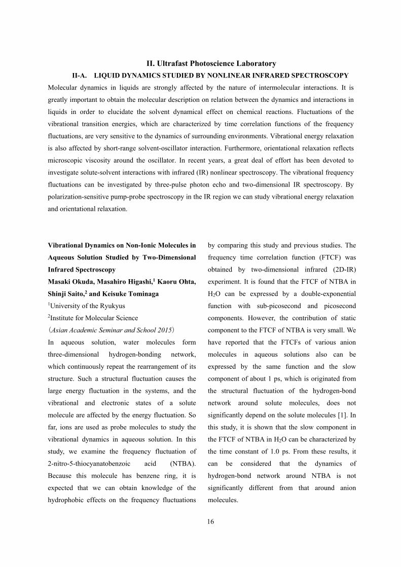
Vibrational Dynamics on Non-Ionic Molecules in
Aqueous Solution Studied by Two-Dimensional
Infrared Spectroscopy
Masaki Okuda, Masahiro Higashi,1 Kaoru Ohta,
Shinji Saito,2 and Keisuke Tominaga
1University of the Ryukyus 2Institute for Molecular Science
(Asian Academic Seminar and School 2015)
In aqueous solution, water molecules form
three-dimensional hydrogen-bonding network,
which continuously repeat the rearrangement of its
structure. Such a structural fluctuation causes the
large energy fluctuation in the systems, and the
vibrational and electronic states of a solute
molecule are affected by the energy fluctuation. So
far, ions are used as probe molecules to study the
vibrational dynamics in aqueous solution. In this
study, we examine the frequency fluctuation of
2-nitro-5-thiocyanatobenzoic acid (NTBA).
Because this molecule has benzene ring, it is
expected that we can obtain knowledge of the
hydrophobic effects on the frequency fluctuations
by comparing this study and previous studies. The
frequency time correlation function (FTCF) was
obtained by two-dimensional infrared (2D-IR)
experiment. It is found that the FTCF of NTBA in
H2O can be expressed by a double-exponential
function with sub-picosecond and picosecond
components. However, the contribution of static
component to the FTCF of NTBA is very small. We
have reported that the FTCFs of various anion
molecules in aqueous solutions also can be
expressed by the same function and the slow
component of about 1 ps, which is originated from
the structural fluctuation of the hydrogen-bond
network around solute molecules, does not
significantly depend on the solute molecules [1]. In
this study, it is shown that the slow component in
the FTCF of NTBA in H2O can be characterized by
the time constant of 1.0 ps. From these results, it
can be considered that the dynamics of
hydrogen-bond network around NTBA is not
significantly different from that around anion
molecules.

17
Figure 1. (a) The 2D-IR spectra of CN stretching
mode of NTBA in H2O at population time T = 0 ps.
The black line in the spectrum indicates centre line
of the spectrum. (b) The CLS curve of NTBA in
H2O is plotted against population time T. The curve
is proportional to the frequency time-correlation
function of the vibrational mode [2]. (Blue)
Experimental data, (Red) Calculated data.
[1] K. Ohta et al., Acc. Chem. Res., 45, 1982 (2012).
[2] K. Kwak et al., J. Chem. Phys., 127, 124503
(2007).
Vibrational Dynamics of Nitrosyl Stretch of Ru
Complex in Aqueous Solution Studied by
Two-Dimensional Infrared Spectroscopy
Kaoru Ohta, Kyoko Aikawa, and Keisuke
Tominaga
(Ultrafast Phenomena 2014)
To understand the molecular origin of vibrational
dynamics in detail, we chose the NO stretching
mode of [RuCl5(NO)]2- as a vibrational probe. Here
we investigated the temperature dependence of the
vibrational frequency fluctuation of the NO
stretching mode in aqueous solution and compared
with our previous studies of a couple of different
ionic probe molecules. Temperature dependence of
the vibrational dynamics of the solute in solution
provides the systematic information about the
coupling between the vibrational mode of the solute
and the bath degrees of the system.
To quantify the frequency-frequency
correlation functions of vibrational transitions, we
used two-dimensional IR spectroscopy. Home-built
optical parametric amplifier and difference
frequency generators were used to produce a
mid-IR pulse at around 1900 cm-1. 2D IR spectra
were measured with a collinear pulse pair
pump-probe geometry. From the FT-IR spectra of
the NO stretching mode of [RuCl5(NO)]2-, the
peaks of the absorption spectrum are located at
around 1882 cm-1 in D2O and H2O. We measured
the vibrational population relaxation for this mode
by ultrafast IR pump-probe spectroscopy. We found
that vibrational relaxation takes place on 31 ps in
D2O and 7.7 ps in H2O, respectively. Similar fast
relaxations in H2O were observed for the other
vibrational modes such as anti-symmetric stretching
modes of N3- and SCN- [2]. This is because the
vibrational bands at around 1900 cm-1 couples
18
strongly with a combination band of the bending
and librational modes of H2O. Spectral overlap of
the solvent vibrational modes enhances the
vibrational energy transfer from solute to solvent.
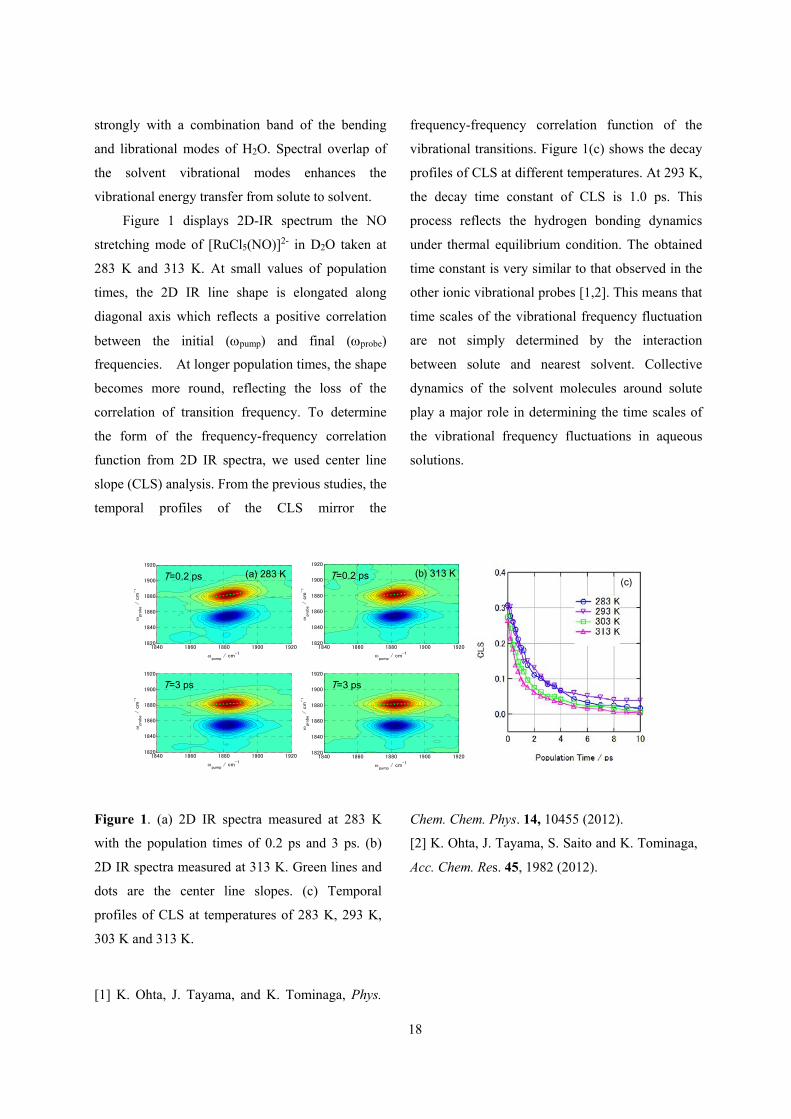
Figure 1 displays 2D-IR spectrum the NO
stretching mode of [RuCl5(NO)]2- in D2O taken at
283 K and 313 K. At small values of population
times, the 2D IR line shape is elongated along
diagonal axis which reflects a positive correlation
between the initial (pump) and final (probe)
frequencies. At longer population times, the shape
becomes more round, reflecting the loss of the
correlation of transition frequency. To determine
the form of the frequency-frequency correlation
function from 2D IR spectra, we used center line
slope (CLS) analysis. From the previous studies, the
temporal profiles of the CLS mirror the
frequency-frequency correlation function of the
vibrational transitions. Figure 1(c) shows the decay
profiles of CLS at different temperatures. At 293 K,
the decay time constant of CLS is 1.0 ps. This
process reflects the hydrogen bonding dynamics
under thermal equilibrium condition. The obtained
time constant is very similar to that observed in the
other ionic vibrational probes [1,2]. This means that
time scales of the vibrational frequency fluctuation
are not simply determined by the interaction
between solute and nearest solvent. Collective
dynamics of the solvent molecules around solute
play a major role in determining the time scales of
the vibrational frequency fluctuations in aqueous
solutions.
pump
/ cm-1
pr
obe
/ c
m-1
1840 1860 1880 1900 19201820
1840
1860
1880
1900
1920
pump
/ cm-1
pr
obe
/ c
m-1
1840 1860 1880 1900 19201820
1840
1860
1880
1900
1920
(a) 283 KT=0.2 ps
T=3 ps
pump
/ cm-1
pr
obe
/ c
m-1
1840 1860 1880 1900 19201820
1840
1860
1880
1900
1920
pump
/ cm-1
pr
obe
/ c
m-1
1840 1860 1880 1900 19201820
1840
1860
1880
1900
1920
(b) 313 KT=0.2 ps
T=3 ps
(c)
Figure 1. (a) 2D IR spectra measured at 283 K
with the population times of 0.2 ps and 3 ps. (b)
2D IR spectra measured at 313 K. Green lines and
dots are the center line slopes. (c) Temporal
profiles of CLS at temperatures of 283 K, 293 K,
303 K and 313 K.
[1] K. Ohta, J. Tayama, and K. Tominaga, Phys.
Chem. Chem. Phys. 14, 10455 (2012).
[2] K. Ohta, J. Tayama, S. Saito and K. Tominaga,
Acc. Chem. Res. 45, 1982 (2012).

19
Solute-solvent interactions of benzonitrile in
solutions studied by sub-picosecond infrared
pump-probe spectroscopy
Motohiro Banno, Ayumi Kotani, Kaoru Ohta, and
Keisuke Tominaga
(Bull. Chem. Soc. Jpn., 2014)
The vibrational energy relaxation of the CN
stretching mode of benzonitrile (BN) in solution has
been investigated by infrared (IR) pump-probe
spectroscopy. The peak wavenumber, lineshape, and
cross section of the ground state absorption band due
to the CN stretching mode depend on the solvent and
concentration. From the experimental results of IR
spectroscopic methods and calculations with the
density functional theory, the structures of
solute-solute or solute-solvent complexes are
suggested. In hexane, BN exists as the monomer or
the dimer in which the two CN groups are arranged
in the anti-parallel way. In ethanol (EtOH), it is
suggested that BN forms the anti-parallel dimer and a
BN-EtOH hydrogen-bonded complex. In
dimethylsulfoxide (DMSO), BN probably forms a
solute-solvent complex. From the results of IR
pump-probe spectroscopy, the decay time constant of
the pump-probe signal shows a probe wavenumber
dependence in hexane and DMSO, which indicates
inhomogeneity of the microscopic environment
around BN in these solvents. In EtOH, the
wavenumber dependence was not observed. The
result suggests that the VER processes for the BN
dimer and the BN-EtOH complex are accelerated
from that for the monomer, and the VER times are
almost identical among the two structures.
II-B. DYNAMICS OF ELECTRONICALLY EXCITED STATE IN CONDENSED PHASES
Understanding of dynamics in the electronically excited state is a key issue to elucidate mechanisms in
various photochemical reactions in condensed phases. It is also important for designing and developing
new materials which have characteristic functions. We employ various kinds of ultrafast technique to
monitor photochemical and photophysical invents in sub-pico- to picoseconds time scales. By femtosecond
fluorescence up-conversion technique, dynamics in the electronically excited state can be observed with a
time resolution up to 100 fs. Vibrational dynamics in the electronically excited can be investigate by
UV/VIS-pump IR probe technique. Moreover, low-frequency responses by photoexcitation are investigated
by UV/VIS-pump THz probe experiment. Such responses include change of low-frequency vibrational
modes induced by photoexcitation and photo-induced changes of charge carrier dynamics.
Vibrational Dynamics of the CO Stretching of
9-Fluorenone Studied by Visible-pump and
Infrared-probe Spectroscopy
Yuki Fukui, Kaoru Ohta, and Keisuke Tominaga
(Faraday Discussion)
In a solution, a solute molecule relaxes to the most
stable state of the electronically excited state (S1)
after the electronic excitation through various

20
relaxation processes. These processes are, for
example, a vibrational energy relaxation, a cooling
process of the locally heated environment around
the solute, and solvation dynamics. They influence
chemical reactions initiated by the electronic
excitation. In a protic solvent, a solute molecule
forms intermolecular hydrogen bonds with
solvents if the solute has hydrogen bonding sites
such as a carbonyl group. The hydrogen bonds
influence various properties of solute such as its
electronic states, the relaxation processes, and so
on. 9-Fluorenone (FL) has a carbonyl group which
can form hydrogen bonds. The vibrational
dynamics of FL in alcohol solutions are previously
reported in the electronically ground state and
excited states. In this study, we studied effects of
hydrogen bond on the vibrational structures and
the vibrational dynamics of the CO stretching
mode of the excited state FL in various solvents
including alcohol by sub-picosecond visible-pump
and IR-probe spectroscopy.
We measured the visible-pump and IR-probe
signals in various solvents including alcohol. The
transient IR spectrum of the CO stretching band in
methanol-d4 has peaks at 1530 cm-1 and1555 cm-1.
We assigned the two bands at 1530 cm-1 and1555
cm-1 to a FL complex with solvents and free FL,
respectively. In all the solvents, the CO stretching
bands show blue-shifts in time. A possible
mechanism for this blue-shift is vibrational cooling.
Vibraional cooling is derived from anharmonic
couplings by some low frequency modes. This
cooling process of the locally heated environment
around the solute influences the blue-shifts.
However, red-shift is observed at later time for the
band at 1530 cm-1 in methanol-d4. A possible
mechanism of this spectral shift is reorganization of
solvent which form hydrogen bonds with FL.
Vibrational Dynamics of the CN Stretching in
the Electronically Excited State by Visible-Pump
and Infrared-Probe Spectroscopy
Sho Hiraoka, Kaoru Ohta, and Keisuke
Tominaga
(Ultrafast Phenomena 2014)
Understanding of mechanisms of photochemical
reactions in solution is one of the important
problems in current chemistry. After
photoexcitation to the electronically exited state of
a solute molecule in solution, various relaxation
processes occur such as vibrational energy
relaxation or solvation dynamics. The fate of the
excited state is strongly influenced by these
relaxation processes, therefore, it is very important
to understand these relaxation processes in detail.
On the other hand, hydrogen-bonding liquids such
as water and alcohol form characteristic network
structures, which affect various properties of a
solute molecule such as the electronic state and
vibrational structures. In this work, we focus on the
vibrational dynamics of a solute in the
electronically excited state in alcohol solutions. The
vibrational mode we investigate is the CN
stretching mode of 3-cyano-7-hydroxy-4-methyl
coumarin (C183m, Figure 1). Generally, coumarin
dyes change their permanent dipole moments
largely by photoexcitation, causing change of the
dielectric interaction between the dyes and
surrounding solvents. We have carried out
visible-pump and infrared (IR)-probe spectroscopic
measurements on this mode to examine influence of

21
the solvent interactions on the vibrational dynamics
of the solute.
Figure 1. Structure of C183m.
The transient IR spectra of the CN stretching
band are shown in Figure 2. There are a bleach
centered at 2225 cm-1 and a transient absorption at
2180 cm-1, and we fit the transient spectra with a
sum of two Gaussian functions to determine the
peak wavenumber of the transient absorption
component. The peak of the CN stretching mode
shows a blue shift with delay time, and the
time-dependence of this blue shift is reproduced
well by a single exponential with a time constant
of about 10 ps. One of the possible mechanisms for
this blue shift is vibrational cooling. Vibrational
cooling resulted from anharmonic couplings
between the high-frequency mode and some
low-frequency modes. Just after the
photoexcitation the local temperature around the
solute is higher than the room temperature because
of the dissipation of the excess energy, causing
population in the higher vibrational states of the
low-frequency modes. This vibrational cooling has
been observed in several transient vibrational
spectroscopic studies [1,2]. Another possible
mechanism for the blue shift is solvation dynamics.
If the photoexcitation accompanies a large change
of the dipole moment of the solute, dielectric
response from the polar solvent causes the
stabilization of the electronic and vibrational states
of the solute. Depending on the directions of the
permanent dipole moments of the S0 and S1 states
and the charge distribution in the normal modes,
the peak wavenumber shifts either the blue side or
red side during the solvation dynamics. We further
carried out UV-pump and IR-probe measurement,
where the third harmonics at 266 nm was used as a
pump pulse. In this case the excess energy is about
12450 cm-1, which is much larger than that of the
second harmonic excitation (1420 cm-1). The
transient absorption spectrum at t=0 ps is further
blue-shifted compared to the second harmonic
excitation case, and the time-dependence of the
blue shift can be described by a similar time
constant to that of the vis-pump case. These results
suggest that the blue shift is caused by vibrational
cooling rather than solvation dynamics, since the
total shift due to vibrational cooling depends on
the excess energy whereas that due to solvation
dynamics does not depend on it so much.
1.2
1.0
0.8
0.6
0.4
0.2
0.0
-0.2
-0.4
Abs
orb
ance
/ m
O.D
.
2300225022002150
Wavenumber /cm-1
5 ps
1 ps
10 ps
20 ps
30 ps
40 ps
Figure 2. Transient absorption spectra of C183m in
CH3OH at different delay times.
[1] P. Hamm, S. M. Ohline, and W. Zinth, J. Chem.
Phys. 106, 519 (1997).

22
[2] K. Iwata and H. Hamaguchi, J. Phys. Chem. A, 101, 632 (1997).
Time-Resolved Fluorescence Spectroscopy Study
of Excited State Dynamics of Alkyl- and
Benzo-Substituted Triphyrin(2.1.1)
Yusuke Iima, Daiki Kuzuhara,1 Zhao-Li Xue,1
Seiji Akimoto, Hiroko Yamada1 and Keisuke
Tominaga 1Nara Institute of Science and Technology
(Phys. Chem. Chem. Phys., 2014)
We have investigated the photophysical properties
of alkyl-substituted triphyrin(2.1.1) (ATp) and
benzotriphyrin(2.1.1) (BTp) by steady-state and
time-resolved fluorescence spectroscopy. We
focused on the effect of NH proton tautomerization,
planarity of the macrocycles, and substituents on
these properties. The fluorescence quantum yields
(Φy) of ATp did not depend on solvent viscosity,
whereas those of BTp increased with solvent
viscosity, reaching a maximum value of 0.17 in
paraffin. Interestingly, analyzing y showed that the
non-radiative rate constant of BTp decreased
sharply as the solvent viscosity increased. These
results suggest that the substituted phenyl groups
play a crucial role in suppressing molecular
distortion, thus leading to decreased non-radiative
relaxation in triphyrin(2.1.1). The hydrogen bond
formed in the inner cavity potentially contributes to
the suppression of the structural distortion, whereas
the pyrrole rings in the macrocycle are close, as in
porphycene.
II-C. MOLECULAR DYNAMICS IN THE TERAHERTZ FREQUENCY REGION IN
CONDENSED PHASES
Vibrational spectroscopy has been widely used to investigate structures, interactions and dynamics of
molecules and molecular complexes. The low-frequency region below several terahertz (THz; 1 THz = 33.3
cm-1) corresponds to intermolecular modes of complexes and intramolecular modes with a weaker potential
force and/or larger reduced mass. Intermolecular interactions such as hydrogen bonding, van der Waals forces
and charge-transfer interactions play important roles in various chemical and biological processes. Moreover,
the low-frequency spectra also reflect molecular dynamics on a time scale from picoseconds to femtoseconds.
There has been dramatic progress in the generation and detection techniques of freely propagating THz
radiation in the past two decades. The examples of the generation technique include photoconductive
switching, optical rectification, and the surface photocurrent of semiconductors. Because the pulse duration of
the THz radiation is in a sub-picosecond time region, it is possible to measure the electric field of the
radiation by coherent detection methods, which consequently allows us to conduct THz time-domain
spectroscopy (TDS). By THz-TDS we can obtain the refractive index and extinction coefficient of a medium
by measuring the phase and amplitude of the radiation. THz-TDS is an attractive method for studying
dynamics in condensed phases with time scales of sub-picoseconds and picoseconds. We have applied

23
THz-TDS to investigate various kinds of condensed materials, including neat liquids and mixtures of liquids,
biological polymers, and charge carrier dynamics in semiconductors and conducting polymers.
Low-frequency dynamics of trehalose-coated
lysozyme studied by terahertz time-domain
spectroscopy
Risa Okada, Naoki Yamamoto1, Atsuo Tamura1,
Keisuke Tominaga 1Kobe University
(ASUD 2014)
Under the extreme conditions such as highly dried
state or very low temperature bioploymers like
proteins usually denature, leading to damage on
organisms. However, some organisms can survive
under such extreme conditions without suffering
any damage. To protect themselves, these
organisms in the dehydrated state contain large
amount of sugar, particularly trehalose. Since
viscosity of trehalose is very high, it prevents
structural changes of biopolymers. When protein
changes its structure, large-amplitude motions in
the low-frequency region are often activated. Such
motions have collective nature and correspond to
low-frequency motions below a hundred
wavenumbers. In this study, we measured the
low-frequency spectra of proteins coated with
trehalose from 83 K to 353 K by terahertz
time-domain spectroscopy (THz-TDS) to study
thermal activation of the low-frequency dynamics
affected by sugar coating.
In THz-TDS system, a pair of
photoconductive antenna was used for generation
and detection of THz electro-magnetic wave. The
sample is trehalose, lysozyme, and
trehalose-coated lysozyme. Other disaccharides
except trehalose (maltose and sucrose) were also
measured. Each of these lyophilized samples in a
cell was pressed at a pressure of 8 Mpa to make
pellet samples.
Trehalose is crystallized by heating. The
absorption coefficient spectra of trehalose are
different from trehalose-coated lysozyme when
heated. THz absorption coefficients increase
linearly with temperature. At around 300 K, the
slope of the absorption coefficient of
trehalose-coated lysozyme becomes larger. This
temperature dependence of trehalose-coated
lysozyme by THz-TDS matches with the
calorimetric glass transition. We measured
complex dielectric constant from GHz to THz by
network analyzer (0.2 ~20 GHz) and THz-TDS
(0.3 ~2 THz) in order to reveal the motions around
glass transition. And we reproduced dielectric
spectra in the sum of Cole-Cole relaxation modes
and underdamped vibrational modes. In the result,
THz dielectric spectra include not relaxation
modes but underdamped vibrational modes. So
THz dielectric spectra of each temperature are
reproduced by two underdamped vibrational
modes. We discuss relation between temperature
dependence of low frequency dynamics and each
of motion.

24
The Low-Frequency Vibration Study of Amino
Acids Using Terahertz Spectroscopy and
Solid-state Density Functional Theory
Feng Zhang, Keisuke Tominaga, Michitoshi
Hayashi1, and Houng-Wei Wang1 1National Taiwan University
(SPIE Photonics Asia)
Understanding the low-frequency normal modes of
amino acids, the building blocks of proteins, is
crucial to reveal the vibration-function relationship in
the macromolecular system. Recent advances in
terahertz spectroscopy and solid-state density
functional theory have ensured the accurate access to
low-frequency modes of amino acids. A new
knowledge people have learnt so far is that the inter-
and intra-molecular vibrations are strongly coupled
with each other in the low-frequency (or THz) region
through the vibrational coordinate mixing. Rich
information is believed embedded in this
phenomenon [1,2].
We will introduce an analytical
mode-decoupling method that allows for the accurate
decomposition of a normal mode of interest into the
three intermolecular translations, three principal
librations and various intrinsic intramolecular
vibrations. We will demonstrate a theoretical analysis
by using the L-alanine system as an example. The
mode-decoupling method helps reveal new
intramolecular vibrational modes on the first hand,
and more importantly, shed light on a new
phenomenon of the intramolecular vibrational
coordinate distribution (IVCD). IVCD describes the
broad distributions of important intramolecular
normal modes such as NH3+ torsion, COO- torsion
and CH3 torsion.
The IVCD concept may imply solutions to a
string of unsettled problems relevant to the
low-frequency molecular vibrations, e.g. the
formation mechanism of the peptide bonds, the
explanation of the line shape of CH3 tunneling in the
small molecular system as well as the first onset of
anharmonicity of CH3 rotation in the protein system.
1. F. Zhang, O. Kambara, K. Tominaga, J. Nishizawa,
T. Sasaki, H.-W. Wang, and M. Hayashi, RSC Adv., 4,
269-278, (2014).
2. F. Zhang, M. Hayashi, H.-W. Wang, K. Tominaga,
O. Kambara, J. Nishizawa, and T. Sasaki, J. Chem.
Phys. 140, 174509 (2014).
Terahertz spectroscopy and solid-state density
functional theory calculation of anthracene: effect
of dispersion force on the vibrational modes
Feng Zhang, Michitoshi Hayashi1, Houng-Wei
Wang1, Keisuke Tominaga, Ohki Kambara2,
Jun-ichi Nishizawa3, and Tetsuo Sasaki2
1National Taiwan University
2Shizuoka University 3Tohoku University
(J. Chem. Phys., 2014)
The phonon modes of molecular crystals in the
terahertz frequency region often feature delicately
coupled inter- and intra-molecular vibrations. Recent
advances in density functional theory such as DFT-D*
have enabled accurate frequency calculation.
However, the nature of normal modes has not been
quantitatively discussed against experimental criteria
such as isotope shift (IS) and correlation field
splitting (CFS). Here, we report an analytical
mode-decoupling method that allows for the

25
decomposition of a normal mode of interest into
intermolecular translation, libration, and
intramolecular vibrational motions. We show an
application of this method using the crystalline
anthracene system as an example. The relationship
between the experimentally obtained IS and the IS
obtained by PBE-D* simulation indicates that two
distinctive regions exist. Region I is associated with a
pure intermolecular translation, whereas region II
features coupled intramolecular vibrations that are
further coupled by a weak intermolecular translation.
We find that the PBE-D* data show excellent
agreement with the experimental data in terms of IS
and CFS in region II; however, PBE-D* produces
significant deviations in IS in region I where strong
coupling between inter- and intra-molecular
vibrations contributes to normal modes. The result of
this analysis is expected to facilitate future
improvement of DFT-D*.
Intramolecular Vibrations in Low-Frequency
Normal Modes of Amino Acids: L-Alanine in Neat
Solid State
Feng Zhang, Houng-Wei Wang1, Keisuke
Tominaga, and Michitoshi Hayashi1 1National Taiwan University
(J. Phys. Chem. A, in press)
This paper presents a theoretical analysis of the
low-frequency phonons of L-alanine by using the
solid-state density functional theory at the gamma
point. We are particularly interested in the
intramolecular vibrations accessing low-frequency
phonons via vibration mixing with intermolecular
vibrations. A new mode-analysis method is
introduced to quantify the vibrational characteristics
of such intramolecular vibrations. We find that the
torsional motions of COO− are involved in
low-frequency phonons, although COO− is
conventionally assumed to undergo localized torsion.
We also find the broad distributions of intramolecular
vibrations relevant to important functional groups of
amino acids, e.g., the COO− and NH3+ torsions, in the
low-frequency phonons. The latter finding is
illustrated by the concept of frequency distribution of
vibrations. These findings may lead to immediate
implications in other amino acid systems.
Conduction in Polyaniline Emeraldine Salt in the
Terahertz Region: A Temperature Dependence
Study
Alvin Karlo G. Tapia and Keisuke Tominaga
(Chem. Phys. Lett., 2014)
The temperature-dependent conductivity of
polyaniline emeraldine salt (PAni-ES) was studied by
terahertz (THz) time-domain spectroscopy from 80 to
290 K to investigate conduction properties in the THz
region. The absorption coefficient and index of
refraction increase with temperature. This reflects an
increasing conductivity, which indicates a thermally
assisted hopping transition. The frequency-dependent
behavior of the conductivity is described by using the
Mott-Davis model. The model fitting parameter, S,
decreases with increasing temperature, indicating a
possible correlated barrier hopping mechanism.
Lastly, the activation energy at THz frequencies
decreases with increasing frequency, suggesting
intraband transitions.

26
Temperature and Hydration Dependence of
Complex Dielectric Spectra of Lysozyme from
GHz to THz Frequency Region
Naoki Yamamoto, Atsuo Tamura1, and Keisuke
Tominaga 1Kobe University
(8th International Conference on Broadband
Dielectric Spectroscopy and its Applications)
Understanding of structures and dynamics of
proteins is a fundamental problem for studies on
protein function. When protein expresses the
function, large structural changes often occur.
These changes are induced by the collective
motions of many atoms in protein, corresponding
to the low-frequency motion below a few tens of
wavenumbers. Therefore, the low-frequency
spectra of protein contain information on the
motions relevant to the function of protein. In this
study we obtained and analyzed complex dielectric
spectra in the THz region. We studied temperature
and hydration dependence on the dielectric spectra
of lysozyme from GHz to terahertz (THz)
frequency regions.
5
4
3
2
1
0
',
''
6040200
Wavenumber/cm-1
83 K 133 K 183 K 243 K 293 K
h = 0.11
6040200
Wavenumber/cm-1
h = 0.34
Figure 1. Temperature dependence of complex
dielectric spectra of the dehydrated state (left) and a
hydrated state (right), respectively. In each panel the
upper and the lower groups correspond to the real
parts and the imaginary parts, respectively. The
hydration degree, which is defined by the value of h
(weight of water divided by weight of protein) is
shown in each figure.
In both dehydrated and hydrated states the complex
dielectric spectra linearly increase as temperature rise
below 180 K as shown in Figure 1. However, at
higher temperatures the spectral components of the
dielectric spectra increase more in the hydrated state
than the dehydrated state. We performed spectral
analysis of the complex dielectric spectra, )( ,
using some model functions such as a sum of an

27
underdamped mode and a Cole-Cole function. We
discuss temperature dependencies of these parameters
obtained from the analysis.
Temperature and Hydration Dependence of
Low-Frequency Spectra of Lipid Bilayers Studied
by Terahertz Time-Domain Spectroscopy
Naoki Yamamoto, Tomoyo Andachi, Atsuo
Tamura1, and Keisuke Tominaga 1Kobe University
(J. Phys. Chem. B, in press)
We have studied temperature and hydration
dependent low-frequency spectra of lipid bilayers of
1,2-dimyristoyl-sn-glycero-3-phosphoryl-3’-rac-glyc
erol (DMPG) and 1,2-dimyristoyl-sn-glycero-
3-phosphocholine (DMPC) by terahertz time-domain
spectroscopy (THz-TDS). We measured X-ray
diffraction patterns and mid-infrared spectra of these
lipid bilayers and found that the lipid bilayer have
two different types of phases, i.e. the gel phase and
the crystalline phase, depending on preparation
methods of the samples. In both the phases a few
distinct bands were observed in the THz region. For
DMPG the peak wavenumbers of the absorption
bands did not change upon hydration, while the
bandwidth in the crystalline phase was smaller than
that in the gel phase. We performed spectral analyses
for the complex dielectric spectra for DMPG and
DMPC with a model function, mainly to determine
the peak wavenumbers of the absorption bands. In
contrast to the case of the DMPG bilayers the peak
wavenumber of the absorption band of the DMPC
bilayer shifts upon hydration. In the hydrated DMPC
bilayer it was suggested fast reorienting water
molecules exist with a relaxation time of
sub-picoseconds. It is suggested that the THz
absorption patterns reflect the lipid packing pattern in
the bilayers. The temperature dependence of the
absorption band was analyzed by an empirical
equation, and the anharmonicity of the vibrational
potential of the low-frequency mode was
quantitatively evaluated.
28
II-D. EXCITATION RELAXATION DYNAMICS OF PHOTOSYNTHETIC SYSTEMS
STUDIED BY TIME-RESOLVED FLUORESENCE SPECTROSCOPY
In photosynthetic systems, solar energy is captured by antenna pigments, such as chlorophylls, carotenoids
and phycobiliproteins, which is followed by the energy transfer among photosynthetic pigments and the
electron transfer in the reaction centers. It is, therefore, of great importance for the clear understanding of
primary processes in photosynthetic reactions to detect signals that reflect ultrafast excited state dynamics
after an excitation of pigments. By using short pulses from a Ti:Sappire laser as light sources for measurements
of time-resolved fluorescence spectra in femtosecond to nanosecond region, ultrafast processes in
photosynthetic systems are examined.
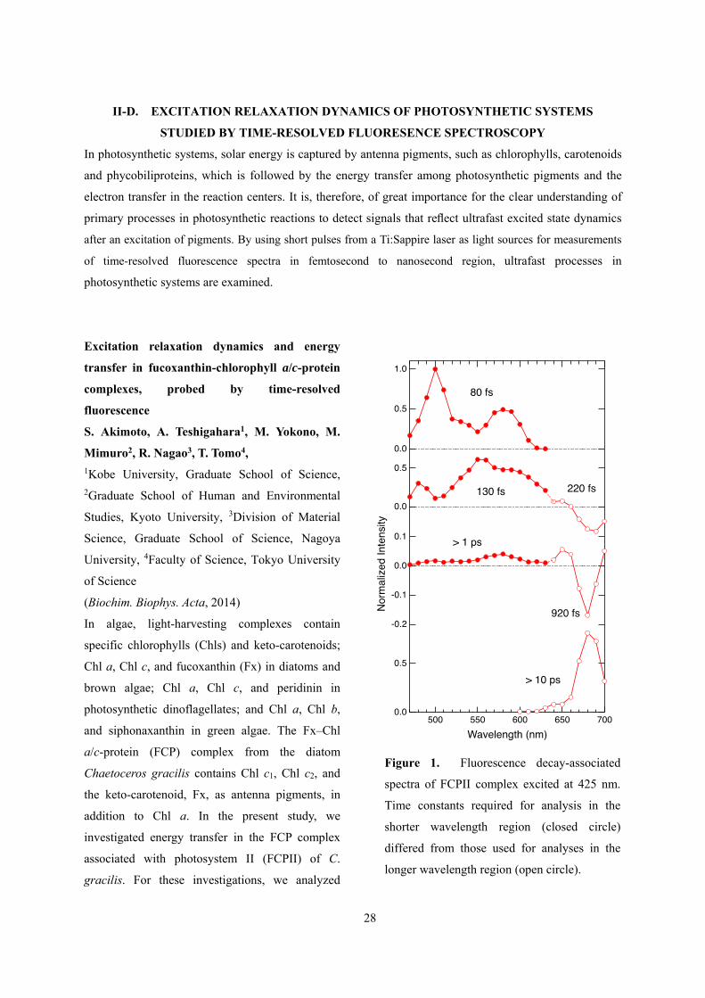
Excitation relaxation dynamics and energy
transfer in fucoxanthin-chlorophyll a/c-protein
complexes, probed by time-resolved
fluorescence
S. Akimoto, A. Teshigahara1, M. Yokono, M.
Mimuro2, R. Nagao3, T. Tomo4, 1Kobe University, Graduate School of Science, 2Graduate School of Human and Environmental
Studies, Kyoto University, 3Division of Material
Science, Graduate School of Science, Nagoya
University, 4Faculty of Science, Tokyo University
of Science
(Biochim. Biophys. Acta, 2014)
In algae, light-harvesting complexes contain
specific chlorophylls (Chls) and keto-carotenoids;
Chl a, Chl c, and fucoxanthin (Fx) in diatoms and
brown algae; Chl a, Chl c, and peridinin in
photosynthetic dinoflagellates; and Chl a, Chl b,
and siphonaxanthin in green algae. The Fx–Chl
a/c-protein (FCP) complex from the diatom
Chaetoceros gracilis contains Chl c1, Chl c2, and
the keto-carotenoid, Fx, as antenna pigments, in
addition to Chl a. In the present study, we
investigated energy transfer in the FCP complex
associated with photosystem II (FCPII) of C.
gracilis. For these investigations, we analyzed
Figure 1. Fluorescence decay-associated
spectra of FCPII complex excited at 425 nm.
Time constants required for analysis in the
shorter wavelength region (closed circle)
differed from those used for analyses in the
longer wavelength region (open circle).

29
time-resolved fluorescence spectra, fluorescence
rise and decay curves, and time-resolved
fluorescence anisotropy data. Chl a exhibited
different energy forms with fluorescence peaks
ranging from 677 nm to 688 nm. Fx transferred
excitation energy to lower-energy Chl a with a
time constant of 300 fs. Chl c transferred excitation
energy to Chl a with time constants of 500–600 fs
(intra-complex transfer), 600–700 fs
(intra-complex transfer), and 4–6 ps (inter-complex
transfer). The latter process made a greater
contribution to total Chl c-to-Chl a transfer in
intact cells of C. gracilis than in the isolated FCPII
complexes. The lower-energy Chl a received
excitation energy from Fx and transferred the
energy to higher-energy Chl a.
Energy transfer processes in chlorophyll
f-containing cyanobacteria using time-resolved
fluorescence spectroscopy on intact cells
T. Tomo1, T. Shinoda1, M. Chen2, S. I.
Allakhverdiev3,4, S. Akimoto 1 Faculty of Science, Tokyo University of Science, 2School of Biological Sciences, University of
Sydney, 3Institute of Plant Physiology, Russian
Academy of Science, 4Institute of Basic Biological
Problems, Russian Academy of Sciences
(Biochim. Biophys. Acta, 2014)
We examined energy transfer dynamics in the
unique chlorophyll (Chl) f-containing
cyanobacterium Halomicronema hongdechloris.
The absorption band of Chl f appeared during
cultivation of this organism under far-red light.
The absorption maximum of Chl f in organic
solvents occurs at a wavelength of approximately
40 nm longer than that of Chl a. In vivo, the cells
display a new absorption band at approximately
730 nm at 298 K, which is at a significantly longer
wavelength than that of Chl a. We primarily
assigned this band to a long wavelength form of
Chl a. The function of Chl f is currently unknown.
We measured the fluorescence of cells using
time-resolved fluorescence spectroscopy in the
picosecond-to-nanosecond time range and found
clear differences in fluorescence properties
between the cells that contained Chl f and the cells
that did not. After excitation, the fluorescence
peaks of photosystem I and photosystem II
Figure 1. Time resolved fluorescence spectra (TRFS)
of H. hongdechloris cells. Left side: normalized TRFS
of the cells cultured under white light. Right hand:
normalized TRFS of the cells under far-red light.

30
appeared quickly but diminished immediately. A
unique fluorescence peak located at 748 nm
subsequently appeared in cells containing Chl f.
This finding strongly suggests that in this alga, Chl
f exists in both photosystems and that Chl f is
located close to Chl a.

31
III Coherent Photoscience Laboratory
III-A. HIGH FIELD ELECTRON SPIN RESONANCE (ESR) STUDIES OF QUANTUM SPIN
SYSTEMS
Quantum spin system is a magnetic system which shows distinct quantum effects due to its strong quantum
fluctuation. Recently, quasi-one dimensional quantum spin systems have attracted much interest, and their
short range order and their ground state at low temperature should be clarified. High frequency high field
ESR turns out to be a powerful means to observe the short-range order and the ground state of the system.
Moreover, frustrated magnetic systems have also attracted much interest recently. The frustration in the
magnetic system suppresses the conventional magnetic order and a unique ground state appears at low
temperature. Especially the competition is expected between the frustration and low dimensionality.
Following the trends from a Grant-in-Aid for Scientific Research on Priority Areas “Novel states of matter
induced by frustration” (No.473, 2007-2011, Headed by Prof. H. Kawamura (Osaka University) and H.
Ohta was a member), we are studying these low dimensional antiferromagnets with frustration and related
multiferroic materials intensively. High Field ESR of multiferroic materialYCrO3 has been performed and
non-conventional AFMR modes, which cannot be interpreted by the molecular field theory, were observed.
The master course student S. Ikeda, who presented the ESR study of YCrO3, received the Excellent
Presentation Award at Young Frontier Meeting organized by the Center for Supports to Research and
Education Activities, Kobe University, and the Best Presentation Award for the Physics Master Course
Presentation at Kobe University. H. Ohta gave an invited presentations at International Conference
“Magnetic Resonance: fundamental research and pioneering applications (MR-70)” (June 23-27, 2014,
Kazan, Russia), “International Symposium Catalytic Systems for Chemical Energy Conversion” (July
23-25, 2014, Mülheim an der Ruhr, Germany), and CONIAPS XVII (January 16-18, 2015, Jaipur, India),
and introduced the recent results of high field ESR in Kobe. Especially H. Ohta received the Fellowship
Award for his contribution to the developments of high field THz ESR from the International Academy of
Physical Sciences during CONIAPS XVII. H. Ohta (Chair), S. Okubo (Secretary), E. Ohmichi, T. Sakurai,
S. Hara have organized the joint symposium APES-IES-SEST2014 (Nov. 12-16, 2014, Nara, Japan). The
symposium was rather successful with 279 participants from 22 countries which turned out to be a record
for APES symposium. In meantime H. Ohta served as the Vice-President of IES (International EPR(ESR)
Society) and was appointed as the President of IES since January, 2015. H. Ohta is also acting as the
Vice-President of APES (Asia-Pacific EPR/ESR Society), the Secretary General of the Japan Society of
Infrared Science and Technology, and the Council Member of SEST. Moreover, in order to strengthen the
pulsed magnetic field researches in western Japan region, we have set up The KOFUC (Kobe-Osaka-Fukui
Universities Centers) Network, which is intended to strengthen the pulsed magnetic field researches in
western Japan region, was approved by the High Magnetic Field Forum of Japan in December, 2014.

32
Disappearance of Ising nature in Ca3ZnMnO6
studied by high-field ESRM
Y Ruan1,2, Z W Ouyang1, Y M Guo1,2, J J Cheng1,2,
Y C Sun1,2, Z C Xia1, G H Rao3, S Okubo4 and H
Ohta4 1Wuhan National High Magnetic Field Center,
Huazhong University of Science and Technology 2School of Physics, Huazhong University of
Science and Technology 3School of Materials Science and Engineering,
Guilin University of Electronic Technology 4Molecular Photoscience Research Center, Kobe
University
(J. Phys.: Condens. Matter 26, 2014)
High-field electron spin resonance measurements
of an antiferromagnet Ca3ZnMnO6 isostructure,
with the Ising-chain multiferroic Ca3ZnMnO6,
have been carried out. Two distinct resonance
modes were observed below TN = 25 K, which is
well explained by conventional antiferromagnetic
resonance theory with easy-plane anisotropy. The
zero-field spin gap is derived to be about 166 GHz,
originating from the easy-plane anisotropy and
exchange interaction.
Our result suggests that the
Dzyaloshinsky–Moriya interaction, which may
induce spin canting, is absent. Disappearance of
Ising anisotropy in Ca3ZnMnO6 suggests that the
Co4+ ion, as well as the Co–Mn superexchange,
plays an important role for the Ising nature in
Ca3ZnMnO6.
Groud state of the spin-1/2 chain of green dioptase
at high fields
Kazuki Matsui, Masashi Fujisawa1, Kenta
Hagiwara, Yukihiro Hoshino, Takayuki Goto*,
Takahiko Sasaki2, Hidekazu Tanaka3, Susumu
Okubo1, and Hitoshi Ohta1
Physics Division, Sophia University 1Molecular Photoscience Research Center, Kobe
University 2Institute for Materials Research, Tohoku University, 3Department of Physics, Tokyo Institute of
Technology
(JPS Conf Proc 3, 2014)
The gem-stone dioptase Cu6Si6O18·6H2O has a chiral
crystal structure of equilateral triangular helices
consisting of Cu-3d spins. It shows an
antiferromagnetic order with an easy axis along c at
TN = 14.5 K under zero field, and a magnetization
jump at HC= 13.5 T when the field is applied along
c-axis. By 29Si-NMR measurements, we have
revealed that the high-field state is essentially the two
sub-lattice structure, and that the component within
ab-plane is collinear. The result indicates no apparent
match with the geometrical pattern of helical spin
chain.

33
III-B. ESR, PL AND MAGNETIC PROPERTY MEASUREMENTS OF MAGNETIC
SEMICONDUCTORS
Rare-earth ions incorporated in semiconductors show the luminescence originated from intra-4f-shell
transition under electrical excitation or photo excitation of the host semiconductor. Especially, as the
wavelength 1.5 m lies in the minimum loss region of silica fibers, the photoluminescence (PL) from Er3+
is very important for applications. ESR measurements have been performed intensively in order to clarify
the functionality of various Er centers in GaAs:Er,O systems. Especially, we have shown that the
Er-concentration dependence of intensities of ESR absorption has a strong correlation with the
Er-concentration dependence of the photoluminescence (PL) intensities of GaAs:Er,O doped with carriers.
The doping effect turns out to be similar to the effect of Zn doping. The analyses of ESR suggest the existence of
exchange interaction between Er sites. This work is in close collaboration with Fujiwara group of Faculty of
Engineering, Osaka University. We have proposed a model for the PL and ESR results and Fatma Elmasry
received a Doctor Degree in March, 2015, for this work.
GdN thin film is studied by ESR, and the SQUID magnetometer described in III-D. SQUID
measurements and the Arrott plot analyses revealed that the Curie temperature of the film is about 32 K.
ESR measurements also showed clear ferromagnetic resonance (FMR) at 4.2 K and the analyses has been
performed. This work is in close collaboration with Kita group of Faculty of Engineering, Kobe University.
The paper by T. Shimokawa on FMR appeared in J. Appl. Phys.
Electron spin resonance study of Er-concentration
effect in GaAs;Er,O containing charge carriers
F. Elmasry1, S. Okubo2, H. Ohta1,2, and Y.
Fujiwara3
1Graduate School of Science, Kobe University
2Molecular Photoscience Research Center, Kobe
University, 3Graduate School of Engineering, Osaka
University
(J. Appl. Phys. 115, 2014)
Er-concentration effect in GaAs;Er,O containing
charge carriers (n-type, high resistance, p-type)
has been studied by X-band Electron spin
resonance (ESR) at low temperature (4.7 K < T <
18 K). Observed A, B, and C types of ESR signals
were identical to those observed previously in
GaAs:Er,O without carrier. The local structure
around Er-2O centers is not affected by carriers
because similar angular dependence of g-values
was observed in both cases (with/without carrier).
For temperature dependence, linewidth and
lineshape analysis suggested the existence of Er
dimmers with antiferromagnetic exchange
interaction of about 7K. Moreover, drastic
decrease of ESR intensity for C signal in p-type
sample was observed and it correlates with the
decrease of photoluminescence (PL) intensity.
Possible model for the Er-2O trap level in
GaAs:Er,O is discussed from the ESR and PL
experimental results.

34
Electronic transitions in GdN band structure
R. Vidyasagar1, T. Kita1, T. Sakurai2, and H. Ohta3
1Department of Electrical and Electronic Engineering,
Graduate School of Engineering, Kobe University
2Centre for Support to Research and Education
Activities, Kobe University
3Molecular Photoscience Research Center and
Graduate School of Science, Kobe University
(J. Appl. Phys. 115, 2014)
Using the near-infrared (NIR) absorbance
spectroscopy, electronic transitions and spin
polarization of the GdN epitaxial film have been
investigated; and the GdN epitaxial film was
grown by a reactive rf sputtering technique. The
GdN film exhibited three broad bands in the NIR
frequency regimes; and those bands are
attributable primarily to the minority and majority
spin transitions at the X-point and an indirect
transition along the Γ-X symmetric direction of
GdN Brillouin zone.
We experimentally observe a pronounced
red-shift of the indirect band gap when cooling
down below the Curie temperature which is
ascribed to the orbital-dependent coulomb
interactions of Gd-5dxy electrons, which tend to
push-up the N-2p bands. On the other hand, we
have evaluated the spin polarization of 0.17
(±0.005), which indicates that the GdN epitaxial
film has almost 100% spin-polarized carriers.
Furthermore, the experimental result of GdN
electronic transitions are consistent with the
previous reports and are thus well-reproduced. The
Arrott plots evidenced that the Curie temperature
of GdN film is 36K and the large spin moment is
explained by the nitrogen vacancies and the
intra-atomic exchange interaction.
Microscopic properties of degradation-free
capped GdN thin films studied by electron spin
resonance
Tokuro Shimokawa1, Yohei Fukuoka2, Masashi
Fujisawa3, Weimin Zhang4, Susumu Okubo4,
Takahiro Sakurai5, Hitoshi Ohta4, Reddithota
Vidyasagar6, Hiroaki Yoshitomi6, Shinya
Kitayama6, and Takashi Kita6 1Center for Collaborative Research and
Technology Development, Kobe University 2Graduate School of Science, Kobe University 3Research Center for Low Temperature Physics,
Tokyo Institute of Technology 4Molecular Photoscience Research Center, Kobe
University 5Center for Supports to Research and Education
Activities, Kobe University
6Department of Electrical and Electronic
Engineering, Graduate School of Engineering,
Kobe University
(J. Appl. Phys. 117, 2015)
The microscopic magnetic properties of
high-quality GdN thin films have been investigated
by electron spin resonance (ESR) and
ferromagnetic resonance (FMR) measurements.
Detailed temperature dependence ESR
measurements have shown the existence of two
ferromagnetic components at lower temperatures,
which was not clear from the previous
magnetization measurements. The temperature,
where the resonance shift occurs for the major
ferromagnetic component, seems to be consistent
with the Curie temperature obtained from the
previous magnetization measurement. On the other

35
hand, the divergence of line width is observed
around 57K for the minor ferromagnetic
component. The magnetic anisotropies of GdN thin
films have been obtained by the analysis of FMR
angular dependence observed at 4.2K. Combining
the X-ray diffraction results, the correlation
between the magnetic anisotropies and the lattice
constants is discussed.
III-C. DEVELOPMENTS OF MULTI-EXTREME ESR SYSTEM, SQUID ESR AND
MICRO-CANTILEVER ESR
High frequency high field ESR measurement has many advantages compared with the conventional X-band
ESR, for instance, the high spectral resolution, measurements beyond a magnetic phase transition, and the
detection of spin systems that have a large zero-field splitting, which is not possible by X-band ESR.
However, THz ESR has the limitation of sensitivity at the moment. Therefore, a new, highly sensitive
detection technique for THz ESR compatible with a high magnetic field is required for its wide application,
and we are developing the highly sensitive THz ESR system using the micro- cantilever. E. Ohmichi is
working on the microfabrication of MEMS cantilever for micro-cantilever ESR and developed a
high-sensitivity cantilever ESR system using a fiber-optic interferometer and a Faraday method. H. Ohta
gave an invited presentations about the multi-extreme ESR system including SQUID ESR and
micro-cantilever ESR at International Conferences “Magnetic Resonance: fundamental research and
pioneering applications (MR-70)” (June 23-27, 2014, Kazan, Russia), “International Symposium Catalytic
Systems for Chemical Energy Conversion” (July 23-25, 2014, Mülheim an der Ruhr, Germany), and
CONIAPS XVII (January 16-18, 2015, Jaipur, India). The master course student A. Ishikawa received the
Best Presentation Award for the Physics Master Course Presentation at Kobe University and the SEST
Student Research Award. S. Okamoto received the APES Poster Award and Dr. Alexey Alfonsov received
the IES Poster Award at APES-IES-SEST2014.
We are also developing SQUID ESR, whose detection method is a longitudinal ESR similar to the
cantilever ESR, after the installation of SQUID magnetometer in 2010. This development is done by a
close collaboration with Dr. T. Sakurai (Center for Supports to Research and Education Activities, Kobe
University). Although the applied field is limited to 5 T, this is a very simple method to perform millimeter
wave ESR. The advantage of SQUID ESR is that we can obtain the absolute value of ESR intensity in the
unit of magnetization. We have shown that SQUID ESR can be also applicable to the ESR measurement
under pressure, and the high pressure up to 1.5 GPa. Moreover, the pressure region is expanded to 2.7 GPa
using the hybrid-type piston-cylinder pressure cell, and the transmission-type high-field ESR system is
developed with the combination of this pressure cell and the cryogen-free 10 T superconducting magnet.
The developments of transmission-type pressure cells are done by the collaboration with Prof. Uwatoko
(ISSP, University of Tokyo). T. Sakurai gave an invited presentations related to SQUID ESR at the meeting
of The Spectroscopical Society of Japan in Osaka in November, 2014. The master course student K.
Kawasaki received the SEST Student Research Award for the related topic.

36
III-D. MAGNETIZATION MEASUREMENTS USING SQUID MAGNETOMETER
The installation of SQUID magnetometer in 2010 by a Grant-in-Aid Creative Scientific Research
“Development of properties and functionalities by precise control of rare-earth doping” (2007-2011, Prof. Y.
Fujiwara (Osaka University)) opened up wide varieties of collaborative researches. In 2014 users of
SQUID magnetometer are Mochida and Takahashi groups, Uchino group, and Eda group (Department of
Chemistry, Kobe University), Sugawara and Matsuoka group (Department of Physics, Kobe University),
Kita group (Faculty of Engineering, Kobe University), Hasegawa group (ISIR, Osaka University), Kikuchi
group (Department of Applied Physics, University of Fukui), T. Sakurai and S. Hara (Center for Supports to
Research and Education Activities, Kobe University). It is also used for the development of SQUID ESR as
described in III-C.
Magnetic hysteresis behavior and magnetic
pinning in a d0 ferromagnet/superconductor
nanostructure
T. Uchino1, Y. Uenaka1, H. Soma1, T. Sakurai2,
and H. Ohta3
1Department of Chemistry, Graduate School of
Science, Kobe University
2Center for Support to Research and Education
Activities, Kobe University
3Molecular Photoscience Research Center and
Graduate School of Science, Kobe University
(J. Appl. Phys. 115, 2014)
We investigate the interaction between
superconductivity and defect-induced d0
ferromagnetism using a composite consisting of
MgB2 and MgO nanocrystals. The composite
exhibits a ferromagnetic hysteresis behavior in the
temperature region from 40 to 300K. Defective
MgO nanocrystals (~20 nm) embedded in the
composite are considered to be responsible for the
observed ferromagnetism. The zero field cool and
field cool magnetization curves show that the
superconducting transition occurs at Tc = 38.6 K,
in agreement with Tc of pure MgB2. In the
temperature region from Tc to 0.9Tc (~35 K), the
magnetization hysteresis curves show a
superposition of ferromagnetic (F) and
superconducting (S) signals. When the temperature
of the system is decreased below 0.65Tc (~25K),
the S signals dominate over the F signals. The
resulting magnetic hysteresis loops are highly
asymmetric and the descending filed branch is
nearly flat, as predicted in the case of surface
pinning. At temperatures below 0.5Tc (~20K), a
sharp peak is developed near zero field in the
magnetization hysteresis curves, implying an
enhancement of superconducting vortex pinning.
The observed pinning enhancement most likely
results from magnetic pinning due to randomly
distributed magnetic MgO grains, which yield the
magnetic inhomogeneity and the related pinning
potential in a length scale of ~100 nm. Thus, the
present ferromagnetic/superconducting composite
provides an ideal model system that demonstrates
the availability of d0 ferromagnetism as a source of
magnetic potential for effective vortex pinning.

37
Decamethyl- and octamethyl-ferrocenium salts of
F1- and F2-TCNQ: Effects of fluorine substitution
on the crystal structures and magnetic
interactions
Y. Funasako1, T. Mochida1, T. Akasaka1,2, T.
Sakurai3, H. Ohta4, Y. Nishio5
1Department of Chemistry, Graduate School of
Science, Kobe University 2Department of Chemistry, Faculty of Science,
Toho University 3Center for Supports to Research and Education
Activities, Kobe University 4Molecular Photoscience Research Center, Kobe
University 5Department of Physics, Faculty of Science, Toho
University
(Inorganica Chimica Acta 419, 2014)
Decamethyl- and octamethyl-ferrocenium salts of
FnTCNQ (n = 1, 2) were prepared and their crystal
structures characterized. The 2,5-F2TCNQ salts
[Fe(C5Me5)2](2,5-F2TCNQ) (1) and
[Fe(C5Me4H)2](2,5-F2TCNQ) (2) exhibit
one-dimensional . . .[D]+[A]-[D]+[A]-. . .
mixed-stack structures, while the salts with polar
acceptors [Fe(C5Me5)2][A] (A = F1TCNQ (3),
2,3-F2TCNQ (4), 2,6-F2TCNQ (5)) and
[Fe(C5Me4H)2] [A] (A = 2,3-F2TCNQ (6),
2,6-F2TCNQ (7)) consist of [D]+[A2]2-[D]+ units
involving a diamagnetic dimer of the acceptors.
These salts are isomorphous to the corresponding
TCNQ salts. 1 and 2 exhibit small ferromagnetic
interactions at low temperatures. 1 undergoes an
antiferromagnetic phase transition at TN = 3.9 K,
which is higher than TN = 2.1 K of the
metamagnetic polymorph of [Fe(C5Me5)2](TCNQ).
Unusual Magnetic Phase Transition of 2D Kagomé
Compound Cu3(CO3)(bpe)32ClO4
H. Kikuchi1, Y. Fujii2, H. Nakata1, T. Taniguchi3,
W. Zhang4, S. Okubo4, H. Ohta4 1Department of Applied Physics, University of
Fukui 2Research Center for Development of Far-Infrared
Region, University of Fukui 3Graduate School of Science, Osaka University 4Molecular Photoscience Research Center, Kobe
University
(JPS Conf. Proc. 3, 2014)
Cu3(CO3)(bpe)3 ・ 2ClO4 (bpe =
1,2-bis(4-pyridyl)etane) is a two dimensional
geometrically frustrated magnet with an S=1/2
slightly distorted kagomé lattice. We measured dc
magnetization, ac magnetic susceptibility, field
dependence of the specific heat, nuclear magnetic
resonance (NMR) and electron spin resonance (ESR)
spectra of powder sample of this compound to
investigate its magnetic properties in detail. Although
dc and ac magnetization data suggest the occurrence
of ferromagnetic order at approximately 8 K, no
anomaly in specific heat was observed at this
temperature. The 1H-NMR spectra of the sample
drastically broaden at temperatures below
approximately 10 K, suggesting the rapid
development of internal magnetic field. Furthermore,
the high field ESR spectra show anomalies below
approximately 20 K. Contrary to conventional

38
magnetic transitions, the magnetic transition in this
compound is not accompanied by a distinct anomaly
of specific heat. We have discussed possible reasons
for the non-observance of specific heat anomaly.
A biferrocenium salt containing paramagnetic
tetracyanoquinodimethane hexamers: Charge
disproportionation via donor-acceptor interaction
T. Mochida1, Y. Funasako1,2, K. Takahashi1, M.
Inokuchi2, T. Sakurai3, S. Ikeda4, H.Ohta5, H.
Mori6, M. Uruichi7 1Department of Chemistry, Graduate School of
Science, Kobe University 2Department of Applied Chemistry, Faculty of
Engineering, Tokyo University of Science 3Center for Supports to Research and Education
Activities, Kobe University 4Department of Physics, Graduate School of
Science, Kobe University 5Molecular Photoscience Research Center, Kobe
University 6Institute for Solid State Physics, The University of
Tokyo 7Institute for Molecular Science
(Chem. Commun., 50, 2014)
Crystal engineering of mixed valence compounds
has been a topic of continuous interest for many
years. Biferrocene is an electron donor (D)
comprised of two ferrocene units exhibiting both
mixedvalence monocation (D+) and
non-mixed-valence dication (D2+) oxidation states.
To date, many biferrocenium salts containing D+
have been prepared, and their valence states have
been extensively investigated. Recently, we
prepared a series of alkyl-biferrocenium salts with
various acceptor (A) molecules, including
tetracyanoquinodimethane (TCNQ) derivatives.
Salts with D/A ratios of 1 : 1–1 : 3 were obtained;
the valency of the donor changes from D+ to D2+
with increasing stoichiometry. In particular, the
intermediate salts contain both D2+ and D+
molecules. The coexistence of dications and
monocations is an interesting phenomenon that has
also been found in viologen salts. Charge
disproportionation, or charge separation, is an
important topic in the physics of charge transfer
salts.

39
Original Papers 発表論文 authors title journal Vol. page yearHiroshi Anzai, Neeraj Kumar Joshi, Mazanori Fuyuki, Akihide Wada
Fourier Transform Two-dimensional Fluorescence Excitation Spectrometer (FT-2DFES) by using Tandem Fabry-Pérot Interferometer
Rev. Sci. Inst. 86 14101 2015
K. Tada, W. Kashihara, M. Baba, T. Ishiwata, E. Hirota, and S. Kasahara
High-resolution laser spectroscopy and magnetic effect of the B 2E' ← X 2A2' transition of 14NO3 radical
J. Chem. Phys. 141 184307 2014
K. Tada, K. Teramoto, T. Ishiwata, E. Hirota, and S. Kasahara
High-resolution laser spectroscopy and magnetic effect of the B 2E' ← X 2A2' transition of the 15N substituted nitrate radical
J. Chem. Phys. in press
Yusuke Iima, Daiki Kuzuhara, Zhao-Li Xue, Seiji Akimoto, Hiroko Yamada, and Keisuke Tominaga
Time-Resolved Fluorescence Spectroscopy Study of Excited State Dynamics of Alkyl- and Benzo-Substituted Triphyrin(2.1.1)
Phys. Chem. Chem. Phys.
16 13129 – 13135
2014
Feng Zhang, Michitoshi Hayashi, Houng-Wei Wang, Keisuke Tominaga, Ohki Kambara, Jun-ichi Nishizawa, and Tetsuo Sasaki
Terahertz spectroscopy and solid-state density functional theory calculation of anthracene: effect of dispersion force on the vibrational modes
J. Chem. Phys. 140 174509 2014
Yuki Fukui, Kaoru Ohta, and Keisuke Tominaga
Vibrational Dynamics of the CO Stretching of 9-Fluorenone Studied by Visible-pump and Infrared-probe Spectroscopy
Faraday Discussion
in press
Naoki Yamamoto, Tomoyo Andachi, Atsuo Tamura, and Keisuke Tominaga
Temperature and hydration dependence of low-frequency spectra of lipid bilayers studied by terahertz time-domain spectroscopy
J. Phys. Chem. B
in press
Feng Zhang, Houng-Wei Wang, Keisuke Tominaga, and Michitoshi Hayashi
Intramolecular Vibrations in Low-Frequency Normal Modes of Amino Acids: L-Alanine in Neat Solid State
J. Phys. Chem. A
in press
R. Nagao, M. Yokono, A. Teshigahara, S. Akimoto, T. Tomo
Light-harvesting ability of the fucoxanthin chlorophyll a/c-binding protein associated with photosystem II from the diatom Chaetoceros gracilis as revealed by picosecond time-resolved fluorescence spectroscopy
J. Phys. Chem. B
118 5093-5100 2014
R. Hayashi, G. Shimakawa, K. Shaku, S. Shimizu, S. Akimoto, H. Yamamoto, K. Amako, T. Sugimoto, M. Tamoi, A. Makino, C. Miyake
O2-dependent large electron flow functions as electron sink, replacing the steady-state electron flux in photosynthesis in the cyanobacterium Synechocystis sp. PCC 6803, but not in the cyanobacterium Synechococcus sp. PCC 7942
Biosci. Biotech. Biochem.
78 384-393 2014
S. Akimoto, M. Yokono, E. Yokono, S. Aikawa, A. Kondo
Short-term light adaptation of a cyanobacterium, Synechocystis sp. PCC 6803, proved by time-resolved fluorescence spectroscopy
Plant Physiol. Biochem.
81 149-154 2014
M. Araki, S. Akimoto, M. Mimuro, T. Tsuchiya
Artificially acquired chlorophyll b is highly acceptable to the thylakoid-lacking cyanobacterium, Gloeobacter violaceus PCC 7421
Plant Physiol. Biochem.
81 155-162 2014

40
T. Tomo, T. Shinoda, M. Chen, S. I. Allakhverdiev, S. Akimoto
Energy transfer processes in chlorophyll f-containing cyanobacteria using time-resolved fluorescence spectroscopy on intact cells
Biochim. Biophys. Acta
1837 1484-1489 2014
S. Akimoto, A. Teshigahara, M. Yokono, M. Mimuro, R. Nagao, T. Tomo
Excitation relaxation dynamics and energy transfer in fucoxanthin-chlorophyll a/c-protein complexes, probed by time-resolved fluorescence
Biochim. Biophys. Acta
1837 1514-1521 2014
H. Yamamoto, K. Ohkubo, S. Akimoto, S. Fukuzumi, A. Tsuda
Control of reaction pathways in the photochemical reaction of a quinone with tetramethylethylene by metal binding
Org. Bio. Chem. 36 7004-7017 2014
R. Nagao, M. Yokono, T. Tomo, S. Akimoto
Control mechanism of excitation energy transfer in a complex consisting of photosystem II and fucoxanthin chlorophyll a/c-binding protein
J. Phys. Chem. Lett
5 2983-2987 2014
Y. Ueno, S. Aikawa, A. Kondo, S. Akimoto
Light adaptation of the unicellular red alga, Cyanidioschyzon merolae, probed by time-resolved fluorescence spectroscopy
Photosynth. Res. in press
K. Niki, S. Aikawa, M. Yokono, A. Kondo, S. Akimoto
Differences in energy transfer of a cyanobacterium, Synechococcus sp. PCC 7002, grown in different cultivation media
Photosynth. Res. in press
M. Iwai, M. Yokono, M. Kondo, K. Noguchi, S. Akimoto, A. Nakano
Light-harvesting complex Lhcb9 confers a green alga-type photosystem I supercomplex to the moss Physcomitrlla patens
Nature Plants in press
A. Onishi, S. Aikawa, A. Kondo, S. Akimoto
Energy transfer in Anabaena variabilis filaments under nitrogen depletion, studied by time-resolved fluorescence
Photosynth. Res. in press
S. Akimoto, T. Shinoda, M. Chen, S. I. Allakhverdiev, T. Tomo
Energy transfer in the chlorophyll f-containing cyanobacterium, Halomicronema hongdechloris, analyzed by time-resolved fluorescence spectroscopies
Photosynth. Res. in press
M. Yokono, A. Takabayashi, S. Akimoto, A. Tanaka
A megacomplex composed of both photosystem reaction centers in higher plants
Nature Communications
accepted
T. Uchino, Y. Uenaka, H. Soma, T. Sakurai, and H. Ohta
Magnetic hysteresis behavior and magnetic pinning in a d0 ferromagnet/superconductor nanostructure
J. Appl. Phys. 115 063910/1-8 2014
F. Elmasry, S. Okubo, H. Ohta, and Y. Fujiwara
Electron spin resonance study of Er-concentration effect in GaAs;Er,O containing charge carriers
J. Appl. Phys. 115 193904/1-7 2014
R. Vidyasagar, T. Kita, T. Sakurai, and H. Ohta
Electronic transitions in GdN band structure
J. Appl. Phys. 115 203717/1-5 2014
Y. Funasako, T. Mochida, T. Akasaka, T. Sakurai, H. Ohta, Y. Nishio
Decamethyl- and octamethyl-ferrocenium salts of F1- and F2-TCNQ: Effects of fluorine substitution on the crystal structures and magnetic interactions
Inorganica Chimica Acta
419 105-110 2014
M Y Ruan, Z W Ouyang, Y M Guo, J J Cheng, Y C Sun, Z C Xia, G H Rao, S Okubo and H Ohta
Disappearance of Ising nature in Ca3ZnMnO6 studied by high-field ESR
J. Phys.: Condens. Matter
26 236001/1-5 2014

41
K. Matsui, M. Fujisawa, K. Hagiwara, Y. Hoshino, T. Goto, T. Sasaki, H. Tanaka, S. Okubo and H. Ohta
Ground state of the spin-1/2 chain of green dioptase at high fields
JPS Conf. Proc. 3 014011/1-6 2014
H. Kikuchi, Y. Fujii, H. Nakata, T. Taniguchi, W. Zhang, S. Okubo, H. Ohta
Unusual Magnetic Phase Transition of 2D Kagom'e Compound Cu3(CO3)(bpe)32ClO4
JPS Conf. Proc. 3 012019 2014
T. Mochida, Y. Funasako, K. Takahashi, M. Inokuchi, T. Sakurai, S. Ikeda, H.Ohta, H. Mori, M. Uruichi
A biferrocenium salt containing paramagnetic tetracyanoquinodimethane hexamers: Charge disproportionation via donor-acceptor interactions
Chem. Commun. 50 13370-13372 2014
T. Shimokawa, Y. Fukuoka, M. Fujisawa, W. Zhang, S. Okubo, T. Sakurai, H. Ohta, R. Vidyasagar, H. Yoshitomi, S. Kitayama, and T. Kita
Microscopic properties of degradation-free capped GdN thin films studied by electron spin resonance
J. Appl. Phys. 117 043909 2015

42
Invited Talks (domestic and international) 招待講演(国内および国際研究集会) 発表者氏名 開催時
期 開催地 plenary
or invite 学会名 講演題目
富永圭介 K. Tominaga
2014.4 Seoul, Korea
invite 5th International THz-Bio Workshop
THz Spectroscopy on Condensed Matter: Molecular Crystals, Aqueous Solutions, and Proteins
2014.8 Los Baňos, Philippines
plenary Symposium on Ultrafast Spectroscopy: Its applications on Life and Environmental Sciences
Terahertz Spectroscopy on Molecular Crystals
2014.12 Kolkata, India
invite Advances in Spectroscopy and Ultrafast Dynamics
Terahertz Spectroscopy on Condensed Matter; Molecular Crystals and Proteins
2015.1 Bangalore, India
invite Temporally and Spatially Resolved Molecular Science, Faraday Discussion 177
Vibrational Dynamics of the CO Stretching of 9-Fluorenone Studied by Visible-pump and Infrared-probe Spectroscopy
2015.1 Bangalore, India
invite Advances in Structure & Dynamics
Low-frequency Vibrational Motions of Condensed Phases Studied by Terahertz Time Domain Spectroscopy
2015.1 Hamamatsu invite International Symposium toward the Future of Advanced Researches in Shizuoka University 2015
Terahertz spectroscopy on condensed matter
2015.2 Taipei, Taiwan
invite 1st Symposium for the Promotion of Applied Research collaboration in Asia (SPARCA 2015)
Characterization of Condensed Material by Terahertz Radiation Spectroscopy
2015.3 Kolkata, India
invite Asian Academic Seminar and School 2015
Terahertz Spectroscopy on Condensed Matter; Molecular Crystals and Proteins
近藤未菜子M. Kondo
2014.8 Los Baňos, Philippines
plenary Symposium on Ultrafast Spectroscopy: Its applications on Life and Environmental Sciences
Dynamics of the H-Bond Complex Formation of the Excited State 4-Aminophthalimide Analogue studied by Time-Resolved Vibrational Spectroscopy
秋本誠志 S. Akimoto
2014.4 仙台 invite 理研シンポジウム「最先端光計測とライフサイエンスの近未来 –バイオ・ラマン2017– [4]」
光合成系における励起エネルギー移動の環境応答
2014.5 奈良 invite 公開シンポジウム「多様な光合成の世界」、第 5 回日本光合成学会年会
クロロフィルの光エネルギー捕集にみられる多様性
2014.6 Pushchino, Russia
invite Photosynthesis Research for Sustainability – 2014
Differences in energy transfer of cyanobacteria grown in different cultivation media
2014.7 名古屋 invite 光合成セミナー2014:反応中心と色素系の多様性
時間分解蛍光分光法を用いた光合成初期過程の観測
太田仁 H. Ohta
2014.5 八王子 invite 第 9 回 ESR 入門セミナー ESR 超入門
2014.5 八王子 invite 第 9 回 ESR 入門セミナー ESR の基礎と原理
2014.5 八王子 invite 第 9 回 ESR 入門セミナー 固体の ESR スペクトル解析法(固体試料の ESR 測定から高分解能 ESR へ)
2014.6 Kazan, Russia
invite Magnetic Resonance: fundamental research and pioneering applications (MR-70)
Developments and applications of multi-extreme THzESR
2014.7 愛知 invite 第 12 回 ESR 夏の学校 電子スピン共鳴(ESR)序論

43
2014.7 Mülheim an der Ruhr, Germany
invite International Symposium Catalytic Systems for Chemical Energy Conversion
Multi-Extreme THz ESR Systems in Kobe
2014.7 Toulouse, France
invite Recent development of multi-extreme high field THz ESR and some applications (セミナー)
Recent development of multi-extreme high field THz ESR and some applications
2015.1 Jaipur, India
invite CONIAPS XVII Developments and Applications of Multi-Extreme THz ESR Systems in Kobe
櫻井敬博 T. Sakurai
2014.11 大阪 invite 日本分光学会関西支部平成 26 年度最近の分光学の進歩に関する講演会-最新デバイスを用いた分光法の進展-
SQUID磁束計を用いたESR測定技術の開発と圧力下測定への展開
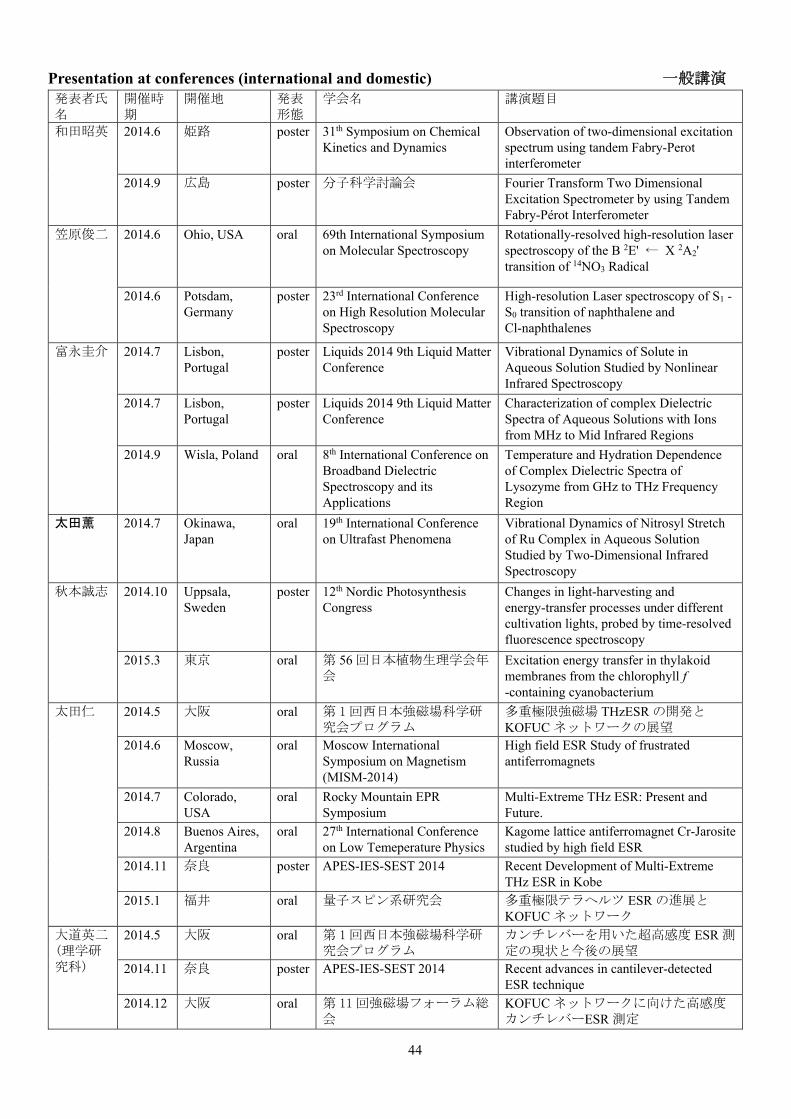
44
Presentation at conferences (international and domestic) 一般講演 発表者氏名
開催時期
開催地 発表形態
学会名 講演題目
和田昭英 2014.6 姫路 poster 31th Symposium on Chemical Kinetics and Dynamics
Observation of two-dimensional excitation spectrum using tandem Fabry-Perot interferometer
2014.9 広島 poster 分子科学討論会 Fourier Transform Two Dimensional Excitation Spectrometer by using Tandem Fabry-Pérot Interferometer
笠原俊二 2014.6 Ohio, USA oral 69th International Symposium on Molecular Spectroscopy
Rotationally-resolved high-resolution laser spectroscopy of the B 2E' ← X 2A2' transition of 14NO3 Radical
2014.6 Potsdam, Germany
poster 23rd International Conference on High Resolution Molecular Spectroscopy
High-resolution Laser spectroscopy of S1 - S0 transition of naphthalene and Cl-naphthalenes
富永圭介 2014.7 Lisbon, Portugal
poster Liquids 2014 9th Liquid Matter Conference
Vibrational Dynamics of Solute in Aqueous Solution Studied by Nonlinear Infrared Spectroscopy
2014.7 Lisbon, Portugal
poster Liquids 2014 9th Liquid Matter Conference
Characterization of complex Dielectric Spectra of Aqueous Solutions with Ions from MHz to Mid Infrared Regions
2014.9 Wisla, Poland oral 8th International Conference on Broadband Dielectric Spectroscopy and its Applications
Temperature and Hydration Dependence of Complex Dielectric Spectra of Lysozyme from GHz to THz Frequency Region
太田薫 2014.7 Okinawa, Japan
oral 19th International Conference on Ultrafast Phenomena
Vibrational Dynamics of Nitrosyl Stretch of Ru Complex in Aqueous Solution Studied by Two-Dimensional Infrared Spectroscopy
秋本誠志 2014.10 Uppsala, Sweden
poster 12th Nordic Photosynthesis Congress
Changes in light-harvesting and energy-transfer processes under different cultivation lights, probed by time-resolved fluorescence spectroscopy
2015.3 東京 oral 第 56 回日本植物生理学会年会
Excitation energy transfer in thylakoid membranes from the chlorophyll f -containing cyanobacterium
太田仁 2014.5 大阪 oral 第 1回西日本強磁場科学研究会プログラム
多重極限強磁場 THzESR の開発とKOFUC ネットワークの展望
2014.6 Moscow, Russia
oral Moscow International Symposium on Magnetism (MISM-2014)
High field ESR Study of frustrated antiferromagnets
2014.7 Colorado, USA
oral Rocky Mountain EPR Symposium
Multi-Extreme THz ESR: Present and Future.
2014.8 Buenos Aires, Argentina
oral 27th International Conference on Low Temeperature Physics
Kagome lattice antiferromagnet Cr-Jarosite studied by high field ESR
2014.11 奈良 poster APES-IES-SEST 2014 Recent Development of Multi-Extreme THz ESR in Kobe
2015.1 福井 oral 量子スピン系研究会 多重極限テラヘルツ ESR の進展とKOFUC ネットワーク
大道英二 (理学研究科)
2014.5 大阪 oral 第 1回西日本強磁場科学研究会プログラム
カンチレバーを用いた超高感度 ESR 測定の現状と今後の展望
2014.11 奈良 poster APES-IES-SEST 2014 Recent advances in cantilever-detected ESR technique
2014.12 大阪 oral 第 11 回強磁場フォーラム総会
KOFUC ネットワークに向けた高感度カンチレバーESR 測定

45
2015.3 東京 oral 日本物理学会 2015 年春季大会
バイメタルカンチレバーを用いた熱的検出高周波 ESR 測定法の開発
大久保晋 2014.5 大阪 oral 第一回西日本強磁場科学研究会プログラム
強磁場 THzESR によるスピン系の研究
2014.9 愛知 oral 日本物理学会 2014 年秋季大会
スピネル化合物 GeCo2O4の単結晶試料による ESR 測定 4
2014.11 奈良 poster APES-IES-SEST 2014 High-field ESR Measurements of Low-dimensional Magnets in Natural Minerals
2014.12 大阪 poster 第 11 回強磁場フォーラム総会
S=1/2 擬1次元フラストレート磁性体NaCuMoO4(OH)の強磁場 ESR 測定
2015.1 福井 oral 量子スピン系研究会 新しい S=1/2 ハニカム格子反強磁性体?ScCu2/3V1/3O3の強磁場 ESR 測定
2015.3 東京 oral 日本物理学会 2015 年春季大会
YCrO3の高周波 ESR 測定 2
櫻井敬博 (研究基盤センター)
2014.5 大阪 oral 第 1回西日本強磁場科学研究会プログラム
圧力下強磁場 ESR の現状と展望
2014.9 愛知 oral 日本物理学会 2014 年秋季大会
圧力下強磁場多周波数 ESR 装置の高感度化と応用
2014.11 奈良 poster APES-IES-SEST 2014 Development of High Pressure and Muti-frequency ESR System and Its Application to Quantum Spin System
2014.12 大阪 poster 第11回強磁場フォーラム総会
高圧下テラヘルツ強磁場 ESR の開発とKOFUC ネットワークへの展開
2015.3 東京 poster 日本物理学会 2015 年春季大会
圧力下強磁場多周波数 ESR 装置の高感度化と応用 II
原茂生(研究基盤センター)
2014.9 愛知 oral 日本物理学会 2014 年秋季大会
新規Co系 1次元鎖酸化物の構造と磁性
2014.11 奈良 poster APES-IES-SEST 2014 Magnetic Anisotropy of the Distorted-Diamond-Chain Compound Cu3(MoO4)2(OH)2
2015.3 東京 poster 日本物理学会 2015 年春季大会
水熱合成による単結晶 M2CoGe2O7の単結晶育成と磁化の異方性測定

46
Presentation by Graduate Students and Postdocs 院生、ポストドクの学会発表 指導教員 発表者氏名 学
年 時期 学会名 講演題目
和田昭英 A. Wada
Neeraj Kumar Joshi
PD 2014.6 31th Symposium on Chemical Kinetics and Dynamics
Polarity Controlled Reaction Path and Kinetics of Thermal Cis-to-Trans Isomerization of 4-Aminoazobenzene
Neeraj Kumar Joshi
PD 2014.9 第 8 回分子科学討論会 On the Branching Ratio of Photoisomerization of 4-Aminoazobenzene
笠原俊二 S. Kasahara
多田康平 D3 2014.5 第 14 回分子分光研究会 15NO3 ラジカルの B-X 遷移 0-0 バンドの高分解能レーザー分光
中野拓海 D3 2014.5 第 14 回分子分光研究会 ナフタレンの S1 状態の高振動状態の高分解能レーザー分光
山本涼 M2 2014.5 第 14 回分子分光研究会 単一モード紫外レーザーによるクロロナフタレンの高分解能分光
多田康平 D3 2014.6 30th Symposium on Chemical Kinetics and Dynamics
High-resolution Laser Spectroscopy of the B-X Transition of Nitrate Radical
中野拓海 D3 2014.6 30th Symposium on Chemical Kinetics and Dynamics
High-resolustion UV laser spectroscopy of vibronic bands of naphthalene S1 - S0 transition
山本涼 M2 2014.6 30th Symposium on Chemical Kinetics and Dynamics
High-resolution UV Laser spectroscopy of S1 - S0 transition of chloronaphthalene
多田康平 D3 2014.6 69th International Symposium on Molecular Spectroscopy
Rotationally-resolved high-resolution laser spectroscopy of the B 2E' ← X 2A2' Transition of 15NO3 Radical
多田康平 D3 2014.9 23rd International Conference on High Resolution Molecular Spectroscopy
Rotationally-resolved high-resolution Laser spectroscopy of 15NO3 Radical
多田康平 D3 2014.9 第 8回分子科学討論会 15NO3 ラジカルの B-X 遷移の高分解能レーザー分光
中野拓海 D3 2014.9 第 8回分子科学討論会 ナフタレン S1-S0 遷移の振電バンドにおける高分解能レーザー分光
山本涼 M2 2014.9 第 8回分子科学討論会 クロロナフタレンの S1←S0 遷移の高分解能レーザー分光~振動励起状態の観測~
多田康平 D3 2014.11 Indo-Japan Joint Workshop on "Frountiers in Molecular Spectroscopy: Fundamentals and Applications to Material and Biology"
High-resolution laser spectroscopy and Zeeman effect of free radical NO3
富永圭介 K. Tominaga
平岡翔 M2 2014.7 19th International Conference on Ultrafast Phenomena
Vibrational Dynamics of the CN Stretching in the Electronically Excited State by UV and Visible-Pump and Infrared-Probe Spectroscopy
奥田真紀 D1 2014.7 Gordon research conference "Water and Aqueous Solution"
The Dynamics of Non-ionic Molecules in Aqueous Solution Studied by Two-dimensional Infrared Spectroscopy
近藤未菜子 PD 2014.7 Gordon research conference "Water and Aqueous Solution"
Hydrogen Bonding Interaction between Acetate Anion and Water Molecule: Influence of cations
近藤未菜子 PD 2014.8」 Symposium on Ultrafast Spectroscopy: Its applications on Life and Environmental Sciences
Dynamics of the H-Bond Complex Formation of the Excited State 4-Aminophthalimide Analogue studied by Time-Resolved Vibrational Spectroscopy

47
岡田梨沙 M2 2014.9 第 8 回分子科学討論会 テラヘルツ時間領域分光法によるトレハロースにコートされたリゾチームの低振動ダイナミクス
松苗康徳 M2 2014.9 第 8 回分子科学討論会 時間分解蛍光分光法による凝集誘起発光の機構に関する研究
難波英里 M1 2014.9 第 8 回分子科学討論会 20 MHz から 4000 cm-1における分光学的測定によるグアニジウムイオンと水の相互作用
近藤未菜子 PD 2014.9 第 8 回分子科学討論会 超高速時間分解赤外分光法による水素結合錯体の振動ダイナミクス
奥田真紀 D1 2014.9 第 8 回分子科学討論会 二次元赤外分光法による水溶液中における非イオン性振動プローブ分子の動的挙動
平岡翔 M2 2014.9 第 8 回分子科学討論会 光ポンプ-テラヘルツプローブ分光装置の開発とその電荷キャリアダイナミクスへの応用
Feng Zhang PD 2014.9 第 8 回分子科学討論会 Phonon Modes of Molecular Crystals Studied by Terahertz Spectroscopy and Solid-state Density Functional Theory; a Concept of Vibrational Coordinate Distribution
Feng Zhang PD 2014.1 SPIE Photonics Asia Low-frequency vibration study of amino acids using terahertz spectroscopy and solid-state density functional theory
奥田真紀 D1 2014.11 Indo-Japan Joint Workshop on “Frontiers in Molecular Spectroscopy: Fundamentals and Applications to Material and Biology
Vibrational Dynamics of Non-Ionic Molecules in Aqueous Solution Studied by Two-Dimensional Infrared Spectroscopy
岡田梨沙 M2 2014.12 Advances in Spectroscopy and Ultrafast Dynamics
Low-frequency dynamics of trehalose-coated lysozyme studied by terahertz time-domain spectroscopy
難波英里 M1 2014.12 若手フロンティア研究会2014
広帯域分光測定によるグアニジウムイオンと水との相互作用
奥田真紀 D1 2015.1 神戸大学先端融合科学シンポジウム『生体分子のダイナミクスを眺める』
二次元赤外分光法による水溶液中における非イオン性振動プローブ分子の動的挙動
平岡翔 M2 2015.1 神戸大学先端融合科学シンポジウム『生体分子のダイナミクスを眺める』
光ポンプ-テラヘルツプローブ分光装置の開発とその電荷キャリアダイナミクスへの応用
松苗康徳 M2 2015.1 神戸大学先端融合科学シンポジウム『生体分子のダイナミクスを眺める』
時間分解蛍光分光法による凝集誘起発光の機構に関する研究
岡田梨沙 M2 2015.1 神戸大学先端融合科学シンポジウム『生体分子のダイナミクスを眺める』
Low-frequency Dynamics of Trehalose-coated Lysozyme Studied by Terahertz Time-Domain Spectroscopy
難波英里 M1 2015.1 神戸大学先端融合科学シンポジウム『生体分子のダイナミクスを眺める』
広帯域分光測定によるグアニジウムイオンと水との相互作用
Feng Zhang PD 2015.1 神戸大学先端融合科学シンポジウム『生体分子のダイナミクスを眺める』
Intramolecular Vibrations in Low-Frequency Normal Modes of Amino Acids: L-Alanine in Neat Solid Stat
48
奥田真紀 D1 2015.3 Asian Academic Seminar and School 2015
Vibrational Dynamics on Non-Ionic Molecules in Aqueous Solution Studied by T wo-Dimensional Infrared Spectroscopy
秋本誠志 S. Akimoto
大西亜弥 M1 2014.6 Photosynthesis Research for Sustainability – 2014
Energy transfer in Anabaena variabilis filaments during heterocyst differentiation studied by time-resolved fluorescence
植野嘉文 M1 2014.6 Photosynthesis Research for Sustainability – 2014
Light adaptation of the primitive red alga Cyanidioschyzon merolae, proved by time-resolved fluorescence spectroscopy
高下友基 M2 2014.7 光合成セミナー2014:反応中心と色素系の多様性
異なる色素組成をもつフィコビリソームにおける励起エネルギー移動過程
大西亜弥 M1 2014.7 光合成セミナー2014:反応中心と色素系の多様性
時間分解蛍光分光法を用いた Anabaena variabilis の窒素欠乏条件下におけるエネルギー移動過程の観測
植野嘉文 M1 2014.7 光合成セミナー2014:反応中心と色素系の多様性
異なる光質下で培養された紅藻Cyanidioschyzon merolae のエネルギー移動過程
植野嘉文 M1 2014.12 若手フロンティア研究会2014
原始紅藻 Cyanidioschyzon merolae のエネルギー移動の培養光質依存性
大西亜弥 M1 2014.12 若手フロンティア研究会2014
窒素欠乏条件における励起エネルギー移動過程の変化
大西亜弥 M1 2015.3 第56回日本植物生理学会年会
Energy transfer changes in Anabaena variabilis filaments under nitrogen depletion
植野嘉文 M1 2015.3 第56回日本植物生理学会年会
Long-term light adaptation of the unicellular red alga Cyanidioschyzon merolae, probed by time-resolved fluorescence spectroscopy
太田仁 H. Ohta
吉田翔太 M1 2014.9 日本物理学会 2014 年秋季大会
2 次元反強磁性体 Sr2MO3X(M=Cr,Ni X=F,Cl)の強磁場 ESR 測定
北原遥子 M1 2014.9 日本物理学会 2014 年秋季大会
擬 1 次元フラストレート磁性体NaCuMoO4(OH)の強磁場 ESR 測定
川崎航平 M1 2014.9 日本物理学会 2014 年秋季大会
マイクロコイルを用いた圧力下での ESR 測定装置の開発
荒川翔 M2 2014.9 日本物理学会 2014 年秋季大会
CuB2O4の ESR による研究
松井亮輔 M2 2014.9 日本物理学会 2014 年秋季大会
圧力下サイクロトロン共鳴装置の開発とディラック電子系への応用
池田将平 M2 2014.9 日本物理学会 2014 年秋季大会
YCrO3とその関連物質における強磁場 ESR測定
肘井敬吾 PD 2014.9 日本物理学会 2014 年秋季大会
4 体相互作用を持つ S=1/2 量子スピン梯子系における基底状態相図についての数値的研究
F. Elmasry D3 2014.11 APES-IES-SEST 2014 Er-concentration amd charge carrier effects on GaAs;Er,O revealed by X-band ESR
松井亮輔 M2 2014.11 APES-IES-SEST 2014 Development of High Pressure Cyclotron Resonance Systems and Application to Dirac Fermion System
川崎航平 M1 2014.11 APES-IES-SEST 2014 Development of high pressure ESR systems using micro-coil
岡本翔 M1 2014.11 APES-IES-SEST 2014 Application of cantilever-detected ESR to biological systems
石川陽帆 M2 2014.11 APES-IES-SEST 2014 High seisitive force detection system for high-frequency ESR measurement
田伏諒 M2 2014.11 APES-IES-SEST 2014 Cantilever-detected ESR measurement using a frequency modulation technique
Alexey Alfonsov
PD 2014.11 APES-IES-SEST 2014 High Frequency ferromagnetic resonance study of Heusler Compounds using a micro-cantilver

49
荒川翔 M2 2014.11 APES-IES-SEST 2014 Magnetic Anisotropy in Chiral System CuB2O4 by ESR
吉田翔太 M1 2014.11 APES-IES-SEST 2014 High Field ESR Measurements of Two Dimensional Antiferromagnets Sr2NiO3X (X=F, Cl)
北原遥子 M1 2014.11 APES-IES-SEST 2014 High Field ESR Measurements of Quasi One Dimensional Frustrated Magnet NaCuMoO4(OH)
池田将平 M2 2014.11 APES-IES-SEST 2014 High-field ESR Measurements of YCrO3 and YCaCrO4
肘井敬吾 PD 2014.11 APES-IES-SEST 2014 Numerical study of dynamical susceptibility in one dimensional trimerized spin systems
吉田翔太 M1 2014.11 日本赤外線学会 第 24 回研究発表会
2 次元反強磁性体 Sr2Nio3X(X=F,C1)のテラヘルツ ESR 測定
北原遥子 M1 2014.11 日本赤外線学会 第 24 回研究発表会
擬一次元フラスとレート磁性体NaCuMoO4(OH)のテラヘルツ ESR 測定
池田将平 M2 2014.11 日本赤外線学会 第 24 回研究発表会
ペロブスカイト化合物 YCrO3のテラヘルツESR 測定
岡本翔 M1 2014.11 日本赤外線学会 第 24 回研究発表会
カンチレバーを用いた高分解能生体 ESR 測定に向けた試み
田伏諒 M2 2014.11 日本赤外線学会 第 24 回研究発表会
バイメタル構造を用いた熱的検出カンチレバーESR 測定法の開発
川崎航平 M1 2014.11 日本赤外線学会 第 24 回研究発表会
ミリ波サブミリ波領域における圧力下 ESR測定技術の開発と応用
川崎航平 M1 2014.12 第11回強磁場フォーラム総会
圧力下強磁場高周波数 ESR 装置の開発と量子スピン系への応用
岡本翔 M1 2014.12 第11回強磁場フォーラム総会
カンチレバーを用いた溶液生体試料の高周波 ESR 測定に向けた試み
吉田翔太 M1 2014.12 第11回強磁場フォーラム総会
2 次元反強磁性体 ScCu2/3V1/3O3の強磁場 ESR測定
池田将平 M2 2014.12 第11回強磁場フォーラム総会
ペロブスカイト化合物 YCrO3の強磁場 ESR測定
Alexey Alfonsov
PD 2014.12 日本物理学会大阪支部講演会
High-field electron spin resonance study of electronic inhomogeneities in correlated transition metal compounds
池田将平 M2 2014.12 若手フロンティア研究会2014
マルチフェロイック物質 YCrO3の強磁場ESR 測定
北原遥子 M2 2014.12 若手フロンティア研究会2014
S=1/2NaCuMoO4(OH)の強磁場 ESR 測定
岡本翔 M1 2014.12 若手フロンティア研究会2014
カンチレバーを用いた高周波 ESR 測定法の生体試料への応用
松井亮輔 M2 2014.12 若手フロンティア研究会2014
圧力下サイクロトロン共鳴装置の開発とディラック電子系への応用
田伏諒 M2 2014.12 若手フロンティア研究会2014
バイメタル構造を用いたカンチレバーESR測定の熱的検出法
吉田翔太 M1 2014.12 若手フロンティア研究会2014
2次元反強磁性体 ScCu2/3V1/3O3のサブミリ波ESR 測定
石川陽帆 M2 2014.12 若手フロンティア研究会2014
カシミール力の高感度測定装置開発
荒川翔 M2 2014.12 若手フロンティア研究会2014
巨大光学磁気電気効果を示す螺旋磁性体CuB2O4の ESR による研究
川崎航平 M1 2014.12 若手フロンティア研究会2014
マイクロコイル ESR 技術の圧力下への応用
肘井敬吾 PD 2015.1 量子スピン系研究会 S=1BQ 模型における付加的な SU(2)対称性とその一般化
肘井敬吾 PD 2015.3 日本物理学会 2015 年春季大会
S=1/2 三量体量子スピン鎖における動的感受率の数値的研究

50
北原遥子 M1 2015.3 日本物理学会 2015 年春季大会
擬一次元フラストレート磁性体NaCuMoO4(OH)の強磁場 ESR 測定 2
吉田翔太 M1 2015.3 日本物理学会 2015 年春季大会
フラストレート 2 次元反強磁性体ScCu2/3V1/3O3の強磁場 ESR 測定
川崎航平 M1 2015.3 日本物理学会 2015 年春季大会
テラヘルツ領域における圧力下 ESR 装置の開発と応用
岡本翔 M1 2015.3 日本物理学会 2015 年春季大会
カンチレバーを用いた高周波 ESR 測定法の金属タンパク質への応用

51
Books 著書 著者(共著者も含む) 書名 出版社名 ページ
数 発行年
秋本誠志 「葉緑体」「光化学系 I」「光化学系 II」in: 「光化学の事典」(光化学協会編)
朝倉書店 322–327 2014
52
Other Publications 参考論文・記事・報告 著者 タイトル 出版物名 巻・号・ページ 発行
年
Feng Zhang, Keisuke Tominaga, Michitoshi Hayashi, and Houng-Wei Wang
Low-frequency Vibration Study of Amino Acids Using Terahertz Spectroscopy and Solid-state Density Functional Theory
SPIE Proceedings Vol. 9275, "Infrared, Millimeter-Wave, and Terahertz Technologies III", Cunlin Zhang, Xi-Cheng Zhang, Masahiko Tani, Editors
Vol. 9275, 92750D-1, -9
2014
Kaoru Ohta, Kyoko Aikawa, and Keisuke Tominaga
Vibrational Dynamics of Nitrosyl Stretch of Ru Complex in Aqueous Solution Studied by Two-Dimensional Infrared Spectroscopy
"Ultrafast Phenomena XIX”, edited by Kaoru Yamanouchi, Steven Cundiff, Regina de Vivie-Riedle, Makoto Kuwata-Gonokami, Louis DiMauro, Springer
pp. 479–482 2015
Sho Hiraoka, Kaoru Ohta and Keisuke Tominaga
Vibrational Dynamics of the CN Stretching in the Electronically Excited State by UV and Visible-Pump and Infrared-Probe Spectroscopy
“Ultrafast Phenomena XIX”, edited by Kaoru Yamanouchi, Steven Cundiff, Regina de Vivie-Riedle, Makoto Kuwata-Gonokami, Louis DiMauro, Springer
pp. 487-491 2015
秋本誠志 生命科学を理解するための蛍光の基礎 2 (時間分解蛍光と異方性)
分光研究 63・2・73-81 2014

53
Lecture to Public 講演、模擬授業など 氏名 講演題目 集会名 日時 場所
富永圭介 Low-frequency motions in condensed phases studied by pulsed terahertz radiation
seminar 12.15. 2014 S. N. Bose National Centre for Basic Sciences
Feng Zhang Low-frequency Vibrations Study of Molecular Crystals Using Terahertz Spectroscopy and Solid-state Density Functional Theory
seminar at Prof. X. C. Shen's group
5.14. 2014 Shanghai Institute of Technical Physics of the Chinese Academy of Science
Feng Zhang Low-frequency Vibrations Study of Molecular Crystals Using Terahertz Spectroscopy and Solid-state Density Functional Theory
seminar at Prof. C. L. Zhang's group
5.16.2014 Capital Normal University, China
Feng Zhang Low-frequency Vibration Study of Molecular Solids Using Terahertz Spectroscopy and Solid-state Density Functional Theory
seminar at Prof. J. P. Wang's group
10.15. 2014 Institute of chemistry, Chinese Academy of Sciences, Beijing
Feng Zhang Low-frequency Vibration Study of Molecular Solids Using Terahertz Spectroscopy and Solid-state Density Functional Theory
seminar at Prof. X. S. Zhao's group
10.16. 2014 Beijing University, Beijing
Feng Zhang Low-frequency Vibration Study of Molecular Solids Using Terahertz Spectroscopy and Solid-state Density Functional Theory
seminar at Prof. Y. X. Weng's group
10.17. 2014 Institute of physics, Chinese Academy of Sciences, Beijing
太田仁 Recent development of multi-extreme high field THz ESR and some applications
Seminar 26-29.7.2014 Laboratoire National des Champs Magnetiques Intenses

54
Awards 受賞 氏名 受賞研究題目 賞名 団体、学会名
太田仁 Fellowship Fellowship award International Academy of Physical Sciences
石川陽帆 Highly sensitive force detection system for high-frequency ESR measurement
SEST 学生優秀研究賞 APES2014-IES-SEST2014
川崎航平 Development of high pressure ESR system using micro-coil
SEST 学生優秀研究賞 APES2014-IES-SEST2014
岡本翔 Application of cantilever-detected ESR to biological systems
APES Poster award APES2014-IES-SEST2014
Alexey Alfonsov
High frequency ferromagnetic resonance study of Heusler compounds using a micro-cantilever
IES Poster award APES2014-IES-SEST2014
池田将平 マルチフェロイック物質 YCrO3の強磁場ESR 測定
優秀賞 若手フロンティア研究会2014

55
Conference Organization 学術集会の開催 氏名 学術集会 共同主催者 場所 時期 参加
者概数
富永圭介(代表) Frontiers in Molecular Spectroscopy: Fundamentals and Applications to Material and Biology
Anunay Samanta (インド側代表)
奈良、東大寺総合文化センター
平成 26 年11 月 25 日-28 日
約 25名
富永圭介 先端融合科学シンポジウム「生体分子のダイナミクスを眺める」
茶谷絵理(代表)、山本直樹、鍔木基成
神戸大学理学研究科
平成 27 年 1月 19、20 日
約 40名
太田仁(代表) APES-IES-SEST2014 (Joint conference of 9th Asia-Pacific EPR/ESR Society Symposium, 1st International EPR(ESR) Society Symposium, 53rd SEST Annual Meeting)
大久保晋(Secretary General) 肘井敬吾(Web)
奈良、東大寺総合文化センター
平成 26 年11 月 12-16日
289 名
APES-IES-SEST2014

56
Molecular Photoscience Seminar 分子フォトサイエンスセミナー Date Name Affiliation Title
7.14. 2014 Prof. Prem B. Bisht Indian Institute of Technology Madras, Chennai
Effect of Surface Plasmon Resonances on Optical Kerr Effect in Graphene-Silver Nanoparticle Composites
7. 15. 2014 篠北啓介博士 University of Groningen Hydrogen bond dynamics in alcohols studied by two-dimensional infrared spectroscopy
11.10.2014 御園雅俊教授 福岡大学理学部物理科学科 光周波数コムを利用した超高分解能レーザー分光
11.4.2014 Prof. Dr. V. Kataev IFW Dresden (分子フォトと国際協定あり)
Interplay of site disorder and magnetic frustration in the quantum spin magnet CoAI2O4
11.18.2014 Prof. Dr. K. Möbius Free University Berlin High-field EPR studies on biomolecules - crossing the gap to NMR
11.2014 Dr. Alexey Alfonsov IFW Dresden, Germany (分子フォトと国際協定あり)
Multi-frequency electron spin resonance spectroscopy study of the FeAs-based superconductors
3.5.2015 Dr. Sergei Zvyagin Dresden High Magnetic Field Laboratory
Spin dynamics in triangular-lattice antiferromagnets: high-field ESR studies

57
Molecular Photoscience Research Center
Kobe University,
Nada, Kobe 657-8501 Japan
Tel: +81-78-803-5761
URL: http://www.kobe-u.ac.jp/mprc/index-e.html (English)
〒657-8501
神戸市灘区六甲台町 1-1
神戸大学分子フォトサイエンス研究センター
電話番号 078-803-5761
URL:http://www.kobe-u.ac.jp/mprc/ (日本語)