ansc 689 physiological chemistry of livestock specids digestion and absorption … ·...
TRANSCRIPT

Handout 4 Carbohydrate Digestion in Monograstrics and Ruminants
1
ANSC 689 PHYSIOLOGICAL CHEMISTRY OF LIVESTOCK SPECIDS
Digestion and Absorption of Carbohydrates in Monogastrics and Ruminants
I. Digestion of carbohydrates: overview
A. Oral digestion
1. Ingested starch is hydrolyzed by α-amylase.
2. pH optimum is 6.7.
B. Stomach
1. Salivary amylase digestion continues.
2. Some fermentation of lactose to lactic acid can occur.
C. Intestinal luminal digestion
1. Pancreatic α-amylase
digests starch to maltose.
2. Branched-chain limit
dextrans also are formed.
D. Intestinal mucosal digestion
1. Lactase, trehalase, and four maltases work on oligosaccharides.
2. Monosaccharides are absorbed into the enterocytes.
E. Poultry (avian species in general)
1. The crop, proventriculus, and gizzard replace the simple stomach of other
monogastrics.
2. The esophagus extends to the cardiac region of the proventriculus.
3. The proventriculus is similar to the stomach, in that typical gastric secretions (mucin,
HCl, and pepsinogen) are produced.

Handout 4 Carbohydrate Digestion in Monograstrics and Ruminants
2
II. The gastrointestinal tracts of pigs and poultry
B. Architecture and secretions of
the gastrointenstinal tract in
nonruminant mammals
1. Oral region – In the mouth, saliva
is secreted from the parotid,
mandibular, and sublingual
glands. α-Amylase in saliva
initiates carbohydrate digestion.
2. Esophageal region – The
esophagus extends from the
pharynx to the esophageal portion
of the stomach.
3. Gastric region – The stomach is
divided into the esophageal region,
the cardiac region, and the fundic
(proper gastric) region. The
cardiac region elaborates mucus,
proteases, and lipase (discussed
later in the semester). The action
of α-amylase stops in the fundus,
when the pH drops below 3.6.
4. Pancreatic region – The
endocrine portion secretes insulin
and glucagon (and other peptide
hormones) from the islets of
Langerhans. The exocrine portion
secretes pancreatic α-amylase in
addition to other digestive
enzymes. These and bile secretion
will be discussed later.
5. Small intestine – The duodenum (4-5%) originates
at the distal end of the stomach (pyloric valve),
whereas the jejunum (88-91%) and ileum (4-5%)
form the lower intestine. Pancreatic α-amylase is
mixed with chyme from the stomach. Alkaline
secretions from Brunner’s glands raise the pH to
approx. 8.0
6. Large intestine – Associated with the proximal end
of the large intestine is a short cecum, which is the
site of microbial fermentation. The colon is long and
highly functional.
Handout 4 Carbohydrate Digestion in Monograstrics and Ruminants
3
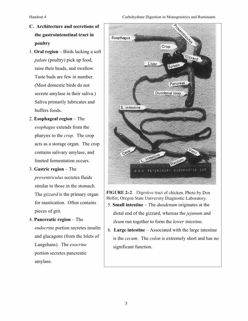
C. Architecture and secretions of
the gastrointenstinal tract in
poultry
1. Oral region – Birds lacking a soft
palate (poultry) pick up food,
raise their heads, and swallow.
Taste buds are few in number.
(Most domestic birds do not
secrete amylase in their saliva.)
Saliva primarily lubricates and
buffers foods.
2. Esophageal region – The
esophagus extends from the
pharynx to the crop. The crop
acts as a storage organ. The crop
contains salivary amylase, and
limited fermentation occurs.
3. Gastric region – The
proventriculus secretes fluids
similar to those in the stomach.
The gizzard is the primary organ
for mastication. Often contains
pieces of grit.
4. Pancreatic region – The
endocrine portion secretes insulin
and glucagons (from the Islets of
Langehans). The exocrine
portion secretes pancreatic
amylase.
5. Small intestine – The duodenum originates at the
distal end of the gizzard, whereas the jejunum and
ileum run together to form the lower intestine.
6. Large intestine – Associated with the large intestine
is the cecum. The colon is extremely short and has no
significant function.

Handout 4 Carbohydrate Digestion in Monograstrics and Ruminants
4
III. Digestion and fermentation of carbohydrates
A. Oral digestion
1. Ingested starch is hydrolyzed by α-amylase.
2. pH optimum is 6.7.
B. Stomach digestion
1. Salivary amylase digestion continues.
2. Some fermentation of lactose to lactic acid can occur. This may contribute acidity to
assist the limited HCl production in infants (important for the formation of milk clots).
3. Diets high in molasses increase fermentation in the stomach, and increase VFA
production.

Handout 4 Carbohydrate Digestion in Monograstrics and Ruminants
5
C. Intestinal luminal digestion
1. Pancreatic α-amylase digests starch to maltose, branched-chain limit dextrans, and
traces of glucose.
2. These products of digestion migrate to the mucosal surface of the duodenum
following a concentration gradient.
D. Intestinal mucosal digestion
1. Lactase, trehalase, and four maltases (including sucrase) work on oligosaccharides.
2. Monosaccharides are absorbed into the enterocytes.
3. Absorption takes place in the duodenum and jejunum.
E. Microbial activity in the small intestine
1. Gut microorganisms can digest nonstarch structural carbohydrates.
2. This is associated with a substantial amount of VFA production.
F. Large intestinal digestion and fermentation
1. Gastrointestinal contents are retained up to 38 h in the large intestine vs less than 6 h
in the stomach and small intestine.
2. Microorganisms in the colon and cecum produce cellulases, hemicellulases, and
pectinsases.
3. Primary products are VFA (acetic, propionic, and butryric acids) and methane.
III. Absorption of monosaccharides
A. Glucose and galactose
1. Both are transported into the enterocyte by a Na-dependent glucose transporter
(Sglt1).
2. Both then are released into the blood by a facilitated sugar transporter (GLUT2).
B. Fructose
1. Fructose is absorbed first by a Na-independent brush border fructose transporter
(GLUT5).
2. Fructose is released into the blood by GLUT2.

Handout 4 Carbohydrate Digestion in Monograstrics and Ruminants
6
B. Omnivores
1. Nonglandular region – no digestive secretions or absorption occurs.
2. Cardiac region – lined with epithelial cells that secrete mucin (prevents lining of the
stomach from being digested).
3. Fundic region – contains parietal cells (secrete HCl), neck chief cells (secrete mucin),
and body chief cells (secrete pepsinogen, and lipase).
Handout 4 Carbohydrate Digestion in Monograstrics and Ruminants
7
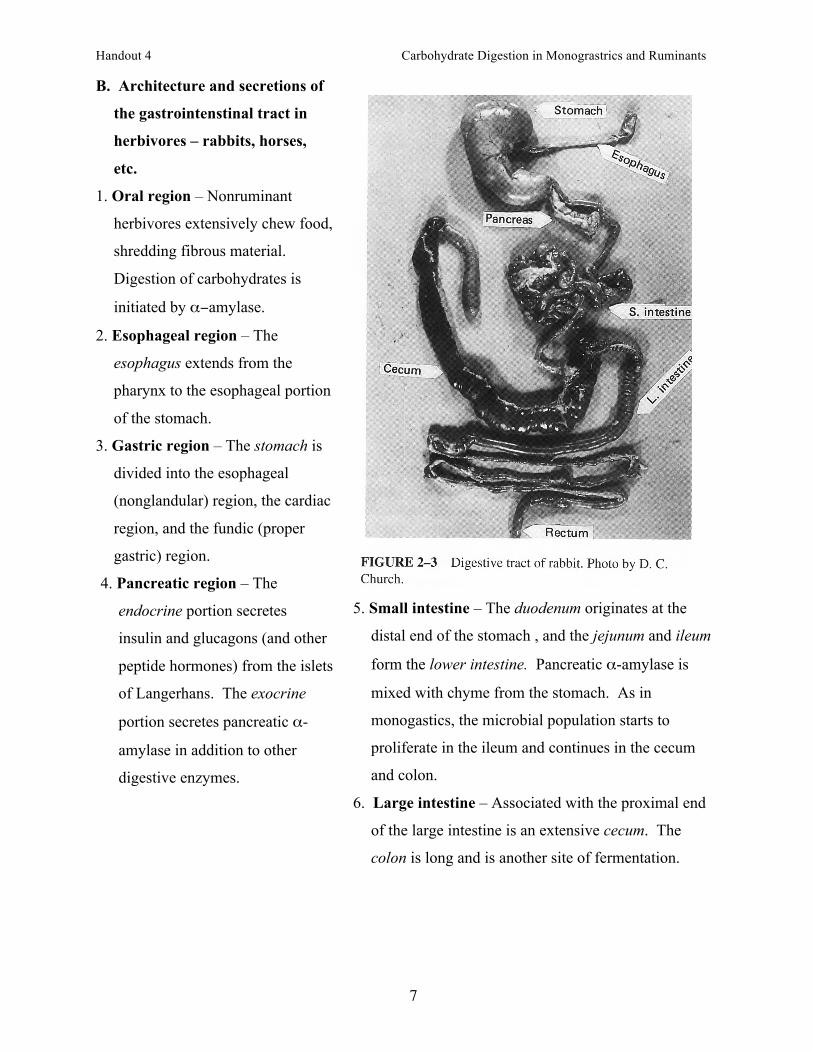
B. Architecture and secretions of
the gastrointenstinal tract in
herbivores – rabbits, horses,
etc.
1. Oral region – Nonruminant
herbivores extensively chew food,
shredding fibrous material.
Digestion of carbohydrates is
initiated by α−amylase.
2. Esophageal region – The
esophagus extends from the
pharynx to the esophageal portion
of the stomach.
3. Gastric region – The stomach is
divided into the esophageal
(nonglandular) region, the cardiac
region, and the fundic (proper
gastric) region.
4. Pancreatic region – The
endocrine portion secretes
insulin and glucagons (and other
peptide hormones) from the islets
of Langerhans. The exocrine
portion secretes pancreatic α-
amylase in addition to other
digestive enzymes.
5. Small intestine – The duodenum originates at the
distal end of the stomach , and the jejunum and ileum
form the lower intestine. Pancreatic α-amylase is
mixed with chyme from the stomach. As in
monogastics, the microbial population starts to
proliferate in the ileum and continues in the cecum
and colon.
6. Large intestine – Associated with the proximal end
of the large intestine is an extensive cecum. The
colon is long and is another site of fermentation.

Handout 4 Carbohydrate Digestion in Monograstrics and Ruminants
8
C. Architecture and secretions of
the gastrointenstinal tract in
herbivores – true ruminants
1. Oral region – Ruminants
masticate their food to a limited
extent.
2. Esophageal region – In
ruminants, the bolus of food can
travel either to the reticulorumen,
or back to the mouth
(rumination).
3. Gastric region – The stomach is
divided into four compartments,
the reticulum, rumen, omasum,
and abomasum (glandular
stomach).
4. Pancreatic region – The
endocrine portion secretes insulin
and glucagons (from the Islets of
Langerhans). The exocrine
portion secretes pancreatic
amylase. Both regions are less
developed in ruminant species
than in monogastrics.
5. Small intestine – The duodenum
originates at the distal end of the
abomasum, and the jejunum and
ileum form the lower intestine.
6. Large intestine – There is a small cecum, and the
large intestine is considerably smaller than in
nonruminant herbivores.

Handout 4 Carbohydrate Digestion in Monograstrics and Ruminants
9
II. Digestion and fermentation of carbohydrates in true ruminants
A. Oral digestion
1. Saliva (150 L/day) contains sodium bicarbonate (buffer).
2. pH optimum is 6.7.
B. Reticulorumen
1. Contains large populations of bacteria and protozoans.
2. Make-up of microorganisms depends on diet (high-forage vs high-grain).
3. Microorganism numbers peak 2 to 3 hours after a high-grain diet and 4 to 5 hours after
a high-forage diet.
4. Sugars and starches are degraded to VFA.
5. Digestion of structural carbohydrates occurs in the extracellular compartment
(hydrolysis to glucose) and intracellular compartment (formation of VFA).
C. Intestinal luminal digestion
1. Pancreatic α-amylase digests starch to maltose, branched-chain limit dextrans, and
traces of glucose.
2. These products of digestion migrate to the mucosal surface of the duodenum
following a concentration gradient.
3. The amount of α-amylase produced by the ruminant pancreas is very low compared to
monogastric animals.
D. Intestinal mucosal digestion
1. Lactase, trehalase, and four maltases (including sucrase) work on oligosaccharides.
2. Monosaccharides are absorbed into the enterocytes.
3. Absorption takes place in the duodenum and jejunum.
E. Microbial activity in the small intestine
1. Gut microorganisms can digest nonstarch structural carbohydrates.
2. This is associated with a small amount of VFA production in ruminants.
F. Large intestinal digestion and fermentation
1. Microorganisms in the colon and cecum produce cellulases, hemicellulases, and
pectinsases.
2. Primary products are VFA (acetic, propionic, and butryric acids) and methane. This is
limited in ruminants.