anthony t. yeung m.d., christopher a. yeung m.d., … · posterolateral selective endoscopic...
TRANSCRIPT
Anthony T. Yeung M.D., Christopher A. Yeung M.D., Yinggang Zheng M.D.
121
Minim
ally Invasive Procedures In Spine Surgery
POSTEROLATERAL SELECTIVE ENDOSCOPIC DISCECTOMY THE YESS
TECHNIQUEAnthony T. Yeung M.D., Christopher A. Yeung M.D., Yinggang Zheng M.D.
1. IntroductionThe intervertebral disc, an important supporting struc-ture of the spinal column, is implicated as a major source of low back pain and sciatica. (1,2) The patho-genesis of disc degeneration and herniation is com-plex and multifactorial, but clearly outlined and doc-umented by Wolfgang Rauschning’s work illustrating the patho-anatomy of degenerative disc disease and degenerative conditions of the lumbar spine. (3) Most disc herniations are not the result of an acute event, but an accumulation of several insults to the spine that lead to degeneration, annular tears, and eventual disc herniation. (4) There are several theories of disc degen-eration including mechanical, chemical, age-related, autoimmune and genetic. Within the mechanical the-ory, the following types of abnormal loads have been proven experimentally to cause disc injury: torsion (5), compression (5, 6), repetitive compressive loading in flex-ion (7), hyper flexion (8), and vibration. (9)
Traditionally disc surgery has been reserved for disc herniations causing radiculopathy or nerve def-icits due to mechanical compression on the spinal nerves. (10) This is due to the inherent morbidity of the posterior surgical approach that must violate and alter the important function of the posterior spinal column. Open posterior discectomy often includes or requires a midline incision, muscle and ligament stripping, prolonged muscle retraction, bone resec-tion of the lamina and facet, and nerve root and du-ral tube retraction. This can cause instability and scarring around the sensitive nerve roots even in a technically perfect operation. The morbidity of the standard posterior approach has therefore limited the use of surgery as an early treatment option in the cascade of disc degeneration and herniation. Thus,
surgery was often not recommended for herniations without neurologic deficits, “small” herniations, cen-tral herniations, and annular tears. The dogma that “disc surgery is really decompressive nerve surgery” dominates the rationale for traditional micro-discec-tomy for herniated discs.
Minimally invasive surgical options that limit the inherent approach related-morbidity are pos-sible with the posterolateral portal. (11-28) This ap-proach to the disc is most challenging at the L5-S1 level due to the prominence of the iliac crest. Most L5-S1 disc spaces are accessible; however, entry into the disc may require foraminal decompression of the lateral facet.
The least invasive of all posterolateral intradiscal techniques is the injection of Chymopapain, a treat-ment option validated by at least two large prospec-tive, randomized double blind studies and numer-ous cohort studies. (29,30) This treatment produced satisfactory results in many studies and came into widespread clinical use in the 1970’s, but lost popu-larity with reports of complications as severe as ana-phylactic shock and transverse myelitis. (31) Although these complications can now be virtually eliminated with pre-operative antigen screening and discogra-phy, the perceived risk has limited its continued use. More recent studies from experienced chymopapain users still tout chymopapain as a valuable adjunct to endoscopic disc surgery. (32,33,34)
The introduction of the operating microscope for discectomy by Yasargil in 1967 and later by Williams encouraged smaller incisions for the standard poste-rior approach. (35,36) The transcanal microscope-assisted technique became the gold standard; however, it still requires retraction of the dural tube and nerve, pe-riosteal stripping of the muscle and ligaments, hemi-
Posterolateral Selective Endoscopic Discectomy The YESS Technique
122
Min
imal
ly In
vasi
ve P
roce
dure
s In
Spi
ne S
urge
ry
laminotomy, and regional or general anesthesia. Tu-bular retractors have recently been developed that can be used with either a microscope or endoscope for this posterior transcanal approach. (37) This utilizes tissue dilation rather than cutting, and minimizes the su-perficial tissue destruction, but still requires the same amount of bone removal and neural manipulation as the standard microscopic posterior discectomy.
The concept of indirect decompression of the spi-nal canal via a posterolateral, extracanal approach was introduced by Kambin in 1973 using a Craig cannula for limited nucleotomy in combination with a tran-scanal approach. (14) In 1975 Hijikata reported the first stand alone nonvisualized posterolateral percutane-ous central nucleotomy. (9)
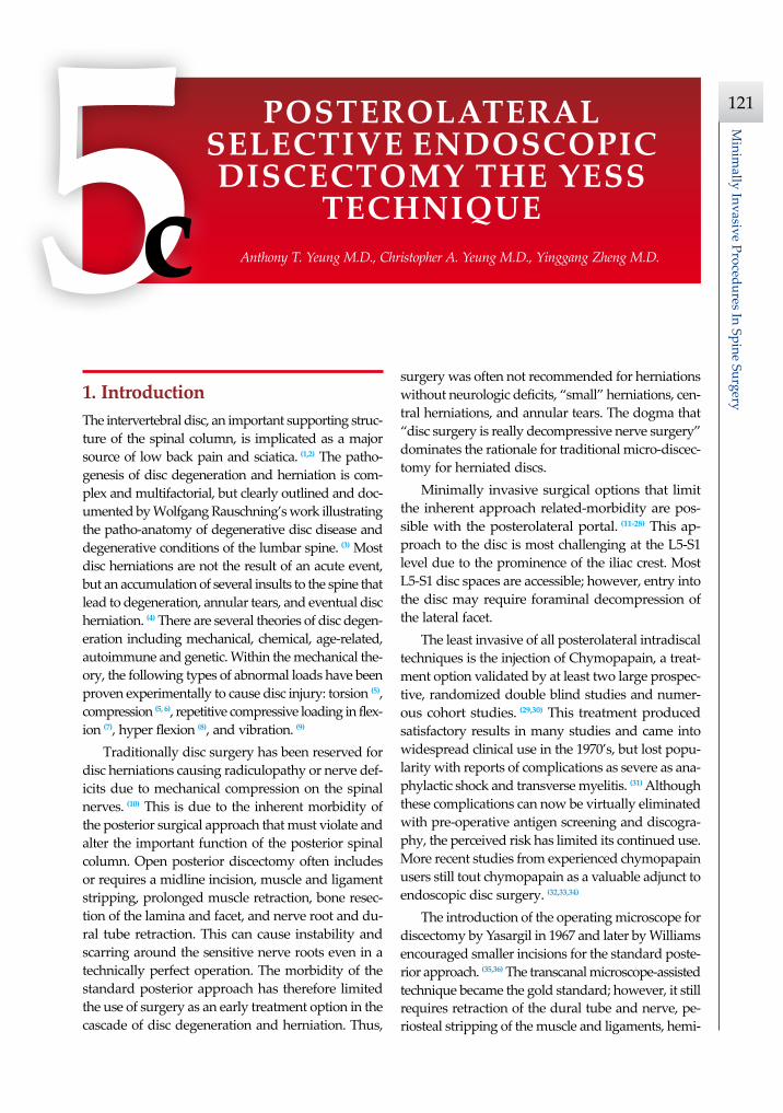
Kambin went on to describe the safe triangular working zone (Kambin’s Triangle) (Figure 1) and re-sults of arthroscopic microdiscectomy, in which ar-throscopic visualization of the herniation via the posterolateral approach was used for discectomy of contained herniations. (11, 14-19) Hermantin et al. re-ported satisfactory results from video assisted ar-throscopic microdiscectomy in 97% of patients com-pared to 93% in traditional microdiscectomy with an average of 31 months follow-up. (11) The arthroscopic group had less narcotic use and less time off from work. The study was prospective and randomized with 30 subjects in each group.
Mayer also showed promising results in a prospec-tive randomized study comparing percutaneous dis-cectomy with microscopic discectomy for contained or slight subligamentous herniations. (21) The percuta-neous group showed comparable or superior results. Long term disability defined by return to work sta-tus, produced statistically significant differences. In the percutaneous group, 95% returned to their pre-vious occupation compared to 72.2% in the microdis-cectomy group. Each group had 20 subjects.
Evolving methodology the 1980’s and early 1990’s allowed for endoscopic lumbar nerve root decompres-sion by a visualized, direct excision of contained and non-contained herniated disc fragments. (19,20,25,28)
Yeung introduced a rigid rod-lens, flow inte-grated, multichannel, wide-angle operating spinal endoscope in 1998 that allowed for even more flexi-bility accessing the disc, traversing and exiting nerve roots, and epidural space. The endoscope configura-tion offered significant visual improvement and the complementary instrument system with specialized slotted and bevel-ended tubular access cannulas al-lowed for same-field viewing of the intradiscal space, annular wall, and epidural space. The design allows for improved access to the posterior disc for visual-ized fragmentectomy, improved access to the under-surface of the superior articular facet for foramin-oplasty, and protection of the neural structures by rotating the cannula. (26,27)
2. Indications and Treatment Rational
• All lumbar disc herniations except migrated/se-questered fragments inaccessible through the fo-ramen
• Annular tears• IDD-Internal disc disruption diagnosed with dis-
cography producing concordant pain and radio-graphic abnormalities
• Foraminal stenosis• Synovial cysts of the facet joint• Discitis
Perhaps the ideal lesion for posterolateral selec-tive endoscopic discectomy is the far lateral, extra-foraminal disc herniation. The exiting nerve is rou-tinely visualized, and the cannula inserts directly at the herniation site. This approach requires less ma-
Figure 1: Kambin’s triangular working zone is the site
of surgical access for posterolateral endoscopic discetomy. It is defined as a right triangle over
the dorsolateral disc. The hypotenuse is the exiting nerve root, the base (width) is the su-perior border of the caudal vertebra, and the
height is the dura/traversing nerve root.
Anthony T. Yeung M.D., Christopher A. Yeung M.D., Yinggang Zheng M.D.
123
Minim
ally Invasive Procedures In Spine Surgery
nipulation of the exiting nerve root than the parame-dian posterior approach.
Any herniation contiguous with the disc space not sequestered and migrated is amenable to endoscopic disc excision. The timing of surgical treatment is sim-ilar to posterior transcanal discectomy. The size and types of herniations chosen by the surgeon for endo-scopic excision will depend on the skill and experience of the surgeon as well as the anatomic considerations in the patient relative to the location of the herniation. Certainly, all contained disc herniations are appropri-ate for endoscopic decompression. With experience ex-truded herniations can be routinely addressed.
The posterolateral endoscopic approach only re-quires tissue dilation to accommodate a 7mm work-ing cannula. This tissue sparing approach offers con-sideration for earlier surgical timing when approach related risk/benefit ratios are factored in after patients fail conservative treatment and continue to have de-bilitating pain without neurologic deficit. Quality of life issues and functional issues associated with chronic discogenic pain can be addressed with this minimally invasive surgical option. Therefore, small disc herniations with predominant leg pain, central disc herniations with predominant back pain, IDD, and annular tears causing chemical sciatica are ame-nable to disc surgery by endoscopic means.
The discectomy decompresses the disc, alleviat-ing pressure on the annulus, and removes any un-stable degenerated disc fragments that could her-niate. Radiofrequency energy can be applied to the annular tears under direct visualization to contract the collagen and ablate ingrown granulation tissue, neoangiogenesis, and sensitized nociceptors. Fre-quently interpositional nuclear tissue is seen within the fibers of the annular tear preventing the tear from healing. This tissue can then be removed to al-low the tear to heal.
Endoscopic foraminoplasty can be readily achieved with bone trephines/rasps and the side firing Holmi-um-YAG laser. (38) The roof of the foramen is formed by the undersurface of the superior articular facet. This is easily visualized and accessed via the endo-scope. The side firing Holmium-YAG laser and bone trephines strip the facet capsule and remove bone to enlarge the foraminal opening. Studies by Panjabi have demonstrated that decompression through the fora-men can be more effective than posterior decompres-sion for foraminal stenosis. The posterior removal of
1/3 of the medial facet produces more instability than posterolateral foraminal decompression. (39) Synovial cysts can also be visualized and removed.
Discitis can be treated with posterolateral endo-scopic discectomy and debridement. Current meth-ods rely on needle aspiration followed by prolonged antibiotic treatment. Needle aspirations are not as reliable as tissue samples from endoscopic debride-ment, and are often negative even in the face of bac-terial discitis. Surgeons are often hesitant to perform open debridement because of the morbidity of the open approach, creation of dead space and devas-cularized tissue, and the concern for spreading the infection in the spinal canal. Endoscopic excisional biopsy and thorough debridement via the postero-lateral portal has provided almost immediate pain relief and a much more reliable tissue sample for laboratory analysis and culture. (40) Since only tis-sue dilation is used, no dead space is created that would allow the infection to spread. Many patients with discitis have co-morbidities, which make them poor open surgical candidates.
3. Surgical Procedures
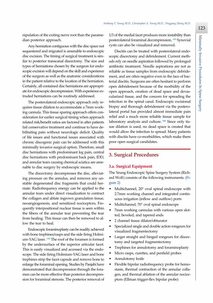
3.a. Surgical EquipmentThe Yeung Endoscopic Spine Surgery System (Rich-ard Wolf) consists of the following instruments. (Fi-gure 2)• Multichannel, 20° oval spinal endoscope with
2.7mm working channel and integrated contin-uous irrigation (inflow and outflow) ports
• Multichannel, 70° oval spinal endoscope• 7mm working cannulas with various open slot-
ted, beveled, and tapered ends• 2 channel tissue dilator/obturator• Specialized single and double action rongeurs for
visualized fragmentectomy• Larger straight and hinged rongeurs for discec-
tomy and targeted fragmentectomy• Trephines for annulotomy and foraminoplasty• Micro rasps, curettes, and penfield probes• Annulotomy knife• Flexible bipolar radiofrequency probe for hemo-
stasis, thermal contraction of the annular colla-gen, and thermal ablation of the annular nocice-ptors (Ellman trigger-flex bipolar probe)
Posterolateral Selective Endoscopic Discectomy The YESS Technique
124
Min
imal
ly In
vasi
ve P
roce
dure
s In
Spi
ne S
urge
ry
Adjunctive equipment• Straight and flexible suction-irrigation shavers
for discectomy (Endius MDS)• Side firing Holmium-YAG laser (Trimedyne)• Fluid pump for continuous irrigation• Video endoscopy tower
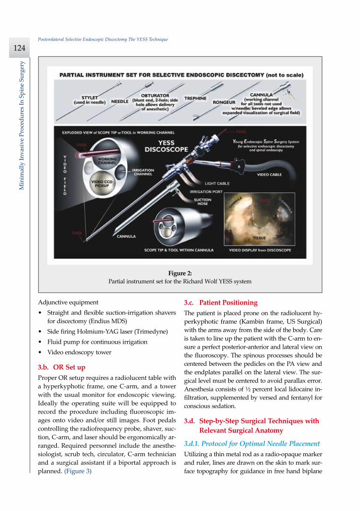
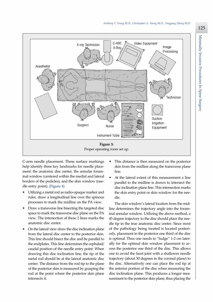
3.b. OR Set upProper OR setup requires a radiolucent table with a hyperkyphotic frame, one C-arm, and a tower with the usual monitor for endoscopic viewing. Ideally the operating suite will be equipped to record the procedure including fluoroscopic im-ages onto video and/or still images. Foot pedals controlling the radiofrequency probe, shaver, suc-tion, C-arm, and laser should be ergonomically ar-ranged. Required personnel include the anesthe-siologist, scrub tech, circulator, C-arm technician and a surgical assistant if a biportal approach is planned. (Figure 3)
3.c. Patient PositioningThe patient is placed prone on the radiolucent hy-perkyphotic frame (Kambin frame, US Surgical) with the arms away from the side of the body. Care is taken to line up the patient with the C-arm to en-sure a perfect posterior-anterior and lateral view on the fluoroscopy. The spinous processes should be centered between the pedicles on the PA view and the endplates parallel on the lateral view. The sur-gical level must be centered to avoid parallax error. Anesthesia consists of ½ percent local lidocaine in-filtration, supplemented by versed and fentanyl for conscious sedation.
3.d. Step-by-Step Surgical Techniques with Relevant Surgical Anatomy
3.d.1. Protocol for Optimal Needle PlacementUtilizing a thin metal rod as a radio-opaque marker and ruler, lines are drawn on the skin to mark sur-face topography for guidance in free hand biplane
Figure 2: Partial instrument set for the Richard Wolf YESS system
Anthony T. Yeung M.D., Christopher A. Yeung M.D., Yinggang Zheng M.D.
125
Minim
ally Invasive Procedures In Spine Surgery
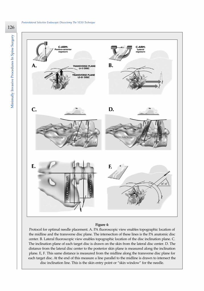
C-arm needle placement. These surface markings help identify three key landmarks for needle place-ment: the anatomic disc center, the annular foram-inal window (centered within the medial and lateral borders of the pedicles), and the skin window (nee-dle entry point). (Figure 4)• Utilizing a metal rod as radio-opaque marker and
ruler, draw a longitudinal line over the spinous processes to mark the midline on the PA view.
• Draw a transverse line bisecting the targeted disc space to mark the transverse disc plane on the PA view. The intersection of these 2 lines marks the anatomic disc center.
• On the lateral view draw the disc inclination plane from the lateral disc center to the posterior skin. This line should bisect the disc and be parallel to the endplates. This line determines the cephalad/caudal position of the needle entry point. When drawing this disc inclination line, the tip of the metal rod should be at the lateral anatomic disc center. The distance from the rod tip to the plane of the posterior skin is measured by grasping the rod at the point where the posterior skin plane intersects it.
• This distance is then measured on the posterior skin from the midline along the transverse plane line.
• At the lateral extent of this measurement a line parallel to the midline is drawn to intersect the disc inclination plane line. This intersection marks the skin entry point or skin window for the nee-dle.The skin window’s lateral location from the mid-
line determines the trajectory angle into the foram-inal annular window. Utilizing the above method, a 45 degree trajectory to the disc should place the nee-dle tip in the true anatomic disc center. Since most of the pathology being treated is located posteri-orly, placement in the posterior one third of the disc is optimal. Thus one needs to “fudge” 1-2 cm later-ally for the optimal skin window placement to ac-cess the posterior one third of the disc. This allows one to avoid the facet joint with a shallower needle trajectory (about 30 degrees in the coronal plane) to the disc. Alternatively one can place the rod tip at the anterior portion of the disc when measuring the disc inclination plane. This produces a longer mea-surement to the posterior skin plane, thus placing the
Figure 3: Proper operating room set up.
Posterolateral Selective Endoscopic Discectomy The YESS Technique
126
Min
imal
ly In
vasi
ve P
roce
dure
s In
Spi
ne S
urge
ry
Figure 4: Protocol for optimal needle placement. A. PA fluoroscopic view enables topographic location of the midline and the transverse disc plane. The intersection of these lines is the PA anatomic disc center. B. Lateral fluoroscopic view enables topographic location of the disc inclination plane. C. The inclination plane of each target disc is drawn on the skin from the lateral disc center. D. The distance from the lateral disc center to the posterior skin plane is measured along the inclination plane. E, F. This same distance is measured from the midline along the transverse disc plane for each target disc. At the end of this measure a line parallel to the midline is drawn to intersect the
disc inclination line. This is the skin entry point or “skin window” for the needle.
Anthony T. Yeung M.D., Christopher A. Yeung M.D., Yinggang Zheng M.D.
127
Minim
ally Invasive Procedures In Spine Surgery
skin window more lateral. This is actually the pre-ferred method. This coordinate system of finding the optimal anatomical landmarks for instrument place-ment will help decrease the steep learning curve for needle placement and eliminate the less accurate “down the tunnel” method favored by radiologists and pain management physicians.
The positive disc inclination plane of the L5-S1 disc is noteworthy. A steep positive inclination line (lordosis) will position the optimal skin window more cephalad from the transverse plane line, avoid-ing the “high iliac crest”. A flatly inclined L5-S1 disc will position the optimal skin window with the iliac crest obstructing the trajectory of the needle. The skin window will have to start more medial to avoid the iliac crest, and sometimes the lateral ¼ of the facet joint must be resected to allow for posterior needle placement in the disc.
The first neutrally aligned disc inclination plane is usually at L4-L5 or L3-L4. A neutrally aligned disc inclination plane is in the same plane as the trans-verse plane line, thus the skin window is in line with the transverse plane line. A negatively inclined disc, often at L1-L2 and L2-L3, places the skin window caudal to the transverse plane line.
3.d.2. Needle PlacementInfiltrate the skin window and subcutaneous tissue with one half percent lidocaine. Insert a six inch long, 18 gauge needle from the skin window at a 25-30 de-gree angle from the coronal plane (reciprocal of 60-65 degrees from the parasaggital plane), anteromedially toward the anatomic disc center. Infiltrate the nee-dle tract with one half percent lidocaine as you are advancing the needle. The superficial portion of the needle trajectory is usually outside of the c-arm view-ing perimeter. Once the needle tip is visible within c-arm viewing perimeter, tilt the c-arm, beam paral-lel to the disc inclination plane, the Ferguson view. Advance the needle toward the target foraminal an-nular window. If minor directional adjustments are necessary, use the plane of the needle bevel and hub pressure to navigate. At the first bony resistance or before the needle tip is advanced medial to the pedi-cle, turn the c-arm to the lateral projection. Do not advance the needle tip medial to the pedicle during the initial approach. Doing so risks inadvertent tra-versing nerve root and dural puncture.
Most frequently the first bony resistance encoun-tered is the lateral facet. Increase the trajectory angle
to aim ventral to the facet and continue the approach toward the foraminal annular window. Turning the needle bevel to face dorsal helps the needle tip skive off the undersurface of the facet. The c-arm lateral projection should confirm the needle tip’s correct an-nular location. In the lateral view the correct needle tip position should be just touching the posterior an-nulus surface. In the postero-anterior view the nee-dle tip should be centered in the foraminal annular window. The above two views of the c-arm confirm that the needle tip has engaged, the safe zone, the center of the foraminal annular widow.
While monitoring the postero-anterior view, ad-vance the needle tip through the annulus to the mid-line (anatomic disc center). Then check the lateral view. If the needle tip is in the center of the disc on the lateral view you have a central needle placement, which is good for a central nucleotomy. Ideally the needle tip will be in the posterior one third of the disc indicating posterior needle placement. This is ideal for accessing the herniations.
3.d.3. Evocative Chromo-discographyPerform confirmatory contrast discography at this time. The following contrast mixture is used: nine cc of Isovue 300 with one cc of indigo carmine dye. This combination of contrast ratio gives readily visi-ble radio-opacity on the discography images, and in-tra-operative light blue chromatization of pathologic nucleus and annular fissures which help guide the targeted fragmentectomy.
Discography is an integral part of selective endo-scopic discectomy. The literature on discography is currently considered controversial. It is controversial partly because of the high inter-observer variability by discographers in reporting the patient’s subjective pain as well as the ailing patient’s inability to give a clear response, especially if pain response is altered by the use of analgesics or sedation during the pro-cedure. The surgeon who is accomplished in endo-scopic spine surgery should do the discography him-self in order to decrease the inter-observer variability in interpreting the patient’s response and thus better select for appropriate patients.
3.d.4. Instrument PlacementInsert a long thin guide wire through the 18 gauge needle channel. Advance the guide wire tip, one to two centimeters deep into the annulus, then remove
Posterolateral Selective Endoscopic Discectomy The YESS Technique
128
Min
imal
ly In
vasi
ve P
roce
dure
s In
Spi
ne S
urge
ry
the needle. Slide the bluntly tapered tissue dilating obturator over the guide wire until the tip of the ob-turator is firmly engaged in the annular window. An eccentric parallel channel in the obturator allows for four quadrant annular infiltration using small incre-mental volumes of one half percent lidocaine in each quadrant, enough to anesthetize the annulus, but not the nerves. Hold the obturator firmly against the an-nular window surface and remove the guide wire. In-filtrate the full thickness of the annulus through the obturator’s center channel using lidocaine.
The next step is the through-and-through fen-estration of the annular window by advancing the bluntly tapered obturator with a mallet. Annular fen-estration is the most painful step of the entire pro-cedure. Advise the anesthesiologist to heighten the sedation level just prior to annular fenestration. Ad-vance the obturator tip deep into the annulus and confirm on the c-arm views. Now slide the beveled access cannula over the obturator toward the disc. Advance the cannula until the beveled tip is deep in the annular window. Remove the obturator and in-sert the endoscope to get a view of the disc nucleus and annulus.
Alternatively if you are worried about further extruding a large disc herniation or you want to in-spect the outer annular fibers before fenestrating the annulus, the surgeon can engage the outer annulus with the blunt obturator. Then the beveled cannula is advanced over the obturator to the annulus. The obturator is removed and the endoscope is inserted. The outer annular fibers can be inspected to ensure that no neural structures are in the path of the can-nula prior to the annulotomy. Then an annulotome or a cutting trephine can be used for the annular fenestration under direct vision. Prominent disc tis-sue can be removed prior to entering the disc with the cannula.
The foraminal annular window is an easily iden-tifiable c-arm and intraoperative anatomic landmark and is the starting location for endoscopic disc excision. Through the endoscope, the surgeon may see various amounts of blue stained nucleus pulposus. The gen-eral purpose access cannula has a bevel hypotenuse of 12 mm and outside diameter of 7 mm. When the cannula is slightly retracted to the midstraddle posi-tion in relationship to the annular wall, the wide an-gle scope visualizes the epidural space, annular wall and the intradiscal space in the same field.
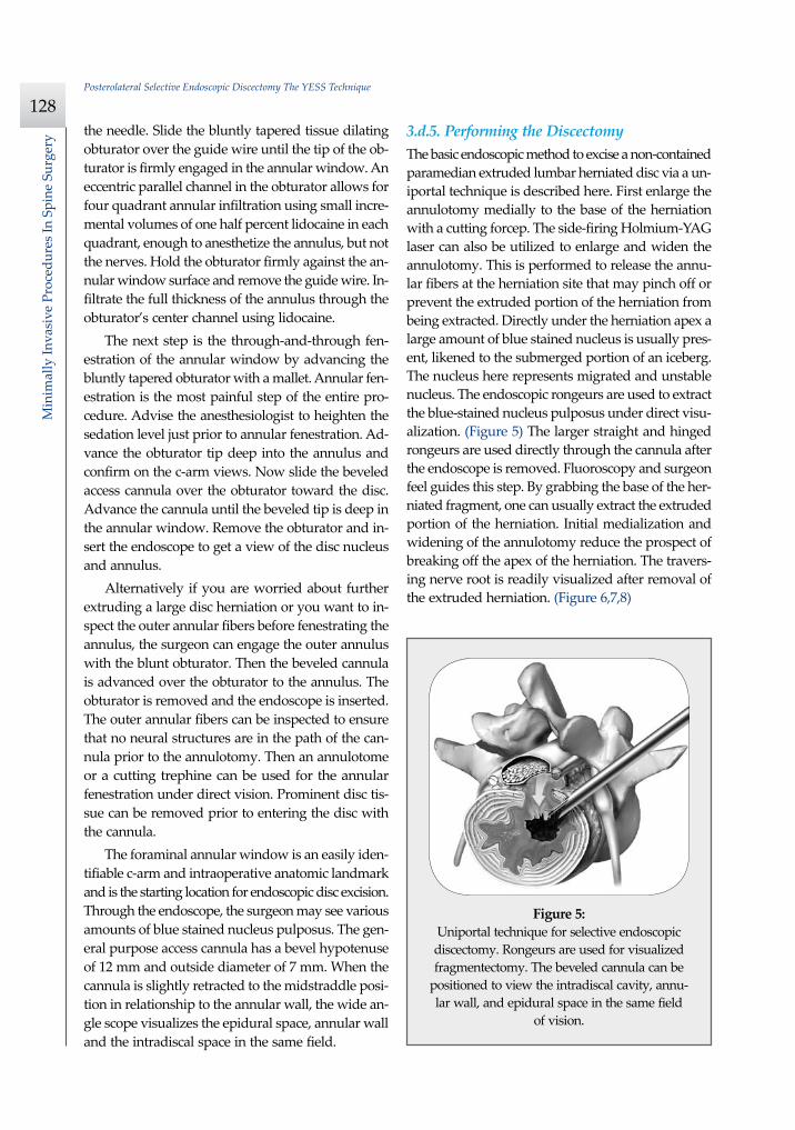
3.d.5. Performing the DiscectomyThe basic endoscopic method to excise a non-contained paramedian extruded lumbar herniated disc via a un-iportal technique is described here. First enlarge the annulotomy medially to the base of the herniation with a cutting forcep. The side-firing Holmium-YAG laser can also be utilized to enlarge and widen the annulotomy. This is performed to release the annu-lar fibers at the herniation site that may pinch off or prevent the extruded portion of the herniation from being extracted. Directly under the herniation apex a large amount of blue stained nucleus is usually pres-ent, likened to the submerged portion of an iceberg. The nucleus here represents migrated and unstable nucleus. The endoscopic rongeurs are used to extract the blue-stained nucleus pulposus under direct visu-alization. (Figure 5) The larger straight and hinged rongeurs are used directly through the cannula after the endoscope is removed. Fluoroscopy and surgeon feel guides this step. By grabbing the base of the her-niated fragment, one can usually extract the extruded portion of the herniation. Initial medialization and widening of the annulotomy reduce the prospect of breaking off the apex of the herniation. The travers-ing nerve root is readily visualized after removal of the extruded herniation. (Figure 6,7,8)
Figure 5: Uniportal technique for selective endoscopic discectomy. Rongeurs are used for visualized fragmentectomy. The beveled cannula can be
positioned to view the intradiscal cavity, annu-lar wall, and epidural space in the same field
of vision.
Anthony T. Yeung M.D., Christopher A. Yeung M.D., Yinggang Zheng M.D.
129
Minim
ally Invasive Procedures In Spine Surgery
Next perform a bulk decompression by using a straight and flexible suction-irrigation shaver (Endius MDS). This step requires shaver head c-arm localiza-tion before power is activated to avoid nerve/dura injury and anterior annular penetration. The cavity thus created is called the working cavity. The deb-ulking process serves two functions. First it decom-presses the disc, reducing the risk for further acute herniation. Second it removes the unstable nucleus material to prevent future reherniation.
Inspect the working cavity. If a non-contained ex-truded disc fragment is still present by finding blue stained nucleus material posteriorly, then these frag-ments are teased into the working cavity with the en-doscopic rongeurs and the flexible radio-frequency trigger-flex bipolar probe (Ellman) and removed. Creation of the working cavity allows the herniated disc tissue to follow the path of least resistance into the cavity. The flexible radio-frequency bipolar probe is used to contract and thicken the annular collagen at the herniation site. It is also used for hemostasis throughout the case.
The vast majority of herniations can be treated via the uniportal technique. Sometimes for a large central herniations the disc needs to be approached from both sides, biportal technique.
4. Potential Complications and Avoidance
As with arthroscopic knee surgery, the risks of seri-ous complications or injury are low—about 1-3% in the author’s experience. The usual risks of infection, nerve injury, dural tears, bleeding, and scar forma-tion are always present as with any surgery. Transient dysesthesia, the most common post-op complaint, oc-curs about 5%-15% of the time and is almost always transient. Its cause is still incompletely understood and may be related to nerve recovery, operating ad-jacent to the dorsal root ganglion of the exiting nerve, or a small hematoma adjacent to the ganglion of the exiting nerve, as it can occur days or even weeks af-ter surgery. Transient dysesthesia can occur even in cases where no adverse events were detected with continuous EMG and SEP neuromonitoring. Thus it cannot be completely avoided. The symptoms are like a variant of complex regional pain syndrome (CRPS), but less severe, and without the skin changes that accompany CRPS. Dysesthesia is readily treated by
transforaminal epidural blocks, rarely sympathetic blocks, and the use of Neurontin titrated up to 1800-3200 mg /day if needed.
Avoidance of complications is enhanced by the ability to clearly visualize normal and patho-anat-omy, the use of local anesthesia and conscious se-dation rather than general or spinal anesthesia, and the use of a standardized needle placement protocol. The entire procedure is usually accomplished with the patient remaining comfortable during the entire procedure and should be done without the patient feeling severe pain except when expected, such as during evocative discography, annular fenestration, or when instruments are manipulated past the ex-iting nerve. Local anesthesia using half percent xy-locaine allows generous use of this dilute anesthetic for pain control and still allows the patient to feel pain when the nerve root is manipulated. Continu-ous EMG and SEP can also help monitor and prevent nerve irritation. This usually correlates well with the patients’ intraoperative feedback.
5. DiscussionEndoscopic spine surgery has a very high learning curve, but is within the grasp of every endoscopic surgeon with proper training. As with any new pro-cedure, the complication rate may be higher during the learning curve, and may vary with each surgeon’s skills and experience. The endoscopic technique is safer for the patient since he is conscious and able to provide immediate input to the surgeon when pain is generated. The surgeon’s ability to perform the surgery without causing the patient undue pain will self select for surgeons who can master the tech-nique to the extent that the surgeon will prefer endo-scopic over traditional surgery for the same condition. For most contained disc herniations and discogenic pain, the experienced endoscopic spine surgeon will opt for the endoscopic approach as the treatment of choice for his patients.
6. Case Presentation
6.a. HistoryA 22 year old male with a two year history of low back pain and intermittent right leg pain sustained an acute worsening of his right leg pain 12 days
Posterolateral Selective Endoscopic Discectomy The YESS Technique
130
Min
imal
ly In
vasi
ve P
roce
dure
s In
Spi
ne S
urge
ry
prior to evaluation. He proportionalized his pain to 5% back and 95% leg pain. He complained of a new onset of weakness, tingling, and constant numbness. The pain and numbness radiated down the poster-olateral leg to the dorsum of the right foot. He was unable to bear weight on the right leg and was us-ing a walking pole for support. He was unable to sleep supine and had to sleep in a recliner to mini-mize the pain. Sitting provided some relief. He de-nied bowel or bladder incontinence, but had consti-pation for the last 12 days.
6.b. Physical ExamPhysical exam revealed an antalgic gait, limited lum-bar extension to 10 degrees, tenderness in the right sciatic notch, positive straight leg raising (SLR) and Lasegue’s tests, positive contralateral SLR, 2+ bilat-eral patella and Achilles deep tendon reflexes, de-creased sensation to light touch over the dorsum of the foot and to a lesser extent the lateral border of the foot, and weakness. The right sided weakness was graded as 4/5 anterior tibialis, 2/5 EHL, 3/5 hip abductor, 4/5 gastroc-soleus.
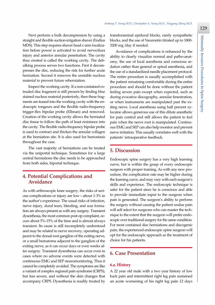
6.c. ImagingMRI revealed a large right paracentral/foraminal ex-truded herniated nucleus pulposus with slight cau-
dal migration causing compression of both the exit-ing and traversing nerve roots (Figure 9).
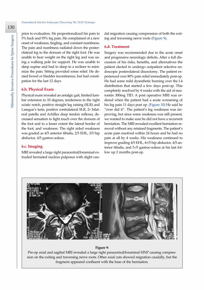
6.d. TreatmentSurgery was recommended due to the acute onset and progressive neurologic deficits. After a full dis-cussion of his risks, benefits, and alternatives the patient elected to undergo outpatient selective en-doscopic posterolateral discectomy. The patient ex-perienced over 80% pain relief immediately post-op. He had some mild dysesthetic burning over the L4 distribution that started a few days post-op. This completely resolved by 4 weeks with the aid of neu-rontin 300mg TID. A post operative MRI was or-dered when the patient had a acute worsening of his leg pain 11 days post op. (Figure 10) He said he “over did it”. The patient’s leg weakness was im-proving, but since some weakness was still present, we wanted to make sure he did not have a recurrent herniation. The MRI revealed excellent herniation re-moval without any retained fragments. The patient’s acute pain resolved within 24 hours and he had no pain at all by 4 weeks. His weakness continued to improve grading 4/5 EHL, 4+/5 hip abductor, 4/5 an-terior tibialis, and 5-/5 gastroc-soleus at his last fol-low up 2 months post-op.
Figure 9: Pre-op axial and sagittal MRI revealed a large right paracentral/foraminal HNP causing compres-sion on the exiting and traversing nerve roots. Other axial cuts showed migration caudally, but the
fragment appeared confluent with the base of the herniation.
Mehmet Zileli M.D., Vehbi Gulmen M.D.
131
Minim
ally Invasive Procedures In Spine Surgery
7. References1. Bogduk N. The Innervation of the Intervertebral
Disc. Chapter 5: 135-149. The Biology of the Inter-vertebral Disc, Vol I. Ghosh P ed. CRC Press, Boca Raton, FL. 1988.
2. Luomaa K, Kilkka R, Luukkonen R, et al. Low Back Pain in Relation to Lumbar Disc Degeneration. Spine 2000;25:487-492
3. Boden S, Herzog R, Rauschning W, Rydevik B. In-structional Course Lecture #249: Lumbar Spine. The Herniated Disc. AAOS 67th Annual Meeting, 2000.
4. Hadjipavlou AG, Simmons JW, Gaitanis IN, Goel VK. Etiopathogenisis of Disc Degeneration and Clin-ical Implication. In: Savitz MH, Chiu JC, Yeung AT, eds. The Practice of Minimally Invasive Spinal Tech-nique. First edition. Richmond, VA. AAMISMS Ed-ucation, LLC; 2000;16:149-170
5. Farfan H. Mechanical Disorders of the Low Back. Philadelphia, PA: Lea & Febiger; 1973
6. Bogduk N. Clinical Anatomy of the Lumbar Spine and Sacrum, 2nd Ed. Melbourn, Australia: Churchill Livingston; 1996
7. Adams MA, Hutton WC. Gradual Disc Prolapse. Spine 1985;10:524-531
8. Adams MA, Hutton WC. Prolapsed Intervertebral Disc—A Hyperflexion Injury. Spine 1982;7:184-191
9. Chaffin DP, Anderson GBJ. Occupational Biome-chanics. New York, NY: Wiley Interscience; 1984:369-412
10. Herzog RJ, Rydevick B, Boden SD. Instructional Course Lecture #310: Lumbar spine. The Herniated Disc. AAOS 70th Annual Meeting; February 7, 2003; New Orleans, LA
11. Hermantin FU, Peters T, Quartararo L, Kambin P. A Prospective, Randomized Study Comparing the Results of Open Discectomy with those of Video-Assisted Arthroscopic Microdiscectomy. JBJS 1999; 81-A:958-965
12. Hijikata S, Yamagishi M, Nakayma T. Percutaneous Discectomy: A New Treatment Method for Lumbar Disc Herniation. J. Tokyo Den-ryoku Hosp1975;5:39-44
13. Hijikata S. Percutaneous Nucleotomy. A New Con-cept Technique and 12 Years’ Experience. Clin. Or-thop 1989;238:9-23
14. Kambin P, Gellman H. Percutaneous Lateral Discec-tomy of the Lumbar Spine. A Preliminary report . Clin. Orthop 1983;174:127-132
15. Kambin P, Sampson S. Posterolateral Percutaneous suction-excision of Herniated Lumbar Interverte-bral Discs. Report of Interim Results. Clin. Orthop 1986;207:37-43
16. Kambin P, Brager M.D. Percutaneous Posterolat-eral Discectomy. Anatomy and Mechanism. Clin. Orthop 1987;223:145-154. 1987.
17. Kambin P, Schaffer JL. Percutaneous Lumbar Dis-cectomy. Review of 100 Patients and Current Prac-tice. Clin. Orthop 1989;238:24-34
18. Kambin P. Arthroscopic Microdiscectomy. Arthros-copy 1992;8:287-295
Figure 10: Post op MRI revealed excellent removal of the herniated disc and decompression of the nerve
roots. The instrument trajectory can be seen within the disc as an area of higher signal on the T2 weighted image.
Microendoscopic Discectomy Using METRx System
132
Min
imal
ly In
vasi
ve P
roce
dure
s In
Spi
ne S
urge
ry
19. Kambin P, O’brien E, Zhou L, Schaffer JL. Arthroscopic Microdiscectomy and Selective Fragmentectomy. Clin Orthop 1998;347:150-167
20. Mathews HH. Transforaminal Endoscopic Micro-discectomy. Neurosurg Clin N Am 1996;7:59-63.
21. Mayer HM, Brock M: Percutaneous Endoscopic Discectomy: Surgical Technique and Preliminary Results Compared to Microsurgical Discectomy. J Neurosurg 1993;78: 216-225
22. Onik G, Helms CA, Ginsberg L, Hooglund FT, Mor-ris J. Percutaneous Lumbar Discectomy Using a New Aspiration Probe. Am. J. Roentgenol 1985;144:1137-1140
23. Schaffer JL, Kambin P. Percutaneous Posterolateral Lumbar Discectomy and Decompression with a 6.9-Millimeter Cannula. Analysis of Operative Failures’ and Complications. JBJS 1991;73-A 822-831
24. Schreiber A, Suezawa Y, Leu H. Does Percutane-ous Nucleotomy with Discoscopy Replace Conven-tional Discectomy? Eight Years of Experience and Results in Treatment of Herniated Lumbar Disc. Clin. Orthop1989;238:35-42
25. Tsou PM, Yeung AT. Transforaminal Endoscopic De-compression for Radiculopathy Secondary to Non-contained Intracanal Lumbar Disc Herniation. Spine Journal 2002; 2:41-48
26. Yeung AT. The Evolution of Percutaneous Spinal Endoscopy and Discectomy: State of Art. Mt. Sinai J Med 2000; 67:327-332
27. Yeung, AT. Minimally Invasive Disc Surgery with the Yeung Endoscopic Spine System (Y.E.S.S.). Sur-gical Technology International VIII 1999;1-11
28. Yeung AT, Tsou PM. Posterolateral Endoscopic Exci-sion for Lumbar Disc Herniation. The Surgical Tech-nique, Outcome and Complications in 307 Consec-utive Cases. Spine 2002;27:722-731
29. Gogan WJ, Fraser RD. Chymopapain. A 10-Year, Double Blind Study. Spine 1992;17:388-94
30. Javid MJ, Norby E. Safety and Efficacy of Chymopa-pain (Chymodiactin) in Herniated Nucleus Pulpo-
sus with Sciatica – Results of a Randomized, Dou-ble Blind Study. JAMA 1983;249:2489-2494
31. Smith L. Chemonucleosis. Personal History, Trials, and Tribulations. Clin Orthop 1993;287:117-124
32. Hoogland T, Scheckenbach C. Percutaneous Lum-bar Nucleotomy with Low-dose Chymopapain, an Ambulatory Procedure. Z Orthop Ihre Grenzgeb 1995;133(2):106-13. German
33. Van de Belt H, Franssen S, Deutman R. Repeat Chemonucleolysis is Safe and Effective. Clin Or-thop 1999;363:121-5
34. Deutman R. The Case for Chemonucleolysis in Discogenic Sciatica. A review. Acta Orthop Scand. 1992;63(5):571-5. Review
35. Yasargil MG. Microsurgical Operation of Herniated Lumbar Disc. In: Wullenweber R, Brock M, Hamer J, Klinger M, Spoerri O, eds. Advances in Neuro-surgery, vol 4. Berlin, New York:Springer-Verlag; 1977:81-94
36. Williams RW. Microlumbar Discectomy: A conser-vative Surgical Approach to the Virgin Herniated Lumbar Disc. Spine 1978;3:175-82
37. Perez-Cruet MJ, Foley KT, Isaacs RE, et al. Microen-doscopic Lumbar Discectomy: Technical Note. Neu-rosurgery 2002;51(5 Suppl):129-36
38. Knight MTN, Goswami AKD. Endoscopic Laser Foraminoplasty. In: Savitz MH, Chiu JC, Yeung AT, eds. The Practice of Minimally Invasive Spinal Tech-nique. First edition. Richmond, VA. AAMISMS Ed-ucation, LLC; 2000;42:337-40
39. Osman SG, Nibu K, Panjabi MM, Marsolais EB, Chaudhary R. Transforaminal and posterior decom-pressions of the lumbar spine. A comparative study of stability and intervertebral foramen area. Spine 1997;22(15):1690-5
40. Ito M, et.al. Transforaminal Surgery for Pyogenic Thoracolumbar Spondylodiscitis. Paper presented at: the American Academy of Minimally Invasive Spinal Medicine and Surgery 3rd World Congress; Dec 8-11, 2002; Phoenix, Arizona