anti-inflammatory and antioxidant properties of casein
TRANSCRIPT

molecules
Article
Anti-Inflammatory and Antioxidant Properties ofCasein Hydrolysate Produced Using High HydrostaticPressure Combined with Proteolytic Enzymes
Fatemeh Bamdad 1, Seulki Hazel Shin 1, Joo-Won Suh 2,*, Chamila Nimalaratne 1
and Hoon Sunwoo 1,*1 Centre for Pharmacy & Health Research, Faculty of Pharmacy and Pharmaceutical Sciences,
University of Alberta, 11361-87 Ave, Edmonton, AB T6G 2E1, Canada; [email protected] (F.B.);[email protected] (S.H.S.); [email protected] (C.N.)
2 Center for Nutraceutical and Pharmaceutical Materials, Myongji University, Yongin,Gyeonggi 449-728, Korea
* Correspondence: [email protected] (J.-W.S.); [email protected] (H.S.);Tel.: +82-31-330-6190 (J.-W.S.); +1-780-492-0547 (H.S.)
Academic Editors: Giancarlo Cravotto and Francisco J. BarbaReceived: 9 March 2017; Accepted: 6 April 2017; Published: 10 April 2017
Abstract: Casein-derived peptides are shown to possess radical scavenging and metal chelatingproperties. The objective of this study was to evaluate novel anti-inflammatory properties of caseinhydrolysates (CH) produced by an eco-friendly process that combines high hydrostatic pressurewith enzymatic hydrolysis (HHP-EH). Casein was hydrolysed by different proteases, includingflavourzyme (Fla), savinase (Sav), thermolysin (Ther), trypsin (Try), and elastase (Ela) at 0.1,50, 100, and 200 MPa pressure levels under various enzyme-to-substrate ratios and incubationtimes. Casein hydrolysates were evaluated for the degree of hydrolysis (DH), molecular weightdistribution patterns, and anti-inflammatory properties in chemical and cellular models. Hydrolysatesproduced using HHP-EH exhibited higher DH values and proportions of smaller peptidescompared to atmospheric pressure-enzymatic hydrolysis (AP-EH). Among five enzymes, Fla-digestedHHP-EH-CH (HHP-Fla-CH) showed significantly higher antioxidant properties than AP-Fla-CH.The anti-inflammatory properties of HHP-Fla-CH were also observed by significantly reduced nitricoxide and by the suppression of the synthesis of pro-inflammatory cytokines in lipopolysaccharide(LPS)-stimulated RAW 264.7 macrophage cells. Liquid chromatography with tandem massspectrometry (LC-MS/MS) revealed that 59% of the amino acids of the peptides in HHP-Fla-CHwere composed of proline, valine, and leucine, indicating the potential anti-inflammatory properties.In conclusion, the HHP-EH method provides a promising technology to produce bioactive peptidesfrom casein in an eco-friendly process.
Keywords: casein; enzymatic hydrolysis; high hydrostatic pressure; antioxidant activity; anti-inflammation
1. Introduction
It is well-recognized that food-derived peptides can exert beneficial biological activities inaddition to their basic nutritional role [1]. These bioactive peptides are relatively short (typically2–20 amino acids) and may possess antioxidant, anti-inflammatory, antihypertensive, antimicrobial,and anticancer properties that have a potential role in maintaining or promoting human health [2].Both anti-inflammatory and antioxidant properties are two of the main topics considered for preventingchronic diseases such as cardiovascular disease and cancer due to oxidative stress, as well as abnormalinflammatory responses. The intake of natural food-derived peptides may delay the onset of diseasesby reducing the oxidative damage and pro-inflammatory responses [3,4].
Molecules 2017, 22, 609; doi:10.3390/molecules22040609 www.mdpi.com/journal/molecules
Molecules 2017, 22, 609 2 of 16
Among other animal-derived food sources, milk proteins are considered as a highly nutritiousfood component with a well-balanced essential amino acid composition, and have also been reportedas a good source of bioactive components [5]. Approximately, casein accounts for 80% of the totalmilk proteins, which are mainly composed of αS1 (~40% of total casein), αS2 (~10% of total casein),β (~35% of total casein), and κ (~15% of total casein) casein sub-units [6]. Accordingly, caseinphosphopeptides derived from gastrointestinal and commercial proteases may act as multifunctionalbioactive peptides with radical scavenging and metal chelating properties, and may play a role inenhancing mineral bioavailability [7].
High hydrostatic pressure (HHP) has been used as an important food preservation techniqueto increase the shelf life of meat and vegetable products by inactivating harmful microbes [8,9].Alternatively, the combined use of HHP and enzymatic hydrolysis (HHP-EH) to produce proteinhydrolysates reduces the amount and reaction time of enzymes, resulting in increased hydrolysateyields [10,11]. Accelerated enzyme activity observed at a certain degree of HHP (50–200 MPa) can be aresult of effective enzyme-substrate contact, achieved through an increased solute concentration due tothe compressed solution and a higher rate of diffusion due to disintegrated cellular compartments [11,12].In addition, partially unfolded proteins under HHP are more susceptible to enzyme cleavage [13,14].Therefore, high hydrostatic pressure combined with enzymatic hydrolysis (HHP-EH) is an eco-friendlyprocess compared to enzymatic hydrolysis under atmospheric pressure (AP-EH), because it usesa lower amount of enzymes and solvents. Several studies have reported the use of extremelyhigh-pressure (400–800 MPa) as a pre-treatment before enzymatic hydrolysis [12,15,16], which mayresult in unacceptable protein denaturation. Previously, HHP-EH was used to obtain bioactive peptidesfrom β-lactoglobulin [17], ovalbumin [18], chickpea protein [19], and pinto bean protein [20]. Recently,our lab developed a novel technology of HHP-EH to increase the degree of hydrolysis of hydrophobicproteins such as phosvitin [21].
As mentioned above, casein-derived peptides possess strong radical scavenging and metalchelating properties. A combined use of high hydrostatic pressure and enzymatic hydrolysis of caseinmay produce lower molecular weight peptides with potential antioxidant and anti-inflammatoryproperties. The main objectives of this study were to optimize the conditions for the hydrolysisof casein using the HHP-EH process to produce bioactive peptides and to assess the effect ofHHP on the antioxidant and anti-inflammatory activities of the casein hydrolysates (CH). Variousenzymes, enzyme-to-substrate (E:S) ratios, and pressure levels were used to hydrolyse casein, inorder to determine the optimal conditions required to achieve the highest efficiency of enzymaticcatalysis. The casein hydrolysates were then tested for antioxidant and anti-inflammatory properties, inchemical and cellular models. The effective peptide sequences were also characterized by LC–MS/MS,to identify the potential amino acids contributing to the antioxidant properties. The one-step hydrolysistechnology developed in this study reduces the processing time, avoids the use of harsh chemicals orsolvents, and conserves their bioactivities.
2. Results and Discussion
2.1. Effect of Different Enzymes and High Hydrostatic Pressure on Casein Hydrolysis
Casein was hydrolysed by elastase (Ela), flavourzyme (Fla), savinase (Sav), thermolysin (Ther),trypsin (Try), under the optimum condition for each enzyme (Table 1). Figure 1a presents the effectof pressure levels (25, 50, 100, and 200 MPa) on the degree of hydrolysis (DH) (at E:S ratio of 1:50,hydrolysed for 1 h) of casein. Among the five enzymes used in our study, Fla and Try showedsignificantly higher DH values at HHP compared to AP (1.6- and 1.5-fold higher at 100 MPa than atAP (0.1 MPa), respectively, p < 0.05). Thus, these enzymes were used to prepare casein hydrolysates atan E:S ratio of 1:50, under 100 MPa pressure, and incubated for 1 h for the characterization studies.

Molecules 2017, 22, 609 3 of 16
Molecules 2017, 22, 609 3 of 16
Figure 1. Degree of hydrolysis of casein hydrolysed under different conditions. (a) under atmospheric (0.1 MPa) and different high hydrostatic pressures (HHP, 50, 100, and 200 MPa) hydrolysed with different enzyme treatments at an enzyme-to-substrate (E:S) ratio of 1:50 for 1h; (b) different incubation times (15 and 30 min, 1 and 2 h) at an E:S ratio of 1:50 under 100 MPa; (c) at E: S ratios of 1:25, 1:50, 1:100, and 1:200 under 100 MPa for 1 h. Error bars are expressed as mean ± standard error with n ≥ 3. In each graph, the bars with different lowercase letters represent significant differences (p < 0.05) in degree of hydrolysis (DH) values.
Table 1. Enzymes applied in high hydrostatic pressure combined with enzymatic hydrolysis (HHP-EH) and atmospheric hydrolysis, and the operational conditions.
Enzyme Sources Proteolytic Activity a Optimum ConditionsElastase Elastase from hog pancreas ≥4 U/mg pH 8; 37 °C
Flavourzyme Protease from Aspergillus oryzae ≥500 U/g pH 7; 50 °C Savinase Protease from Bacillus sp. ≥16 U/g pH 7; 55 °C
Thermolysin Protease from Bacillus thermoproteolyticus 14 U/mg pH 7; 50 °C Trypsin Protease derived from porcine pancreas 30 U/g pH 7; 37 °C
a Minimum proteolytic activity of the enzyme at optimum pH and temperature.
In general, HHP (up to 100 MPa) changes the exposure of functional groups, which affects the protein volume (compressibility) and hydration shell surrounding the protein molecule, leading to a higher susceptibility of protein to enzymatic catalysis [10,22–24]. A higher efficiency of cellulase and β-amylase under 100 MPa was observed in the extraction of saponins from ginseng root (1.5- and 1.4-fold increase in saponin yield, respectively) compared to the AP condition [10]. Similar results were also reported by Garcia-Mora et al. [25] in the HHP assisted proteolysis of lentil proteins. They observed a significant increase in the short peptide content of hydrolysates obtained by commercial proteases. Higher pressure levels up to 150 MPa may disrupt larger protein aggregations and thereby increase the substrate accessibility, resulting in increased enzyme efficiency [26]. The pressure levels exceeding 150 MPa may cause the re-association of dissociated aggregates and enzyme inactivation [26,27]; however, at higher pressure levels (>200 MPa), the dissociation is irreversible
Figure 1. Degree of hydrolysis of casein hydrolysed under different conditions. (a) under atmospheric(0.1 MPa) and different high hydrostatic pressures (HHP, 50, 100, and 200 MPa) hydrolysed withdifferent enzyme treatments at an enzyme-to-substrate (E:S) ratio of 1:50 for 1h; (b) different incubationtimes (15 and 30 min, 1 and 2 h) at an E:S ratio of 1:50 under 100 MPa; (c) at E: S ratios of 1:25, 1:50,1:100, and 1:200 under 100 MPa for 1 h. Error bars are expressed as mean ± standard error with n ≥ 3.In each graph, the bars with different lowercase letters represent significant differences (p < 0.05) indegree of hydrolysis (DH) values.
Table 1. Enzymes applied in high hydrostatic pressure combined with enzymatic hydrolysis (HHP-EH)and atmospheric hydrolysis, and the operational conditions.
Enzyme Sources Proteolytic Activity a Optimum Conditions
Elastase Elastase from hog pancreas ≥4 U/mg pH 8; 37 ◦CFlavourzyme Protease from Aspergillus oryzae ≥500 U/g pH 7; 50 ◦C
Savinase Protease from Bacillus sp. ≥16 U/g pH 7; 55 ◦CThermolysin Protease from Bacillus thermoproteolyticus 14 U/mg pH 7; 50 ◦C
Trypsin Protease derived from porcine pancreas 30 U/g pH 7; 37 ◦Ca Minimum proteolytic activity of the enzyme at optimum pH and temperature.
In general, HHP (up to 100 MPa) changes the exposure of functional groups, which affects theprotein volume (compressibility) and hydration shell surrounding the protein molecule, leading toa higher susceptibility of protein to enzymatic catalysis [10,22–24]. A higher efficiency of cellulaseand β-amylase under 100 MPa was observed in the extraction of saponins from ginseng root (1.5- and1.4-fold increase in saponin yield, respectively) compared to the AP condition [10]. Similar results werealso reported by Garcia-Mora et al. [25] in the HHP assisted proteolysis of lentil proteins. They observeda significant increase in the short peptide content of hydrolysates obtained by commercial proteases.Higher pressure levels up to 150 MPa may disrupt larger protein aggregations and thereby increasethe substrate accessibility, resulting in increased enzyme efficiency [26]. The pressure levels exceeding

Molecules 2017, 22, 609 4 of 16
150 MPa may cause the re-association of dissociated aggregates and enzyme inactivation [26,27];however, at higher pressure levels (>200 MPa), the dissociation is irreversible and leads to smallercaseinate particles [28]. The disruption of electrostatic bridges leaves more charged groups on proteinchains that attract water molecules. The diffusion of water to the protein cavities leads to a higher rateof mass transfer and efficient protein-enzyme contact [16].
2.2. Effect of Incubation Time on Casein Hydrolysis
Optimum casein hydrolysis at 100 MPa was evaluated by performing a series of experimentsat different incubation times (15 min, 30 min, 1 h, and 2 h) and an E:S ratio of 1:50. Hydrolysis for alonger period resulted in a significant increase in the DH values in all samples compared to 15 minhydrolysis (Figure 1b). Hydrolysis for 2 h did not further increase the DH compared to 1 h in Fla, Sav,and Try-hydrolysed samples. This is in agreement with the previous report, which stated that the DHof Try hydrolysates of casein increases rapidly in the initial 20 min, reaching a plateau after 45 min,before remaining constant for the rest of the hydrolysis [29].
2.3. Effect of Enzyme-to-Substrate (E:S) Ratio on Casein Hydrolysis
Optimum casein hydrolysis at 100 MPa was evaluated at different E:S ratios (1:25, 50, 100, and200) for 1 h. A higher amount of enzymes (E:S ratios of 1:25, 1:50) led to a higher DH compared to anE:S ratio of 1:200 (Figure 1c). In other words, with increased amounts of substrate in the mixture, theefficiency of the enzymes decreased. For four out of five of the enzymes used in the study (Ela, Sav,Ther, and Try), both the E:S ratios of 1:25 and 1:50 yielded similar DH values. Therefore, an E:S ratio of1:50 was selected for further experiments, to reduce the cost and the usage of excess enzymes.
2.4. Molecular Weight Distribution of CH
Figure 2 illustrates the size exclusion chromatograms of intact casein and casein hydrolysatesproduced by Fla and Try. The intact casein chromatogram showed three peaks at 14, 18, and 22 min.The first and second peaks represent oligomers of casein molecules as their MW (670 and 91 kDa) arelarger than individual caseins (MW range: 19–25 kDa). Enzymatic hydrolysis caused a pronouncedshift of the peaks towards a lower MW range. Fla completely digested the intact casein peaks andproduced short peptides with an MW value of less than 3 kDa. However, Try resulted in the partialhydrolysis of casein, which produced two peaks corresponding to 25 and 7 kDa peptides.
Molecules 2017, 22, 609 4 of 16
and leads to smaller caseinate particles [28]. The disruption of electrostatic bridges leaves more charged groups on protein chains that attract water molecules. The diffusion of water to the protein cavities leads to a higher rate of mass transfer and efficient protein-enzyme contact [16].
2.2. Effect of Incubation Time on Casein Hydrolysis
Optimum casein hydrolysis at 100 MPa was evaluated by performing a series of experiments at different incubation times (15 min, 30 min, 1 h, and 2 h) and an E:S ratio of 1:50. Hydrolysis for a longer period resulted in a significant increase in the DH values in all samples compared to 15 min hydrolysis (Figure 1b). Hydrolysis for 2 h did not further increase the DH compared to 1 h in Fla, Sav, and Try-hydrolysed samples. This is in agreement with the previous report, which stated that the DH of Try hydrolysates of casein increases rapidly in the initial 20 min, reaching a plateau after 45 min, before remaining constant for the rest of the hydrolysis [29].
2.3. Effect of Enzyme-to-Substrate (E:S) Ratio on Casein Hydrolysis
Optimum casein hydrolysis at 100 MPa was evaluated at different E:S ratios (1:25, 50, 100, and 200) for 1 h. A higher amount of enzymes (E:S ratios of 1:25, 1:50) led to a higher DH compared to an E:S ratio of 1:200 (Figure 1c). In other words, with increased amounts of substrate in the mixture, the efficiency of the enzymes decreased. For four out of five of the enzymes used in the study (Ela, Sav, Ther, and Try), both the E:S ratios of 1:25 and 1:50 yielded similar DH values. Therefore, an E:S ratio of 1:50 was selected for further experiments, to reduce the cost and the usage of excess enzymes.
2.4. Molecular Weight Distribution of CH
Figure 2 illustrates the size exclusion chromatograms of intact casein and casein hydrolysates produced by Fla and Try. The intact casein chromatogram showed three peaks at 14, 18, and 22 min. The first and second peaks represent oligomers of casein molecules as their MW (670 and 91 kDa) are larger than individual caseins (MW range: 19–25 kDa). Enzymatic hydrolysis caused a pronounced shift of the peaks towards a lower MW range. Fla completely digested the intact casein peaks and produced short peptides with an MW value of less than 3 kDa. However, Try resulted in the partial hydrolysis of casein, which produced two peaks corresponding to 25 and 7 kDa peptides.
Figure 2. Size exclusion chromatograms of casein hydrolysates using different enzyme treatments at an E:S ratio of 1:50 under 100 MPa for 1 h.
Matrix-assisted laser desorption/ionization time-of-flight (MALDI–TOF) mass spectra of samples showed that the MW of short peptides presented a wide range of values (500 to >2000 Da). Table 2
Figure 2. Size exclusion chromatograms of casein hydrolysates using different enzyme treatments atan E:S ratio of 1:50 under 100 MPa for 1 h.
Molecules 2017, 22, 609 5 of 16
Matrix-assisted laser desorption/ionization time-of-flight (MALDI–TOF) mass spectra of samplesshowed that the MW of short peptides presented a wide range of values (500 to >2000 Da). Table 2summarizes the relative area under the peaks categorized in the three MW ranges (500–1000, 1000–2000,and >2000 Da). It should be noted that, for a better accuracy and to avoid the background noises, onlythe peptides from 500 to 5000 Da were detected in the MALDI–TOF analysis. Therefore, the peptidessmaller than 500 Da are not included in this peptide distribution. Fla-treated samples showed a veryhigh content of 1–2 kDa peptides after digestion (Table 2). Moreover, remarkable increases occurred inthe 500–1000 Da fraction under the HHP condition, especially in the Try sample. The Try-digestedsample had higher proportions of 1–2 kDa and >2 kDa, and the relative area of the small peptidefraction (<1 kDa) increased to 26.5% under HHP conditions, compared to the 4.1% value which wasobtained under AP digestion. Garcia-Mora et al. also observed a similar trend of the percentagedistribution of peptide masses for HHP (at 300 MPa) and AP hydrolysis [25].
Table 2. Relative area (%) of the peptide peaks obtained in matrix-assisted laser desorption/ionizationtime-of-flight (MALDI–TOF) mass spectra of casein hydrolysates produced under an atmospheric andhigh pressure condition.
SamplesAtmospheric Hydrolysis High Pressure Hydrolysis
500–1000 Da 1000–2000 Da >2000 Da 500–1000 Da 1000–2000 Da >2000 Da
Fla-CH 0.3 98.6 1.1 1.9 95.9 2.2Try-CH 4.1 46.5 49.4 26.5 23.7 49.8
In a previous report, Morato et al. [30] studied the optimal conditions for the enzymatic hydrolysisof casein with a higher di- and tri-peptide content, to be used in dietary supplements. They obtained ahydrolysate containing only 14% of peptides and a molecular mass higher than 800 Da with a 4% E:Sratio, which is equivalent to 1:25, using subtilisin for 5 h hydrolysis. Fla is a commercial mixture ofendo- and exo-peptidases obtained from a fungus (Aspergillus oryzae) and has been used to obtain lowmolecular weight peptides [31]. Rossini et al. (2009) also showed that casein hydrolysed by Fla resultedin a higher concentration of free amino acids and low molecular weight peptides, and also exhibitedgreater antioxidant properties [31]. In the current work, the use of a high hydrostatic pressure duringcasein hydrolysis by Fla resulted in a hydrolysate with significantly higher antioxidant properties anddegree of hydrolysis compared to the hydrolysate obtained under atmospheric pressure. This studyalso demonstrated the anti-inflammatory properties of HHP-Fla-CH through its ability to suppress theNO production and synthesis of pro-inflammatory cytokines in lipopolysaccharide (LPS)-stimulatedRAW 264.7 macrophage cells.
2.5. Antioxidant Capacity of CH
The antioxidant activity of hydrolysates may be attributed to the radical scavenging propertiesor the metal chelating capacity of peptides, or a combined effect of both. In this study, to betterunderstand the antioxidant capacity of casein hydrolysates, we used 1,1-diphenyl-2-picryl Hydrazyl(DPPH) and superoxide scavenging assays to determine the radical scavenging capacity. For themetal chelating ability, iron chelating and ferric reducing antioxidant power (FRAP) assays were used.The enzymatic hydrolysis of casein under AP and HHP improved the antioxidant properties of CH(Figure 3).
2.5.1. 1,1-Diphenyl-2-picryl Hydrazyl (DPPH) Radical Scavenging Capacity
DPPH is a relatively stable free radical, which is widely used for the in vitro evaluation ofantioxidant compounds [32]. Free radicals are stabilized by hydrogen or electron donations fromantioxidant peptides, leading to stable non-reactive compounds which cannot trigger the oxidationchain reactions [33]. As presented in Figure 3a, CH showed a dose-dependent DPPH scavenging
Molecules 2017, 22, 609 6 of 16
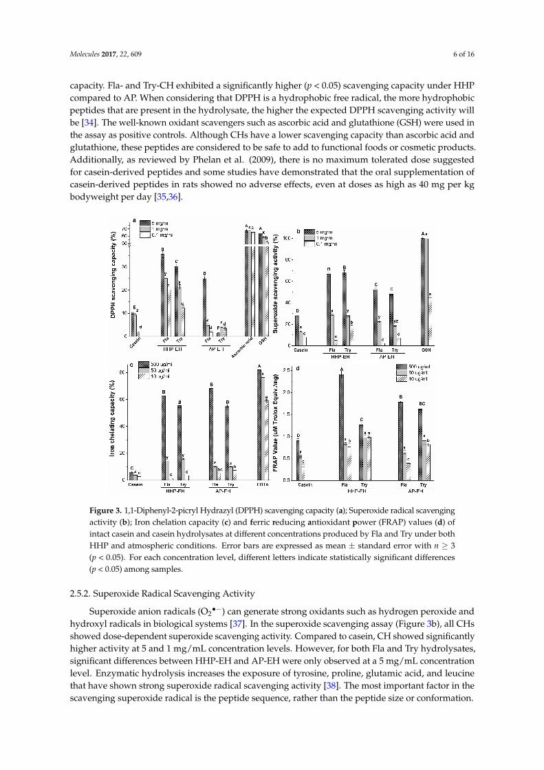
capacity. Fla- and Try-CH exhibited a significantly higher (p < 0.05) scavenging capacity under HHPcompared to AP. When considering that DPPH is a hydrophobic free radical, the more hydrophobicpeptides that are present in the hydrolysate, the higher the expected DPPH scavenging activity willbe [34]. The well-known oxidant scavengers such as ascorbic acid and glutathione (GSH) were used inthe assay as positive controls. Although CHs have a lower scavenging capacity than ascorbic acid andglutathione, these peptides are considered to be safe to add to functional foods or cosmetic products.Additionally, as reviewed by Phelan et al. (2009), there is no maximum tolerated dose suggestedfor casein-derived peptides and some studies have demonstrated that the oral supplementation ofcasein-derived peptides in rats showed no adverse effects, even at doses as high as 40 mg per kgbodyweight per day [35,36].
Molecules 2017, 22, 609 6 of 16
in the assay as positive controls. Although CHs have a lower scavenging capacity than ascorbic acid and glutathione, these peptides are considered to be safe to add to functional foods or cosmetic products. Additionally, as reviewed by Phelan et al. (2009), there is no maximum tolerated dose suggested for casein-derived peptides and some studies have demonstrated that the oral supplementation of casein-derived peptides in rats showed no adverse effects, even at doses as high as 40 mg per kg bodyweight per day [35,36].
Figure 3. 1,1-Diphenyl-2-picryl Hydrazyl (DPPH) scavenging capacity (a); Superoxide radical scavenging activity (b); Iron chelation capacity (c) and ferric reducing antioxidant power (FRAP) values (d) of intact casein and casein hydrolysates at different concentrations produced by Fla and Try under both HHP and atmospheric conditions. Error bars are expressed as mean ± standard error with n ≥ 3 (p < 0.05). For each concentration level, different letters indicate statistically significant differences (p < 0.05) among samples.
2.5.2. Superoxide Radical Scavenging Activity
Superoxide anion radicals (O2•−) can generate strong oxidants such as hydrogen peroxide and hydroxyl radicals in biological systems [37]. In the superoxide scavenging assay (Figure 3b), all CHs showed dose-dependent superoxide scavenging activity. Compared to casein, CH showed significantly higher activity at 5 and 1 mg/mL concentration levels. However, for both Fla and Try hydrolysates, significant differences between HHP-EH and AP-EH were only observed at a 5 mg/mL concentration level. Enzymatic hydrolysis increases the exposure of tyrosine, proline, glutamic acid, and leucine that have shown strong superoxide radical scavenging activity [38]. The most important factor in the scavenging superoxide radical is the peptide sequence, rather than the peptide size or conformation.
2.5.3. Iron Chelating Activity
Iron and copper are the most important pro-oxidant metal ions. They facilitate oxidation reactions by catalysing hydroperoxide decomposition that results in hydroxyl radicals as the most reactive oxygen species [39,40]. Therefore, metal ion chelation plays an important role in the
Figure 3. 1,1-Diphenyl-2-picryl Hydrazyl (DPPH) scavenging capacity (a); Superoxide radical scavengingactivity (b); Iron chelation capacity (c) and ferric reducing antioxidant power (FRAP) values (d) ofintact casein and casein hydrolysates at different concentrations produced by Fla and Try under bothHHP and atmospheric conditions. Error bars are expressed as mean ± standard error with n ≥ 3(p < 0.05). For each concentration level, different letters indicate statistically significant differences(p < 0.05) among samples.
2.5.2. Superoxide Radical Scavenging Activity
Superoxide anion radicals (O2•−) can generate strong oxidants such as hydrogen peroxide and
hydroxyl radicals in biological systems [37]. In the superoxide scavenging assay (Figure 3b), all CHsshowed dose-dependent superoxide scavenging activity. Compared to casein, CH showed significantlyhigher activity at 5 and 1 mg/mL concentration levels. However, for both Fla and Try hydrolysates,significant differences between HHP-EH and AP-EH were only observed at a 5 mg/mL concentrationlevel. Enzymatic hydrolysis increases the exposure of tyrosine, proline, glutamic acid, and leucinethat have shown strong superoxide radical scavenging activity [38]. The most important factor in thescavenging superoxide radical is the peptide sequence, rather than the peptide size or conformation.

Molecules 2017, 22, 609 7 of 16
2.5.3. Iron Chelating Activity
Iron and copper are the most important pro-oxidant metal ions. They facilitate oxidation reactionsby catalysing hydroperoxide decomposition that results in hydroxyl radicals as the most reactiveoxygen species [39,40]. Therefore, metal ion chelation plays an important role in the antioxidantmechanism of the peptides. Furthermore, recent evidence has shown that long chain peptidescan entrap the metal ions in a cage-like structure [39]. The CH showed dose-dependent activitywith the highest iron chelating activity (55–68%) being viewed at 500 µg/mL, which is comparableto ethylenediaminetetraacetic acid (EDTA) (60% at 10 µg/mL, p < 0.05) (Figure 3c). There are nodifferences in the iron chelating capacity of the HHP- and AP-EH samples, or among the Fla- andTry-CH at a higher concentration level (500 µg/mL).
2.5.4. Reducing Capacity (FRAP assay)
A FRAP assay measures the electron-donor capacity of antioxidant compounds. In this assay, Fe3+
is reduced to Fe2+ by electron-donation from electron-rich amino acid side chains [41]. As presentedin Figure 3d, at a 500 µg/mL concentration of peptides, only the HHP-Fla-CH sample showed asignificantly higher FRAP value than AP compared with Try-CH (p < 0.05).
Possibly, smaller peptides could exhibit better reducing activity towards ferric ions, and suppresstheir pro-oxidant effect. Small peptides have a higher charge density (charge-to-mass ratio) thanlarger peptides, due to their more exposed electron-rich side chains. Similar results were observed byLin et al. when studying the reducing capacity of small peptides (<1 kDa) obtained from egg whitehydrolysates [42].
2.6. Anti-Inflammatory Properties of CH
2.6.1. Cell Viability Assay
The RAW 264.7 cell viability was evaluated to demonstrate the effect of CH on macrophageproliferation. The results (Figure 4a) showed that cells treated with all HHP-EH-CH (1 mg/mL)had >90% viability, indicating that CH did not influence RAW cell proliferation and thus, may beconsidered safe to the cells. It was previously demonstrated that whey protein hydrolysates producedunder high pressure are well-tolerated (over 80% viability) by epithelial cells at peptide concentrationsup to 1 mg/mL [43].
After 24 h incubation, both HHP- and AP-EH-CH did not show significant variations in cellviability. The detailed mechanisms of the uptake of peptides and the effect of peptides on cellularmetabolism requires further investigation to ensure the safe use of peptides.
2.6.2. Determination of NO Production by Macrophage Cells
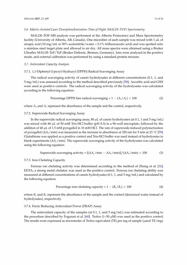
Antioxidant peptides can reduce oxidative signaling, which is a part of inflammatory signalingcascades [43]. Oxidative stress stimulates the production of pro-inflammatory cytokines [44].Nitric oxide is an important mediator of inflammatory processes. The overproduction of NO isassociated with diabetes mellitus, neurodegenerative disorders, and other inflammatory conditions [45].Therefore, in our study, the ability of HHP-EH-CH to attenuate NO production was estimated usingLPS-induced macrophage cells (Figure 4b). The content of NO in a RAW 264.7 cell supernatant wasevaluated after pre-incubation with HHP-EH-CH before LPS treatment (Figure 4b). The negativecontrol showed a minimal content of NO (0.03 µM NO in cell supernatant), while LPS significantlystimulated NO production up to 7.8 µM NO. Pre-incubation with HHP-EH-CH significantly (p < 0.05)suppressed NO production to 3.5–6.5 µM NO, while non-hydrolysed casein showed no effect on NOproduction. Although both HHP-Fla-CH and HHP-Try-CH significantly reduced the production of NO,HHP-Try-CH was the most efficient in the down-regulation of NO production in macrophage cells.
In a previous study, Alcalase-hydrolysed soy protein showed an 18–35% inhibition in NOproduction in LPS-induced macrophage cells [46]. Different mechanisms have been proposed for

Molecules 2017, 22, 609 8 of 16
the inhibition of LPS-induced effects on macrophage cell lines by proteins and peptides. Binding tothe lipid A moiety of LPS and interference with the LPS-CD14 interaction by competence with theLPS-binding protein have been suggested [47]. Other factors, such as the amino acid sequence, alsoinfluence the cell internalization of peptides, which may change their inhibitory effect [48].
2.6.3. Gene Expression of Pro-Inflammatory Cytokines in LPS-Stimulated Macrophages
Pro-inflammatory cytokine gene expression in macrophage cells was analysed to confirm theanti-inflammatory effect of HHP-Fla-CH. The mRNA level of pro-inflammatory cytokines (tumornecrosis factor (TNF-α) and interleukin (IL)1β (IL-1β)) increased in LPS-stimulated cells (Figure 4c).Without LPS-stimulation, the HHP-Fla-CH showed a minor effect on gene expression of TNF-α andIL-1β. Exposure to LPS strongly induced TNF-α and IL-1β gene expression by 32 and 500-times,respectively. The effect of HHP-Fla-CH on TNF-α and IL-1β gene expression was significantly higher(p < 0.05) than intact casein, and reduced the TNF-α and IL-1 β gene expression by 83 and 85%,respectively. The present data demonstrated that HHP-Fla-CH exhibited a remarkable inhibitory effecton TNF-α and IL-1β gene expression (p < 0.05).
Molecules 2017, 22, 609 8 of 16
2.6.3. Gene Expression of Pro-Inflammatory Cytokines in LPS-Stimulated Macrophages
Pro-inflammatory cytokine gene expression in macrophage cells was analysed to confirm the anti-inflammatory effect of HHP-Fla-CH. The mRNA level of pro-inflammatory cytokines (tumor necrosis factor (TNF-α) and interleukin (IL)1β (IL-1β)) increased in LPS-stimulated cells (Figure 4c). Without LPS-stimulation, the HHP-Fla-CH showed a minor effect on gene expression of TNF-α and IL-1β. Exposure to LPS strongly induced TNF-α and IL-1β gene expression by 32 and 500-times, respectively. The effect of HHP-Fla-CH on TNF-α and IL-1β gene expression was significantly higher (p < 0.05) than intact casein, and reduced the TNF-α and IL-1 β gene expression by 83 and 85%, respectively. The present data demonstrated that HHP-Fla-CH exhibited a remarkable inhibitory effect on TNF-α and IL-1β gene expression (p < 0.05).
Figure 4. (a) Viability of RAW 264.7 cells; (b) Nitric oxide content of RAW 264.7 cell supernatant in the presence of intact casein or casein hydrolysates (at 1 mg/mL) produced under the HHP condition and (c) mRNA expression of pro-inflammatory cytokines (TNF-α and IL-1β) relative to glyceraldehyde 3-phosphate dehydrogenase (GAPDH) (∆∆Ct) in LPS-stimulated RAW 264.7 macrophage cells in presence of intact casein (10 μg/mL) or Fla-hydrolysed casein (10 μg/mL). Values with different lower case letters are significantly (p < 0.05) different. Error bars are expressed as mean ± standard error with n ≥ 3 (p < 0.05).
2.7. Peptide Sequencing of the Most Potent CH
HHP-Fla-CH was selected as the most potent sample for peptide sequencing due to the higher degree of hydrolysis, and high antioxidant and anti-inflammatory properties. A total of nine peptides characterized by the MS/MS and NCBI protein database search are shown in Table 3. Peptides with 6–14 amino acids ranging from 590 to 1500 Da mainly contain hydrophobic residues, making up about 59% of the sequence. The majority of antioxidant peptides have a molecular weight range of
Figure 4. (a) Viability of RAW 264.7 cells; (b) Nitric oxide content of RAW 264.7 cell supernatant in thepresence of intact casein or casein hydrolysates (at 1 mg/mL) produced under the HHP condition and(c) mRNA expression of pro-inflammatory cytokines (TNF-α and IL-1β) relative to glyceraldehyde3-phosphate dehydrogenase (GAPDH) (∆∆Ct) in LPS-stimulated RAW 264.7 macrophage cells inpresence of intact casein (10 µg/mL) or Fla-hydrolysed casein (10 µg/mL). Values with different lowercase letters are significantly (p < 0.05) different. Error bars are expressed as mean ± standard error withn ≥ 3 (p < 0.05).

Molecules 2017, 22, 609 9 of 16
2.7. Peptide Sequencing of the Most Potent CH
HHP-Fla-CH was selected as the most potent sample for peptide sequencing due to the higherdegree of hydrolysis, and high antioxidant and anti-inflammatory properties. A total of ninepeptides characterized by the MS/MS and NCBI protein database search are shown in Table 3.Peptides with 6–14 amino acids ranging from 590 to 1500 Da mainly contain hydrophobic residues,making up about 59% of the sequence. The majority of antioxidant peptides have a molecularweight range of 500–1800 Da [49]. Peptide identification by LC–MS/MS indicated that the mainsource of milk-derived antioxidant peptides is β-casein, which contains the most hydrophobicresidues. A high content of hydrophobic residues such as alanine, valine, leucine, and isoleucineshows a positive influence on scavenging free radicals [50]. Antioxidant peptides isolated fromsquid skin gelatin contained proline, alanine, valine, and leucine, which contribute the most toantioxidant activity [51]. All of the peptides identified in HHP-Fla-CH have more than one prolineresidue in their sequence. Proline has an electron-rich nitrogen-containing pyrrolidone ring thatstabilizes the radical peptide formed after electron donation [52]. Proline-rich peptides are identifiedas multifunctional, with antioxidant, antimicrobial, and immunomodulatory properties, and thusmay play a promising role in peptide-based food/supplement products [53]. Several studies havecharacterized antioxidant peptides with the amino acid sequence YFYPEL from αS1 casein [38],and QKALNEINQF, TKKTKLTEEEKNRL [54], FALPQYLK, and PYVRYL [55] from αS2 casein.The antioxidant properties of these peptides were mainly attributed to the presence of aromatic aminoacids such as tyrosine and phenylalanine [55]. Among the identified sequences, five peptides haveglutamic acid and glutamine as the terminal or penultimate amino acids, which can act as redox-activeresidues [52,56]. Therefore, our findings suggest that a proper balance between hydrophobic and polaramino acids may positively contribute to the antioxidant activity of casein peptides.
Table 3. Amino acid sequences of potent peptides in Fla-hydrolysed casein identified by LC–MS/MSand their respective protein fragments.
Peptide Sequence Ion (m/z) Observed Mass Calculated Mass Source (Fragment)
PGPIPN 594.33 593.33 593.32 β-Casein (78–83)PFPGPIPN 838.44 837.43 837.44 β-Casein (76–83)YPFPGPIP 887.47 886.47 886.46 β-Casein (75–82)
VYPFPGPIPN 1100.55 1099.55 1099.57 β-Casein (74–83)MPFPKYPVEP 610.82 (2) 1219.62 1219.59 β-Casein (124–133)
EPVLGPVRGPFP 632.87 (2) 1263.73 1263.70 β-Casein (210–221)QEPVLGPVRGPFP 696.90 (2) 1391.78 1391.76 β-Casein (209–221)TPVVVPPFLQPE 661.87 (2) 1321.73 1321.72 β-Casein (95–106)
TQTPVVVPPFLQPE 776.42 (2) 1550.83 1550.83 β -Casein (93–106)
3. Materials and Methods
3.1. Materials
Sodium caseinate powder from bovine milk, flavourzyme (Fla), savinase (Sav), thermolysin(Ther), and phosvitin were purchased from Sigma-Aldrich (Oakville, ON, Canada). Trypsin (Try) wasobtained from Thermo Fisher Scientific (Waltham, MA, USA). Elastase (Ela) was purchased from MPBiomedicals (Santa Ana, CA, USA). The mouse macrophage cell line, RAW 264.7, was obtained fromthe American Type Culture Collection (Rockville, MD, USA) and cultured in Dulbecco’s modifiedEagle’s medium supplemented with 10% fetal bovine serum and penicillin/streptomycin/fungizone.LPS from E. coli 0111:B4 was from Sigma-Aldrich. Cell culture reagents (DMEM, FBS, PenicillinStreptomycin Glutamine, and Dulbecco’s Phosphated Buffered Saline), Ultrapure Distilled Water, andthe TRIzol reagent were purchased from Invitrogen (Carlsbad, CA, USA). 5× All-In-One RT MasterMixand EvaGreen 2× qPCR MasterMix-Low ROX were from Applied Biological Materials (Richmond, BC,

Molecules 2017, 22, 609 10 of 16
Canada). All primers were ordered from IDT (Coralville, IA, USA). All other chemicals and reagentswere of analytical grade and purchased from commercial sources.
3.2. Apparatus
A portable high hydrostatic pressure instrument (TFS-2L, Toyo-Koatsu, Hiroshima, Japan)was used for the HHP-EH process. The vessel was filled with ultrapure water as a fluid of lowcompressibility and the internal pressure reached 50 and 100 MPa in about 1 and 2 min, respectively.The HHP instrument was designed with a fast decompression of the vessel for accurate pressurecontrol. The protein-enzymes mixture was kept in a sealed pouch during the treatment, and theinternal temperature of the vessel was automatically controlled by the computerized automaticpressure and temperature controlling system during the operation.
3.3. Enzymatic Hydrolysis of Casein
Casein was hydrolysed by Ela, Fla, Sav, Ther, and Try. Briefly, a caseinate solution (10 mgprotein/mL distiller water) was mixed with each enzyme at four different enzyme:substrate (E:S)ratios (1:25, 1:50, 1:100, and 1:200 w/w). The pH of the mixtures was adjusted by the addition of0.1 M HCl or NaOH, according to the optimum pH for each enzyme (Table 1). Each mixture was thenplaced in the HHP-EH chamber set at pressures of 50, 100, and 200 MPa for 15, 30, 60, and 120 min atthe enzyme’s optimum temperature. Casein solution was also hydrolysed under atmospheric pressure(AP, 0.1 MPa) for comparison. The above procedures were performed in the absence of enzymesto act as experimental controls. After hydrolysis, the samples were heated at 100 ◦C for 10 min toinactivate the enzyme and were then centrifuged (4000× g, 30 min) to remove the insoluble parts.The supernatants were stored at −20 ◦C prior to analysis. The protein content of the casein sampleswas determined by the Bradford method using the Bio-Rad Proteins Assay kit (Bio-Rad, Mississauga,ON, Canada).
3.4. Determination of Degree of Hydrolysis (DH)
The DH was determined by the quantitative determination of the free amino groups releasedduring hydrolysis, using an O-phthaldialdehyde (OPA) fluorometric assay [57]. The OPA solution(200 µL) was added to 20 µL of standard, samples, and control within the wells of a black microplate.The fluorometer readings were performed using a Synergy H1 Multi-Mode Reader (BioTek, Winooski,VT, USA) at excitation and emission wavelengths of 350 and 450 nm, respectively. The readings werecorrected with unhydrolyzed casein to eliminate the effect of terminal amino groups. Deionizedwater was used as the control and L-serine was used as a standard. The DH was calculated by thefollowing equation:
DH= (h/htot) × 100 (1)
where the hydrolysis equivalent (h) is the amount of free amino groups produced during hydrolysis,expressed as millimoles of serine equivalents per gram protein and htot is the total amount of aminogroups present in totally hydrolysed casein with 6 N HCl at 110 ◦C for 24 h.
3.5. Size Exclusion High Performance Liquid Chromatography (SE-HPLC)
The average molecular weight (MW) of the casein hydrolysates was determined by SE–HPLCusing a Shimadzu 10 AVP HPLC system equipped with a Biosuite 125/5 mm HR-SEC column(7.8 × 300 mm, Waters, Milford, MA, USA). Phosphate buffer (100 mM) containing 100 mM NaCl wasused as the mobile phase at a flow rate of 0.5 mL/min at 25 ◦C. Protein in the effluent was monitored at220 nm. A calibration curve was made from the log MW of the standard markers and their respectiveelution times (R2 = 0.99).
Molecules 2017, 22, 609 11 of 16
3.6. Matrix-Assisted Laser Desorption/Ionization–Time of Flight (MALDI–TOF) Spectrometry
MALDI–TOF–MS analysis was performed at the Alberta Proteomics and Mass Spectrometryfacility (University of Alberta, AB, Canada). One microliter of each sample was mixed with 1 µL ofsinapic acid (10 mg/mL in 50% acetonitrile/water + 0.1% trifluoroacetic acid) and was spotted ontoa stainless steel target plate and allowed to air dry. All mass spectra were obtained using a BrukerUltraflex MALDI–ToF/ToF (Bruker Daltonic, Bremen, Germany). Ions were analysed in the positivemode, and external calibration was performed by using a standard protein mixture.
3.7. Antioxidant Capacity Analysis
3.7.1. 1,1-Diphenyl-2-picryl Hydrazyl (DPPH) Radical Scavenging Assay
The radical scavenging activity of casein hydrolysates at different concentrations (0.1, 1, and5 mg/mL) was assessed according to the method described previously [58]. Ascorbic acid and GSHwere used as positive controls. The radical scavenging activity of the hydrolysates was calculatedaccording to the following equation:
Percentage DPPH free radical scavenging = 1 − (As/Ac) × 100 (2)
where As and Ac represent the absorbance of the sample and the control, respectively.
3.7.2. Superoxide Radical Scavenging Assay
In the superoxide radical scavenging assay, 80 µL of casein hydrolysates (at 0.1, 1 and 5 mg/mL)was mixed with 80 µL of 50 mM Tris-HCl buffer (pH 8.3) in a 96-well microplate, followed by theaddition of 40 µL of 1.5 mM pyrogallol in 10 mM HCl. The rate of superoxide-induced polymerizationof pyrogallol (∆As/min) was measured as the increase in absorbance at 320 nm for 5 min at 23 ◦C [59].Glutathione was applied as a positive control and Tris-HCl buffer was used instead of hydrolysates inblank experiments (∆Ac/min). The superoxide scavenging activity of the hydrolysates was calculatedusing the following equation:
Superoxide scavenging activity = [(∆Ac/min − ∆As/min)]/(∆Ac/min) × 100 (3)
3.7.3. Iron Chelating Capacity
Ferrous ion chelating activity was determined according to the method of Zhang et al. [32].EDTA, a strong metal chelator, was used as the positive control. Ferrous ion chelating ability wasmeasured at different concentrations of casein hydrolysates (0.1, 1, and 5 mg/mL) and calculated bythe following equation:
Percentage iron chelating capacity = 1 − (Bs/Bc) × 100 (4)
where Bs and Bc represent the absorbance of the sample and the control (deionized water instead ofhydrolysates), respectively.
3.7.4. Ferric Reducing Antioxidant Power (FRAP) Assay
The antioxidant capacity of the samples (at 0.1, 1, and 5 mg/mL) was estimated according tothe procedure described by Fogarasi et al. [60]. Trolox (1–50 µM) was used as the positive control.The results were expressed as micromoles of Trolox equivalent (TE) per mg of sample (µmol TE/mg).
Molecules 2017, 22, 609 12 of 16
3.8. Anti-Inflammatory Properties of CH
3.8.1. Cell Viability Assay
The proliferation of Raw 264.7 cells was evaluated using the 3-(4,5-dimethylthiazol-2-yl)-2,5-diphenyltetrazolium bromide (MTT) assay [61]. Briefly, cells were seeded in 96-well microplates at adensity of 1× 10⁴ cells/mL and incubated at 37 ◦C overnight. After treatment with casein hydrolysates(1 mg/mL) for 20 h and washing the cells with Hank’s buffered salt solution (HBSS), 100 µL of MTTreagent (0.5 mg/mL in media) was added and the cell was incubated in the dark for 4 h. Formazancrystals were solubilized in 200 µL dimethyl sulfoxide, and the color intensity was measured at 570 nm.The results were expressed as a percentage of the absorbance in the test wells compared to that in thecontrol wells (media without peptide).
3.8.2. Determination of Nitric Oxide (NO) Production (Griess Assay)
The effect of casein hydrolysates on NO production by stimulated macrophages was evaluatedby the determination of accumulated nitrite (NO2
−) in the culture media [62]. RAW 264.7 cells werecultured at 1 × 10⁵ cells/mL and incubated overnight. Cells were treated with casein hydrolysates(1 mg/mL) for 4 h. After washing cells with HBSS, bacterial LPS (1 µg/mL) was added to stimulatethe macrophage cells and was incubated for 20 h. Then, 100 µL of cell supernatant was mixed (1:1 v/v)with the Griess reagent, followed by 15 min incubation at ambient temperature. The absorbance wasrecorded at 540 nm. The growing cells without any treatment acted as the negative control, while thepositive controls were prepared by stimulating cells with LPS. Sodium nitrite at different concentrationlevels (0.1–100 µM) was used to make the standard curve (R2 = 0.9998).
3.8.3. Real-Time Polymerase Chain Reaction (RT-PCR) Analysis for Cytokine Gene Expression
RAW 264.7 cells were plated in 6-wells at 5× 10⁴ cells/cm2. After 24 h incubation at 37 ◦C, cells weretreated using LPS (1 µg/mL) with casein and Fla-hydrolysed casein (10 µg/mL). Cells incubated inmedia served as the negative control. Cells treated only with LPS (1 µg/mL) were taken as the positivecontrol. Glyceraldehyde 3-phosphate dehydrogenase (GAPDH) was used as the endogenous control.RNA was extracted by the TRIzol-chloroform method from RAW 264.7 cells stimulated with LPS(1 µg/mL) and then treated with Fla-treated casein (10 µg/mL) for 24 h. RNA purity was measuredby a spectrophotometric method. The total RNA was reverse-transcripted into cDNA (1 µg of RNAsample in 20 µL of RT-reaction mixture) using the 5× All-In-One RT MasterMix for RT-PCR. qRT-PCRwas performed in a Quantstudio III (Applied Biosystems, Carlsbad, CA, USA). The 10 µL qPCRreaction mixture consisted of 2.5 ng cDNA, 300 nM of each primer (tumor necrosis factor (TNF)-α,forward: 5′-TAC TGA ACT TCG GGG TGA TCG GTC C-3′, reverse: 5′-CAG CCT TGT CCC TTGAAG AGA ACC -3′; interleukin (IL)1β, forward: 5′-GGA GAA CCA AGC AAC GAC AAA ATA CC-3′,reverse: 5′-TGG GGA ACT CTG CAG ACT CAA AC-3′; GAPDH, forward: 5′-ACT TTG TAC AGCTCA TTT CC-3′, reverse: 5′-TGC AGC GAA CTT TAT TGA TG-3′) and 5 µL of EvaGreen 2× qPCRMasterMix-Low Rox. Water was used as the control. Each mRNA expression was normalized againstGAPDH mRNA (∆∆Ct-method) and all data are presented as the fold change against the unstimulatedcontrol [63].
3.9. Liquid Chromatography-Tandem Mass Spectrometry (LC-MS/MS)
Hydrolysates were subjected to LC-MS/MS analysis on a nanoAcquity UPLC (Waters,)connected to a Q-TOF mass spectrometer equipped with an electrospray ionization source (Waters).Five microliters of the peptide were loaded onto a C18 PepMap 100 Nano-Precolumn (300 µm × 1 mm,Dionex, Sunnyvale, CA, USA) and nano analytical column (75 µm × 150 mm, C18 acclaim PepMap100 column, Dionex). Desalting on the peptide trap was achieved by flushing trap with 1% acetonitrileand 0.1% formic acid in water, at a flow rate of 10 µL/min for 1 to 3 min. Peptides were separated witha gradient of 1–60% solvent B (acetonitrile, 0.1% formic acid) over 55 min, at a flow rate of 350 nL/min.
Molecules 2017, 22, 609 13 of 16
The MS/MS data were analysed through proteomic software called Mascot software (version 2.2.,Matrix Science, Boston, MA, USA). For the database search, the mass tolerance values for the parention and fragment ion were set at 0.1 and 0.2 Da, respectively. Up to two missed cleavages were selected.Oxidation on methionine was selected as the variable modification. Confidence of positive proteinidentification was judged by high protein and peptide scores in the search results. A manual inspectionof the original MS/MS spectra was performed to assure that major peaks in the MS/MS spectra werematched and explained.
3.10. Statistical Analysis
All experiments were performed in at least three independent trials. The results are reported asmeans± SD. The results were subjected to two-way analysis of variance followed by Duncan’s multiplerange test using SAS software version 9.3 (SAS Institute, Cary, NC, USA). Statistical significance ofdifferences was defined at the 5% level (p < 0.05).
4. Conclusions
Enzymatic hydrolysis of casein under high hydrostatic pressure (at 100 MPa for 1 h at E:S ratio of1:50) improved the DH and resulted in hydrolysates with improved antioxidant properties comparedto atmospheric pressure hydrolysis. The molecular weight distribution of hydrolysates reflectedin SEC–HPLC and MALDI–TOF analysis confirmed that the HHP-EH method produces a greaterproportion of short peptides compared to AP-EH. In general, high pressure processing significantlyimproved the DH, proportion of smaller peptides, and antioxidant properties of hydrolysates.The effect of high pressure was most notable on Try-hydrolysed casein, which showed a significantincrease (4.1–26.5%) in the proportion of small peptides (500 up to >2000 Da), and showed anincreased DPPH and superoxide radical scavenging capacity under HHP compared to AP. Overall,Fla hydrolysates resulted in higher DH and better antioxidant properties when considering thefive enzymes tested. Casein hydrolysates were cyto-compatible and did not adversely influencemacrophage cell growth. A significant reduction in the NO level after pre-incubation with caseinhydrolysates indicates the inhibitory effect of the hydrolysates in inflammation. Therefore, the HHP-EHmethod provides a promising technology to produce bioactive peptides from various protein sourcesunder mild hydrolysis conditions. In addition to the improved hydrolysis efficiency, HHP-EH isa simple, environmentally friendly, and economic alternative to conventional protein hydrolysismethods. The multifunctional peptides produced from casein through HHP-EH can be used inpreparing therapeutic supplements or natural health products with antioxidant/anti-inflammatoryproperties, aiming to prevent/treat chronic diseases such as cardiovascular disease and cancers.Further investigations are required to study the in-depth structural characteristics of hydrolysatesobtained from the HHP-EH digestion of proteins. Currently, we are working on the effect of HHP-EHon allergenicity and the functional properties of casein-derived peptides with regard to the structuralchanges which occur during the hydrolysis process.
Acknowledgments: This work was supported by the Canadian Food Innovators under Grant #CFI-009 andCooperative Research Program for Agriculture Science and Technology Development (Project No. PJ01128901),Rural Development Administration, Republic of Korea.
Author Contributions: H.S. conceived and designed the research plan, and supported the study. J.W.S.contributed the analysis tools and supported the study. S.H.S and F.B. performed the experiments. F.B. analysedthe data and wrote the paper. C.N. analysed the data.
Conflicts of Interest: The authors declare no conflict of interest.
References
1. Kitts, D.D.; Weiler, K. Bioactive proteins and peptides from food sources. Applications of bioprocesses usedin isolation and recovery. Curr. Pharm. Des. 2003, 9, 1309–1323. [CrossRef] [PubMed]
2. Shahidi, F.; Zhong, Y. Bioactive peptides. J. AOAC Int. 2008, 91, 914–931. [PubMed]

Molecules 2017, 22, 609 14 of 16
3. Halliwell, B. Free radicals and antioxidants: Updating a personal view. Nutr. Rev. 2012, 70, 257–265.[CrossRef] [PubMed]
4. Chakrabarti, S.; Jahandideh, F.; Wu, J.P. Food-derived bioactive peptides on inflammation and oxidativestress. BioMed Res. Int. 2014, 1–11. [CrossRef] [PubMed]
5. Clare, D.A.; Swaisgood, H.E. Bioactive milk peptides: A prospectus. J. Dairy Sci. 2000, 83, 1187–1195.[CrossRef]
6. Farrell, H.M.; Jimenez-Flores, R.; Bleck, G.T.; Brown, E.M.; Butler, J.E.; Creamer, L.K.; Hicks, C.L.;Hollar, C.M.; Ng-Kwai-Hang, K.F.; Swaisgood, H.E. Nomenclature of the proteins of cows’ milk—Sixthrevision. J. Dairy Sci. 2004, 87, 1641–1674. [CrossRef]
7. Kitts, D.D. Antioxidant properties of casein-phosphopeptides. Trends Food Sci. Technol. 2005, 16, 549–554.[CrossRef]
8. Garriga, M.; Grebol, N.; Aymerich, M.T.; Monfort, J.M.; Hugas, M. Microbial inactivation after high-pressureprocessing at 600 Mpa in commercial meat products over its shelf life. Innov. Food Sci. Emerg. Technol. 2004,5, 451–457. [CrossRef]
9. Barba, F.; Esteva, M.J.; Frigola, A. High pressure treatment effect of physicochemical and nutritionalproperties of fluid foods during storage. Compr. Rev. Food Sci. Food Saf. 2012, 11, 307–322. [CrossRef]
10. Sunwoo, H.H.; Gujral, N.; Huebl, A.C.; Kim, C.T. Application of high hydrostatic pressure and enzymatichydrolysis for the extraction of ginsenosides from fresh ginseng root (Panax ginseng CA Myer). Food BioprocessTechnol. 2014, 7, 1246–1254. [CrossRef]
11. Barba, F.; Terefe, N.S.; Buckow, R.; Knorr, D.; Orlien, V. New opportunities and perspectives of high pressuretreatment to improve health and safety attributes of foods. Food Res. Int. 2015, 77, 725–742. [CrossRef]
12. Schettino, V.; Bini, R. Constraining molecules at the closest approach: Chemistry at high pressure.Chem. Soc. Rev. 2007, 36, 869–880. [CrossRef] [PubMed]
13. Rivalain, N.; Roquain, J.; Demazeau, G. Development of high hydrostatic pressure in biosciences: Pressureeffect on biological structures and potential applications in biotechnologies. Biotechnol. Adv. 2010, 28, 659–672.[CrossRef] [PubMed]
14. Hendrickx, M.; Denys, S.; Indrawati; Ludikhuyze, L.; Van den Broeck, I.; Weemaes, C. Effect of combinedhigh pressure-elevated temperatures on food quality affecting enzymes. Vtt Symp. 1998, 186, 21–25.
15. Weingand-Ziade, A.; Ribes, F.; Renault, F.; Masson, P. Pressure- and heat-induced inactivation ofbutyrylcholinesterase: Evidence for multiple intermediates and the remnant inactivation process. Biochem. J.2001, 356, 487–493. [CrossRef] [PubMed]
16. Ludikhuyze, L.; van Loey, A.; Indrawati; Smout, C.; Hendrickx, M. Effects of combined pressure andtemperature on enzymes related to quality of fruits and vegetables: From kinetic information to processengineering aspects. Crit. Rev. Food Sci. Nutr. 2003, 43, 527–586. [CrossRef] [PubMed]
17. Van Willige, R.W.G.; Fitzgerald, R.J. Tryptic and chymotryptic hydrolysis of beta-lactoglobulin A, B and ABat ambient and high-pressure. Milchwissenschaft 1995, 50, 183–186.
18. Quiros, A.; Chichon, R.; Recio, I.; Lopez-Fandino, R. The use of high hydrostatic pressure to promote theproteolysis and release of bioactive peptides from ovalbumin. Food Chem. 2007, 104, 1734–1739. [CrossRef]
19. Zhang, T.; Jiang, B.; Miao, M.; Mu, W.M.; Li, Y.H. Combined effects of high-pressure and enzymatic treatmentson the hydrolysis of chickpea protein isolates and antioxidant activity of the hydrolysates. Food Chem. 2012,135, 904–912. [CrossRef] [PubMed]
20. Garcia-Mora, P.; Penas, E.; Frias, J.; Zielinski, H.; Wiczkowski, W.; Zielinska, D.; Martinez-Villaluenga, C.High-pressure-assisted enzymatic release of peptides and phenolics increases angiotensin converting enzymeI inhibitory and antioxidant activities of pinto bean hydrolysates. J. Agric. Food Chem. 2016, 64, 1730–1740.[CrossRef] [PubMed]
21. Yoo, H.; Bamdad, F.; Gujral, N.; Suh, J.W.; Sunwoo, H. High hydrostatic pressure-assisted enzymatictreatment improves antioxidant and anti-inflammatory properties of phosvitin. Curr. Pharm. Biotechnol. 2017,18, 158–167. [CrossRef]
22. Lopez-Fandino, R. High pressure-induced changes in milk proteins and possible applications in dairytechnology. Int. Dairy J. 2006, 16, 1119–1131. [CrossRef]
23. Mozhaev, V.V.; Heremans, K.; Frank, J.; Masson, P.; Balny, C. High pressure effects on protein structure andfunction. Proteins 1996, 24, 81–91. [CrossRef]

Molecules 2017, 22, 609 15 of 16
24. Zeece, M.; Huppertz, T.; Kelly, A. Effect of high-pressure treatment on in vitro digestibility ofbeta-lactoglobulin. Innov. Food Sci. Emerg. Technol. 2008, 9, 62–69. [CrossRef]
25. Garcia-Mora, P.; Penas, E.; Frias, J.; Gomez, R.; Martinez-Villaluenga, C. High-pressure improves enzymaticproteolysis and the release of peptides with angiotensin I converting enzyme inhibitory and antioxidantactivities from lentil proteins. Food Chem. 2015, 171, 224–232. [CrossRef] [PubMed]
26. Hendrickx, M.; Ludikhuyze, L.; van den Broeck, I.; Weemaes, C. Effects of high pressure on enzymes relatedto food quality. Trends Food Sci. Technol. 1998, 9, 197–203. [CrossRef]
27. Rastogi, N.K.; Raghavarao, K.S.M.S.; Balasubramaniam, V.M.; Niranjan, K.; Knorr, D. Opportunities andchallenges in high pressure processing of foods. Crit. Rev. Food Sci. 2007, 47, 69–112. [CrossRef] [PubMed]
28. Dalgleish, D.G.; Corredig, M. The structure of the casein micelle of milk and its changes during processing.Annu. Rev. Food Sci. T 2012, 3, 449–467. [CrossRef] [PubMed]
29. Wang, J.S.; Su, Y.J.; Jia, F.; Jin, H.L. Characterization of casein hydrolysates derived from enzymatic hydrolysis.Chem. Cent. J. 2013, 7, 62–69. [CrossRef] [PubMed]
30. Morato, A.F.; Carreira, R.L.; Junqueira, R.G.; Silvestre, M.P.C. Optimization of casein hydrolysis for obtaininghigh contents of small peptides: Use of subtilisin and trypsin. J. Food Compos. Anal. 2000, 13, 843–857.[CrossRef]
31. Rossini, K.; Norena, C.P.Z.; Cladera-Olivera, F.; Brandelli, A. Casein peptides with inhibitory activity onlipid oxidation in beef homogenates and mechanically deboned poultry meat. LWT Food Sci. Technol. 2009,42, 862–867. [CrossRef]
32. Zhang, J.H.; Zhang, H.; Wang, L.; Guo, X.N.; Wang, X.G.; Yao, H.Y. Antioxidant activities of the riceendosperm protein hydrolysate: Identification of the active peptide. Eur. Food Res. Technol. 2009, 229,709–719. [CrossRef]
33. Bamdad, F.; Chen, L.Y. Antioxidant capacities of fractionated barley hordein hydrolysates in relation topeptide structures. Mol. Nutr. Food Res. 2013, 57, 493–503. [CrossRef] [PubMed]
34. Rajapakse, N.; Mendis, E.; Byun, H.G.; Kim, S.K. Purification and in vitro antioxidative effects of giant squidmuscle peptides on free radical-mediated oxidative systems. J. Nutr. Biochem. 2005, 16, 562–569. [CrossRef][PubMed]
35. Phelan, M.; Aherne, A.; FitzGerald, R.J.; O’Brien, N.M. Casein-derived bioactive peptides: Biological effects,industrial uses, safety aspects and regulatory status. Int. Dairy J. 2009, 19, 643–654. [CrossRef]
36. Doorten, A.Y.P.S.; vd Wiel, J.A.G.; Jonker, D. Safety evaluation of an IPP tripeptide-containing milk proteinhydrolysate. Food Chem. Toxicol. 2009, 47, 55–61. [CrossRef] [PubMed]
37. Saito, K.; Jin, D.H.; Ogawa, T.; Muramoto, K.; Hatakeyama, E.; Yasuhara, T.; Nokihara, K. Antioxidativeproperties of tripeptide libraries prepared by the combinatorial chemistry. J. Agric. Food Chem. 2003, 51,3668–3674. [CrossRef] [PubMed]
38. Suetsuna, K.; Ukeda, H.; Ochi, H. Isolation and characterization of free radical scavenging activities peptidesderived from casein. J. Nutr. Biochem. 2000, 11, 128–131. [CrossRef]
39. Bamdad, F.; Wu, J.P.; Chen, L.Y. Effects of enzymatic hydrolysis on molecular structure and antioxidantactivity of barley hordein. J. Cereal Sci. 2011, 54, 20–28. [CrossRef]
40. Ayala, A.; Munoz, M.F.; Arguelles, S. Lipid peroxidation: Production, metabolism, and signaling mechanismsof malondialdehyde and 4-hydroxy-2-nonenal. Oxid. Med. Cell. Longev. 2014, 2014, 360438. [CrossRef][PubMed]
41. Smolskaite, L.; Venskutonis, P.R.; Talou, T. Comprehensive evaluation of antioxidant and antimicrobialproperties of different mushroom species. LWT Food Sci. Technol. 2015, 60, 462–471. [CrossRef]
42. Lin, S.Y.; Jin, Y.; Liu, M.Y.; Yang, Y.; Zhang, M.S.; Guo, Y.; Jones, G.; Liu, J.B.; Yin, Y.G. Research on thepreparation of antioxidant peptides derived from egg white with assisting of high-intensity pulsed electricfield. Food Chem. 2013, 139, 300–306. [CrossRef] [PubMed]
43. Iskandar, M.M.; Dauletbaev, N.; Kubow, S.; Mawji, N.; Lands, L.C. Whey protein hydrolysates decreaseIL-8 secretion in lipopolysaccharide (LPS)-stimulated respiratory epithelial cells by affecting LPS binding totoll-like receptor 4. Br. J. Nutr. 2013, 110, 58–68. [CrossRef] [PubMed]
44. Elmarakby, A.A.; Sullivan, J.C. Relationship between oxidative stress and inflammatory cytokines in diabeticnephropathy. Cardiovasc. Ther. 2012, 30, 49–59. [CrossRef] [PubMed]
45. Sharma, J.N.; Al-Omran, A.; Parvathy, S.S. Role of nitric oxide in inflammatory diseases. Inflammopharmacology2007, 15, 252–259. [CrossRef] [PubMed]

Molecules 2017, 22, 609 16 of 16
46. Martinez-Villaluenga, C.; Dia, V.P.; Berhow, M.; Bringe, N.A.; de Mejia, E.G. Protein hydrolysatesfrom beta-conglycinin enriched soybean genotypes inhibit lipid accumulation and inflammation in vitro.Mol. Nutr. Food Res. 2009, 53, 1007–1018. [CrossRef] [PubMed]
47. Elass-Rochard, E.; Legrand, D.; Salmon, V.; Roseanu, A.; Trif, M.; Tobias, P.S.; Mazurier, J.; Spik, G. Lactoferrininhibits the endotoxin interaction with CD14 by competition with the lipopolysaccharide-binding protein.Infect. Immun. 1998, 66, 486–491. [PubMed]
48. Maiolo, J.R.; Ferrer, M.; Ottinger, E.A. Effects of cargo molecules on the cellular uptake of arginine-richcell-penetrating epeptides. BBA Biomembranes 2005, 1712, 161–172. [CrossRef] [PubMed]
49. Ranathunga, S.; Rajapakse, N.; Kim, S.K. Purification and characterization of antioxidative peptide derivedfrom muscle of conger eel (Conger myriaster). Eur. Food Res. Technol. 2006, 222, 310–315. [CrossRef]
50. Alashi, A.M.; Blanchard, C.L.; Mailer, R.J.; Agboola, S.O.; Mawson, A.J.; He, R.; Girgih, A.; Aluko, R.E.Antioxidant properties of australian canola meal protein hydrolysates. Food Chem. 2014, 146, 500–506.[CrossRef] [PubMed]
51. Mendis, E.; Rajapakse, N.; Byun, H.G.; Kim, S.K. Investigation of jumbo squid (Dosidicus gigas) skin gelatinpeptides for their in vitro antioxidant effects. Life Sci. 2005, 77, 2166–2178. [CrossRef] [PubMed]
52. Bamdad, F.; Ahmed, S.; Chen, L.Y. Specifically designed peptide structures effectively suppressed oxidativereactions in chemical and cellular systems. J. Funct. Foods 2015, 18, 35–46. [CrossRef]
53. Vitali, A. Proline-rich peptides: Multifunctional bioactive molecules as new potential therapeutic drugs.Curr. Protein Pept. Sci. 2015, 16, 147–162. [CrossRef] [PubMed]
54. Srinivas, S.; Prakash, V. Bioactive peptides from bovine milk alpha-casein: Isolation, characterization andmultifunctional properties. Int. J. Pept. Res. Ther. 2010, 16, 7–15. [CrossRef]
55. Lopez-Exposito, I.; Quiros, A.; Amigo, L.; Recio, I. Casein hydrolysates as a source of antimicrobial,antioxidant and antihypertensive peptides. Lait 2007, 87, 241–249. [CrossRef]
56. Udenigwe, C.C.; Aluko, R.E. Chemometric analysis of the amino acid requirements of antioxidant foodprotein hydrolysates. Int. J. Mol. Sci. 2011, 12, 3148–3161. [CrossRef] [PubMed]
57. Benkhelifa, H.; Bengoa, C.; Larre, C.; Guibal, E.; Popineau, Y.; Legrand, J. Casein hydrolysis by immobilizedenzymes in a torus reactor. Process Biochem. 2005, 40, 461–467. [CrossRef]
58. Tang, X.Y.; He, Z.Y.; Dai, Y.F.; Xiong, Y.L.L.; Xie, M.Y.; Chen, J. Peptide fractionation and free radicalscavenging activity of zein hydrolysate. J. Agric. Food Chem. 2010, 58, 587–593. [CrossRef] [PubMed]
59. Sakanaka, S.; Tachibana, Y. Active oxygen scavenging activity of egg-yolk protein hydrolysates and theireffects on lipid oxidation in beef and tuna homogenates. Food Chem. 2006, 95, 243–249. [CrossRef]
60. Fogarasi, A.L.; Kun, S.; Tanko, G.; Stefanovits-Banyai, E.; Hegyesne-Vecseri, B. A comparative assessment ofantioxidant properties, total phenolic content of einkorn, wheat, barley and their malts. Food Chem. 2015,167, 1–6. [CrossRef] [PubMed]
61. Carmichael, J.; Degraff, W.B.; Gamson, J.; Gazdar, A.F.; Minna, J.D.; Mitchell, J.B. Chemosentivity testing ofhuman-lung cancer cell-lines using a colorimetric assay. Br. J. Cancer 1987, 55, 335.
62. Green, L.C.; Wagner, D.A.; Glogowski, J.; Skipper, P.L.; Wishnok, J.S.; Tannenbaum, S.R. Analysis of nitrate,nitrite, and [n-15]-labeled nitrate in biological-fluids. Anal. Biochem. 1982, 126, 131–138. [CrossRef]
63. Chanput, W.; Mes, J.; Vreeburg, R.A.M.; Sayelkoul, H.F.J.; Wichers, H.J. Transcription profiles ofLPS-stimulated THP-1 monocytes and macrophages: A tool to study inflammation modulating effectsof food-derived compounds. Food Funct. 2010, 1, 254–261. [CrossRef] [PubMed]
Sample Availability: Samples of the compounds are not available.
© 2017 by the authors. Licensee MDPI, Basel, Switzerland. This article is an open accessarticle distributed under the terms and conditions of the Creative Commons Attribution(CC BY) license (http://creativecommons.org/licenses/by/4.0/).