anti-inflammatory effect of different propolis extracts...
TRANSCRIPT
Australian Journal of Basic and Applied Sciences, 6(6): 29-40, 2012 ISSN 1991-8178
Corresponding Author: Karim M. Ahmed, Eman M. Saleh, Department of Biochemistry, Faculty of Science, Ain Shams University, Abbassia, Cairo, Egypt 11566 Phone Number: +202 6828641; Mobile: +2 (0100) 1648959; Fax: +202 26853561 E-mail: [email protected]
29
Anti-Inflammatory Effect Of Different Propolis Extracts In Thioacetamide-Induced Hepatotoxicity In Male Rat
1Karim M. Ahmed, Eman M. Saleh, 2Eglal M. Sayed and 1Kamal A. F. Shalaby
1Department of Biochemistry, Faculty of Science, Ain Shams University, Abbassia, Cairo, Egypt
11566 2Research and Training Center on Vectors of Diseases (RTC), Faculty of Science, Ain Shams
University, Abbassia, Cairo, Egypt 11566 Abstract: Propolis is a natural product has anti-inflammatory and antioxidant activities. The present study aimed to investigate the therapeutic and anti-inflammatory effects of both aqueous extract (AEP) and oil extract of propolis (OEP) against thioacetamide (TAA) induced hepatotoxicity in rats. Seventy male rats were divided into 7 groups and treated for successive 8 weeks. Group1was kept as control; Group 2 was ingested orally with sunflower oil; Group 3 was injected intraperitoneally (ip) twice a week with TAA (200 mg/kg b.wt.); Group 4 was treated orally with AEP (100 mg/kg b.wt./day); Group 5 was treated orally with OEP (100 mg/kg b.wt./day); Group 6 was injected ip with TAA (200 mg/kg b.wt.) twice a week plus oral ingestion of AEP (100 mg/kg b.wt./day); Group 7 was injected ip with TAA (200 mg/kg b.wt.) twice a week plus oral ingestion of OEP (100 mg/kg b.wt./day). Thioacetamide induced liver damage in rats manifested by the significant rise in serum levels of AST, ALT, ALP, GGT, bilirubin, total cholesterol, triglyceride and LDL with decrease in the levels of total proteins, albumin and HDL compared with control. Both AEP and OEP reduced the toxic effect of TAA indicated by a decrease in the levels of previously elevated liver parameters with an increase in the levels of total proteins, albumin and HDL. On the other hand, TAA significantly decreased the levels of IL-6 and TNF-α and increased CRP, however, AEP and OEP counteracted this effect of TAA. These results revealed the anti-inflammatory effects of the different propolis extracts. AEP was more potent than OEP as an anti-inflammatory and anti-hepatotoxic extract. The histopathologic results supported the biochemical and immunological results. Key words: Aqueous extract of propolis (AEP), Oil extract of propolis (OEP), Thioacetamide (TAA), Hepatotoxicity, IL-6, TNF-α, CRP.
INTRODUCTION Acute and chronic liver diseases constitute a global concern, medical treatments for these diseases are often
difficult to handle, and have limited efficiency (Lee, et al., 2007). Therefore, there has been considerable interest in the role of complementary and alternative medicines for the treatment of liver disease. Developing therapeutically effective agents from natural products may reduce the risk of toxicity when the drug is used clinically (Shen, et al., 2009). Cirrhosis is defined pathologically by the loss of normal microscopic lobular architecture with fibrosis and nodular regeneration. Chronic liver disease, including cirrhosis, is currently the twelfth leading cause of death in the United States (Minino and Smith, 2001). Current treatment for patients with cirrhosis includes removing the injury-causing stimulus, antiviral therapy, and liver transplantation. For end-stage cirrhosis, transplantation is a highly successful treatment with a high survival rate. However, due to the increased demand, limited availability of organs, compatibility issues, as well as other factors, this option is not available for all patients. The therapeutic effect of folk medicine is greatly considered for the treatment of liver cirrhosis and alleviation of hepatocellular damage caused by different toxins and inflammatory factors (Yang, et al., 2008).
As the major site of xenobiotic metabolism, the liver plays a central role in preventing accumulation of a wide range of compounds by converting them into a form suitable for elimination. As the process of xenobiotic metabolism requires multiple biochemical transformations, and the fact that some intermediates mediate toxic responses, the liver is potentially susceptible to injury during the act of performing its function (Merrick, 2006). Hepatotoxins are widely used in animal models to induce acute liver damage (Bélanger and Butterworth, 2005). The toxic effects of thioacetamide (TAA) have been attributed to the metabolic products that result from its bioactivation. TAA undergoes a two-step bioactivation that is mediated by the microsomal cytochrome P450 isozyme (CYP2E1) to thioacetamide sulphoxide, and further, to a reactive metabolite, thioacetamide-S, S-dioxide (Chilakapati, et al., 2005). Liver cirrhosis is commonly studied using animal models, the rat models for liver cirrhosis are usually generated by bile duct ligation (BDL) (Kountouras, et al., 1984) or administration of
Aust. J. Basic & Appl. Sci., 6(6): 29-40, 2012
30
hepatotoxins such as carbon tetrachloride (CCl4) (Weber, et al., 2003) or thioacetamide (TAA) (Li, et al., 1990).
Propolis is a resinous substance with varying colors and consistencies, collected by Apis mellifera bees from several vegetal sources. The word propolis comes from the Greek pro meaning ‘in defense of’ and polis ‘city’, i.e. defense of beehives (Almeida and Menezes, 2002). More than 250 individual compounds have been established as the constituents of propolis. Propolis contains mainly resins, flavonoids and phenolic aldehydes (polyphenols), waxes and fatty acids, essential oils, pollen, other organics and minerals. It also contains B-complex, C and E vitamins, important minerals such as Zn2+, Mg2+, Cu2+, Fe2+, Mn2+, Ni2+ and Ca2+ and trace elements (Hegazi, et al., 2000). Propolis compounds such as flavonoids, phenolic acids and its esters have anti-inflammatory, antibacterial, antiviral, immunomodulatory, antioxidant and antiproliferative effects (Kim, et al., 2006). At least 38 flavanoids have been found in propolis (Dobrowolski, et al., 1991). Bioflavanoids are antioxidant molecules that play very important role in the scavenging of free radicals, which are produced in degenerative heart diseases, atherosclerosis, aging and effects of toxic substances. In particular, caffeic acid phenethyl ester (CAPE) is also a phenolic antioxidant and an active anti-inflammatory component of propolis (Michaluart, et al., 1999).
Propolis is a low-cost potential anti-inflammatory agent for both acute and chronic stages (Borrelli, et al., 2002). Propolis activates immune cells that produce cytokines. Many researches proved its antiviral, antibacterial, anti-inflammatory and immunostimulating activities (Wang, et al., 2005). Several studies have demonstrated that propolis exerts have anti-inflammatory effects via inhibiting the release of arachidonic acid from cell membranes and suppressing cyclooxygenase COX-1 and COX-2 enzyme activities (Michaluart, et al., 1999).
The aim of the present study was to evaluate the prospective protection and anti-inflammatory effect of different bee propolis extracts against hepatotoxicity induced by thioacetamide in male rats.
MATERIALS AND METHODS
Chemicals:
Thioacetamide (TAA) was purchased from Sigma-Aldrich Company, Los Angeles, USA. Crude propolis was obtained from honey bee, Apis mellifera carnica, colonies situated at the apiary of Faculty of Agriculture at Fayoum, Egypt. All other chemicals used in the experiment were of analytical grade. Preparation of TAA:
TAA was prepared freshly by dissolving in distilled water. The resultant suspension was administered ip to animals twice a week at a doses of 200 mg/kg b.wt. (Toyama, et al., 2004). Preparation Of Propolis Extracts:
Crude propolis was kept dry and cold (-40C) until used. Propolis extracts were freshly prepared and
administered to animals by gavage at a dose of 100 mg/kg b.wt.
A. Aqueous extract of propolis (AEP): Propolis was dissolved in distilled water, heated gently, shaken at room temperature and filtered through
gauze and then through Whatman No:1 filter paper to obtain final concentration (100mg/kg b.wt.) (El-Khayat, et al., 2009).
B. Oil extract of propolis (OEP):
Extraction was carried out with moderate shaking for 15min at 85C in sunflower oil. This extract was filtered through gauze and then through Whatman No:1 filter paper and used for oral administration (Ziaran, et al., 2005). Experimental animals:
Seventy adult male Swiss albino rats weighing 120-150 g of the same age (6 weeks) were used throughout this study. Animals were obtained from El-Nile Company for Pharmaceuticals, Amireya, Cairo, Egypt. Animals were maintained under standard conditions of ventilation, temperature (252C), humidity (60-70%) and light/dark condition (12/12h). The rats were housed in stainless steel cages and provided with free access to food and drinking water ad libitum. After two weeks of acclimatization, animals were divided into 7 groups (n=10). Rats were administered their respective doses throughout the study during successive 8 weeks. The local committee approved the design of the experiments, and the protocol conforms the guidelines of the National Institute of Health (NIH).
Aust. J. Basic & Appl. Sci., 6(6): 29-40, 2012
31
Experimental design: Animals were divided into 7 groups of 10 animals each. Control group (group 1); rats were fed on the
standard synthetic diet with oral administeration of distilled water. Sunflower oil group (group 2); rats were orally administered with sunflower oil. Thioacetamide group (group 3); rats were injected intraperitoneally (ip) with TAA at a dose of (200 mg /kg b.wt.) twice a week. AEP group (group 4); rats were daily administered with AEP (100 mg/kg b.wt./day). OEP group (group 5); animals of this group were orally administered with OEP at a dose of (100 mg/kg b.wt./day). Prophylactic group of AEP (group 6); rats were orally administered with TAA at a dose of (200 mg/kg b.wt.) twice a week plus AEP at a dose of (100 mg/kg b.wt./day). Prophylactic group of OEP (group 7); animals of this group were orally administered with TAA at a dose of (200 mg/kg b.wt.) twice a week plus OEP at a dose of (100 mg/kg b.wt./day). Biochemical analysis:
At the end of the experiment period, animals were weighed then anesthetized using anesthetic ether. Then animals were sacrificed by decapitation after 24 hours fasting period from the final administration. Blood samples were collected into clean dry centrifuge tubes, left at room temperature for 15 minutes to clot, centrifuged at 1000g for 10 minutes to separate blood serum. Serum was carefully aspirated and transferred into dry clean Eppindorf tubes using Pasteur pipette and then kept frozen at - 20° C till analysis. Liver were removed and weighed for tissue analyses. Histopathological studies:
Liver were immediately excised, washed using chilled saline solution, blotted, and weighed. A small piece of each was immediately fixed in 10% formalin. These formalin-fixed tissues were embedded in paraffin, sectioned (5m), stained with hematoxylin and eosin (H&E), and examined under a light microscope for histopathological assessment. Blood Parameters:
Serum was used to estimate the following liver enzymes, alanine transaminase (ALT; EC 2.6.1.2) and aspartate transaminase (AST; EC 2.6.1.1) (Reitman and Frankel, 1957), alkaline phosphatase (ALP; EC 3.1.3.1) (Bessey, et al., 1946) and gamma glutamyl transferase (GGT; EC 2.3.2.2) (Rosalki, 1975) using BioMed kit, Hannover, Germany. Total and direct bilirubin concentrations (Pearlman and Lee, 1974) using Diamond Diagnostics kit, Hannover, Germany as per the manufacturer’s instructions. Serum total proteins contents (Lowry, et al., 1951), albumin concentration (Doumas, et al., 1971), total cholesterol (Allain, et al., 1974), triglycerides (Fossati and Prencipe 1982) and HDL-cholesterol (Castelli, et al., 1977) were quantified using BioMed kit, Hannover, Germany. Finally, serum LDL-cholesterol was calculated using the equation of [total cholesterol-(triglycerides/5)-HDL-cholesterol]. Anti-inflammatory parameters:
Serum levels of IL-6 and TNF-α were measured using a commercially available enzyme-linked immunosorbent assay (ELISA) kit supplied by Cortez Diagnostics, USA, as per the manufacturer’s instructions. CRP was measured using the commercial kit of Biomed Diagnostics according to the method of MacLeod and Avery (1950). Statistical analysis:
Data are expressed as mean standard deviation (S.D.) of ten replicate determinations. Statistical analysis was performed using one-way analysis of variance (ANOVA) to assess significant differences among different groups (Sokal and Rohlf, 1969). The results are considered to be significant when p0.05. All statistical analyses were performed using SPSS software program version 16 (SPSS Inc., USA). Results:
No mortality was seen in animals during the study. The dose of propolis did not initiate any side effects for the animals whereas many side effects were observed in animals treated with TAA such as yellowish body hair, loosing of body weight, general weakness (completely loss of activity), abdominal edema and enlargement of testes.
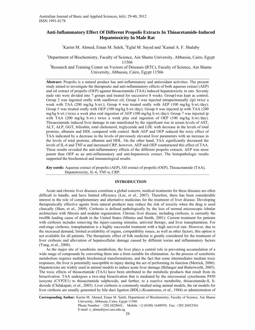
Total proteins content was significantly decreased (p<0.05) and albumin was decreased in rats received TAA alone when compared with the control group (Figure 1). However, the same parameters were significantly increased (p<0.05) in rats received a combination of TAA and AEP or TAA and OEP (prophylactic groups) compared with rats received TAA alone. Also, treatment with AEP or OEP alone significantly increased (p<0.05) serum total proteins and albumin concentrations compared with control group as shown in Figure 1.
Serum total cholesterol, triglycerides and LDL-C levels were significantly increased (p<0.05) while HDL-C was significantly lowered (p<0.05) in rats treated with TAA compared with control rats (Table 1), in the
Aust. J. Basic & Appl. Sci., 6(6): 29-40, 2012
32
meantime, AEP and OEP when combined with TAA (groups 6 &7) could significantly (p<0.05) modulate the lipid contents in rats by decreasing total lipids, total cholesterol, triglycerides and LDL-C levels with significantly favorable increase (p<0.05) in HDL-C level (Table 1). Administration of either AEP or OEP alone didn’t change the lipid profile compared with control group as shown in Table 1.
Figure 2 demonstrates the significant increase (p<0.05) in total and direct bilirubin levels in animals treated with TAA compared with control group. Meanwhile, there was a significant decrease (p<0.05) in the levels of total and direct bilirubin in AEP prophylactic group compared with TAA group, while OEP combined with TAA significantly reduced (p<0.05) only total bilirubin compared with TAA group. The data summarized in Figure 3 indicates that, AST, ALT, ALP and GGT levels were significantly increased (p<0.05) in rats received TAA either alone or in combination with AEP or OEP compared with the control group. However, the same parameters in rats received AEP or OEP combined with TAA were significantly decreased (p<0.05) compared with TAA group. Administration of either AEP or OEP alone didn’t induce noticeable change in the levels of liver enzymes compared with control group as shown in Figure 3.
IL-6 level was significantly decreased (p<0.05) when TAA was administered either alone or combined with AEP or OEP compared with control group (Figure 4). However, administration of either AEP or OEP in prophylactic groups significantly increased (p<0.05) IL-6 level compared with TAA group (Figure 4). Administration of either AEP or OEP alone significantly (p<0.05) decreased IL-6 level compared with control group (Figure 4). Administration of TAA caused a significant decrease (p<0.05) in the level of TNF-α compared with control group (Figure 5). On the other hand, administration of AEP or OEP either alone or combined with TAA showed non-significant increase in TNF-α level compared with TAA group (Figure 5). However, administration of OEP either alone or combined with TAA showed significant (p<0.05) decrease in TNF-α level compared with control while AEP either alone or combined with TAA showed non-significant decrease compared with control group (Figure 5). The level of CRP showed a significant (p<0.05) increase in all treated groups compared with control group, meanwhile administration of AEP combined with TAA showed significant (p<0.05) decrease in CRP compared with TAA group (Figure 6).
Histopathology:
Histology of the liver sections of normal animals showed normal hepatic architecture and liver lobular with well-preserved cytoplasm, prominent nucleus (Figure 7). Liver of animals treated with oil showed vascular degeneration of hepatocytes (fatty changes) and mild mononuclear cell infiltration of portal triads (Figure 8). Liver of animals treated with AEP or OEP showed an almost normal architecture of hepatic lobules which are surrounded by thin fibrous strands (Figures 9 & 10). The liver sections of TAA-treated animals showed hepatic cells with severe toxicity characterized by centrilobular necrosis, preiportal hepatocyte vacoulation with clearing of cytoplasm, scattered inflammation and cell transformation (Figure 11). AEP or OEP mixed with TAA appeared to significantly reduce TAA-induced toxicity as evidenced by less inflammation changes and necrosis (Figures 12 & 13).
Fig. 1: Serum levels of total proteins (g/dl) and albumin (g/dl) of male albino rats treated with TAA, AEP, OEP and combination of them. Values are expressed as meansSD; n=10 for each treatment group. Significant difference from the control group at *p0.05 Significant difference from the TAA group at #p0.05
contro
lOil
TAAAEP
OEP
AEP pro
phylac
tic
OEP pro
phylac
tic
0
2
4
6
8Total ProteinsAlbumin
* **
*
**
* *#*#
*#
*#
*#
*#
g/d
L

Aust. J. Basic & Appl. Sci., 6(6): 29-40, 2012
33
Table 1: Changes in the concentrations of serum total cholesterol (mg/dl), triglycerides (mg/dl), HDL (mg/dl) and LDL (mg/dl) of male albino rats treated with TAA, AEP, OEP and combination of them.
Group Parameter
Group 1 (Control)
Group 2 (Oil)
Group 3 (TAA)
Group 4 (AEP)
Group 5 (OEP)
Group 6 (AEP Prophylactic)
Group 7 (OEP Prophylactic)
Total Cholesterol
114.747.02
108.66017.34
209.9857.01*
109.3415.92
117.069.30
128.3922.12*#
135.9618.02*#
Triglycerides
65.968.042
69.936.95 110.6613.51*
71.8510.60 77.8213.47
88.697.78*# 95.106.15*#
HDL 42.494.77 43.024.42 25.132.15* 47.674.59 36.903.47 40.082.16# 32.431.77*# LDL 33.920.56 35.234.64 63.893.29* 31.130.56* 36.910.28 40.141.04*# 49.250.80*#
Values are expressed as meansSD; n=10 for each treatment group. Significant difference from the control group at *p0.05 Significant difference from the TAA group at #p0.05
Fig. 2: Concentration of total and direct bilirubin (mg/dl) of male albino rats treated with TAA, AEP, OEP and combination of them. Values are expressed as meansSD; n=10 for each treatment group. Significant difference from the control group at *p0.05 Significant difference from the TAA group at #p0.05
contro
lOil
TAAAEP
OEP
AEP pro
phylac
tic
OEP pro
phylac
tic0
50
100
150AST
ALT
* *
*** * *#
*# *#*#
ALP
GGT
*
*
*# *#
*
*
*#*#
IU/L
Fig. 3: Changes in the activities of serum AST (IU/L), ALT (IU/L), ALP (IU/L) and GGT (IU/L) of male albino rats treated with TAA, AEP, OEP and combination of them.
Values are expressed as meansSD; n=10 for each treatment group. Significant difference from the control group at *p0.05 Significant difference from the TAA group at #p0.05
contro
lOil
TAAAEP
OEP
AEP pro
phylac
tic
OEP p
rophyl
actic
0.0
0.5
1.0
1.5Total BilirubinDirect Bilirubin
mg
/dL
*
**# *#
#

Aust. J. Basic & Appl. Sci., 6(6): 29-40, 2012
34
IL-6
contro
lOil
TAAAEP
OEP
AEP pro
phylac
tic
OEP pro
phylac
tic
0
50
100
150
200
250
*
**# *#
pg
/ml
*
Fig. 4: Changes in the level of serum IL-6 of male albino rats treated with TAA, AEP, OEP and combination of them.
Values are expressed as meansSD; n=10 for each treatment group. Significant difference from the control group at *p0.05 Significant difference from the TAA group at #p0.05
TNF-Alpha
contro
lOil
TAAAEP
OEP
AEP pro
phylac
tic
OEP pro
phylac
tic
0
500
1000
1500
***
*
pg
/ml
Fig. 5: Changes in the level of serum TNF-α of male albino rats treated with TAA, AEP, OEP and combination of them.
Values are expressed as meansSD; n=10 for each treatment group. Significant difference from the control group at *p0.05 Significant difference from the TAA group at #p0.05
CRP
contro
lOil
TAAAEP
OEP
AEP pro
phylac
tic
OEP pro
phylac
tic
0
10
20
30
40
*
*
*
*#
*
mg
/L
Fig. 6: Changes in the level of serum CRP of male albino rats treated with TAA, AEP, OEP and combination of them.
Values are expressed as meansSD; n=10 for each treatment group. Significant difference from the control group at *p0.05 Significant difference from the TAA group at #p0.05
Aust. J. Basic & Appl. Sci., 6(6): 29-40, 2012
35
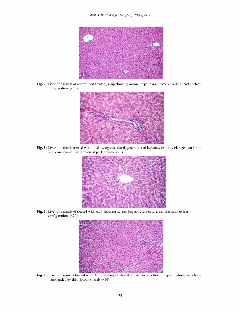
Fig. 7: Liver of animals of control non-treated group showing normal hepatic architecture, cellular and nuclear configuration. (x10)
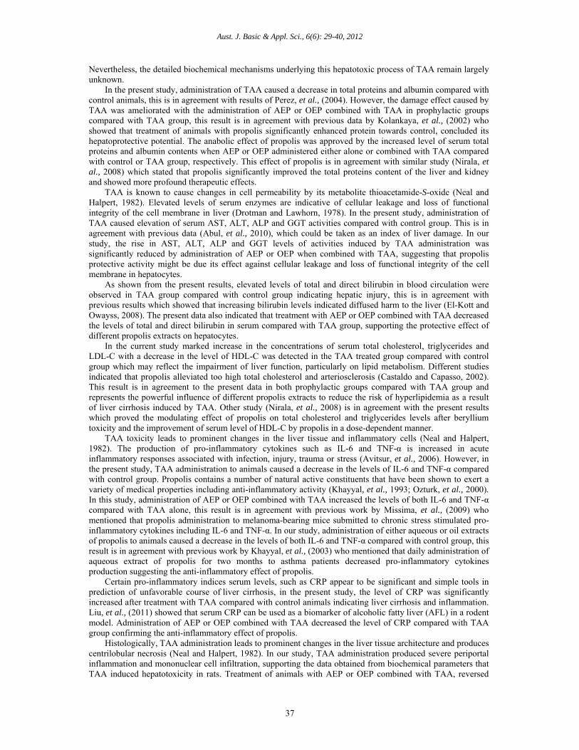
Fig. 8: Liver of animals treated with oil showing vascular degeneration of hepatocytes (fatty changes) and mild mononuclear cell infiltration of portal triads (x20).
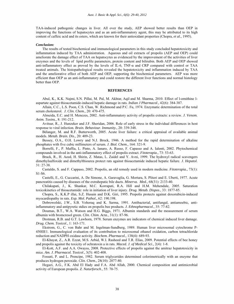
Fig. 9: Liver of animals of treated with AEP showing normal hepatic architecture, cellular and nuclear configuration. (x20)
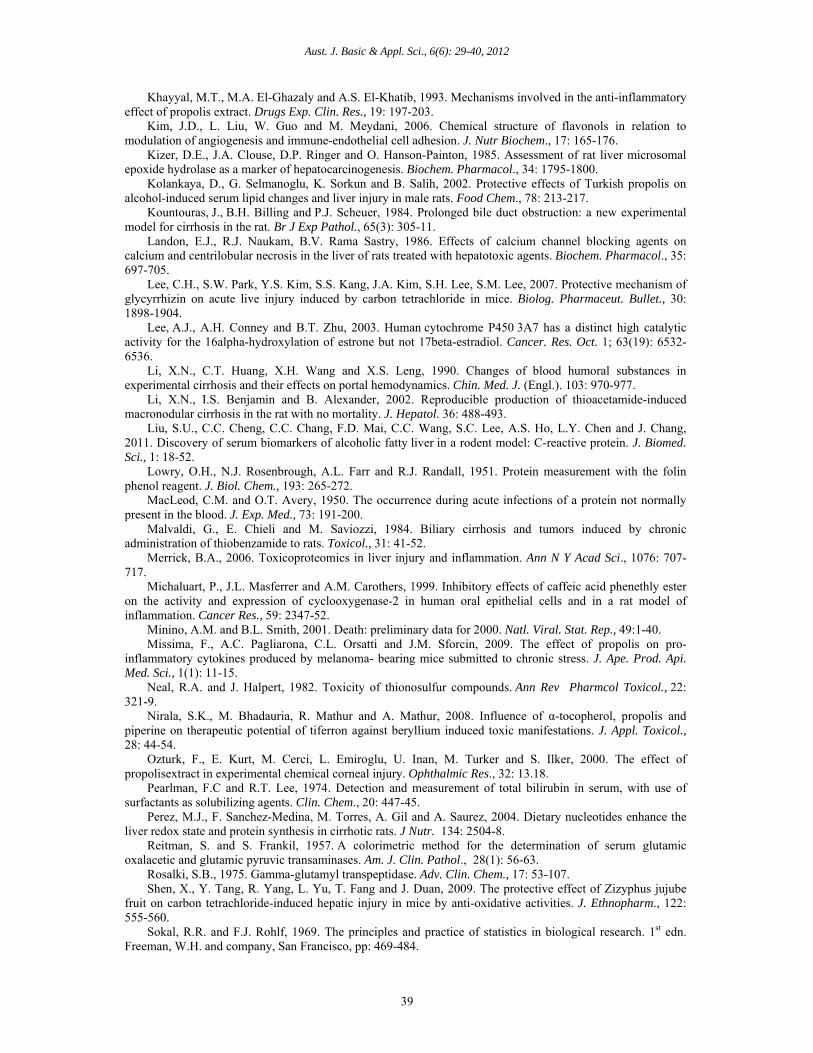
Fig. 10: Liver of animals treated with OEP showing an almost normal architecture of hepatic lobules which are surrounded by thin fibrous strands (x10).

Aust. J. Basic & Appl. Sci., 6(6): 29-40, 2012
36
Fig. 11: Liver of animals treated with TAA showing fibrosis of portal triads and dense collagenous matrix, fibroblasts and intense mononuclear cell infiltration (x40).
Fig. 12: Liver of animals treated with TAA and AEP showing congestion of portal blood vesselmild perivascular fibroplasia with formation of thin fibrous strands which extend to encircle the adjacent lobule for some distance (x20).
Fig. 13: Liver of animals treated with TAA and OEP showing separation of hepatic lobules by loosely arranged edematous fibrous strands (x10). Discussion:
Thioacetamide (TAA) was chosen for this experiment because it consistently produces liver cirrhosis in rats with histological appearance that is more akin to human cirrhosis (Li, et al., 2002). Besides, oral and intraperitoneal administrations of TAA are both established methods in the generation of fibrosis and cirrhosis models in rats (Zhao, et al., 2002). TAA is a sulfur containing compound that is necrogenic (Landon, et al., 1986) and carcinogenic (Kizer, et al., 1985). It is commonly used for inducing fulminant hepatic failure (Bruck, et al., 1999) and liver cirrhosis in animal models (Li, et al., 2002). During the biotransformation of TAA, both flavin-containing monooxygenase (FMO) (Malvaldi, et al., 1984) and cytochrome P450 (Lee, et al., 2003) reduce dioxygen to superoxide anion, which is then catalyzed (Ekström, et al., 1989) to form hydrogen peroxide (H2O2). Therefore, biotransformation of TAA precedes oxidative damage associated liver injury.
Aust. J. Basic & Appl. Sci., 6(6): 29-40, 2012
37
Nevertheless, the detailed biochemical mechanisms underlying this hepatotoxic process of TAA remain largely unknown.
In the present study, administration of TAA caused a decrease in total proteins and albumin compared with control animals, this is in agreement with results of Perez, et al., (2004). However, the damage effect caused by TAA was ameliorated with the administration of AEP or OEP combined with TAA in prophylactic groups compared with TAA group, this result is in agreement with previous data by Kolankaya, et al., (2002) who showed that treatment of animals with propolis significantly enhanced protein towards control, concluded its hepatoprotective potential. The anabolic effect of propolis was approved by the increased level of serum total proteins and albumin contents when AEP or OEP administered either alone or combined with TAA compared with control or TAA group, respectively. This effect of propolis is in agreement with similar study (Nirala, et al., 2008) which stated that propolis significantly improved the total proteins content of the liver and kidney and showed more profound therapeutic effects.
TAA is known to cause changes in cell permeability by its metabolite thioacetamide-S-oxide (Neal and Halpert, 1982). Elevated levels of serum enzymes are indicative of cellular leakage and loss of functional integrity of the cell membrane in liver (Drotman and Lawhorn, 1978). In the present study, administration of TAA caused elevation of serum AST, ALT, ALP and GGT activities compared with control group. This is in agreement with previous data (Abul, et al., 2010), which could be taken as an index of liver damage. In our study, the rise in AST, ALT, ALP and GGT levels of activities induced by TAA administration was significantly reduced by administration of AEP or OEP when combined with TAA, suggesting that propolis protective activity might be due its effect against cellular leakage and loss of functional integrity of the cell membrane in hepatocytes.
As shown from the present results, elevated levels of total and direct bilirubin in blood circulation were observed in TAA group compared with control group indicating hepatic injury, this is in agreement with previous results which showed that increasing bilirubin levels indicated diffused harm to the liver (El-Kott and Owayss, 2008). The present data also indicated that treatment with AEP or OEP combined with TAA decreased the levels of total and direct bilirubin in serum compared with TAA group, supporting the protective effect of different propolis extracts on hepatocytes.
In the current study marked increase in the concentrations of serum total cholesterol, triglycerides and LDL-C with a decrease in the level of HDL-C was detected in the TAA treated group compared with control group which may reflect the impairment of liver function, particularly on lipid metabolism. Different studies indicated that propolis alleviated too high total cholesterol and arteriosclerosis (Castaldo and Capasso, 2002). This result is in agreement to the present data in both prophylactic groups compared with TAA group and represents the powerful influence of different propolis extracts to reduce the risk of hyperlipidemia as a result of liver cirrhosis induced by TAA. Other study (Nirala, et al., 2008) is in agreement with the present results which proved the modulating effect of propolis on total cholesterol and triglycerides levels after beryllium toxicity and the improvement of serum level of HDL-C by propolis in a dose-dependent manner.
TAA toxicity leads to prominent changes in the liver tissue and inflammatory cells (Neal and Halpert, 1982). The production of pro-inflammatory cytokines such as IL-6 and TNF-α is increased in acute inflammatory responses associated with infection, injury, trauma or stress (Avitsur, et al., 2006). However, in the present study, TAA administration to animals caused a decrease in the levels of IL-6 and TNF-α compared with control group. Propolis contains a number of natural active constituents that have been shown to exert a variety of medical properties including anti-inflammatory activity (Khayyal, et al., 1993; Ozturk, et al., 2000). In this study, administration of AEP or OEP combined with TAA increased the levels of both IL-6 and TNF-α compared with TAA alone, this result is in agreement with previous work by Missima, et al., (2009) who mentioned that propolis administration to melanoma-bearing mice submitted to chronic stress stimulated pro-inflammatory cytokines including IL-6 and TNF-α. In our study, administration of either aqueous or oil extracts of propolis to animals caused a decrease in the levels of both IL-6 and TNF-α compared with control group, this result is in agreement with previous work by Khayyal, et al., (2003) who mentioned that daily administration of aqueous extract of propolis for two months to asthma patients decreased pro-inflammatory cytokines production suggesting the anti-inflammatory effect of propolis.
Certain pro-inflammatory indices serum levels, such as CRP appear to be significant and simple tools in prediction of unfavorable course of liver cirrhosis, in the present study, the level of CRP was significantly increased after treatment with TAA compared with control animals indicating liver cirrhosis and inflammation. Liu, et al., (2011) showed that serum CRP can be used as a biomarker of alcoholic fatty liver (AFL) in a rodent model. Administration of AEP or OEP combined with TAA decreased the level of CRP compared with TAA group confirming the anti-inflammatory effect of propolis.
Histologically, TAA administration leads to prominent changes in the liver tissue architecture and produces centrilobular necrosis (Neal and Halpert, 1982). In our study, TAA administration produced severe periportal inflammation and mononuclear cell infiltration, supporting the data obtained from biochemical parameters that TAA induced hepatotoxicity in rats. Treatment of animals with AEP or OEP combined with TAA, reversed
Aust. J. Basic & Appl. Sci., 6(6): 29-40, 2012
38
TAA-induced pathogenic changes in liver. All over the study, AEP showed better results than OEP in improving the functions of hepatocytes and as an anti-inflammatory agent, this may be attributed to its high content of caffeic acid and its esters, which are known for their antioxidant properties (Chopra, et al., 1995). Conclusion:
The results of tested biochemical and immunological parameters in this study concluded hepatotoxicity and inflammation induced by TAA administration. Aqueous and oil extracts of propolis (AEP and OEP) could ameliorate the damage effect of TAA on hepatocytes as evidenced by the improvement of the activities of liver enzymes and the levels of lipid profile parameters, protein content and bilirubin. Both AEP and OEP showed anti-inflammatory effect as proved by the levels of IL-6, TNF-α and CRP compared with control or TAA treated animals. The histopathological results revealed the hepatotoxicity and inflammation induced by TAA and the ameliorative effect of both AEP and OEP, supporting the biochemical parameters. AEP was more efficient than OEP as an anti-inflammatory and could restore the different liver functions and normal histology better than OEP.
REFERENCES
Abul, K., K.K. Najmi, S.N. Pillai, M. Pal, M. Akhtar, Aqil and M. Sharma, 2010. Effect of l-ornithine l-
aspartate against thioacetamide-induced hepatic damage in rats. Indian J Pharmacol., 42(6): 384-387. Allain, C.C., L.S. Poon, C.S. Chan, W. Richmond and P.C. Fu, 1974. Enzymatic determination of the total
serum cholesterol. J. Clin. Chem., 20: 470-475. Almeida, E.C. and H. Menezes, 2002. Anti-inflammatory activity of propolis extracts: a review. J. Venom.
Anim. Toxins., 8: 191-212. Avitsur, R., J. Hunzeker and J.F. Sheridan, 2006. Role of early stress in the individual differences in host
response to viral infection. Brain. Behaviour. Immunity., 20: 339-348. Bélanger, M. and R.F. Butterworth, 2005. Acute liver failure: a critical appraisal of available animal
models. Metab. Brain. Dis., 20: 409-23. Bessey, O.A., O.H. Lowry and N.J. Brock, 1946. A method for the rapid determination of alkaline
phosphates with five cubic millimeters of serum. J. Biol. Chem., 164: 321-9. Borrelli, F., P. Maffia, L. Pinto, A. Ianaro, A. Russo, F. Capasso and A. Ialenti, 2002. Phytochemical
compounds involved in the anti-inflammatory effect of propolis extract. Fitoterapia., 73: 53-63. Bruck, R., H. Aeed, H. Shirin, Z. Matas, L. Zaidel and Y. Avni, 1999. The hydroxyl radical scavengers
dimethylsulfoxide and dimethylthiourea protect rats against thioacetamide induced hepatic failure. J. Hepatol. 31: 27-38.
Castaldo, S. and F. Cappaso, 2002. Propolis, an old remedy used in modern medicine. Fitoterapia., 73(1): S1-S6.
Castelli, E., G. Cuccarini, A. De Simone, A. Garavaglia, G. Mortara, S. Pliteri and E. Uberti, 1977. Acute pancreatitis caused by diseases of the extrahepatic bile ducts. Minerva. Med., 68(31): 2133-40.
Chilakapati, J., K. Shankar, M.C. Korrapati, R.A. Hill and H.M. Mehendale, 2005. Saturation toxicokinetics of thioacetamide: role in initiation of liver injury. Drug. Metab. Dispos., 33: 1877-85.
Chopra, S., K.K.P illai, S.Z. Husain and D.K. Giri, 1995. Propolis protects against doxorubicin-induced myocardiopathy in rats. Exp. Mol. Pathol., 62: 190.198.
Dobrowolski, J.W., S.B. Vohoraq and K. Sarma, 1991. Antibacterial, antifungal, antiamoebic, anti-inflammatory and antipyretic stdies on propolis bee products. J. Ethnopharmacol., 35: 77-82.
Doumas, B.T., W.A. Watson and H.G. Biggs, 1971. Albumin standards and the measurement of serum albumin with bromcresol green. Clin. Chim. Acta., 31(1): 87-96.
Drotman, R.B. and G.T. Lawhorn, 1978. Serum enzymes are indicators of chemical induced liver damage. Drug. Chem. Toxicol., 1: 163-171.
Ekstrom, G., C. von Bahr and M. Ingelman-Sundberg, 1989. Human liver microsomal cytochrome P-450IIE1. Immunological evaluation of its contribution to microsomal ethanol oxidation, carbon tetrachloride reduction and NADPH oxidase activity. Biochem. Pharmacol., 138(4): 689-93.
El-Khayat, Z., A.R. Ezzat, M.S. Arbid, W.I. Rasheed and T.R. Elias, 2009. Potential effects of bee honey and propolis against the toxicity of ochratoxin a in rats. Maced. J. of Medical Sci., 2(4): 1-8.
El-Kott, A.F. and A.A. Owayss, 2008. Protective effects of propolis against the amitraz hepatotoxicity in mice. Am. J. Pharmacol. Toxicol., 3(5): 402-408.
Fossati, P. and L. Prencipe, 1982. Serum triglycerides determined colorimetrically with an enzyme that produces hydrogen peroxide. Clin. Chem., 28(10): 2077-80.
Hegazi, A.G., F.K. Abd El Hady and F.A. Abd Allah, 2000. Chemical composition and antimicrobial activity of European propolis. Z. Naturforsch., 55: 70-75.
Aust. J. Basic & Appl. Sci., 6(6): 29-40, 2012
39
Khayyal, M.T., M.A. El-Ghazaly and A.S. El-Khatib, 1993. Mechanisms involved in the anti-inflammatory effect of propolis extract. Drugs Exp. Clin. Res., 19: 197-203.
Kim, J.D., L. Liu, W. Guo and M. Meydani, 2006. Chemical structure of flavonols in relation to modulation of angiogenesis and immune-endothelial cell adhesion. J. Nutr Biochem., 17: 165-176.
Kizer, D.E., J.A. Clouse, D.P. Ringer and O. Hanson-Painton, 1985. Assessment of rat liver microsomal epoxide hydrolase as a marker of hepatocarcinogenesis. Biochem. Pharmacol., 34: 1795-1800.
Kolankaya, D., G. Selmanoglu, K. Sorkun and B. Salih, 2002. Protective effects of Turkish propolis on alcohol-induced serum lipid changes and liver injury in male rats. Food Chem., 78: 213-217.
Kountouras, J., B.H. Billing and P.J. Scheuer, 1984. Prolonged bile duct obstruction: a new experimental model for cirrhosis in the rat. Br J Exp Pathol., 65(3): 305-11.
Landon, E.J., R.J. Naukam, B.V. Rama Sastry, 1986. Effects of calcium channel blocking agents on calcium and centrilobular necrosis in the liver of rats treated with hepatotoxic agents. Biochem. Pharmacol., 35: 697-705.
Lee, C.H., S.W. Park, Y.S. Kim, S.S. Kang, J.A. Kim, S.H. Lee, S.M. Lee, 2007. Protective mechanism of glycyrrhizin on acute live injury induced by carbon tetrachloride in mice. Biolog. Pharmaceut. Bullet., 30: 1898-1904.
Lee, A.J., A.H. Conney and B.T. Zhu, 2003. Human cytochrome P450 3A7 has a distinct high catalytic activity for the 16alpha-hydroxylation of estrone but not 17beta-estradiol. Cancer. Res. Oct. 1; 63(19): 6532-6536.
Li, X.N., C.T. Huang, X.H. Wang and X.S. Leng, 1990. Changes of blood humoral substances in experimental cirrhosis and their effects on portal hemodynamics. Chin. Med. J. (Engl.). 103: 970-977.
Li, X.N., I.S. Benjamin and B. Alexander, 2002. Reproducible production of thioacetamide-induced macronodular cirrhosis in the rat with no mortality. J. Hepatol. 36: 488-493.
Liu, S.U., C.C. Cheng, C.C. Chang, F.D. Mai, C.C. Wang, S.C. Lee, A.S. Ho, L.Y. Chen and J. Chang, 2011. Discovery of serum biomarkers of alcoholic fatty liver in a rodent model: C-reactive protein. J. Biomed. Sci., 1: 18-52.
Lowry, O.H., N.J. Rosenbrough, A.L. Farr and R.J. Randall, 1951. Protein measurement with the folin phenol reagent. J. Biol. Chem., 193: 265-272.
MacLeod, C.M. and O.T. Avery, 1950. The occurrence during acute infections of a protein not normally present in the blood. J. Exp. Med., 73: 191-200.
Malvaldi, G., E. Chieli and M. Saviozzi, 1984. Biliary cirrhosis and tumors induced by chronic administration of thiobenzamide to rats. Toxicol., 31: 41-52.
Merrick, B.A., 2006. Toxicoproteomics in liver injury and inflammation. Ann N Y Acad Sci., 1076: 707-717.
Michaluart, P., J.L. Masferrer and A.M. Carothers, 1999. Inhibitory effects of caffeic acid phenethly ester on the activity and expression of cyclooxygenase-2 in human oral epithelial cells and in a rat model of inflammation. Cancer Res., 59: 2347-52.
Minino, A.M. and B.L. Smith, 2001. Death: preliminary data for 2000. Natl. Viral. Stat. Rep., 49:1-40. Missima, F., A.C. Pagliarona, C.L. Orsatti and J.M. Sforcin, 2009. The effect of propolis on pro-
inflammatory cytokines produced by melanoma- bearing mice submitted to chronic stress. J. Ape. Prod. Api. Med. Sci., 1(1): 11-15.
Neal, R.A. and J. Halpert, 1982. Toxicity of thionosulfur compounds. Ann Rev Pharmcol Toxicol., 22: 321-9.
Nirala, S.K., M. Bhadauria, R. Mathur and A. Mathur, 2008. Influence of α-tocopherol, propolis and piperine on therapeutic potential of tiferron against beryllium induced toxic manifestations. J. Appl. Toxicol., 28: 44-54.
Ozturk, F., E. Kurt, M. Cerci, L. Emiroglu, U. Inan, M. Turker and S. Ilker, 2000. The effect of propolisextract in experimental chemical corneal injury. Ophthalmic Res., 32: 13.18.
Pearlman, F.C and R.T. Lee, 1974. Detection and measurement of total bilirubin in serum, with use of surfactants as solubilizing agents. Clin. Chem., 20: 447-45.
Perez, M.J., F. Sanchez-Medina, M. Torres, A. Gil and A. Saurez, 2004. Dietary nucleotides enhance the liver redox state and protein synthesis in cirrhotic rats. J Nutr. 134: 2504-8.
Reitman, S. and S. Frankil, 1957. A colorimetric method for the determination of serum glutamic oxalacetic and glutamic pyruvic transaminases. Am. J. Clin. Pathol., 28(1): 56-63.
Rosalki, S.B., 1975. Gamma-glutamyl transpeptidase. Adv. Clin. Chem., 17: 53-107. Shen, X., Y. Tang, R. Yang, L. Yu, T. Fang and J. Duan, 2009. The protective effect of Zizyphus jujube
fruit on carbon tetrachloride-induced hepatic injury in mice by anti-oxidative activities. J. Ethnopharm., 122: 555-560.
Sokal, R.R. and F.J. Rohlf, 1969. The principles and practice of statistics in biological research. 1st edn. Freeman, W.H. and company, San Francisco, pp: 469-484.
Aust. J. Basic & Appl. Sci., 6(6): 29-40, 2012
40
Toyama, T., H. Nakamura, Y. Harano, N. Yamauchi, A. Morita, T. Kirishima, M. Minami, Y. Itoh, T. Okanoue, 2004. PPA Ralpha ligands activate antioxidant enzymes and suppress hepatic fibrosis in rats. Biochem. Biophys. Res. Commun., 324(2): 697-704.
Wang, D., D.B. Xiang, Y.J. He, Z.P. Li, X.H. Wu, J.H. Mou, H.L. Xiao and Q.H. Zhang, 2005. Effect of caffeic acid phenethyl ester on proliferation and apoptosis of colorectal cancer cells in vitro. World J. Gastroenterol., 11: 4008-4012.
Weber, L.W., M. Boll and A. Stampfl, 2003. Hepatotoxicity and mechanism of action of haloalkanes: carbon tetrachloride as a toxicological model.Crit. Rev. Toxicol., 33: 105-136.
Yang, M.L., Z.Y. Hong, B.G. Wei, S.K. Qian, D. Min, P.X. He and J.Z. Shi, 2008. Therapeutic effect of traditional Chinese medicine on coagulation disorder and accompanying intractable jaundice in hepatitis B virus-related liver cirrhosis patients. World J Gastroenterol., 14(39): 6060-6064.
Zhao, G., K. Nakano, K. Chijiiwa, J. Ueda, M. Tanaka, 2002. Inhibited activities in CCAAT/enhancer-binding protein, activating protein-1 and cyclins after hepatectomy in rats with thioacetamide-induced liver cirrhosis. Biochem. Biophys. Res. Commun., 292: 474-481.
Ziaran, H.R., H.R. Rahmani and J. Pourreza, 2005. “Effect of dietary oil extract of propolis on immune response and broiler performance”. Pakist J. Biol. Sci., 8(10): 1485-1490.