anti-mu¨llerian hormone (amh) as a predictive marker …¨llerian hormone (amh) as a predictive...
TRANSCRIPT

...........................................................................................................................
Anti-Mullerian hormone (AMH) as apredictive marker in assistedreproductive technology (ART)A. La Marca1,3, G. Sighinolfi1, D. Radi1, C. Argento1, E. Baraldi1,A. Carducci Artenisio2, G. Stabile2, and A. Volpe1
1Mother-Infant Department, Section of Obstetrics and Gynecology, University of Modena and Reggio Emilia, Policlinico of Modena, Via delPozzo, 71, 41100 Modena, Italy 2Department of Medicine and Pharmacology, University of Messina, Messina, Italy
3Correspondence address. Tel: þ39-059-422-4379; Fax: þ39-059-422-4394; E-mail: [email protected]
table of contents
† Introduction† Methods† AMH in female fertility
AMH in ovarian physiologyFactors modulating AMH levels in womenPrediction of quantitative ovarian response in ARTPrediction of qualitative ovarian response in ARTAMH in ovarian reserve testing
† AMH in male fertilityAMH in testicular physiologyValue of AMH measurement in infertile men
† Conclusions
background: In women, anti-Mullerian hormone (AMH) levels may represent the ovarian follicular pool and could be a useful markerof ovarian reserve. The clinical application of AMH measurement has been proposed in the prediction of quantitative and qualitative aspectsin assisted reproductive technologies (ART). In men AMH is secreted in both the serum and seminal fluid. Its measurement may be useful inclinical evaluation of the infertile male.
methods: The PubMed database was systematically searched for studies published until the end of January 2009, search criteria relevantto AMH, ovarian reserve, ovarian response to gonadotrophin stimulation, spermatogenesis and azoospermia were used.
results: AMH seems to be a better marker in predicting ovarian response to controlled ovarian stimulation than age of the patient, FSH,estradiol and inhibin B. A similar performance for AMH and antral follicular count has been reported. In clinical practice, AMH measurementmay be useful in the prediction of poor response and cycle cancellation and also of hyper-response and ovarian hyperstimulation syndrome.In the male, the wide overlap of AMH values between controls and infertile men precludes this hormone from being a useful marker ofspermatogenesis.
conclusions: As AMH may permit the identification of both the extremes of ovarian stimulation, a possible role for its measurementmay be in the individualization of treatment strategies in order to reduce the clinical risk of ART along with optimized treatment burden. It isfundamental to clarify the cost/benefit of its use in ovarian reserve testing. Regarding the role of AMH in the evaluation of infertile men, AMHas single marker of spermatogenesis does not seem to reach a satisfactory clinical utility.
Key words: AMH / ART / COS / poor response / OHSS / azoospermia
& The Author 2009. Published by Oxford University Press on behalf of the European Society of Human Reproduction and Embryology. All rights reserved.For Permissions, please email: [email protected]
Human Reproduction Update, Vol.16, No.2 pp. 113–130, 2010
Advanced Access publication on September 30, 2009 doi:10.1093/humupd/dmp036

IntroductionAnti-Mullerian hormone (AMH) is a dimeric glycoprotein, a memberof the transforming growth factor-beta superfamily (Jost, 1946; Cateet al., 1986), which acts on tissue growth and differentiation. AMHwas originally identified because of its fundamental role in male sexdifferentiation. Indeed, expressed in the Sertoli cells of fetal testis,AMH induces the regression of the Mullerian ducts. In the absenceof AMH, Mullerian ducts evolved into uterus, fallopian tubes and theupper part of the vagina (Munsterberg and Lovell-Badge, 1991; Leeand Donahoe, 1993; Josso et al., 2001).
In women AMH is produced by granulosa cells, from pre-antral andantral follicles (Weenen et al., 2004) and the main physiological role ofAMH in the ovary seems to be limited to the inhibition of the earlystages of follicular development (Themmen, 2005; Visser andThemmen, 2005).
AMH is secreted by the ovary into circulation, hence AMH ismeasurable in serum. As serum AMH levels essentially reflect theovarian follicular pool, reduction in the number of small growingfollicles may be followed by a reduction in circulating AMH. Recently,AMH has been evaluated by several groups as a potential novelclinical marker of ovarian reserve and of response to gonadotrophins(Seifer et al., 2002; Van Rooij et al., 2002; Fanchin et al., 2003a, b;Muttukrishna et al., 2004; Eldar-Geva et al., 2005; Hazout et al.,2004; Penarrubia et al., 2005; Tremellen et al., 2005; Ficiciogluet al., 2006; La Marca et al., 2007). In particular in the last few yearsseveral large prospective studies have been published reportingextremely interesting new data on the possible clinical application ofAMH measurement in the prediction of quantitative and qualitativeovarian response in assisted reproductive technologies (ART).
In the male, AMH is the earliest Sertoli cell specific protein expressedby the gonad (Tran et al., 1977). It is secreted by the testis from theeighth week of pregnancy and remains secreted at high-level untilpuberty, when Sertoli cell maturation is characterized by decreasedAMH production (Rajpert-De Meyts et al., 1999). Paralleling the situ-ation in women, the main physiological role of AMH in the adult maleseems to be limited to the paracrine control of testicular function.
In the adult man, AMH is secreted both in serum and in seminalfluid (Fenichel et al., 1999) and, being a specific marker of Sertolicell function, its measurement may be useful to obtain informationon spermatogenesis in infertile men. In the last few years severalstudies have been published on the possible clinical use of AMHassay in the diagnostic work-up of patients with oligoasthenoterato-zoospermia (OAT) and azoospermia (Fenichel et al., 1999; Fujisawaet al., 2002; Al-Qahtani et al., 2005; Appasamy et al., 2007;Muttukrishna et al., 2007) and in particular on the predictive valueof AMH for the successful sperm retrieval in azoospermic patients (Isi-koglu et al., 2006; Mostafa et al., 2007; Duvilla et al., 2008; Gouliset al., 2009).
In this review the main findings of published studies have been sum-marized and some conclusions on the clinical application of AMHmeasurement in both the infertile male and female have been drawn.
MethodsPubMed database was systematically searched for studies published untilthe end of January 2009, using search criteria relevant to AMH, ovarian
reserve, ovarian response to gonadotrophin stimulation, spermatogenesisand azoospermia. Specifically the following search terms were used: AMH,Mullerian Inhibiting Substance, ovarian reserve, ovarian ageing, poorresponse, poor responder, hyper-response, hyper-responder, ovarianhyperstimulation syndrome (OHSS), ART, IVF, ICSI, sperm, spermatogen-esis, seminal fluid, azoospermia, oligozoospermia, OAT, TESE and TESA.Cross-references picked up during the review search were also selectedif they were not included initially. Both prospective and retrospectivearticles were considered. Methods for selecting and synthesizing thedata were based on personal experience.
AMH in female fertility
AMH in ovarian physiologyAMH is produced by granulosa cells from pre-antral and antral fol-licles, restricting expression to growing follicles, until they havereached the size and differentiation state at which they are selectedfor dominance by the action of pituitary FSH (Weenen et al., 2004)(Fig. 1). In the human this occurs in antral follicles of size 4–6 mm.AMH is not expressed in atretic follicles and theca cells. OvarianAMH expression has been observed as early as 36 weeks’ gestationin the humans’ fetus (Raypert-De Meyts et al., 1999). Recent studiesshow that in adult rat ovaries FSH and estradiol may down-regulateAMH expression (Baarends et al., 1995).
AMH exerts its biological effects through a transmembrane serine/threonine kinase typeII receptor (AMHRII), which is specificallyexpressed in the gonads and in the mesenchymal cells adjacent tothe Mullerian ducts (Di Clemente et al., 2003). In adult female rats,AMH and AMHRII mRNAs are mainly expressed in granulosa cellsfrom pre-antral and smaller antral follicles (Baarends et al., 1995). Inaddition, AMHRII mRNA expression was observed in theca cells ofpre-antral and small antral follicles. Besides the exclusive AMHRII,three candidate AMH type I receptors have been identified to beinvolved in AMH-induced Mullerian duct regression (Visser, 2003).These type I receptors, ALK2, ALK3 and ALK 6, are shared withthe bone morphogenetic proteins (BMPs). Subsequently, similar toBMPs, AMH signalling is mediated through the downstream signallingmolecules Smad1, Smad5 and Smad8 (Visser, 2003). However, therelative contribution of these three type I receptors to AMH signallingin the ovary remains to be determined.
The main physiological role of AMH in the ovary seems to belimited to the inhibition of the early stages of follicular development(Themmen, 2005; Visser and Themmen, 2005), since both in vivoand in vitro experiments have indicated that the transition from primor-dial into growing follicles becomes enhanced in absence of AMH,leading to early exhaustion of the primordial follicle pool (Durlingeret al., 2001; Visser and Themmen, 2005). In vitro culture of mouseneonatal ovaries and human cortical strips has confirmed the inhibitoryrole of AMH in primordial follicle recruitment (Carlsson et al., 2006).Moreover it has been suggested that follicles are more sensitive to FSHin the absence of AMH. The effects of AMH on FSH sensitivity of fol-licles was tested in a in vivo model in which the follicle dynamics werecompared with wild-type and AMH null mice in the presence of lowand high FSH serum concentrations (Durlinger et al., 2001). Thestudy shows that more growing follicles were found in AMH nullmice than in wild-type mice, both in term of numbers and in termsof developmental stage (Durlinger et al., 2001).
114 La Marca et al.
Recently, ovaries from rats placed in organ culture and incubated inthe absence and presence of AMH, show that AMH alters theexpression of several hundred genes (Nilsson et al., 2007). Theoverall effects of AMH exposure was to decrease the expression ofstimulatory factors, increase the expression of inhibitory factors andregulate cellular pathways that result in the inhibition of primordial fol-licle development (Nilsson et al., 2007).
Current theories also suggest a role for AMH as a co-regulator ofsteroidogenesis in granulosa cells, as AMH levels appear to berelated to estradiol levels in follicular fluid from small antral follicles(Andersen and Byskov, 2006). This is confirmed by a recent studywhich showed that polymorphisms in the gene for AMH or AMHreceptor type II seem to be related to follicular phase estradiollevels, suggesting a role for AMH in the FSH-induced steroidogenesisin the human ovary (Kevenaar et al., 2007).
Factors modulating AMH levels in womenAMH is produced and secreted by the gonads into the circulation,and AMH is measurable in serum from both men and women.Serum AMH levels from women are lower than those in men through-out life. In women AMH levels are almost undetectable at birth with asubtle increase within the first 2 or 4 years of age, after that AMHappears to be stable until adulthood but found to decrease as a signof follicular reserve exhaustion becoming undetectable at menopause(Fig. 2) (Lee et al., 1996; Guibourdenche et al., 2003; La Marca et al.,2005a; Van Rooij et al., 2005; Bergada et al., 2006; Shin et al., 2008;Robertson et al., 2008; La Marca 2009a). Interestingly, in women cir-culating AMH appears to be solely of ovarian origin since AMH isundetectable 3–5 days following bilateral ovariectomy (La Marca
et al., 2005a). As AMH levels essentially reflect the follicular ovarianpool, reduction in the number of small growing follicles may be fol-lowed by a reduction in circulating AMH. The reduction in ovarianreserve is a physiological process occurring in the late reproductiveperiod and consistently associated with a decrease in AMH levels(Van Rooij et al., 2005; Robertson et al., 2008). The strong correlationexisting between AMH levels and the resting pool of follicles hasrecently been highlighted by some papers showing that AMHmeasurement may be used to predict the occurrence of menopause(Sowers et al., 2008; Van Disseldorp et al., 2008).
Non-significant variations of AMH throughout the menstrual cyclehave been reported by our group (La Marca et al., 2006a) and con-firmed by a number of independent studies (Hehenkamp et al.,2006; Tsepelidis et al., 2007; Streuli et al., 2008) (Fig. 2). Othershave reported significant cyclical fluctuations in AMH levels with arapid decrease in the early luteal phase (Wunder et al., 2008; Streuliet al., 2009). However, excursions from mean levels of þ3% to219% have been calculated (Wunder et al., 2008; Streuli et al.,2009). These variations are similar to reported inter-cycle variabilityfor AMH (Fanchin et al., 2005a, b; Streuli et al., 2008). Hence in theclinical setting the inter- and intra-cycle variability in serum AMHlevels may be considered to be low enough to permit randomtiming of AMH measurement during the menstrual cycle.
In women, AMH levels seem to be unmodified under conditionsin which endogenous gonadotrophin release is substantially dimin-ished, such as during pregnancy (La Marca et al., 2005b), GnRHagonist treatment (Mohamed et al., 2006) and short-term oral con-traceptive administration (Arbo et al., 2007; Somunkiran et al., 2007;Streuli et al., 2008), indicating that non-cyclic FSH-independent
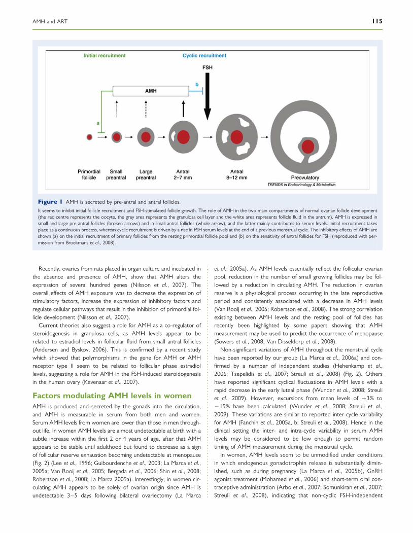
Figure 1 AMH is secreted by pre-antral and antral follicles.
It seems to inhibit initial follicle recruitment and FSH-stimulated follicle growth. The role of AMH in the two main compartments of normal ovarian follicle development(the red centre represents the oocyte, the grey area represents the granulosa cell layer and the white area represents follicle fluid in the antrum). AMH is expressed insmall and large pre-antral follicles (broken arrows) and in small antral follicles (whole arrow), and the latter mainly contributes to serum levels. Initial recruitment takesplace as a continuous process, whereas cyclic recruitment is driven by a rise in FSH serum levels at the end of a previous menstrual cycle. The inhibitory effects of AMH areshown (a) on the initial recruitment of primary follicles from the resting primordial follicle pool and (b) on the sensitivity of antral follicles for FSH (reproduced with per-mission from Broekmans et al., 2008).
AMH and ART 115

ovarian activity persists even when pituitary FSH secretion issuppressed.
Women with polycystic ovary syndrome (PCOS) show increaseddevelopment of antral follicles compared with normal women (Pignyet al., 2006). On histological examination, polycystic ovaries (PCO)exhibit a normal number of primordial follicles, whereas the numberof developing follicles is double compared with normal ovaries(Webber et al., 2003). Accordingly circulating AMH levels in womenwith PCOS are two to three times higher than healthy controls(Fallat et al., 1997; Cook et al., 2002; Pigny et al., 2003; La Marcaet al., 2004a, b; Laven et al., 2004; Mulders et al., 2004; Eldar-Gevaet al., 2005; Piltonen et al., 2005; Wachs et al., 2007). In womenwith PCOS, increased AMH levels may not only be due to excessiveaccumulation of antral follicles (Wang et al., 2007) but also toincreased granulosa cell AMH secretion (Mulders et al., 2004).Indeed, levels of AMH are on average 75 times higher in granulosacells from PCO, compared with levels in granulosa cells fromnormal ovaries (Pellatt et al., 2007).
AMH levels appear to be related to the severity of the syndrome,since levels have been observed to be higher in insulin-resistantPCOS women than in patients with normal insulin sensitivity(Fleming et al., 2006). Similarly AMH is higher in amenorrheic com-pared with oligomenorrheic women with PCOS (La Marca et al.,2004a, b), which could indicate a role for AMH in the pathogenesisof PCOS-related anovulation. The relationship between AMH levelsand the severity of the syndrome seems to be confirmed by studiesdemonstrating that PCOS patients ovulating during a weightloss-programme had AMH levels lower than women remaining ano-vulatory (Moran et al., 2007; Thomson et al., 2009). Interestingly, inone study no significant changes in AMH levels were observed ineither responders or non-responders during the weightloss-programme (Thomson et al., 2009). In order to clarify thecomplex relationship existing between insulin resistance, androgenexcess and high levels of AMH, a prospective, randomized, double-blind 26 week-long study was undertaken in women with PCOS
(Carlsen et al., 2009). All patients received diet and lifestyle counsel-ling, and metformin. Concomitantly, they were randomized to eitherdexamethasone or placebo. The study clearly demonstrated that cir-culating AMH concentrations were unaffected by 6 months of life-style counselling with metformin and placebo treatment. AMHlevels were also unaffected by 6 months of androgen suppressionwith dexamethasone in addition. These results may indicate thathigh serum AMH levels in PCOS may be more strongly related tothe presence of PCO than to the full spectrum of the syndrome(PCOS) as modifications in androgens and insulin sensitivity arenot followed by changes in ovarian AMH output (Carlsen et al.,2009).
Finally, AMH measurement has been found to offer a relatively highspecificity and sensitivity (92 and 67%, respectively) as a diagnosticmarker for PCO (Pigny et al., 2006). On this basis it has been pro-posed that in situations where accurate ultrasound data are not avail-able, AMH could be used instead of the follicle count as a diagnosticcriterion for PCOS (Pigny et al., 2006).
Obesity has been associated with reduced fertility, even in the pres-ence of ovulatory menstrual cycles, and to increased probability ofmiscarriage compared with normal weight women (Rich-Edwardset al., 2002; Fedorcsak et al., 2004). Non-PCOS obese womenshow reduced levels of inhibin B and AMH (Gracia et al., 2005;Freeman et al., 2007) suggesting that obesity may be associated withimpaired ovarian reserve. However, a recent study (Su et al., 2008)examined the correlation of obesity with hormonal and ultrasound-derived markers of ovarian reserve and found that serum AMHlevels are lower in obese women compared with age-matchedwomen of normal weight, despite similar antral follicular count. Thissuggests that AMH levels in obese women may be lower for physio-logical reasons related to obesity itself and may not be necessarilyindicative of impaired ovarian reserve (Su et al., 2008). Otherfactors related to reduced AMH levels are smoking (Freour et al.,2008), alcohol use (Nardo et al., 2007) and race or ethnicity (Seiferet al., 2008).
Figure 2 Left: Mean serum AMH levels show a reduction throughout reproductive life. Undetectable AMH levels after spontaneous menopausehave been reported (constructed graphic). Right: Circulatory pattern of AMH during the menstrual cycle of young healthy women aged 18–24years. Serum AMH levels have been shown to be stable throughout the menstrual cycle. Day 0 ¼ day of LH surge (reproduced with permissionfrom La Marca et al., 2006a).
116 La Marca et al.

Prediction of quantitative ovarian responsein ARTAMH levels seem to decline gradually during gonadotrophin adminis-tration as a part of controlled ovarian stimulation (COS) (Fanchinet al., 2003a, b; La Marca et al., 2004a, b). The reduction of AMHlevels during COS could be due to a negative direct or indirecteffect of FSH on ovarian AMH secretion. During exogenous adminis-tration of FSH there is an increase in estradiol levels, which could be areason for decreased AMH. Indeed estradiol has been implicated inthe down-regulation of AMH and AMHII mRNA in the ovary (Baar-ends et al., 1995). Stimulation with FSH induces growth of folliclesthat enlarge and lose their AMH expression, and this is probably themain reason for AMH reduction. Hence, due to the reduction ofAMH levels during FSH administration, AMH measurement topredict the ovarian response to FSH should not be performedduring gonadotrophin treatment, but some months to some daysprior commencing FSH treatment.
Much data show a strong and positive correlation between basalAMH serum levels and the number of retrieved oocytes in womenundergoing ovarian stimulation (Table I). In the evaluation of AMHas a marker of ovarian response to FSH, the first article to reportan association between circulating AMH and ovarian response to gon-adotrophin was by Seifer and colleague segues (Seifer et al., 2002).The authors observed that higher AMH on Day 3 of the stimulationprotocol was associated with a greater number of retrieved
oocytes. In particular, AMH levels were 2.5-fold higher in patientswith at least 11 oocytes compared with those with six oocytes orfewer retrieved. Results from this study were successively confirmedby several retrospective and prospective studies by different indepen-dent groups.
In Table I all retrospective and prospective studies that have found acorrelation between the number of retrieved oocytes and AMH levelshave been summarized. The majority of authors compared AMH withage and other hormonal markers (FSH, estradiol and Inhibin B), butonly a few studies also compared AMH levels with ultrasoundmarkers of ovarian reserve. The balance of the published studiesseems to indicate that AMH is a better marker in predicting ovarianresponse to COS than age of the patient, Day 3 FSH, estradiol andinhibin B.
Almost all of these studies found a significant correlation betweenAMH and antral follicular count, but very few studies have comparedthe performance of the two markers in the prediction of the numberof retrieved oocytes. Only Ficicioglu et al. (2006) and McIlveen et al.(2007) concluded that AMH is better than AFC, whereas twostudies found AFC to be superior to AMH (Eldar-Geva et al., 2005;Kwee et al., 2007) and five studies reported a similar performanceof the two markers (Van Rooij et al., 2002; Muttukrishna et al.,2005; Elgindy et al., 2007; Lekamge et al., 2007; Jayaprakasan et al.,2008).
Hence, it may be concluded that AFC and AMH perform withsimilar power in the prediction of the number of retrieved oocytes.
.......................................................................................................
.............................................................................................................................................................................................
Table I Studies on AMH as marker of ovarian response to controlled ovarian stimulation (COS)
Author n R with oocytes* AMH better than
AFC Ov. Vol d3 FSH d3 E2 d3 inhB Age
Seifer et al. (2002) 107 0.48p p
Van Rooij et al. (2002) 130 0.57 5p p p p
Fanchin et al. (2003a, b) 93 0.43
Muttukrishna et al. (2004) 69 0.69p p
Hazout et al. (2004) 109 0.38p p p p
Muttukrishna et al. (2005) 108 0.5 5p
Eldar-Geva (2005) 56 0.64 Xp p
Silberstein et al. (2006) 257 0.33p
Ficicioglu et al. (2006) 50 0.56p p p p
Lekamge et al. (2007) 126 0.34 5
La Marca et al. (2007) 48 0.7
Kwee et al. (2007) 110 0.63 Xp p p
Nakhuda et al. (2007) 77 0.63p
McIlveen et al. (2007) 84 0.78p p p
5p
Nelson et al. (2007) 340 0.71p p
Elgindy et al. (2008) 33 0.88 5p p
Lie Fong et al. (2008) 125 0.47
Jee et al. (2008) 59 0.53 X
Jayaprakasan et al. (2008) 135 0.47 5p p p p
Wunder et al. (2008) 276 0.35p
X
Comparison with other predictors.*R with oocytes: correlation between serum AMH levels and the number of retrieved oocytes;
p, better than; X, worse than; ¼, equal to.
AMH and ART 117
This was confirmed by a recent meta-analysis in which the value ofserum AMH levels as a test to predict ovarian response in IVF in com-parison to the performance of the AFC was been assessed (Broeret al., 2008). A total of 13 studies were analyzed reporting on AMHand 17 on AFC. The ROC curves for the prediction of ovarianresponse indicated no significant difference between the performancesof AMH and AFC. Hence it may be concluded that at present AMHappears to offer at least the same level of accuracy and clinical valuefor the prediction of ovarian response as AFC (Broer et al., 2008).
Prediction of poor response and cycle cancellationA proportion of women (2–30%) undergoing COS experience poorresponse (Hendriks et al., 2005) for which there is no universallyaccepted definition. Numerous criteria have been used to characterizepoor response. The number of developed follicles and the number ofretrieved oocytes are two of the most important criteria for definingpoor response. The proposed number varies among different authorsand ranges from less than three to less than five dominant follicles onthe day of hCG, and from less than three to less than five retrievedoocytes (reviewed in Tarlatzis et al., 2003). More logically, poorresponse is generally considered to have occurred if the cycle is can-celled due to an inadequate ovarian response to stimulation. What-ever definition is used, poor responders have definitely lowerpregnancy rates compared with normal responders of similar age(El-Toukhy et al., 2002; Ulug et al., 2003; Kailasam et al., 2004; Galey-Fontaine et al., 2005; Klinkert et al., 2005; Saldeen et al., 2007).
In the clinical setting it may be useful to correctly predict the occur-rence of poor response as this may lead to avoiding treatment inwomen destined not to respond to COS, thus contributing to redu-cing the cycle cancellation rate, the treatment costs and psychologicalstress for the couple. Finally improved counselling for the prediction ofpoor response may ameliorate disappointment and distress.
A large number of clinical parameters have been shown to predictthe poor ovarian response to stimulation with exogenous gonado-trophins and have been introduced in the clinical practice. Theseinclude age, basal serum FSH and inhibin B levels, antral folliclecount, ovarian volume, a number of dynamic tests and more recentlyAMH (Navot et al., 1987; Fanchin et al., 1994; Faddy and Gosden,1996; Lass et al., 1997; Tomas et al., 1997; Hall et al., 1999; Ravhinet al., 2000; Bancsi et al., 2002; Broekmans et al., 2006).
Several authors investigated the utility of AMH in the prediction ofpoor response to FSH. Reported sensitivity and specificity rangedbetween 44–97% and 41–100%, respectively (Table II). Sensitivity–specificity points for all studies reporting on the performance ofAMH in the prediction of poor response are reported in Fig. 3. It isclear that not all studies found an optimal sensitivity (.0.75) andspecificity (.0.85) for AMH predicting poor response (Fig. 3).However, as will be discussed later, if AMH is measured with theaim of refraining bad prognosis couples from IVF, then in order tohave a low number of false positive results, specificity more than sen-sitivity should be taken into consideration. On this basis it should behighlighted that more than half of the studies on AMH have reporteda specificity higher than 0.85 (Fig. 3).
One of the main advantages of AMH with respect to the other hor-monal markers of ovarian reserve is the possibility to be used as amenstrual cycle-independent marker since AMH seems to be stableand to have very low inter- and intra-cycle variability. In the first
published study based on a single random measurement of AMH, ithas been calculated a sensitivity of 80% and specificity of 93% forthe prediction of poor response (La Marca et al., 2007).
Variable predictive performance for AMH was reported in thevarious studies and this has been considered by some authors(Seifer and Maclaughlin, 2007; Nakhuda, 2008) to be partly due tothe use of different variants of AMH assay. Two different kits havebeen developed for AMH measurement [Immunotech–BeckmanCoulter and Diagnostic System Laboratories (DSL)]. The main differ-ence between the two assays is in the antibodies which have beenobtained by using different standard proteins, thus leading to differ-ences in the assay sensitivities. Initial studies comparing the twoassays have shown that AMH levels appear to be 4–5-fold lowerwith the DSL assay compared with the Immunotech–Beckman assay(Bersinger et al., 2007; Freour et al., 2007). In their report, Bersingerand colleagues (2007) alluded to problems inherent to AMH measure-ments that stem from residual matrix effects and instabilities of certainantigenic determinants. However, although developed independently,these assays are now both produced by a single company (Beckman–Coulter), and cross-referencing has shown that the correlationbetween the two assays is very high as confirmed by recent studiesthat found similar AMH values with both assays (Taieb et al., 2008;Streuli et al., 2009), therefore suggesting that the methodological pro-blems mentioned by Bersinger and colleagues (2007) should havebeen addressed and solved by the assay manufacturer. Both kits arelikely to remain in production over the next few years as approxi-mately half of researchers are using the DSL assay and the otherhalf the Immunotech–Beckman product. However, it is anticipatedthat within 2 years, an automated system for AMH measurementwill become available, and industry sources indicate that it is likelythat this will be calibrated to the Immunotech–Beckman kit.
Most importantly, the performance of any test of ovarian reserve,including AMH, is strictly dependent on the prevalence of thedisease (poor response) we want to identify. Throughout the pub-lished studies the prevalence of poor response may vary on thebasis of the percentage of older (high incidence of poor response)and younger (low incidence of poor response) patients included inthe study and, of course, on the basis of the adopted definition forpoor response. As a consequence, the same test, measured at thesame laboratory, will have different predictive performance if the pro-portion of older patients and the definition of poor response willchange.
In conclusion the balance of all the clinical studies on AMH seems tosuggest that AMH measurement, prior to gonadotrophin secretion,may be useful in the prediction of women at risk for poor-responseor no response to gonadotrophins. Moreover the absence of modifi-cations in serum AMH levels throughout the menstrual cycle permitsclinicians to have a reliable serum marker of ovarian reserve that canbe measured independently of the day of the cycle.
Prediction of hyper-response and OHSSOvarian hyper-response is the opposite end of the spectrum ofovarian reserve and might lead to a potentially life threatening con-dition, the OHSS.
OHSS refers to an exaggerated ovarian response to gonadotrophintreatment. The syndrome has a broad spectrum of clinical manifes-tations, from mild illness needing only careful observation to severe
118 La Marca et al.
illness requiring hospitalization and intensive care, being a potentiallylife-threatening condition. Mild and moderate forms of OHSS mayoccur in 15–20% of all ovarian stimulation cycles, however, thesevere form of the syndrome has been reported as frequently as1–3% (Practice Committee of ASRM, 2008).
The specific risk factors for OHSS include young age, low BMI, signsof PCOS, previous history of OHSS and high estradiol on the day ofhCG (Macklon et al., 2006; Fauser et al., 2008; Practice Committeeof ASRM, 2008). The key to preventing OHSS is the recognition ofrisk factors for OHSS leading to an individualization of gonadotrophinstarting dose which should be the minimum dose necessary to achievethe therapeutical goal. However, the accurate prediction of OHSS inan individual IVF cycle remains a difficult task. Indeed, PCOS (themain risk factor used in the prediction of OHSS) is present only in
20% of women undergoing COH and in ,20% of patients developingsymptoms of impending OHSS (Bellver et al., 2003; Tummon et al.,2005).
The recognition of a dose–response relationship between AMHand ovarian response to FSH leads to the hypothesis that hyper-response to ovulation induction might result from high AMH. In thiscontext high basal AMH may be associated with an increased risk ofdeveloping OHSS.
At present few studies have been published reporting on this issue(Table III). However, it seems that hyper-response and OHSS may beassociated with significantly higher mean basal AMH levels (Eldar-Geva2005; Tremellen et al., 2005; Nakhuda et al., 2006; La Marca et al.,2007; Nelson et al., 2007; Lee et al., 2008; Nardo et al., 2008).Recently, four prospective studies performed on large number of
.............................................................................................................................................................................................
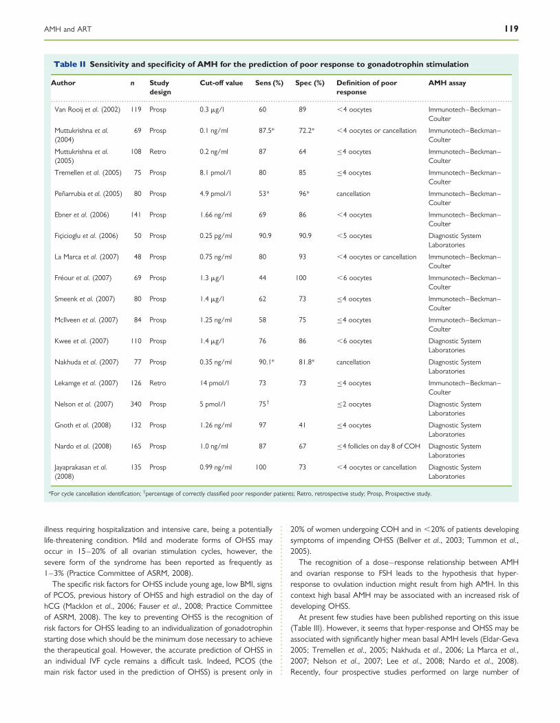
Table II Sensitivity and specificity of AMH for the prediction of poor response to gonadotrophin stimulation
Author n Studydesign
Cut-off value Sens (%) Spec (%) Definition of poorresponse
AMH assay
Van Rooij et al. (2002) 119 Prosp 0.3 mg/l 60 89 ,4 oocytes Immunotech–Beckman–Coulter
Muttukrishna et al.(2004)
69 Prosp 0.1 ng/ml 87.5* 72.2* ,4 oocytes or cancellation Immunotech–Beckman–Coulter
Muttukrishna et al.(2005)
108 Retro 0.2 ng/ml 87 64 �4 oocytes Immunotech–Beckman–Coulter
Tremellen et al. (2005) 75 Prosp 8.1 pmol/l 80 85 �4 oocytes Immunotech–Beckman–Coulter
Penarrubia et al. (2005) 80 Prosp 4.9 pmol/l 53* 96* cancellation Immunotech–Beckman–Coulter
Ebner et al. (2006) 141 Prosp 1.66 ng/ml 69 86 ,4 oocytes Immunotech–Beckman–Coulter
Ficicioglu et al. (2006) 50 Prosp 0.25 pg/ml 90.9 90.9 ,5 oocytes Diagnostic SystemLaboratories
La Marca et al. (2007) 48 Prosp 0.75 ng/ml 80 93 ,4 oocytes or cancellation Immunotech–Beckman–Coulter
Freour et al. (2007) 69 Prosp 1.3 mg/l 44 100 ,6 oocytes Immunotech–Beckman–Coulter
Smeenk et al. (2007) 80 Prosp 1.4 mg/l 62 73 �4 oocytes Immunotech–Beckman–Coulter
McIlveen et al. (2007) 84 Prosp 1.25 ng/ml 58 75 �4 oocytes Immunotech–Beckman–Coulter
Kwee et al. (2007) 110 Prosp 1.4 mg/l 76 86 ,6 oocytes Diagnostic SystemLaboratories
Nakhuda et al. (2007) 77 Prosp 0.35 ng/ml 90.1* 81.8* cancellation Diagnostic SystemLaboratories
Lekamge et al. (2007) 126 Retro 14 pmol/l 73 73 �4 oocytes Immunotech–Beckman–Coulter
Nelson et al. (2007) 340 Prosp 5 pmol/l 75†�2 oocytes Diagnostic System
Laboratories
Gnoth et al. (2008) 132 Prosp 1.26 ng/ml 97 41 �4 oocytes Diagnostic SystemLaboratories
Nardo et al. (2008) 165 Prosp 1.0 ng/ml 87 67 �4 follicles on day 8 of COH Diagnostic SystemLaboratories
Jayaprakasan et al.(2008)
135 Prosp 0.99 ng/ml 100 73 ,4 oocytes or cancellation Diagnostic SystemLaboratories
*For cycle cancellation identification; †percentage of correctly classified poor responder patients; Retro, retrospective study; Prosp, Prospective study.
AMH and ART 119

subjects have been published (Kwee et al., 2007; Nelson et al., 2007;Lee et al., 2008; Nardo et al., 2008) reporting relevant value for AMHfor the prediction of hyper response and OHSS (Table IV).
Particularly the studies by Lee et al. (2008) and Nardo et al. (2008)have independently calculated a similar performance of AMH for theprediction of hyper response and OHSS. The reported cut-off valueis of about 3.5 ng/ml, above which hyper-response/OHSS may beanticipated. In the study by Lee et al. (2008), a cohort of 262 IVFcycles was investigated, in order to evaluate the predictive value forOHSS by means of age, BMI, estradiol and AMH levels. Authorsfound that the ROC of the basal AMH was larger than age andBMI, and works equally well as the number of follicles and estradiollevels on the day of hCG. Basal AMH levels predicted OHSS with a
sensitivity of 90.5% and specificity of 81.3%. Interestingly the cut-offvalue calculated (3.36 ng/ml) corresponded to the highest quartileof the AMH values in their population, suggesting that hyper-responseand OHSS may be caused by gonadotrophin administration to womenwith ‘enhanced ovarian reserve’ (Lee et al., 2008). This was alsoevident in a previous study by our group (La Marca et al., 2007) inwhich all cases with ovarian hyper-response to COS where in thegroup of patients with basal AMH levels in the highest AMH quartile.Considering that PCOS has been associated with high AMH levels, it islogical to conclude that the prevalence of PCOS patients amongwomen with AMH levels in the highest AMH quartile may beincreased thus in part explaining the observed high rate of OHSS inthis group of women.
In conclusion, AMH measurement prior to gonadotrophin stimu-lation could provide useful information to direct the application ofmild patient-friendly stimulation protocols in order to avoid OHSS.
Prediction of qualitative ovarian responsein ARTIt is extensively recognized that pregnancy in ART is mostly related tothe qualitative than quantitative aspects of IVF. As the status of theovarian reserve includes both the quantity and quality of ovarian fol-licle pool, AMH may reflect not only quantitative but also qualitativeovarian responsiveness. Indeed several authors have found a significantpositive correlation between AMH levels, oocyte quality (Hazoutet al., 2004; Ebner et al., 2006; Silberstein et al., 2006; Cupisti et al.,2007; Fanchin et al., 2007, Lekamge et al., 2007) and embryo mor-phology (Silberstein et al., 2006). However, this relationship has notbeen confirmed by others (Smeenk et al., 2007; Lie Fong et al.,2008). In order clarify the complex relationship between AMH andoocyte quality, embryo quality and implantation and pregnancy rate,we should separately comment on studies of AMH in the follicularfluid and in serum.
Studies on AMH in the follicular fluidIn an elegant study AMH was measured in the follicular fluid obtainedfrom both small and large follicles on the day of oocyte retrieval
Figure 3 Sensitivity–specificity points for all studies reporting onthe performance for AMH in the prediction of poor response.
Reference lines indicate a desired level for sensitivity (0.75) and specificity(0.85).
................................................................................................
.............................................................................................................................................................................................
Table III Basal AMH levels in women with normal response, hyper-response to controlled ovarian stimulation (COS) andovarian hyperstimulation syndrome (OHSS)
Author Design n Mean AMH levels
Normal response Excessive response OHSS
Tremellen et al. (2005) Prosp 75 15.47 pmol/l 21.53 pmol/la
Eldar-Geva et al. (2005) Prosp 56 14.1 pmol/l 37.8 pmol/lb
Nakhuda et al. (2006) Retro 30 0.63 ng/ml 3.6 ng/ml
La Marca et al. (2007) Prosp 48 5.98 ng/ml 10.13 ng/mlc
Nelson et al. (2007) Prosp 340 10 pmol/l 27 pmol/ld
Nardo et al. (2008) Prosp 165 3.04 ng/ml 5.56 ng/mlb
Retro: retrospective study; Prosp: prospective study.aExcessive response if �18 oocytes retrieved.bExcessive response if �20 oocytes retrieved.cExcessive response if �16 oocytes retrieved.dExcessive response if �21 oocytes retrieved.
120 La Marca et al.

(Fanchin et al., 2005a, b). AMH levels in follicular fluid were found tobe roughly three times higher in small than in large follicles confirmingthe hypothesis that AMH production by granulosa cells probablydeclines during final follicular maturation. Moreover in both smalland large follicles, follicular fluid AMH levels correlated positivelywith the number of early antral follicles on cycle Day 3 before COS,growing follicles on the day of hCG administration and oocytesretrieved. This interesting finding may indicate that peripheral AMHlevels are not exclusively dependent on the number of follicles; theyare also modulated by individual follicular ability to produce AMH.Hence, elevated peripheral AMH levels indicate not only that thenumber of antral follicles is increased, but also that each follicle prob-ably produces more AMH individually. This offers us a new under-standing of the reported association between peripheral AMH levelsand the ovarian fertility potential, and leads the authors to speculatethat serum AMH measurement could reflect not only quantitativebut also qualitative ovarian responsiveness to COS (Fanchin et al.,2005a, b).
In a successive study by the same group (Fanchin et al., 2007), 118monodominant follicle cycles were prospectively studied. AMH wasmeasured in the follicular fluid and the fate of oocytes and embryosgenerated was observed. It was found that embryo implantation, clini-cal pregnancy and ongoing pregnancy rate increase dramatically fromthe low to the high follicular fluid AMH groups. The embryo mor-phology was similar within the groups, indicating that AMH in follicularfluid may be an additional factor in the selection of the oocyte (Fanchinet al., 2007). This is particularly relevant in countries with restrictivelaws limiting the number of oocytes that may be inseminated.A recent study on a large number of subjects (n ¼ 276) confirmedthe previous finding that levels of AMH in follicular fluid were signifi-cantly increased in women who became pregnant in the respectiveIVF /ICSI treatment cycle (Wunder et al., 2008).
Studies on circulating AMHAlthough studies on follicular fluid seem to indicate that AMH may beuseful in the prediction of oocyte and embryo quality and finally preg-nancy, the same could not be said for circulating AMH. At presentonly few studies concluded that serum AMH measurement may beable to give relevant information on gametes and embryo qualityand on the outcome of the treatment cycle.
Silberstein and colleagues (2006) found that serum AMH measuredon the day of hCG correlated with the quality of embryos obtained
thus allowing discrimination between embryos with high- and low-implantation potential. Consequently implantation rate, but notpregnancy rate, was higher in the group with high basal AMH levels(Silberstein et al., 2006). However, the lack of a consistent correlationbetween serum AMH and embryo morphology and embryo aneu-ploidy rate, which is not in favour of a direct relationship betweenoocyte quantity and embryo quality, has been clearly demonstrated(Lie Fong et al., 2008). Hence serum AMH seems not to be an ade-quate marker for embryo quality.
The vast majority of the studies investigating the performance ofserum AMH in the prediction of pregnancy occurrence following IVFreported that AMH measurement is not useful in the prediction ofsuccess. Only few studies reported a significant cut-off for AMHlevels able to distinguish between pregnancy and non-pregnancy. Itshould be noted that the only two positive prospective studies (Eldar-Geva et al., 2005; Elgindy et al., 2008) were limited by very smallnumbers of subjects (n ¼ 56 and 33, respectively). Conversely thelargest study (n ¼ 109) concluding that serum AMH may be predictiveof pregnancy had a retrospective design, hence limiting the scientificsoundness of the finding (Lekamge et al., 2007). However, the studyby Lekamge and colleagues (2007) analyzed for the first time thecumulative pregnancy rate from both fresh and freezed/thawedembryos. As a consequence of the relationship between serumAMH and the quantitative ovarian response to COS, women withlow AMH levels yielded fewer oocytes and generated fewerembryos, culminating in halving of the cumulative pregnancy rate com-pared with the high AMH group (Lekamge et al., 2007). Hence thehigher pregnancy rate observed in the group of patients with highbasal AMH levels, when compared with those with low AMH levels,may be explained on the basis of an increased availability of oocytes.
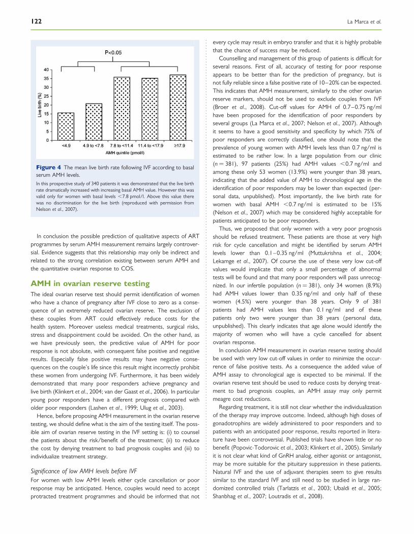
Until now only one study has been published relating serum AMHlevels to the live birth rate following IVF (Nelson et al., 2007). In thislarge prospective study of 340 patients it was demonstrated that thelive birth rate dramatically increased with increasing basal AMHvalues (Fig. 4). However, this was valid only for women with basallevels ,7.8 pmol/l. Above this value there was no discriminationfor the live birth. Basal AMH does not seem to predict pregnancyor non-pregnancy, but simply enables patients to be identified asbeing at a low or high probability of pregnancy after IVF. As concludedby the same authors, this finding may, at least in part, be explained bythe very good correlation existing between basal AMH and thenumber of retrieved oocytes (Nelson et al., 2007).
.............................................................................................................................................................................................
Table IV AMH cut-off values for the prediction of hyper-response to COS and OHSS
Author n Study design Cut-off value Sensitivity (%) Specificity (%) Prediction ofhyper-response
Predictionof OHSS
Kwee et al. (2007) 110 Prosp 5 mcg/l 53 91pa
Nelson et al. (2007) 340 Prosp 25 pmol/l 60 94.9pb
Lee et al. (2008) 262 Prosp 3.36 ng/ml 90.5 81.3p
Nardo et al. (2008) 165 Prosp 3.5 ng/ml 88 70pa
Prosp: prospective study.aExcessive response if .20 oocytes retrieved.bExcessive response if �21 oocytes retrieve.
AMH and ART 121
In conclusion the possible prediction of qualitative aspects of ARTprogrammes by serum AMH measurement remains largely controver-sial. Evidence suggests that this relationship may only be indirect andrelated to the strong correlation existing between serum AMH andthe quantitative ovarian response to COS.
AMH in ovarian reserve testingThe ideal ovarian reserve test should permit identification of womenwho have a chance of pregnancy after IVF close to zero as a conse-quence of an extremely reduced ovarian reserve. The exclusion ofthese couples from ART could effectively reduce costs for thehealth system. Moreover useless medical treatments, surgical risks,stress and disappointment could be avoided. On the other hand, aswe have previously seen, the predictive value of AMH for poorresponse is not absolute, with consequent false positive and negativeresults. Especially false positive results may have negative conse-quences on the couple’s life since this result might incorrectly prohibitthese women from undergoing IVF. Furthermore, it has been widelydemonstrated that many poor responders achieve pregnancy andlive birth (Klinkert et al., 2004; van der Gaast et al., 2006). In particularyoung poor responders have a different prognosis compared witholder poor responders (Lashen et al., 1999; Ulug et al., 2003).
Hence, before proposing AMH measurement in the ovarian reservetesting, we should define what is the aim of the testing itself. The poss-ible aim of ovarian reserve testing in the IVF setting is: (i) to counselthe patients about the risk/benefit of the treatment; (ii) to reducethe cost by denying treatment to bad prognosis couples and (iii) toindividualize treatment strategy.
Significance of low AMH levels before IVFFor women with low AMH levels either cycle cancellation or poorresponse may be anticipated. Hence, couples would need to acceptprotracted treatment programmes and should be informed that not
every cycle may result in embryo transfer and that it is highly probablethat the chance of success may be reduced.
Counselling and management of this group of patients is difficult forseveral reasons. First of all, accuracy of testing for poor responseappears to be better than for the prediction of pregnancy, but isnot fully reliable since a false positive rate of 10–20% can be expected.This indicates that AMH measurement, similarly to the other ovarianreserve markers, should not be used to exclude couples from IVF(Broer et al., 2008). Cut-off values for AMH of 0.7–0.75 ng/mlhave been proposed for the identification of poor responders byseveral groups (La Marca et al., 2007; Nelson et al., 2007). Althoughit seems to have a good sensitivity and specificity by which 75% ofpoor responders are correctly classified, one should note that theprevalence of young women with AMH levels less than 0.7 ng/ml isestimated to be rather low. In a large population from our clinic(n ¼ 381), 97 patients (25%) had AMH values ,0.7 ng/ml andamong these only 53 women (13.9%) were younger than 38 years,indicating that the added value of AMH to chronological age in theidentification of poor responders may be lower than expected (per-sonal data, unpublished). Most importantly, the live birth rate forwomen with basal AMH ,0.7 ng/ml is estimated to be 15%(Nelson et al., 2007) which may be considered highly acceptable forpatients anticipated to be poor responders.
Thus, we proposed that only women with a very poor prognosisshould be refused treatment. These patients are those at very highrisk for cycle cancellation and might be identified by serum AMHlevels lower than 0.1–0.35 ng/ml (Muttukrishna et al., 2004;Lekamge et al., 2007). Of course the use of these very low cut-offvalues would implicate that only a small percentage of abnormaltests will be found and that many poor responders will pass unrecog-nized. In our infertile population (n ¼ 381), only 34 women (8.9%)had AMH values lower than 0.35 ng/ml and only half of thesewomen (4.5%) were younger than 38 years. Only 9 of 381patients had AMH values less than 0.1 ng/ml and of thesepatients only two were younger than 38 years (personal data,unpublished). This clearly indicates that age alone would identify themajority of women who will have a cycle cancelled for absentovarian response.
In conclusion AMH measurement in ovarian reserve testing shouldbe used with very low cut-off values in order to minimize the occur-rence of false positive tests. As a consequence the added value ofAMH assay to chronological age is expected to be minimal. If theovarian reserve test should be used to reduce costs by denying treat-ment to bad prognosis couples, an AMH assay may only permitmeagre cost reductions.
Regarding treatment, it is still not clear whether the individualizationof the therapy may improve outcome. Indeed, although high doses ofgonadotrophins are widely administered to poor responders and topatients with an anticipated poor response, results reported in litera-ture have been controversial. Published trials have shown little or nobenefit (Popovic-Todorovic et al., 2003; Klinkert et al., 2005). Similarlyit is not clear what kind of GnRH analog, either agonist or antagonist,may be more suitable for the pituitary suppression in these patients.Natural IVF and the use of adjuvant therapies seem to give resultssimilar to the standard IVF and still need to be studied in large ran-domized controlled trials (Tarlatzis et al., 2003; Ubaldi et al., 2005;Shanbhag et al., 2007; Loutradis et al., 2008).
Figure 4 The mean live birth rate following IVF according to basalserum AMH levels.
In this prospective study of 340 patients it was demonstrated that the live birthrate dramatically increased with increasing basal AMH value. However this wasvalid only for women with basal levels ,7.8 pmol/l. Above this value therewas no discrimination for the live birth (reproduced with permission fromNelson et al., 2007).
122 La Marca et al.
In conclusion AMH measurement, when low AMH values arefound, may have an added value to chronological age only in the coun-selling of the patients. Doubts remain regarding both the possiblereduction of costs (consequent to IVF refusing) and the possibleimprovement of the outcome (consequent to the individualization ofthe treatment).
Significance of normal AMH levels before IVFWomen with normal AMH levels are most probably normal respon-ders and a good prognosis may be anticipated. Currently, there isno evidence to modify the normal strategy based on the standardlong protocol (Daya, 2000). In recent years, mild ovarian stimulationprotocols for predicted good responder women have been proposedas a valid and alternative standard treatment in order to achieve cost-effective, patient-friendly regimens which optimize the balancebetween outcomes and risks of treatment (Hohmann et al., 2001,2003; Heijnen et al., 2007; Verberg et al., 2009). However, further,sufficiently-powered prospective studies applying novel mild treatmentregimens are required.
Significance of high AMH levels before IVFWomen with high AMH levels are considered to be at risk for hyper-response and OHSS. Hence these women should be informed aboutthis risk. Women with high AMH levels are those who may reallybenefit from the individualization of the treatment. Indeed a lowFSH starting dose followed by the use of GnRH antagonists (AlInany et al., 2006; Doldi et al., 2006; Griesinger et al., 2006) havebeen shown to reduce the incidence of OHSS and may be proposedas a first line treatment for patients with high serum AMH levels.Moreover the use of GnRH antagonist permits the triggering of ovu-lation by means of GnRH agonist instead of hCG and this practicehas been recognized as useful in the prevention of OHSS (Olivenneset al., 2002; Engmann et al., 2006; Griesinger et al., 2006; Orvietoet al., 2006; Kol and Solt, 2008).
In conclusion it seems that AMH measurement, when high AMHvalues are found, may have relevant clinical value for the specialist.Indeed such information may improve the counselling of the patients(about the increased risk for OHSS), and permit an individualizationof the treatment with the aim of reducing the incidence of OHSS,and finally, AMH measurement may reduce the costs linked tohospitalization.
AMH in male fertility
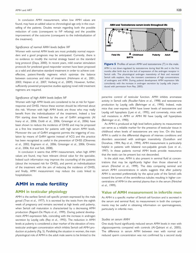
AMH in testicular physiologyAMH is the earliest Sertoli cell specific protein expressed by the malegonad (Tran et al., 1977). It is secreted by the testis from the eighthweek of pregnancy and remains secreted at high levels until puberty,when Sertoli cell maturation is characterized by a decreasing AMHproduction (Rajpert-De Meyts et al., 1999). During puberal develop-ment AMH expression falls, coinciding with the increase in androgensecretion by Leydig cells (Rey et al., 1993). The reduction in AMHlevels at puberty is considered a clear marker of the elevation of intra-testicular androgen concentration which inhibits Sertoli cell AMH pro-duction at puberty (Fig. 5). Paralleling the situation in women, the mainphysiological role of AMH in the adult male seems to be limited to the
paracrine control of testicular function. AMH inhibits aromataseactivity in Sertoli cells (Rouiller-Fabre et al., 1998) and testosteroneproduction by Leydig cells (Behringer et al., 1990). Indeed, malemice that over-express AMH have lower levels of testosterone andLeydig cell hypoplasia (Lyet et al., 1995), and conversely, mice withnull mutations in AMH or AMH RII have Leydig cell hyperplasia(Behringer et al., 1990).
As AMH is produced at high level before puberty its measurementcan serve as a reliable marker for the presence of testicular tissue inchildhood when levels of testosterone are very low. On this basisAMH is useful in the differential diagnosis of intersex conditions anddisorders associated with androgen insensitivity (Gustafson andDonahoe, 1994; Rey et al., 1994). AMH measurement is particularlyhelpful in patients with bilateral non-palpable gonads (Lee et al.,1997). In these patients normal AMH levels provide reassurancethat the testis can be present but not descended.
In the adult man, AMH is also present in seminal fluid at concen-trations that may be significantly higher than those observed inserum (Fenichel et al., 1999). The data comparing seminal andserum AMH concentrations in adults suggests that after pubertyAMH is secreted preferentially by the apical pole of the Sertoli cellstoward the lumen of the seminiferous tubules resulting in higher con-centrations of AMH in the seminal plasma than in the serum (Fenichelet al., 1999).
Value of AMH measurement in infertile menAs AMH is a specific marker of Sertoli cell function and is secreted inthe serum and seminal fluid, its measurement in both the compart-ments may be useful in obtaining information on spermatogenesis,particularly in infertile men.
Studies on serum AMHOne study found significantly reduced serum AMH levels in men witholigozoospermia compared with controls (Al-Qahtani et al., 2005).The difference in serum AMH between men with normal andreduced sperm concentration was not confirmed by a second study
Figure 5 Profiles of serum AMH and testosterone (T) in the male.
AMH is not down-regulated by testosterone during fetal life and in the firstmonths after birth due to the lack of expression of the androgen receptor inSertoli cells. The physiological androgen insensitivity of fetal and neonatalSertoli cells explains, thus, the transient coexistence of high concentrationsof androgens and AMH. During puberal development AMH expression fallscoincidently with the increase in androgen secretion by Leydig cells (repro-duced with permission from Rey, 2005).
AMH and ART 123

by the same group (Appasamy et al., 2007), however, a correlation ofserum AMH levels with sperm count and serum FSH levels has beenreported (Appasamy et al., 2007).
In the largest study to date, performed on 199 men, no significantdifferences were found in serum AMH levels between controls andmen with oligozoospermia (Tuttelmann et al., 2009), confirming thatserum AMH is not of diagnostic significance in men with impairedspermatogenesis. Serums AMH levels have been found to be signifi-cantly lower in non-obstructive azoospermic (NOA) than in obstruc-tive azoospermic (OA) patients and normal fertile men (Muttukrishnaet al., 2007). However, the wide overlapping of values between con-trols and infertile men prevents this hormone from being a useful diag-nostic marker.
Other studies have investigated whether serum AMH levels may bepredictive of the presence of sperm in testis from NOA patients (Isi-koglu et al., 2006; Goulis et al., 2009). It has been clearly demon-strated that serum AMH could not predict the presence of sperm infine-needle aspiration (Goulis et al., 2009) or in testicular spermextraction (TESE) (Isikoglu et al., 2006) performed in men with NOA.
Studies on AMH in the seminal fluidAfter puberty, AMH is preferentially secreted by the apical side ofSertoli cells, into the seminiferous tubules, explaining the higher con-centration of AMH in the seminal fluid when compared with theserum in adult men (Fenichel et al., 1999). This observation suggestsa closer link between spermatogenesis and seminal AMH thanserum AMH. Seminal AMH correlated with sperm concentrationand, as a consequence, seminal AMH levels in controls were signifi-cantly higher than in oligozoospermic men (Fujisawa et al., 2002;Duville et al., 2008; Mostafa et al., 2007). As expected, seminalAMH levels have been reported to be significantly lower in azoosper-mic men than in oligozoospermic and healthy men (Duville et al.,2008; Mostafa et al., 2007). In particular AMH was not detectable insemen from OA patients (Fenichel et al., 1999; Mostafa et al., 2007)whereas it was detectable in 39–57.5% of NOA patients (Fenichelet al., 1999; Mostafa et al., 2007).
When evaluating the predictive value of seminal AMH on TESEoutcome in NOA patients, all studies confirmed that AMH measure-ment in the seminal fluid is not useful in distinguishing between caseswith positive and negative outcome (Fenichel et al., 1999; Isikogluet al., 2006; Mostafa et al., 2007; Duvilla et al., 2008). This is not sur-prising as, similarly to other endocrinological markers of testicularfunction (FSH and inhibin B), variations in AMH levels can occur forreasons unrelated to spermatogenesis.
ConclusionsRecent studies have indicated that AMH may constitute an importantnovel measure of ovarian reserve. Serum AMH levels show areduction throughout reproductive life and are undetectable aftermenopause (Van Rooij et al., 2004; Van Rooij et al., 2005; La Marcaet al., 2005a, b; Robertson et al., 2008; Shin et al., 2008; Van Dissel-dorp et al., 2008). Similarly, early ovarian ageing and prematureovarian failure have been associated with very low or undetectableserum levels, respectively (La Marca et al., 2006b; De Koning et al.,2008; Knauff et al., 2008). Furthermore AMH levels do not significantlychange during the menstrual cycle (Hehenkamp et al., 2006; La Marca
et al., 2006a; Streuli et al., 2008), whereas all other hormonessecreted by the ovary show significant variations throughout thecycle. The stability and consistency of its levels indicate that AMHcould be used as the most reliable single marker of ovarian ageingand ovarian response to COS.
For women who want to become pregnant by means of ART, it isimportant to offer counselling about the optimal balance betweenbenefit and risk. Since these outcomes are highly dependent onovarian reserve, much effort has been put into identifying good clinicalmarkers of ovarian reserve regarding individual prognosis for successand to design appropriate stimulation protocols.
Although AMH measurement is of course more expensive than ageevaluation as a single marker of ovarian reserve, it clearly performsbetter in the prediction of both poor and hyper-response to COS(Table V). Furthermore, AMH ease of measurement confers a relevantadvantage to FSH which is cycle-dependent and less powerful. AMHmay also be informative on ovarian reserve in women during GnRHagonist treatment or hormonal contraception that consequentlyexhibit suppressed FSH levels. Finally, it seems that poor responsemay be predicted by AMH with a performance which is similar tothe AFC. Conversely AMH seems superior to AFC in the predictionof hyper response (Nardo et al., 2008). Although AFC is a verycommon and useful measurement it may be sometimes technicallychallenging and operator-dependent. Considering all these peculiarcharacteristics, it may be concluded that AMH is a candidate proposedas the ideal test for the ovarian reserve evaluation (La Marca et al.,2006a, b, c; 2009b) (Table V).
One new interesting field of application for AMH measurement, maybe its use in the individualization of ovarian stimulation regimens. Inmany centres, the starting FSH dose for the first IVF is often selectedon the basis of age and possibly also BMI of the patient. Someauthors have recently proposed adjusting the treatment strategy onthe basis of AMH levels (Nelson et al., 2007, 2009; Gnoth et al., 2008).
As low and high AMH values are predictive of poor- and high-response to gonadotrophins, respectively, it has been proposed thatthe daily dose of FSH is tailored according to the pre-IVF AMHlevels, and independently of the age and BMI of the patient (Nelsonet al., 2007, 2009; Gnoth et al., 2008).
........................................................................................
Table V Comparison of characteristics of the mostwidely used markers of ovarian reserve
Characteristics for a goodmarker
Age AMH FSH AFC
Prediction of poor response þ þþþ þþ þþþ
Prediction of hyper response þ þþþ 2 þþ
Low inter-cycle variability þþþ þþ 2 þþ
Low intra-cycle variability þþþ þþ 2 þþ
Blinded to the operator þþþ þþþ þþþ 2
Applicable to all patients (a) þþþ þþþ þ þ
Cheapness þþþ 2 2 2
(a) FSH and antral follicle count (AFC) are not informative in patients on hormonalcontraception or GnRH agonist treatment. Moreover the count of antral follicles may bedifficult in women with ovarian cysts or with previous pelvic surgery.
124 La Marca et al.

If AMH measurement is proposed to all women before entering anIVF programme, a clear definition of cut-off values for the prediction ofpoor- and hyper-response is required. Similarly the treatment strat-egies for the various groups of patients should be elaborated. Finallyan analysis of cost and benefit of this programme is mandatory.Most of these aspects have been addressed in a recent prospectivestudy in which the COS strategies have been based only on serumAMH levels (Nelson et al., 2009). More than five hundred patientswere divided into four groups on the basis of AMH levels: the pre-dicted negligible response category (AMH , 1 pmol/l), the predictedreduced response category (AMH � 1, ,5 pmol/l), the predictednormal response category (AMH � 5, ,15 pmol/l) and the predictedhigh response category (AMH � 15 pmol/l). Different stimulationprotocols were then applied only on the basis of this stratification,independently of the age of the patients. In particular, women withlow AMH received a high starting FSH dose followed by the GnRHantagonist, women with normal AMH levels received the standardlong protocol and women with high AMH received a low FSH dosefollowed by the GnRH antagonist. The authors found that this AMH-based strategy of COS was associated with a significant reduction ofexcessive response to stimulation and in reduced treatment burden,reduced cycle cancellation and a trend towards increased clinical effi-cacy. Even if the study by Nelson and colleagues (2009) has severallimitations such as the non-randomized design and the non-randomuse of different gonadotrophin formulations, it clearly demonstratesthat a single AMH assay may be used to individualize treatment strat-egies for IVF, potentially resulting in reduced clinical risks, along withoptimized treatment burden and clinical pregnancy rate (Nelsonet al., 2009).
Finally AMH may be incorporated into a more complex predictivecalculation of response like the CONSORT formula (Olivenneset al., 2009). The CONSORT dosing algorithm individualizes recombi-nant FSH doses for ART according to certain patient characteristics:basal FSH, body mass index, age and antral follicle count. The useof the CONSORT algorithm seems to achieve an adequate oocyteyield and good pregnancy rates (Olivennes et al., 2009). Adjustmentof the algorithm by incorporating further powerful markers such asAMH may, in turn, increase the clinical efficacy of the formula.
In summary, published studies indicate a relevant role for AMHmeasurement in the identification of both the extremes of ovarianresponse to stimulation, and probably in the consequent individualiza-tion of treatment strategies in order to possibly reduce the incidenceof cycle cancellation and OHSS. It still remains to clarify the cost/benefit of its use as a single assay before beginning an IVF cycle andwhether the AMH-determined strategy of COS for assisted con-ception may be associated with improved live birth rate.
Concerning the role of AMH in the evaluation of infertile men, itshould be highlighted that much research has been focused in thelast years on the identification of serum and seminal markers able toimprove the understanding of germinal deficiency and to allow dis-crimination between absent, incomplete or reduced spermatogenesis.AMH seems to be a good candidate marker since it is of testicularorigin, it is specifically secreted by Sertoli cells, it is correlated withspermatogenesis and is present in both serum and seminal fluid indetectable concentrations. Serum AMH seems to be significantlylower in men with NOA than OA and controls, however, the wideoverlapping of values between subjects prevents this hormone from
being of clinical utility. On the contrary AMH is undetectable inseminal fluid for men with obstructive azoospermia, thus beinguseful to formulate the, not always easy, diagnosis of obstructiveazoospermia. Unfortunately in the studies to date, the seminal AMHpredictive value on TESE outcome in case of NOA is not optimal inthe identification of men with successful sperm retrieval. Inclusion ofAMH in an equation obtained by multivariate logistic regression analy-sis and including other preoperative factors may be a strategy toobtain a satisfactory clinical use of AMH determination in men.
ReferencesAl-Inany HG, Abou-Setta AM, Aboulghar M. Gonadotrophin-releasing
hormone antagonists for assisted conception. Cochrane Database SystRev 2006;3:CD001750.
Al-Qahtani A, Muttukrishna S, Appasamy M, Johns J, Cranfield M,Visser JA, Themmen AP, Groome NP. Development of a sensitiveenzyme immunoassay for anti-Mullerian hormone and the evaluationof potential clinical applications in males and females. Clin Endocrinol(Oxf) 2005;63:267–273.
Andersen CY, Byskov AG. Estradiol and regulation of anti-Mullerianhormone, inhibin-A, and inhibin-B secretion: analysis of small antraland preovulatory human follicles’ fluid. J Clin Endocrinol Metab 2006;91:4064–4069.
Appasamy M, Muttukrishna S, Pizzey AR, Ozturk O, Groome NP,Serhal P, Jauniaux E. Relationship between male reproductivehormones, sperm DNA damage and markers of oxidative stress ininfertility. Reprod Biomed Online 2007;14:159–165.
Arbo E, Vetori DV, Jimenez MF, Freitas FM, Lemos N, Cunha-Filho JS.Serum anti-Mullerian hormone levels and follicular cohortcharacteristics after pituitary suppression in the late luteal phase withoral contraceptive pills. Hum Reprod 2007;22:3192–3196.
Baarends WM, Uilenbroek JT, Kramer P, Hoogerbrugge JW, vanLeeuwen EC, Themmen AP, Grootegoed JA. Anti-Mullerian hormoneand anti-Mullerian hormone type II receptor messenger ribonucleicacid expression in rat ovaries during postnatal development, theestrous cycle and gonadotropin-induced follicle growth. Endocrinology1995;136:4951–4962.
Bancsi LF, Broekmans FJ, Eijkemans MJ, de Jong FH, Habbema JD,te Velde ER. Predictors of poor ovarian response in in vitrofertilization: a prospective study comparing basal markers of ovarianreserve. Fertil Steril 2002;77:328–336.
Behringer RR, Cate RL, Froelick GJ, Palmiter RD, Brinster RL. Abnormalsexual development in transgenic mice chronically expressing mullerianinhibiting substance. Nature 1990;345:167–170.
Bellver J, Munoz EA, Ballesteros A, Soares SR, Bosch E, Simon C,Pellicer A, Remohı J. Intravenous albumin does not preventmoderate-severe ovarian hyperstimulation syndrome in high-risk IVFpatients: a randomized controlled study. Hum Reprod 2003;18:2283–2288.
Bergada I, Milani C, Bedecarras P, Andreone L, Ropelato MG, Gottlieb S,Bergada C, Campo S, Rey RA. Time course of the serum gonadotropinsurge, inhibins, and anti-Mullerian hormone in normal newborn malesduring the first month of life. J Clin Endocrinol Metab 2006;91:4092–4098.
Bersinger NA, Wunder D, Birkhauser MH, Guibourdenche J.Measurement of anti-Mullerian hormone by Beckman Coulter ELISAand DSL ELISA in assisted reproduction: differences between serumand follicular fluid. Clin Chim Acta 2007;384:174–175.
AMH and ART 125

Broekmans FJ, Kwee J, Hendriks DJ, Mol BW, Lambalk CB. A systematicreview of tests predicting ovarian reserve and IVF outcome. HumReprod Update 2006;12:685–718.
Broekmans FJ, Visser JA, Laven JS, Broer SL, Themmen AP, Fauser BC.Anti-Mullerian hormone and ovarian dysfunction. Trends EndocrinolMetab 2008;19:340–347.
Broer SL, Mol BW, Hendriks D, Broekmans FJ. The role of anti-Mullerianhormone in prediction of outcome after IVF: comparison with the antralfollicle count. Fertil Steril 2008; [Epub ahead of print].
Carlsson IB, Scott JE, Visser JA, Ritvos O, Themmen AP, Hovatta O.Anti-Mullerian hormone inhibits initiation of growth of humanprimordial ovarian follicles in vitro. Hum Reprod 2006;21:2223–2227.
Carlsen SM, Vanky E, Fleming R. Anti-Mullerian hormone concentrationsin androgen-suppressed women with polycystic ovary syndrome. HumReprod 2009;24:1732–1738.
Cate RL, Mattaliano RJ, Hession C, Tizard R, Farber NM, Cheung A,Ninfa EG, Frey AZ, Gash DJ, Chow EP. Isolation of the bovine andhuman genes for Mullerian inhibiting substance and expression of thehuman gene in animal cells. Cell 1986;45:685–698.
Cook CL, Siow Y, Brenner AG, Fallat ME. Relationship between serummullerian-inhibiting substance and other reproductive hormones inuntreated women with polycystic ovary syndrome and normalwomen. Fertil Steril 2002;77:141–146.
Cupisti S, Dittrich R, Mueller A, Strick R, Stiegler E, Binder H,Beckmann MW, Strissel P. Correlations between anti-Mullerianhormone, inhibin B, and activin A in follicular fluid in IVF/ICSI patientsfor assessing the maturation and developmental potential of oocytes.Eur J Med Res 2007;12:604–608.
Daya S. Gonadotropin releasing hormone agonist protocols for pituitarydesensitization in in vitro fertilization and gamete intrafallopian transfercycles. Cochrane Database Syst Rev 2000;2:CD001299.
de Koning CH, McDonnell J, Themmen AP, de Jong FH, Homburg R,Lambalk CB. The endocrine and follicular growth dynamicsthroughout the menstrual cycle in women with consistently orvariably elevated early follicular phase FSH compared with controls.Hum Reprod 2008;23:1416–1423.
di Clemente N, Josso N, Gouedard L, Belville C. Components of theanti-Mullerian hormone signaling pathway in gonads. Mol CellEndocrinol 2003;211:9–14.
Doldi N, Persico P, Di Sebastiano F, Marsiglio E, Ferrari A.Gonadotropin-releasing hormone antagonist and metformin fortreatment of polycystic ovary syndrome patients undergoing in vitrofertilization-embryo transfer. Gynecol Endocrinol 2006;22:235–238.
Durlinger AL, Gruijters MJ, Kramer P, Karels B, Kuman TR, Matzuk MM,Rose UM, de Jong FH, Uilenbroek JT, Grootegoed JA et al.Anti-Mullerian hormone attenuates the effects of FSH on follicledevelopment in the mouse ovary. Endocrinology 2001;142:4891–4899.
Duvilla E, Lejeune H, Trombert-Paviot B, Gentil-Perret A, Tostain J,Levy R. Significance of inhibin B and anti-Mullerian hormonein seminal plasma: a preliminary study. Fertil Steril 2008;89:444–448.
Ebner T, Sommergruber M, Moser M, Shebl O, Schreier-Lechner E,Tews G. Basal level of anti-Mullerian hormone is associatedwith oocyte quality in stimulated cycles. Hum Reprod 2006;21:2022–2026.
Eldar-Geva T, Ben-Chetrit A, Spitz IM, Rabinowitz R, Markowitz E,Mimoni T, Gal M, Zylber-Haran E, Margalioth EJ. Dynamic assays ofinhibin B, anti-Mullerian hormone and estradiol following FSHstimulation and ovarian ultrasonography as predictors of IVF outcome.Hum Reprod 2005;20:3178–3183.
Elgindy EA, El-Haieg DO, El-Sebaey A. Anti-Mullerian hormone:correlation of early follicular, ovulatory and midluteal levels with
ovarian response and cycle outcome in intracytoplasmic sperminjection patients. Fertil Steril 2008;89:1670–1676.
El-Halawaty S, Rizk A, Kamal M, Aboulhassan M, Al-Sawah H, Noah O,Al-Inany H. Clinical significance of serum concentration ofanti-Mullerian hormone in obese women with polycystic ovarysyndrome. Reprod Biomed Online 2007;15:495–499.
El-Toukhy T, Khalaf Y, Hart R, Taylor A, Braude P. Young age does notprotect against the adverse effects of reduced ovarian reserve—aneight year study. Hum Reprod 2002;17:1519–1524.
Engmann L, Siano L, Schmidt D, Nulsen J, Maier D, Benadiva C. GnRHagonist to induce oocyte maturation during IVF in patients at high riskof OHSS. Reprod Biomed Online 2006;13:639–644.
Faddy MJ, Gosden RG. A model conforming the decline in follicle numbersto the age of menopause in women. Hum Reprod 1996;11:1484–1486.
Fallat ME, Siow Y, Marra M, Cook C, Carrillo A. Mullerian-inhibitingsubstance in follicular fluid and serum: a comparison of patients withtubal factor infertility, polycystic ovary syndrome, and endometriosis.Fertil Steril 1997;67:962–965.
Fanchin R, de Ziegler D, Olivennes F, Taieb J, Dzik A, Frydman R.Exogenous follicle stimulating hormone ovarian reserve test (EFORT):a simple and reliable screening test for detecting ‘poor responders’ inin-vitro fertilization. Hum Reprod 1994;9:1607–1611.
Fanchin R, Schonauer LM, Righini C, Frydman N, Frydman R, Taieb J.Serum anti-Mullerian hormone dynamics during controlled ovarianhyperstimulation. Hum Reprod 2003a;18:328–332.
Fanchin R, Schonauer LM, Righini C, Guibourdenche J, Frydman R, Taieb J.Serum anti-Mullerian hormone is more strongly related to ovarianfollicular status than serum inhibin B, estradiol, FSH and LH on day 3.Hum Reprod 2003b;18:323–327.
Fanchin R, Louafi N, Mendez Lozano DH, Frydman N, Frydman R, Taieb J.Per-follicle measurements indicate that anti-mullerian hormonesecretion is modulated by the extent of follicular development andluteinization and may reflect qualitatively the ovarian follicular status.Fertil Steril 2005a;84:167–173.
Fanchin R, Taieb J, Lozano DH, Ducot B, Frydman R, Bouyer J. Highreproducibility of serum anti-Mullerian hormone measurementssuggests a multi-staged follicular secretion and strengthens its role inthe assessment of ovarian follicular status. Hum Reprod 2005b;20:923–927.
Fanchin R, Mendez Lozano DH, Frydman N, Gougeon A, di Clemente N,Frydman R, Taieb J. Anti-Mullerian hormone concentrations in thefollicular fluid of the preovulatory follicle are predictive of theimplantation potential of the ensuing embryo obtained by in vitrofertilization. J Clin Endocrinol Metab 2007;92:1796–1802.
Fauser BC, Diedrich K, Devroey P. Predictors of ovarian response:progress towards individualized treatment in ovulation induction andovarian stimulation. Hum Reprod Update 2008;14:1–14.
Fedorcsak P, Dale PO, Storeng R, Ertzeid G, Bjercke S, Oldereid N,Omland AK, Abyholm T, Tanbo T. Impact of overweight andunderweight on assisted reproduction treatment. Hum Reprod 2004;19:2523–2528.
Fenichel P, Rey R, Poggioli S, Donzeau M, Chevallier D, Pointis G.Anti-Mullerian hormone as a seminal marker for spermatogenesisin non-obstructive azoospermia. Hum Reprod 1999;14:2020–2024.
Ficicioglu C, Kutlu T, Baglam E, Bakacak Z. Early follicular antimullerianhormone as an indicator of ovarian reserve. Fertil Steril 2006;85:592–596.
Fleming R, Deshpande N, Traynor I, Yates RW. Dynamics of FSH-inducedfollicular growth in subfertile women: relationship with age, insulinresistance, oocyte yield and anti-Mullerian hormone. Hum Reprod2006;21:1436–1441.
126 La Marca et al.

Freeman EW, Gracia CR, Sammel MD, Lin H, Lim LC, Strauss JF 3rd.Association of anti-mullerian hormone levels with obesity in latereproductive-age women. Fertil Steril 2007;87:101–106.
Freour T, Mirallie S, Bach-Ngohou K, Denis M, Barriere P, Masson D.Measurement of serum anti-Mullerian hormone by Beckman CoulterELISA and DSL ELISA: comparison and relevance in assistedreproduction technology (ART). Clin Chim Acta 2007;375:162–164.
Freour T, Masson D, Mirallie S, Jean M, Bach K, Dejoie T, Barriere P.Active smoking compromises IVF outcome and affects ovarianreserve. Reprod Biomed Online 2008;16:96–102.
Fujisawa M, Yamasaki T, Okada H, Kamidono S. The significance ofanti-Mullerian hormone concentration in seminal plasma forspermatogenesis. Hum Reprod 2002;17:968–970.
Galey-Fontaine J, Cedrin-Durnerin I, Chaıbi R, Massin N, Hugues JN. Ageand ovarian reserve are distinct predictive factors of cycle outcome inlow responders. Reprod Biomed Online 2005;10:94–99.
Gnoth C, Schuring AN, Friol K, Tigges J, Mallmann P, Godehardt E.Relevance of anti-Mullerian hormone measurement in a routine IVFprogram. Hum Reprod 2008;23:1359–1365.
Goulis DG, Tsametis C, Iliadou PK, Polychronou P, Kantartzi PD,Tarlatzis BC, Bontis IN, Papadimas I. Serum inhibin B andanti-Mullerian hormone are not superior to follicle-stimulatinghormone as predictors of the presence of sperm in testicularfine-needle aspiration in men with azoospermia. Fertil Steril 2009;91:1279–1284.
Gracia CR, Freeman EW, Sammel MD, Lin H, Nelson DB. The relationshipbetween obesity and race on inhibin B during the menopause transition.Menopause 2005;12:559–566.
Griesinger G, Diedrich K, Devroey P, Kolibianakis EM. GnRH agonist fortriggering final oocyte maturation in the GnRH antagonist ovarianhyperstimulation protocol: a systematic review and meta-analysis.Hum Reprod Update 2006;12:159–168.
Guibourdenche J, Lucidarme N, Chevenne D, Rigal O, Nicolas M,Luton D, Leger J, Porquet D, Noel M. Anti-Mullerian hormone levelsin serum from human foetuses and children: pattern and clinicalinterest. Mol Cell Endocrinol 2003;211:55–63.
Gustafson ML, Donahoe PK. Male sex determination: current concepts ofmale sexual differentiation. Annu Rev Med 1994;45:505–524.
Hall JE, Welt CK, Cramer DW. Inhibin A and inhibin B reflect ovarianfunction in assisted reproduction but are less useful at predictingoutcome. Hum Reprod 1999;14:409–415.
Hazout A, Bouchard P, Seifer DB, Aussage P, Junca AM, Cohen-Bacrie P.Serum antimullerian hormone/mullerian-inhibiting substance appears tobe a more discriminatory marker of assisted reproductive technologyoutcome than follicle-stimulating hormone, inhibin B, or estradiol.Fertil Steril 2004;82:1323–1329.
Hehenkamp WJ, Looman CW, Themmen AP, de Jong FH, Te Velde ER,Broekmans FJ. Anti-Mullerian hormone levels in the spontaneousmenstrual cycle do not show substantial fluctuation. J Clin EndocrinolMetab 2006;91:4057–4063.
Heijnen EM, Eijkemans MJ, De Klerk C, Polinder S, Beckers NG,Klinkert ER, Broekmans FJ, Passchier J, Te Velde ER, Macklon NSet al. A mild treatment strategy for in-vitro fertilisation: a randomisednon-inferiority trial. Lancet 2007;369:743–749.
Hendriks DJ, Mol BW, Bancsi LF, Te Velde ER, Broekmans FJ. Antralfollicle count in the prediction of poor ovarian response andpregnancy after in vitro fertilization: a meta-analysis and comparisonwith basal follicle-stimulating hormone level. Fertil Steril 2005;83:291–301.
Hohmann FP, Laven JS, de Jong FH, Eijkemans MJ, Fauser BC. Low-doseexogenous FSH initiated during the early, mid or late follicular phase
can induce multiple dominant follicle development. Hum Reprod.2001;16:846–854.
Hohmann FP, Macklon NS, Fauser BC. A randomized comparison of twoovarian stimulation protocols with gonadotropin-releasing hormone(GnRH) antagonist cotreatment for in vitro fertilization commencingrecombinant follicle-stimulating hormone on cycle day 2 or 5 with thestandard long GnRH agonist protocol. J Clin Endocrinol Metab 2003;88:166–173.
Isikoglu M, Ozgur K, Oehninger S, Ozdem S, Seleker M. Serumanti-Mullerian hormone levels do not predict the efficiency oftesticular sperm retrieval in men with non-obstructive azoospermia.Gynecol Endocrinol 2006;22:256–260.
Jayaprakasan K, Campbell B, Hopkisson J, Johnson I, Raine-Fenning N.A prospective, comparative analysis of anti-Mullerian hormone,inhibin-B, and three-dimensional ultrasound determinants of ovarianreserve in the prediction of poor response to controlled ovarianstimulation. Fertil Steril 2008; [Epub ahead of print].
Jee BC, Ku SY, Suh CS, Kim KC, Lee WD, Kim SH. Serum anti-Mullerianhormone and inhibin B levels at ovulation triggering day can predict thenumber of immature oocytes retrieved in in vitro fertilization cycles.J Korean Med Sci 2008;23:657–661.
Josso N, di Clemente N, Gouedard L. Anti-Mullerian hormone and itsreceptors. Mol Cell Endocrinol 2001;179:25–32.
Jost A. Recherches sur la differenciation sexuelle de l’embryon de lapin,Arch. Anat. Microsc Morphol Exp 1946;36:271–315.
Kailasam C, Keay SD, Wilson P, Ford WC, Jenkins JM. Defining poorovarian response during IVF cycles, in women aged ,40 years, and itsrelationship with treatment outcome. Hum Reprod 2004;19:1544–1547.
Kevenaar ME, Themmen AP, Laven JS, Sonntag B, Fong SL,Uitterlinden AG, de Jong FH, Pols HA, Simoni M, Visser JA.Anti-Mullerian hormone and anti-Mullerian hormone type II receptorpolymorphisms are associated with follicular phase estradiol levels innormo-ovulatory women. Hum Reprod 2007;22:1547–1554.
Klinkert ER, Broekmans FJ, Looman CW, Te Velde ER. A poor response inthe first in vitro fertilization cycle is not necessarily related to a poorprognosis in subsequent cycles. Fertil Steril 2004;81:1247–1253.
Klinkert ER, Broekmans FJ, Looman CW, Habbema JD, te Velde ER.Expected poor responders on the basis of an antral follicle count donot benefit from a higher starting dose of gonadotrophins in IVFtreatment: a randomized controlled trial. Hum Reprod 2005;20:611–615.
Knauff EA, Eijkemans MJ, Lambalk CB, Ten Kate-Booij MJ, Hoek A,Beerendonk CC, Laven JS, Goverde AJ, Broekmans FJ, Themmen APet al. Anti Mullerian hormone, inhibin B, and antral follicle count inyoung women with varying degrees of hypergonadotropic ovarianfailure. J Clin Endocrinol Metab 2008; [Epub ahead of print].
Kol S, Solt I. GnRH agonist for triggering final oocyte maturation in patientsat risk of ovarian hyperstimulation syndrome: still a controversy? J AssistReprod Genet 2008;25:63–66.
Kwee J, Schats R, McDonnell J, Themmen A, de Jong F, Lambalk C.Evaluation of anti-Mullerian hormone as a test for the prediction ofovarian reserve. Fertil Steril 2007; [Epub ahead of print].
La Marca A, Malmusi S, Giulini S, Tamaro LF, Orvieto R, Levratti P,Volpe A. Anti-Mullerian hormone plasma levels in spontaneousmenstrual cycle and during treatment with FSH to induce ovulation.Hum Reprod 2004a;19:2738–2741.
La Marca A, Orvieto R, Giulini S, Jasonni VM, Volpe A, De Leo V.Mullerian-inhibiting substance in women with polycystic ovarysyndrome: relationship with hormonal and metabolic characteristics.Fertil Steril 2004b;82:970–972.
La Marca A, De Leo V, Giulini S, Orvieto R, Malmusi S, Giannella L,Volpe A. Anti-Mullerian hormone in premenopausal women and after
AMH and ART 127

spontaneous or surgically induced menopause. J Soc Gynecol Investig2005a;12:545–548.
La Marca A, Giulini S, Orvieto R, De Leo V, Volpe A. Anti-Mullerianhormone concentrations in maternal serum during pregnancy. HumReprod 2005b;20:1569–1572.
La Marca A, Stabile G, Artenisio AC, Volpe A. Serum anti-Mullerianhormone throughout the human menstrual cycle. Hum Reprod 2006a;21:3103–3107.
La Marca A, Pati M, Orvieto R, Stabile G, Carducci Artenisio A, Volpe A.Serum Anti-Mullerian Hormone levels in women with secondaryamenorrhea. Fertil Steril 2006b;85:1547–1549.
La Marca A, Volpe A. Anti-Mullerian hormone (AMH) in femalereproduction: is measurement of circulating AMH a useful tool? ClinEndocrinol (Oxf) 2006c;64:603–610.
La Marca A, Giulini S, Tirelli A, Bertucci E, Marsella T, Xella S, Volpe A.Anti-Mullerian hormone measurement on any day of the menstrualcycle strongly predicts ovarian response in assisted reproductivetechnology. Hum Reprod 2007;22:766–771.
La Marca A, Marzotti S, Brozzetti A, Stabile G, Carducci Artenisio A,Bini V, Giordano R, De Bellis A, Volpe A, Falorni A; on behalf of theItalian Addison Network. Primary ovarian insufficiency due tosteroidogenic cell autoimmunity is associated with a preserved pool offunctioning follicles. J Clin Endocrinol Metab 2009a. [Epub ahead of print].
La Marca A, Broekmans FJ, Volpe A, Fauser BC, Macklon NS; ESHRESpecial Interest Group for Reproductive Endocrinology–AMH RoundTable. Anti-Mullerian hormone (AMH): what do we still need toknow? Hum Reprod 2009b;24:2264–2275.
Lashen H, Ledger W, Lopez-Bernal A, Barlow D. Poor responders toovulation induction: is proceeding to in-vitro fertilization worthwhile?Hum Reprod 1999;14:964–969.
Lass A, Skull J, McVeigh E, Margara R, Winston RM. Measurement ofovarian volume by transvaginal sonography before ovulation inductionwith human menopausal gonadotrophin for in-vitro fertilization canpredict poor response. Hum Reprod 1997;12:294–297.
Laven JS, Mulders AG, Visser JA, Themmen AP, De Jong FH, Fauser BC.Anti-Mullerian hormone serum concentrations in normoovulatory andanovulatory women of reproductive age. J Clin Endocrinol Metab 2004;89:318–323.
Lee MM, Donahoe PK. Mullerian inhibiting substance: a gonadal hormonewith multiple functions. Endocr Rev 1993;14:152–164.
Lee MM, Donahoe PK, Hasegawa T, Silverman B, Crist GB, Best S,Hasegawa Y, Noto RA, Schoenfeld D, MacLaughlin DT. Mullerianinhibiting substance in humans: normal levels from infancy toadulthood. J Clin Endocrinol Metab 1996;81:571–576.
Lee MM, Donahoe PK, Silverman BL, Hasegawa T, Hasegawa Y,Gustafson ML, Chang YC, MacLaughlin DT. Measurements of serummullerian inhibiting substance in the evaluation of children withnonpalpable gonads. N Engl J Med 1997;336:1480–1486.
Lee TH, Liu CH, Huang CC, Wu YL, Shih YT, Ho HN, Yang YS, Lee MS.Serum anti-Mullerian hormone and estradiol levels as predictors ofovarian hyperstimulation syndrome in assisted reproductiontechnology cycles. Hum Reprod 2008;23:160–167.
Lekamge DN, Barry M, Kolo M, Lane M, Gilchrist RB, Tremellen KP.Anti-Mullerian hormone as a predictor of IVF outcome. ReprodBiomed Online 2007;14:602–610.
Lie Fong S, Baart EB, Martini E, Schipper I, Visser JA, Themmen AP, deJong FH, Fauser BJ, Laven JS. Anti-Mullerian hormone: a marker foroocyte quantity, oocyte quality and embryo quality? Reprod BiomedOnline 2008;16:664–670.
Loutradis D, Vomvolaki E, Drakakis P. Poor responder protocols forin-vitro fertilization: options and results. Curr Opin Obstet Gynecol2008;20:374–378.
Lyet L, Louis F, Forest MG, Josso N, Behringer RR, Vigier B. Ontogeny ofreproductive abnormalities induced by deregulation of anti-mullerianhormone expression in transgenic mice. Biol Reprod 1995;52:444–454.
Macklon NS, Stouffer RL, Giudice LC, Fauser BC. The science behind 25years of ovarian stimulation for in vitro fertilization. Endocr Rev 2006;27:170–207.
McIlveen M, Skull JD, Ledger WL. Evaluation of the utility of multipleendocrine and ultrasound measures of ovarian reserve in theprediction of cycle cancellation in a high-risk IVF population. HumReprod 2007;22:778–785.
Mohamed KA, Davies WA, Lashen H. Antimullerian hormone andpituitary gland activity after prolonged down-regulation with goserelinacetate. Fertil Steril 2006;86:1515–1517.
Moran LJ, Noakes M, Clifton PM, Norman RJ. The use of anti-mullerianhormone in predicting menstrual response after weight loss inoverweight women with polycystic ovary syndrome. J Clin EndocrinolMetab 2007;92:3796–3802.
Mostafa T, Amer MK, Abdel-Malak G, Nsser TA, Zohdy W, Ashour S,El-Gayar D, Awad HH. Seminal plasma anti-Mullerian hormone levelcorrelates with semen parameters but does not predict success oftesticular sperm extraction (TESE). Asian J Androl 2007;9:265–270.
Mulders AG, Laven JS, Eijkemans MJ, de Jong FH, Themmen AP,Fauser BC. Changes in anti-Mullerian hormone serum concentrationsover time suggest delayed ovarian ageing in normogonadotrophicanovulatory infertility. Hum Reprod 2004;19:2036–2042.
Munsterberg A, Lovell-Badge R. Expression of the mouse anti-Mullerianhormone gene suggests a role in both male and female sexualdifferentiation. Development 1991;113:613–624.
Muttukrishna S, Suharjono H, McGarrigle H, Sathanandan M. Inhibin B andanti-Mullerian hormone: markers of ovarian response in IVF/ICSIpatients? BJOG 2004;111:1248–1253.
Muttukrishna S, McGarrigle H, Wakim R, Khadum I, Ranieri DM, Serhal P.Antral follicle count, anti-mullerian hormone and inhibin B: predictors ofovarian response in assisted reproductive technology? BJOG 2005;112:1384–1390.
Muttukrishna S, Yussoff H, Naidu M, Barua J, Arambage K, Suharjono H,Sathanandan M. Serum anti-Mullerian hormone and inhibin B indisorders of spermatogenesis. Fertil Steril 2007;88:516–518.
Nakhuda GS. The role of mullerian inhibiting substance in femalereproduction. Curr Opin Obstet Gynecol 2008;20:257–264.
Nakhuda GS, Chu MC, Wang JG, Sauer MV, Lobo RA. Elevated serummullerian-inhibiting substance may be a marker for ovarianhyperstimulation syndrome in normal women undergoing in vitrofertilization. Fertil Steril 2006;85:1541–1543.
Nakhuda GS, Sauer MV, Wang JG, Ferin M, Lobo RA. Mullerian inhibitingsubstance is an accurate marker of ovarian response in women ofadvanced reproductive age undergoing IVF. Reprod Biomed Online2007;14:450–454.
Nardo LG, Christodoulou D, Gould D, Roberts SA, Fitzgerald CT, Laing I.Anti-Mullerian hormone levels and antral follicle count in womenenrolled in in vitro fertilization cycles: Relationship to lifestyle factors,chronological age and reproductive history. Gynecol Endocrinol 2007;24:1–8.
Nardo LG, Gelbaya TA, Wilkinson H, Roberts SA, Yates A, Pemberton P,Laing I. Circulating basal anti-Mullerian hormone levels as predictor ofovarian response in women undergoing ovarian stimulation for in vitrofertilization. Fertil Steril 2008; [Epub ahead of print].
Navot D, Rosenwaks Z, Margalioth EJ. Prognostic assessment of femalefecundity. Lancet 1987;2:645–647.
Nelson SM, Yates RW, Fleming R. Serum anti-Mullerian hormone andFSH: prediction of live birth and extremes of response in stimulated
128 La Marca et al.

cycles– implications for individualization of therapy. Hum Reprod 2007;22:2414–2421.
Nelson SM, Yates RW, Lyall H, Jamieson M, Traynor I, Gaudoin M,Mitchell P, Ambrose P, Fleming R. Anti-Mullerian hormone-basedapproach to controlled ovarian stimulation for assisted conception.Hum Reprod 2009; [Epub ahead of print].
Nilsson E, Rogers N, Skinner MK. Actions of anti-Mullerian hormone onthe ovarian transcriptome to inhibit primordial to primary follicletransition. Reproduction 2007;134:209–221.
Olivennes F, Cunha-Filho JS, Fanchin R, Bouchard P, Frydman R. The useof GnRH antagonists in ovarian stimulation. Hum Reprod Update 2002;8:279–290.
Olivennes F, Howles CM, Borini A, Germond M, Trew G, Wikland M,Zegers-Hochschild F, Saunders H, Alam V, CONSORT study group.Individualizing FSH dose for assisted reproduction using a novelalgorithm: the CONSORT study. Reprod Biomed Online 2009;18:195–204.
Orvieto R, Rabinson J, Meltzer S, Zohav E, Anteby E, Homburg R.Substituting HCG with GnRH agonist to trigger final follicularmaturation—a retrospective comparison of three different ovarianstimulation protocols. Reprod Biomed Online 2006;13:198–201.
Pellatt L, Hanna L, Brincat M, Galea R, Brain H, Whitehead S, Mason H.Granulosa cell production of anti-Mullerian hormone is increased inpolycystic ovaries. J Clin Endocrinol Metab 2007;92:240–245.
Penarrubia J, Fabregues F, Manau D, Creus M, Casals G, Casamitjana R,Carmona F, Vanrell JA, Balasch J. Basal and stimulation day 5anti-Mullerian hormone serum concentrations as predictors of ovarianresponse and pregnancy in assisted reproductive technology cyclesstimulated with gonadotropin-releasing hormone agonist–gonadotropin treatment. Hum Reprod 2005;20:915–922.
Pigny P, Merlen E, Robert Y, Cortet-Rudelli C, Decanter C, Jonard S,Dewailly D. Elevated serum level of anti-mullerian hormone inpatients with polycystic ovary syndrome: relationship to the ovarianfollicle excess and to the follicular arrest. J Clin Endocrinol Metab 2003;88:5957–5962.
Pigny P, Jonard S, Robert Y, Dewailly D. Serum Anti-Mullerian Hormoneas a Surrogate for Antral Follicle Count for Definition of thePolycystic Ovary Syndrome. J Clin Endocrinol Metab 2006;91:941–945.
Piltonen T, Morin-Papunen L, Koivunen R, Perheentupa A, Ruokonen A,Tapanainen JS. Serum anti-Mullerian hormone levels remain high untillate reproductive age and decrease during metformin therapy inwomen with polycystic ovary syndrome. Hum Reprod 2005;20:1820–1826.
Popovic-Todorovic B, Loft A, Bredkjaeer HE, Bangsbøll S, Nielsen IK,Andersen AN. A prospective randomized clinical trial comparing anindividual dose of recombinant FSH based on predictive factors versusa ‘standard’ dose of 150 IU/day in ‘standard’ patients undergoingIVF/ICSI treatment. Hum Reprod 2003;18:2275–2282.
Practice Committee of American Society for Reproductive Medicine.Ovarian hyperstimulation syndrome. Fertil Steril 2008;90:S188–S193.
Rajpert-De Meyts E, Jørgensen N, Graem N, Muller J, Cate RL,Skakkebaek NE. Expression of anti-Mullerian hormone during normaland pathological gonadal development: association with differentiationof Sertoli and granulosa cells. J Clin Endocrinol Metab 1999;84:3836–3844.
Rey R. Anti-Mullerian hormone in disorders of sex determination anddifferentiation. Arq Bras Endocrinol Metabol. 2005;49:26–36.
Rey R, Lordereau-Richard I, Carel JC, Barbet P, Cate RL, Roger M,Chaussain JL, Josso N. Anti-mullerian hormone and testosteroneserum levels are inversely during normal and precocious pubertaldevelopment. J Clin Endocrinol Metab 1993;77:1220–1226.
Rey R, Mebarki F, Forest MG, Mowszowicz I, Cate RL, Morel Y,Chaussain JL, Josso N. Anti-mullerian hormone in children withandrogen insensitivity. J Clin Endocrinol Metab 1994;79:960–964.
Rich-Edwards JW, Spiegelman D, Garland M, Hertzmark E, Hunter DJ,Colditz GA, Willett WC, Wand H, Manson JE. Physical activity, bodymass index, and ovulatory disorder infertility. Epidemiology 2002;13:184–190.
Robertson DM, Hale GE, Fraser IS, Hughes CL, Burger HG. A proposedclassification system for menstrual cycles in the menopause transitionbased on changes in serum hormone profiles. Menopause 2008;15:1139–1144.
Rouiller-Fabre V, Carmona S, Merhi RA, Cate R, Habert R, Vigier B. Effectof anti-Mullerian hormone on Sertoli and Leydig cell functions in fetaland immature rats. Endocrinology 1998;139:1213–1220.
Saldeen P, Kallen K, Sundstrom P. The probability of successful IVFoutcome after poor ovarian response. Acta Obstet Gynecol Scand2007;86:457–461.
Seifer DB, Maclaughlin DT. Mullerian Inhibiting Substance is an ovariangrowth factor of emerging clinical significance. Fertil Steril 2007;88:539–546.
Seifer DB, MacLaughlin DT, Christian BP, Feng B, Shelden RM. Earlyfollicular serum mullerian-inhibiting substance levels are associatedwith ovarian response during assisted reproductive technology cycles.Fertil Steril 2002;77:468–471.
Seifer DB, Golub ET, Lambert-Messerlian G, Benning L, Anastos K,Watts DH, Cohen MH, Karim R, Young MA, Minkoff H et al.Variations in serum mullerian inhibiting substance between white,black, and Hispanic women. Fertil Steril 2008; [Epub ahead of print].
Shanbhag S, Aucott L, Bhattacharya S, Hamilton MA, McTavish AR.Interventions for ‘poor responders’ to controlled ovarianhyperstimulation (COH) in in-vitro fertilisation (IVF). CochraneDatabase Syst Rev 2007;1:CD004379.
Shin SY, Lee JR, Noh GW, Kim HJ, Kang WJ, Kim SH, Chung JK. Analysisof serum levels of anti-Mullerian hormone, inhibin B, insulin-like growthfactor-I, insulin-like growth factor binding protein-3, andfollicle-stimulating hormone with respect to age and menopausalstatus. J Korean Med Sci 2008;23:104–110.
Silberstein T, MacLaughlin DT, Shai I, Trimarchi JR, Lambert-Messerlian G,Seifer DB, Keefe DL, Blazar AS. Mullerian inhibiting substance levels atthe time of HCG administration in IVF cycles predict both ovarianreserve and embryo morphology. Hum Reprod 2006;21:159–163.
Smeenk JM, Sweep FC, Zielhuis GA, Kremer JA, Thomas CM, Braat DD.Antimullerian hormone predicts ovarian responsiveness, but notembryo quality or pregnancy, after in vitro fertilization orintracyoplasmic sperm injection. Fertil Steril 2007;87:223–226.
Somunkiran A, Yavuz T, Yucel O, Ozdemir I. Anti-Mullerian hormonelevels during hormonal contraception in women with polycystic ovarysyndrome. Eur J Obstet Gynecol Reprod Biol 2007;134:196–201.
Sowers MR, Eyvazzadeh AD, McConnell D, Yosef M, Jannausch ML,Zhang D, Harlow S, Randolph JF Jr. Anti-mullerian hormone andinhibin B in the definition of ovarian aging and the menopausetransition. J Clin Endocrinol Metab 2008;93:3478–3483.
Streuli I, Fraisse T, Pillet C, Ibecheole V, Bischof P, de Ziegler D. Serumantimullerian hormone levels remain stable throughout the menstrualcycle and after oral or vaginal administration of synthetic sex steroids.Fertil Steril 2008;90:395–400.
Streuli I, Fraisse T, Chapron C, Bijaoui G, Bischof P, de Ziegler D. Clinicaluses of anti-Mullerian hormone assays: pitfalls and promises. Fertil Steril2009;91:226–230.
Su HI, Sammel MD, Freeman EW, Lin H, DeBlasis T, Gracia CR. Body sizeaffects measures of ovarian reserve in late reproductive age women.Menopause 2008;15:857–861.
AMH and ART 129

Taieb J, Belville C, Coussieu J, Guibourdenche J, Picard JY, di Clemente N.Deux dosages de l’hormone antimullerienne: performances analytiqueset cliniques. Annales de Biologie Clinique 2008;66:537–547.
Tarlatzis BC, Zepiridis L, Grimbizis G, Bontis J. Clinical management of lowovarian response to stimulation for IVF: a systematic review. HumReprod Update 2003;9:61–76.
Themmen AP. Anti-Mullerian hormone: its role in follicular growthinitiation and survival and as an ovarian reserve marker. J Natl CancerInst Monogr 2005;34:18–21.
Thomson RL, Buckley JD, Moran LJ, Noakes M, Clifton PM, Norman RJ,Brinkworth GD. The effect of weight loss on anti-Mullerianhormone levels in overweight and obese women with polycysticovary syndrome and reproductive impairment. Hum Reprod 2009;24:1976–1981.
Tomas C, Nuojua-Huttunen S, Martikainen H. Pretreatment transvaginalultrasound examination predicts ovarian responsiveness togonadotrophins in in-vitro fertilization. Hum Reprod 1997;12:220–223.
Tran D, Muesy-Dessole N, Josso N. Anti-Mullerian hormone is afunctional marker of foetal Sertoli cells. Nature 1977;269:411–412.
Tremellen KP, Kolo M, Gilmore A, Lekamge DN. Anti-mullerian hormoneas a marker of ovarian reserve. Aust N Z J Obstet Gynaecol 2005;45:20–24.
Tsepelidis S, Devreker F, Demeestere I, Flahaut A, Gervy Ch, Englert Y.Stable serum levels of anti-Mullerian hormone during the menstrualcycle: a prospective study in normo-ovulatory women. Hum Reprod2007;22:1837–1840.
Tummon I, Gavrilova-Jordan L, Allemand MC, Session D. Polycysticovaries and ovarian hyperstimulation syndrome: a systematic review.Acta Obstet Gynecol Scand 2005;84:611–616.
Tuttelmann F, Dykstra N, Themmen AP, Visser JA, Nieschlag E, Simoni M.Anti-Mullerian hormone in men with normal and reduced spermconcentration and men with maldescended testes. Fertil Steril 2009;91:1812–1819.
Ubaldi FM, Rienzi L, Ferrero S, Baroni E, Sapienza F, Cobellis L, Greco E,Management of poor responders in IVF. Reprod Biomed Online 2005;10:235–246.
Ulug U, Ben-Shlomo I, Turan E, Erden HF, Akman MA, Bahceci M.Conception rates following assisted reproduction in poor responderpatients: a retrospective study in 300 consecutive cycles. ReprodBiomed Online 2003;6:439–443.
van der Gaast MH, Eijkemans MJ, van der Net JB, de Boer EJ, Burger CW,van Leeuwen FE, Fauser BC, Macklon NS. Optimum number of oocytesfor a successful first IVF treatment cycle. Reprod Biomed Online 2006;13:476–480.
Van Disseldorp J, Faddy MJ, Themmen AP, de Jong FH, Peeters PH, vander Schouw YT, Broekmans FJ. Relationship of serum antimullerianhormone concentration to age at menopause. J Clin Endocrinol Metab2008;93:2129–2134.
Van Rooij IA, Broekmans FJ, te Velde ER, Fauser BC, Bancsi LF, Jong FH,Themmen AP. Serum anti-Mullerian hormone levels: a novel measure ofovarian reserve. Hum Reprod 2002;17:3065–3071.
Van Rooij IA, Tonkelaar I, Broekmans FJ, Looman CW, Scheffer GJ,de Jong FH, Themmen AP, te Velde ER. Anti-mullerian hormone is apromising predictor for the occurrence of the menopausal transition.Menopause 2004;11:601–606.
van Rooij IA, Broekmans FJ, Scheffer GJ, Looman CW, Habbema JD,de Jong FH, Fauser BJ, Themmen AP, te Velde ER. Serumantimullerian hormone levels best reflect the reproductive declinewith age in normal women with proven fertility: a longitudinal study.Fertil Steril 2005;83:979–987.
Verberg MF, Macklon NS, Nargund G, Frydman R, Devroey P,Broekmans FJ, Fauser BC. Mild ovarian stimulation for IVF. HumReprod Update 2009;15:13–29.
Visser JA. AMH signaling: from receptor to target gene. Mol Cell Endocrinol2003;211:65–73.
Visser JA, Themmen AP. Anti-Mullerian hormone and folliculogenesis. MolCell Endocrinol 2005;234:81–86.
Wachs DS, Coffler MS, Malcom PJ, Chang RJ. Serum anti-mullerianhormone concentrations are not altered by acute administration offollicle stimulating hormone in polycystic ovary syndrome and normalwomen. J Clin Endocrinol Metab 2007;92:1871–1874.
Wang JG, Nakhuda GS, Guarnaccia MM, Sauer MV, Lobo RA. Mullerianinhibiting substance and disrupted folliculogenesis in polycystic ovarysyndrome. Am J Obstet Gynecol 2007;196:77e1–75.
Webber LJ, Stubbs S, Stark J, Trew GH, Margara R, Hardy K, Franks S.Formation and early development of follicles in the polycystic ovary.Lancet 2003;362:1017–1021.
Weenen C, Laven JS, Von Bergh AR, Cranfield M, Groome NP, Visser JA,Kramer P, Fauser BC, Themmen AP. Anti-Mullerian hormoneexpression pattern in the human ovary: potential implications forinitial and cyclic follicle recruitment. Mol Hum Reprod 2004;10:77–83.
Wunder DM, Bersinger NA, Yared M, Kretschmer R, Birkhauser MH.Statistically significant changes of antimullerian hormone and inhibinlevels during the physiologic menstrual cycle in reproductive agewomen. Fertil Steril 2008;89:927–933.
Submitted on February 11, 2009; resubmitted on August 21, 2009; accepted onAugust 26, 2009
130 La Marca et al.