anti-wrinkle effects of water extracts of teas in hairless ... · in this study, the anti-wrinkle...
TRANSCRIPT
283
Toxicol. Res.Vol. 30, No. 4, pp. 283-289 (2014)
http://dx.doi.org/10.5487/TR.2014.30.4.283plSSN: 1976-8257 eISSN: 2234-2753 Original Article
Open Access
Anti-wrinkle Effects of Water Extracts of Teas in Hairless Mouse
Kyung Ok Lee1, Sang Nam Kim2 and Young Chul Kim2
1Department of Beauty Art, Howon University, Gunsan, Korea2Department of Public Health, Keimyung University, Daegu, Korea
(Received November 29, 2014; Revised December 11, 2014; Accepted December 16, 2014)
Tea flavonoids and polyphenols are well known for their extraordinary antioxidant activity which is con-
sidered important for anti-aging processes in animals. This study evaluated the anti-wrinkle effects of three
different kinds of tea (Camellia sinensis) water extracts (CSWEs) including green, white, and black teas
using a photoaged hairless mouse model. Data showed that the CSWE-treatment greatly improved skin
conditions of mice suffering from UVB-induced photoaging, based on the parameters including the skin
erythema index, moisture capacity, and transepidermal water loss. In addition, the wrinkle measurement
and image analysis of skin replicas indicated that CSWEs remarkably inhibited wrinkle formation. In his-
tological examination, the CSWE-treated mice exhibited diminished epidermal thickness and increased
collagen and elastic fiber content, key signatures for skin restoration. Furthermore, the reduced expression
of MMP-3, a collagen-degradative enzyme, was observed in the skin of CSWE-treated animals. Interest-
ingly, comparative data between green, white, and black tea indicated that the anti-wrinkle activity of
white tea and black tea is equally greater than that of green tea. Taken together, these data clearly demon-
strated that CSWEs could be used as an effective anti-wrinkle agent in photoaged animal skin, implying
their extended uses in therapeutics.
Key words: Aging, Antioxidant, Camellia sinensis, MMP-3, Polyphenols
INTRODUCTION
Ultraviolet (UV) irradiation results in diverse clinical skin
changes such as wrinkling, sunburn, immune-suppression,
cancer, and premature skin aging (photoaging) (1). UV
exposure to skin induces extensive generation of reactive
oxygen species (ROS). In vivo, ROS partly play a positive
role such as energy production, phagocytosis, regulation of
cell growth, and intracellular signaling. On the other hand,
ROS can react with DNA, proteins, fatty acids, and saccha-
rides causing oxidative damage. Such injuries result in a
number of harmful effects: disturbed cell metabolism, mor-
phological and ultrastructural changes, attack on the regula-
tion pathways and alterations in the differentiation,
proliferation and apoptosis of skin cells (2). Photodamaged
skin is characterized by epidermal hyperplasia and altered
biomechanical properties of the dermis, ultimately leading
to wrinkle formation. Qualitative and/or quantitative alter-
ations of the dermal extracellular matrix components
involved in the photoagaing process are represented by
accumulation of elastotic material and immunohistochemi-
cal changes in collagen, the main macro-molecular compo-
nent of the skin (3). Acute exposure of human skin to UV
irradiation in vivo up-regulates synthesis of several matrix
metalloproteinases (MMPs) including MMP-1 (interstitial
collagenase), MMP-3 (stromelysin-1), and MMP-9 (92-kd
gelatinase B), all of which are involved in degradation of
skin collagen (4).
Retinoids are known to induce the proliferation of kerati-
nocytes and fibroblasts, promote collagen synthesis, and
decrease the MMPs expression level in the dermis (5). Plant
extracts have been widely used as topical applications for
wound-healing, anti-aging, and disease treatments (6). In
particular, the antioxidant activity of herbal phenolics,
namely phenolic acids and flavonoids, has been given much
attention. Tea leaves (Camelia sinensis L.) are a rich source
for polyphenols, natural compounds consisting of flavonoids,
mainly catechins [(−)-epigallocatechin-3-gallate (EGCG),
(−)-epicatechin-3-gallte (ECG), (−)-epigallocatechin (EGC),
Correspondence to: Young Chul Kim, Department of PublicHealth, Keimyung University, 1095 Dalgubeol-daero, Daegu 704-701, KoreaE-mail: [email protected]
This is an Open-Access article distributed under the terms of theCreative Commons Attribution Non-Commercial License (http://creativecommons.org/licenses/by-nc/3.0) which permits unrestrictednon-commercial use, distribution, and reproduction in anymedium, provided the original work is properly cited.
284 K.O. Lee et al.
(−)-epicatechin (EC)], which undergo oxidation to form
theaflavins and thearubigens in the manufacturing of black
tea (7,8). The epicatechin derivatives, which are commonly
called polyphenols, are the active ingredients in green tea
and possess antioxidant, anti-inflammatory and anti-car-
cinogenic properties (9). EGCG has been shown to sup-
press skin cancer caused by ultraviolet irradiation or
carcinogens (10) and reduce the expression of extracellular
matrix degradation (11). Polyphenol in green tea has a skin
anti-aging effect through stimulating the proliferation of
keratinocyte (12). White tea manufactured by a minimal
process is also known to have a skin photo-protective effect
(13). Black tea reduces the number of sunburn cells caused
by ultraviolet irradiation (14), and shows skin cancer sup-
pression, anti-inflammatory (15), anti-oxidation and antimi-
crobial activity (16). Despite the well-established health
benefits, the effects of white and black teas on skin aging
have not been extensively investigated so far.
In this study, the anti-wrinkle effects of three different tea
(Camellia sinensis) water extracts (CSWEs) including green,
white, and black teas were evaluated based on physiologi-
cal and histological observation and MMP expression anal-
ysis using a photoaged hairless mouse model.
MATERIALS AND METHODS
Preparation of tea extracts. Green, white, and black
teas were obtained from an oriental medicinal herb market,
Daegu, Korea. Six-hundred gram of green tea, white tea and
black tea each suspended in 6 L distilled water was boiled
for 2 hr in a heating extractor (COSMOS-660, Kyungseo
Machine Co., Korea) and concentrated. The aqueous extracts
were then lyophilized into powder. These specimens were
dissolved to 2% concentrations in the vehicle [propylene
glycol : ethanol : water (5 : 3 : 2)] for the downstream experi-
ments.
Experimental animal. Seven-weeks-old female SKH-1
hairless mice were purchased from Charles River (Japan).
The animals were acclimatized for 1 wk before use and
maintained throughout at standard conditions: 22 ± 1oC,
50 ± 5% relative humidity, and 12/12 hr light/dark cycle.
Animals were allowed free access to water and diet. The
mice were divided into six groups seven mice each, which
consisted of no UV irradiation group as the normal group
(N), UV irradiation group as the control group (C), UV irra-
diation and 0.01% retinoic acid treatment group as the posi-
tive control group (RA), and UV irradiation and 2% green
tea (GT), 2% white tea (WT), or 2% black tea (BT) treat-
ment group. The skin was extracted, a part of the skin was
fixed with 10% neutral buffered formalin solution for histo-
logical analysis, and the rest of the skin was stored in deep
freezer (−80oC) for MMP-3 analysis. Both animal care and
the protocol for this study were in accordance with IACUC
(Institutional Animal Care and Use Committee) and OECD
guidelines.
UV irradiation. The mice were irradiated dorsally using
an UVB radiation (302 nm sunlamp) for 12 wks, three
times a week. The irradiation dose was one MED (minimal
erythemal dose: 60 mJ/cm2) in the 1st wk, two MED (120
mJ/cm2) in the 2nd wk, three MED (180 mJ/cm2) in the 3rd
wk, and four MED (240 mJ/cm2) between the 4th and 12th
wk. Four MED irradiation was applied once a week during
the period of sample application.
Application of the test compounds. After wrinkle
induction using an UVB irradiation for 12 wks, saline and
CSWEs were applied 200 µl each time, for four weeks, five
times a wk (2%: 0.26 g/kg BW/day). RA was diluted to
0.01% with polyethylene glycol.
Measurement of skin conditions. Skin moisture capac-
ity, erythema index, and transepidermal water loss (TEWL)
were measured using Corneometer® CM825, Mexameter®
MX18, and Tewameter® TM300 (Courage & Khazaka,
Köln, Germany), respectively.
Measurement of skin wrinkle. At the end of the exper-
iment, skin replicas were made from the back skin of the
hairless mice using a SILFLO impression material (FLEX-
ICO, England). The Visioline® VL650 (Courage & Khaz-
aka, Koln, Germany) was used to assess the skin surface.
Total wrinkle area (mm2) was calculated.
Histological observation. After fixation with 10%
neutral buffered formalin solution for 24 hr at room tem-
perature, the extracted skin tissue was subjected to the pro-
cesses of washing, dehydration, clearing, and infiltration,
and was embedded with paraffin to be sliced into a section
of 4 µm in thickness. After staining the section with H&E,
Masson’s trichrome, Verhoeff's and Toluidine, the patterns
of changes in skin tissue were observed under the optical
microscope.
Reverse transcription-polymerase chain reaction (RT-PCR). The total RNA was extracted from the dorsal skin
samples by homogenization with 400 µl of lysis/binding
buffer per 50-mg samples using a high pure RNA tissue kit
(Roche, Germany) per the manufacturer instructions. The
purity was checked by measuring the optical density (OD)
at 260 nm and 280 nm with a spectrometer to confirm the
A260/A280 ratio was between 1.8 and 2.0. The total RNA
(1 µg/µl) was used to synthesize cDNA utilizing a Cycle
Script RT PreMix (dT20) kit with the PCR-cycling parame-
ters of 95oC for 5 min followed by 12 cycles of 30oC for
1 min, and 50oC for 4 min. MMP-3 was amplified using an
AccupowerTM PCR PreMix kit with 2 µl template, 15.2 µl

Anti-wrinkle Effects of Water Extracts of Teas in Hairless Mouse 285
sterile water, and 1.4 µl of 100 pmol/µl each of gene-spe-
cific primers (mouse MMP-3 forward 5'-TAGCAGGT-
TATCCTAAAAGCA-3', reverse 5'-CCAGCTATTGCTCT-
TCAAT-3'; GAPDH forward 5'-CCCACTAACATCAAAT-
GGGG-3', reverse 5'-ACACATTGGGGGTAGGAACA-3')
with the following PCR conditions: 35 cycles of 94oC for
45 s, 60oC for 45 s, and 72oC for 60 s. The amplification
products were run on a 1.5% agarose gel and visualized
with ethidium bromide staining. The DNA band densities
were evaluated with the KODAK Gel Logic 100 image
analysis system.
Statistical analysis. Differences between the groups
were evaluated statistically using one-way analysis of vari-
ance (ANOVA) followed by the Duncan's multiple range
test as a post hoc comparison using SPSS WIN (v20.0). Sta-
tistical significance was set at p < 0.05, p < 0.01 and p <
0.001.
RESULTS
Skin conditions. The skin moisture capacity of all the
treatment groups (i.e., RA, GT, WT, and BT) was signifi-
cantly higher compared to the control group (C) throughout
the experiment period (Table 1). At wk 4, the moisture
capacity of RA, GT, WT, and BT groups was 207%, 196%,
233%, and 219%, respectively, significantly (p < 0.001)
higher than that of C group. While the skin erythema index
increases over time in C group which didn’t receive any
treatment, it gradually decreased in the 4 treatment groups.
At wk 4, the skin erythema index of RA, GT, WT, and BT
groups was 32%, 41%, 46%, and 42%, respectively, signifi-
cantly (p < 0.001) lower than that of C group. Overall, the
skin TEWL of all the treatment groups was at the basal lev-
els similar to N group in contrast to C group that displayed
a significant increase of TEWL over time. At wk 4, the skin
TEWL of RA, GT, WT, and BT groups was 58%, 82%,
87%, and 86%, respectively, significantly (p < 0.001) lower
than that of C group.
Replica image analysis. There was no wrinkle forma-
tion in N group, whereas varying degrees of fine wrinkles
were observed on the replica images of the control and
treatment groups (Fig. 1). In UV-irradiated animals, there
Table 1. Changes in moisture capacity, erythema index and transepidermal water loss (TEWL) of SKH-1 hairless mice
Index Week N C RA 0.01% GT 2% WT 2% BT 2%
Moisture
capacity
1 060.48 ± 9.53b 039.86 ± 1.59a 057.99 ± 7.31b 057.04 ± 10.01b 064.37 ± 2.25b 064.78 ± 2.39b
2 051.46 ± 9.66b
031.02 ± 1.75a
065.70 ± 5.75c
069.09 ± 9.84c
076.16 ± 2.66c
075.82 ± 1.31c
3 036.31 ± 7.25b
018.52 ± 3.09a
071.42 ± 5.82cd
069.61 ± 4.22c
078.79 ± 1.71de
080.90 ± 0.81e
4 046.06 ± 5.95b 028.90 ± 2.10a 088.59 ± 4.87cd 085.67 ± 5038c 095.89 ± 2.42de 098.55 ± 5.98e
Erythema
index
1 240.67 ± 30.67a 0346.9 ± 14.48d 314.38 ± 17.33c 279.76 ± 11.72b 274.17 ± 5.87b 275.86 ± 5.64b
2 246.33 ± 23.30a
357.52 ± 14.46c
301.10 ± 16.60b
267.81 ± 12.00a
255.24 ± 8.46a
274.10 ± 11.51ab
3 261.71 ± 21.60a
377.43 ± 13.36c
294.43 ± 13.14b
260.93 ± 11.09a
236.75 ± 12.71a
261.31 ± 9.58a
4 285.50 ± 34.20b 413.43 ± 12.21c 282.64 ± 15.50b 245.50 ± 12.07a 223.64 ± 4.24a 238.86 ± 8.52a
TEWL
1 008.29 ± 2.07a 019.16 ± 2.92bc 024.16 ± 4.49c** 014.67 ± 3.01b** 014.21 ± 2.03b** 013.81 ± 1.68b**
2 011.08 ± 2.37a
022.86 ± 2.71b
018.33 ± 4.85b**
011.66 ± 2.24a
010.79 ± 5.47a
009.84 ± 1.53a
3 011.90 ± 1.26b
024.79 ± 1.51d
018.76 ± 3.66c
009.86 ± 1.83ab
008.76 ± 1.45ab
007.74 ± 1.20a
4 015.77 ± 3.06b 040.27 ± 2.24c 016.90 ± 3.59b 007.39 ± 0.85a 005.34 ± 1.50a 005.53 ± 0.66a
Unit: Moisture capacity and erythema index (arbitrary unit), TEWL (g/m2/h). N: saline-treated normal group, C: UVB-irradiated control group,RA 0.01%: 0.01% retinoic acid-treated group, GT 2%: 2% green tea-treated group, WT 2%: 2% white tea-treated group, BT 2%: 2% black tea-treated group. Values represent the mean ± SD of 7 mice. a,b,c,d,eValues with different superscripts in the same raw are significantly different(p < 0.001) by ANOVA and Duncan’s multiple range test. **p < 0.01 significantly different from the control group.
Fig. 1. Comparison in replica images of SKH-1 hairless miceskin applied with test compounds for 4 wks. N: saline-treatednormal group, C: UVB-irradiated control group, RA 0.01%: 0.01%retinoic acid-treated group, GT 2%: 2% green tea-treated group,WT 2%: 2% white tea-treated group, BT 2%: 2% black tea-treated group. As compared with C group, RA and CSWE groupsreduced wrinkle formation in a pattern of shallow furrows andthin and narrow crests.

286 K.O. Lee et al.
was a tendency toward decrease of wrinkle formation in the
treatment groups over time, with the least amount of visible
wrinkle at wk 4. As compared with C group, RA and
CSWE groups (GT, WT, and BT) greatly improved wrinkle
conditions (i.e., shallow furrows and thin and narrow
crests). The C group showed significantly (p < 0.001) wider
wrinkle area than N group by 110%, and RA, GT, WT, and
BT groups showed significantly (p < 0.001) narrower area
of wrinkle than C group by 49%, 37%, 46%, and 45%,
respectively (Fig. 2).
Histological observation. Microscopic examination
was performed in the skin. The epidermis and dermis of C
group were remarkably thickened by UV exposure and a
moderate number of inflammatory cells including lympho-
cytes, neutrophils, and macrophages were infiltrated in the
dermis. Inflammatory cells were rarely found in the dermis
of RA and CSWE groups. The thick epidermal layer found
in the C group is shown in Fig. 3A. Collagen fibers in N
group displayed highly dense regular arrangement while
they were severely broken and reduced in the structural
density in the dermis of C group. As expected, collagen
fibers in the dermis of RA and CSWE groups were almost
intact with a regular arrangement (Fig. 3B). Similarly, elas-
Fig. 2. Comparison of wrinkle area of SKH-1 hairless mice skinapplied with test compounds for 4 wks. Values represent themean ± SD of 7 mice. a,b,cValues with different superscripts aresignificantly different (p < 0.001) by ANOVA and Duncan’s multi-ple range test. A significant decrease in wrinkle area wasobserved in RA and CSWE groups.
Fig. 3. Histological observation on SKH-1 hairless mice skin applied with test compounds for 4 wks. (A) H&E stain, ×200, (B) Masson’strichrome stain, ×200, (C) Verhoeff’s stain, ×200, (D) Toluidine blue stain, ×200 magnification. N: saline-treated normal group, C: UVB-irradiated control group, RA 0.01%: 0.01% retinoic acid-treated group, GT 2%: 2% green tea-treated group, WT 2%: 2% white tea-treated group, BT 2%: 2% black tea-treated group. Scale bar 100 µm. The epidermis and dermis of control group were remarkablythickened in comparison with normal group. A moderate large numbers of neutrophil and lymphocyte were infiltrated in the dermisof control group (A), and collagen fibers (B) and elastic fibers (C) in the dermis were arranged irregularly in control groups. A largenumber of mast cells were found in the dermis and hypodermis of control group and their degranulation was severe, whereas a fewnumbers of mast cells were observed in the dermis of RA and CSWE groups (D).
Anti-wrinkle Effects of Water Extracts of Teas in Hairless Mouse 287
tic fibers in N group had a regular arrangement but an elas-
tosis with denatured and tangled elastic fibers was observed
in C group. Both the RA and CSWE groups featured less
denaturalization of elastic fibers was observed compared to
the C group (Fig. 3C). A large number of mast cells and
prominent degranulation were found in the C group. In con-
trast, relatively less number of mast cells were found in the
RA and CSWE groups and the degree of degranulation was
negligible (Fig. 3D).
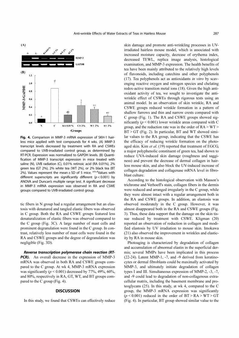
Reverse transcription polymerase chain reaction (RT-PCR). An overall decrease in the expression of MMP-3
mRNA was observed in both RA and CSWE groups com-
pared to the C group. At wk 4, MMP-3 mRNA expression
was significantly (p < 0.001) decreased by 77%, 49%, 60%,
and 80%, respectively in RA, GT, WT, and BT groups com-
pared to the C group (Fig. 4).
DISCUSSION
In this study, we found that CSWEs can effectively reduce
skin damage and promote anti-wrinkling processes in UV-
irradiated hairless mouse model, which is associated with
increased moisture capacity, decrease of erythema index,
decreased TEWL, replica image analysis, histological
examination, and MMP-3 expression. The health benefits of
tea have been mainly attributed to the relatively high levels
of flavonoids, including catechins and other polyphenols
(17). Tea polyphenols act as antioxidants in vitro by scav-
enging reactive oxygen and nitrogen species and chelating
redox-active transition metal ions (18). Given the high anti-
oxidant activity of tea, we sought to investigate the anti-
wrinkle effect of CSWEs through rigorous tests using an
animal model. In an observation of skin wrinkle, RA and
CSWE groups reduced wrinkle formation in a pattern of
shallow furrows and thin and narrow crests compared with
C group (Fig. 1). The RA and CSWE groups showed sig-
nificantly (p < 0.001) lower wrinkle areas compared with C
group, and the reduction rate was in the order of RA > WT >
BT > GT (Fig. 2). In particular, BT and WT showed simi-
lar values to the RA group, indicating that the CSWE has
the efficacy of reducing wrinkle formation on the photo-
aged skin. Kim et al. (19) reported that treatment of EGCG,
a major polyphenolic constituent of green tea, had shown to
reduce UVA-induced skin damage (roughness and saggi-
ness) and prevent the decrease of dermal collagen in hair-
less mouse skin, and also block the UV-induced increase of
collagen degradation and collagenase mRNA level in fibro-
blast culture.
According to the histological observation with Masson’s
trichrome and Verhoeff's stain, collagen fibers in the dermis
were reduced and arranged irregularly in the C group, while
they were almost intact with a regular arrangement both in
the RA and CSWE groups. In addition, an elastosis was
observed moderately in the C group. However, it was
almost disappeared both in the RA and CSWE groups (Fig.
3). Thus, these data support that the damage on the skin tis-
sue reduced by treatment with CSWE. Kligman (20)
reported an observation of reduction in collagen and modi-
fied elastosis by UV irradiation to mouse skin. Imokawa
(21) also observed the improvement in wrinkles and elastic-
ity by RA in mouse skin.
Photoaging is characterized by degradation of collagen
and accumulation of abnormal elastin in the superficial der-
mis; several MMPs have been implicated in this process
(22-24). Latent MMP-1, -7, and -9 derived from keratino-
cytes or dermal fibroblasts could be maximally activated by
MMP-3, and ultimately initiate degradation of collagen
types I and III. Simultaneous expression of MMP-2, -3, -7,
and -9 could lead to degradation of non-collagenous extra-
cellular matrix, including the basement membrane and pro-
teoglycans (25). In this study, at wk 4, compared to the C
group, the MMP-3 mRNA expression was significantly
(p < 0.001) reduced in the order of BT > RA > WT > GT
(Fig. 4). In particular, BT group showed similar value to the
Fig. 4. Comparison in MMP-3 mRNA expression of SKH-1 hair-less mice applied with test compounds for 4 wks. (A) MMP-3transcript levels decreased by treatment with RA and CSWEscompared to UVB-irradiated control group, as determined byRT-PCR. Expression was normalized to GAPDH levels. (B) Quanti-fication of MMP-3 transcript expression in mice treated withsaline (N), UVB radiation (C), 0.01% retinoic acid (RA 0.01%), 2%green tea (GT 2%), 2% white tea (WT 2%), or 2% black tea (BT2%). Values represent the mean ± SD of 5 mice. a,b,c,dValues withdifferent superscripts are significantly different (p < 0.001) byANOVA and Duncan’s multiple range test. A significant decreasein MMP-3 mRNA expression was observed in RA and CSWEgroups compared to UVB-irradiated control group.
288 K.O. Lee et al.
RA group. The RA and CSWE groups also showed remark-
ably lower MMP-2 and MMP-9 activities than that of the C
group (data not shown). Vayalil et al. (22) reported that top-
ical application of EGCG had been shown to reduce UV-
induced production of MMP-2, -3, -7, and -9, which are
known to degrade collagen and lead to photodamage.
In this study, the degree of erythema was measured to
examine the inflammatory reaction. The skin erythema indi-
ces of all the groups were significantly (p < 0.001) lower
compared to the C group during the whole period of experi-
ment. At wk 4, compared to the C group, the skin erythema
indices were significantly (p < 0.001) reduced in the order
of WT > BT > GT > RA (Table 1). Elamets et al. (26)
reported that the EGCG and ECG polyphenolic fractions
were most efficient at inhibiting erythema, whereas EGC
and EC had little effect. In an observation of H&E stain, a
moderate number of inflammatory cells including macro-
phages, neutrophils, and lymphocytes were infiltrated in the
thick dermis of the C group, whereas only a few inflamma-
tory cells were found in the dermis of RA and CSWE
groups. Dona et al. (27) and Katiyar et al. (28) reported that
the topical application of EGCG before UVB exposure
reduced the number of CD11b+ monocytes/macrophages
and neutrophils that infiltrated in the exposed skin. In an
observation of toluidine blue stain, the number and degree
of degranulation of mast cells were reduced in the RA and
CSWE groups, compared with C group. Thus, it is affirmed
that the inflammation on the skin tissue reduced by treat-
ment with CSWEs.
Human skin acts as a barrier between the internal and the
external environment, protecting the body from mechanical
damage, noxious substances, penetration by pathogens and
radiation. The skin also plays a vital role in regulating body
homeostasis by reducing TEWL to a minimum via the stra-
tum corneum (SC). The TEWL is used for assessing epider-
mal barrier function. Therefore, in the current study, TEWL
was used as one of the parameters for evaluating the anti-
aging properties. The skin moisture capacity of all the
groups was significantly (p < 0.001) higher compared to the
C group throughout the experiment period. At wk 4, com-
pared to the C group, the skin moisture capacity was signifi-
cantly (p < 0.001) increased in the order of BT > WT > RA >
GT (Table 1). At wk 4, compared to the C group, the skin
TEWLs were significantly (p < 0.001) reduced in the order
of WT > BT > GT > RA. It was found that the damage to
the skin barrier in the CSWE groups decreased, because the
skin moisture capacity was significantly higher and the
TEWL was significantly lower, compared to the C group.
Green tea polyphenols have been reported to have many
effects on cellular and molecular responses in the epider-
mis, but more important effect of this general class of ingre-
dients is on TEWL. For instance, polyphenols, such as
catechins when ingested in beverages, have been shown to
improve SC barrier function (29), although at much higher
concentrations compared with the current skin topical appli-
cation study. Puch et al. (30) demonstrated enhancement of
the skin barrier function in human volunteers after con-
sumption of green tea containing milk.
All together, the findings of this study indicated that
CSWEs derived from green, white and black teas were
effective in the wrinkle improvement by reducing dermal
extracellular matrix damage, and alleviating inflammation
and skin barrier damage in an in vivo hairless mouse photo-
aged model. These anti-wrinkle activities are likely attribut-
able to the high levels of EGCG and polyphenols in green
tea and white tea, and to theaflavins and thearubigins formed
during the oxidation of black tea. In particular, the water
extracts from white and black teas had a better effect than
green tea on the wrinkle improvement. Interestingly, despite
differences in the processing between white tea and black
tea, they did not significantly differ each other in the ability
to improve photoaged skin. In conclusion, this study sug-
gests a high possibility of the practical use of white and
black teas as anti-wrinkle agents.
REFERENCES
1. Fisher, G.J., Wang, Z.Q., Datta, S.C., Varani, J., Kang, S. and
Voorhees, J.J. (1997) Pathophysiology of premature skin
aging induced by ultraviolet light. N. Engl. J. Med., 337,
1419-1428.
2. Zhaorigetu, S., Yanaka, N., Sasaki, M., Watanabe, H. and
Kato, N. (2003) Inhibitory effects of silk protein, sericin on
UVB-induced acute damage and tumor promotion by reduc-
ing oxidative stress in the skin of hairless mouse. J. Photo-
chem. Photobiol. B, 71, 11-17.
3. Chaquour, B., Seité, S., Coutant, K., Fourtanier, A., Borel, J.P.
and Bellon, G. (1995) Chronic UVB and all trans retinoic acid
induced qualitative and quantitative changes in hairless mouse
skin. J. Photochem. Photobiol. B, 28, 125-135.
4. Honda, A., Abe, R., Makino, T., Norisugi, O., Fujita, Y.,
Watanabe, H., Nishihira, J., Iwakura, Y., Yamagishi, S., Shi-
mizu, H. and Shimizu, T. (2008) Interleukin-1beta and macro-
phage migration inhibitory factor (MIF) in dermal fibroblasts
mediate UVA-induced matrix metalloproteinase-1 expression.
J. Dermatol. Sci., 49, 63-72.
5. Varani, J., Gendimenico, G.J., Shah, B., Gibbs, D., Capetola,
R.J., Mezick. J.A. and Voorhees, J.J. (1991) A direct compari-
son of pharmacologic effects of retinoids on skin cells in vitro
and in vivo. Skin Pharmacol. Physiol., 4, 254-261.
6. Hsu, S. (2005) Green tea and the skin. J. Am. Acad. Derma-
tol., 52, 1049-1059.
7. Yen, G.C. and Chen, H.Y. (1995) Antioxidant activity of vari-
ous tea extracts in relation to their antimutagenicity. J. Agric.
Food Chem., 43, 27-32.
8. Ho, C.T., Chen, Q., Shi, H., Zhang, K.Q. and Rosen, R.T.
(1992) Antioxidative effect of polyphenol extract prepared
from various Chinese teas. Prev. Med., 21, 520-525.
9. Katiyar, S.K., Afaq, F., Perez, A. and Mukhtar, H. (2001)
Green tea polyphenol (−)-epigallocatechin-3-gallate treatment
of human skin inhibits ultraviolet radiation-induced oxidative
Anti-wrinkle Effects of Water Extracts of Teas in Hairless Mouse 289
stress. Carcinogenesis, 22, 287-294.
10. Wang, Z.Y., Agarwal, R., Bickers, D.R. and Mukhtar, H.
(1991) Protection against ultraviolet B radiation-induced pho-
tocarcinogenesis in hairless mice by green tea polyphenols.
Carcinogenesis, 12, 1527-1530.
11. Lee, J.H., Chung, J.H. and Cho, K.H. (2005) The effects of
epigallocatechin-3-gallate on extracellular matrix metabo-
lism. J. Dermatol. Sci., 40, 195-204.
12. Hsu, S., Bollag, W.B., Lewis, J., Huang, Q., Singh, B.,
Sharawy, M., Yamamoto, T. and Schuster, G. (2003) Green tea
polyphenols induce differentiation and proliferation in epider-
mal keratinocytes. J. Pharmacol. Exp. Ther., 306, 29-34.
13. Camouse, M.M., Domingo, D.S., Swain, F.R., Conrad, E.P.,
Matsui, M.S., Maes, D., Declercq, L., Cooper, K.D., Stevens,
S.R. and Baron, E.D. (2009) Topical application of green and
white tea extracts provides protection from solar-simulated
ultraviolet light in human skin. Exp. Dermatol., 18, 522-526.
14. Record, I.R. and Dreosti, I.E. (1998) Protection by black tea
and green tea against UVB and UVA + B induced skin cancer
in hairless mice. Mutat. Res. Fundam. Mol. Mech. Mutagen.,
422, 191-199.
15. Ratnasooriya, W.D. and Fernancho, T.S.P. (2009) Anti-inflam-
matory activity of Sri Lankan black tea (Camellia sinensis L.)
in rats. Pharmacogn. Res., 1, 11-20.
16. Bancirova, M. (2010) Comparison of the antioxidant capacity
and the antimicrobial activity of black and green tea. Food
Res. Int., 43, 1379-1382.
17. Shahidi, F. (2000) Antioxidants in food and food antioxi-
dants. Nahrung, 44, 158-163.
18. Frei, B. and Higdon, J.V. (2003) Antioxidant activity of tea
polyphenols in vivo: evidence from animal. J. Nutr., 133,
3275S-3284S.
19. Kim, J., Hwang, J.S., Cho, Y.K., Han, Y., Jeon, Y.J. and Yang,
K.H. (2001) Protective effects of (−)-epigallocatechin-3-gal-
late on UVA- and UVB-induced skin damage. Skin Pharma-
col. Appl. Skin Physiol., 14, 11-19.
20. Kligman, L.H. (1996) The hairless mouse model for photoa-
ging. Clin. Dermatol., 14, 183-195.
21. Imokawa, G. (2008) Recent advances in characterizing biolog-
ical mechanisms underlying UV-induced wrinkles: a pivotal
role of fibrobrast-derived elastase. Arch. Dermatol. Res., 300
Suppl 1, S7-S20.
22. Vayalil, P.K., Mittal, A., Hara, Y., Elmets, C.A. and Katiyar,
S.K. (2004) Green tea polyphenols prevent ultraviolet light-
induced oxidative damage and matrix metalloproteinases
expression in mouse skin. J. Invest. Dermatol., 122, 1480-
1487.
23. Inomata, S., Matsunaga, Y., Amano, S., Takada, K., Kobayashi,
K., Tsunenaga, M., Nishiyama, T., Kohno, Y. and Fukuda, M.
(2003) Possible involvement of gelastinases in basement
membrane damage and wrinkle formation in chronically ultra-
violet B-exposed hairless mouse. J. Invest. Dermatol., 120,
128-134.
24. Fisher, G.J., Kang, S., Varani, J., Bata-Csorgo, Z., Wan, Y.,
Datta, S. and Voorhees, J.J. (2002) Mechanisms of photoa-
ging and chronological skin aging. Arch. Dermatol., 138,
1462-1470.
25. Fisher, G.J. and Voorhees, J.J. (1998) Molecular mechanisms
of photoaging and its prevention by retinoic acid: ultraviolet
irradiation induces MAP kinase signal transduction cascades
that induce AP-1-regulated matrix metalloproteinases that
degrade human skin in vivo. J. Invest. Dermatol. Symp. Proc.,
3, 61-68.
26. Elamets, C.A., Singh, D., Tubesing, K., Matsui, M., Katiyar,
S. and Mukhtar, H. (2001) Cutaneous photoprotection from
ultraviolet injury by green tea polyphenols. J. Am. Acad. Der-
matol., 44, 425-432.
27. Donà, M., Dell’Aica, I., Calabrese, F., Benelli, R., Morini, M.,
Albini, A. and Garbisa, S. (2003) Neutrophil restraint by
green tea: inhibition of inflammation, associated angiogene-
sis, and pulmonary fibrosis. J. Immunol., 170, 4335-4341.
28. Katiyar, S.K. and Mukhtar, H. (2001) Green tea polyphenol
(−)-epigallocatechin-3-gallate treatment to mouse skin pre-
vents UVB-induced infiltration of leukocytes, depletion of
antigen-presenting cells, and oxidative stress. J. Leukocyte
Biol., 69, 719-726.
29. Heinrich, U., Neukam, K., Tronnier, H., Sies, H. and Stahl, W.
(2006) Long-term ingestion of high flavanol cocoa provides
photoprotection against UV-induced erythema and improves
skin condition in women. J. Nutr., 136, 1565-1569.
30. Puch, F., Samson-Villeger, S., Guyonnet, D., Blachon, J.L.,
Rawlings, A.V. and Lassel, T. (2008) Consumption of func-
tional fermented milk containing borage oil, green tea and
vitamin E enhances skin barrier function. Exp. Dermatol., 17,
668-674.