antibacterial activity of oleoyl-chitosan nanoparticles: a novel antibacterial dispersion system
TRANSCRIPT
Available online at www.sciencedirect.com
www.elsevier.com/locate/carbpol
Carbohydrate Polymers 74 (2008) 114–120
Antibacterial activity of oleoyl-chitosan nanoparticles:A novel antibacterial dispersion system
Ke Xing a, Xi Guang Chen a,*, Yan Yan Li a, Cheng Sheng Liu a,Chen Guang Liu a, Dong Su Cha b, Hyun Jin Park b
a College of Marine Life Science, Ocean University of China, 5# Yushan Road, Qingdao 266003, PR Chinab The Graduate School of Biotechnology, Korea University, Seoul 136-701, South Korea
Received 11 December 2007; received in revised form 22 January 2008; accepted 23 January 2008Available online 1 February 2008
Abstract
A novel chitosan antibacterial dispersion system was prepared by oleoyl-chitosan (O-chitosan) nanoparticles (OCNP), and the anti-bacterial activity against Escherichia coli and Staphylococcus aureus was investigated. Results showed that OCNP could be well distrib-uted in nutrient broth and had strong antibacterial activity. The minimum inhibitory concentrations (MICs) of all OCNP samples rangedfrom 31.25 to 125 mg/l against E. coli. For S. aureus, the MIC of all samples was 125 mg/l. OCNP of low chitosan molecular weight(MW) appeared most effective against E. coli. For S. aureus, the effect of chitosan MW on the antibacterial activity of OCNP wasnot pronounced. E. coli was most susceptible to OCNP of O-chitosan with degrees of substitution (DS) 5%, while no marked differencewas found among OCNP of O-chitosan with different DS against S. aureus. OCNP exhibited the most pronounced antibacterial activityat pH 6.0 in the experimental range. The integrity of cell membranes was destroyed when bacterial suspensions were treated with OCNP.� 2008 Elsevier Ltd. All rights reserved.
Keywords: Chitosan; Oleoyl-chitosan nanoparticles; Antibacterial dispersion system
1. Introduction
Chitosan, a-(1–4)-2-amino-2-deoxy-b-D-glucan, is anatural nontoxic biopolymer derived by partially deacety-lated of chitin, a major component of the crustaceanshells. It has attracted considerable interest due to itsunique biological activity, such as antimicrobial activity(Li et al., 2007a, 2007b; No, Park, Lee, & Meyers,2002; Park, Je, Byun, Moon, & Kim, 2004; Rabea,Badawy, Stevens, Smagghe, & Steurbaut, 2003; Yoshihikoet al., 2003), antitumor activity (Koide, 1998; Mitra,Gaur, Ghosh, & Maitra, 2001; Qin et al., 2004; Suzukiet al., 1986), and immune enhancing effect (Jeon &Kim, 2001). The antibacterial activity of chitosan is influ-enced by a number of factors that included the species of
0144-8617/$ - see front matter � 2008 Elsevier Ltd. All rights reserved.
doi:10.1016/j.carbpol.2008.01.024
* Corresponding author. Tel./fax: +86 0532 82032586.E-mail address: [email protected] (X.G. Chen).
bacteria (No et al., 2002), concentration (Liu et al., 2006;Zheng & Zhu, 2003), pH (Liu, Guan, Yang, Li, & Yao,2001), the solvent, molecular weight (MW) (Liu et al.,2006) and so on. However, chitosan is only soluble inacidic media like acetic acid, which also has the antibac-terial activity on bacteria. Besides this, the precipitationoccurs upon addition of chitosan solution to the culturemedium, which makes it difficult to investigate the anti-bacterial activity and antibacterial mechanism of chitosancorrectly. Therefore, a novel dispersion system of chitosanis necessary to be established and be used to evaluate theantibacterial action of chitosan.
Chitosan-based nanoparticles can be easily formedthrough self-aggregation. There have been many reportsof hydrophobic modifications of chitosan and nanoparticleformation by self-aggregation in aqueous solution (Chen,Lee, & Park, 2003; Kim et al., 2001; Liu, Desai, Chen, &Park, 2005). These modifications can introduce hydropho-
K. Xing et al. / Carbohydrate Polymers 74 (2008) 114–120 115
bic groups into chitosan and produced chitosan amphi-philic polymers. Some of these chitosan amphiphilic deriv-atives can form nanosized self-aggregation in aqueousmedia (Janes, Fresneau, Marazuela, Fabra, & Alonso,2001). In our previous study, oleoyl-chitosan (O-chitosan)nanoparticles (OCNP) were prepared using an O/W emul-sification method based on O-chitosan, which were synthe-sized by grafting oleoyl onto the –NH2 at C-2 in thechitosan molecule. Different from chitosan, nanoparticlessystems of chitosan could be well distributed in aqueoussolution and less affected by pH of the solution. Thesecharacteristics could make it a novel potential antibacterialdispersion system to study the antibacterial mode. How-ever, few investigations have been focused on the antibac-terial dispersion system of self-assembled chitosannanoparticles.
In this paper, OCNP as a novel antibacterial dispersionsystem were prepared. Escherichia coli (Gram-negative)and Staphylococcus aureus (Gram-positive) were chosento be models for the antibacterial assay of OCNP. Theinfluences of chitosan MW, degrees of substitution (DS)of O-chitosan, concentration of the nanoparticles and pHof the solution on the antibacterial activity of OCNP wereexamined. Additionally, the integrity of the cell membranesof E. coli and S. aureus influenced by OCNP wasinvestigated.
2. Materials and methods
2.1. Materials
Chitosan (MW = 1100 kDa), degree of deacetylation82%, was made from crab shell and obtained from BiotechCo. Oleoyl chloride, pyridine, chloroform, methylene chlo-ride, acetic acid, and sodium tripolyphosphate (STPP) werepurchased from Sigma Chemicals and used without furtherpurification.
2.2. Preparation of OCNP
Chitosan was degraded by the method of acetic acidhydrolysis, and OCNP with different chitosan MW andDS of O-chitosan were prepared in our pervious study(Li et al., 2006, 2007a, 2007b). Different OCNP samplesused in this paper are shown in Table 1.
Table 1OCNP with Different MW of chitosan and DS of O-chitosan
series OCNP samples MW of chitosan (kDa) DS of O-chitosan (%)
I A 38 2.5B 38 5C 38 11
II B 38 5D 300 5E 1100 5
2.3. Dispersion of OCNP in the culture medium
In order to explain the possible interaction betweenOCNP and the culture medium when mixed together, sam-ple B was selected to test its dispersion in nutrient broth atdifferent concentrations. The solution of chitosan (chitosanMW = 38 kDa), O-chitosan (chitosan MW = 38 kDa,O-chitosan with DS 5%) and sample B were first preparedwith 0.1 M acetic acid. Different concentrations of chito-san, O-chitosan and sample B were prepared with two-foldserial broth dilution (Qi, Xu, Jiang, Hu, & Zou, 2004). Anumber of test bottles each containing 10 ml of sterilenutrient broth were prepared. To the first bottle, 10 ml ofsamples (1000 mg/l) was added. After mixing, 10 ml ofthe mixture was transferred to the second bottle, and sim-ilar transformations were repeated. Hence, each bottle con-tained a test sample solution with half of the concentrationof the previous one. The final pH of solutions was adjustedto 5.0 or 6.0 with 10% NaOH solution. The controls were0.1 M acetic acid solution of chitosan, O-chitosan and sam-ple B (1000 mg/l), respectively. The transmittance of thesolution was recorded on a 1601 UV–vis spectrophotome-ter (Shimadzu, Tokyo, Japan), using a quartz cell with anoptical path length of 1 cm at 610 nm.
2.4. Cultivation of microorganisms
Escherichia coli (ATCC 25992) and S. aureus (ATCC25923) were used as the test organisms. A representativebacteria colony was picked off with a wire loop, placed innutrient broth, and then incubated at 37 �C for 12 h. Byappropriately diluting with sterile distilled water, the cul-tures of E. coli and S. aureus containing �107 CFU/mlwere prepared and used for the antibacterial test.
2.5. Evaluation of the antibacterial activity in vitro
The solution of OCNP (1000 mg/l) was first preparedwith 0.1 M acetic acid then adjusted to pH 6.0 with 10%NaOH solution. The MIC of OCNP was determined bytwo-fold serial broth dilution (Qi et al., 2004) describedabove and optical density method. The control only con-tained nutrient broth and 0.1 M acetic acid without OCNP.After adjusting to pH 6.0 with 10% NaOH solution, all ofthe samples were inoculated under aseptic conditions with50 ll of the inoculums of bacteria and incubated at 37 �Cfor 24 h, then mensurated the turbidity of the culturedmedium at 610 nm. The lowest concentration of OCNPthat inhibited the growth of bacteria was considered asthe MIC.
To determine the effect of pH, the solution of OCNP(1000 mg/l) was adjusted to pH 4.0, 4.5, 5.0, 5.5 and 6.0with 10% NaOH solution. In this experiment, the concen-trations of E. coli and S. aureus were adjusted to�104 CFU/ml with sterile distilled water, respectively.4 ml OCNP solution was added to 1 ml of the cell suspen-sions. The same volume of sterile distilled water was added
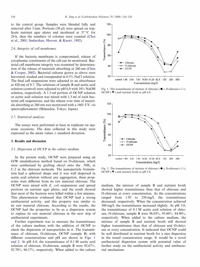
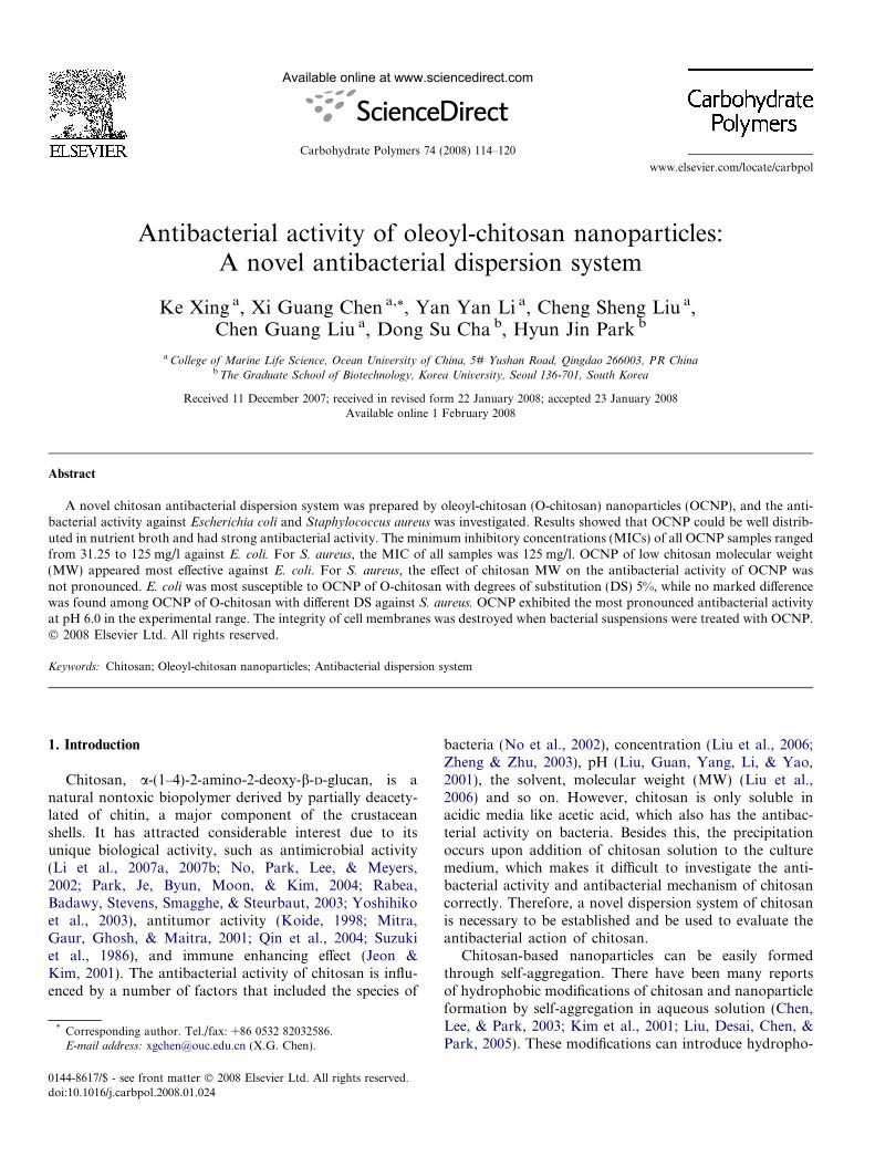
Fig. 1. The transmittance of mixture of chitosan (-d-), O-chitosan (-s-),OCNP (-.-) and nutrient broth at pH 6.0.
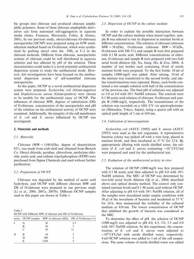
Fig. 2. The transmittance of mixture of chitosan (-d-), O-chitosan (-s-),OCNP (-.-) and nutrient broth at pH 5.0.
116 K. Xing et al. / Carbohydrate Polymers 74 (2008) 114–120
to the control group. Samples were blended fully andremoved after 5 min. Portions (50 ll) were spread on trip-licate nutrient agar plates and incubated at 37 �C for24 h, then the numbers of colonies were counted (Choiet al., 2001; Sudarshan, Hoover, & Knorr, 1992).
2.6. Integrity of cell membranes
If the bacteria membrane is compromised, release ofcytoplasmic constituents of the cell can be monitored. Bac-terial cell membrane integrity was examined by determina-tion of the release of materials absorbing at 260 nm (Chen& Cooper, 2002). Bacterial cultures grown as above wereharvested, washed and resuspended in 0.5% NaCl solution.The final cell suspensions were adjusted to an absorbanceat 420 nm of 0.7. The solutions of sample B and acetic acidsolution (control) were adjusted to pH 6.0 with 10% NaOHsolution, respectively. A 1.5-ml portion of OCNP solutionor acetic acid solution was mixed with 1.5 ml of each bac-terial cell suspensions, and the release over time of materi-als absorbing at 260 nm was monitored with a 1601 UV–visspectrophotometer (Shimadzu, Tokyo, Japan).
2.7. Statistical analyses
The assays were performed at least in triplicate on sep-arate occasions. The data collected in this study wereexpressed as the mean values ± standard deviation.
3. Results and discussion
3.1. Dispersion of OCNP in the culture medium
In the present study, OCNP were prepared using anO/W emulsification method based on O-chitosan, whichwere synthesized by grafting oleoyl onto the –NH2 atC-2 in the chitosan molecule. The nanoparticle formula-tion had a spherical shape and it was well dispersed inacetic acid solution without any aggregation, these prop-erties were different from its raw material chitosan. TheOCNP were mixed with E. coli suspensions and spreadportions on nutrient agar plates, and the result showedthat most of the bacteria were killed within a few minutes.This phenomenon evidenced that OCNP had a strongantibacterial activity, and this property was similar toits raw material chitosan. According to the results, theOCNP had the properties to be as a dispersion systemto replace its raw material chitosan in the next step ofantibacterial experiments.
Further experiment was to measure the transmittanceof the culture medium with the addition of OCNP tocheck the dispersion of nanoparticles in it. The transmit-tance of chitosan, O-chitosan, OCNP (sample B) withdifferent concentrations and pH are shown in Figs. 1and 2. At pH 6.0, the transmittance of 0.1 M acetic acidsolution of chitosan, O-chitosan, sample B were 92.67%,92.70%, 66.17%, respectively. When added to the culture
medium, the mixture of sample B and nutrient brothshowed higher transmittance than that of chitosan andO-chitosan at every concentration. As the concentrationsranged from 1.95 to 250 mg/l, the transmittancedecreased, respectively. When the concentration achieved500 mg/l, the transmittance increased slightly. At pH 5.0,the transmittance of 0.1 M acetic acid solution of chito-san, O-chitosan, sample B were 96.03%, 93.60%, 84.90%,respectively. When added to the culture medium, themixture of sample B and nutrient broth still showedhigher transmittance than that of chitosan and O-chito-san at every concentration. It indicated that OCNP couldbe well distributed in nutrient broth for a nice dispersionin the tested concentration range. It would be a novelantibacterial dispersion system with potential value offurther study on the antibacterial activity and antibacte-rial mechanism.
K. Xing et al. / Carbohydrate Polymers 74 (2008) 114–120 117
3.2. Effect of concentration on the antibacterial activity of
OCNP
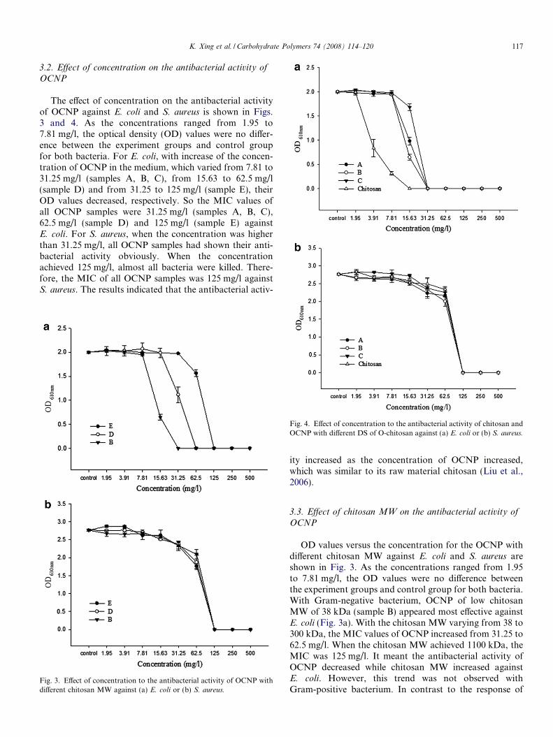
The effect of concentration on the antibacterial activityof OCNP against E. coli and S. aureus is shown in Figs.3 and 4. As the concentrations ranged from 1.95 to7.81 mg/l, the optical density (OD) values were no differ-ence between the experiment groups and control groupfor both bacteria. For E. coli, with increase of the concen-tration of OCNP in the medium, which varied from 7.81 to31.25 mg/l (samples A, B, C), from 15.63 to 62.5 mg/l(sample D) and from 31.25 to 125 mg/l (sample E), theirOD values decreased, respectively. So the MIC values ofall OCNP samples were 31.25 mg/l (samples A, B, C),62.5 mg/l (sample D) and 125 mg/l (sample E) againstE. coli. For S. aureus, when the concentration was higherthan 31.25 mg/l, all OCNP samples had shown their anti-bacterial activity obviously. When the concentrationachieved 125 mg/l, almost all bacteria were killed. There-fore, the MIC of all OCNP samples was 125 mg/l againstS. aureus. The results indicated that the antibacterial activ-
Fig. 3. Effect of concentration to the antibacterial activity of OCNP withdifferent chitosan MW against (a) E. coli or (b) S. aureus.
Fig. 4. Effect of concentration to the antibacterial activity of chitosan andOCNP with different DS of O-chitosan against (a) E. coli or (b) S. aureus.
ity increased as the concentration of OCNP increased,which was similar to its raw material chitosan (Liu et al.,2006).
3.3. Effect of chitosan MW on the antibacterial activity of
OCNP
OD values versus the concentration for the OCNP withdifferent chitosan MW against E. coli and S. aureus areshown in Fig. 3. As the concentrations ranged from 1.95to 7.81 mg/l, the OD values were no difference betweenthe experiment groups and control group for both bacteria.With Gram-negative bacterium, OCNP of low chitosanMW of 38 kDa (sample B) appeared most effective againstE. coli (Fig. 3a). With the chitosan MW varying from 38 to300 kDa, the MIC values of OCNP increased from 31.25 to62.5 mg/l. When the chitosan MW achieved 1100 kDa, theMIC was 125 mg/l. It meant the antibacterial activity ofOCNP decreased while chitosan MW increased againstE. coli. However, this trend was not observed withGram-positive bacterium. In contrast to the response of

Fig. 5. Effect of pH on the antibacterial activity of OCNP against (a)E. coli or (b) S. aureus.
118 K. Xing et al. / Carbohydrate Polymers 74 (2008) 114–120
E. coli, growth of S. aureus was almost suppressed byOCNP with different chitosan MW at 125 mg/l (Fig. 3b).Therefore, the effect of chitosan MW on the antibacterialactivity of OCNP against S. aureus was not as pronouncedas that observed with E. coli.
Until now, there have been many investigations of therelationship between MW and the antibacterial activity ofchitosan. However, little information about relationshipsof chitosan MW and the antibacterial activity by chitosannanoparticles has been reported. Liu et al. (2006) reportedthat the antibacterial activity of low MW chitosan is stron-ger than that of the high MW chitosan against E. coli.Effect of chitosan MW on the antibacterial activity ofOCNP was similar to that of chitosan. No et al. (2002)reported that among one series of chitosan with MW rang-ing from 28 to 1671 kDa, chitosan of 470 kDa appearedmost effective against S. aureus. It was found that OCNPwith high chitosan MW and low chitosan MW showedan equal antibacterial activity for S. aureus. It meant thatthe effect of chitosan MW on the antibacterial activity ofOCNP was not as pronounced as chitosan against S.
aureus.
3.4. Effect of DS of O-chitosan on the antibacterial activity
of OCNP
The effect of DS of O-chitosan on the antibacterial activ-ity of OCNP against E. coli and S. aureus is shown inFig. 4. No marked difference was found among threeOCNP samples tested in relation to S. aureus, while sampleB formed by O-chitosan with DS 5% exhibited the mostpronounced antibacterial activity against E. coli. It wasfound that OCNP and chitosan (chitosan MW = 38 kDa)exhibited an equal antibacterial activity against S. aureus,whereas OCNP presented inferior inhibitory activity thanchitosan (chitosan MW = 38 kDa) against E. coli. It wasinteresting to find that the antibacterial activity of OCNPincreased upon increasing the DS of O-chitosan from2.5% to 5% and decreased with further increase to 11%.In our previous study, Li et al. (2007a, 2007b) reported thatthe increase of DS might facilitate OCNP self-aggregation,but in the high DS sample with high viscosity and low sol-ubility nanoparticles were not readily formed. Solubility ofO-chitosan was decreased as the DS increased from 5% to11%, therefore, it could be suggested that the effective con-centration of OCNP (sample C) with DS 11% might be alittle lower than the concentration set in this paper. Thedecreased antibacterial activity observed with the OCNP(sample C) may be attributed to the decrease in solubilityof O-chitosan with DS 11%.
3.5. Effect of pH on the antibacterial activity of OCNP
The effect of pH on the antibacterial activity of OCNPagainst E. coli and S. aureus is shown in Fig. 5. The anti-bacterial activity increased as the pH increased from 4.0to 6.0 and reached a maximum at pH 6.0 for both E. coli
and S. aureus. OCNP could be well distributed in aceticacid solution as the pH varied from 4.0 to 6.0. Weattempted to test the antibacterial activity of OCNP in neu-tral and alkaline pH but were frustrated in the poor stabil-ity under such conditions (data not shown). Therefore, theupper pH value studied was limited to 6.0 to make surethat OCNP could keep their nice dispersivity. There aremany studies regarding the effect of pH on the antibacterialactivity of chitosan. Several workers (Fujimoto, Tsuchiya,Terao, Nakamura, & Yamamoto, 2006; No et al., 2002)reported that acidic pH increased the antibacterial effectof chitosan. While Liu et al. (2001) evidenced that the anti-bacterial activity of chitosan decreased gradually with pHvarying from 6.3 to 4.0. Effect of pH on the antibacterialactivity of OCNP was similar to that of chitosan reportedby Liu et al. (2001). Chitosan cannot dissolve in water butin acetic acid solution that also had the antibacterial activ-ity (Liu et al. 2006). This property cannot be ignored.OCNP with strong antibacterial activity at pH 6.0 couldreduce the effect of acetic acid on the bacterial growthgreatly.

Fig. 7. Release of 260 nm absorbing material from E. coli suspensionstreated with 300 mg/l (-d-), 150 mg/l (-s-) of OCNP (sample B) and pH6.0 acetic acid solution (-.-).
K. Xing et al. / Carbohydrate Polymers 74 (2008) 114–120 119
3.6. Integrity of bacterial cell membranes
The cytoplasmic cell membrane is a structural compo-nent, which may become damaged and functionally invalidwhen bacterial suspensions are exposed to antimicrobialagents. If bacterial membrane became compromised, smallions such as K+ and PO4
3� tend to leach out first, and fol-lowed by large molecules such as DNA, RNA and othermaterials. The release of these intracellular componentswith strong UV absorption at 260 nm is an indication ofmembrane damage (Chen & Cooper, 2002).
According to the results above, OCNP could exhibitpronounced antibacterial activity when the concentrationswere higher than the MIC values at pH 6.0. Sample Bwas selected for the assay of cell membranes integritybecause of its stronger antibacterial activity. The releaseof intracellular components upon addition of OCNP toS. aureus suspensions is shown in Fig. 6. When S. aureus
suspensions were treated with OCNP, the absorbance ofthe suspensions at 260 nm dramatically increased up to60 min then at a decreasing rate up to 120 min. Since S.
aureus did not have the outer membrane (OM) to preventthe influx of foreign molecules, it was not surprising tosee A260 increased as soon as OCNP mixed with bacterialcell suspensions. In the case of E. coli (Fig. 7), there wasa lag of about 5 min before intracellular components weredetected at 260 nm because E. coli had the OM to preventthe influx of foreign molecules. Then the A260 increasedrapidly up to 80 min. Thereafter the absorbance wasalmost unchanged in suspensions treated with 300 mg/lOCNP, while the absorbance increased at a decreasing rateup to 120 min in suspensions treated with 150 mg/l OCNP.OCNP induced a significant amount of release of 260 nmabsorbing material from these Gram-negative bacteria.Furthermore, A260 values were greater in suspensions trea-ted with 300 mg/l than with 150 mg/l OCNP. Therefore,the release rate of intracellular components caused by
Fig. 6. Release of 260 nm absorbing material from S. aureus suspensionstreated with 300 mg/l (-d-), 150 mg/l (-s-) of OCNP (sample B) and pH6.0 acetic acid solution(-.-).
OCNP was concentration-dependent, which was alsoagreeable with the previous findings for antibacterial activ-ity. In control suspensions, a lag of about 20 min was fol-lowed by a relatively slowly release of intracellularcomponents up to 120 min.
Antibacterial activity of chitosan as related to mem-branes permeability has been evaluated in the literature(Liu, Du, Wang, & Sun, 2004). They studied on the integ-rity of cell membranes using chitosan against E. coli and S.
aureus. Their results showed that release of 260 nm absorb-ing materials quickly increased, and the damage of cellmembranes was concentration-dependent. Our results evi-denced that OCNP could induce the release of intracellularcomponent via destroying the integrity of bacterial cellmembranes, which was similar to its raw material chitosan.
4. Conclusions
In this study, OCNP as a novel antibacterial dispersionsystem were prepared, which could be well distributed innutrient broth for a nice dispersion. OCNP showed strongantibacterial activity against E. coli and S. aureus. The con-centration of the nanoparticles, MW of chitosan, DS of O-chitosan and pH of the solution affected the antibacterialactivity of OCNP. For E. coli, the MIC values of OCNPranged from 31.25 to 125 mg/l as chitosan MW increasedfrom 38 to 1100 kDa. In case of S. aureus, the MIC of allOCNP samples was 125 mg/l. OCNP of low chitosanMW of 38 kDa appeared most effective against E. coli. Itwas also found that OCNP of O-chitosan with DS 5%exhibited the most pronounced antibacterial activityagainst E. coli. While the effect of chitosan MW and DSof O-chitosan on the antibacterial activity of OCNPagainst S. aureus was not so pronounced. Furthermore,OCNP showed increased antibacterial activity when pHincreased from 4.0 to 6.0 and reached a maximum at pH6.0. When E. coli and S. aureus suspensions treated with
120 K. Xing et al. / Carbohydrate Polymers 74 (2008) 114–120
sample B, the release of intracellular component increasedvia destroying the integrity of bacterial cell membranes.OCNP, as a novel antibacterial dispersion system, still keepthe original antibacterial activity of chitosan and have thepotential value in the determination of the exact antibacte-rial mechanism of chitosan.
Acknowledgements
This work was supported by grants from NSFC(30770582), ISTCP (2006DFA33150) and the ShandongScience Foundation (Y2006C110).
References
Chen, C. Z., & Cooper, S. L. (2002). Interactions between dendrimer
biocides and bacterial membranes. Biomaterials, 23, 3359–3368.Chen, X. G., Lee, C. M., & Park, H. J. (2003). O/W emulsification for the
self-aggregation and nanoparticle formation of linoleic acid-modified
chitosan in the aqueous system. Journal of Agricultural and Food
Chemistry, 51, 3135–3139.Choi, B. K., Kim, K. Y., Yoo, Y. J., Oh, S. J., Choi, J. H., & Kim, C. Y.
(2001). In vitro antimicrobial activity of a chitooligosaccharide
mixture against Actinobacillus actinomycetimcomitans and Streptococ-
cus mutans. International Journal of Antimicrobial Agents, 18, 553–557.Fujimoto, T., Tsuchiya, Y., Terao, M., Nakamura, N., & Yamamoto, M.
(2006). Antibacterial effects of chitosan solution� against Legionella
pneumophila, Escherichia coli, and Staphylococcus aureus. International
Journal of Food Microbiology, 112, 96–101.Janes, K. A., Fresneau, M. P., Marazuela, A., Fabra, A., & Alonso, M. J.
(2001). Chitosan nanoparticles as delivery systems for doxorubicin.Journal of Controlled Release, 73, 255–267.
Jeon, Y. J., & Kim, S. K. (2001). Potential immuno-stimulating effect of
antitumoral fraction of chitosan oligosaccharides. Journal of Chitin
and Chitosan, 6, 163–167.Kim, Y. H., Gihm, S. H., Park, C. R., Lee, K. Y., Kim, T. W., Kwon, I.
C., et al. (2001). Structural characteristics of size-controlled self-
aggregates of deoxycholic acid-modified chitosan and their application
as a DNA delivery carrier. Bioconjugate Chemistry, 12, 932–938.Koide, S. S. (1998). Chitin–chitosan: Properties, benefits and risks.
Nutrition Research, 18, 1091–1101.Li, Y. Y., Chen, X. G., Liu, C. S., Cha, D. S., Park, H. J., & Lee, C. M.
(2007a). Effect of the molecular mass and degree of substitution of
oleoylchitosan on the structure, rheological properties, and formation
of nanoparticles. Journal of Agricultural and Food Chemistry, 55,4842–4847.
Li, Y. Y., Chen, X. G., Yu, L. M., Wang, S. X., Sun, G. Z., & Zhou, H. Y.(2006). Aggregation of hydrophobically modified chitosan in solution
and at the air–water interface. Journal of Applied Polymer Science, 102,1968–1973.
Li, Y., Chen, X. G., Liu, N., Liu, C. S., Liu, C. G., Meng, X. H., et al.(2007b). Physicochemical characterization and antibacterial property
of chitosan acetates. Carbohydrate Polymers, 67, 227–232.Liu, C. G., Desai, K. G., Chen, X. G., & Park, H. J. (2005). Linolenic
acid-modified chitosan for formation of self-assembled nanoparticles.Journal of Agricultural and Food Chemistry, 53, 437–441.
Liu, H., Du, Y. M., Wang, X. H., & Sun, L. P. (2004). Chitosan kills
bacteria through cell membrane damage. International Journal of Food
Microbiology, 95, 147–155.Liu, N., Chen, X. G., Park, H. J., Liu, C. G., Liu, C. S., Meng, X. H.,
et al. (2006). Effect of MW and concentration of chitosan on
antibacterial activity of Escherichia coli. Carbohydrate Polymers, 64,60–65.
Liu, X. F., Guan, Y. L., Yang, D. Z., Li, Z., & Yao, K. D. (2001).Antibacterial action of chitosan and carboxymethylated chitosan.Journal of Applied Polymer Science, 79, 1324–1335.
Mitra, S., Gaur, U., Ghosh, P. C., & Maitra, A. N. (2001). Tumour
targeted delivery of encapsulated dextran-doxorubicin conjugate using
chitosan nanoparticles as carrier. Journal of Controlled Release, 74,317–323.
No, H. K., Park, N. Y., Lee, S. H., & Meyers, S. P. (2002).Antibacterial activity of chitosan and chitosan oligomers with
different molecular weights. International Journal of Food Microbi-
ology, 74, 65–72.Park, P. J., Je, J. Y., Byun, H. G., Moon, S. H., & Kim, S. K. (2004).
Antimicrobial activity of hetero-chitosan and their oligosaccharides
with different molecular weights. Journal of Microbiology and Biotech-
nology, 14, 317–323.Qi, L. F., Xu, Z. R., Jiang, X., Hu, C. H., & Zou, X. F. (2004).
Preparation and antibacterial activity of chitosan nanoparticles.Carbohydrate Research, 339, 2693–2700.
Qin, C. Q., Zhou, B., Zeng, L. T., Zhang, Z. H., Liu, Y., & Du, Y. M.(2004). The physicochemical properties and antitumor activity of
cellulase-treated chitosan. Food Chemistry, 84, 107–115.Rabea, E. I., Badawy, M. E. T., Stevens, C. V., Smagghe, G., & Steurbaut,
W. (2003). Chitosan as antimicrobial agent: Applications and mode of
action. Biomacromolecules, 4, 1457–1465.Sudarshan, N. R., Hoover, D. G., & Knorr, D. (1992). Antibacterial
action of chitosan. Food Biotechnology, 6, 257–272.Suzuki, K., Mikami, T., Okawa, Y., Tokoro, A., Suzuki, S., & Suzuki, M.
(1986). Antitumor effect of hexa-N-acetychtohexaose and chitohexa-ose. Carbohydrate Research, 151, 403–408.
Yoshihiko, O., Mayumi, S., Takahiro, A., Hiroyuki, S., Yoshihiro, S.,Ichiro, N., et al. (2003). Antimicrobial activity of chitosan with
different degrees of acetylation and molecular weights. Biocontrol
Science, 8, 25–30.Zheng, L. Y., & Zhu, J. F. (2003). Study on antimicrobial activity of
chitosan with different molecular weights. Carbohydrate Polymers, 54,527–530.