antigen antibody reactions or - judoctors –antibody reactions or ... –far ag excess (no ppt....
TRANSCRIPT

Antigen – Antibody Reactions
or Serological Reactions

Antigen-Antibody interactions
Characterized as:
Non-covalent interaction (similar to “lock and
key” fit of enzyme-substrate)
Do not lead to irreversible alteration of Ag or Ab
This exact and specific interaction has led to
many immunological assays that are used to:
detect Ag or Ab
diagnose disease
measure magnitude of humoral IR
identify molecules of biological and
medical interest

IntroductionAg – Ab reactions are one of the most specific noncovalent biochemical reactions known
The forces that hold the reactants together are:
- van der Waals forces
- Electrostatic forces
- Hydrophobic forces
They can be represented by the simple formula:
Ag + Ab ↔ AgAb
The reaction is driven to the right but it is reversible


Strength of Reaction
The strength of the reaction (how far it is driven
to the right) is referred to as affinity
Antibody affinity
- A quantitative measure of binding strength
- Combined strength of the noncovalent
interactions between a binding site on an Ab &
monovalent Ag
- Affinity varies broadly among immunoglobulins

Strength of ReactionAntibody avidity
- Avidity is often used to describe the collective affinity of multiple binding sites on an antibody molecule
- True strength of the Ab -Ag interaction within
biological systems
- The interaction at one site will increase the possibility
of reaction at a second site
- High avidity can compensate for low affinity (secreted
pentameric IgM has a higher avidity than IgG )

CROSS REACTIVITYAntibody elicited by one Ag can cross-react with a related Ag.
Occurs if two different Ags share identical or very similar epitope
1- Vaccinia virus and smallpox virus
2- Rabies & JE vaccine
3- Streptococcus pyogenes infection: heart &
Kidney damage following infection
4- Original antigenic sin.
5- Bacterial Ag and sugars on RBC

STAGES OF Ag - Ab REACTIONS
Primary reactions Vs secondary reactions: Small Ag -Ab complexes Vs large complexes (The Latticehypothesis)
Development of macroscopic manifestations reactions (e.g. immunoprecipitation)
Ag – Ab reactions involving IgM are confined to the blood stream, while those of lower molecular weight (IgG and IgE) can leave the vasculature and enter tissues
Time required is hours to days for precipitin formation leading to irreversible immunoprecipitates

LATTICE THEORY
Lattice formation (visible Ag - Ab aggregates)
occurs when:
– Ag is multivalent (contains more than 2
identical epitopes)
– Cross-linking of Ags by specific Abs (2 or
more antigen-binding sites)
– Molar ratios of epitopes and antigen-binding
sites are optimal (zone of equivalence)

Zone of
equivalence

LATTICE THEORYZones of lattice formation
– Far Ag excess (no ppt. formed; free Ag in
supernatant) -- “postzone”
– Ag excess (sub-optimal ppt.; free Ag in spnt.)
– Zone of equivalence (maximum ppt.; no Ag or Ab in
spnt.)
– Ab excess (sub-optimal ppt; Ab in spnt.)
– Far Ab excess (no ppt; Ab in spnt.) -- “prozone”

ZONES OF PRECIPITIN FORMATION
Precipitin Curve

METHODS THAT DETECT Ag- Ab REACTIONS
Primary Reactions:
- Immunofluorescence (IF)
- Radioimmunoassay (RIA)
- Enzyme immunoassay (EIA)
- Immunonephelometry (measures picogram to
nanogram quantities of analyte)
Secondary Reactions
- Agglutination Techniques
- Precipitation Techniques ± Electrophoresis


PrecipitationPrecipitation can take place in capillary tubes, test tubes, and in gel

Precipitation in gel
- Double diffusion
- Single (radial) diffusion
- Combination of diffusion in gel and
electrophoresis
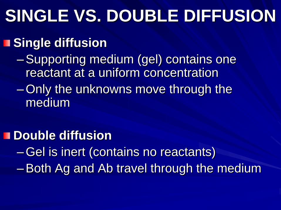
SINGLE VS. DOUBLE DIFFUSION
Single diffusion
– Supporting medium (gel) contains one reactant at a uniform concentration
– Only the unknowns move through the medium
Double diffusion
– Gel is inert (contains no reactants)
– Both Ag and Ab travel through the medium

The region of
equivalence

RADIAL IMMUNODIFFUSION
Ab uniformly distributed in gel; Ag diffuses outward
from a well (single diffusion)
Ag- Ab complexes form as concentric rings around the
well at zone of equivalence
At a set time, ring diameters are measured
[Ag] is directly proportional to the ring d2
Unknown value is determined by comparing to a 3-
standard curve

RADIAL IMMUNODIFFUSION
Standard Curve
Precipitin RingsA B C a b c
Standards Samples

RADIAL IMMUNODIFFUSIONFahey method (kinetic)– Read at 18 hours
– Plot [std] vs. ring diameter on semi-log paper
Mancini method (endpoint)– Read at 48 or 72 hours
– Plot [std] vs. ring diameter squared on graph paper
Results reliable only if the ring size is within the range of the standards; if greater than highest std, dilute and repeat test
Used to measure IgM, IgG, C4,C3,transferrin, CRP, others


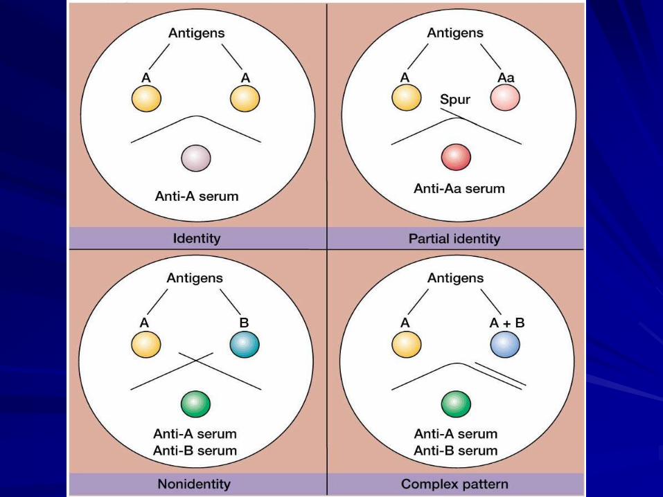
OUCHTERLONY DOUBLE DIFFUSION
Ag & Ab placed in wells cut into an agarose gel (both reactants diffuse)
Precipitin line (or arc) indicates Ab has specificity for Ag
Position of precipitin between wells depends on MW and concentration of reactants
3 possible patterns of reaction: identity, non-identity, partial identity


OUCHTERLONY DOUBLE DIFFUSION
Ouchterlony Plates Precipitin Patterns


ELECTROIMMUNOASSAY (ROCKET)
Electrophoresis hastens movement of Ag (placed in wells) through Ab -imbedded gel (single diffusion)
Selected pH (8.6) keeps Abs at their isoelectric point; they will not move
Rocket-shaped precipitin bands will form at zone of equivalence (changes as reactants move)
[Ag] proportional to length of rocket
Unknowns compared to standards

ELECTROIMMUNOASSAY (ROCKET)

ELECTROIMMUNOASSAY (ROCKET)
May be used to quantitate plasma proteins such
as coagulation factors, alpha-fetoprotein, C3,
C4, CRP, haptoglobin
Compared with RID:
– faster
– similar sensitivity
– requires electrophoretic equipment and more
technological finesse
Largely replaced by immunonephelometry

IMMUNONEPHELOMETRY
Ag + Ab AgAb microscopic Ag - Ab complexes
Microcomplexes cause light moving through the suspending solution to scatter
Nephelometer detects light scattered at a 90o angle
Amount of light scattered at 90o is proportional to Ag -Ab complexes formed
Sensitive and quantitative technique used for measurement of many serum proteins

IMMUNOELECTROPHORESIS (IEP)
Electrophoresis and double diffusion
2 stages
– Proteins separated by electrophoresis
– Antiserum placed in trough parallel to separated
proteins; all reactants diffuse in all directions
– Precipitin forms at zones of equivalence
Trough may be filled with simple or complex antisera
yielding simple to complex patterns
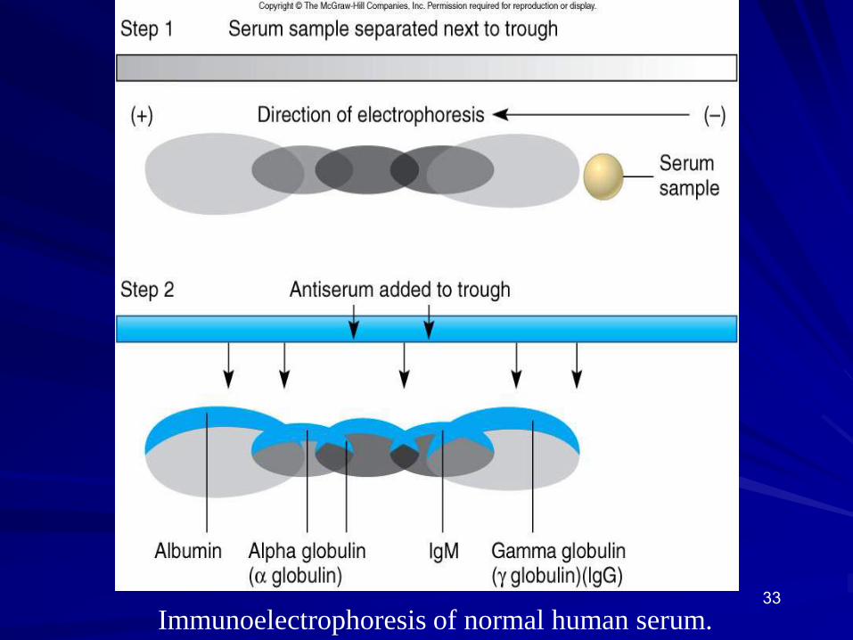
33
Immunoelectrophoresis of normal human serum.


IMMUNOELECTROPHORESIS (IEP)
Qualitative to semi-quantitative
Serum, urine, or CSF may be analyzed
Complex patterns may be difficult to interpret
Useful to detect:– missing proteins
– abnormal proteins
– normal proteins in abnormal concentrations
Used to evaluate conditions such as multiple myeloma
Largely replaced by immunofixation

IMMUNOFIXATION
ELECTROPHORESIS (IFE)
Proteins that were separated by electrophoresis are exposed to Ab directly, instead of through diffusion
Steps:
– Electrophoresis of protein mixture in gel (use serum or urine samples)
– Paper strips imbedded with specific Ab are “blotted” onto gel; Ags transfer to paper and bind to Abs
– Strips washed (unbound material washes away)
– Strips stained to reveal precipitin bands

IFE

IMMUNOFIXATION
ELECTROPHORESIS (IFE)
Used to detect the presence of Igs in conditions like multiple myeloma
Fairly sensitive - Ab is highly specific, electrophoresis leaves Ag isolated and accessible
Faster and easier to interpret than IEP
Only 1 Ab may be used per strip

WESTERN BLOTTING
Similar to IFE but the unknown is Ab rather than Ag
Steps:
– Separation of complex antigenic material (eg., viral
proteins) by electrophoresis
– Separated components transferred from gel to
nitrocellulose paper by “blotting”
– Unknown (or control) sera (which may have Abs)
incubated with paper strips; Ag - Ab complexes ppt.
at site of transfer
– Strips washed; staining reveals complexes

WESTERN BLOTTING

42The Western blot procedure.

FLOCCULATION
Immunoprecipitation (or agglutination) of insoluble particles
Characterized by very sharp pro- and postzones
No precipitin formed in zones of Ab or Ag excess, only in zone of equivalence
Clinically important examples, VDRL and RPR tests (screening tests for syphilis)

FLOCCULATION VS. IMMUNOPRECIPITATION

Flocculation Tests
- VDRL (Venereal Disease Research Lab.) test
- RPR (Rapid Plasma Reagin) test

AgglutinationTiter
Zeta potential
Types of Agglutination
- Direct agglutination or hemagglutination
- Indirect (passive) agglutination or hemagglutination
- Agglutination or hemagglutination inhibition
The Coombs test
- Direct
- Indirect

Agglutination Reactions

Agglutination
• Qualitative slide agglutination
- identification of bacteria with antisera directed against
O, H, K antigens

Agglutination
• Latex agglutination
• Coagglutination

Agglutination
• Tube agglutination tests:
- Gruber-Widal: typhoid fever (S. typhi)
- Weil-Felix: typhus (Rickettsia)
- Wright: brucellosis
Identify and titrate antibodies in the patient’s
serum.
Titre: is defined as the reciprocal of the
highest dilution of serum showing agglutination.

1:100 1:200 1:400
Titer


Agglutination inhibition

Hemagglutination Inhibition Test
To Detect Antibodies (Rubella)
- Serum (Ab)+ HA +RBCs= No Hemagglutination
= Positive Test
- Serum (No Ab)+ HA + RBCs =Hemagglutination
=Negative Test
To Detect Antigen (HBsAg)
- Serum (HBsAg) +Anti HBsAG + HBsAg coated RBCs =
No Hemagglutination = Positive Test
- Serum (No HBsAg)+ Anti HBsAG + HBsAg coated
RBCs = Hemagglutination =Negative Test

Use of Labels in Ag – Ab Reactions
Immunoassays
- Radioimmunoassay (RIA)
- Enzyme Immunoassys (EIA)
Immunofluorescence (IF)
- Direct IF
- Indirect IF
Flow cytometry and Cell Sorting (FACS)


Immunologic Tests
4) Radioimmunoassay (RIA)– a very sensitive test;
used for measuring hormones, serum proteins, drugs,
etc. at low concentrations (≤ 0.001ug/ml)
measures “competitive binding” of radiolabelled Ag
+ unlabelled (test) Ag to high affinity Ab

ELISA

ELISA tests
Depend on enzyme conjugated to 2 Ab reacting with a
specific substrate to produce a color reaction.
Variations of ELISA’s: Allows for qualitative or
quantitative testing. Each one can be used for
qualitative detection of Ag or Ab
Also, a standard curve based on known
concentrations of Ag/Ab can be prepared and an
unknown concentration can be determined
Indirect ELISA
Sandwich ELISA
Competitive ELISA


Direct and indirect
Immunofluorescence

ImmunoprecipitationProvides a quick and
sensitive test for finding
proteins/Ag’s especially
in low concentrations
Binds Ab to synthetic
bead support
centrifuged
Or 2° Ab with bead or
magnetic bead and
collect by magnetism


Distribution of selected markers on some leukemia
cell types → Immunophenotyping using
“flow cytometry & mAb”

Sensitivity of various immunoassays