antigen loading of mhc class i molecules in the endocytic ... · to mhc class ii molecules, occur...
TRANSCRIPT
Traffic 2001 2: 124–137Munksgaard International Publishers
Antigen Loading of MHC Class I Molecules in theEndocytic Tract
Monique J. Kleijmeera,*, Jean-Michel Escolab,
Fons G.C.M. UytdeHaagc, Eva Jakobsond,
Janice M. Griffitha, Albert D.M.E. Osterhause,
Willem Stoorvogela, Cornelis J.M. Melieff,
Catherine Rabouilleg and Hans J. Geuzea
a Department of Cell Biology, Institute of Biomembranes,UMC, Utrecht, The Netherlandsb Department of Biochemistry, Sciences II, Geneva,Switzerlandc IntroGene, Leiden, The Netherlandsd Department of Laboratory Medicine, Karolinska Institute &Hospital, Swedene Institute of Virology, Erasmus University Rotterdam, TheNetherlandsf Immunohaematology and Blood Bank, LUMC, Leiden, TheNetherlandsg Institute of Cell and Molecular Biology, The MichaelSwann Building, University of Edinburgh, UK* Corresponding author: M.J. Kleijmeer,[email protected]
Major histocompatibility complex (MHC) class I
molecules bind antigenic peptides that are translocated
from the cytosol into the endoplasmic reticulum by the
transporter associated with antigen processing. MHC
class I loading independent of this transporter also exists
and involves peptides derived from exogenously ac-
quired antigens. Thus far, a detailed characterization of
the intracellular compartments involved in this pathway
is lacking. In the present study, we have used the model
system in which peptides derived from measles virus
protein F are presented to cytotoxic T cells by B-
lymphoblastoid cells that lack the peptide transporter.
Inhibition of T cell activation by the lysosomotropic drug
ammoniumchloride indicated that endocytic compart-
ments were involved in the class I presentation of this
antigen. Using immunoelectron microscopy, we demon-
strate that class I molecules and virus protein F co-local-
ized in multivesicular endosomes and lysosomes.
Surprisingly, these compartments expressed high levels
of class II molecules, and further characterization iden-
tified them as MHC class II compartments. In addition,
we show that class I molecules co-localized with class II
molecules on purified exosomes, the internal vesicles of
multivesicular endosomes that are secreted upon fusion
of these endosomes with the plasma membrane. Finally,
dendritic cells, crucial for the induction of primary im-
mune responses, also displayed class I in endosomes
and on exosomes.
Key words: B cells, dendritic cells, endosomes, exo-
somes, MHC class I
Received 31 August 2000, revised and accepted for publi-
cation 9 November 2000
Major histocompatibility complex (MHC) class I and class IImolecules have evolved to capture peptides proteolyticallyderived from endogenous or exogenous protein sources,respectively (1–3). Endogenous antigenic peptides are gen-erated in the cytosol by the proteasome (4), and thesepeptides enter the lumen of the endoplasmic reticulum (ER)by translocation through the transporter associated with anti-gen processing (TAP) (5–7). MHC class I molecules, consist-ing of heavy chain (HC)/b2-microglobulin (b2m) heterodimers,require stabilization by antigenic peptides to allow transportfrom the ER to the Golgi complex and plasma membrane forpresentation to CD8+ T cells. Proteolytic processing of ex-ogenous proteins, as well as binding of generated peptidesto MHC class II molecules, occur in the endocytic system(8,9). MHC class II molecules, consisting of two transmem-brane glycoproteins a and b, are expressed by antigen-pre-senting cells (APCs), like B cells, macrophages and dendriticcells (DCs). The majority of intracellular MHC class IImolecules is located in endosomal/lysosomal compartments(10), which have been termed MIICs, for MHC class II-en-riched compartments (11,12). Peptide loading can occur indifferent types of endocytic compartments, comprising earlyendosomes (EEs), late endosomes (LEs) and lysosomes,depending on cell type and source of antigen (8,13–15).
In the last few years, several studies have shown that exoge-nous peptides can also be presented in the context of classI molecules, a phenomenon with major implications for thepeptide repertoire that can be presented to cytotoxic T cells(16–18). Antigen, taken up via phagocytosis or macropinocy-tosis by either macrophages or DCs, can enter the classicalpathway for presentation by class I (19–21), either by possi-ble membrane rupture of the phagosome (17) or by a selec-tive transport mechanism (22). This presentation pathwayrequires the proteasome machinery, TAP, and newly synthe-sized class I molecules (23,24). On the other hand, therehave been several reports on TAP-independent processing ofantigens for CD8+ T cell recognition (25–27), as demon-strated for peptides derived from exogenously applied inacti-vated virus particles, virus-like particles and glycopeptides(16,18). In contrast to TAP-dependent presentation, this path-way requires only low-antigen doses to elicit a response andis inhibited by an increase of the endosomal pH, i.e. by thelysosomotropic drug ammoniumchloride (NH4Cl), and inde-pendent of newly synthesized class I molecules. These ob-servations suggest a role for endocytic compartments inpeptide binding to recycling class I molecules.
The purpose of the present study was to explore the pres-ence and function of class I molecules in the endocyticsystem. We used human B-lymphoblastoid cell lines (B-
124
MHC class I in Endosomes and on Exosomes
LCLs) that were infected with measles virus (MV). Recently,in this system, TAP-independent presentation of the viralenvelope fusion protein F (MV-F) in the context of class IMHC has been demonstrated (28). In addition, NH4Cl inhib-ited presentation of MV-F. Our analysis of the endocyticcompartments in B-LCLs by immunoelectron microscopy(IEM) indicated that class I molecules co-localized with MV-Fprotein throughout the endocytic pathway, most prominentlywithin multivesicular LEs. We did not find clearly distinctendosomal subtypes for either class I or II molecules, indicat-ing that MIICs contain class I molecules. As we have demon-strated for class II molecules (29), one possible pathway forclass I molecules to reach the cell surface may be by fusionof multivesicular LEs with the plasma membrane. Here weshow that exosomes containing peptide-loaded class Imolecules were indeed released by B cells. The presence offunctional class I molecules in endosomes and on exosomesmay greatly enhance the peptide-repertoire that can be pre-sented to cytotoxic T cells, especially since we found thatalso blood-derived DCs, the most potent APCs, expressedclass I molecules in endosomes and on exosomes.
Results
TAP-independent presentation of MV protein F by MHC
class I molecules and sensitivity to the lysosomotropic
drug NH4Cl
As a model system to study the subcellular compartmentsinvolved in TAP-independent presentation of exogenouslyacquired antigens by class I molecules, we used the presen-tation of MV-F protein from MV. B-LCLs BM28.7 (parent cellline to BM36.1), TAP2 mutant BM36.1, and the TAP2 recon-
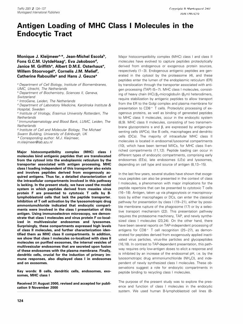
stituted BM36.1 were all transfected for stable expression ofHLA-B27. Cytolytic T lymphocyte (CTL) clone WH-F40 spe-cifically recognizes peptides from MV-F in the context ofclass I human leukocyte antigen (HLA)-B27 molecules (30).When pulsed with the specific peptide, all three cell typeswere killed with equal efficiency by CTL clone WH-F40, asshown for different effector: target ratios (Figure 1). Interest-ingly, a significant percentage of BM36.1 cells were lysedafter infection with MV, despite the fact that they lack TAP2(Figure 1B). In TAP2 reconstituted BM36.1 cells, the lysispercentage was similar to the parent cell line (Figure 1C).These results suggest that, apart from TAP-dependent pre-sentation, an alternative pathway, independent of TAP, existsto present MV-F to CTL.
To investigate whether endocytic compartments were in-volved in the presentation of MV-F, we examined the effectof the lysosomotropic drug NH4Cl on presentation efficiencyin a proliferative assay. This drug neutralizes endosomal pH,thereby affecting protein recycling from endosomes and dis-turbing endosomal antigen processing (31). Because NH4Clprevents killing by CTLs, and thus cannot be used in thekilling assay described above, we had to monitor T cellactivation in a proliferation assay. The B-LCLs 8.16, 95.3, andJP were infected with MV in the presence or absence ofNH4Cl and, after fixation, used to stimulate the T cell clonesWH-F24 and JPIII.8. MV infection of cells occurred at neutralpH and was not affected by NH4Cl (32, 33, and data notshown). Table 1 shows that NH4Cl inhibited presentation ofMV-F protein to class I-restricted T cells in different B celllines, most dramatically in JP cells with 84% inhibition of Tcell proliferation. We found a similar effect on presentation in
Figure 1: TAP-independent pre-
sentation of the MV-F protein by
HLA-B27 molecules to human CTL.
Parent cell line BM28.7 (A), TAP2mutant BM36.1 (B), as well as theTAP2 reconstituted BM36.1 (C), alltransfected for stable expression ofHLA-B27, were infected with MV(closed circles), mock-infected (trian-gles), or pulsed with MV-F peptide(squares) as described. Cells weresubsequently labeled with 51Cr andused as targets for killing by the MV-F-specific and HLA-B27-restrictedCTL clone WH-F40 at effector:target(E:T) ratios of 2:5. On the y-axis, thepercentages of specific lysis (9SD)are depicted.
125Traffic 2001: 2: 124–137
Kleijmeer et al.
Table 1: Inhibition of MV-F presentation by NH4Cl
Medium MV MV+NH4Cl % Inhibitioncpm×103 cpm×103 cpm×103
1.1 14.390.6 9.391.29.5.3 341.0 17.490.3 8.193.6 538.1.60.1 18.892.7 2.990.3JP 84
Different HLA-B27+ B-LCLs were infected with MV in theabsence or presence of NH4Cl. After fixation, cells were used asAPCs in a proliferative assay with T cell clones WH-F24, WH-F40, or JPIII.8. Numbers represent 3H-thymidine uptake (cpm×103) of representative experiments. The right column shows thepercentage of inhibition of T cell proliferation by NH4Cl.
Characterization of class I-positive endosomes
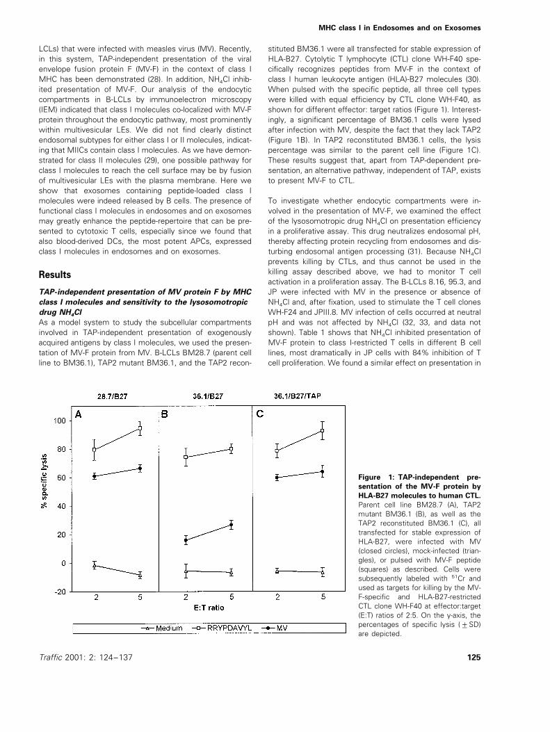
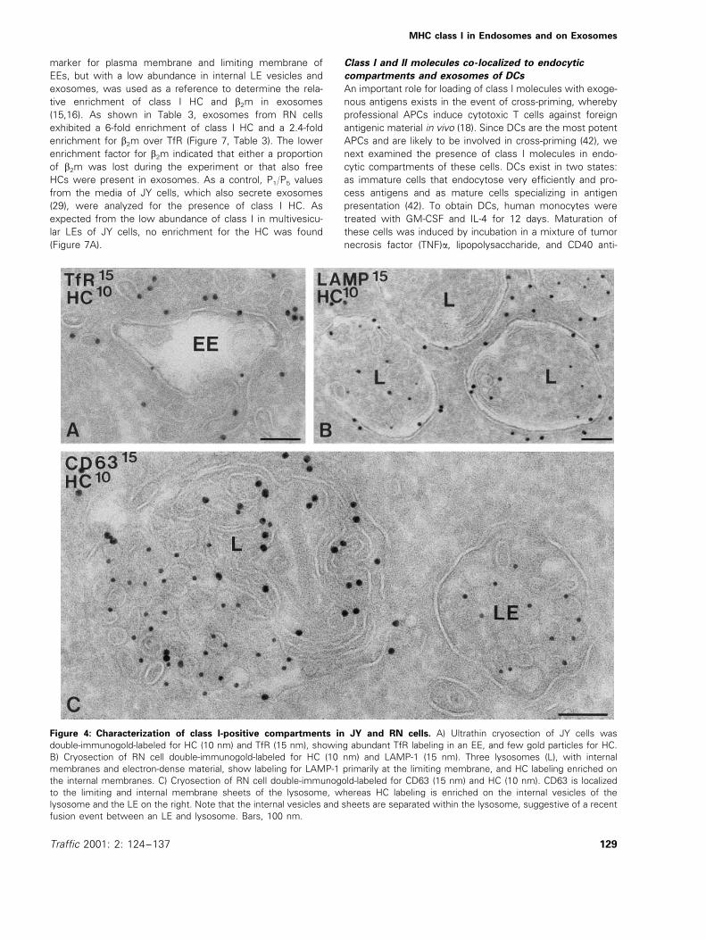
In a previous IEM study on the human B-LCL JY, we wereunable to detect appreciable amounts of class I molecules inlate endocytic compartments. In the light of the present data,we further explored the occurrence of class I molecules inendocytic compartments of the B cell lines JP and RN, andreinvestigated JY cells. By morphological criteria and markerdistribution, three major types of endocytic compartmentswere distinguished: EEs, LEs, and lysosomes (see 12 fordetailed characterization). The most prominent class I-posi-tive structure in JP (Figure 2B,C, Figure 3B) and RN cells(Figures 4 and 5) were multivesicular LEs, but in additionEEs, identified by double-labeling for the EE marker transfer-rin receptor (TfR) (Figure 4A), and lysosomes (Figure 4C)contained significant class I labeling (Table 2). Importantly,the labeling patterns of b2m and HC were similar (Figure 5A,inset), suggesting that functional class I molecules could beformed in endosomes. In JY cells, the labeling of class I inendosomes was very low, of which most was found in EEs(Table 2). The data confirm our previous findings in thesecells (11). Multivesicular LEs were distinguished from lyso-somes mainly by morphological criteria, i.e. the presence ofmany internal membrane vesicles or membrane sheets (12),respectively, and the arrival time of endocytic tracers (12,29).Furthermore, the lysosomal membrane proteins LAMP-1 andCD63, although present in both compartments, were moreabundant on lysosomal membranes (Figure 4B,C). The rela-tive distribution of class I molecules over plasma membrane,biosynthetic organelles, and endosomal compartments indi-cated that, in the RN cells, 5% of the total gold particleswere present in endosomes and lysosomes, while 3% werelocated in ER and Golgi membranes. As expected, the major-ity (92%) were present at the cell surface. In contrast, count-ings on BM36.1 showed that 61% of the total class I waspresent on membranes of the ER and Golgi complex, due toinsufficient peptide loading and retention of class I in thebiosynthetic pathway in the absence of TAP. Still, we foundthat 34% of the labeling was present on the plasma mem-brane and 3% in endosomes and lysosomes, indicating thatalso in TAP mutant cells, class I molecules can reachendosomes.
Presence of class I molecules in MIICs
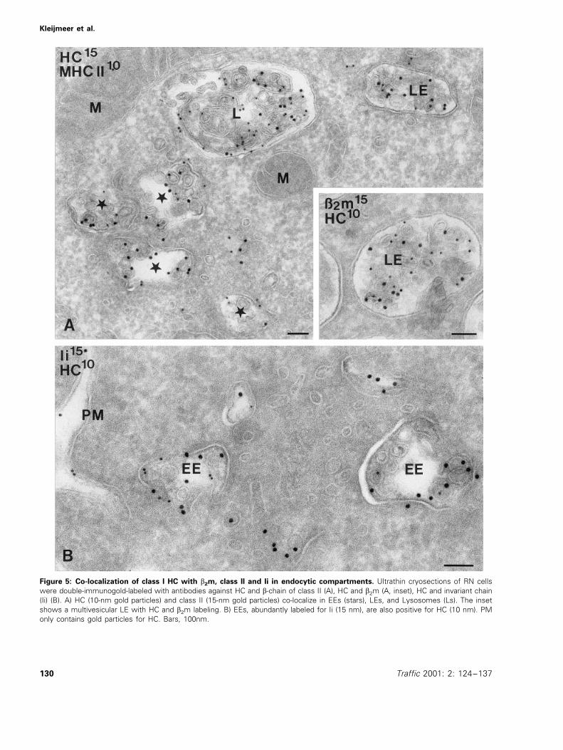
Endocytic compartments in human B cells are enriched inclass II molecules, and are termed MIICs (11,12). The obser-vation that many of the endocytic compartments in B cellswere class I-positive prompted us to examine whether classI and II molecules localized to the same compartments or ifsubtypes existed. As indicated above, in JY cells, only EEsdisplayed significant class I labeling and the overlap withclass II labeling was minute. However, in JP (Figure 3B) andRN cells (Figure 5), class I and II molecules co-localized in alltypes of endocytic compartments, including EEs, LEs, andlysosomes. Class I molecules also co-localized with invariantchain, a chaperone that mediates transport of newly synthe-sized class II molecules to the endocytic pathway (Figure5B), suggesting that class I and II molecules come togetherin the same endocytic compartments.
the presence of the lysosomotropic drug chloroquine (datanot shown), and together, these data demonstrate that endo-somes play an important role in the presentation of MV-Fprotein by class I. Control experiments showed that NH4Cldid not have any effect on the viability of the APCs, nor onthe expression of class I at the cell surface as measured byflow cytometry analysis (not shown). Similarly, total DNA andprotein synthesis as measured by 3[H]-TdR and L-[4,5-3H]leucine incorporation, respectively, were not affected(data not shown).
Co-localization of MV-F and class I in endosomes
To study the endosomal distribution of class I and MV-F, weused IEM on JP cells, which showed the most dramaticreduction of MV-F presentation by NH4Cl (Table 1). JP cells,infected with MV for 24 h, were fixed and ultrathin cryosec-tions were labeled with antibodies for class I HC and MV-F.We found that class I and MV-F co-localized at the cellsurface (Figure 2A) and in multivesicular endosomes (Figure2B,C). Also, class I molecules in the TAP mutant cellsBM36.1 were located in endosomes (data not shown), sug-gesting that these compartments may indeed play a role inthe generation of MV-F peptides and their loading onto classI molecules. Since class I was frequently observed in clathrin-coated pits and vesicles in close proximity to the cell surface(Figure 3A), the endosomal class I molecules might be inter-nalized from the plasma membrane. To investigate the originof endosomal class I further, JP cells were treated withcycloheximide for 6 h to block the supply of newly synthe-sized class I molecules (16,18). Quantitation of class I im-munogold labeling revealed that the number of gold particlesfor class I in the endocytic tract did not change upon cyclo-heximide treatment. Both in control and cycloheximide-treated cells, the average number of gold particles perLE/lysosome was eight, whereas the biosynthetic pathwayin cycloheximide-treated cells was devoid of class I labeling,and cell surface expression had decreased by 40%. In con-trast, class II labeling (illustrated for non-treated JP cells inFigure 3B) in the same cell samples had decreased by 50%in endosomal/lysosomal compartments. These data suggestthat, unlike class II molecules, the majority of endosomalclass I molecules did not derive from de novo synthesis andrepresented recycling molecules.
126 Traffic 2001: 2: 124–137

MHC class I in Endosomes and on Exosomes
Transport of class I from endosomes to the
extracellular milieu via exocytosis of LEs
We have previously described that in B cells, multivesicularLEs can fuse with the plasma membrane (29). As a conse-quence, class II molecules present in the limiting membraneof LEs are incorporated in the plasma membrane, whereasthe internal vesicles of LEs, which also contain functionalclass II molecules, are released into the extracellular mediumas so-called exosomes (29,34). To determine whether endo-somal class I molecules follow a similar exocytic pathway,we analyzed the class I content of exosomes by IEM andbiochemically. The term exosomes was first described inreticulocytes, which release 50–80-nm small-membranevesicles during their maturation into red blood cells (35). In
reticulocytes secretion of these vesicles, which originatefrom multivesicular compartments of the endocytic pathway,is a way to eliminate internalized plasma membrane proteins,such as TfR (36,37). Recent studies show that exosomes ofB-LCLs and DCs, as well as the internal vesicles of LEs, areenriched in tetraspan proteins (CD37, CD53, CD63, CD81,CD82), class II molecules, and heat-shock protein hsc73, butare poor in LAMP-1 and HLA-DM, which are primarily locatedat the limiting membranes of LEs and lysosomes (34, 38, 39,unpublished observations, M.J. Kleijmeer and M. Marsh,MRC Laboratory for Molecular Cell Biology, UCL, London).Recently, a study by Zitvogel et al. (40) showed that exo-somes from mouse DCs have strong anti-tumor effects,probably mediated by class I presentation to cytotoxic T cells.
Figure 2: Subcellular localization of MV-F and class I heavy chain (HC) in JP cells. Ultrathin cryosections of MV-infected JP cellswere double-immunolabeled for class I HC and MV-F with 10- and 15-nm gold particles, as indicated in the figures. A) Class I HC and MV-Fare both present on the plasma membrane (PM). The arrowheads point at virus particles present at the extracellular face of the PM.B and C) Co-localization of MV-F with class I HC is observed in multivesicular compartments (stars). A and B 10 nm gold particle labelsClass 1 HC, 15 nm gold particle labels MV-F. C 15 nm gold particle labels MV-F, 10 nm gold particle labels Class 1 HC. Bars, 100 nm.
127Traffic 2001: 2: 124–137
Kleijmeer et al.
Figure 3: Class I in clathrin-coated pits and co-localization of class I and II in JP cells. A) Single-immunolabeling of class I HC onJP cells shows the presence of HC in a clathrin-coated pit and vesicle (arrowheads). PM, plasma membrane. B) Double-immunolabel-ing of HC (15 nm) and class II (10 nm) shows abundant labeling for both class I and II in multivesicular LE and multilaminar lysosomal(L) compartments. G, Golgi complex, M, mitochondrion. Bars, 100 nm.
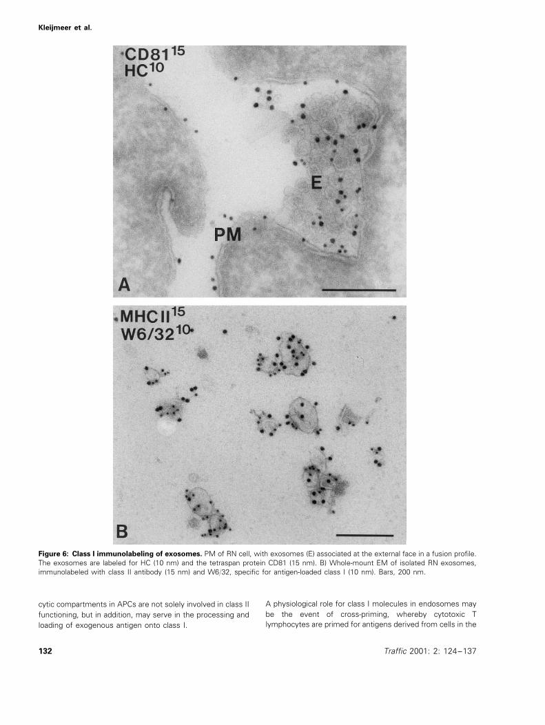
Class I molecules have not been demonstrated on B cell-derived exosomes before, and interestingly, by IEM, RN cellsshowed many class I-carrying exosomes in exocytic profilesat the plasma membrane (Figure 6A). CD81, one of theexosomal tetraspan proteins (Figure 6A), localized to thesame exosomal vesicles. To further explore these findings,exosomes were isolated from culture media of RN cells bydifferential centrifugation as described before (29,34). Theexosome-enriched pellet (P5, obtained after a final centrifuga-tion step at 70000×g) was analyzed by whole-mount IEMand biochemically. Double-immunolabeling with antibody
W6/32, specifically recognizing complexed heterodimericclass I molecules (41) and with class II antibody showed thepresence of both molecules on typical 50–80-nm exosomes(Figure 6B). These results indicate that exosomes containfunctional class I molecules. Next, pellets P1 (cells) to P5
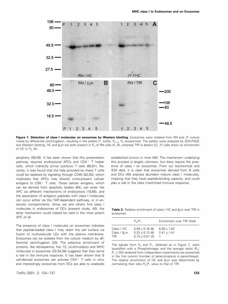
(exosomes) were subjected to sodium dodecyl sulfate-poly-acrylamide gel electrophoresis (SDS-PAGE) and Westernblotting to study the presence of class I HC, b2m, and TfR(Figure 7A,B,C, respectively). The amounts of these proteinsrecovered in P1 and P5 were determined and the ratios P5/P1
were calculated. The P5/P1 value for TfR, an established
128 Traffic 2001: 2: 124–137
MHC class I in Endosomes and on Exosomes
marker for plasma membrane and limiting membrane ofEEs, but with a low abundance in internal LE vesicles andexosomes, was used as a reference to determine the rela-tive enrichment of class I HC and b2m in exosomes(15,16). As shown in Table 3, exosomes from RN cellsexhibited a 6-fold enrichment of class I HC and a 2.4-foldenrichment for b2m over TfR (Figure 7, Table 3). The lowerenrichment factor for b2m indicated that either a proportionof b2m was lost during the experiment or that also freeHCs were present in exosomes. As a control, P1/P5 valuesfrom the media of JY cells, which also secrete exosomes(29), were analyzed for the presence of class I HC. Asexpected from the low abundance of class I in multivesicu-lar LEs of JY cells, no enrichment for the HC was found(Figure 7A).
Class I and II molecules co-localized to endocytic
compartments and exosomes of DCs
An important role for loading of class I molecules with exoge-nous antigens exists in the event of cross-priming, wherebyprofessional APCs induce cytotoxic T cells against foreignantigenic material in vivo (18). Since DCs are the most potentAPCs and are likely to be involved in cross-priming (42), wenext examined the presence of class I molecules in endo-cytic compartments of these cells. DCs exist in two states:as immature cells that endocytose very efficiently and pro-cess antigens and as mature cells specializing in antigenpresentation (42). To obtain DCs, human monocytes weretreated with GM-CSF and IL-4 for 12 days. Maturation ofthese cells was induced by incubation in a mixture of tumornecrosis factor (TNF)a, lipopolysaccharide, and CD40 anti-
Figure 4: Characterization of class I-positive compartments in JY and RN cells. A) Ultrathin cryosection of JY cells wasdouble-immunogold-labeled for HC (10 nm) and TfR (15 nm), showing abundant TfR labeling in an EE, and few gold particles for HC.B) Cryosection of RN cell double-immunogold-labeled for HC (10 nm) and LAMP-1 (15 nm). Three lysosomes (L), with internalmembranes and electron-dense material, show labeling for LAMP-1 primarily at the limiting membrane, and HC labeling enriched onthe internal membranes. C) Cryosection of RN cell double-immunogold-labeled for CD63 (15 nm) and HC (10 nm). CD63 is localizedto the limiting and internal membrane sheets of the lysosome, whereas HC labeling is enriched on the internal vesicles of thelysosome and the LE on the right. Note that the internal vesicles and sheets are separated within the lysosome, suggestive of a recentfusion event between an LE and lysosome. Bars, 100 nm.
129Traffic 2001: 2: 124–137
Kleijmeer et al.
Figure 5: Co-localization of class I HC with b2m, class II and Ii in endocytic compartments. Ultrathin cryosections of RN cellswere double-immunogold-labeled with antibodies against HC and b-chain of class II (A), HC and b2m (A, inset), HC and invariant chain(Ii) (B). A) HC (10-nm gold particles) and class II (15-nm gold particles) co-localize in EEs (stars), LEs, and Lysosomes (Ls). The insetshows a multivesicular LE with HC and b2m labeling. B) EEs, abundantly labeled for Ii (15 nm), are also positive for HC (10 nm). PMonly contains gold particles for HC. Bars, 100nm.
130 Traffic 2001: 2: 124–137

MHC class I in Endosomes and on Exosomes
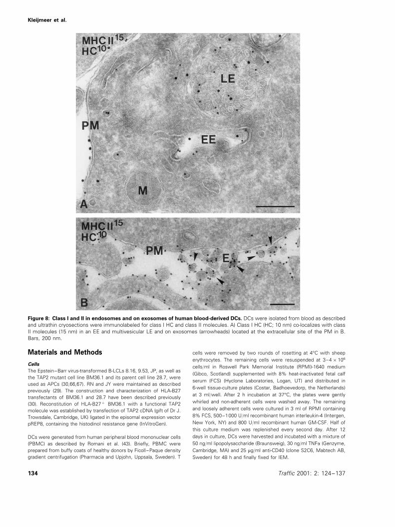
body for 48 h (43). Immunolabeling showed that class I HCwas present in both EEs and multivesicular LEs, togetherwith class II molecules (Figure 8A, Table 2). Of the total classI labeling present in these cells, 9% was located in endo-somes. In agreement with the presence of class I in LEs,DCs are able to secrete exosomes (39,40). We show nowthat class I and II molecules were located together on DCexosomes (Figure 8B). Since class I was also present in thelimiting membrane of multivesicular LEs (Figure 8), exocyto-sis of these compartments represents a pathway of insertionof endosomal class I molecules into the plasma membraneof DCs.
Discussion
TAP-independent peptide loading of class I molecules hasbeen reported to involve endocytic compartments(16,18,25,28,44–48). We have studied the presentation ofMV-F via class I by B-LCLs to further explore the role andcharacteristics of such compartments. Stimulation of MV-F-specific CTL was shown to be partially independent fromTAP, and required acidic endocytic compartments. IEMshowed that class I molecules were present throughout theendocytic tract, i.e. in EEs, multivesicular LEs, and lyso-somes, together with MV-F and class II molecules. Multi-vesicular LEs, containing both class I and II molecules, wereshown to fuse with the plasma membrane and externalizethe internal vesicles as exosomes. Secreted exosomes con-tained class I molecules, as shown by IEM and biochemicalanalysis. Importantly, also in DCs, the most potent type ofAPC, class I and II co-localized in endosomes and onexosomes.
MV-F protein is synthesized as a type I transmembraneglycoprotein, and is presented predominantly by class I MHC
molecules to CD8+ T cells in a TAP-dependent fashion (30).Using TAP2 mutant cells, we demonstrate here that presen-tation can also occur independently of TAP, which is inagreement with data obtained on presentation of MV-F bythe TAP-deficient cell line T2 (28). The weak base NH4Clinhibited class I-restricted MV-F presentation. Furthermore,class I HC, b2m, and MV-F co-localized in endosomes, sug-gesting that MV-F could be processed and bound to class Isomewhere in the endocytic tract. What fraction of theendosomal class I molecules is functional or destined fordegradation (49) remains to be determined. The antibody thatwe used against class I HC did not allow us to distinguishbetween free or complexed HCs and antibodies recognizingpeptide-loaded class I molecules failed to react in immunola-beling on ultrathin cryosections. However, a subcellular frac-tionation study on MelJuSo cells (28) has shown that LEsand lysosomes contain mature class I molecules by immuno-precipitation with conformation specific W6/32 antibody,which recognizes mature heterodimeric class I complexes.This antibody also showed reactivity in our whole-mount IEMon exosomes, implying that class I molecules in multivesicu-lar LEs contain peptide. Another strong indication that class Imolecules on exosomes are functional comes from a studyon DC-derived exosomes, which are capable of stimulatingCD8+ T cells in vitro (40).
Endosomal class I molecules are probably not derived fromthe biosynthetic pathway (50,51), but represent recyclingsurface molecules (52–56), since a blockade in protein syn-thesis for 6 h with cycloheximide did not lower the level ofclass I in endocytic compartments. In addition, class I waspresent in clathrin-coated pits of the plasma membrane. Thisis in agreement with observations that an endosomal path-way of class I-restricted presentation is not affected bycycloheximide, nor by brefeldin A, an agent interfering withtransport of newly synthesized proteins from the ER(16,44,47,57). MHC class I loading with MV-F-derived pep-tides in endosomes most likely involves peptide exchange ofpre-loaded class I. Indeed, that class I molecules are able toexchange peptides at acidic pH is indicated by the fact that atpH 5, which corresponds with the pH of LEs and earlylysosomal compartments, peptide-receptive ‘empty’ class Imolecules can be generated (28).
LEs and lysosomes play a major role in class II antigenpresentation, a reason why these compartments in APCshave been collectively termed MIICs (11). MIICs were firstdescribed in the B cell line JY, which harbors almost noendosomal class I molecules, suggesting an exclusive role ofthese compartments in class II presentation (11). However,our present observations show that JY cells are exceptionalamong 10 different B cell lines with respect to the lowabundance of class I in endosomes (present study, C.Rabouille, unpublished observations). Changes in rates ofendocytosis and degradation of class I molecules might beinfluenced by the cell’s state of differentiation, which mayexplain differences in the amount of endosomal class Imolecules (52). Since DCs also contain class I molecules inendosomes (40, present study), it is more likely that endo-
Table 2: Distribution of MHC class I heavy chain (HC) in endo-cytic compartments
Gold %Gold%Gold%
JP 60 791 483 5894 296 3594RN 41 1791 137 5693 66 2794
8249411599128JY 1694DC 69 3390 108 5294 20 1695
Endocytic compartments were subdivided in three major types:early endosomes (EE), multivesicular late endosomes (LE) andlysosomes (L). The columns on the left below each compart-ment show the total number of gold particles counted in 20 cellprofiles on this compartment. The respective percentages9SEM, based on three countings of 20 cell profiles, are displayedin the right hand columns.
131Traffic 2001: 2: 124–137
Kleijmeer et al.
Figure 6: Class I immunolabeling of exosomes. PM of RN cell, with exosomes (E) associated at the external face in a fusion profile.The exosomes are labeled for HC (10 nm) and the tetraspan protein CD81 (15 nm). B) Whole-mount EM of isolated RN exosomes,immunolabeled with class II antibody (15 nm) and W6/32, specific for antigen-loaded class I (10 nm). Bars, 200 nm.
cytic compartments in APCs are not solely involved in class IIfunctioning, but in addition, may serve in the processing andloading of exogenous antigen onto class I.
A physiological role for class I molecules in endosomes maybe the event of cross-priming, whereby cytotoxic Tlymphocytes are primed for antigens derived from cells in the
132 Traffic 2001: 2: 124–137
MHC class I in Endosomes and on Exosomes
Figure 7: Detection of class I molecules on exosomes by Western blotting. Exosomes were isolated from RN and JY culturemedia by differential centrifugation, resulting in the pellets P1 (cells), P2–4, P5 (exosomes). The pellets were analyzed by SDS-PAGEand Western blotting. HC and b2m are both present in P5 of RN cells (A, B), whereas TfR is absent (C). JY cells show no enrichmentof HC in P5 (A).
periphery (58,59). It has been shown that this presentationpathway requires professional APCs and CD4+ T helpercells, which indirectly prime cytotoxic T cells (60,61). Re-cently, it was found that the help provided by these T cellscould be replaced by signaling through CD40 (62,63), whichimplicates that APCs may directly cross-present cellularantigens to CD8+ T cells. These cellular antigens, whichcan be derived from apoptotic bodies (64), can enter theAPC via different mechanisms of endocytosis (18,65), andthe association of antigenic peptides with class I moleculescan occur either via the TAP-dependent pathway, or in en-docytic compartments. Since we and others find class Imolecules in endosomes of DCs (present study, 40), thelatter mechanism could indeed be valid in the most potentAPC of all.
The presence of class I molecules on exosomes indicatesthat peptide-loaded class I may reach the cell surface viafusion of multivesicular LEs with the plasma membrane.Exosomes can be isolated from the culture medium by dif-ferential centrifugation (29). The selective enrichment ofproteins, like tetraspanins, hsc 73, co-stimulatory and MHCmolecules in exosomes (29,34,39) suggests that they servea role in the immune response. It has been shown that Bcell-derived exosomes can activate CD4+ T cells in vitro,and interestingly exosomes from DCs are able to eradicate
established tumors in mice (40). The mechanism underlyingthis process is largely unknown, but does require the pres-ence of class I on exosomes. From our biochemical andIEM data, it is clear that exosomes derived from B cellsand DCs (40) express abundant mature class I molecules,implying that they have peptide-binding capacity, and couldplay a role in the class I-restricted immune response.
Table 3: Relative enrichment of class I HC and b2m over TfR inexosomes
Enrichment over TfR (fold)P5/P1
0.5990.16 (6)Class I HC 6.0991.62Class I b2m 0.2390.13 (6) 2.4191.41
0.1090.01 (3) 1TfR
The signals from P5 and P1, obtained as in Figure 7, werequantified with a PhosphoImager and the average ratios (P5/P19SD) obtained from independent experiments are presentedin the first column (number of determinations in parentheses).The relative enrichment of HC and b2m was determined bynormalizing their ratio P5/P1 value to that of TfR.
133Traffic 2001: 2: 124–137
Kleijmeer et al.
Figure 8: Class I and II in endosomes and on exosomes of human blood-derived DCs. DCs were isolated from blood as describedand ultrathin cryosections were immunolabeled for class I HC and class II molecules. A) Class I HC (HC; 10 nm) co-localizes with classII molecules (15 nm) in an EE and multivesicular LE and on exosomes (arrowheads) located at the extracellular site of the PM in B.Bars, 200 nm.
Materials and Methods
Cells
The Epstein–Barr virus-transformed B-LCLs 8.16, 9.53, JP, as well asthe TAP2 mutant cell line BM36.1 and its parent cell line 28.7, wereused as APCs (30,66,67). RN and JY were maintained as describedpreviously (29). The construction and characterization of HLA-B27transfectants of BM36.1 and 28.7 have been described previously(30). Reconstitution of HLA-B27+ BM36.1 with a functional TAP2molecule was established by transfection of TAP2 cDNA (gift of Dr J.Trowsdale, Cambridge, UK) ligated in the episomal expression vectorpREP8, containing the histodinol resistance gene (InVitroGen).
DCs were generated from human peripheral blood mononuclear cells(PBMC) as described by Romani et al. (43). Briefly, PBMC wereprepared from buffy coats of healthy donors by Ficoll–Paque densitygradient centrifugation (Pharmacia and Upjohn, Uppsala, Sweden). T
cells were removed by two rounds of rosetting at 4°C with sheeperythrocytes. The remaining cells were resuspended at 3–4×10⁶
cells/ml in Roswell Park Memorial Institute (RPMI)-1640 medium(Gibco, Scotland) supplemented with 8% heat-inactivated fetal calfserum (FCS) (Hyclone Laboratories, Logan, UT) and distributed in6-well tissue-culture plates (Costar, Badhoevedorp, the Netherlands)at 3 ml/well. After 2 h incubation at 37°C, the plates were gentlywhirled and non-adherent cells were washed away. The remainingand loosely adherent cells were cultured in 3 ml of RPMI containing8% FCS, 500–1000 U/ml recombinant human interleukin-4 (Intergen,New York, NY) and 800 U/ml recombinant human GM-CSF. Half ofthis culture medium was replenished every second day. After 12days in culture, DCs were harvested and incubated with a mixture of50 ng/ml lipopolysaccharide (Braunsweig), 30 ng/ml TNFa (Genzyme,Cambridge, MA) and 25 mg/ml anti-CD40 (clone S2C6, Mabtech AB,Sweden) for 48 h and finally fixed for IEM.
134 Traffic 2001: 2: 124–137
MHC class I in Endosomes and on Exosomes
Antibodies
The following monoclonal antibodies were used: anti-b2m BBM-1(kind gift of Dr J.J. Neefjes, NKI, Amsterdam, the Netherlands),H68.4 against TfR (Zymed Lab., San Francisco, CA), CLB gran 1/2,435 against CD63 (CLB, Amsterdam, the Netherlands), H4A3 againstLAMP-1 (CD107a; Pharmingen, San Diego, CA), and W6/32 (DAKO,Glostup, Denmark) against MHC class I heterodimeric complexes.The rabbit polyclonal antibodies were: anti-TfR (kind gift of Dr A.Schwartz), 631 69 to b2m, anti-MHC class I HC (both kind gifts of DrJ.J. Neefjes, NKI), anti-MHC class II (kind gift of Dr H.L. Ploegh,Department of Pathology, Harvard Medical School, Boston, MA),anti-luminal epitope of invariant chain antibody ICC5 (68; kind gift ofDr P.A. Morton, Monsanto, St Louis, MO), K9 against MV-F (obtainedfrom RIVM, Bilthoven, the Netherlands), and anti-IgG from DAKO.
Presentation assays
MV-infected or peptide-pulsed APCs were used as target cells in a51Cr-release assay or, after fixation, as stimulator cells in a prolifera-tive T cell assay as described (30). The CD8+ CTL clones WH-F24and WH-F40 were HLA-B*2705-restricted and MV-F protein (aa se-quence RRYPDAVYL)-specific (30). JP III.8 was a CD8+, class I-re-stricted MV-F protein-specific T cell clone obtained from a patientwith acute measles (30). To investigate the effects of lysosomotropicdrugs on MHC class I-restricted presentation of the MV-F protein, theHLA-B27+ APCs 8.16, 9.53 and the autologous JP B-LCLs wereinfected with MV in the presence or absence of NH4Cl (20 mM inRPMI-1640, 1% FBS) for 1 h, washed twice, and further cultured inRPMI, 10% FBS, and the indicated concentration of NH4Cl, whereafter the cells were fixed with paraformaldehyde (30).
IEM
Cells were fixed and prepared for ultrathin cryosectioning and im-munolabeling as described previously (69). Briefly, cells were fixed in2% paraformaldehyde or in a mixture of 2% paraformaldehyde/0.2%glutaraldehyde in phosphate buffer. After washing with phosphatePBS and PBS/50 mM glycine, cell pellets were embedded in 10%gelatin, cut in small blocks, and infiltrated with 2.3 M sucrose at 4°Cfor 4 h. Finally, the blocks were mounted and frozen in liquid nitro-gen. Ultrathin cryosections were indirectly immunolabeled with 10-nm protein A gold particles in single-immunolabeling experimentsand with 10 and 15 nm in double-immunolabeling experiments.Sections were embedded in a mixture of 2% methyl cellulose and0.4% uranyl. When indicated, JP cells were incubated with 100 mg/mlcycloheximide for 6 h and then fixed and processed for IEM asdescribed.
Semi-quantitative analysis of HC distribution
Ultrathin cryosections of JY and RN cells, and mature cytokine-derived DCs were single-immunolabeled with HC antibody and 10-nm protein A gold particles. In 3×20 cell profiles of each cell type,gold particles present on endocytic compartments were countedwhen within a 20-nm range of a membrane and designated to EEs,multivesicular LEs, or lysosomes, as detailed in Table 2. To determinethe relative distribution of class I molecules, gold particles werecounted on membranes of ER, Golgi complex, plasma membrane,and endosomes. Ultrathin cryosections of control and cycloheximide-treated JP cells were immunolabeled with HC antibody and, in eachquantitation, 50 endosomes/lysosomes were randomly selected.Plasma membrane length was measured by a point-hit method.
Isolation of exosomes
Exosomes were isolated by differential centrifugation as previouslydescribed (29,34). Briefly, RN cells were washed by centrifugationand re-cultured in fresh medium for 18 h. Cell culture media (35 ml)
containing about 5×10⁷ cells were centrifuged once for 10 min at200×g (pellet P1), twice for 10 min at 500×g (pellet P2), twice for15 min at 2000×g (pellet P3), once for 30 min at 10000×g (pellet P4),and finally once for 60 min at 70000×g (pellet P5) using a rotor SW27(Beckman Instruments, Fullerton, CA). P1 corresponds to cells and P5
to the fraction enriched in exosomes. P1-P5 were directly solubilizedin reducing or non-reducing SDS sample buffer, incubated for 5 minat 95°C, and submitted to SDS-PAGE and Western blotting. Forwhole-mount EM, membranes from P5 were floated into a sucrosegradient, adhered to a grid, fixed in 2% paraformaldehyde and dou-ble-immunolabeled for class I and II molecules.
Immunoblotting
After SDS-PAGE, the proteins were transferred to Immobilon-P mem-brane (Millipore, Bedford, MA). The membranes were then blockedfor 90 min in PBS containing 5% (w/v) non-fatty dry milk Protivar(Nutricia, Zoetermeer, the Netherlands) with 0.1% (w/v) Tween 20(blocking buffer) and reacted for 90 min with the primary antibody,followed by detection with 0.1 mg/ml of 125I-labeled recombinantprotein-G (Zymed Lab.) for 90 min. For detection of monoclonalantibodies, a rabbit anti-IgG was used as an intermediate. 125I wasdetected and analyzed using a PhosphoImager (Molecular Dynamics,Sunnyvale, CA).
Acknowledgments
The authors thank Dr G. Raposo for helpful discussions and M.Poelen and R. Leckie for technical assistance. R.M.C Scriwanek andM.K. Niekerk are gratefully acknowledged for their excellent photo-graphical work. This work was supported by a grant from the Eu-ropean Community (to H.J.G.) (nr. ERB4050PL940675). M.J.K. wasthe recipient of grants (nrs. 901-09-241/805-48-014) from the Neder-landse Organisatie voor Wetenschappelijk Onderzoek (NWO).
References
1. Townsend A, Bodmer H. Antigen recognition by class I-restricted Tlymphocytes. Ann Rev Immunol 1989;7: 601–624.
2. Germain RN, Margulies DH. The biochemistry and cell biology ofantigen processing and presentation. Annu Rev Immunol 1993;11:403–450.
3. Wolf PR, Ploegh HL. How MHC class II molecules acquire peptide-cargo: biosynthesis and trafficking through the endocytic pathway.Annu Rev Cell Dev Biol 1995;11: 267–306.
4. Rock KL, Gramm C, Rothstein L, Clark K, Stein R, Dick L, Hwang D,Goldberg AL. Inhibitors of the proteasome block non-lysosomaldegradation and the generation of peptides presented on MHC-class I molecules. Cell 1994;78: 761–771.
5. Trowsdale J, Hanson I, Mockbridge I, Beck S, Townsend A, Kelly A.Sequences encoded in the class II region of the MHC related to the‘‘ABC’’ superfamily of transporters. Nature 1990;348: 741–744.
6. Spies T, Cerundolo V, Colonna M, Cresswell P, Townsend A,DeMars B. Presentation of viral antigen by MHC class I molecules isdependent on a putative peptide transporter heterodimer. Nature1992;355: 644–646.
7. Kleijmeer MJ, Kelly A, Geuze HJ, Slot JW, Townsend A, TrowsdaleJ. Location of MHC-encoded transporters in the endoplasmicreticulum and cis-Golgi. Nature 1992;357: 342–344.
8. Watts C. Capture and processing of exogenous antigens for presen-tation on MHC molecules. Annu Rev Immunol 1997;15: 821–850.
9. Cresswell P. Assembly, transport, and function of MHC class IImolecules. Annu Rev Immunol 1994;12: 259–293.
10. Geuze HJ. The role of endosomes and lysosomes in MHC class IIfunctioning. Immunol Today 1998;19: 282–287.
11. Peters PJ, Neefjes JJ, Oorschot V, Ploegh HL, Geuze HJ. Segrega-tion of MHC class II molecules from MHC class I molecules in the
135Traffic 2001: 2: 124–137
Kleijmeer et al.
Golgi complex for transport to lysosomal compartments. Nature1991;349: 669–676.
12. Kleijmeer MJ, Morkowsky S, Griffith J, Rudensky AY, Geuze HJ.MHC class II compartments represent conventional endocytic com-partments in human and mouse B lymphoblasts. J Cell Biol1997;139: 639–649.
13. Amigorena S, Drake JR, Webster P, Mellman I. Transient accumula-tion of new class II MHC molecules in a novel endocytic compart-ment in B lymphocytes. Nature 1994;369: 113–120.
14. West MA, Lucocq JM, Watts C. Antigen processing and class IIMHC peptide-loading compartments in human B-lymphoblastoidcells. Nature 1994;369: 147–151.
15. Castellino F, Germain RN. Extensive trafficking of MHC class II-in-variant chain complexes in the endocytic pathway and appearanceof peptide-loaded class II in multiple endocytic compartments.Immunity 1995;2: 73–88.
16. Jondal M, Schirmbeck R, Reimann J. MHC class I-restricted CTLresponses to exogenous antigens. Immunity 1996;5: 295–302.
17. Rock KL. A new foreign policy: MHC class I molecules monitor theoutside world. Immunol Today 1996;17: 131–137.
18. Yewdell JW, Norbury CC, Bennik JR. Mechanism of exogenousantigen presentation by MHC class I molecules in vitro and in vivo:implications for generating CD8+ T cell responses to infectiousagents, tumors, transplants and vaccines. Adv Immunol 1999;73:1–77.
19. Pfeifer JD, Wick MJ, Roberts RL, Fidlay K, Normark SJ, Harding CV.Phagocytic processing of bacterial antigens for class I MHC presen-tation to T cells. Nature 1993;361: 359–362.
20. Reis e Sousa C, Germain R. Major histocompatibility complex classI presentation of peptides derived from soluble exogenous antigenby a subset of cells engaged in phagocytosis. J Exp Med 1995;182:841–851.
21. De Bruijn ML, Jackson MR, Peterson PA. Phagocyte-induced anti-gen-specific activation of unprimed CD8+ T cells in vitro. Eur JImmunol 1995;25: 1274–1285.
22. Rodriguez A, Regnault A, Kleijmeer M, Ricciardi-Castagnoli P,Amigorena S. Selective transport of internalized antigens to thecytosol for MHC class I presentation in dendritic cells. Nature CellBiol 1999;1: 362–368.
23. Kovacsovics-Bankowski M, Rock KL. A phagosome-to-cytosolpathway for exogenous antigens presented on MHC class Imolecules. Science 1995;267: 243–246.
24. Norbury CC, Hewlett LJ, Prescott AR, Shastri N, Watts C. Class IMHC presentation of exogenous soluble antigen via macropinocyto-sis in bone marrow macrophages. Immunity 1995;3: 783–791.
25. Zhou X, Glas R, Liu T, Ljunggren H-G, Jondal M. Antigen processingmutant T2 cells present viral antigen restricted through H-2Kb. Eur JImmunol 1993;23: 1802–1808.
26. Schirmbeck R, Melber K, Reimann J. Hepatitis B virus small surfaceantigen particles are processed in a novel endosomal pathway formajor histocompatibility complex class I-restricted epitope presen-tation. Eur J Immunol 1995;25: 1063–1070.
27. Bachmann MF, Oxenius A, Pircher H, Hengartner H, Ashton-Richardt PA, Zinkernagel R. TAP1-independent loading of class Imolecules by exogenous viral proteins. Eur J Immunol 1995;25:1739–1743.
28. Gromme M, UytdeHaag FGCM, Janssen H, Calafat J, vanBinnendijk RS, Kenter MJH, Tulp A, Verwoerd, Neefjes JJ. RecyclingMHC class I molecules and endosomal peptide loading. Proc NatlAcad Sci USA 1999;96: 10326–10331.
29. Raposo G, Nijman HW, Stoorvogel W, Leijendekker RL, Harding CV,Melief CJM, Geuze HJ. B lymphocytes secrete antigen presentingvesicles. J Exp Med 1996;183: 1–12.
30. van Binnendijk RS, van Baalen CA, Poelen MCM, De Vries P, BoesJ, Cerundolo V, Osterhaus ADME, UytdeHaag FGCM. Measles virustransmembrane fusion protein synthesized de novo or presented inimmunostimulating complexes is endogenously processed for HLA-class I- and HLA-class II-restricted cytotoxic T cell recognition. JExp Med 1992;176: 119–128.
31. Ziegler HK, Unanue ER. Decrease in macrophage antigencatabolism caused by ammonia and chloroquine is associated with
inhibition of antigen presentation to T cells. Proc Natl Acad Sci USA1982;79: 175–178.
32. Lamb RA, Kolakofsky D. Paramyxoviridae: the viruses and theirreplication. In Fields BN, Knipe DM, Howley PM (Eds.), FieldsVirology, 3rd edition. 1996: 1177–1204.
33. Griffin DE, Bellini WJ. Measles virus. In Fields BN, Knipe DM,Howley PM (Eds.), Fields Virology, 3rd edition. 1996: 1177–1204.
34. Escola JM, Kleijmeer MJ, Stoorvogel W, Griffith J, Yoshie O, GeuzeHJ. Selective enrichment of tetraspan proteins on the internal vesi-cles of multivesicular endosomes and on exosomes secreted byhuman B-lymphocytes. J Biol Chem 1998;273: 20121–20127.
35. Johnstone RM, Adam M, Hammond JR, Orr L, Turbide C. Vesicleformation during reticulocyte maturation. Association of plasmamembrane activities with released vesicles (exosomes). J Biol Chem1987;262: 9412–9420.
36. Harding C, Heuser J, Stahl P. Endocytosis and intracellular process-ing of transferrin and colloidal gold-transferrin in rat reticulocytes:demonstration of a pathway for receptor shedding. Eur J Cell Biol1984;35: 256–263.
37. Pan BT, Teng K, Wu C, Adam M, Johnstone RM. Electron micro-scopic evidence for externalization of the transferrin receptor invesicular form in sheep reticulocytes. J Cell Biol 1985;101: 942–948.
38. Hammond C, Denzin LK, Pan M, Geuze HJ, Cresswell P. Thetetraspan protein CD82 is a resident of MHC class II compartments(MIICs) where it associates with HLA-DR, DM and DO molecules. JImmunol 1998;161: 3282–3291.
39. Thery C, Regnault A, Garin J, Wolfers J, Zitvogel L, Ricciardi-Cas-tagnoli P, Raposo G, Amigorena S. Molecular characterization ofdendritic cell-derived exosomes: selective accumulation of the heatshock protein hsc73. J Cell Biol 1999;147: 599–610.
40. Zitvogel L, Regnault A, Lozier A, Wolfers J, Flament C, Tenze D,Ricciardi-Castagnoli P, Raposo G, Amigorena S. Eradication ofestablished murine tumors using a novel cell-free vaccine: dendriticcell-derived exosomes. Nature Med 1998;4: 594–600.
41. Barnstable CJ, Bodmer WF, Brown G, Galfre BB, Milstein C,Williams AF, Ziegler A. Production of monoclonal antibodies togroup A erythrocytes, HLA and other cell surface antigens: newtools for genetic analysis. Cell 1997;14: 9–20.
42. Banchereau J, Steinman RM. Dendritic cells and the control ofimmunity. Nature 1998;392: 245–252.
43. Romani N, Gruner S, Brang D, Kampgen E, Lenz A, TrockenbacherB, Konwalinka G, Fritsch PO, Steinman RM, Schuler G. Proliferatingdendritic cell progenitors in human blood. J Exp Med 1994;180:83–93.
44. Schirmbeck R, Reimann J. Peptide transporter-independent, stressprotein-mediated endosomal processing of endogenous proteinantigens for major histocompatibility complex class I presentation.Eur J Immunol 1994;24: 1478–1486.
45. Liu T, Zhou X, O8 rvell C, Lederer E, Ljunggren H-G, Jondal M.Heat-inactivated Sendai virus can enter multiple MHC class I pro-cessing pathways and generate cytotoxic T lymphocyte responsesin vivo. J Immunol 1995;154: 3147–3155.
46. Ulmer JB, Donnelly JJ, Liu MA. Presentation of an exogenousantigen by major histocompatibility complex class I molecules. Eur JImmunol 1994;24: 1590–1596.
47. Liu T, Chambers B, Diehl AD, Van Kear L, Jondal M, Ljunggren H-G.TAP peptide transporter-independent presentation of heat-killedSendai virus antigen on MHC class I molecules by splenic antigen-presenting cells. J Immunol 1997;159: 5364–5371.
48. Campbell DJ, Serwold T, Shastri N. Bacterial proteins can beprocessed by macrophages in a transporter associated with antigenprocessing-independent, cysteine protease-dependent manner forpresentation by MHC class I molecules. J Immunol 2000;164: 168–175.
49. Neuman E, Huleatt JW, Vargas H, Rupp EE, Jack RM. Regulation ofMHC class I synthesis and expression by human neutrophils. JImmunol 1992;148: 3520–3527.
50. Sugita M, Brenner MB. Association of the invariant chain with majorhistocompatibility complex class I molecules directs trafficking toendocytic compartments. J Biol Chem 1995;270: 1443–1448.
136 Traffic 2001: 2: 124–137

MHC class I in Endosomes and on Exosomes
51. Vigna JJ, Smith KD, Lutz CT. Invariant chain association with MHCclass I: preference for HLA class I/beta 2-microglobulin het-erodimers, specificity and influence of hte MHC peptide-bindinggroove. J Immunol 1996;157: 4503–4510.
52. Tse DB, Pernis B. Spontaneous internalization of class I majorhistocompatibility complex molecules in T lymphoid cells. J ExpMed 1984;159: 193–207.
53. Aragnol D, Malissen B, Schiff C, Piron M-A, Leserman LD. Endocy-tosis of MHC molecules by B cell-B lymphoma and B cell-Tlymphoma hybrids. J Immunol 1986;137: 3347–3353.
54. Dasgupta JD, Watkins S, Slayter H, Yunis EJ. Receptor-like natureof class I HLA: endocytosis via coated pits. J Immunol 1988;141:2577–2580.
55. Vega MA, Strominger JL. Constitutive endocytosis of HLA class Iantigens requires a specific portion of the intracytoplasmic tail thatshares structural features with other endocytosed molecules. ProcNatl Acad Sci USA 1989;86: 2688–2692.
56. Reid PA, Watts C. Cycling of cell-surface MHC glycoproteinsthrough primaquine-sensitive intracellular compartments. Nature1990;346: 655–657.
57. Schirmbeck R, Bohm W, Melber K, Reimann J. Processing ofexogenous heat-aggregated (denatured) and particulate (native)hepatitis B surface antigen for class I-restricted epitope presenta-tion. J Immunol 1995;155: 4676–4684.
58. Bevan MJ. Cross-priming for a secondary response to minor Hantigens with H-2 congenic cells which do not cross-react in thecytotoxic assay. J Exp Med 1976;143: 1283–1288.
59. Matzinger P, Bevan M. Induction of H-2-restricted cytotoxic T cells:in vivo induction has the appearance of being unrestricted. CellImmunology 1977;33: 92–100.
60. Huang AYC, Golumbeck P, Ahmadzadeh M, Jaffee E, Pardoll D,Levitsky H. Role of bone marrow-derived cells in presenting MHCclass I-restricted tumor antigens. Science 1994;264: 961–965.
61. Keene J, Forman J. Helper activity is required for the in vivogeneration of cytotxic T lymphocytes. J Exp Med 1982;155: 768–782.
62. Schoenberger SP, Toes REM, van der Voort EIH, Offringa R, MeliefCJM. T help for cytotoxic T lymphocytes is mediated by CD40-CD40L interactions. Nature 1998;393: 480–483.
63. Ridge JP, Di Rosa F, Matzinger P. A conditioned dendritic cell canbe a temporal bridge between a CD4+ T-helper and a T-killer cell.Nature 1998;393: 474–478.
64. Albert ML, Sauter B, Bhardwaj N. Dendritic cells acquire antigenfrom apoptotic cells and induce class I-restricted CTLs. Nature1998;392: 86–89.
65. Arnold-Schild D, Hanau D, Spehner D, Schmid C, Rammensee H-G,de la Salle H, Schild H. Receptor-mediated endocytosis of heat-shock proteins by professional antigen presenting cells. J Immunol1999;162: 3757–3760.
66. Urban RG, Chicz RM, Lane WS, Strominger JL, Rehm A, Kenter MJ,UytdeHaag FG, Ploegh HL, Uchanska-Ziegler B, Ziegler A. A subsetof HLA-B27 molecules contains peptides much longer than non-amers. Proc Natl Acad Sci USA 1994;91: 1534–1538.
67. Mellins E, Smith L, Arp B, Cotner T, Celis E, Pious D. Defectiveprocessing and presentation of exogenous antigens in mutants withnormal class II genes. Nature 1990;343: 71–74.
68. Morton PA, Zacheis ML, Giacoletto KS, Manning JA, Schwartz BD.Delivery of nascent MHC class II-invariant chain complexes tolysosomal compartments and proteolysis of invariant chain by cys-teine proteases precedes peptide binding in B-lymphoblastoid cells.J Immunol 1995;154: 137–150.
69. Raposo G, Kleijmeer MJ, Posthuma G, Slot JW, Geuze HJ. Im-munogold labeling of ultrathin cryosections: application in immunol-ogy. In Herzenberg LA, Weir DM, Herzenberg LA, Blackwell C (Eds.),Weirs Handbook of Experimental Immunology. Malden, MA: Black-well Science, 1997: 1–11. 208 pp.
137Traffic 2001: 2: 124–137