antigens and antibodies
TRANSCRIPT
Antigens and Antibodies
Dr. Deepak K Gupta

Antigen
• Any substance which, when introduced parentrally into the body stimulates the production of antibody specifically.
• This is a traditional definition since it had got • This is a traditional definition since it had got some exception– Polio vaccine – oral administration
– Some antigen may not produce antibodies
• Specificity is the hallmark of all immunological reactions

Function of Antigen
• Immunogenicity : induction of immune response• Immunological Reactivity: specific reaction with
antibodies or sensitized cells• Based on this function antigen is classified as– Complete antigen: induce antibody formation and – Complete antigen: induce antibody formation and
specific immune response– Partial Antigen (Hapten): a specific non-protein
substance incapable of inducing antibody formation itself but elicit immune response when couple with carrier protein• Complex hapten : polyvalent – 2 or more antibody
combining site• Simple Hapten: univalent

Structure of Antigen
• Smallest unit of antigenicity (antigenic detrminant) – epitope.
• Consist of 4-5 amino residues or monosacharideresidues
• Posses a specific chemical structure, electric • Posses a specific chemical structure, electric charge and spatial configration
• Capable of sensitizing of an imunocytes
• Paratope: area on antibody molecule on which epitope binds– This produces specificity of antigenic reaction

Biological Classes of Antigen
• Depending on the ability to produce antibody formation, antigen are classified as
– T cell dependent (TD): structurally more complex
– T cell independent (TI): simple, limited number of – T cell independent (TI): simple, limited number of repeating epitopes
• Although antibody is produced by B-lymphocytes, but it requires cooperation of T-lymphocytes

Properties of Antigen• Size
– Antigenicity is related to molecular size– Larger molecules are highly antigenic where as haptens are low
molecular weight having low antigenicity
• Chemical Nature– Antigenicity is directly proportional to degree of structural
diversitydiversity– Amino acid> monosachharide> lipid> nucleic acid
• Susceptibility– Only substance metabolized by tissue enzyme to epitope
fragments – potential to produce antigen– Polystyrene not antigenic– Also antigen rapidly broken down doesn’t show Antigenicity
properties– D-amino acids are not metabolized in the body where as L-
amino acids do - prevention of autoimmunity
Properties of Antigen
• Foreignness– Described by Ehrlich– Normal body contain numerous antigens or antigen
producing substance which may not produce antibody.antibody.
– But introduction of this into other body produces immune response
– This response is directly related to degree of foreignness – antigen from same species shows less Antigenicity than others
• Specificity

Specificity
• Antigen shows various types of specificity. They are mainly
– Antigenic specificity
– Species specificity– Species specificity
– Iso-specificity
– Auto-specificity
– Organ specificity
– Heterogenic specificity

Specificity
• Antigenic specificity• Chemical nature of antigen makes it different from other
• Its may arise on the difference of group attaches on ortho, meta or para position
• Also on the basis of spatial arrangement – dextro, levo and mesomeso
• Species specificity• All individual contain species-specific antigens
• This has been used in tracing evolutionary relationship
• Ex: a individual sensitive to horse serum will be sensitive to other equine but not with bovine

Specificity
• Iso-specificity
– Antigens found in the some but not all members of species
– Blood grouping system is based on this– Blood grouping system is based on this
– Genetically determined
• Auto-specificity
• Sometime autologous antigens starts showing antigenicity
• Autoimmunity disorders like Rheumatoid arthritis, SjogrenSyndrome

AntibodiesAntibodies

Introduction
• An antibody is an immunoglobulin (glycoprotein) molecule formed by the immune system in response to an antigenic stimulus.– Found in blood, bodily secretions or on mucous
surface – Binds to the specific antigen responsible for its – Binds to the specific antigen responsible for its
production, thereby inactivating it
• Immunoglobulins provides a structural and chemical concepts.
• “Antibody” is a biological and functional concept.• All antibodies are immunoglobulins, but all
immunoglobulins are not antibodies.
Antibodies
• Constitute the gamma globulin portion of blood proteins - 20-25 % of total serum protein.
• Are soluble proteins secreted by activated B cells and plasma cells in response to an cells and plasma cells in response to an antigen
• Are capable of binding specifically with that antigen

• Consists of four looping polypeptide chains linked together with disulfide bonds– Two identical heavy (H) chains and two identical
light (L) chains
• The four chains bound together form an
Antibody Structure
• The four chains bound together form an antibody monomer
• Each chain has a variable (V) region at one end and a constant (C) region at the other
• Variable regions of the heavy and light chains combine to form the antigen-binding site

Antibody Structure

• Antibodies responding to different antigens have different V regions
• C region (either kappa or lambda unit) is the same for all antibodies in a given class
• C regions form the stem of the Y-shaped antibody and:
Antibody Structure
• C regions form the stem of the Y-shaped antibody and:
– Determine the class of the antibody
– Serve common functions in all antibodies
– Dictate the cells and chemicals that the antibody can bind to
– Determine how the antibody class will function in elimination of antigens

• Based on physiochemical and antigenic difference antibodies are classified as:– IgG : IgG1, IgG2, IgG3, IgG4
– IgA : Serum IgA or Secretory IgA and IgA1 or IgA2
– IgM
– IgD
– IgE
Classes of Antibodies
IgE
*** Decreasing order of their concentration.

IgG
• Major serum immunoglobulin – 80 % of total
• Molecular weight – 7s
• It exist in polymerised form
• Distributed equally in intravascular and • Distributed equally in intravascular and extravascular compartment
• Half life – 25 days
• Raised in chronic disease like Kala azar, chronic malaria or myeloma.
• Normal serum concentration: 8-16 mg per ml

IgG• Only maternal immunoglobulin – normally
transported across the placenta and provides natural passive immunity in the new born
• Not synthesized by fetus
• The function of IgG: general purpose antibody• The function of IgG: general purpose antibody– Immunological reactions such as complement fixation,
precipitation and neutralization of toxins and viruses
– Protective against infectious agents – active in blood and tissues
– Binds to MCO and enhances their phaogocytosis
– Surface recognition – Fc fragments

IgA
• 2nd most abundant class – 10-13 % of serum immunoglobulin
• Norma serum level: 0.6-4.2 mg/ml
• Half life – 6-8 days• Half life – 6-8 days
• Found in colostrum, saliva and tears
• It occurs in two forms– Serum IgA: principally
monomeric 7s molecule
– Secretory IgA: two monomericunit joined together by J-chain
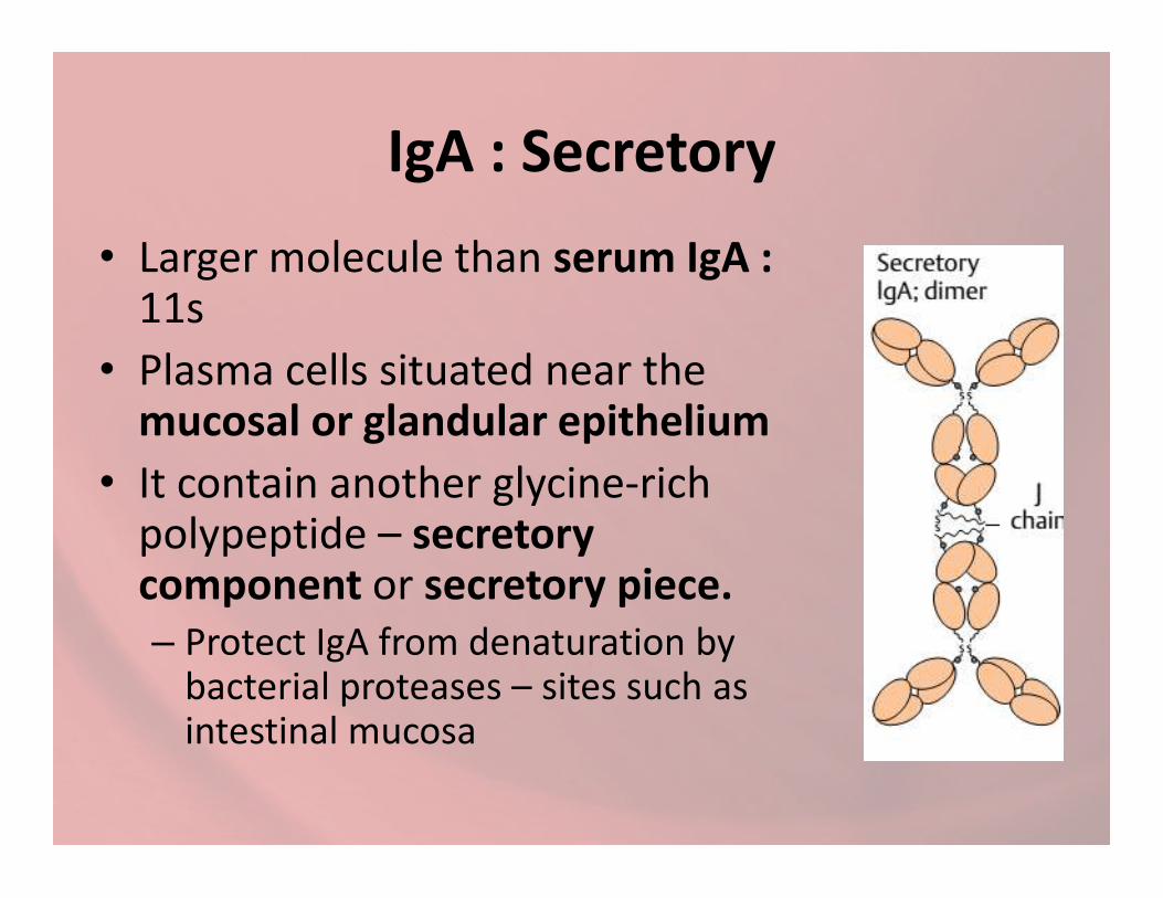
IgA : Secretory
• Larger molecule than serum IgA : 11s
• Plasma cells situated near the mucosal or glandular epithelium
• It contain another glycine-rich polypeptide – secretorycomponent or secretory piece.– Protect IgA from denaturation by
bacterial proteases – sites such as intestinal mucosa

IgA
• Function of IgA
• local immunity – against respiratory and intestinal pathogens
• inhibits adherens of MCO to surface • inhibits adherens of MCO to surface mucosa
• Activate the alternative complement pathway
• Promotes phagocytosis and intracellular killing of MCO

IgM
• 5-8 % of serum immunoglobulins
• Normal level 0.5 – 2 mg/ml
• Hlaf life – 5 days
• Heavy molecule – 19s – millionaire molecule• Heavy molecule – 19s – millionaire molecule
• Polymer of the four-polypeptide joined by J-chain.
• Most of IgM – intravascular (80%)
• Phylogenetically oldest immunoglobulin class – earliest immunoglobulin synthesized by fetus (20 weeks of age)

IgM
• Its not transported across the placenta
• Presence of IgM in fetus or newborn: syphilis, rubella, newborn: syphilis, rubella, HIV infection and toxoplasmosis
• Relatively short lived – hence its serum demonstration indicates acute infection

IgM• Its highly potent as compares to IgG– 1000 times : immune hemolysis– 500-1000 times : Opsonisation– 100 times : bacteriocidal– 20 times : bacterial agglutination
• Less active than IgG : neutralisation of toxins and • Less active than IgG : neutralisation of toxins and viruses
• Largely confined – intravascular space• Functions• Protection – blood invasion MCO• Monomeric IgM – major antibody receptor on the surface of
lymphocytes for antigen recognition
• IgM deficiency - septicemia

IgD
• It resembles IgG structurally
• Serum concentration : 3 mg/100 ml mostly intravascular
• Half life : 3 days• Half life : 3 days
• Recognition receptor for antigen on B Lymphocytes as that of IgM
• Also it causes stimulation of the B-cell –activation and cloning to produce antibody or supression
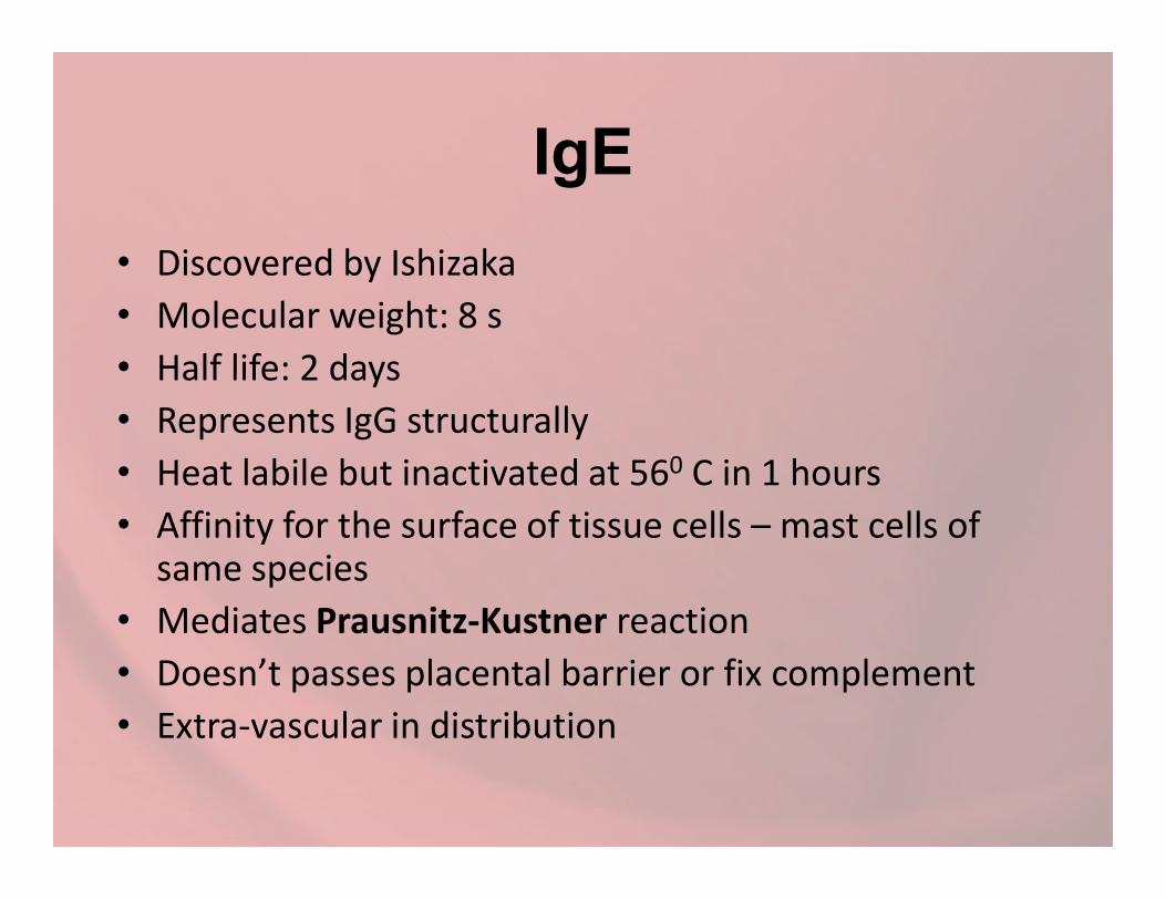
IgE
• Discovered by Ishizaka
• Molecular weight: 8 s
• Half life: 2 days
• Represents IgG structurally
Heat labile but inactivated at 560 C in 1 hours• Heat labile but inactivated at 560 C in 1 hours
• Affinity for the surface of tissue cells – mast cells of same species
• Mediates Prausnitz-Kustner reaction
• Doesn’t passes placental barrier or fix complement
• Extra-vascular in distribution

IgE
• There is very trace (few ng) serum level in normal condition, but elevated in – Atopic (type 1 allergic): asthma, hay fever and eczema– Intestinal parasitic infection
• IgE chiefly produced in the linings of respiratory and intestinal tracts
• IgE chiefly produced in the linings of respiratory and intestinal tracts
• IgE deficiency – undue susceptibility to infection.• It produces• Anaphylacttic type of hypersensitivity• Protection against pathogen by mast cell degranulation and
release of inflammatory mediators• Defense in helminthic infection

IgE

Summary
• In general, antibodies protects by
– IgG: body fluid
– IgA: body surface
– IgM: blood stream– IgM: blood stream
– IgD: recognition molecule on the surface of B-lymphocytes
– IgE: mediates antigenic hypersensitivity

Antigen-Antibody ReactionAntigen-Antibody Reaction

Use of Antigen-Antibody Reactions
• In Body– It forms the basis of antibody
mediates immunity against infectious disease
– Tissue injury like Hypesensitivityand autoimmunityand autoimmunity
• In the Lab – Serological Reaction– Diagnosis of infection– Identification of infectious
agents and difference between non infectious antigen
– Quantification of antigen or antibody

Stages of Antigen-Antibody reactions• It occurs mainly in 3 stages
– Primary Stages• Initial interaction without any visible effect• Occurs very rapidly – obeys the laws of chemistry• Reversible • Can be represented by estimating free and bound antibodies –
physical and chemical method includind markers like radioistopte, fluorescent dyes etc fluorescent dyes etc
– Secondary Stages: demonstrates the events like• Precipitation• Agglutination• Lysis of Cell• Neutralisation of toxins and other biologically active antigen• Complement fixation• Immobilisation of motile organism• Enhancement of Phagocytosis
– Tertiary Stages : Complete destruction of injurious antigen and tissue damage including humoral immunity

Features of Antigen-Antibodies Reaction
• Specific– antigen combines only with its homologus antibody,– Sometimes cross-reactivity may occur due to antigenic
similarity
• Completeness: entire molecules reacts and not fragmentsfragments
• No denaturation of antigen or anotibodies• Combination occurs on the surface• The reaction is firm but reversible• Varying proportion
• both antigen and antibodies are multivalent• Antigen have valencies up to 100• Antibody are generally bivalent – though IgM: 5-10 combining sites


Precipitation Reaction
• When a soluble antigen combines with its antibody in the presence of electrolytes (NaCl) at suitable temperature and pH forms insoluble precipitate. It either– Settles down – Sedimentation– Settles down – Sedimentation
– Suspended as floccules – Flocculation
• It can be carried out either in liquid or gel media.
• Amount of precipitate is greatly influenced by antigen/antibody ratio

Precipitation Reaction
• Based on the antigen/antibody ratio, the precipitation reactions can be classified in 3 zones i.e Zone phenomenon– Prozone : Antibody is in excess. False negative
precipitation may occur.Prozone : Antibody is in excess. False negative precipitation may occur.
– Zone of equivalence : equal proportion of antigen and antibody i.e. optimal proportion, most rapid and abundant reaction
– Postzone: Antigen is in excess. Precipitation is again weak or even absent

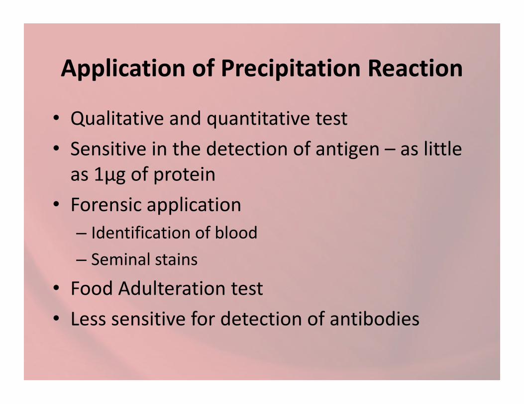
Application of Precipitation Reaction
• Qualitative and quantitative test
• Sensitive in the detection of antigen – as little as 1μg of protein
• Forensic application • Forensic application
– Identification of blood
– Seminal stains
• Food Adulteration test
• Less sensitive for detection of antibodies
Precipitation Reaction
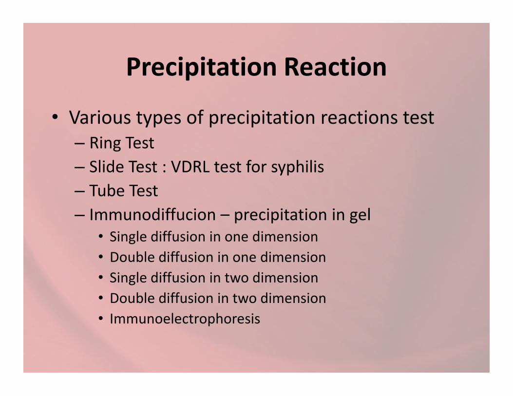
• Various types of precipitation reactions test– Ring Test
– Slide Test : VDRL test for syphilis
– Tube Test – Tube Test
– Immunodiffucion – precipitation in gel• Single diffusion in one dimension
• Double diffusion in one dimension
• Single diffusion in two dimension
• Double diffusion in two dimension
• Immunoelectrophoresis

Agglutination Reaction
• When a particulate antigen is mixed with its antibody in the presence of electrolytes at a suitable temperature and pH, the particles are clumped or agglutinated
• More sensitive for antibodies detection• More sensitive for antibodies detection
• Same Zonal principle applies for agglutination as that of precipitation
• Incomplete or monovalent antibodies doesn’t cause agglutination – combine with antigen : Blocking antibodies

Agglutination Reaction
• Positive reaction is flowed by clumping together of particles
• There are various application for agglutination reactions– Slide Agglutination– Slide Agglutination– Tube agglutionation: Widal test for Typhoid, Paul
Bunnel Test – mononucleosis, Cold agglutination test -mycoplasma pneumonia
– Antiglobulin (Coombs) test – Rh antibody– Passive agglutination test – Rose Waaler test for
Rheumatoid arthritis

Complement Fixation Test (CFT)
• Complement takes part in many immunological reactions.
• In the presence of antibodies complement– Lyses and kills erythrocytes and bacteria
– Immobilises motile organism
– Promotes phagocytosis and immune adherence
– Hypersensitivity
• The ability of antigen-antibody complexes to fix complement is made use of the Complement Fixation Test

Complement Fixation Test (CFT)
• Very versatile and sensitive test – detecting as little as 0.04 mg of antibody and 0.1 mg of antigen.
• Gold standard test for detection of Treponemapallidum and Vibrio choleraepallidum and Vibrio cholerae
• Its carried out in 2 steps and uses five reagents– Antigen– Antibody– Complement– Sheep Erythrocytes– Amboceptor – rabbit antibody to sheep erythrocytes

Complement Fixation Test (CFT)• 1ST Step– Inactivated serum of patient is incubated for 370 C for 1
hr with Wasserman Antigen and fixed amount of complement (Guinea pig)
– If the serum contains antibody – it will utilizes complement and there will be no complement left for further reaction complement and there will be no complement left for further reaction
– If it doesn’t contain any antibody – no reaction will occur
• 2nd Step• Sensitized cell (Sheep Erythrocytes + Amboceptor) is added to the
mixture and incubated.• Lysis of erythrocyte will indicate – complement was not fixed in 1st
step therefore no antibody was present : CFT NEGATIVE • CFT POSITIVE – no lysis will occur

Neutralisation test
• Neutralisation of antigen by its antibody can be demonstrated by various system
• It can be
– Virus neutralisation– Virus neutralisation
– Bacterial Neutralisation
– Toxin Neutralisation – in vivo or in vitro
• Ex: Shick Test – Diptheria, , Antistreptolysin ‘O’ ASO test

Oppsonisation
• Given by Wright (1903)
• Heat labile substance present in fresh normal sera – facilitated phagocytosis
• Opsonic index• Opsonic index
– study the progress of resistance during the course of diseas
– Ratio of the phagocytic activity of the patient’s blood for a given bacterium to the phagocyticactivity of blood from a normal individual
Immunofluorescence
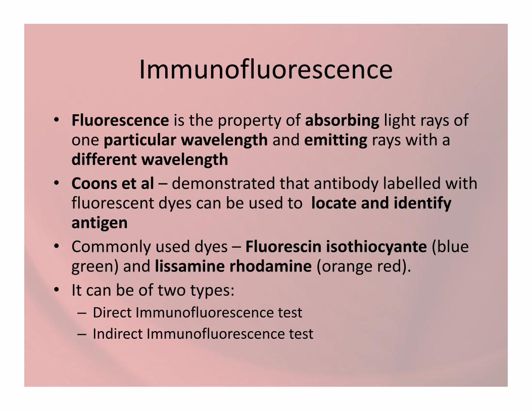
• Fluorescence is the property of absorbing light rays of one particular wavelength and emitting rays with a different wavelength
• Coons et al – demonstrated that antibody labelled with fluorescent dyes can be used to locate and identify fluorescent dyes can be used to locate and identify antigen
• Commonly used dyes – Fluorescin isothiocyante (blue green) and lissamine rhodamine (orange red).
• It can be of two types:– Direct Immunofluorescence test
– Indirect Immunofluorescence test

Direct Immunofluorescence test• Specific antibody labeled with fluorescent dye –
detection of unknown antigen in a specimen
• If antigen is present it reacts with labeled antibodies and fluorescence can be observed in UV light of fluorescent microscopelight of fluorescent microscope
• Example– Bacteria, Virus or other antigen in blood, CSF, urine
faeces, tissues and other specimens
– Rabies detection in brain
• Disadvantage: Separate fluorescent antibodies preparation is required for each antigen detection

Direct Immunofluorescence test

Indirect Immunofluorescence test
• A known antigen is fixed in a slide with unknown antibody (Serum),
• If its positive there will be a reaction to form globullin coating on antigenglobullin coating on antigen
• After this the smear is treated with fluorescent labeled antiserum to humman gammaglobulin
• This will react with antibody globulin bound to antigen
– Slide is examine under UV-microscope

Indirect Immunofluorescence test
• Advantage: a single antihuman globulin fluorescent conjugate can be employed for can be employed for detection of antibody in any antigen
• No separate conjugation with each antibody is required

Radioimmunoassay (RIA)
• highly sensitive and quantitative procedure that utilizes radioactively labeled antigenor antibody
• S. A. Berson and Rosalyn Yalow (Nobel Prize) : • S. A. Berson and Rosalyn Yalow (Nobel Prize) : levels of insulin–anti-insulin complexes in diabetics
• Principle – Competitive binding of radiolabeled antigen and
unlabeled antigen to a high-affinity antibody.

Enzyme Linked Immunoassay (ELISA)
• It is one of immunoassay method used to detection of– Antibodies– Proteins– Peptides– Biomolecules– Biomolecules
• An absorbing material specific for either one of the above components
• Ex: Cellulose, agarose or solid phase such as polysterene, polyvinyl or polycarbonate tubes or microwells or membranes or discs of polyacrylamide, paper or plastics
ELISA
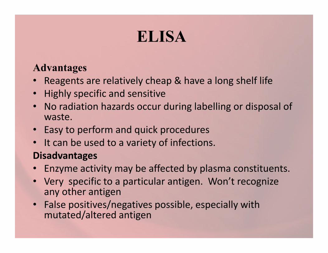
Advantages • Reagents are relatively cheap & have a long shelf life • Highly specific and sensitive• No radiation hazards occur during labelling or disposal of
waste.• Easy to perform and quick procedures • Easy to perform and quick procedures • It can be used to a variety of infections.Disadvantages• Enzyme activity may be affected by plasma constituents.• Very specific to a particular antigen. Won’t recognize
any other antigen• False positives/negatives possible, especially with
mutated/altered antigen

•Direct ELISA•Indirect ELISA•Sandwich ELISA
Types of ELISA
NON -COMPETETIVE ELISA
•Sandwich ELISA•Competitive ELISA•Ogives ELISA

Direct ELISA• The direct detection method
uses a labeled primary antibody that reacts directly with the antigen.
• Direct detection can be performed with antigen that is performed with antigen that is directly immobilized on the assay plate .
• Direct detection is not widely used in ELISA
• But is quite common for immunohistochemicalstaining of tissues and cells.

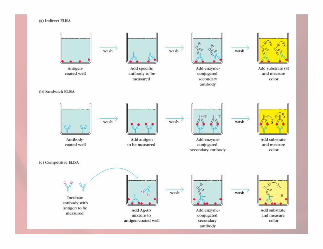
• The indirect ELISA utilizes an unlabeled primary antibody in conjunction with a labeled secondary labeled secondary antibody.
• The secondary antibody has specificity for the primary antibody

Direct and Indirect ELISA

Sandwich ELISA
• The sandwich measures the amount of antigen between two layers of antibodies.
• They are especially useful if the concentration of antigens is low
.
of antigens is low
• Or they are contained in a mix of high concentrations of contaminating protein
• Major advantages - the antigen does not need to be purified prior to use, due to its high specificity

Sandwich ELISA1. Antibody (capture) is bound to a microtiter plate well. 2. Antigen is then added and bound to the antibody and
unbound products are then removed3. A second antibody is added (detection) linked to enzyme4. Lastly substrate to this enzyme is added and converted by
enzyme into colored productenzyme into colored product5. The rate of color formation is proportional to amount of
antibody
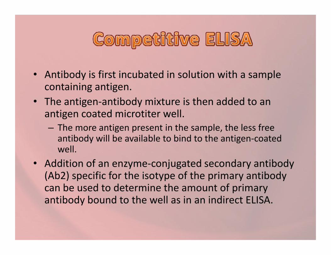
• Antibody is first incubated in solution with a sample containing antigen.
• The antigen-antibody mixture is then added to an antigen coated microtiter well. – The more antigen present in the sample, the less free – The more antigen present in the sample, the less free
antibody will be available to bind to the antigen-coated well.
• Addition of an enzyme-conjugated secondary antibody (Ab2) specific for the isotype of the primary antibody can be used to determine the amount of primary antibody bound to the well as in an indirect ELISA.

COMPETETIVE ELISA
Solid phase coated with antibody
Add unknown amount of unlabeled antigen and known amount of labeled antigen
Free and labeled antigen are capturedFree and labeled antigen are captured
Color formation by oxidation of substrate into a colored compound
Under standard condition ,the enzyme activity measured is proportional to the proportion of labeled antigen in the mixture of labeled and unlabled antigen.

• A newer technique uses an solid phase made up of an immuno-sorbent immuno-sorbent polystyrene rod with 8-12 protruding o-gives.

ELISA RESULTSThe ELISA assay yields three different types of data output:
Quantitative: ELISA data can be interpreted in
comparison to a standard curve in order to preciselycalculate the concentrations of antigen in various samples.
Qualitative : ELISAs can also be used to achieve a yesQualitative : ELISAs can also be used to achieve a yes
or no answer indicating whether a particular antigen ispresent in a sample.
Semi-quantitative: ELISAs can be used to compare
the relative levels of antigen

Application
• Screening donated blood for evidence of viral contamination by – HIV-1 and HIV-2 (presence of anti-HIV antibodies) – Hepatitis C (presence of antibodies) – Hepatitis B (testing for both antibodies and a viral antigen)
• Measuring hormone levels – HCG (as a test for pregnancy) – HCG (as a test for pregnancy) – LH (determining the time of ovulation)– TSH, T3 and T4 (for thyroid function)
• Detecting infections – Sexually-transmitted agents like HIV, syphilis and chlamydia– Hepatitis B and C – Toxoplasma gondii
• Detecting illicit drugs.• Detecting allergens in food and house dust
