antioxidant therapies for acute spinal cord injury · review antioxidant therapies for acute spinal...
TRANSCRIPT

REVIEW
Antioxidant Therapies for Acute Spinal Cord Injury
Edward D. Hall
1Spinal Cord & Brain Injury Research Center, University of Kentucky College of Medicine, Biomedical Biological ScienceResearch Building, Lexington, Kentucky 40506; 2Neurotrauma Research, Anatomy & Neurobiology, Neurology, Neurosurgery andPhysical Medicine & Rehabilitation, University of Kentucky Chandler Medical Center, Lexington, Kentucky 40506; and 3741 S.
Limestone St, Biomedical & Biological Science Research Building 483, Lexington, Kentucky 40536-0509
Summary: One of the most investigated molecular mechanismsinvolved in the secondary pathophysiology of acute spinal cordinjury (SCI) is free radical-induced, iron-catalyzed lipidperoxidation (LP) and protein oxidative/nitrative damage tospinal neurons, glia, and microvascular cells. The reactivenitrogen species peroxynitrite and its highly reactive freeradicals are key initiators of LP and protein nitration in theinjured spinal cord, the biochemistry, and pathophysiology ofwhich are first of all reviewed in this article. This is followed by apresentation of the antioxidant mechanistic approaches andpharmacological compounds that have been shown to haveneuroprotective properties in preclinical SCI models. Two of
these, which act by inhibition of LP, are high-dose treatment withthe glucocorticoid steroid methylprednisolone (MP) and thenonglucocorticoid 21-aminosteroid tirilazad, have beendemonstrated in the multicenter NASCIS clinical trials toproduce at least a modest improvement in neurological recoverywhen administered within the first 8 hours after SCI. Althoughthese results have provided considerable validation of oxidativedamage as a clinically practical neuroprotective target, there is aneed for the discovery of safer and more effective antioxidantcompounds for acute SCI. Key Words: Spinal cord injury, freeradicals, lipid peroxidation, peroxynitrite, antioxidants,neuroprotection.
INTRODUCTION
One of the best validated secondary injury mechanismsin acute spinal cord injury (SCI) concerns the post-traumatic generation of reactive oxygen species (ROS)and their resulting oxygen free radical-induced oxidativedamage. Indeed, the only evidence from clinical trials todate that supports the potential for an acute pharmacolog-ical neuroprotective effect in SCI involves two compoundsthat appear to work by interference with oxidative damageand the process of free radical-induced, iron-catalyzed lipidperoxidation (LP), in particular. This reviewwill present thebasics of the biochemistry of ROS and free radicalgeneration, and the oxidative damage, evidence for itsinvolvement in the biochemistry and pathophysiology ofacute SCI and relationship to other secondary injurymechanisms, and the preclinical and clinical pharmacologyof antioxidant neuroprotection.
CHEMISTRY OF FREE RADICAL PRODUCTION,LIPID PEROXIDATION, AND MECHANISMSFOR ITS PHARMACOLOGICAL INHIBITION
The chemistry of free radical formation and LP hasbeen previously discussed in a prior review of “Anti-Antioxidant Therapies for Traumatic Brain Injury,” anarticle recently published in this journal [1]. However,some updated portions of that article are re-presentedhere to provide the needed background for the discussionof oxidative damage and its prevention in the context ofacute SCI.
Superoxide radicalIn the injured spinal cord, a number of sources of the
primordial free radical superoxide (O2•-) may be operative
within the first minutes and hours after injury including thearachidonic acid cascade (i.e., by product of prostaglandinsynthase and 5-lipoxygenase activity), either enzymatic(by-product of monoamine oxidase-B activity), or auto-oxidation of biogenic amine neurotransmitters (e.g.,dopamine, norepinephrine, 5-hydroxytryptamine),“mitochondrial leak,” xanthine oxidase activity, and theoxidation of extravasated hemoglobin. Activated microglia
Electronic supplementary material The online version of this article(doi:10.1007/s13311-011-0026-4) contains supplementary material,which is available to authorized users.
Edward D. Hall,1,2,3
Address correspondence and reprint requests to: Edward D. Hall, Ph.D.,741 S. Limestone St, Biomedical Science Research Building 483,Lexington, KY 40536-0509. E-mail: [email protected].
Neurotherapeutics: The Journal of the American Society for Experimental NeuroTherapeutics
Vol. 8, 152Y167, April 2011* The American Society for Experimental NeuroTherapeutics, Inc.152

and infiltrating neutrophils and macrophages provideadditional sources of O2
•- radicals.Superoxide, which is formed by the single electron
reduction of oxygen, may act as either an oxidant orreductant. Although O2
•- itself is reactive, its directoxidative reactivity toward biological substrates inaqueous environments is relatively weak. In fact, itstendency to give up its unpaired electron (i.e., acting as areductant) gives it the potential to act as an anti-oxidantinstead of an oxidant. Moreover, once formed, O2
•-
undergoes spontaneous dismutation to form H2O2 in areaction that is markedly accelerated by the enzymesuperoxide dismutase: O2-
• -+O2_+2H+ → H2O2+O2
[2]. In solution, O2•- actually exists in equilibrium with
the hydroperoxyl radical (HO2.): O2
• -+H+ → HO2•,which is considerably more lipid soluble and is a farmore powerful oxidizing or reducing agent. Because thepKa of the O2
•-/HO2• is 4.8, as the pH of a solution falls(i.e., tissue acidosis), the equilibrium between O2
• - andHO2
. shifts in favor of HO2•, which is much more
reactive than O2•- particularly toward lipids.
Iron-dependent hydroxyl radical formationThe central nervous system (CNS), including the spinal
cord, is an extremely rich source of iron and its regionaldistribution varies in parallel with the sensitivity ofvarious regions to oxidative damage [3]. Under normalcircumstances, low molecular weight forms of redoxactive iron in the spinal cord are maintained at extremelylow levels. In plasma, the iron transport protein transferrintightly binds iron in the ferric (Fe+++) form. Intracellularly,Fe+++ is sequestered by the iron storage protein ferritin.Although both ferritin and transferrin have very highaffinity for iron at neutral pH, and effectively maintainiron in a noncatalytic state [2], both proteins readily giveup their iron at pH values of 6.0 or less, which is a level ofacidosis that has been shown to be reached in the injuredspinal cord. In the case of ferritin, its iron can also bereleased by reductive mobilization by O2
• -. Once ironis released from ferritin or transferrin, it can activelycatalyze oxygen radical reactions. Therefore, within thetraumatized spinal tissue, where pH is typicallylowered, conditions are favorable for the potentialrelease of iron from storage proteins [2].A second source of catalytically active iron is
hemoglobin. Hemorrhage resulting from mechanicaltrauma releases hemoglobin, as well as other blood-borne molecules, into the injured tissue. Althoughhemoglobin itself has been reported to stimulate oxygenradical reactions, it is more likely that iron released fromhemoglobin is responsible for hemoglobin-mediatedoxidative damage [4, 5]. Iron is released from hemoglo-bin by H2O2 or by lipid hydroperoxides (LOOH; see asfollows) [16], and this release is further enhanced as thepH falls to 6.5 or below.
Free iron or iron chelates participate in free radicalreactions at two levels. The spontaneous oxidation(auto-oxidation) of ferrous iron (Fe++) results in theformation of O2
•- [2]: Fe++ + O2 → Fe+++ + O2• -.
Secondly, Fe++ is also oxidized in the presence of H2O2 toform hydroxyl radical (•OH) (Fenton reaction): Fe++ +H2O2 → Fe+++ + •OH + OH-.
PeroxynitriteNearly 20 years ago, Beckman [6] introduced the
theory that the principal ROS involved in producingtissue injury in a variety of neurological disorders is the“reactive nitrogen species” (RNS) peroxynitrite (PN),which is formed by the combination of nitric oxidesynthase (NOS)-generated nitric oxide (∙NO) radical andO2
•- as follows: O2•-+•NO → ONOO-. Peroxynitrite-
mediated oxidative damage is actually caused by PNdecomposition products that possess potent free radicalcharacteristics. These are formed in 1 of 2 ways. The firstinvolves the protonation of PN to form peroxynitrousacid (ONOOH), which can undergo homolyticdecomposition to form the highly reactive nitrogendioxide radical (∙NO2) and ∙OH; (ONOOH → ∙NO2+∙OH). Perhaps more important in the physiologicalcontext, PN will react with carbon dioxide to formnitrosoperoxocarbonate (ONOOCO2), which candecompose into ∙NO2 and carbonate radical (∙CO3);(ONOOCO2 → ∙NO2+ ∙CO3). FIG. 1 summarizes thisbiochemistry of free radical formation as it relates toacute SCI.Each of the PN-derived radicals (∙OH, ∙NO2, and
∙CO3) can initiate LP cellular damage by abstraction ofan electron from a hydrogen atom bound to an allyliccarbon in polyunsaturated fatty acids or can cause proteincarbonylation by reaction with susceptible amino acids(e.g., lysine, cysteine, arginine). Additionally, ∙NO2 cannitrate the 3-position of tyrosine residues in proteins; 3-nitrotyrosine (3-NT) is a specific footprint of PN-inducedcellular damage. Recent work strongly suggests that PNis a major contributor to secondary oxidative tissuedamage in the injured spinal cord and compounds thatscavenge either PN or its derived free radicals areneuroprotective (see as follows).
Lipid peroxidationThe process of LP involves three chemical reaction
phases: initiation, propagation, and fragmentation. Ini-tiation of LP occurs when a free radical species (R•)oxidatively attacks and removes a hydrogen atom(including its single electron) from an allylic carbon(carbon surrounded by two adjacent carbons via doublebonds) of a polyunsaturated fatty acid (LH). In theprocess (LH+R•→L•+RH), the initiating radical isquenched by receipt of an electron (hydrogen) from thepolyunsaturated fatty acid. However, this converts the
153ANTIOXIDANT NEUROPROTECTION IN SCI
Neurotherapeutics, Vol. 8, No. 2, 2011

latter into a lipid or alkyl radical (L•). In turn, this setsthe stage for a series of chain-like propagation reactionsthat begin when the alkyl radical takes on a mole ofoxygen (O2) creating a lipid peroxyl radical (LOO•; L•+O2 → LOO•). The LOO• radical then reacts with aneighboring LH within the membrane and steals itselectron forming a LOOH and a second alkyl radical (L•;LOO•+LH → LOOH+L•).Once LP begins, iron may participate in driving the
process as LOOH decomposes by reactions catalyzed byeither Fe++ or Fe+++. In the case of Fe++, the reactionresults in formation of a lipid alkoxyl radical (LO•;LOOH + Fe++ → LO• + OH- + Fe+++). If, however, thereaction involves Fe+++, the LOOH is converted backinto a lipid peroxyl radical (LOOH + Fe+++ → LOO• +Fe++). Both of the reactions of LOOH with iron haveacidic pH optima making them more likely to occur in anacidic environment, which as noted earlier has beenshown to be present in injured spinal cord tissue due to ametabolic shift toward anaerobic glucose metabolism andthe generation lactic acid [7, 8]. Either alkoxyl (LO.) orperoxyl (LOO.) radicals arising from LOOHdecomposition by iron can initiate so-called lipidhydroperoxide-dependent lipid peroxidation resulting in“chain branching” reactions: (LOO• + LH → LOOH +L• or LO• + LH → LOH + L•).Ultimately, the LP process leads to fragmentation or
“scission” reactions in which the peroxidized polyunsa-turated fatty acid (e.g., arachidonic acid) breaks down togive rise to the neurotoxic aldehydes 4-hydroxynonenal(4-HNE) or 2-propanal (acrolein). Although LP disruptsthe normal phospholipid architecture of cellular and
subcellular organellar membranes, the end-products ofLP 4-HNE and acrolein can bind to proteins, damagingtheir structure and function. On the other hand, primaryradical-mediated oxidative damage can also occur inproteins. For instance, iron-catalyzed, •OH mechanismscan target certain basic amino acids (e.g., lysine,arginine, histidine) leading to the formation of “proteincarbonyl” moieties. However, it should be noted that notall of the protein carbonyl that is measured analytically isdue to primary protein oxidation, but also includescarbonyl containing 4-HNE and acrolein that is boundto proteins. Still another form of protein oxidativedamage involves the oxidation of cysteine sulfhydrylgroups, which can lead to the formation of abnormaldisulfide bridges and changes in protein structure andfunction [2].Nucleic acids (both DNA and RNA) are also suscep-
tible to oxidative medication by inorganic and organic(i.e., lipid) radicals. The most common measure of thisform of damage involves the measurement of guanineoxidation product 8-hydroxyguanine. In addition tointerfering with DNA replication, transcription andmRNA translation, DNA oxidative damage also triggersDNA repair mechanisms that can greatly stress cellularfunction and survival. One such mechanism concerns theactivation of poly-ADP ribose polymerase whose actioncan lead to severe depletion of cellular stores ofadenosine triphosphate (ATP). In addition, DNA-proteincross linking can occur (e.g., thymine–tyrosine) [2].However, compared to the numerous studies that havedocumented post-traumatic LP in SCI, very little exami-nation of nucleic acid oxidation has occurred.
FIG. 1. Biochemistry of oxygen radical formation via the peroxynitrite pathway and the dismutation/Fenton reaction pathway.Nomenclature: CO2 = carbon dioxide; •CO3 = carbonate radical; Fe2+ = ferrous iron; Fe3+ = ferric iron; GSH PX = glutathioneperoxidase; H+ = proton; H2O2 = hydrogen peroxide; Hb = hemoglobin; NO• = nitric oxide or nitrogen monoxide; •NO2 = nitrogendioxide; O2
-. = superoxide radical; •OH = hydroxyl radical; ONOO- = peroxynitrite anion; ONOOCO2- = nitrosoperoxocarbonate;
ONOOH = peroxynitrous acid; PG=prostaglandin; SOD = superoxide dismutase.
154 HALL
Neurotherapeutics, Vol. 8, No. 2, 2011

EVIDENCE OF FREE RADICAL FORMATIONAND OXIDATIVE DAMAGE IN THE ACUTELY
INJURED SPINAL CORD
Increased free radical formationLiu et al. [9] provided the most direct evidence of an
increase in free radical formation in the injured spinalcord. First, they documented an increase in O2
•-
production in the injured spinal cord by microcannulaperfusion with cytochrome C, which reacts with O2
•-
forming reduced cytochrome C, a well known approachfor measuring O2
•-. Second, they used a combination ofmicrodialysis, salicylate trapping methods, and analyticaltechniques to demonstrate an early increase in •OH levelsin the contused rat spinal cord [10]. Moreover, in otherexperiments in which they infused Fe++ and H2O2
(Fenton’s reagent) into the injured spinal cord in aquantity sufficient to produce levels of •OH, similar towhat they measured after contusion SCI, they observedthat the resulting histological damage was similar to whatthey documented after contusion injury [10]. Others havesimilarly shown that microinjection of Fe++ (FeCl2) intothe cat spinal cord produces a histological lesion that isnearly indistinguishable from that produced by contusionSCI [11].
Increased lipid peroxidationThe first evidence of LP occurring in the experimentally
injured spinal cord was provided by Milvy et al. [12] whodemonstrated an increase free radical species by electronspin resonance spectrometry and in the levels of the LPproduct malondialdehyde (MDA) in the contused cat spinalcord by spectrofluorescent methods [13]. Subsequently, thepresent author’s laboratory confirmed the early post-traumatic increase in MDA, along with an increase incyclic guanosine monophosphate (cGMP) levels hypothe-sized to be due to the free radical and LOOH-inducedactivation of guanylate cyclase activation as early as 1 hpost-SCI [14]. Others have confirmed that MDA levelsincrease in the first 2 h [15]. An increase in LP that appearsin the injured cord within the first hour has also beenevidenced in terms of a depletion of endogenous antiox-idants, including ascorbic acid [16], alpha-tocopherol (alsoknown as vitamin E) [17], glutathione [18], and ubiquinol-9and ubiqunol-10 [19]. More contemporary immunohisto-chemical and immunoblotting methods have been recentlyused, which reveals an impressive increase in the levels ofthe LP-derived aldehydic breakdown products 4-HNE [20–23] and acrolein [24, 25].As noted earlier, free iron is an important catalyst
for both •OH generation and the propagation of LPreactions. Indeed, changes in free iron content in theinjured spinal cord have been shown to correlate withthe levels of LP products formed during the first hoursafter SCI [26].
Increased protein oxidationMultiple laboratories have demonstrated an increase in
protein oxidation products in the injured spinal cord usingthe dinitrophenylhydrazine (DNPH) trapping method,which reacts with the protein carbonyl groups found onlysine, arginine, and histidine residues due to iron-inducedoxidation. Using immunoblotting techniques that identifyDNPH-modified protein carbonyl moieties, an increase inprotein oxidation has been documented by at least threelaboratories [23, 27, 28]. However, it must be noted that theDNPH-based carbonyl assay does not distinguish betweenprotein carbonyl groups produced by direct iron-drivenamino acid oxidation and protein binding of LP-generatedaldehydic breakdown products, which can also leaveexposed carbonyl groups that can react with DNPH [2].Thus, the protein carbonyl assay is both a measure of LPand direct protein oxidation.
Increased peroxynitrite-mediated oxidative damageIncreasing evidence suggests that PN plays at least an
equal role with iron-induced Fenton chemistry as a source ofdamaging free radicals in the injured spinal cord. Asexplained earlier in this article, PN can produce LP andprotein oxidative damage via its derived free radicals •OH,•NO2, and •CO3. However, the indictment of PN as a playerin secondary damage in spinal cord tissue is derived fromanalytic or immunological measurements of •NO2-mediatednitration of protein tyrosine residues (3-NT). Severallaboratories have now documented an increase in 3-NTcontent in the injured spinal cord, beginning within the firsthour in rat contusion SCI models [23, 28–31]. Usingmicrodialysis, Liu et al. (31) have been able to estimate thelevels of 3-NT produced in the contused spinal cord. Thiswas followed by their demonstration that microcannulainfusion of PN or the PN-generating compound 3-morpholinosydnonimine (SIN-1) into the noninjuredspinal cord in sufficient quantities to achieve the levels ofPN and 3-NT observed in the contused spinal cord resultedin neuronal damage similar to that observed after SCI [32–35]. The author’s laboratory, using immunoblotting andimmunohistochemical approaches, has shown that thetemporal and spatial time courses of lipid peroxidative (4-HNE) and protein nitrative (3-NT) damage in the injuredspinal cord are nearly identical, suggesting that PN is amajor initiator of both forms of post-SCI oxidative damage.FIG. 2 displays the overlap of 4-HNE and 3-NT immunos-taining during the first 72 h after SCI.
INVOLVEMENT OF OXIDATIVE DAMAGEIN THE ACUTE PATHOPHYSIOLOGY OF SCI
Free radicals and oxidative damage have been linkedto a number of aspects of the secondary pathophysiologyof acute SCI. Most notable among these are disruption of
155ANTIOXIDANT NEUROPROTECTION IN SCI
Neurotherapeutics, Vol. 8, No. 2, 2011

spinal neuronal ion homeostasis, mitochondrial dysfunc-tion, enhancement of glutamate-mediated excitotoxicity,and microvascular perfusion deficits.
Role in disruption of ion homeostasisOne of the most established components of post-
traumatic SCI pathophysiology is the occurrence ofmassive disruptions in neuronal ionic physiology relatedto sodium (Na+), potassium (K+), calcium (Ca++). Althoughthe initial changes in ion distributions are triggered bymechanical depolarization, opening of voltage-dependention channels and release of excitatory neurotransmitters(e.g., glutamate and aspartate), oxidative membranedamage has been linked to subsequent exacerbations ofion homeostatic dysfunction. Perhaps most critical of theseoxidative damage-enhanced ionic disturbances concernsthe ability of ROS to increase intracellular Ca++ overloaddue in large part to the sensitivity of the plasma andendoplasmic reticular membrane Ca++-ATPase (i.e., Ca++
pump) to LP-induced damage [36, 37]. Inhibition of thisenzyme interferes with Ca++ extrusion. Induction of LP inisolated synaptosomes has been demonstrated to rapidlyincrease Ca++ uptake, which was blocked by antioxidantcompounds, but not by Ca++ channel blockers [38].
Similarly, LP-mediated oxidative inactivation of themembrane Na+/K+-ATPase [37] worsens intracellular Na+accumulation, which will then reverse the direction of theNa+/Ca++ exchanger (anti-porter) and further exacerbateintracellular Ca++ accumulation. Inhibition of Na+/K+-ATPase has been repeatedly demonstrated in injuredspinal cord tissue in parallel with an increase in LPproducts [14, 39].
Role in mitochondrial dysfunctionMitochondrial dysfunction plays an especially critical
role in the post-traumatic cell death cascade in the injuredspinal cord. It is clear that this is directly related to theintracellular accumulation of Ca++ ions, whichcompromise mitochondrial function and increase ROSproduction. Following SCI, there is a loss ofmitochondrial homeostasis together with increasedmitochondrial ROS production and LP-induceddisruption of synaptic homeostasis [40–43]. Evidencehas accumulated, which shows a particularly importantROS that is being formed by Ca++-stressed mitochondriais PN. Nitric oxide has been shown to be present inmitochondria derived from a mitochondrial NOS isoform[44, 45]. Exposure of mitochondria to Ca++, which is
FIG. 2. (a) Representative examples showing the post-traumatic time course of oxidative damage as revealed by 3-nitrotyrosine (3-NT)and 4-hydroxynonenal (4-HNE) immunostaining. Images are representative coronal sections at the epicenter (T10) at 24 and 72 h, and at1 and 2 weeks post-injury. Adjacent sections from the same animal were stained for 3-NT and 4-HNE. All sections are counterstainedwith nuclear fast red. Sham animal showed minimal staining for both markers. By 3-h post injury, staining was substantially elevated,encompassing all of the gray matter and extending into the white matter. It peaked for 3-NT and 4-HNE around 24- to 72-h postinjurywith only a small rim of white matter remaining unstained. By 1 week, 3-NT staining had nearly disappeared, whereas the elevation of 4-HNE persisted throughout the gray matter out to at least 2-week post injury. Scale bar=500 μm. (b) Higher power views of 3-NT oxidativedamage in the injured spinal cord in the 24-h post-injury spinal cross section at 1-mm caudal to the epicenter, which shows intenseoxidative damage staining of gray matter microvessels (arrows), as well as surrounding parenchymal elements, the same 24-h 3-NT crosssection from (a). A collage of FIGS. 1 and 5, and for more detail see Carrico et al. [22], reproduced with permission.
156 HALL
Neurotherapeutics, Vol. 8, No. 2, 2011

known to cause them to become dysfunctional, leads toPN generation, which in turn triggers mitochondrial Ca++
release (i.e., limits the Ca++ uptake or buffering capacity)[46]. Both PN forms, ONOO- and ONOOCO2, have beenshown to deplete mitochondrial antioxidant stores and tocause protein nitration [47]. The relationship of PNgeneration to mitochondrial dysfunction in the injuredspinal cord has been recently documented by studies thathave shown the timing of post-SCI mitochondrialdysfunction (i.e., respiratory and Ca++ bufferingimpairment) is correlated with an increase in PN-induced 3-NT and 4-HNE and protein carbonyl contentin mitochondrial proteins [28]. The loss of mitochondrialfunction and the increase in oxidative damage markers,including 3-NT, is antagonized by early in vivo post-SCItreatment with the PN radical scavenger tempol [23].Other studies have demonstrated that the exposure ofnormal CNS mitochondria to 4-HNE rapidly impairstheir respiratory function and that spinal cordmitochondria are 10-fold more sensitive to this effectthan brain mitochondria [48].
Enhancement of glutamate-mediated excitotoxicityIt is well known that glutamate-mediated excitotoxic
mechanisms play an important role in secondary injuryafter SCI [49–52]. Also, the fact that there is a reciprocallink between the post-traumatic neurotoxicity of gluta-mate and free radical-induced oxidative damage is wellestablished. First, the detrimental effect of LP on intra-cellular Ca++ (e.g., Ca++ permeabilities, extrusionmechanisms, and mitochondrial/endoplasmic reticularbuffering) will act to increase glutamate-mediatedpostsynaptic excitotoxicity, which is largely mediatedby intracellular Ca++ overload. Second, free radicalmechanisms have been demonstrated to potentiateglutamate release, an effect that is antagonized by freeradical scavenging agents [53]. Third, the accumulationof LP products such as 4-HNE in synaptosomalmembranes harvested from injured spinal cord tissue isassociated with an impairment of synaptosomalglutamate uptake [21]. Additionally, it has beendemonstrated in other in vitro studies that induction ofLP in synaptosomes [54] or spinal neuronal cultures [55]impairs amino acid uptake mechanisms. Fourth, andmost convincing in regard to the involvement ofoxidative damage in excitotoxicity, is the finding thatLP inhibitors have been shown to attenuate glutamateand N-methyl-D-aspartate (NMDA)-induced damage inneuronal cultures, confirming the involvement of LP inglutamate-mediated cell death [56].
Impairment of microvascular perfusionDemopoulos et al. [57] in their seminal studies on
post-SCI LP demonstrated that LP-associated injuryincludes early (2-3 h) damage to spinal microvascular
endothelium in the form of crater formation, adherenceof platelets and leukocytes, and microemboli coincidentwith a reduction in spinal cord white matter blood flow(SCBF). Pharmacological confirmation of the associationof LP-induced spinal microvascular damage with adecrease in SCBF was provided by the present authorwho showed that pre-SCI treatment with high doses ofthe LP inhibitor, vitamin E or ascorbic acid, significantlyattenuated the drop in white matter SCBF during the first4-h postinjury [58]. Additionally, acute postinjuryadministration of LP inhibitors has been shown toprevent the progressive decline in SCBF, if administeredwithin the first hour after SCI [59, 60].
NEUROPROTECTIVE EFFECTSOF ANTIOXIDANT DRUGS IN SCI INJURY
Overview of antioxidant mechanistic approachesBased on the previously outlined steps involved in
oxygen radical-induced oxidative damage, and LP inparticular, a number of potential mechanisms for itsinhibition are apparent. These fall into 4 generalcategories.The first category includes compounds that inhibit the
initiation of LP and other forms of oxidative damage bypreventing the formation of ROS or RNS species. Forinstance, nitric oxide synthesis inhibitors by limiting •NOproduction can limit PN formation. However, they alsohave the potential to interfere with the physiological rolesfor which •NO is responsible, including antioxidanteffects, due to its important role as a scavenger of lipidperoxyl radicals (e.g., LOO•+•NO →LOONO) [61].Another approach to blocking post-traumatic radicalformation is the inhibition of the arachidonic acidcascade, during which the formation of O2
•- occurs as aby-product of prostaglandin synthase and 5-lipoxygenasebiochemistry.The second indirect LP inhibitory approach involves
chemically scavenging the initiating radical species (e.g.,O2
•-, •OH, •NO2, •CO3) before they have a chance tosteal an electron from a polyunsaturated fatty acid andthus initiate LP. The use of pharmacologicallyadministered SOD represents an example of thisstrategy. Another example concerns the use of thenitroxide antioxidant tempol, which has been shown tocatalytically scavenge the PN-derived free radicals •NO2
and •CO3 [62]. In either case, a general limitation ofthese first two approaches is that they would be expectedto have a short therapeutic window such that theinhibiting drugs would have to be administered rapidlyto have a chance to interfere with the initial post-traumatic, free radical production that has beendocumented in SCI models [10, 63]. Although it isbelieved that ROS and RNS production persists for some
157ANTIOXIDANT NEUROPROTECTION IN SCI
Neurotherapeutics, Vol. 8, No. 2, 2011

hours or days after injury, the major portion of inorganicnonlipid radical formation is an early event (oftenreferred to as “free radical burst”) that peaks tooquickly to pharmacologically inhibit, unless theantioxidant compound is already on board when theinjury occurs or is available for administrationimmediately thereafter.In contrast to these indirect-acting antioxidant mech-
anisms, the third category involves stopping the chainreaction propagation of LP once it has begun. One wayto accomplish this would be with a drug that canintercalate (i.e., insert itself) within the neural cell (e.g.,neurons, vascular cells, glia) membranes among thephospholipids of the membrane bi-layer and physico-chemically inhibiting membrane fluidity and the prop-agation of LP reactions by blocking the interaction ofLOO• and LO• with adjacent polyunsaturated fatty acids.An example of this kind of antioxidant effect is seen withthe administration of high doses of the glucocorticoidsteroid MP detailed later in this review.The fourth and most frequently demonstrated way to
accomplish this is by directly scavenging LOO• or LO•radicals. The endogenous, and thus prototypical, scav-enger of these lipid radicals is alpha-tocopherol (vitaminE [vit E]), which inhibits LP by donating an electronfrom its phenolic hydroxyl (OH) moiety to quench aLOO• radical. However, this scavenging process is onlystoichiometric (1 vit E can only quench 1 LOO•), and inthe process vit E loses its antioxidant efficacy becominga vit E radical (LOO•+vit E → LOOH+vit E•).Although vit E• is relatively harmless, it also cannotscavenge another LOO• until it is reduced back to itsactive form by receiving an electron from anotherendogenous reducing agent, such as ascorbic acid(vitamin C) or glutathione (GSH). Although this tripartiteLOO• antioxidant defense system (vit E, vitamin C,GSH) works fairly effectively in the absence of a majorpost-traumatic oxidative stress, studies have shown thateach of these antioxidants is rapidly consumed during theearly minutes and hours after SCI [16–18]. Thus, it haslong been recognized that more effective pharmacolog-ical LOO• and LO• scavengers are needed than whatendogenous biochemistry has provided. Furthermore, itis expected that compounds that could interrupt the LPprocess after it has begun would be able to exert a morepractical neuroprotective effect (i.e., possess longerantioxidant therapeutic window).
Prophylactic neuroprotection with high-dosevitamin EAs already mentioned, acute spinal cord compression
injury in cats results in a rapid depletion of endogenoustissue vit E levels by 80% below the levels in noninjuredspinal tissue by 4 h postinjury [64, 65], due to its rapidconsumption during intense post-SCI LP reactions. On
the other hand, high-dose oral vit E supplementation for5 days prior to SCI acts to attenuate the progressive post-traumatic decrease in white matter SCBF [58] togetherwith a significant enhancement in hind-limb motorfunction compared to non-vit E supplemented animals[66]. This effect has been replicated in a rat compressionSCI model [67]. In contrast, vit E deficiency has beenshown to increase post-SCI LP and to attenuate motorfunctional recovery [68]. However, despite these effects,the value of vit E as an acute treatment for SCI is limitedby the fact that it require weeks to achieve a significantincrease in parenchymal CNS tissue levels [69]. On theother hand, long-term, oral high-dose supplementationwith vit E may provide effective prophylactic neuro-protection against SCI. Unfortunately, none of theexperimental SCI studies have defined the dose responsefor prophylactic vit E neuroprotection. As a possibleguide for human use of vit E supplementation to limitpost-SCI neurological impairment, it has been shown thatthe dose required to lessen baseline LP product levels inhuman plasma after 20 weeks of daily supplementation is800 I.U./day [70].
Antioxidant neuroprotection with high-dosemethylprednisolone therapyIncreasing knowledge of the post-traumatic LP mech-
anism in the 1970 s and early 1980 s [57, 71] promptedthe search for a neuroprotective pharmacologic strategyaimed at antagonizing oxygen radical-induced LP in asafe and effective manner. Attention was focused on thehypothetical possibility that glucocorticoid steroids mightbe effective inhibitors of post-traumatic LP, based ontheir high lipid solubility and known ability to intercalateinto artificial membranes between the hydrophobicpolyunsaturated fatty acids of the membrane phospholi-pids and to thereby limit the propagation of LP chainreactions throughout the phospholipid bilayer [57, 72–74]. The author’s laboratory tested whether the earlyadministration of high-dose glucocorticoid MP might beable to inhibit post-traumatic spinal cord LP. In an initialset of experiments in cats, it was observed that theadministration of an intravenous bolus of MP couldindeed inhibit post-traumatic LP in spinal cord tissue[73], but that the doses required for this effect weresurprisingly high (30 mg/kg). Further experimentalstudies, also conducted in cat SCI models, showed thatthe 30 mg/kg dose of MP not only prevented LP, but inparallel, it inhibited post-traumatic spinal cord ischemia[59, 75] and supported aerobic energy metabolism (i.e.,reduced lactate and improved ATP levels) [8, 76, 77],improved recovery of extracellular calcium (i.e., reducedintracellular overload) [75], and attenuated calpain-mediated neurofilament degradation [77]. However, thecentral effect in this protective scenario is the inhibitionof post-traumatic LP. With many of these therapeutic
158 HALL
Neurotherapeutics, Vol. 8, No. 2, 2011

parameters (i.e., LP, secondary ischemia, aerobic energymetabolism), the dose response for MP follows a sharpU-shaped pattern. The neuroprotective and vasoprotec-tive effect is partial with a dose of 15 mg/kg, optimal at30 mg/kg, and fully diminished at 60 mg/kg [72].The antioxidant neuroprotective action of MP is closely
linked to the tissue pharmacokinetics of the drug [8, 72, 78,79]. For instance, when MP tissue levels are at their peakfollowing administration of a 30 mg/kg intravenous dose,lactate levels in the injured cord are suppressed. Whentissue MP levels decline, spinal tissue lactate rises.However, the administration of a second dose (15 mg/kgi.v.), at the point at which the levels after the first dose havedeclined by 50%, acts to maintain the suppression of lactateseen at the peak of the first dose and to more effectivelymaintain ATP generation and energy charge and protectspinal cord neurofilaments from degradation [8, 77]. Thisprompted the hypothesis that prolonged MP therapymight better suppress the secondary injury process andlead to better outcomes compared with the effects of asingle, large intravenous dose. Indeed, subsequentexperiments in a cat spinal injury model demonstratedthat animals treated with MP using a 48-hour anti-oxidant dosing regimen improved recovery of motorfunction during a 4-week period [64, 80].Although it is believed that much of the vaso- and
neuroprotective effect of MP is indeed due to inhibitionof LP, this glucocorticoid has also been reported todecrease oligodendroglial apoptosis in the injured ratspinal cord via a mechanism related to glucocorticoidreceptor activation because a glucocorticoid receptorantagonist blocks the oligodendroglial protection [81].It has also been proposed that some of the protectiveeffects of MP may be due to its well-known anti-inflammatory effects. However, those actions are seenat much lower doses than those required to protect theinjured spinal cord via LP inhibitory effects [82].
NASCIS II efficacy of 24-h high-dose MPThe experimental studies with high-dose MP inspired
the testing of its efficacy in human SCI in the secondNational Acute Spinal Cord Injury Study (NASCIS II)[83], even though an earlier NASCIS trial, which came tobe known as NASCIS I, had failed to show any efficacyof lower MP doses when administered for a 10-dayperiod [84, 85]. The NASCIS II trial compared 24-hdosing with MP vs placebo for the treatment of acuteSCI. A priori trial hypotheses included the prediction thatSCI patients treated within the first 8-h postinjury wouldrespond better to pharmacotherapy than patients treatedafter 8 h. Indeed, the results demonstrated the effective-ness of 24-h, intensive MP dosing (30 mg/kg i.v. bolusplus a 23-h infusion at 5.4 mg/kg/h) when treatment wasinitiated within 8 h. A significant benefit was observed inindividuals with both neurologically complete (i.e.,
plegic) and incomplete (i.e., paretic) injuries. Moreover,the functional benefits were sustained at 6-week, 6-month, and 1-year follow-ups [83, 86–88]. The high-dose regimen actually improved function below the levelof the injury and lowered the level of the functionalinjury [87]. Subsequent to NASCIS II, 2 other groups ofinvestigators in Japan [89] and France [90] reportedsuccessful replications of the therapeutic efficacy of theNASCIS II MP protocol in SCI patients.Although predictable side effects of steroid therapy
were noted in NASCIS II (including gastrointestinalbleeding, wound infections, and delayed healing),these were not significantly more frequent than thoserecorded in placebo-treated patients [83]. Anotherfinding was the fact that delay in the initiation of MPtreatment until after 8 h was actually associated withdecreased neurological recovery [87]. Thus, treatmentwithin the 8-h window is beneficial, whereas dosingafter 8 h can be detrimental for reasons that arediscussed in detail elsewhere [82].
Tirilazad: A 21-aminosteroid antioxidant devoidof glucocorticoid side effectsMethylprednisolone is a potent glucocorticoid that
possesses a number of glucocorticoid receptor-mediatedanti-inflammatory and cytoprotective actions. Despite thepossible role of these effects of MP in the injured spinalcord, the principal neuroprotective mechanism appears to bethe inhibition of post-traumatic LP that is not mediated viaglucocorticoid receptor-mediated activity [91–93]. Thisknowledge prompted the hypothesis that modifying thesteroid molecule to enhance the anti-LP effect, whileeliminating the glucocorticoid effects of the steriod wouldresult in more targeted antioxidant therapy devoid of thetypical side effects of steroid therapy. This led to thediscovery and development of more potent steroidal LPinhibitors, the 21-aminosteroids or “lazaroids,” which lackthe glucocorticoid receptor-mediated side effects that limitthe clinical use of high-doseMP. One of these, tirilazad (alsoknown as U-74006F), was selected for development basedon its beneficial effects in animal SCI models [60, 94, 95].
NASCIS III extension of high-dose MPfrom 24- to 48-h and comparison with tirilazadThe demonstrated efficacy of a 24-h dosing regimen of
MP in human SCI in NASCIS II [83] and the discoveryof tirilazad [91–93] prompted the conduct of NASCIS III[96, 97]. In the NASCIS III trial, 3 groups of patientswere evaluated. The first (active control) group wastreated with the 24-h MP dosing regimen that had beenpreviously shown to be effective in NASCIS II. Thesecond group was also treated with MP, except that theduration of the MP infusion was prolonged to 48 h. Thepurpose was to determine whether extension of the MPinfusion from 24- to 48-h resulted in greater improve-
159ANTIOXIDANT NEUROPROTECTION IN SCI
Neurotherapeutics, Vol. 8, No. 2, 2011

ment in neurological recovery in acute SCI patients. Thethird group of patients was treated with a single 30 mg/kgintravenous bolus of MP followed by the 48-h adminis-tration of tirilazad. No placebo group was included becauseit was deemed ethically inappropriate to withhold at leastthe initial large bolus of MP. Another objective of the studywas to ascertain whether treatment initiation within 3 hfollowing injury was more effective than when therapy wasdelayed until 3- to 8-h post-SCI.When the NASCIS III trial was complete, it was found
that all 3 treatment arms produced comparable degrees ofrecovery when treatment was begun within the shorter 3-h window. When the 24-h dosing of MP was begun morethan 3 h post-SCI, recovery was poorer in comparisonwith the cohort treated within 3 h following SCI.However, in the 3- to 8-h post-SCI cohort, when MPdosing was extended to 48 h, significantly betterrecovery was observed than with the 24-h dosing. Inthe comparable tirilazad cohort (3-8 h post-SCI), recov-ery was slightly but not significantly better than in the24-h MP group, and it was poorer than in the 48-h MPgroup. These results showed that: 1) initiation of high-dose MP treatment within the first 3 h is optimal; 2) thenonglucocorticoid tirilazad is as effective as 24-h MPtherapy; and 3) if treatment is initiated more than 3 hpost-SCI, extension of the MP dosing regimen isindicated from 24 to 48 h. However, in comparison withthe 24-h dosing regimen, significantly more glucocorti-coid-related immunosuppression-based side effects wereseen with more prolonged dosing (i.e., the incidence ofsevere sepsis and pneumonia significantly increased). Incontrast, tirilazad showed no evidence of steroid-relatedside effects, suggesting that this nonglucocorticoid 21-aminosteroid would be safer for extension of dosingbeyond the 48-h limit used in NASCIS III [96, 97].It should be noted that subsequent to the demonstra-
tion of significantly increased side effects of 48-h dosingwith MP in NASCIS III, a controversy has developedconcerning the risk-to-benefit ratio for MP in human SCI.For a more complete discussion of the pros and consinvolved in this controversy and specific recommenda-tions for MP use in acute SCI, the reader is referredelsewhere [82]. Moreover, Table 1 summarizes thedesign and results of NASCIS I, II, and III clinical trials.
Efficacy of peroxynitrite scavengers in SCI modelsIn view of the well-validated role of ROS, RNS, and
oxygen radical-induced LP in the pathophysiology of post-traumatic secondary SCI, and the demonstrated benefits ofLP inhibitors such as high-dose MP and tirilazad, it hasbeen logical to pursue the development of improvedantioxidant compounds that could more safely and effec-tively inhibit post-traumatic LP. Accumulating evidencestrongly suggests that the most important reactive oxygenspecies in acute SCI is probably PN [30, 34]. Consistent
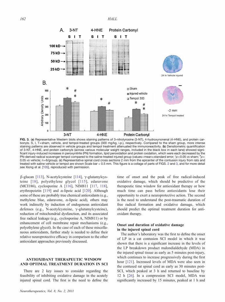
with this view, several compounds that scavenge either PN,including uric acid [98] and the PN decomposition catalyst5, 10, 15, 20-tetrakis (4-sulfanatophenyl) porphyrin iron IIIcloride (FeTSPP) [99], or that scavenger PN-derived freeradicals such as tempol, a catalytic scavenger of PN-derived•NO2 and •CO3 [62, 100–102] have been reported to haveneuroprotective and/or neurological recovery-promotingeffects in SCI models. A major mechanism of the protectiveeffect of tempol appears to be preservation of spinal cordmitochondrial function from post-traumatic PN-mediatedimpairment [101]. FIG. 3 displays the effects of early (15minutes) post-SCI tempol administration on mitochondrialoxidative damage in rat thoracic spinal cord tissue subjectedto contusion SCI 24 h earlier [105].Two broad spectrum antioxidant compounds, melatonin
[103, 104] and the metalloporphyrin compound Mn (III)tetrakis (4-benzoic acid) porphyrin (MnTBAP) [105, 106]have been reported to be beneficial in SCI models in parallelwith a reduction in PN-derived 3-NT levels in the injuredspinal cord, suggesting that their overall protective effectsare largely due to interference with PN oxidative damage.Although the decrease in PN-induced nitrative damage (3-NT) is no doubt therapeutically important, it must be notedthat across all SCI studies carried out with variousantioxidant compounds, the most consistent finding is areduction in LP, as assessed by the levels of LP-derivedproducts such as 4-HNE and/or acrolein. Moreover, recentwork has shown that lipid peroxyl radicals (LOO•) play akey role in the chemical nitration of protein tyrosine residuesby PN-derived •NO2 [107].
Scavenging of neurotoxic lipid peroxidation-derivedaldehydes in SCI modelsAs previously discussed, 4-HNE and acrolein, which are
derived from peroxidized polyunsaturated fatty acidsprovide useful markers of post-traumatic LP in the injuredspinal cord. However, these 2 products have been repeat-edly shown to be cytotoxic. For example, Hamann et al.[25, 108, 109] have demonstrated that 4-HNE and acroleinare neurotoxic in SCImodels, and that chemical scavengingof these toxic LP-derived aldehydes with certain hydrazine-containing compounds that can covalently bind them isneuroprotective. The author’s laboratory has also demon-strated that mitochondrial function is exquisitely sensitiveto impairment by 4-HNE, and moreso to acrolein, and alsothat spinal cord mitochondria are 10-fold more sensitivethan brain mitochondria [48].
Efficacy of miscellaneous antioxidants in SCI modelsFurther pharmacological validation of the neuropro-
tective benefits of inhibition of oxidative damage in theinjured spinal cord is inicated by several compoundsheretofore unmentioned, which have been reported toattenuate oxidative damage in SCI models. These includemethylene blue [110], mexilitine [111], thiopental [112],
160 HALL
Neurotherapeutics, Vol. 8, No. 2, 2011

Tab
le1.
Summaryof
NASC
ISClin
ical
Trialsof
High-DoseMethylpredn
isolon
ean
dTirilazad.
Trial
TreatmentGroups
TreatmentWindo
wPatients/Centers
End
points
Results
Refs
NASCIS
IMP:10
0mgor
1000
mgi.v.
bolusfor15
minutes/day
for10
days
24h
330/10
Motor
andsensory
(pin
prick,
light
touch)
functio
nat
6mon
ths
MP10
0mgvs
MP10
00mg:
nosign
ificant
difference
inmotor
orsensoryfunctio
n
84,85
Adverse
reactio
nsIncreasedwound
infectionand
trendtowardincreasedmortality
in10
00-m
gdo
segroup
NASCIS
IIPlacebo
14h:
aprioriplan
was
tostratify
thedata
atthenearestwho
lenu
mberto
themedian
enrollm
enttim
e(8
hafterinjury)
487/10
Motor
andsensory
(pin
prick,
light
touch)
functio
nat
6weeks,
6mon
ths,and1year
MPvs
Placebo
:83
,86
,87
,88
MP30
mg/kg
i.v.bo
lus
over
15min,45
min.
pause+
5.4mg/kg/hr
for23
hr
Significant
improv
ementin
motor
functio
nat
6weeks,6mon
ths,and
1year,andsensoryfunctio
nat
6weeks
and6mon
ths,andin
both
completeandincompletepatients
treatedwith
in8h,
butnotin
patients
treatedafter8h;
noincrease
inadverseeffects
Nalox
one5.4mg/kg
i.v.
bolusov
er15
min,45
min
pause+
5.4mg/kg/hr
for23
hr
Adverse
reactio
nsNaloxonevs
Placebo
:Non
significant
trendtowardim
prov
edmotor
andsensoryfunctio
nin
incomplete
patientstreatedwith
in8h;
noincrease
inadverseeffects
NASCIS
III
MP30
mg/kg
i.v.bo
lus
for15
minutes,45
-min
pause+
5.4mg/kg/h
for23
h
8h:
aprioriplan
was
tostratify
data
accordingto
treatm
entwith
infirst3hor
between3and8h
499/16
Motor
andsensory
(pin
prick,
light
touch)
functio
nat
6weeks,
6mon
ths,and1year
Patientstreatedwith
in3h:
96,97
24-h
MP,
48-h
MP,
and48
-hTgrou
psshow
edequivalent
degreesof
neurological
functio
nalrecovery
MP30
mg/kg
i.v.bo
lus
for15
minutes,with
a45
-minutepause+
5.4mg/kg/h
for47
h
FIM
Patientstreatedbetween3and8h:
48-h
MPproduced
sign
ificantly
greater
improv
ementin
sensoryandmotor
functio
nandnearly
sign
ificant
improv
ement
inFIM
comparedwith
24-h
MP;48
-hT
show
edfunctio
nalrecovery
between24
and48
-hMP
MP30
mg/kg
i.v.bo
lusfor
15minutes,with
a45-m
inute
pause+
T1.5mg/kg/q
6h
for47
h
Adverse
reactio
ns48-h
MPresultedin
significantly
high
erincidenceof
severe
sepsisandpn
eumonia
vs24
-hMP;48
-hTproduced
lowest
incidenceof
adversereactio
ns
FIM
=functio
nalindepend
ence
measure;i.v.=intravenou
s;M=methy
lpredn
isolon
e;NASCIS=NationalAcute
SpinalCordInjury
Study
;T=tirilazad.
161ANTIOXIDANT NEUROPROTECTION IN SCI
Neurotherapeutics, Vol. 8, No. 2, 2011
β-glucan [113], N-acetylcysteine [114], γ-glutamylcys-teine [18], polyethylene glycol [115], edaravone(MCI186), cyclosporine A [116], NIM811 [117, 118],erythropoietin [119] and α-lipoic acid [120]. Althoughsome of these are probably true chemical antioxidants (e.g.,methylene blue, edaravone, α-lipoic acid), others maywork indirectly by induction of endogenous antioxidantdefenses (e.g., N-acetylcysteine, γ-glutamylcysteine),reduction of mitochondrial dysfunction, and its associatedfree radical leakage (e.g., cyclosporine A, NIM811) or byenhancement of cell membrane repair mechanisms (e.g.,polyethylene glycol). In the case of each of these miscella-neous antioxidants, further study is needed to define theirrelative neuroprotective efficacy in comparison to the otherantioxidant approaches previously discussed.
ANTIOXIDANT THERAPEUTIC WINDOWAND OPTIMAL TREATMENT DURATION IN SCI
There are 2 key issues to consider regarding thefeasibility of inhibiting oxidative damage in the acutelyinjured spinal cord. The first is the need to define the
time of onset and the peak of free radical-inducedoxidative damage, which should be predictive of thetherapeutic time window for antioxidant therapy or howmuch time can pass before antioxidants lose theiropportunity to exert a neuroprotective action. The secondis the need to understand the post-traumatic duration offree radical formation and oxidative damage, whichshould predict the optimal treatment duration for anti-oxidant therapy.
Onset and duration of oxidative damagein the injured spinal cordThe author’s laboratory was the first to define the onset
of LP in a cat contusion SCI model in which it wasshown that there is a significant increase in the levels ofthe LP breakdown product malondialdehyde (MDA) inthe injured spinal tissue as early as 5 minutes post-injury,which continues to increase progressively during the firsthour [121]. Increased levels of MDA were also seen inthe contused rat spinal cord as early as 30 minutes post-SCI, which peaked at 3 h and returned to baseline by12 h [26]. In a compression SCI model, MDA wassignificantly increased by 15 minutes, peaked at 1 h and
FIG. 3. (a) Representative Western blots shows staining patterns of 3-nitrotyrosine (3-NT), 4-hydroxynonenal (4-HNE), and protein car-bonyls. S, I, T=sham, vehicle, and tempol-treated groups (300 mg/kg, i.p.), respectively. Compared to the sham group, more intensestaining patterns are observed in vehicle groups and tempol treatment attenuated the immunoreactivity. (b) Densitometric quantificationof 3-NT, 4-HNE, and protein carbonyls (across various molecular weight ranges, included in the black box in each lane) showed signi-ficant injury-induced increases in peroxynitrite (PN) formation, lipid peroxidation and protein oxidation, which were each decreased by thePN-derived radical scavenger tempol compared to the saline-treated injured group (values=mean±standard error; *p<0.05 vs sham; #p<0.05 vs vehicle; n=6/group). (c) Representative spinal cord cross sections 2 mm from the epicenter of the contusion injury from rats andtreated with saline vehicle or tempol are shown Scale bar = 0.5 mm. This figure is a collage of parts of FIGS. 2 and 3, and for more detailsee Xiong et al. [105], reproduced with permission.
162 HALL
Neurotherapeutics, Vol. 8, No. 2, 2011

then fell thereafter [122], suggesting that there is notmuch difference between the cat contusion and ratcompression models at least as far as the onset of post-SCI LP. However, two unfortunate limitations of thesestudies were the choice of MDA, which is a biomarker ofenzymatic LP during the arachidonic acid cascade, aswell as free radical-induced LP, and the fact thatpostinjury times longer than 12 h were not included.However, subsequent studies using one of the morecontemporary rat contusion SCI paradigms and moresensitive immunoblotting of the immunohistochemicalassay methods have more completely and specificallydefined the time course of LP (4-HNE, acrolein) andprotein oxidation (protein carbonyl), and nitration (3-NT) following SCI. The first of these showed a peakincrease in 4-HNE immunostaining at 24 h [21] or48 h [20] after SCI. A more recent and more extendedimmunoblotting/immunohistochemical time coursestudy in the rat contusion model has confirmed thatthe increase in 4-HNE occurs as early as 1 h, peaks at24 h, and remains significantly elevated for at least7 days [23]. However, 4-HNE immunohistochemicalstaining at 14 days after SCI reveals a persistentelevation [22]. The species or type of SCI model doesnot seem to make a difference, because in a guinea pigcompression injury model the 4-HNE levels in injuredspinal tissue also peaked at 24 h and persisted for atleast 7 days [24].Interestingly, the time course of PN-mediated 3-NT
after rat contusion SCI mirrors the timing of LP-related4-HNE levels in terms of early onset, peak at 24 h, andpersistence for at least 7 days, although by day 14 the 3-NT immunostaining has waned much more than that for4-HNE [22]. A re-examination of FIG. 2 reveals thespatial and temporal superimposition of 4-HNE and 3-NT in the contused spinal cord at least out 7 days, whichincludes both microvascular and parenchymal elements.This strongly implies that much of the oxidative damagein that timeframe is produced by PN. In contrast, thelonger persistence of the LP marker 4-HNE may be dueto a later contribution of other inflammatory or iron-catalyzed free radical mechanisms, as previously dis-cussed in this review. Alternatively, the persistence of 4-HNE modified proteins may simply be due to theirslower clearance via proteasomal degradation vs thepossibly faster repair of nitrated proteins by denitraseenzymes. Further study is needed to test these 2possibilities. In any event, the oxidative time coursestudies collectively indicate that antioxidant treatment 1)needs to begin as soon as possible within the first hoursafter SCI; 2) needs to be optimally maintained for at leastthe first 24 to 48 h to cover the peak of spinal cordoxidative damage, and 3) may need to be maintainedeven longer if in fact the persistence of LP and proteinnitrative damage out 7 to 14 days is demonstrated to
represent ongoing oxidative damage, as opposed tosimply the persistent presence of unrepaired oxidativedamage that mainly transpired during the first 24 to 48 h.Clearly, the present evidence suggests that treatment forat least the first 24 h is critically important because 24 his also the time at which the peak of spinal cordmitochondrial oxidative damage and functional impair-ment occurs [28].
Antioxidant therapeutic window and optimaltreatment duration derived from the NASCIS trialsAs already mentioned, the NASCIS II clinical trial
revealed that a 24-h high-dose MP regimen significantlyimproved motor functional recovery if initiated withinthe first 8 h, but not if delayed until after that post-SCItime window [83, 86, 87]. If high-dose MP is exerting itsneuroprotective effects by inhibition of LP, as theorized,then this indicates that 8 h is the therapeutic window inSCI. In subsequent studies in the cat spinal cordcompression SCI model, the 21-aminosteroid tirilazad,which works solely by inhibiting LP, was shown tosignificantly improve motor functional recovery if a48-h long treatment regimen was initiated within thefirst 4 h, and the degree of recovery was just as greatas when it was started within the first 30 minutes.Even after a delay of 8 h, an improvement in recoverywas still seen, although it did not quite reach statisticalsignificance [95]. The similarity between the 8-h MPwindow defined in NASCIS II, and the similar windowobserved with tirilazad in the cat SCI model furthersupports the idea that the window for neuroprotectionby an antioxidant mechanism in SCI is approximately8 hours.In the 3-arm NASCIS III clinical trial, which involved a
comparison of 24-h MP vs either 48-h MP or 48-h tirilazad,it was shown that if treatment was initiated within the first3-h post-SCI, the neurological recoveries of the 3 treatmentcohorts was statistically identical [96, 97]. Thus, 24-h MPadministration at a dose demonstrated repeatedly to inhibitpost-traumatic LP in the injured cord was able to produce asmuch of an effect as 48-h antioxidant dosing, whether withhigh-dose MP or the selective antioxidant tirilazad. How-ever, if treatment initiation was delayed until 3- to 8-h post-SCI, then extension of the MP treatment duration to 48 hresulted in a significantly higher degree of neurologicalrecovery than if it was confined to only 24 h. This latterobservation indicates that a more delayed initiation ofantioxidant administration until after the first 3 h after SCInecessitates a longer duration of antioxidant dosing to havean optimal effect. This is presumably due to the likelihoodthat the longer delay leads to a higher degree of free radicalformation and LP chain reactions that would logically takea more aggressive dosing strategy to shut off. Nevertheless,the confirmation of this apparent 8-h antioxidant therapeu-tic window, the optimal treatment onset within the first few
163ANTIOXIDANT NEUROPROTECTION IN SCI
Neurotherapeutics, Vol. 8, No. 2, 2011

hours, the adequacy of a 24-treatment duration if thisoccurs, and the need for more prolonged dosing if drugadministration does not begin until 3 to 8 h, awaits furtherdetailed preclinical study with newer, more potent andselective antioxidant drugs.
CONCLUSIONS
Free radical-induced oxidative damage is arguably one ofthe best-validated mechanisms involved in the complexsecondary injury cascade, which occurs following acute SCI.Although free radical-mediated damage can occur in alltypes of biomolecules, LP appears to play a dominant roledue to the high concentration of peroxidation-sensitivepolyunsaturated fatty acids and high levels of the LP catalystiron. Most convincingly, various LP inhibiting compoundsand other antioxidants have been shown to be neuro-protective in acute SCI models. Two of these, high-doseMP and tirilazad have been shown in multi-center phase IIISCI clinical trials (i.e., NASCIS II and III) to improveneurological recovery, albeit to a moderate degree. However,in the case of MP, the potential for glucocorticoid-relatedside effects has driven the continued search for safer andmore effective antioxidant agents for acute SCI. One suchmechanistic approach would be to scavenge PN or itsderived free radicals that are critically involved in the post-SCI oxidative damage. Second, there is increased interest inattempting to scavenge the neurotoxic aldehydic LP products4-HNE and acrolein, which are key contributors to theoverall effects of LP in the injured spinal cord. The availablepreclinical and clinical data indicate that antioxidant neuro-protection in acute SCI needs to be initiated within the first8 h, preferably within the first 3 h, and that it should becontinued for at least the first 24 to 48 h. However, carefuldefinition of the extended time course of post-traumatic LPand other forms of oxidative damage in the injured rat spinalcord far beyond the first 48 h may reflect a need for moreprolonged antioxidant treatment after acute SCI, which is notfeasible with high-dose MP and its associated side-effectprofile. Accordingly, more potent and highly selectiveantioxidant compounds, which can simultaneously protectspinal cord microvessels (i.e., glia and neurons) are needed.
Acknowledgments: Full conflict of interest disclosure isavailable in the electronic supplementary material for thisarticle. Some of the work reviewed in this aricle was supportedby grants from the Kentucky Spinal Cord & Head InjuryResearch Trust.
REFERENCES
1. Hall ED, Vaishnav RA, Mustafa AG. Antioxidant therapies fortraumatic brain injury. Neurotherapeutics 2010;7:51-61.
2. Halliwell B, Gutteridge J. Free Radicals in Biology and Medicine,3 rd ed. Oxford University Press, 2008.
3. Zaleska MM, Floyd RA. Regional lipid peroxidation in rat brain invitro: possible role of endogenous iron. Neurochem Res 1985;10:397-410.
4. Sadrzadeh SM, Graf E, Panter SS, Hallaway PE, Eaton JW.Hemoglobin: a biologic fenton reagent. J Biol Chem1984;259:14354-14356.
5. Sadrzadeh SM, Eaton JW. Hemoglobin-mediated oxidant damageto the central nervous system requires endogenous ascorbate. J ClinInvest 1988;82:1510-1515.
6. Beckman JS. The double-edged role of nitric oxide in brainfunction and superoxide-mediated injury. J Dev Physiol1991;15:53-59.
7. Anderson DK, Means ED, Waters TR, Spears CJ. Spinal cordenergy metabolism following compression trauma to the felinespinal cord. J Neurosurg 1980;53:375-380.
8. Braughler JM, Hall ED. Lactate and pyruvate metabolism ininjured cat spinal cord before and after a single large intravenousdose of methylprednisolone. J Neurosurg 1983;59:256-261.
9. Liu D, Sybert TE, Qian H, Liu J. Superoxide production afterspinal injury detected by microperfusion of cytochrome c. FreeRadic Biol Med 1998;25:298-304.
10. Bao F, Liu D. Hydroxyl radicals generated in the rat spinal cord atthe level produced by impact injury induce cell death by necrosisand apoptosis: protection by a metalloporphyrin. Neuroscience2004;126:285-295.
11. Anderson DK, Means ED. Iron-induced lipid peroxidation in spinalcord: protection with mannitol and methylprednisolone. J FreeRadic Biol Med 1985;1:59-64.
12. Milvy P, Kakari S, Campbell JB, Demopoulos HB. Paramagneticspecies and radical products in cat spinal cord. Ann N Y Acad Sci1973;222:1102-1111.
13. Seligman ML, Flamm ES, Goldstein BD, Poser RG, DemopoulosHB, Ransohoff J. Spectrofluorescent detection of malonaldehyde asa measure of lipid free radical damage in response to ethanolpotentiation of spinal cord trauma. Lipids 1977;12:945-950.
14. Hall ED, Braughler JM. Effects of intravenous methylprednisoloneon spinal cord lipid peroxidation and Na+ + K+)-ATPase activity.Dose-response analysis during 1st hour after contusion injury in thecat. J Neurosurg 1982;57:247-253.
15. Qian H, Liu D. The time course of malondialdehyde productionfollowing impact injury to rat spinal cord as measured by micro-dialysis and high pressure liquid chromatography. Neurochem Res1997;22:1231-1236.
16. Pietronigro DD, Hovsepian M, Demopoulos HB, Flamm ES. Lossof ascorbic acid from injured feline spinal cord. J Neurochem1983;41:1072-1076.
17. Hall ED, Yonkers PA, Andrus PK, Cox JW, Anderson DK.Biochemistry and pharmacology of lipid antioxidants in acute brainand spinal cord injury. J Neurotrauma 1992;(9 suppl 2):S425-S442.
18. Lucas JH, Wheeler DG, Guan Z, Suntres Z, Stokes BT. Effect ofglutathione augmentation on lipid peroxidation after spinal cordinjury. J Neurotrauma 2002;19:763-775.
19. Lemke M, Frei B, Ames BN, Faden AI. Decreases in tissue levelsof ubiquinol-9 and -10, ascorbate and alpha-tocopherol followingspinal cord impact trauma in rats. Neurosci Lett 1990;108:201-206.
20. Baldwin SA, Broderick R, Osbourne D, Waeg G, Blades DA,Scheff SW. The presence of 4-hydroxynonenal/protein complex asan indicator of oxidative stress after experimental spinal cordcontusion in a rat model. J Neurosurg 1998;88:874-883.
21. Springer JE, Azbill RD, Mark RJ, Begley JG, Waeg G, MattsonMP. 4-hydroxynonenal, a lipid peroxidation product, rapidlyaccumulates following traumatic spinal cord injury and inhibitsglutamate uptake. J Neurochem 1997;68:2469-2476.
22. Carrico KM, Vaishnav R, Hall ED. Temporal and spatial dynamicsof peroxynitrite-induced oxidative damage after spinal cordcontusion injury. J Neurotrauma 2009;26:1369-1378.
23. Xiong Y, Rabchevsky AG, Hall ED. Role of peroxynitrite insecondary oxidative damage after spinal cord injury. J Neurochem2007;100:639-649.
24. Luo J, Uchida K, Shi R. Accumulation of acrolein-protein adductsafter traumatic spinal cord injury. Neurochem Res 2005;30:291-295.
164 HALL
Neurotherapeutics, Vol. 8, No. 2, 2011

25. Hamann K, Durkes A, Ouyang H, Uchida K, Pond A, Shi R.Critical role of acrolein in secondary injury following ex vivo spinalcord trauma. J Neurochem 2008;107:712-721.
26. Liu JB, Tang TS, Xiao DS. Changes of free iron contents and itscorrelation with lipid peroxidation after experimental spinal cordinjury. Chin J Traumatol 2004;7:229-232.
27. Aksenova M, Butterfield DA, Zhang SX, Underwood M, GeddesJW. Increased protein oxidation and decreased creatine kinase BBexpression and activity after spinal cord contusion injury. JNeurotrauma 2002;19:491-502.
28. Sullivan PG, Krishnamurthy S, Patel SP, Pandya JD, RabchevskyAG. Temporal characterization of mitochondrial bioenergetics afterspinal cord injury. J Neurotrauma 2007;24:991-999.
29. Scott GS, Jakeman LB, Stokes BT, Szabo C. Peroxynitriteproduction and activation of poly (adenosine diphosphate-ribose)synthetase in spinal cord injury. Ann Neurol 1999;45:120-124.
30. Xu J, Gyeong-Moon K, Chen S, et al. iNOS and nitrotyrosineexpression after spinal cord injury. J Neurotrauma 2001;18:523-532.
31. Liu D, Ling X, Wen J, Liu J. The role of reactive nitrogen species insecondary spinal cord injury: formation of nitric oxide, peroxynitrite,and nitrated protein. J Neurochem 2000;75:2144-2154.
32. Bao F, DeWitt DS, Prough DS, Liu D. Peroxynitrite generated inthe rat spinal cord induces oxidation and nitration of proteins:reduction by Mn (III) tetrakis (4-benzoic acid) porphyrin. JNeurosci Res 2003;71:220-227.
33. Bao F, Liu D. Peroxynitrite generated in the rat spinal cord inducesneuron death and neurological deficits. Neuroscience2002;115:839-849.
34. Bao F, Liu D. Peroxynitrite generated in the rat spinal cord inducesapoptotic cell death and activates caspase-3. Neuroscience2003;116:59-70.
35. Liu D, Bao F, Prough DS, Dewitt DS. Peroxynitrite generated at thelevel produced by spinal cord injury induces peroxidation ofmembrane phospholipids in normal rat cord: reduction by ametalloporphyrin. J Neurotrauma 2005;22:1123-1133.
36. Rohn TT, Hinds TR, Vincenzi FF. Ion transport ATPases as targetsfor free radical damage. Protection by an aminosteroid of the Ca2+pump ATPase and Na+/K+pump ATPase of human red blood cellmembranes. Biochem Pharmacol 1993;46:525-534.
37. Rohn TT, Hinds TR, Vincenzi FF. Inhibition of Ca2+-pumpATPase and the Na+/K+-pump ATPase by iron-generated freeradicals. Protection by 6,7-dimethyl-2,4-DI-1- pyrrolidinyl-7 H-pyrrolo[2,3-d] pyrimidine sulfate (U-89843D), a potent, novel,antioxidant/free radical scavenger. Biochem Pharmacol1996;51:471-476.
38. Braughler JM, Duncan LA, Chase RL. Interaction of lipidperoxidation and calcium in the pathogenesis of neuronal injury.Cent Nerv Syst Trauma 1985;2:269-283.
39. Clendenon NR, Allen N, Gordon WA, Bingham WG, Jr. Inhibitionof Na+-K+-activated ATPase activity following experimentalspinal cord trauma. J Neurosurg 1978;49:563-568.
40. Azbill RD, Mu X, Bruce-Keller AJ, Mattson MP, Springer JE.Impaired mitochondrial function, oxidative stress and alteredantioxidant enzyme activities following traumatic spinal cordinjury. Brain Res 1997;765:283-290.
41. Sullivan PG, Thompson MB, Scheff SW. Cyclosporin A attenuatesacute mitochondrial dysfunction following traumatic brain injury.Exp Neurol 1999;160:226-234.
42. Sullivan PG, Bruce-Keller AJ, Rabchevsky AG, et al. Exacerbationof damage and altered NF-kappaB activation in mice lacking tumornecrosis factor receptors after traumatic brain injury. J Neurosci1999;19:6248-6256.
43. Matsushita M, Xiong G. Projections from the cervical enlargementto the cerebellar nuclei in the rat, studied by anterograde axonaltracing. J Comp Neurol 1997;377:251-261.
44. Lopez-Figueroa MO, Caamano C, Morano MI, Ronn LC, Akil H,Watson SJ. Direct evidence of nitric oxide presence withinmitochondria. Biochem Biophys Res Commun 2000;272:129-133.
45. Zanella B, Calonghi N, Pagnotta E, Masotti L, Guarnieri C.Mitochondrial nitric oxide localization in H9c2 cells revealed byconfocal microscopy. Biochem Biophys Res Commun2002;290:1010-1014.
46. Bringold U, Ghafourifar P, Richter C. Peroxynitrite formed bymitochondrial NO synthase promotes mitochondrial Ca2+ release.Free Radic Biol Med 2000;29:343-348.
47. Valdez LB, Alvarez S, Arnaiz SL, et al. Reactions of peroxynitritein the mitochondrial matrix. Free Radic Biol Med 2000;29:349-356.
48. Vaishnav RA, Singh IN, Miller DM, Hall ED. Lipid peroxidation-derived reactive aldehydes directly and differentially impair spinalcord and brain mitochondrial function. J Neurotrauma2010;27:1311-1320.
49. Park E, Velumian AA, Fehlings MG. The role of excitotoxicity insecondary mechanisms of spinal cord injury: a review with anemphasis on the implications for white matter degeneration. JNeurotrauma 2004;21:754-774.
50. Xu GY, Hughes MG, Ye Z, Hulsebosch CE, McAdoo DJ.Concentrations of glutamate released following spinal cord injurykill oligodendrocytes in the spinal cord. Exp Neurol 2004;187:329-336.
51. Stys PK. White matter injury mechanisms. Curr Mol Med2004;4:113-130.
52. Alessandri B, Bullock R. Glutamate and its receptors in thepathophysiology of brain and spinal cord injuries. Prog Brain Res1998;116:303-330.
53. Pellegrini-Giampietro DE, Cherici G, Alesiani M, Carla V, MoroniF. Excitatory amino acid release and free radical formation maycooperate in the genesis of ischemia-induced neuronal damage. JNeurosci 1990;10:1035-1041.
54. Braughler JM. Lipid peroxidation-induced inhibition of gamma-aminobutyric acid uptake in rat brain synaptosomes: protection byglucocorticoids. J Neurochem 1985;44:1282-1288.
55. Zhang JR, Scherch HM, Hall ED. Direct measurement of lipidhydroperoxides in iron-dependent spinal neuronal injury. J Neuro-chem 1996;66:355-361.
56. Monyer H, Hartley DM, Choi DW. 21-Aminosteroids attenuateexcitotoxic neuronal injury in cortical cell cultures. Neuron1990;5:121-126.
57. Demopoulos HB, Flamm ES, Pietronigro DD, Seligman ML. Thefree radical pathology and the microcirculation in the major centralnervous system disorders. Acta Physiol Scand Suppl 1980;492:91-119.
58. Hall ED, Wolf DL. A pharmacological analysis of the pathophy-siological mechanisms of posttraumatic spinal cord ischemia. JNeurosurg 1986;64:951-961.
59. Hall ED, Wolf DL, Braughler JM. Effects of a single large dose ofmethylprednisolone sodium succinate on experimental posttrau-matic spinal cord ischemia. Dose-response and time-action analy-sis. J Neurosurg 1984;61:124-130.
60. Hall ED. Effects of the 21-aminosteroid U74006F on posttraumaticspinal cord ischemia in cats. J Neurosurg 1988;68:462-465.
61. Hummel SG, Fischer AJ, Martin SM, Schafer FQ, Buettner GR.Nitric oxide as a cellular antioxidant: a little goes a long way. FreeRadic Biol Med 2006;40:501-506.
62. Carroll RT, Galatsis P, Borosky S, et al. 4-Hydroxy-2,2,6,6-tetramethylpiperidine-1-oxyl (Tempol) inhibits peroxynitrite-medi-ated phenol nitration. Chem Res Toxicol 2000;13:294-300.
63. Liu D, McAdoo DJ. Methylprednisolone reduces excitatory aminoacid release following experimental spinal cord injury. Brain Res1993;609:293-297.
64. Anderson DK, Saunders RD, Demediuk P, et al. Lipid hydrolysisand peroxidation in injured spinal cord: partial protection withmethylprednisolone or vitamin E and selenium. Cent Nerv SystTrauma 1985;2:257-267.
65. Hall ED, Yonkers PA, Horan KL, Braughler JM. Correlationbetween attenuation of posttraumatic spinal cord ischemia andpreservation of tissue vitamin E by the 21-aminosteroid U74006F:evidence for an in vivo antioxidant mechanism. J Neurotrauma1989;6:169-176.
66. Anderson DK, Waters TR, Means ED. Pretreatment with alphatocopherol enhances neurologic recovery after experimental spinalcord compression injury. J Neurotrauma 1988;5:61-67.
67. Iwasa K, Ikata T, Fukuzawa K. Protective effect of vitamin E onspinal cord injury by compression and concurrent lipid peroxida-tion. Free Radic Biol Med 1989;6:599-606.
165ANTIOXIDANT NEUROPROTECTION IN SCI
Neurotherapeutics, Vol. 8, No. 2, 2011

68. Taoka Y, Ikata T, Fukuzawa K. Influence of dietary vitamin Edeficiency on compression injury of rat spinal cord. J Nutr SciVitaminol (Tokyo) 1990;36:217-226.
69. Machlin L, Gabriel E. Kinetics of tissue alpha tocopherol uptakeand depletion following administration of high levels of vitamin E.Ann N Y Acad Sci 1982;393:48-59.
70. Roberts LJ, 2nd, Oates JA, Linton MF, et al. The relationshipbetween dose of vitamin E and suppression of oxidative stress inhumans. Free Radic Biol Med 2007;43:1388-1393.
71. Demopoulos HB, Flamm ES, Seligman ML, Pietronigro DD,Tomasula J, DeCrescito V. Further studies on free-radical pathologyin the major central nervous system disorders: effect of very high dosesof methylprednisolone on the functional outcome, morphology, andchemistry of experimental spinal cord impact injury. Can J PhysiolPharmacol 1982;60:1415-1424.
72. Hall ED. The neuroprotective pharmacology of methylpredniso-lone. J Neurosurg 1992;76:13-22.
73. Hall ED, Braughler JM. Acute effects of intravenous glucocorticoidpretreatment on the in vitro peroxidation of cat spinal cord tissue.Exp Neurol 1981;73:321-324.
74. Hall ED, Braughler JM. Glucocorticoid mechanisms in acute spinalcord injury: a review and therapeutic rationale. Surg Neurol1982;18:320-327.
75. Young W, Flamm ES. Effect of high-dose corticosteroid therapy onblood flow, evoked potentials, and extracellular calcium inexperimental spinal injury. J Neurosurg 1982;57:667-673.
76. Anderson DK, Means ED, Waters TR, Green ES. Microvascularperfusion and metabolism in injured spinal cord after methylpred-nisolone treatment. J Neurosurg 1982;56:106-113.
77. Braughler JM, Hall ED. Effects of multi-dose methylprednisolonesodium succinate administration on injured cat spinal cord neurofilamentdegradation and energy metabolism. J Neurosurg 1984;61:290-295.
78. Braughler JM, Hall ED. Correlation of methylprednisolone levelsin cat spinal cord with its effects on (Na+ + K+)-ATPase, lipidperoxidation, and alpha motor neuron function. J Neurosurg1982;56:838-844.
79. Braughler JM, Hall ED. Uptake and elimination of methylpredniso-lone from contused cat spinal cord following intravenous injection ofthe sodium succinate ester. J Neurosurg 1983;58:538-542.
80. Braughler JM, Hall ED, Means ED, Waters TR, Anderson DK.Evaluation of an intensive methylprednisolone sodium succinatedosing regimen in experimental spinal cord injury. J Neurosurg1987;67:102-105.
81. Lee J-M, Yang, P, Xiao, Q, Chen, S, Lee, K-Y, Hsu, CY, Xu, J.Methyprednisolone protects oligodendrocytes but not neurons afterspinal cord injury. J Neurosci 2008;28:3141-3149.
82. Hall ED, Springer JE. Neuroprotection and acute spinal cord injury:a reappraisal. NeuroRx 2004;1:80-100.
83. Bracken MB, Shepard MJ, Collins WF, et al. A randomized,controlled trial of methylprednisolone or naloxone in the treatmentof acute spinal-cord injury. Results of the Second National AcuteSpinal Cord Injury Study. N Engl J Med 1990;322:1405-1411.
84. Bracken MB, Shepard MJ, Hellenbrand KG, et al. Methylpredni-solone and neurological function 1 year after spinal cord injury.Results of the National Acute Spinal Cord Injury Study. JNeurosurg 1985;63:704-713.
85. Bracken MB, Collins WF, Freeman DF, et al. Efficacy of methyl-prednisolone in acute spinal cord injury. JAMA 1984;251:45-52.
86. Bracken MB, Shepard MJ, Collins WF, Jr., et al. Methylpredniso-lone or naloxone treatment after acute spinal cord injury: 1-yearfollow-up data. Results of the second National Acute Spinal CordInjury Study. J Neurosurg 1992;76:23-31.
87. Bracken MB, Holford TR. Effects of timing of methylprednisoloneor naloxone administration on recovery of segmental and long-tractneurological function in NASCIS 2. J Neurosurg 1993;79:500-507.
88. Bracken MB. Pharmacological treatment of acute spinal cord injury:current status and future projects. J EmergMed 1993;(11 suppl 1):43-48.
89. Otani K AH, Kadoya S. Beneficial effect of methylprednisolonesodium succinate in the treatment of acute spinal cord injury.Sekitsui Sekizui (in Japanese) 1994;7:633-647.
90. Petitjean ME, Pointillart, V, Dixmerias F. Medical treatment ofspinal cord injury in the acute stage. Ann Fr Anesth Reanim (inFrench) 1998;17:114-122.
91. Hall ED. Lazaroid: mechanisms of action and implications fordisorders of the CNS. The Neuroscientist 1997;3:42-51.
92. Hall ED, McCall JM, Means ED. Therapeutic potential of thelazaroids (21-aminosteroids) in acute central nervous systemtrauma, ischemia and subarachnoid hemorrhage. Adv Pharmacol1994;28:221-268.
93. Braughler JM, Chase RL, Neff GL, et al. A new 21-aminosteroidantioxidant lacking glucocorticoid activity stimulates adrenocorti-cotropin secretion and blocks arachidonic acid release from mousepituitary tumor (AtT-20) cells. J Pharmacol Exp Ther1988;244:423-427.
94. Anderson DK, Braughler JM, Hall ED, Waters TR, McCall JM,Means ED. Effects of treatment with U-74006 F on neurologicaloutcome following experimental spinal cord injury. J Neurosurg1988;69:562-567.
95. Anderson DK, Hall ED, Braughler JM, McCall JM, Means ED.Effect of delayed administration of U74006F (tirilazad mesylate)on recovery of locomotor function after experimental spinal cordinjury. J Neurotrauma 1991;8:187-192.
96. Bracken MB, Shepard MJ, Holford TR, et al. Administration ofmethylprednisolone for 24 or 48 hours or tirilazad mesylate for 48hours in the treatment of acute spinal cord injury. Results of theThird National Acute Spinal Cord Injury Randomized ControlledTrial. National Acute Spinal Cord Injury Study. JAMA1997;277:1597-1604.
97. Bracken MB, Shepard MJ, Holford TR, et al. Methylpredniso-lone or tirilazad mesylate administration after acute spinal cordinjury: 1-year follow up. Results of the third National AcuteSpinal Cord Injury randomized controlled trial. J Neurosurg1998;89:699-706.
98. Scott GS, Cuzzocrea S, Genovese T, Koprowski H, Hooper DC.Uric acid protects against secondary damage after spinal cordinjury. Proc Natl Acad Sci U S A 2005;102:3483-3488.
99. Genovese T, Mazzon E, Esposito E, et al. Beneficial effects ofFeTSPP, a peroxynitrite decomposition catalyst, in a mousemodel of spinal cord injury. Free Radic Biol Med 2007;43:763-780.
100. Hillard VH, Peng H, Zhang Y, et al. Tempol, a nitroxideantioxidant, improves locomotor and histological outcomes afterspinal cord contusion in rats. J Neurotrauma 2004;21:1405-1414.
101. Xiong Y, Hall ED. Pharmacological evidence for a role ofperoxynitrite in the pathophysiology of spinal cord injury. ExpNeurol 2009;216:105-114.
102. Xiong Y, Singh IN, Hall ED. Tempol protection of spinal cordmitochondria from peroxynitrite-induced oxidative damage. FreeRadic Res 2009;43:604-612.
103. Liu JB, Tang TS, Yang HL, Xiao DS. Antioxidation of melatoninagainst spinal cord injury in rats. Chin Med J (Engl)2004;117:571-575.
104. Genovese T, Mazzon E, Muia C, Bramanti P, De Sarro A,Cuzzocrea S. Attenuation in the evolution of experimental spinalcord trauma by treatment with melatonin. J Pineal Res2005;38:198-208.
105. Hachmeister JE, Valluru L, Bao F, Liu D. Mn (III) tetrakis (4-benzoic acid) porphyrin administered into the intrathecal spacereduces oxidative damage and neuron death after spinal cordinjury: a comparison with methylprednisolone. J Neurotrauma2006;23:1766-1778.
106. Ling X, Liu D. Temporal and spatial profiles of cell loss afterspinal cord injury: Reduction by a metalloporphyrin. J NeurosciRes 2007;85:2175-2185.
107. Mustafa AG, Singh IN, Wang J, Carrico KM, Hall ED.Mitochondrial protection after traumatic brain injury byscavenging lipid peroxyl radicals. J Neurochem 2010;114:271-280.
108. Hamann K, Nehrt G, Ouyang H, Duerstock B, Shi R. Hydralazineinhibits compression and acrolein-mediated injuries in ex vivospinal cord. J Neurochem 2008;104:708-718.
109. Hamann K, Shi R. Acrolein scavenging: a potential novelmechanism of attenuating oxidative stress following spinal cordinjury. J Neurochem 2009;111:1348-1356.
110. Bardakci H, Kaplan S, Karadeniz U, et al. Methylene bluedecreases ischemia-reperfusion (I/R)-induced spinal cord injury:
166 HALL
Neurotherapeutics, Vol. 8, No. 2, 2011
an in vivo study in an I/R rabbit model. Eur Surg Res 2006;38:482-488.
111. Kaptanoglu E, Caner HH, Surucu HS, Akbiyik F. Effect ofmexiletine on lipid peroxidation and early ultrastructural findingsin experimental spinal cord injury. J Neurosurg 1999;91:200-204.
112. Kaptanoglu E, Sen S, Beskonakli E, et al. Antioxidant actions andearly ultrastructural findings of thiopental and propofol in exper-imental spinal cord injury. J NeurosurgAnesthesiol 2002;14:114-122.
113. Kayali H, Ozdag MF, Kahraman S, et al. The antioxidant effect ofbeta-Glucan on oxidative stress status in experimental spinal cordinjury in rats. Neurosurg Rev 2005;28:298-302.
114. KaynarMY, Erdincler P, Tadayyon E, Belce A, Gumustas K, Ciplak N.Effect of nimodipine and N-acetylcysteine on lipid peroxidation afterexperimental spinal cord injury. Neurosurg Rev 1998;21:260-264.
115. Luo J, Borgens R, Shi R. Polyethylene glycol immediately repairsneuronal membranes and inhibits free radical production afteracute spinal cord injury. J Neurochem 2002;83:471-480.
116. Diaz-Ruiz A, Rios C, Duarte I, et al. Cyclosporin-A inhibits lipidperoxidation after spinal cord injury in rats. Neurosci Lett1999;266:61-64.
117. McEwen ML, Sullivan PG, Springer JE. Pretreatment with thecyclosporin derivative, NIM811, improves the function of synapticmitochondria following spinal cord contusion in rats. J Neuro-trauma 2007;24:613-624.
118. Ravikumar R, McEwen ML, Springer JE. Post-treatment withthe cyclosporin derivative, NIM811, reduced indices of celldeath and increased the volume of spared tissue in the acuteperiod following spinal cord contusion. J Neurotrauma2007;24:1618-1630.
119. Yazihan N, Uzuner K, Salman B, Vural M, Koken T, Arslantas A.Erythropoietin improves oxidative stress following spinal cordtrauma in rats. Injury 2008;39:1408-1413.
120. Toklu HZ, Hakan T, Celik H, et al. Neuroprotective effects ofalpha-lipoic acid in experimental spinal cord injury in rats. JSpinal Cord Med;33:401-409.
121. Hall ED, Braughler JM. Free radicals in CNS injury. Res PublAssoc Res Nerv Ment Dis 1993;71:81-105.
122. Barut S, Canbolat A, Bilge T, Aydin Y, Cokneseli B, Kaya U.Lipid peroxidation in experimental spinal cord injury: time-levelrelationship. Neurosurg Rev 1993;16:53-59.
167ANTIOXIDANT NEUROPROTECTION IN SCI
Neurotherapeutics, Vol. 8, No. 2, 2011