antitumor activity of a pyrrole-imidazole polyamide
TRANSCRIPT

Antitumor activity of a pyrrole-imidazole polyamideFei Yanga, Nicholas G. Nickolsa,b, Benjamin C. Lia, Georgi K. Marinovc, Jonathan W. Saidd, and Peter B. Dervana,1
aDivision of Chemistry and Chemical Engineering, and cDivision of Biology, California Institute of Technology, Pasadena, CA 91125; and bDepartment ofRadiation Oncology and dDepartment of Pathology and Laboratory Medicine, David Geffen School of Medicine of the University of California, Los Angeles,CA 90095
Contributed by Peter B. Dervan, December 17, 2012 (sent for review October 19, 2012)
Many cancer therapeutics target DNA and exert cytotoxicitythrough the induction of DNA damage and inhibition of transcrip-tion. We report that a DNA minor groove binding hairpin pyrrole-imidazole (Py-Im) polyamide interferes with RNA polymerase II(RNAP2) activity in cell culture. Polyamide treatment activates p53signaling in LNCaP prostate cancer cells without detectable DNAdamage. Genome-wide mapping of RNAP2 binding shows re-duction of occupancy, preferentially at transcription start sites,but occupancy at enhancer sites is unchanged. Polyamide treat-ment results in a time- and dose-dependent depletion of theRNAP2 large subunit RPB1 that is preventable with proteasomeinhibition. This polyamide demonstrates antitumor activity ina prostate tumor xenograft model with limited host toxicity.
minor groove binder | small molecule transcription inhibitor | ChIP-Seq
Several chemotherapeutics, including the anthracyclines andcisplatin, exert part of their cytotoxicity through the in-
hibition of transcription (1). Transformed cells often requireconstant expression of antiapoptotic genes for survival, makingtranscription inhibition a relevant therapeutic strategy in oncol-ogy (1, 2). Many radio- and chemotherapy treatments that targetDNA, including UV irradiation, cisplatin, and the topoisomeraseinhibitors, introduce obstacles to RNA polymerase II (RNAP2)elongation by generating bulky or helix-distorting lesions (3–5).In cell culture experiments, transcription blockade has beenshown to induce degradation of the RNAP2 large subunit(RPB1), and function as a signal for p53-mediated apoptosis (6,7). Although many DNA-targeted therapeutics effectively inhibittranscription and induce apoptosis, clinical treatment with gen-otoxic agents can also damage DNA in normal cells, increasingsymptomatic toxicity and potentially leading to secondary can-cers (8). The question arises whether high-affinity, noncovalentDNA-binding ligands offer an approach to transcription in-hibition without DNA damage.Hairpin pyrrole-imidazole (Py-Im) polyamides are synthetic
oligomers with programmable sequence recognition that bindthe minor groove of DNA with high affinity (9). Py-Im poly-amide-DNA binding induces allosteric changes in the DNA helixthat can interfere with protein–DNA interactions (10, 11). Py-Impolyamides have been used as molecular probes in cell culture tomodulate inducible gene-expression pathways (12–15). Inrodents, eight-ring hairpin Py-Im polyamides circulate in bloodfor several hours after administration and affect changes in geneexpression in tissues (16–18).We have previously reported that polyamide 1 (Fig. 1), which
targets the sequence 5′-WGWWCW-3′ found in the androgenresponse element, inhibited a subset of dihydrotestosterone(DHT)-induced genes in LNCaP cells (12). In this article weexplore the effects of this polyamide on the RNAP2 transcriptionmachinery. We find that RNAP2 is preferentially reduced fromtranscription start sites genome-wide without significant pertur-bation at enhancer loci. This reduction is accompanied by pro-teasome-dependent degradation of RPB1. Polyamide treatmentinduces p53 accumulation that is consistent with what is observedfor other transcription inhibitors that interact with DNA (4, 5), butwithout evidence of DNA damage. This polyamide demonstrates
efficacy in vivo against prostate cancer xenografts in mice withlimited host toxicity.
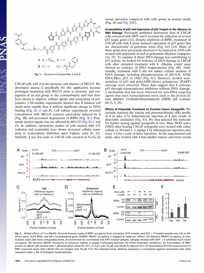
ResultsEffects of Polyamide 1 on Global Occupancy of RNAP2. Polyamide 1was previously shown to inhibit the induction of a subset ofDHT-driven genes in LNCaP cell culture (12). We interrogatedthe effects of 1 on the RNAP2 transcription machinery bymapping the global occupancy of RNAP2 using ChIP-seq. UnderDHT induction, select androgen receptor (AR)-driven genes,such as KLK3, showed increased RNAP2 occupancy over genicregions, but this was decreased in the presence of 1 (Fig. 2A).Although RNAP2 occupancy across constitutively expressedgenes, such as GAPDH, did not change with DHT induction,cotreatment with 1 reduced RNAP2 occupancy across thesegenes (Fig. 2B). This reduction in RNAP2 occupancy by 1 was inthe context of a global decrease of RNAP2 occupancy acrossgenic regions (Fig. S1), particularly at transcription start sites(Fig. 2C). However, 1 did not significantly change RNAP2 oc-cupancy at enhancer loci (Fig. 2D), suggesting 1 may affect theactive elongation of RNAP2 without disturbing the transcriptionapparatus anchored at enhancers, and that the observed differ-ences in RNAP2 occupancy are not a result of technical variationin ChIP success between experiments. Reduction in DNA oc-cupancy of RNAP2 has also been reported in cells treated withα-amanitin, a cyclic octapeptide inhibitor of RPB1 (19).Inhibition of RNAP2 elongation can be caused by a multitude
of genotoxic agents and often results in the degradation of theRPB1 subunit (3, 20, 21). Indeed, in addition to reduced RNAP2DNA occupancy, immunoblot analysis of LNCaP cells treatedwith 1 shows depletion of RPB1 in a time- and concentration-dependent manner (Fig. 2E). To examine if the effect on RPB1protein were a result of decreased transcription of this gene, wemeasured levels of RPB1 mRNA (Fig. 2F). The expression ofRPB1 modestly increased with polyamide treatment, suggestingthis depletion is posttranscriptional.
Polyamide Cytotoxicity Is Reduced by Proteasomal Inhibition andSerum Starvation. Inhibition of RNAP2 has been reported to in-duce apoptosis (4, 6, 22), and may contribute to polyamide cy-totoxicity observed in LNCaP cells cultured with 1 (Fig. 3A). Aprevious study with trabectidin, a DNA minor groove alkylatorthat causes RPB1 degradation, showed the toxicity induced bythe molecule can be reduced by cotreatment with the protea-some inhibitor MG132 (22). To evaluate if polyamide-inducedtoxicity was also reducible by proteasomal inhibition we treated
Author contributions: F.Y., N.G.N., G.K.M., and P.B.D. designed research; F.Y., N.G.N.,B.C.L., and J.W.S. performed research; F.Y., N.G.N., and B.C.L. contributed newreagents/analytic tools; F.Y., N.G.N., B.C.L., G.K.M., and P.B.D. analyzed data; and F.Y.,N.G.N., and P.B.D. wrote the paper.
The authors declare no conflict of interest.
Freely available online through the PNAS open access option.
Data deposition: The data reported in this paper have been deposited in the Gene Ex-pression Omnibus (GEO) database, www.ncbi.nlm.nih.gov/geo (accession no. GSE43253).1To whom correspondence should be addressed. E-mail: [email protected].
This article contains supporting information online at www.pnas.org/lookup/suppl/doi:10.1073/pnas.1222035110/-/DCSupplemental.
www.pnas.org/cgi/doi/10.1073/pnas.1222035110 PNAS | January 29, 2013 | vol. 110 | no. 5 | 1863–1868
MED
ICALSC
IENCE
S
LNCaP cells with 2 in the presence and absence of MG132. Wedeveloped analog 2 specifically for this application becauseprolonged incubation with MG132 alone is cytotoxic, and con-jugation of an aryl group to the γ-aminobutyric acid turn havebeen shown to improve cellular uptake and cytotoxicity of pol-yamides. Cell-viability experiments showed that 2 induced celldeath more rapidly than 1 without significant change to DNAbinding (Fig. S2 A and B). Cell culture experiments revealedcoincubation with MG132 reduced cytotoxicity induced by 2(Fig. 3B) and prevented degradation of RPB1 (Fig. 3C). Poly-amide nuclear uptake was not affected by MG132 (Fig. S2 C andD). In addition, cytotoxicity studies of cells treated with UVradiation and α-amanitin have shown increased cellular sensi-tivity to transcription inhibition upon S-phase entry (6, 23).Similarly, 2 was less toxic to LNCaP cells arrested in G1/G0 by
serum starvation compared with cells grown in normal media(Fig. 3D and Fig. S2E).
Accumulation of p53 and Expression of p53 Targets in the Absence ofDNA Damage. Previously published microarray data of LNCaPcells cotreated with DHT and 1 revealed the induction of severalp53 target genes (12). Despite depletion of RPB1, treatment ofLNCaP cells with 1 alone induced expression of p53 genes thatare characteristic of genotoxic stress (Fig. 4A) (24). Many ofthese genes were previously observed to be induced in A549 cellstreated with polyamide as well as polyamide-alkylator conjugates(14, 25). To examine if direct DNA damage was contributing top53 activity, we looked for evidence of DNA damage in LNCaPcells after extended treatment with 1. Alkaline comet assayshowed no evidence of DNA fragmentation (Fig. 4B). Addi-tionally, treatment with 1 did not induce cellular markers ofDNA damage, including phosphorylation of γH2A.X, ATM,DNA-PKcs, p53, or Chk2 (Fig. 4C). However, modest accu-mulation of p53 and poly(ADP-ribose) polymerase (PARP)cleavage were observed. These data suggest that 1 activatesp53 through transcriptional inhibition without DNA damage,a mechanism that has been observed for non-DNA targetingagents that exert transcriptional stress such as the protein ki-nase inhibitor 5,6-dichlorobenzimidazole (DRB) and α-amani-tin (5, 6, 26).
Effects of Polyamide Treatment on Prostate Cancer Xenografts. Werecently reported the toxicity and pharmacokinetic (PK) profileof 1 in mice (17). Subcutaneous injection of 1 also results indetectable circulation (Fig. S3). We thus selected this moleculefor further testing against xenografts in vivo. Male NOD scid-γ(NSG) mice bearing LNCaP xenografts were treated with eithervehicle or 20 nmol (∼1 mg/kg) 1 by subcutaneous injection onceevery 3 d for a cycle of three injections. At the experimental endpoint, mice treated with 1 had smaller tumors and lower serum
N
N
HN
N
N
O
OHN
NO
NH
O
N
N
NH
O
N
HN
O
O
HNNHHN
HN
NH
N
NO
NH
O
O
O O
R2
R1
H
H
=
=
=
=
1
2
NH3
HN
O
O
+R2
R2
R1
R1
Fig. 1. Structure of polyamides 1 and 2.
A BScale chr19: 2 kb hg1951,358,000 51,365,000
KLK3
_
__
__
_
GAPDH
Scale chr12: 2 kb hg196,644,000 6,648,000
NT
DHT
DHT+1
NT
DHT
DHT+1
EC
Sig
nal (
A.U
.)
0.3
0.2
0.1
0.0
5000
100
300
-500
-100
-300
TSS
(H3K4me3 regions with no H3K4me1)
NontreatedDHTDHT + 1
Position Relative to Peak
Sig
nal (
A.U
.)
Enhancers
(H3K4me1 regions with no H3K4me3)0.12
0.10
0.08
0.06
0.04
F
RPB1:
Dox (μM):1 (μM):
12 10 20
IIOIIA
β-actin: 0
1
2
3
4
5
24h 48h 72hRPB1 m
RN
A ex
pres
sion
D
RPB1: IIOIIA
β-actin:
48hr72hr
0
5
0
5
0
5
_
__
__
_0
10
0
10
0
10
NontreatedDHTDHT + 1
5000
100
300
-500
-100
-300
Position Relative to Peak
Fig. 2. Global effects of 1 on RNAP2. Genome browser tracks of RPB1 occupancy from untreated, DHT-treated, and DHT + 1-treated samples over (A) an AR-driven gene, KLK3 (PSA), and (B) a housekeeping gene, GAPDH. RNAP2 occupancy is mapped as reads per million. (C) Genomic RNAP2 occupancy at tran-scription start sites show comparable levels of enrichment for nontreated and DHT treated samples. Samples treated with DHT + 1 exhibited much loweroccupancy. (D) Genomic RNAP2 occupancy at enhancer regions is largely unchanged between the three treatment conditions. (E) Immunoblot of RPB1protein in LNCaP cells treated with 1 μM doxorubicin (dox) for 16 h, or 1 at 2 μM, 10 μM, and 20 μM for 48 and 72 h. (F) Quantitative RT-PCR measurement ofRPB1 transcript levels after LNCaP cells are treated with 10 μM 1 for the indicated times. Relative expression is normalized against nontreated cells. Datarepresent mean ± SD of biological quadruplicates.
1864 | www.pnas.org/cgi/doi/10.1073/pnas.1222035110 Yang et al.
prostate-specific antigen (PSA) compared with vehicle controls(Fig. 5 A and B). Immunohistological analysis of selected tumorsshowed evidence of cell death by TUNEL stain (Fig. 5C). Al-though tumor-free NSG mice treated with 1 under this regimenshowed no signs of distress or weight loss, LNCaP tumor-bearingNSG mice exhibited weight loss by the experimental end point(Fig. S4). This weight loss was accompanied by an elevation inserum uric acid that was not observed in either control group(Fig. 5D).
DiscussionDNA targeting agents, including cisplatin, the anthracyclines,minor groove binders, and UV radiation have been demon-strated to affect a multitude of DNA-dependent enzymes, suchas the RNA polymerases, DNA polymerase, topoisomerases, andhelicases (21, 27). Our research group and others have usedpolyamides as molecular tools to modulate gene-expressionprograms (12–15). The programmable sequence specificity of Py-Im polyamides offers a unique mechanism to target specifictranscription factor–DNA interfaces and thereby modulate par-ticular gene-expression pathways. In previous studies we havefocused our analysis on specific changes to inducible pathways ofgene expression. For example, we have shown polyamide 1affects ∼30% of the DHT-induced transcripts in LNCaP cells,which may result from inhibition of the transcription factor AR-DNA interface (12). However, the cellular cytotoxicity of thispolyamide may not only be a result of inhibition of DHT-inducedgene expression because analogs of 1 exhibit toxicity in a varietyof cancer cells (28). It is more likely that polyamides perturbmultiple DNA-dependent cellular processes (transcription, rep-lication) that contribute to cytotoxicity. In this study we showthat 1 interferes with RNAP2 elongation resulting in the
degradation of RPB1, activation of p53, and triggering of apo-ptosis, without detectable genomic damage.Our previous study has shown polyamide 1 decreased the ex-
pression of a large number of genes in LNCaP cells (12). Toexamine the effect of 1 on the transcription machinery, we per-formed genome-wide mapping of RNAP2 occupancy by ChIP-seq. We found that although DHT induction increased RNAP2occupancy at select AR-driven genes, cotreatment with 1 causeda genome-wide decrease of RNAP2 occupancy across genicregions. The effect was most pronounced at transcription startsites. Interestingly, RNAP2 occupancy at enhancer loci, wherethe transcription assemblies may be attached via contactsthrough other proteins, was not significantly affected by poly-amide treatment. This finding suggests polyamide 1 may pref-erentially affect RNAP2 loading at regions where RNAP2 isactively engaged, a mechanism that has been previously pro-posed for the gene regulatory activity of polyamides (29).The displacement of RNAP2 from DNA is caused by many
DNA damaging agents that pose an impediment to RNAP2elongation. This effect is normally coupled with the degradationof the large RNAP2 subunit RPB1. Indeed, the cellular level of
Per
cent
of u
ntre
ated
0
20
40
60
80
100
120
140
0.01 0.1 1 10 100
IC50=7.0 ± 2.8μM
A B
Per
cent
of u
ntre
ated
[μM 1]
0
20
40
60
80
100
120
140
0.001 0.01 0.1 1 10
2
2 + MG132
MG132:2: +
++
+
RPB1: IIOIIA
β-actin:
C
[μM 2]
0
20
40
60
80
100
120
0.001 0.01 0.1 1 10
10% FBS0.5% FBS
Per
cent
of u
ntre
ated
[μM 2]
D
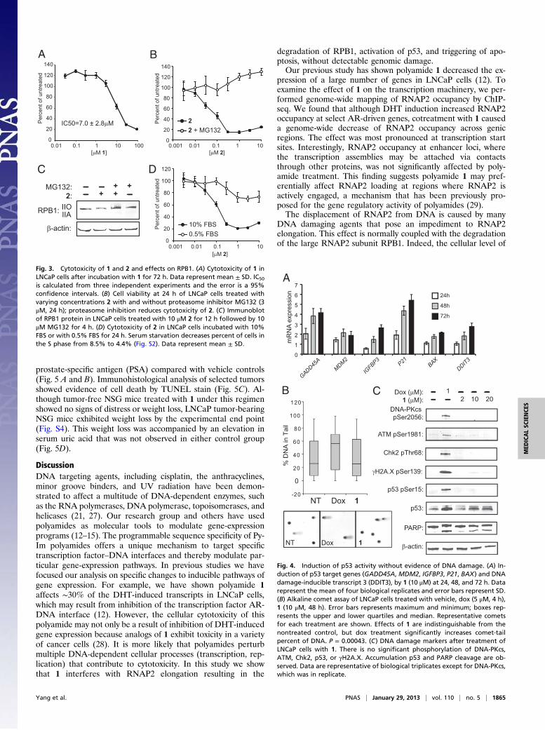
Fig. 3. Cytotoxicity of 1 and 2 and effects on RPB1. (A) Cytotoxicity of 1 inLNCaP cells after incubation with 1 for 72 h. Data represent mean ± SD. IC50
is calculated from three independent experiments and the error is a 95%confidence intervals. (B) Cell viability at 24 h of LNCaP cells treated withvarying concentrations 2 with and without proteasome inhibitor MG132 (3μM, 24 h); proteasome inhibition reduces cytotoxicity of 2. (C) Immunoblotof RPB1 protein in LNCaP cells treated with 10 μM 2 for 12 h followed by 10μM MG132 for 4 h. (D) Cytotoxicity of 2 in LNCaP cells incubated with 10%FBS or with 0.5% FBS for 24 h. Serum starvation decreases percent of cells inthe S phase from 8.5% to 4.4% (Fig. S2). Data represent mean ± SD.
P21
GADD45A
MDM2
IGFBP3
DDIT3
BAX
0
1
2
3
4
5
6
7m
RN
A ex
pres
sion 24h
48h
72h
-20
0
20
40
60
80
100
120
NT Dox 1
% D
NA
in T
ail
NT Dox 1
Chk2 pThr68:
DNA-PKcs pSer2056:
ATM pSer1981:
γH2A.X pSer139:
p53 pSer15:
p53:
β-actin:
PARP:
Dox (μM):1 (μM):
12 10 20
A
B C
Fig. 4. Induction of p53 activity without evidence of DNA damage. (A) In-duction of p53 target genes (GADD45A, MDM2, IGFBP3, P21, BAX) and DNAdamage-inducible transcript 3 (DDIT3), by 1 (10 μM) at 24, 48, and 72 h. Datarepresent the mean of four biological replicates and error bars represent SD.(B) Alkaline comet assay of LNCaP cells treated with vehicle, dox (5 μM, 4 h),1 (10 μM, 48 h). Error bars represents maximum and minimum; boxes rep-resents the upper and lower quartiles and median. Representative cometsfor each treatment are shown. Effects of 1 are indistinguishable from thenontreated control, but dox treatment significantly increases comet-tailpercent of DNA. P = 0.00043. (C) DNA damage markers after treatment ofLNCaP cells with 1. There is no significant phosphorylation of DNA-PKcs,ATM, Chk2, p53, or γH2A.X. Accumulation p53 and PARP cleavage are ob-served. Data are representative of biological triplicates except for DNA-PKcs,which was in replicate.
Yang et al. PNAS | January 29, 2013 | vol. 110 | no. 5 | 1865
MED
ICALSC
IENCE
S

RPB1 in LNCaP cells was found to decrease in both a time- andconcentration-dependent manner when treated with polyamide1. Polyamide 2, a more cytotoxic analog of 1, also reduced cel-lular RPB1 in LNCaP cells and induced cell death. Cotreatmentof 2 with a proteasomal inhibitor MG132 was able to prevent thedegradation of RPB1 and reduce the toxicity of 2 in cell culture.In addition, the cytotoxic effects of other RNAP2 inhibitors arereported to be attenuated by preventing S-phase entry. LNCaPcells arrested in G0/G1 by serum starvation also exhibited re-duced sensitivity to 2 compared with cells grown in normal me-dia. The finding that cytotoxicity is partially rescued by MG132treatment and G0/G1 arrest suggests RPB1 degradation con-tributes to cytotoxicity; however, contributions from other DNA-dependent processes are not ruled out.Although transcription inhibition can activate p53 signaling,
both events can be caused by DNA damage. Analysis of pre-viously published microarray data revealed the induction ofseveral p53 target genes in LNCaP cells cotreated with DHT and1 (12). Further validation of transcript levels of the genes in thisstudy also showed a time-dependent increase in the expression ofGADD45A, MDM2, IGFBP3, P21, BAX, and DDIT3 (Fig. 4A).Because these genes are also markers of genotoxic stress (24)and were found to be induced in A549 cells treated with alky-lating polyamide derivatives (25), we searched for signs of DNAdamage to determine if it was causing transcription inhibitionand p53 activation. Interestingly, both comet assay and immun-blot analysis of cellular DNA damage markers showed no sig-nificant signs of DNA damage. Although faint phosphorylationof γH2A.X was visible, it is likely caused by cellular apoptosis asindicated by the concurrent PARP cleavage. These data areconsistent with studies in yeast mutants that are hypersensitive toDNA damage, which showed no increased sensitivity to poly-amide treatment, suggesting these reversible DNA binders donot compromise genomic integrity (30).The activation of p53 by transcription inhibition in the absence
of DNA damage has been observed for DNA-independentinhibitors of RNAP2, such as DRB, α-amanitin, and variousRNAP2-targeted antibodies (5, 6, 26). Distamycin A, the naturalproduct that provided the structural inspiration for Py-Im poly-amides, inhibits the initiation of RNA synthesis in cell-free assays(27). In cell culture, distamycin also induces degradation ofRPB1 and activates p53 (31, 32). However, low antitumor po-tency and poor stability limit its utility.
To assess the therapeutic potential of polyamide 1 as an an-titumor agent, LNCaP xenografts in a murine model were trea-ted with 1 or PBS vehicle. After three rounds of treatment,tumor growth was reduced by 64% in the treated group. Al-though treatment with 1 alone did not cause changes in animalbody weight or obvious signs of toxicity in tumor-free animals,treatment in tumor-bearing animals resulted in weight loss afterthree treatments. The accompanied elevation in serum uric acidmay be an indication of tumor lysis syndrome (33), which is as-sociated with rapid tumor cell turnover upon polyamide treat-ment. We anticipate that Py-Im polyamides could also demonstrateefficacy in additional xenograft models.
MethodsCompounds and Reagents. Py-Im polyamides 1, 2, and 3 were synthesized onoxime resin, as described previously (28, 34, 35). (R)-MG132 (MG132) wasfrom Santa Cruz Biotechnology.
Cell Viability Assays. LNCaP cells were plated in clear bottom 96-well plates at5,000–7,500 cells per well. The cells were allowed to adhere for 24–36 hbefore compounds were added in fresh media. Cell viability was determinedby the WST-1 assay (Roche) for 1 and 2 after 24- or 72-h incubation with cells.Cells in cytotoxicity rescue experiments were treated with 2 alone or with 3μM MG132 for 24 h. For cell-cycle arrest experiments, LNCaP cells wereseeded at 2,500–5,000 cells per well in normal media and allowed to adherefor 24–36 h. The media was replaced with normal media or media supple-mented with 0.5% (vol/vol) FBS and incubated for 48 h before treatmentwith compound.
In Vivo Xenograft Experiments. All mice experiments were conducted underan approved protocol by the Institutional Animal Care and Use Committee ofthe California Institute of Technology. Male NSG mice were purchased fromThe Jackson Laboratory. The animals were individually caged andmaintainedon a standard light-dark cycle. NSGmice were engrafted with LNCaP cells (2.5million cells) in a mixture of 1:1 media and matrigel in the left flank. Tumorswere grown to ∼100 mm3 (L × W2) before beginning treatment with com-pound or vehicle. Py-Im polyamide 1 was administered once every 3 d at 20nmol per animal (∼1 mg/kg) in a 5% (vol/vol) DMSO:PBS vehicle solutionuntil the experiment endpoint.
Serum Measurements. To investigate if polyamide 1 could be detected inperipheral blood after subcutaneous injections, 120 nmol of 1 [in 5% (vol/vol) DMSO/PBS] was injected into the right flank of four C57BL/6J mice.Blood was collected from anesthetized mice via retroorbital collection at 5min, 4 h, and 12 h after injection, then processed by methods previouslydescribed and analyzed by HPLC (36). For measurement of serum PSA (KLK3)and uric acid, blood was collected from anesthetized mice via retroorbital
Veh 1
Tum
or m
ass
(mg)
Veh 1
Pretreatment PosttreatmentVeh 1
PS
A in
ser
um (n
g/m
l)
0
5
10
15
20
25
0
100
200
300
400
500
Veh 1
Ser
um u
ric a
cid
(mg/
dL)
0
5
10
15
20
25
DBA
Tumor H & E (20X) TUNEL (100X)
Veh
1
1
Tumorbearing
Tumorfree
C
Posttreatment
Fig. 5. Polyamide 1 demonstrates antitumor activity in prostate cancer xenografts. (A) Male immunocompromised mice were engrafted with LNCaP cells andobserved until tumors reached ∼100 mm3. Tumor-bearing mice were then treated with 20 nmol 1 (n = 12) or vehicle (n = 13) by subcutaneous injections intothe flank distal to the tumor once every 3 d for a total of three injections. Mice were killed and tumors resected and weighed 2 d after the final injection.Tumors from mice treated with 1 were smaller (mean: 112 mg; median: 94 mg; range: 47–201 mg) than those of vehicle treated mice (mean: 310 mg; median:292 mg; range: 173–440 mg). Error bars represents maximum and minimum; boxes represents the upper and lower quartiles and median. P = 1.6E-5. (B) SerumPSA measured by ELISA pre- and posttreatment. Serum PSA is lower in the posttreatment serum of mice treated with 1 compared with vehicle. P = 0.024. (C)Selected tumors and histological stains of tumor cross-sections from mice treated with vehicle or 1. (D) Treatment of LNCaP tumor bearing mice with 1increases serum uric acid compared with vehicle controls and polyamide-treated, nontumor-bearing mice. P = 3.2E-9.
1866 | www.pnas.org/cgi/doi/10.1073/pnas.1222035110 Yang et al.
collection at experimental endpoint and serum was separated from blood bycentrifugation. Serum PSA (KLK3) was measured by ELISA (R&D Systems)according to the manufacturer’s instructions. Uric acid was measured aspreviously described (37).
Chromatin Immunoprecipitation. Genomic occupancy of RNAP2 was de-termined by ChIP with the 4H8 antibody (Abcam). LNCaP cells were plated at35 million cells per plate in RPMI supplemented with 10% (vol/vol) CTFBS andallowed to adhere for 24–36 h. The cells were treated with compound 1 infresh media (10% CTFBS) for 48 h. Cells treated and untreated with 1 wereincubated with 1 nM DHT for 6 h. Two-step cross-linking was performed aspreviously described (38). After DSG removal, chromatin was immunopre-ciated by previously published methods (39). DNA was harvested by phenolchloroform extraction and purified with the QIAquick purification kit (Qia-gen). Quantitative PCR was used to validate enrichment at the GAPDHtranscription start site (Primers: F-GGTTTCTCTCCGCCCGTCTT, R-TGTTCGA-CAGTCAGCCGCAT) compared with an internal negative locus (Primers: F-TAGAAGGGGGATAGGGGAAC, R-CCAGAAAACTGGCTCCTTCTT). Each sam-ple was immunoprecipated as five technical replicates. The three mostconsistent samples were combined and submitted for sequencing on anIllumina genome analyzer. Biological replicates were acquired.
Data Processing and Analysis. Sequencing reads were trimmed down to 36 bpand then mapped against the male set of human chromosomes (excluding allrandom chromosomes and haplotypes) using the hg19 version of the humangenome as a reference. Bowtie 0.12.7 was used for aligning reads (40), withthe following settings: “-v 2 -t–best–strata”. Signal profiles over genomiclocations were generated using custom written python scripts; the refSeqannotation was used for gene coordinates. Enhancers and promoters weredefined using previously published histone marker data (41). ChIP-seq peakswere called using MACS2 with default settings (42). Enhancers were definedas H3K4me1+ regions that did not intersect with H3K4me3+ regions andpromoters as H3K4me3+ regions that did not intersect with H3K4me1+
regions. Clustering was performed with Cluster 3.0 (43) and visualized withJava TreeView (44).
Comet Assay. LNCaP cells were plated at 1 million cells per 10-cm plate andallowed to adhere for 24–36 h. Cells were then incubated with either 10 μM 1for 48 h or 5 μM doxorubicin for 4 h. DNA damage was assayed using theTrevigen CometAssay system and samples were prepared from harvestedcells according to the manufacture protocol. Comets were imaged ona confocal microscope (Exciter, Zeiss) at 10× magnification. Percentage ofDNA in the tail was determined using Comet Assay Lite IV (PerceptiveInstruments). More than 100 comets were scored for each condition.
Immunoblot Assay. Samples for immunoblot analysis were prepared byplating LNCaP or DU145 cells at 1 million cells per 10-cm plate. Cells wereallowed to adhere for 24–36 h before incubation with compound. After theappropriate incubation time, cells were washed once with ice-cold PBS andharvested in ice-cold 125 μL lysis buffer (50 mM Tris•HCl pH 7.4, 150 mMNaCl, 1 mM EDTA, 1% Triton X 100) containing protease inhibitor mixture(Roche), 1 mM PMSF (Sigma), and phosphatase inhibitors (Sigma). Sampleswere incubated on ice for 10 min with vortexing once every 3 min. Cellulardebris was pelleted by spinning at 21,000 × g for 15 min to collect the su-pernatant. Samples were then quantified for protein content with theBradford assay (Bio-Rad) and boiled with 4× sample buffer (Li-Cor) for 5 min.Protein electrophoresis was performed in 4–20% precast Tris•glycine SDSgels (Bio-Rad) and transferred to PVDF membranes. Membrane blocking was
done with Odyssey Blocking Buffer (Li-Cor). The following antibodies used toprobe changes in protein levels or phosphorylation states: RBP1 (Santa CruzBiotechnology; N20), p53 (Santa Cruz Biotechnology; DO1), phospho-Chk2-Thr68 (Cell Signaling Technology), Phospho-p53-Ser15 (Cell Signaling Tech-nology), phospho-H2A.X-Ser139 (Cell Signaling Technology), phosphor-ATM-Ser1981 (Abcam), phospho-DNA-PKcs-Ser2056 (Abcam), and β-actin(Abcam). Near-IR secondary antibodies (Li-Cor) were used for imaging.Experiments were performed in biological triplicate except for DNA-PKcs (replicate).
Flow Cytometry. To determine cell cycle distribution of LNCaP cells grown innormal media or under serum-starved conditions, 1 million cells were seededto each 10-cm plate and allowed to adhere for 24–36 h. Media was thenreplaced with fresh normal media [10% (vol/vol) FBS] or serum-starvedmedia [0.5% (vol/vol) FBS] and incubated for an additional 48 h. Cells werethen trypsinized and prepared for analysis as previously described (45).Samples were analyzed in biological triplicate on a FACSCalibur (Becton-Dickinson) instrument. Data analysis was performed using FlowJo 7.6.5.
Quantitative RT-PCR. RNA was extracted using RNEasy columns (Qiagen)according to the manufacturer’s protocols. cDNA was generated from RNAby reverse transcriptase (Transcriptor First Strand cDNA kit; Roche). Quan-titative real-time RT-PCR was performed using SYBR Green PCR Master Mix(Applied Biosystems) on an ABI 7300 instrument. mRNA was measured rel-ative to β-glucuronidase as an endogenous control. Experiments were per-formed in biological quadruplicates. For primer sequences see Table S1.
Confocal Microscopy. Cells were plated in 35-mm optical dishes (MatTek) anddosed with polyamide 3 at 2 μM for 24 h with or without 3μM MG132. Cellswere then washed with PBS and imaged on a confocal microscope (Exciter;Zeiss) using a 63× oil immersion lens. Confocal imaging was performedfollowing established protocols (34).
Histology and Immunohistochemistry. Tumors were resected immediatelyafter euthanasia and fixed in neutral buffered formalin. Selected sampleswere embedded in paraffin, sectioned and stained with H&E. Selected sec-tions were assessed by TUNEL, as previously described (46).
Thermal Denaturation Assays. Polyamides 1 and 2 were incubated with du-plex DNA 5′-CGATGTTCAAGC-3′, which contains the predicted target site forthese compounds (underlined). Melting temperature analyses were per-formed on a Varian Cary 100 spectrophotometer as described (47). Meltingtemperatures were defined as a maximum of the first derivative of absor-bance at 260 nm over the range of temperatures.
Statistical Analysis. Statistical significance was calculated using the Student ttest with two tailed variance. Results were considered significant whenP < 0.05.
ACKNOWLEDGMENTS. We thank Rochelle Diamond of the Caltech FlowCytometry Cell Sorting Facility for help with flow cytometry experimentalsetup and data acquisition; and Dr. Janet Baer, Dr. Karen L. Lencioni, andGwen E. Williams for helpful discussions and technical assistance with animalexperiments. This work was supported in part by the National Institutes ofHealth Grants GM27681 and HG004576, and by the Prostate CancerFoundation. N.G.N. acknowledges the Jonsson Cancer Center Foundationat University of California at Los Angeles for continued support.
1. Derheimer FA, Chang CW, Ljungman M (2005) Transcription inhibition: A potentialstrategy for cancer therapeutics. Eur J Cancer 41(16):2569–2576.
2. Koumenis C, Giaccia A (1997) Transformed cells require continuous activity of RNApolymerase II to resist oncogene-induced apoptosis. Mol Cell Biol 17(12):7306–7316.
3. Jung Y, Lippard SJ (2006) RNA polymerase II blockage by cisplatin-damaged DNA.Stability and polyubiquitylation of stalled polymerase. J Biol Chem 281(3):1361–1370.
4. Ljungman M, Zhang FF (1996) Blockage of RNA polymerase as a possible trigger foru.v. light-induced apoptosis. Oncogene 13(4):823–831.
5. Ljungman M, Zhang FF, Chen F, Rainbow AJ, McKay BC (1999) Inhibition of RNApolymerase II as a trigger for the p53 response. Oncogene 18(3):583–592.
6. Arima Y, et al. (2005) Transcriptional blockade induces p53-dependent apoptosis as-sociated with translocation of p53 to mitochondria. J Biol Chem 280(19):19166–19176.
7. Nguyen VT, et al. (1996) In vivo degradation of RNA polymerase II largest subunittriggered by alpha-amanitin. Nucleic Acids Res 24(15):2924–2929.
8. Arseneau JC, et al. (1972) Nonlymphomatous malignant tumors complicating Hodg-kin’s disease. Possible association with intensive therapy. N Engl J Med 287(22):1119–1122.
9. Dervan PB, Edelson BS (2003) Recognition of the DNA minor groove by pyrrole-im-
idazole polyamides. Curr Opin Struct Biol 13(3):284–299.10. Chenoweth DM, Dervan PB (2009) Allosteric modulation of DNA by small molecules.
Proc Natl Acad Sci USA 106(32):13175–13179.11. Chenoweth DM, Dervan PB (2010) Structural basis for cyclic Py-Im polyamide allosteric
inhibition of nuclear receptor binding. J Am Chem Soc 132(41):14521–14529.12. Nickols NG, Dervan PB (2007) Suppression of androgen receptor-mediated gene ex-
pression by a sequence-specific DNA-binding polyamide. Proc Natl Acad Sci USA 104
(25):10418–10423.13. Nickols NG, Jacobs CS, Farkas ME, Dervan PB (2007) Modulating hypoxia-inducible
transcription by disrupting the HIF-1-DNA interface. ACS Chem Biol 2(8):561–571.14. Muzikar KA, Nickols NG, Dervan PB (2009) Repression of DNA-binding dependent
glucocorticoid receptor-mediated gene expression. Proc Natl Acad Sci USA 106(39):
16598–16603.15. Raskatov JA, et al. (2012) Modulation of NF-κB-dependent gene transcription using
programmable DNA minor groove binders. Proc Natl Acad Sci USA 109(4):1023–1028.
Yang et al. PNAS | January 29, 2013 | vol. 110 | no. 5 | 1867
MED
ICALSC
IENCE
S

16. Matsuda H, et al. (2011) Transcriptional inhibition of progressive renal disease bygene silencing pyrrole-imidazole polyamide targeting of the transforming growthfactor-β1 promoter. Kidney Int 79(1):46–56.
17. Synold TW, et al. (2012) Single-dose pharmacokinetic and toxicity analysis of pyrrole-imidazole polyamides in mice. Cancer Chemother Pharmacol 70(4):617–625.
18. Raskatov JA, et al. (2012) Gene expression changes in a tumor xenograft by a pyrrole-imidazole polyamide. Proc Natl Acad Sci USA 109(40):16041–16045.
19. Palstra RJ, et al. (2008) Maintenance of long-range DNA interactions after inhibitionof ongoing RNA polymerase II transcription. PLoS ONE 3(2):e1661.
20. Bregman DB, et al. (1996) UV-induced ubiquitination of RNA polymerase II: A novelmodification deficient in Cockayne syndrome cells. Proc Natl Acad Sci USA 93(21):11586–11590.
21. Ratner JN, Balasubramanian B, Corden J, Warren SL, Bregman DB (1998) Ultravioletradiation-induced ubiquitination and proteasomal degradation of the large subunitof RNA polymerase II. Implications for transcription-coupled DNA repair. J Biol Chem273(9):5184–5189.
22. Aune GJ, et al. (2008) Von Hippel-Lindau-coupled and transcription-coupled nucleo-tide excision repair-dependent degradation of RNA polymerase II in response totrabectedin. Clin Cancer Res 14(20):6449–6455.
23. McKay BC, Becerril C, Spronck JC, Ljungman M (2002) Ultraviolet light-induced ap-optosis is associated with S-phase in primary human fibroblasts. DNA Repair (Amst)1(10):811–820.
24. el-Deiry WS (1998) Regulation of p53 downstream genes. Semin Cancer Biol 8(5):345–357.
25. Kashiwazaki G, et al. (2012) Synthesis and biological properties of highly sequence-specific-alkylating N-methylpyrrole-N-methylimidazole polyamide conjugates. J MedChem 55(5):2057–2066.
26. Derheimer FA, et al. (2007) RPA and ATR link transcriptional stress to p53. Proc NatlAcad Sci USA 104(31):12778–12783.
27. Puschendorf B, Petersen E, Wolf H, Werchau H, Grunicke H (1971) Studies on theeffect of distamycin A on the DNA dependent RNA polymerase system. Biochem Bi-ophys Res Commun 43(3):617–624.
28. Meier JL, Montgomery DC, Dervan PB (2012) Enhancing the cellular uptake of Py-Impolyamides through next-generation aryl turns. Nucleic Acids Res 40(5):2345–2356.
29. Carlson CD, et al. (2010) Specificity landscapes of DNA binding molecules elucidatebiological function. Proc Natl Acad Sci USA 107(10):4544–4549.
30. Marini NJ, et al. (2003) DNA binding hairpin polyamides with antifungal activity.Chem Biol 10(7):635–644.
31. Zhang Z, et al. (2009) Tanshinone IIA triggers p53 responses and apoptosis by RNApolymerase II upon DNA minor groove binding. Biochem Pharmacol 78(10):1316–1322.
32. Hirota M, Fujiwara T, Mineshita S, Sugiyama H, Teraoka H (2007) Distamycin A en-
hances the cytotoxicity of duocarmycin A and suppresses duocarmycin A-induced
apoptosis in human lung carcinoma cells. Int J Biochem Cell Biol 39(5):988–996.33. Coiffier B, Altman A, Pui CH, Younes A, Cairo MS (2008) Guidelines for the man-
agement of pediatric and adult tumor lysis syndrome: An evidence-based review. J
Clin Oncol 26(16):2767–2778.34. Best TP, Edelson BS, Nickols NG, Dervan PB (2003) Nuclear localization of pyrrole-
imidazole polyamide-fluorescein conjugates in cell culture. Proc Natl Acad Sci USA
100(21):12063–12068.35. Puckett JW, Green JT, Dervan PB (2012) Microwave assisted synthesis of Py-Im poly-
amides. Org Lett 14(11):2774–2777.36. Raskatov JA, Hargrove AE, So AY, Dervan PB (2012) Pharmacokinetics of Py-Im pol-
yamides depend on architecture: Cyclic versus linear. J Am Chem Soc 134(18):
7995–7999.37. Dai KS, et al. (2005) An evaluation of clinical accuracy of the EasyTouch blood uric acid
self-monitoring system. Clin Biochem 38(3):278–281.38. Nowak DE, Tian B, Brasier AR (2005) Two-step cross-linking method for identification
of NF-kappaB gene network by chromatin immunoprecipitation. Biotechniques 39(5):
715–725.39. Reddy TE, et al. (2009) Genomic determination of the glucocorticoid response reveals
unexpected mechanisms of gene regulation. Genome Res 19(12):2163–2171.40. Langmead B, Trapnell C, Pop M, Salzberg SL (2009) Ultrafast and memory-efficient
alignment of short DNA sequences to the human genome. Genome Biol 10(3):R25.41. Yu JD, et al. (2010) An integrated network of androgen receptor, polycomb, and
TMPRSS2-ERG gene fusions in prostate cancer progression. Cancer Cell 17(5):443–454.42. Zhang Y, et al. (2008) Model-based Analysis of ChIP-Seq (MACS). Genome Biol 9(9):
R137.43. de Hoon MJ, Imoto S, Nolan J, Miyano S (2004) Open source clustering software.
Bioinformatics 20(9):1453–1454.44. Saldanha AJ (2004) Java Treeview—Extensible visualization of microarray data. Bio-
informatics 20(17):3246–3248.45. Diamond RA, DeMaggio S (2000) In Living Color: Protocols in Flow Cytometry and Cell
Sorting (Springer, Berlin, New York), pp xxv, 800 pp.46. Zisman A, et al. (2003) LABAZ1: A metastatic tumor model for renal cell carcinoma
expressing the carbonic anhydrase type 9 tumor antigen. Cancer Res 63(16):
4952–4959.47. Dose C, Farkas ME, Chenoweth DM, Dervan PB (2008) Next generation hairpin pol-
yamides with (R)-3,4-diaminobutyric acid turn unit. J Am Chem Soc 130(21):
6859–6866.
1868 | www.pnas.org/cgi/doi/10.1073/pnas.1222035110 Yang et al.