antitumor activity of bortezomib alone and in combination
TRANSCRIPT

Fax +41 61 306 12 34E-Mail [email protected]
Original Paper
Acta Haematol 806 DOI: 10.1159/000XXXXXX
Antitumor Activity of Bortezomib Alone and in Combination with Trail in Human Acute Myeloid Leukemia
Concetta Conticello a, b Luana Adamo c Luisa Vicari c Raffaella Giuffrida c
Gioacchin Iannolo c Gabriele Anastasi c Laura Caruso b Gaetano Moschetti b
Alessandra Cupri a Giuseppe Antonio Palumbo a Massimo Gulisano c
Ruggero De Maria c, d Rosario Giustolisi a Francesco Di Raimondo a
a Department of Biomedical Sciences, Hematology Section, University of Catania, Catania , b Department of Experimental Oncology, Mediterranean Institute of Oncology and c IOM Ricerca, Viagrande, and d Department of Hematology, Oncology and Molecular Medicine, Istituto Superiore di Sanita’, Rome , Italy
resistant AML cells for enhanced TRAIL-mediated killing. These results suggest that a combination of proteasome in-hibitors and TRAIL could be effective for treating AML pa-tients, even patients who are refractory to conventional chemotherapy. Copyright © 2008 S. Karger AG, Basel
Introduction
Acute myeloid leukemia (AML) is a malignant neo-plasm of hematopoietic cells characterized by abnormal proliferation of clonal neoplastic myeloid precursor cells and an impairment of normal hematopoiesis. Although significant advances in understanding the molecular pathogenesis of AML have been made in recent decades, and more specifically targeted agents have been devel-oped for treating AML, only about 20–30% of patients actually enjoy long-term disease survival and the out-come of AML is frequently fatal [1, 2] . Such a severe out-come demands the development of novel treatment strat-egies against this disease.
Key Words
Acute myeloid leukemia � Apoptosis � Bortezomib � TRAIL
Abstract
Acute myeloid leukemia (AML) is a malignant disease char-acterized by abnormal proliferation of clonal precursor cells. Although different strategies have been adopted to obtain complete remission, the disease actually progresses in about 60–70% of patients. Bortezomib has been used in multiple myeloma and other lymphoid malignancies because of its antitumor activity. Here we examined the sensitivity of bone marrow cells from AML patients to bortezomib alone or in combination with TRAIL, a member of the TNF family that induces apoptosis in tumor cells while sparing normal cells (34 patients: 25 newly diagnosed, 4 relapsed, 5 refractory). Bortezomib induced cell death in blasts from each patient sample. The cytotoxic effect was dose- and time-dependent (concentration from 0.001 to 10 � M for 24 and 48 h) and was associated with a downregulation of Bcl-xL and Mcl-1, an up-regulation of TRAIL-R1, TRAIL-R2, p21, activation of execu-tioner caspases and a loss of the mitochondrial membrane potential. Moreover, low doses of bortezomib primed TRAIL-
Received: January 16, 2008 Accepted after revision: June 14, 2008 Published online: $ $ $
Prof. Francesco Di Raimondo, MD Department of Biomedical Sciences, Hematology Section University of Catania, Via Citelli 7 IT–95100 Catania (Italy) Tel. +39 095 743 5911, Fax +39 095 743 5913, E-Mail [email protected]
© 2008 S. Karger AG, Basel0001–5792/08/0000–0000$24.50/0
Accessible online at:www.karger.com/aha
AHA806.indd 1AHA806.indd 1 04.08.2008 11:30:3804.08.2008 11:30:38

Conticello et al.
Acta Haematol 8062
Because of the pivotal role of proteasome inhibitors in apoptosis, bortezomib, either alone or in combination with other apoptosis-inducing compounds, seems to have potential for treating AML. In particular, the proteasome regulates the levels of cyclins and cyclin-dependent ki-nase inhibitors, and of pro- and antiapoptotic proteins such as Bcl-2 family members. It also controls the levels of tumor suppressor genes such as p53, oncogenes, and the activities of signal transduction pathways such as the NF- � B pathway by degrading the inhibitor of nuclear factor- � B (I � B) [3] . Proteasome inhibitors can induce misregulation of numerous proteins leading to cell death, and this occurs preferentially in transformed cells. Re-cently, it has been shown in several tumor models that proteasome inhibitors can block proliferation and induce apoptosis in tumor cells both in vitro and in vivo; there-fore, molecules such as bortezomib represent a potential-ly powerful anticancer therapy [4] . These compounds can also induce tumor regression, alone or in combination with cytotoxic drugs, in xenograft murine models of hu-man lymphoma, myeloma [5] and different carcinomas [6–12] . The antitumor effect against leukemic cells of a combination of proteasome inhibitors with other drugs has already been described by Guzman et al. [13] and Tan [14] on T-ALL and primary AML cells. Remarkably, bort-ezomib has recently been approved for therapeutic use in progressive multiple myeloma [15] , and large-scale phase I [16, 17] and II programs for various neoplastic diseases are currently in progress [18] . Since modulation of pro-teasome function can enhance the efficacy of chemother-apeutics, proteasome inhibition by bortezomib is a ratio-nal target for chemosensitization.
Another new candidate for AML treatment is tumor necrosis factor-related apoptosis-inducing ligand or Apo2 ligand (TRAIL/Apo2L). TRAIL is a member of the TNF family that has recently been proposed as a novel antican-cer agent because of its ability to induce apoptosis in ma-lignant cells while sparing normal cells. TRAIL acts by interacting with its surface death receptors, TRAIL-R1 (also known as DR4) and TRAIL-R2 (also known as DR5), which contain a conserved death domain (DD) motif. After binding to TRAIL ligand, these receptors trimerize and cluster, leading to the formation of the death-inducing signaling complex and recruitment of the adaptor molecule FADD, which in turn recruits caspase-8 and activates apoptotic signaling. While TRAIL/Apo2L is not toxic to most normal human cells in vitro or to TRAIL-treated animals, it induces apoptosis in tumor cells of diverse origins both in vitro and in vivo and in various in vivo tumor models [19, 20] . Although previous
studies have shown that AML blasts are resistant to TRAIL, recent reports indicate that bortezomib sensitiz-es cancer cells to this ligand by inducing the expression of TRAIL receptors in solid tumor cell lines [21–24] .
In this study we have examined the effects of bortezo-mib alone or in combination with TRAIL on AML pri-mary cells. We found that bortezomib has a consistent cytotoxic effect on AML cells, higher than that of stan-dard chemotherapeutic agents currently used to treat AML patients. Moreover, this cytotoxic effect increases significantly when bortezomib is combined with TRAIL, probably because of the modulation of TRAIL receptors after proteasome inhibition.
Materials and Methods
Primary Blast Cells Fresh leukemic blasts from 34 AML patients diagnosed and
treated at the Division of Hematology, Ospedale Ferrarotto, Uni-versity of Catania were obtained after informed consent during routine examination. Bone marrow (BM) cells were isolated by Ficoll-Hypaque density gradient centrifugation. Cells were main-tained in RPMI 1640 medium (Gibco) supplemented with 2 m M L -glutamine and 100 U/ml penicillin-streptomycin and 10% heat-inactivated fetal bovine serum (Invitrogen, Carlsbad, Calif., USA). Cells were kept in a 5% CO 2 atmosphere at a density of 5 ! 10 5 cells/ml. Blasts constituted 1 70% for all samples (from 70 to 100%). A BM biopsy for histological diagnosis was obtained from all AML patients at presentation.
Reagents Antineoplastic agents were purchased from Sigma-Aldrich
(St. Louis, Mo., USA) and resuspended in DMSO (etoposide) or in water (cytarabine and daunorubicin). Bortezomib (also known as PS-341) was purchased from Millennium Pharmaceuticals (Cam-bridge, Mass., USA). Bortezomib (0.1 � M ), cytarabine (1 � g/ml), doxorubicin (5 � M ) and etoposide (0.5 � M ) were used in vitro at doses compatible with the plasma levels reached in vivo during cancer treatment. Human recombinant LZ-TRAIL was purchased from Alexis (Lausen, Switzerland) and the caspase inhibitorz-VAD-fmk from Bachem.
Cell Viability Assay Cell viability was determined using the CellTiter 96 AQ ueous
One Solution Cell Proliferation Assay (Promega, Madison, Wisc., USA) according to the manufacturer’s instructions. This assay is based on the reduction of 3-(4,5-dimethylthiazol-2-yl)-5-(3-car-boxymethoxyphenyl)-2-(4-sulfophenyl)-2H-tetrazolium, inner salt (MTS), to a colored formazan product. The formazan is mea-sured by the absorbance at 490 nm, which is directly proportion-al to the number of living cells. Briefly, cells were seeded into 3 wells of 96-well plates at 5 ! 10 3 cells/200 � l/well and treated with different combinations of the chemotherapeutic drugs test-ed. After 24 and 48 h of incubation the culture medium was re-moved and the cells were washed with PBS (pH 7.4); 100 � l of fresh
AHA806.indd 2AHA806.indd 2 04.08.2008 11:30:5304.08.2008 11:30:53

Bortezomib and TRAIL in AML Acta Haematol 806 3
culture medium without drugs and 20 � l of MTS were added to each well and the cells were incubated for 3 h. The plates were read on a Microplate Reader (Synergy HT, BIO-TEK). Survival was ex-pressed as the percentage of viable cells in the treated sample rel-ative to the untreated control cells.
HALO/COMET Assay Genomic DNA fragmentation was detected by the HALO/
COMET assay according to Godard et al. [25] . Cells were col-lected by scraping, washed in PBS, and resuspended as 10 ! 10 5 cells/10 � l of PBS. The samples were analyzed in duplicate: one for the HALO assay and one for the COMET assay. The first group of samples was subjected to denaturation with high pH buffer (300 m M NaOH, 1 m M EDTA, pH 12.8) for 20 min followed by neutralization (0.4 M Tris-HCl, pH 7.5) for 5 min. Staining with ethidium bromide (2 � g/ml) and analysis with a Leica fluores-cence microscope were performed. The second series of samples, after denaturation, was electrophoresed and treated as well as the HALO samples. The run was performed in the same denaturation buffer for 30 min at 20 V on ice bath under semidark conditions. For each slide in the two types of assays, 50 images (correspond-ing to 50 cells) were analyzed.
Flow Cytometry 1 ! 10 5 cells were used for flow cytometry. Cells were washed
with cold PBS containing 1% bovine serum albumin (BSA) and incubated for 1 h at 4 ° C with control or specific primary antibod-ies: goat anti-human TRAIL-R1 and TRAIL-R2 (both 1: 50, R&D Systems, Minneapolis, Minn., USA). After washing, the cells were incubated for 40 min at 4 ° C with phycoerythrin-conjugated anti-goat secondary antibodies (1: 100, Jackson Laboratories, West Grove, Pa., USA). Labeled cells were washed twice with PBS/BSA and the fluorescence intensity was evaluated by a FACScan (Beck-man Coulter, Fullerton, Calif., USA).
Analysis of Mitochondrial Membrane Potential The mitochondrial membrane potential (MMP) of intact cells
was measured by flow cytometry with the lipophilic cationic probe 5,5 � ,6,6 � -tetrachloro-1,1 � ,3,3 � -tetraethylbenzimidazolcar-bocyanine iodide (JC-1, Molecular Probes, Leiden, The Nether-lands). JC-1 was added directly to the cell culture medium (1 � M final concentration) including cells (0.5 ! 10 6 /ml) and incubated for 10 min at 22 ° C. The medium was then replaced with PBS, and the cells were quantitated for J-aggregate fluorescence intensity by using a FACScan flow cytometer (Beckman Coulter).
Immunostaining Procedure Immunofluorescence staining was performed on cells seeded
on glass slides. Cells were washed in PIPES buffer (80 m M PIPES pH 6.8, 5 m M EGTA and 2 m M MgCl 2 ) and fixed with 4% para-formaldehyde for 10 min. This step was followed by block-per-meabilization in PBS containing 0.2% BSA and 0.1% Triton X-100 for 10 min, followed by exposure to PBS containing 1% BSA for1 h in order to reduce nonspecific staining. The cells were then incubated with rabbit anti-human NF- � B/p65 (1: 100, Santa Cruz) or goat anti-human TRAIL-R1 or TRAIL-R2 (both 1: 50, R&D Systems) for 1 h at room temperature. After three washes in PBS the cells were incubated with rhodamine-conjugated mouse anti-rabbit immunoglobulins (1: 500 Jackson Laboratories) or donkey anti-goat immunoglobulins (1: 400, Jackson Laboratories). Nuclei
were stained with PI (Sigma). Immunohistochemical staining was performed on 2- � m-thick paraffin-embedded AML BM and co-lon cancer sections (used as positive control). After a deparaffina-tion-hydration step, sections were permeabilized with PBS con-taining 0.4% Triton X-100 for 30 min and nonspecific staining was blocked with PBS containing 5% BSA for 30 min. Specimens were incubated for 1 h with goat anti-human TRAIL-R1 and TRAIL-R2 (both 1: 50, R&D Systems). Washed sections were treated with biotinylated anti-goat immunoglobulins and then incubated in streptavidin conjugated with peroxidase (Dako, Carpintera, Calif., USA). Staining was detected using diamino-benzidine (DAB). Sections were counterstained with hematoxy-lin, then dehydrated and mounted in permanent nonaqueous mounting medium.
Caspase Assay Apo-ONE Homogeneous Caspase-3/7 buffer and substrate
(Promega) were mixed and then added to cultured cells, untreat-ed or treated with bortezomib and TRAIL. Sequential cleavage of the substrate by caspase-3/7 activity in the sample yields an in-tensely fluorescent product, the amount of which is proportional to the amount of caspase-3/7 activity in the sample. The fluores-cence was measured spectrophotometrically at 485 nm.
Western Blotting Cell pellets were washed twice with cold PBS and lysed on ice
for 10 min in JS buffer (50 m M Hepes pH 7.4, 150 m M NaCl, 5 m M EGTA, 1% glycerol, 1% Triton X-100, 1.5 m M MgCl 2 ) supplement-ed with protease inhibitors: 0.1 mg/ml phenylmethylsulfonylfluoride and 1 � l/ml each of aprotinin, leupeptin and pepstatin. After centrifugation at 2,000 rpm for 15 min at 4 ° C to remove cell debris, the protein content was determined using the Bio-Rad protein assay (Bio-Rad Laboratories, Richmond, Calif., USA). Af-ter denaturation, aliquots of 30 � l total protein were loaded on to 10 or 12% SDS-polyacrylamide gel. The gels were electroblotted onto nitrocellulose membranes (Hybond C-extra, Amersham Biosciences, Little Chalfont, UK). Filters were blocked for 1 h in 5% nonfat dry milk in TBS-T (2.5 m M Tris-HCl, 15 m M NaCl, 0.05% Tween 20), then incubated for 1 h with the following pri-mary antibodies: mouse monoclonals anti-Bcl-xL (1: 100, Santa Cruz Biotechnology), anti-Smac (1: 1,000, Santa Cruz Biotechnol-ogy), anti-actin (1: 10,000, Sigma), anti-caspase-7 (1: 1,000, Pharmingen, San Diego, Calif., USA), hamster anti-Bcl2 (1: 100, Pharmingen, San Diego, Calif., USA), and the polyclonals goat anti-IAP1 (1: 50, Santa Cruz Biotechnology), rabbit anti-P21 (1: 500; Santa Cruz Biotechnology), rat anti-FLIP (1: 15, Alexis, San Diego, Calif., USA), anti-Mcl-1 (1: 500; Santa Cruz Biotechnolo-gy), anti-caspase-3 (1: 500, Santa Cruz Biotechnology). Filters were then rinsed with TBS-T buffer and incubated for 1 h with the corresponding secondary antibody conjugated with peroxidase (Amersham Biosciences). After washing, proteins were detected by the enhanced chemiluminescent method (ECL Plus; Amer-sham Biosciences).
Statistical Analysis A paired t test was used to analyze the statistical significance
of the results. Values of p ! 0.05 were considered significant. Data are presented as mean values 8 SD of the mean.
AHA806.indd 3AHA806.indd 3 04.08.2008 11:30:5304.08.2008 11:30:53

Conticello et al.
Acta Haematol 8064
Results
Samples A total of 34 samples from patients with AML at diag-
nosis or resistant/refractory AML were examined. Pa-tients’ characteristics such as FAB classification, cytoge-netic features, blast percentage and prior therapies are detailed in table 1 .
AML Blasts Exhibit a Greater Dose-Dependent Sensitivity to Bortezomib than to Conventional Chemotherapy Since previous studies on multiple myeloma have
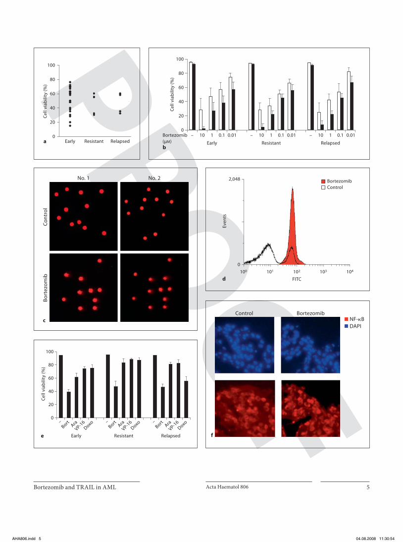
shown that the mean concentration of bortezomib in the plasma after a standard dose (1.3 mg/m 2 ) reaches 1.8 � M (509 ng/ml), with a range of 0.3–3.4 � M (109–1,300 ng/ml) [26] , we evaluated the sensitivity to bortezomib-mediated cell death in AML blasts derived from the 34 patients us-ing a low concentration of bortezomib (0.1 � M ). Cell vi-ability was greatly reduced in all AML blasts without cor-relation with the status of the patients from whom the blast cells were derived (at diagnosis, resistant or relapsed) ( fig. 1 a). In addition, most AML cells showed consistent reduction in cell viability in a dose- and time-dependent manner ( fig. 1 b). COMET assay evidenced an increased COMET score in cells from patients treated for 24 h with 0.1 � M bortezomib indicating that bortezomib is able to induce apoptosis in AML blasts ( fig. 1 c). Furthermore, we examined MMP on 3 samples from AML patients at di-agnosis and we observed loss of MMP after bortezomib treatment ( fig. 1 d). These biologically active concentra-tions are fully compatible with those achieved in vivo in the clinical setting, suggesting that clinical use of bortezo-mib for treating AML may be possible. In a recent report [27] AML blasts were subdivided into two subgroups ac-cording to their sensitivity to bortezomib [low-sensitivity group with ! 50% dying cells and high-sensitivity group (HSG) with 1 50% dying cells]. The sensitivity to bortezo-mib-mediated apoptosis correlated with the differentia-tion properties of AML blasts: the majority of the HSG cases corresponded to the M4 and M5 FAB subtypes, while the majority of the low-sensitivity group corre-sponded to M1 and M2 FAB subtypes. Similarly, we have found that the majority (8/34) of AML blasts that belong to HSG cases have morphologic and immunophenotypic features of the M4 FAB subtype while neither leukocyte cell count, nor specific karyotype or mutations, such as FLT3, are correlated with bortezomib sensitivity.
Since cytarabine, doxorubicin and etoposide are cur-rently used for treating AML, we compared the in vitro
efficacy of high concentrations of these cytotoxic drugs (comparable with the highest levels of achievable plasma peaks) with low doses of bortezomib. AML cells were ex-posed to bortezomib (0.1 � M ), cytarabine (1 � g/ml), doxorubicin (5 � M ), and etoposide (0.5 � M ) for 48 h. Cell viability was assessed by trypan blue exclusion (data not shown) and MTS assays. After 48 h treatment, most AML cells exposed to bortezomib showed reduction in cell vi-ability, while all the other drugs showed only minor ac-tivity ( fig. 1 e). Many AML cells from relapsed or refrac-tory patients were resistant to all the compounds tested with the exception of bortezomib. These results suggest that AML blasts have considerable sensitivity to bortezo-mib, even when they are resistant to high doses of con-ventional chemotherapeutic drugs. AML cells are char-acterized by constitutive abnormal activation of NF- � B and this activation is commonly associated with resis-tance to spontaneous apoptosis, thus favoring the sur-vival and growth of AML blasts [28] . NF- � B has been shown to be constitutively active only when it resides in the nucleus. Since proteasome inhibition interferes with NF- � B status and we have demonstrated a high sensitiv-ity of blast cells to bortezomib-induced cell death, we ex-
Fig. 1. Cytotoxic effect of bortezomib on AML blasts from 34 pa-tients (at diagnosis, resistant or relapsed). a AML blasts were ex-posed to 0.1 � M bortezomib for 48 h. Cell viability was measured by MTS and is expressed as percentage of the corresponding un-treated sample. The results represent the mean average of 3 inde-pendent experiments carried on each AML sample of 34 patients. b AML blasts were incubated in vitro for 24 ()) and 48 h ($) in the presence of increasing concentrations (0.01–10 � M ) of bort-ezomib. Cell viability was measured by MTS. The results repre-sent the mean 8 SD of 34 independent experiments for each AML sample. c AML blasts from newly diagnosed patients were stained by the COMET assay after exposure to 0.1 � M bortezomib for24 h. Two representative experiments are shown. d Flow cytom-etry analysis showed loss of MMP in blasts from 3 AML patients at diagnosis after 0.1 � M bortezomib treatment for 24 h. Mito-chondrial depolarization is indicated by the increase in green flu-orescence after bortezomib treatment (red). One of three experi-ments is shown. e Percentage of cell viability of myeloid blasts treated for 48 h with different compounds. AML cells were treat-ed for 48 h with bortezomib (Bort; 0.1 � M ), cytarabine (Ara;1 � g/ml), doxorubicin (Doxo; 5 � M ) or etoposide (VP-16; 0.5 � M ). The percentage of apoptotic cells was determined using the MTS assay. The results show the mean 8 SD of four independent ex-periments. f Immunofluorescence analysis of 10 AML samples untreated (control) or treated with 0.1 � M bortezomib. Cells were stained with anti-NF- � B (red). DAPI staining (blue) indicates the position of the nuclei in the corresponding view. Original magni-fication ! 20. Two representative experiments of 10 AML patients at diagnosis are shown.
AHA806.indd 4AHA806.indd 4 04.08.2008 11:30:5404.08.2008 11:30:54
Bortezomib and TRAIL in AML Acta Haematol 806 5
0
20
40
60
80
100
Cel
l via
bili
ty (%
)
a Early Resistant Relapsedb
Early
– 10 1 0.1 0.01 – 10 1 0.1 0.01 – 10 1 0.1 0.01
Resistant Relapsed
0
20
40
60
80
100
Cel
l via
bili
ty (%
)
Bortezomib(μM)
d
Even
ts
2,048
0
101100
FITC
104102 103
BortezomibControl
Con
trol
Bo
rtez
omib
c
No. 1 No. 2
e
0
20
40
60
80
100
Cel
l via
bili
ty (%
)
RelapsedEarly Resistant
–Bort Ara
VP-16
Doxo –Bort Ara
VP-16
Doxo –Bort Ara
VP-16
Doxo
Control Bortezomib
f
NF-�BDAPI
AHA806.indd 5AHA806.indd 5 04.08.2008 11:30:5404.08.2008 11:30:54

Conticello et al.
Acta Haematol 8066
plored the NF- � B status in AML blasts from 10 patients (7 newly diagnosed, 2 relapsed, 1 refractory) before and after bortezomib treatment. We observed that NF- � B/p65 is constitutively expressed in the nuclei of all samples but its localization is not affected by bortezomib treat-
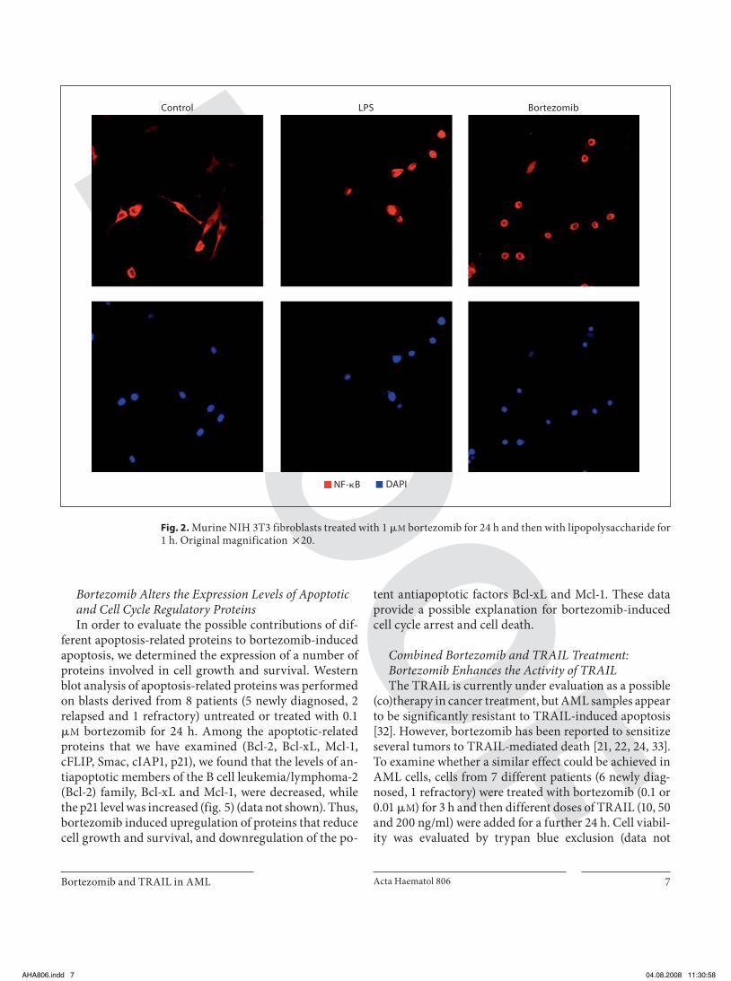
ment in myeloid blast cells ( fig. 1 f). After 24 h of bortezo-mib treatment we were unable to detect any cytosolic NF- � B/p65 by immunofluorescence or by Western blot by isolating cytoplasmic and nuclear lysates, suggesting that this drug acts independently of NF- � B status. Murine NIH 3T3 fibroblasts treated with 1 � M bortezomib for24 h and then with lipopolysaccharide for 1 h were used as a positive control of NF- � B cytoplasmic and nuclear localization ( fig. 2 ). These findings indicate that other ef-fects of the drug might contribute to AML cell death.
Proteasome Inhibitor-Induced Apoptosis Is Mostly Caspase Independent Since bortezomib activates effector caspases in differ-
ent tumor cells, we investigated whether this pathway was involved in the apoptosis induced by bortezomib in AML cells. The activity of the effector caspases was as-sessed 24 and 48 h after treatment with 0.1 � M bortezo-mib. Activation of caspase-3 and caspase-7 was measured by monitoring the cleavage of a fluorogenic caspase sub-strate, Ac-DEVD-AMC, using a fluorometric assay. Treatment of AML blasts from 10 patients (8 newly diag-nosed, 1 relapsed and 1 refractory) with 0.1 � M bortezo-mib resulted in consistent caspase activation, as indicated by the significant formation of the fluorescent cleavage product AMC after 24 and 48 h ( fig. 3 a). A similar but less marked result was also obtained with 0.01 � M bortezo-mib (data not shown). To determine whether such activ-ity involved the processing of executioner caspases, we evaluated the presence of the active forms of caspase-3 and caspase-7 by Western blotting, which confirmed that bortezomib activates the executioner caspases in myeloid blast cells ( fig. 3 b). These data suggest that bortezomib induces caspase activation in AML blasts.
Furthermore, to determine whether caspase activation contributes to bortezomib-induced decrease of viability of AML blasts, we treated the cells with 0.1 or 0.01 � M bortezomib in the presence or absence of the pan-caspase inhibitor zVAD-fmk. Although caspase-3 and 7 activa-tion was found to be strongly inhibited by zVAD ( fig. 3 c), the cytotoxic activity of proteasome inhibition was only slightly reduced by this inhibitor ( fig. 3 d) and this mini-mal inhibition was not found to be statistically signifi-cant, suggesting that caspases contribute only minimally to the cytotoxic activity of bortezomib in AML cells and that other unidentified apoptotic mediators are involved. The Jurkat cell line was used as a known [29–31] positive control where bortezomib induces cell death and DEV-Dase activity and zVAD blocks this effect, showing a cas-pase-dependent death prevented by zVAD ( fig. 4 ).
Table 1. Sample characteristics
Pa-tient
FAB type Blasts%
Cytogenetics Status
1 M2 90 46, XX n.d.2 M1 90 47, XY, + 15 first relapse3 M2 50 not determined n.d.4 therapy-
related/AML80 not determined n.d.
5 M4 60 46, XY, i (16)(q11) n.d.6 MDS/AML 60 not determined n.d.7 M2 42 46, XY n.d. 8 M4 Eo 70–80 not determined n.d.9 MDS/AML 70 46, XY n.d.
10 MDS/AML 20 not determined n.d.11 M4 90 46, XX, inv (16) n.d.12 MDS/AML 67 45, XX, der (3)t (3;?),
del (5); -7n.d.
13 MDS/AML 80 not determined n.d.14 MO 50 not determined n.d.15 M2 90 47, XY, +4 n.d.16 M4 40 46, XY, t (6;9) n.d.17 M4 90 not determined n.d.18 M4 99 46, XY n.d.19 M2 78 47, XY, t (8;21); +13 n.d.20 M1 90 not determined n.d.21 M2 98 not determined n.d.22 M2 60 46, XX n.d.23 M4 50 46, XY n.d.24 M0 90 not determined n.d.25 M4 95 46, XX n.d.26 M2 73 46, XY, del (1), del (3),
–7, +8; 46 XY dic (1;1)refractory
27 M6 76 not determined refractory28 M2 90 not determined n.d.29 M2 65 46, XY first relapse30 M2 40 46, XX, del (5) first relapse31 M2 95 not determined refractory32 M0 80 46, XY, del (9)(q22) refractory33 M1 70 45, XX, t(3;3), -7 refractory34 MDS/AML 89 46, XY first relapse
Relapsed patients: relapsed after the first line of chemotherapy with cytarabine, doxorubicin and etoposide. Resistant or refrac-tory patients: resistant to 2 or more lines of chemotherapy includ-ing cytarabine, doxorubicin and etoposide, high-dose cytarabine, mitoxantrone, and sequential doses of etoposide.
FAB = French-American-British classification; MDS = myelo-dysplastic syndrome; n.d. = newly diagnosed.
AHA806.indd 6AHA806.indd 6 04.08.2008 11:30:5804.08.2008 11:30:58
Bortezomib and TRAIL in AML Acta Haematol 806 7
Bortezomib Alters the Expression Levels of Apoptotic and Cell Cycle Regulatory Proteins In order to evaluate the possible contributions of dif-
ferent apoptosis-related proteins to bortezomib-induced apoptosis, we determined the expression of a number of proteins involved in cell growth and survival. Western blot analysis of apoptosis-related proteins was performed on blasts derived from 8 patients (5 newly diagnosed, 2 relapsed and 1 refractory) untreated or treated with 0.1 � M bortezomib for 24 h. Among the apoptotic-related proteins that we have examined (Bcl-2, Bcl-xL, Mcl-1, cFLIP, Smac, cIAP1, p21), we found that the levels of an-tiapoptotic members of the B cell leukemia/lymphoma-2 (Bcl-2) family, Bcl-xL and Mcl-1, were decreased, while the p21 level was increased ( fig. 5 ) (data not shown). Thus, bortezomib induced upregulation of proteins that reduce cell growth and survival, and downregulation of the po-
tent antiapoptotic factors Bcl-xL and Mcl-1. These data provide a possible explanation for bortezomib-induced cell cycle arrest and cell death.
Combined Bortezomib and TRAIL Treatment: Bortezomib Enhances the Activity of TRAIL The TRAIL is currently under evaluation as a possible
(co)therapy in cancer treatment, but AML samples appear to be significantly resistant to TRAIL-induced apoptosis [32] . However, bortezomib has been reported to sensitize several tumors to TRAIL-mediated death [21, 22, 24, 33] . To examine whether a similar effect could be achieved in AML cells, cells from 7 different patients (6 newly diag-nosed, 1 refractory) were treated with bortezomib (0.1 or 0.01 � M ) for 3 h and then different doses of TRAIL (10, 50 and 200 ng/ml) were added for a further 24 h. Cell viabil-ity was evaluated by trypan blue exclusion (data not
NF-�B DAPI
LPSControl Bortezomib
Fig. 2. Murine NIH 3T3 fibroblasts treated with 1 � M bortezomib for 24 h and then with lipopolysaccharide for 1 h. Original magnification ! 20.
AHA806.indd 7AHA806.indd 7 04.08.2008 11:30:5804.08.2008 11:30:58

Conticello et al.
Acta Haematol 8068
shown) and the MTS assay ( fig. 6 a). Whereas treatment with TRAIL as a single agent was ineffective, cells treated with bortezomib alone or in association with TRAIL ex-hibited many of the morphological characteristics of apoptosis (cells shrinking and fragmenting in multiple membrane-bound apoptotic bodies) as confirmed by the COMET assay ( fig. 6 b). The effects of bortezomib and TRAIL were additive and occurred within 24 h in all the samples of primary AML cells examined. The use of zVAD did not significantly modify the toxicity induced by TRAIL plus bortezomib (data not shown).
The effect found was statistically significant and the combination of the two compounds made it possible to
use low doses of bortezomib and TRAIL to produce a considerable cytotoxic effect. This indicates that bortezo-mib treatment makes AML cells permissive to TRAIL-mediated cytotoxicity.
Expression of TRAIL Receptors in AML Primary Cells Mechanistically, it is possible that resistance of AML
blasts to TRAIL-induced cell death is due to the low ex-pression of the death-inducing receptors TRAIL-R1 and TRAIL-R2. Since proteasome inhibitors have been shown to increase TRAIL-R1 and TRAIL-R2 expression levels in a variety of cell lines, the action of bortezomib could be linked to this upregulation. Immunohistochemistry
0
1
2
3
4
5C
asp
ase-
3/7
(fol
d o
ver N
T)
48 h24 ha
Control
Bortezomib
b
Bortezomib–
<
Caspase-3
Caspase-7
Actin
0
1
2
3
4
5
6
48 h24 hc
Cas
pas
e-3/
7 (f
old
ove
r NT
)
ControlzVADBortezomibzVAD + bortezomib
d
0
20
40
60
80
100
Cel
l via
bili
ty (%
)
Bortezomib (μM)zVAD
–+
––
0.1–
0.01–
0.1+
0.01+
Fig. 3. Activation of caspases in bortezomib-treated AML cells. Cells derived from 10 different patients were incubated in the presence of 0.1 � M bortezomib for 24 and 48 h. Caspase activity was evaluated by cleavage of the fluorogenic substrate Ac-DEVD-AMC using a fluorometric caspase assay ( a ) and by Western blot-ting ( b ). Data show mean 8 SD of ten independent experiments. c AML cells were exposed to 0.1 � M bortezomib for 24 and 48 h
and caspase activity was evaluated in the absence or presence of 20 � M zVAD-FMK by cleavage of the fluorogenic substrate Ac-DEVD-AMC. NT = $ $ $ . d AML cells were exposed to different doses of bortezomib for 24 h in the presence or absence of 20 � M zVAD-FMK. Percentage of cell viability was determined by the MTS assay. Data show mean 8 SD of six independent experi-ments.
AHA806.indd 8AHA806.indd 8 04.08.2008 11:30:5904.08.2008 11:30:59

Bortezomib and TRAIL in AML Acta Haematol 806 9
on formalin-fixed paraffin-embedded sections of four AML BM specimens confirmed the lack of TRAIL recep-tors, in contrast with the high expression detected in ep-ithelial colon carcinoma cells ( fig. 7 a).
We therefore evaluated the surface expression of TRAIL receptors after bortezomib treatment in samples from 8 AML patients (5 newly diagnosed, 1 relapsed, 2 refractory) to determine whether the differences in tu-mor cell sensitivity to TRAIL correlate with TRAIL re-ceptor variations. Most of these samples had already shown high sensitivity to the TRAIL-sensitizing effect of bortezomib. Flow-cytometric analysis revealed that the pattern of cell surface expression of TRAIL receptors in AML cells was significantly (p ! 0.05) altered when the cells were treated with bortezomib ( fig. 7 b). Exposure of blast cells to bortezomib significantly increased the sur-face expression of both TRAIL-R1 and TRAIL-R2 in all
the primary samples. This increased receptor expression, particularly TRAIL-R2, was confirmed by immunofluo-rescence (data not shown).
Furthermore, samples where TRAIL receptors were upregulated by bortezomib treatment were more sensi-tive to low-dose bortezomib plus TRAIL exposure than the other samples.
a
Bortezomib (μM)zVAD
–+
––
0.1–
0.1+
0
20
40
60
80
100C
ell v
iab
ility
(%)
b
Bortezomib (μM)zVAD
–+
––
0.1–
0.1+
0
2
4
6
8
10
12
14
Cas
pas
e-3/
7 (f
old
ove
r NT
)
a
– Bortezomib
p21
Actin
Bcl-xL
Actin
Actin
Mcl-1
–2b
–1
0
1
2
3
Prot
ein
exp
ress
ion
(fol
d in
crea
se)
Bcl-xL Mcl-1p21
Fig. 4. Jurkat cell line was incubated in the presence of 0.1 � M bortezomib for 24 h in the presence or absence of 20 � M zVAD-FMK. a Cell viability was measured by MTS. b Caspase activity was evaluated by cleavage of the fluorogenic substrate Ac-DEVD-AMC. Mean 8 SD of three independent experiments is shown. NT = $ $ $ .
Fig. 5. Immunoblot analysis of expression of pro- and antiapo-ptotic proteins in AML cells untreated or treated with bortezo-mib. a Expression of Bcl-xL, p21 and Mcl-1 in AML cells treated for 24 h with 0.1 � M bortezomib. One representative experiment of eight performed is shown. b Densitometric analysis of immu-noblot experiments in AML cells treated for 24 h with 0.1 � M bortezomib. Data represent mean 8 SD of eight independent ex-periments.
AHA806.indd 9AHA806.indd 9 04.08.2008 11:31:0004.08.2008 11:31:00

Conticello et al.
Acta Haematol 80610
––+–+
––++–
–+––+
–+–+–
+–––+
+––+–
––––+
–––+–
––+––
–+–––
+––––
–––––
TRAIL 10 ng/ml
Bortezomib 0.01 μMBortezomib 0.1 μM
TRAIL 200 ng/mlTRAIL 50 ng/ml
0
20
a
40
60
80
100
Cel
l via
bili
ty (%
)
****
Control
b
Bortezomib plus TRAIL
a
Colon cancer AML
TRA
IL-R
1TR
AIL
-R2
b
0
1
2
3
4
TRA
IL-R
s (M
FI ra
tio)
8765431 28765431 2Samples
TRAIL-R1TRAIL-R2
Fig. 6. Bortezomib-mediated sensitization to TRAIL in AML pri-mary cells. a AML cells were incubated with 0.01–0.1 � M bortezo-mib for 3 h prior to the addition of various concentrations (10, 20 and 50 ng/ml) of TRAIL. Bortezomib was present the whole 24 h of incubation with TRAIL. After 24 h, cell viability was assessed
by MTS assay. The values are averages of seven independent ex-periments. b AML blasts from 2 patients at diagnosis and 1 resis-tant patient were stained by COMET assay after exposure to 0.1 � M bortezomib plus 200 ng/ml TRAIL for 24 h. One representa-tive experiment of three is shown. * p ! 0.05.
Fig. 7. Proteasome inhibition modulates surface expression of TRAIL receptors. a Immunohistochemical analysis of TRAIL re-ceptors 1 and 2 (TRAIL-R1 and TRAIL-R2) on formalin-fixed paraffin-embedded sections of AML BM and colon cancer speci-mens. The results are representative of four independent experi-ments with samples from different AML patients. Original mag-nification ! 40. b AML cells were treated with 0.1 � M bortezomib for 24 h and subsequently analyzed for surface expression of TRAIL-R1 and TRAIL-R2 by flow cytometry as described in Ma-
terials and Methods. Data are expressed as mean fluorescence in-tensity (MFI) ratio between the specific staining of treated cells and control staining of untreated cells where ratio ! 1 means a decrease in expression and ratio 1 1 means an increase in expres-sion of TRAIL-Rs. These data represent results from 8 indepen-dent experiments. Samples 1, 2, 3, 5, 6 are from newly diagnosed patients, samples 4 and 8 are from resistant patients, sample 7 is from a relapsed patient.
AHA806.indd 10AHA806.indd 10 04.08.2008 11:31:0104.08.2008 11:31:01

Bortezomib and TRAIL in AML Acta Haematol 806 11
Discussion
Our data on AML cells from de novo and relapsed/re-fractory patients indicate that bortezomib, at doses fully compatible with those reached in vivo , displays a consid-erably higher cytotoxic activity than the conventional chemotherapeutic agents currently used for treating AML. Moreover, we have demonstrated that low-dose bortezomib sensitizes AML cells to TRAIL-mediated apoptosis, indicating that the two molecules may act to-gether in eradicating AML.
The antitumor effect against leukemic cells of a com-bination of proteasome inhibitors with other drugs such as anthracyclin [34] , arsenic trioxide [35] and more re-cently TRAIL itself has already been described by differ-ent authors. Particularly, Riccioni et al. [27] showed that M4 and M5 AML display a high sensitivity to bortezo-mib-mediated apoptosis and this effect is increased when TRAIL is added.
Proteasome inhibitors are generally held to block NF- � B activation by inhibiting I � B degradation and conse-quently preventing the translocation of NF- � B to the nu-cleus. However, this mechanism does not seem to be im-portant in killing AML blasts. In fact, we found that NF- � B remains in the nucleus in AML samples after bortezomib treatment. We observed that AML cells ex-posed to bortezomib show marked reductions in Bcl-xL and Mcl-1 together with activation of executioner cas-pases such as caspase-3 and 7. Pretreatment of cells with zVAD inhibits caspase activation but failed to block cell death suggesting that caspase activation may not be re-quired for the cytotoxic effect exerted by bortezomib. Since we have examined MMP by JC-1 assay and we have observed loss of MMP after bortezomib treatment, the intrinsic mitochondrial pathway could be critical in ex-ecuting cell death in AML samples.
We also observed a sustained upregulation of the cy-clin-dependent kinase inhibitor p21 in all the AML cells after bortezomib treatment. This probably causes the cells to accumulate in the G2-M phase of the cell cycle, as in other tumor cells exposed to bortezomib [36] . Thus, bortezomib targets AML cells by affecting both prolif-eration and survival.
The discovery that TRAIL potently and selectively tar-gets neoplastic cells while sparing normal cells sparked widespread interest in its use as a novel anticancer mol-ecule. A synergistic apoptotic response has been observed when TRAIL is combined with chemotherapeutic agents or with proteasome inhibitors such as bortezomib. Spe-cifically, treatment with bortezomib sensitizes resistant
prostate, colon and bladder cancer cell lines to TRAIL-induced apoptosis by upregulating TRAIL death recep-tors. Leukemic blasts have been shown to be invariably resistant to TRAIL-mediated apoptosis even if the TRAIL death pathway is present and can function. Lack of sen-sitivity to the TRAIL apoptotic pathway can be explained by different mechanisms involving reduced expression of the death receptors (only a minority of the AML cells ex-press TRAIL-R1 and TRAIL-R2; or there is overexpres-sion of internal regulators of the apoptotic machinery such as cFLIP, which inhibits caspase-8 activation, or Bcl-2/Bcl-xL, which inhibits Bax/Bak-mediated release of cy-tochrome c). Thus, the ability of bortezomib to upregu-late TRAIL-R1 and TRAIL-R2 while reducing the ex-pression of Bcl-2 and Bcl-xL may well explain its capacity to sensitize AML cells to TRAIL-induced apoptosis.
This is consistent with findings in other cell lines treated with bortezomib in combination with TRAIL. The increase of TRAIL receptor expression could be pro-moted by the lack of protein degradation that follows pro-teasome inhibition or may be due to the stabilization of p53, which is known to regulate DR expression in many cell types [21–23, 37] . Therefore, although bortezomib alone is sufficient to induce death in AML blasts, the combination of the two compounds may allow lower dos-es of bortezomib to be used while maintaining a high cy-totoxic effect.
Conclusion
Our data demonstrate that bortezomib exerts a con-siderable cytotoxic activity on AML cells, which is in-creased by combination with TRAIL. It is noteworthy that both proteasome inhibitors and TRAIL have previ-ously been reported as relatively nontoxic toward normal hematopoietic cells. Although further preclinical studies are required to assess the possible use of bortezomib and TRAIL in the treatment of AML, such a high cytotoxic activity, together with the good in vivo tolerability of the two molecules, may provide the basis for future studies aimed at improving the clinical outcome of AML pa-tients.
Acknowledgments
The authors thank the Associazione Italiana per la Ricerca sul Cancro (AIRC) for funding this work.
AHA806.indd 11AHA806.indd 11 04.08.2008 11:31:0404.08.2008 11:31:04
Conticello et al.
Acta Haematol 80612
References
1 Stone RM, O’Donnell MR, Sekeres MA: Acute myeloid leukemia. Hematology Am Soc He-matol Educ Program 2004; 98–117.
2 Aribi A, Ravandi F, Giles F: Novel agents in acute myeloid leukemia. Cancer J 2006; 12: 77–91.
3 Adams J: The development of proteasome in-hibitors as anticancer drugs. Cancer Cell 2004; 5: 417–421.
4 Cusack JC: Rationale for the treatment of sol-id tumors with the proteasome inhibitor bort-ezomib. Cancer Treat Rev 2003; 29(suppl 1):21–31.
5 LeBlanc R, Catley LP, Hideshima T, Lentzsch S, Mitsiades CS, Mitsiades N, Neuberg D, Go-loubeva O, Pien CS, Adams J, Gupta D, Rich-ardson PG, Munshi NC, Anderson KC: Pro-teasome inhibitor PS-341 inhibits human myeloma cell growth in vivo and prolongs survival in a murine model. Cancer Res 2002; 62: 4996–5000.
6 Orlowski RZ, Eswara JR, Lafond-Walker A, Grever MR, Orlowski M, Dang CV: Tumor growth inhibition induced in a murine model of human Burkitt’s lymphoma by a protea-some inhibitor. Cancer Res 1998; 58: 4342–4348.
7 Nawrocki ST, Sweeney-Gotsch B, Takamori R, McConkey DJ: The proteasome inhibitor bortezomib enhances the activity of docetaxel in orthotopic human pancreatic tumor xeno-grafts. Mol Cancer Ther 2004; 3: 59–70.
8 Kamat AM, Karashima T, Davis DW, Lash-inger L, Bar-Eli M, Millikan R, Shen Y, Dinney CP, McConkey DJ: The proteasome inhibitor bortezomib synergizes with gemcitabine toblock the growth of human 253JB-V bladder tumors in vivo. Mol Cancer Ther 2004; 3: 279–290.
9 Williams S, Pettaway C, Song R, Papandreou C, Logothetis C, McConkey DJ: Differential effects of the proteasome inhibitor bortezo-mib on apoptosis and angiogenesis in human prostate tumor xenografts. Mol Cancer Ther 2003; 2: 835–843.
10 Sunwoo JB, Chen Z, Dong G, Yeh N, Crowl Bancroft C, Sausville E, Adams J, Elliott P, Van Waes C: Novel proteasome inhibitor PS-341 inhibits activation of nuclear factor-kappa B, cell survival, tumor growth, and angiogen-esis in squamous cell carcinoma. Clin Cancer Res 2001; 7: 1419–1428.
11 Cusack JC Jr, Liu R, Houston M, Abendroth K, Elliott PJ, Adams J, Baldwin AS Jr: Enhanced chemosensitivity to CPT-11 with proteasome inhibitor PS-341: implications for systemic nuclear factor-kappaB inhibition. Cancer Res 2001; 61: 3535–3540.
12 Russo SM, Tepper JE, Baldwin AS Jr, Liu R, Adams J, Elliott P, Cusack JC Jr: Enhancement of radiosensitivity by proteasome inhibition: implications for a role of NF-kappaB. Int J Ra-diat Oncol Biol Phys 2001; 50: 183–193.
13 Guzman ML, Swiderski CF, Howard DS, Grimes BA, Rossi RM, Szilvassy SJ, Jordan CT: Preferential induction of apoptosis for prima-ry human leukemic stem cells. Proc Natl Acad Sci USA 2002; 99: 16220–16225.
14 Tan C, Waldmann TA: Proteasome inhibitor PS-341, a potential therapeutic agent for adult T-cell leukemia. Cancer Res 2002; 62: 1083–1086.
15 Bross PF, Kane R, Farrell AT, Abraham S, Ben-son K, Brower ME, Bradley S, Gobburu JV, Goheer A, Lee SL, Leighton J, Liang CY, Los-tritto RT, McGuinn WD, Morse DE, Rahman A, Rosario LA, Verbois SL, Williams G, Wang YC, Pazdur R: Approval summary for bort-ezomib for injection in the treatment of mul-tiple myeloma. Clin Cancer Res 2004; 10: 3954–3964.
16 Orlowski RZ, Stinchcombe TE, Mitchell BS, Shea TC, Baldwin AS, Stahl S, Adams J, Essel-tine DL, Elliott PJ, Pien CS, Guerciolini R, An-derson JK, Depcik-Smith ND, Bhagat R, Lehman MJ, Novick SC, O’Connor OA, Soig-net SL: Phase I trial of the proteasome inhibi-tor PS-341 in patients with refractory hemato-logic malignancies. J Clin Oncol 2002; 20: 4420–4427.
17 Cortes J, Thomas D, Koller C, Giles F, Estey E, Faderl S, Garcia-Manero G, McConkey D, Ruiz SL, Guerciolini R, Wright J, Kantarjian H: Phase I study of bortezomib in refractory or relapsed acute leukemias. Clin Cancer Res 2004; 10: 3371–3376.
18 Montagut C, Rovira A, Albanell J: The protea-some: a novel target for anticancer therapy. Clin Transl Oncol 2006; 8: 313–317.
19 Walczak H, Krammer PH: The CD95 (APO-1/Fas) and the TRAIL (APO-2L) apoptosis systems. Exp Cell Res 2000; 256: 58–66.
20 Yagita H, Takeda K, Hayakawa Y, Smyth MJ, Okumura K: TRAIL and its receptors as tar-gets for cancer therapy. Cancer Sci 2004; 95: 777–783.
21 Kabore AF, Sun J, Hu X, McCrea K, Johnston JB, Gibson SB: The TRAIL apoptotic pathway mediates proteasome inhibitor induced apo-ptosis in primary chronic lymphocytic leuke-mia cells. Apoptosis 2006; 11: 1175–1193.
22 Sayers TJ, Murphy WJ: Combining protea-some inhibition with TNF-related apoptosis-inducing ligand (Apo2L/TRAIL) for cancer therapy. Cancer Immunol Immunother 2006; 55: 76–84.
23 Johnson TR, Stone K, Nikrad M, Yeh T, Zong WX, Thompson CB, Nesterov A, Kraft AS: The proteasome inhibitor PS-341 overcomes TRAIL resistance in Bax and caspase 9-nega-tive or Bcl-xL overexpressing cells. Oncogene 2003; 22: 4953–4963.
24 Conticello C AL, Giuffrida R, Vicari L, Zeu-ner A, Eramo A, Anastasi G, Memeo L, Giuf-frida D, Iannolo G, Gulisano M, De Maria R: Proteasome inhibitors synergize with TRAIL to induce anaplastic thyroid carcinoma cell death. J Clin Endocrinol Metab 2007; 92: 1938–1942.
25 Godard T, Deslandes E, Lebailly P, Vigreux C, Sichel F, Poul JM, Gauduchon P: Early detec-tion of staurosporine-induced apoptosis by comet and annexin V assays. Histochem Cell Biol 1999; 112: 155–161.
26 Jung L, Holle L, Dalton WS: Discovery, devel-opment, and clinical applications of bortezo-mib. Oncology (Williston Park) 2004; 18: 4–13.
27 Riccioni R, Senese M, Diverio D, Riti V, Buf-folino S, Mariani G, Boe A, Cedrone M, Lo-Coco F, Foà R, Peschle C, Testa U: M4 and M5 acute myeloid leukaemias display a high sen-sitivity to Bortezomib-mediated apoptosis. Br J Haematol 2007; 139: 194–205.
28 Guzman ML, Neering SJ, Upchurch D, Grimes B, Howard DS, Rizzieri DA, Luger SM, Jordan CT: Nuclear factor-kappaB is constitutively activated in primitive human acute myeloge-nous leukemia cells. Blood 2001; 98: 2301–2307.
29 Bossy-Wetzel E, Green DR: Caspases induce cytochrome c release from mitochondria by activating cytosolic factors. J Biol Chem 1999; 274: 17484–17490.
30 Wang P, Song JH, Song DK, Zhang J, Hao C: Role of death receptor and mitochondrial pathways in conventional chemotherapy drug induction of apoptosis. Cell Signal 2006; 18: 1528–1535.
31 Amarante-Mendes GP, Finucane DM, Martin SJ, Cotter TG, Salvesen GS, Green DR: Anti-apoptotic oncogenes prevent caspase-depen-dent and independent commitment for cell death. Cell Death Differ 1998; 5: 298–306.
32 Riccioni R, Pasquini L, Mariani G, Saulle E, Rossini A, Diverio D, Pelosi E, Vitale A, Chi-erichini A, Cedrone M, Foà R, Lo Coco F,Peschle C, Testa U: TRAIL decoy receptors mediate resistance of acute myeloid leukemia cells to TRAIL. Haematologica 2005; 90: 612–624.
33 Lashinger LM, Zhu K, Williams SA, Shrader M, Dinney CP, McConkey DJ: Bortezomib abolishes tumor necrosis factor-related apo-ptosis-inducing ligand resistance via a p21-dependent mechanism in human bladder and prostate cancer cells. Cancer Res 2005; 65: 4902–4908.
34 Pigneux A, Mahon FX, Moreau-Gaudry F, Uhalde M, de Verneuil H, Lacombe F,Reiffers J, Milpied N, Praloran V, Belloc F: Proteasome inhibition specifically sensitizes leukemic cells to anthracyclin-induced apo-ptosis through the accumulation of Bim and Bax Pro-apoptotic proteins. Cancer Biol Ther 2007; 6: 603–611.
35 Yan H, Wang YC, Li D, Liu W, Wu YL, Chen GQ: Arsenic trioxide and proteasome inhibi-tor bortezomib synergistically induce apopto-sis in leukemic cells: the role of protein kinase Cdelta. Leukemia 2007; 21: 1488–1495.
36 Ling YH, Liebes L, Jiang JD, Holland JF, El-liott PJ, Adams J, Muggia FM, Perez-Soler R: Mechanisms of proteasome inhibitor PS-341-induced G(2)-M-phase arrest and apoptosis in human non-small cell lung cancer cell lines. Clin Cancer Res 2003; 9: 1145–1154.
37 Liu X, Yue P, Khuri FR, Sun SY: p53 upregu-lates death receptor 4 expression through an intronic p53 binding site. Cancer Res 2004; 64: 5078–5083.
AHA806.indd 12AHA806.indd 12 04.08.2008 11:31:0404.08.2008 11:31:04