ap biology circulation and gas exchange
TRANSCRIPT

CHAPTER 42: CIRCULATION & CHAPTER 42: CIRCULATION & GAS EXCHANGEGAS EXCHANGE

What’s the function of a circulatory system?
• A circulatory system facilitates the exchange of gases like O2 & CO2, the absorption of nutrients, and the disposal of wastes between cells and the environment

Invertebrate Circulation• Some inverts have gastrovascular cavities (GVC), which are not
true circulatory systems
– The cavity is used for both digestion & circulation
– These animals have a 2-cell thick body wall separating the inside of the animal from the outside
• The inner layer of cells have direct contact with the GVC. The outer layer of cells exchange nutrients & waste via diffusion

Invertebrate CirculationOpen Vs. Closed circulatory systems
OPEN CLOSED
*Bloodaka circ-ulatory fluid
*Vesselsaka tubes
*Heartaka pump
Blood directly bathesorgans
Blood is mixed with lymph=hemolymph
One or more hearts pumpfluid into sinuses
EX: arthropods
Blood is in vessels
Blood & lymph are separate
One or more hearts pump fluid through smaller vessels which exchange materials w/cells
EX: All verts, octopi, & earthworms

The Basics of Mammalian Circulation• Mammals have a cardiovascular system that’s a double pathway
– The pulmonary circuit cycles blood between the heart and the lungs for gas exchange between blood & lungs– The systemic circuit delivers oxygen rich blood throughout the body in exchange for CO2 waste
which is delivered back to the lungs for disposal (exhalation)
*Red=oxygenated Blue =deoxygenated bloodANIMATION:http://wps.aw.com/bc_campbell_biology_7/26/6669/1707335.cw/index.html

The Mammalian Heart• The heart pumps in rhythmic cycles of contractions
& relaxation– When the heart contracts, it’s called systole
– When it relaxes & fills with blood, it’s called diastole
Valves between cavities shut to prevent the backflow of blood. Ex: Atrioventricular valve & semilunar valves
ANIMATION:http://www.nhlbi.nih.gov/health/dci/Diseases/hhw/hhw_pumping.html

Mammalian Heart: Maintaining a Rhythm• The cardiac muscles of the heart need to be coordinated in order
for the heart to function properly
– The sinoatrial (SA) node is the heart’s pacemaker.
• Electrical impulses are generated at the SA node and then spread from cell to cell throughout the heart
• There’s a delay at the AV node to allow the atria to empty completely before the ventricles contract
ANIMATION
http://www.nhlbi.nih.gov/health/dci/Diseases/hhw/hhw_electrical.html

The Structure of Blood Vessels• Arteries carry O2-
rich blood away from the heart– Capillaries are
small branches that carry blood to & from tissues
• Veins carry O2-deficient blood back
Blood Pressure• Stroke volume is the amount of blood pumped out
the left ventricle during each contraction
• Blood pressure is the pressure that the flowing blood exerts on the walls of the blood vessels
http://www.healthcentral.com/high-blood-pressure/introduction-47-115.html

Blood is a Connective Tissue with Cells Suspended in Plasma
• The liquid part of blood = plasma– Plasma is 90% water– Human plasma pH is maintained at 7.4– Plasma carries nutrients, wastes, proteins such as fibrinogen
which aid in blood clotting, and a variety of blood cells
RBCs are the most numerous of the cell types. They are made of the iron-containing protein, hemoglobin, which has a high affinity for O2

Gas Exchange• Gas exchange (not to be confused with cellular
respiration) is the intake of O2 from the environment and the discharge of CO2 into the environment
– This exchange between the inside of the organism and the environment is vital for cellular respiration (the production of ATP)

The Role of Pressure Gradients in Gas Exchange
• Gases diffuse from regions of high pressure to low pressure
• PO2 =the partial pressure
of oxygen
• PCO2 = the partial pressure
of carbon dioxide
In lungs
In tissue

Gas Exchange• Red blood cells are used to transport oxygen
– RBCs contain hemoglobin
– Hemoglobin has 4 subunits, each with its own iron which can bind to an oxygen molecule
• Therefore, 1 hemoglobin molecule can carry 4 oxygen molecules
• Hemoglobin must be able to bind & unbind to oxygen
• When 1 O2 binds to hemoglobin, the shape changes to increase its affinity for binding to more O2. When 1 O2 is unloaded, the shape of the hemoglobin molecule is changed to facilitate the unloading of the other 3 O2

Gas Exchange• Hemoglobin of RBCs carry oxygen & release it to cells as needed
– The Bohr Shift-when cells are releasing lots of CO2 (such as during exercise), the CO2 combines with water in the plasma and forms carbonic acid. Because plasma is mostly water, CO2 is commonly carried in the plasma in the form of carbonic acid
• This decrease in pH changes the shape of hemoglobin protein & causes it to have less of an affinity for O2, which can then be used for cell respiration
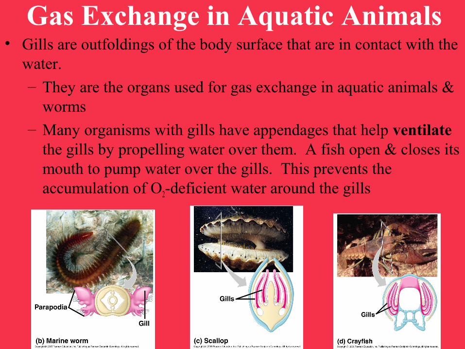
Gas Exchange in Aquatic Animals• Gills are outfoldings of the body surface that are in contact with the
water.
– They are the organs used for gas exchange in aquatic animals & worms
– Many organisms with gills have appendages that help ventilate the gills by propelling water over them. A fish open & closes its mouth to pump water over the gills. This prevents the accumulation of O2-deficient water around the gills
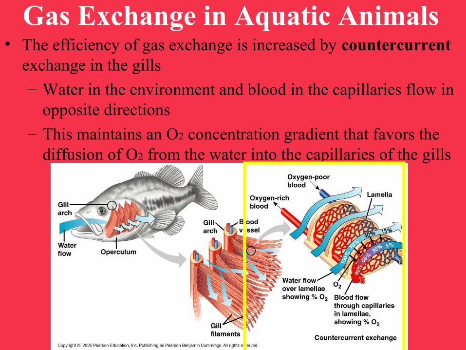
Gas Exchange in Aquatic Animals• The efficiency of gas exchange is increased by countercurrent
exchange in the gills
– Water in the environment and blood in the capillaries flow in opposite directions
– This maintains an O2 concentration gradient that favors the diffusion of O2 from the water into the capillaries of the gills
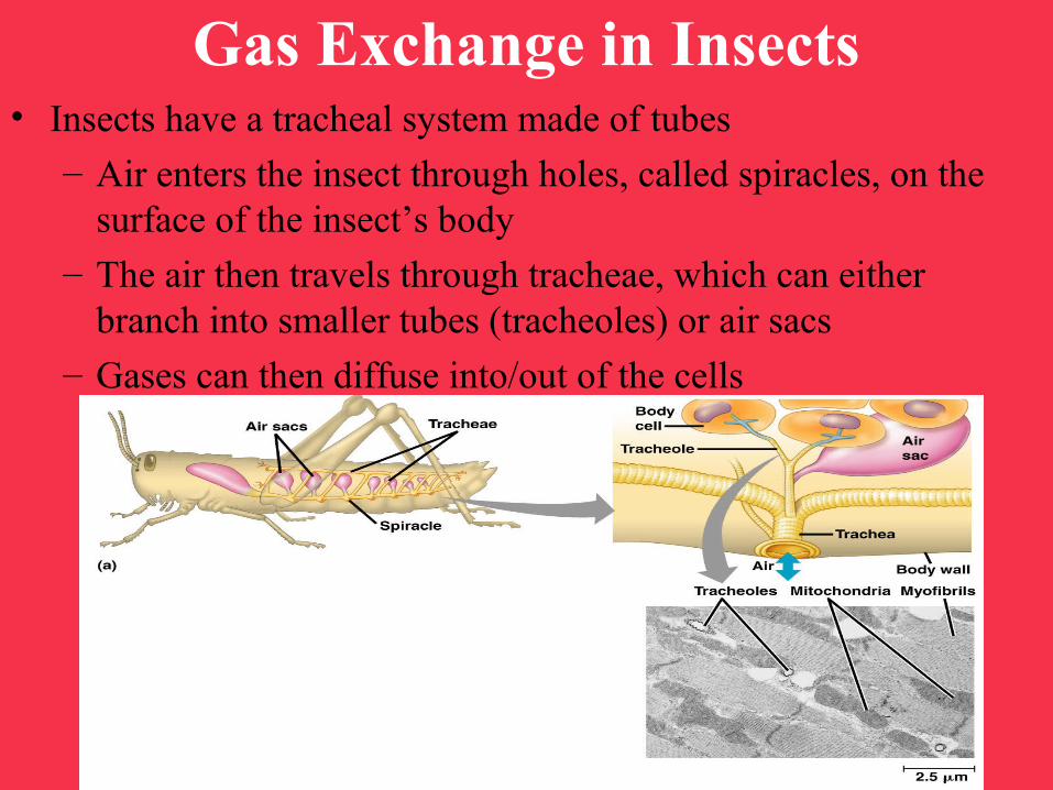
Gas Exchange in Insects• Insects have a tracheal system made of tubes
– Air enters the insect through holes, called spiracles, on the surface of the insect’s body
– The air then travels through tracheae, which can either branch into smaller tubes (tracheoles) or air sacs
– Gases can then diffuse into/out of the cells
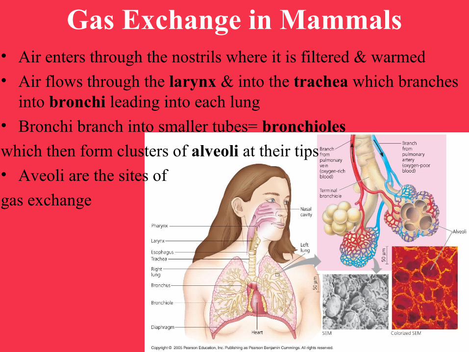
Gas Exchange in Mammals• Air enters through the nostrils where it is filtered & warmed
• Air flows through the larynx & into the trachea which branches into bronchi leading into each lung
• Bronchi branch into smaller tubes= bronchioles
which then form clusters of alveoli at their tips
• Aveoli are the sites of
gas exchange
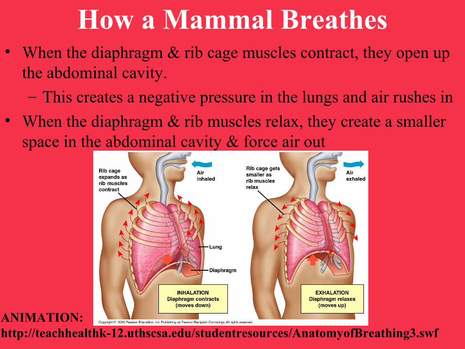
How a Mammal Breathes• When the diaphragm & rib cage muscles contract, they open up
the abdominal cavity.
– This creates a negative pressure in the lungs and air rushes in
• When the diaphragm & rib muscles relax, they create a smaller space in the abdominal cavity & force air out
ANIMATION:http://teachhealthk-12.uthscsa.edu/studentresources/AnatomyofBreathing3.swf