apheresis - ccrod.cancer.gov
TRANSCRIPT

4/26/2021
1
ApheresisSURGERY BRANCH APHERESIS UNIT
Presentation Objectives Brief history of Apheresis
Different devices
What we use today
Different Apheresis procedure types
Surgery Branch Protocols
Surgery Branch Cell Processing
Types of Vascular Access
Pre‐Procedure considerations
Scheduling
Anticoagulant
1
2

4/26/2021
2
Presentation ObjectivesSide Effects and Adverse Reactions
Troubleshooting
Post procedure instructions and considerations for the Research team
History of ApheresisThe term “apheresis” a Greek word for “to separate” or “to remove”
In the early 1950s doctors were looking for a way to separate donor plasma during the whole blood donation process. The desired component needed to be separated.
One of the early designs was the “Latham bowl” which was first marketed in a device for washing or deglycerolizing RBCs (Haemonetics Model 10). It was later adapted for “intermittent‐flow” apheresis used for platelets.
In mid 1960 at NCI a continuous‐flow centrifugal blood separator was developed. This was used for WBC collection and depletion but also of exchanging plasma or RBCs.
In the 1970s another line of continuous‐flow devices by Fenwal and the National Institutes of Health were developed and paved the way for manufacturing of these seallesscentrifuge/separators.
3
4

4/26/2021
3
History As the instruments became more advanced the apheresis devices were updated to make procedures safer, more efficient and easier for operators.
The extracorporeal volume (the volume of blood outside the patient’s body during apheresis) was lowered to reduce volume depletion
Alarms and monitors were improved
Programs became more automated
The size of machines were reduced to make them more portable
The capacity of devices to perform different procedures types such as exchanges, cellular collections, cellular depletions
Haemonetics – MCS +
https://www.medicalexpo.com/prod/haemonetics/product‐78504‐520938.html
5
6

4/26/2021
4
Fenwal CS‐3000 Blood Separator
Cobe Spectra
7
8

4/26/2021
5
Spectra Optia
9
10
4/26/2021
6
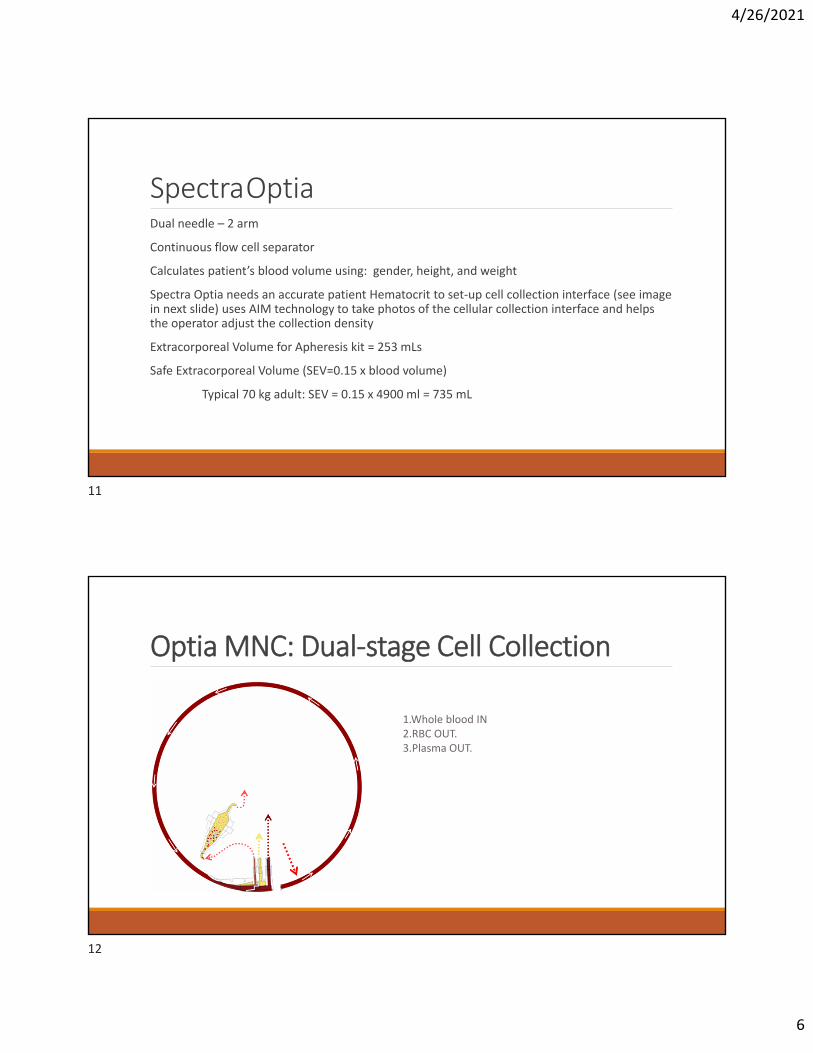
SpectraOptiaDual needle – 2 arm
Continuous flow cell separator
Calculates patient’s blood volume using: gender, height, and weight
Spectra Optia needs an accurate patient Hematocrit to set‐up cell collection interface (see image in next slide) uses AIM technology to take photos of the cellular collection interface and helps the operator adjust the collection density
Extracorporeal Volume for Apheresis kit = 253 mLs
Safe Extracorporeal Volume (SEV=0.15 x blood volume)
Typical 70 kg adult: SEV = 0.15 x 4900 ml = 735 mL
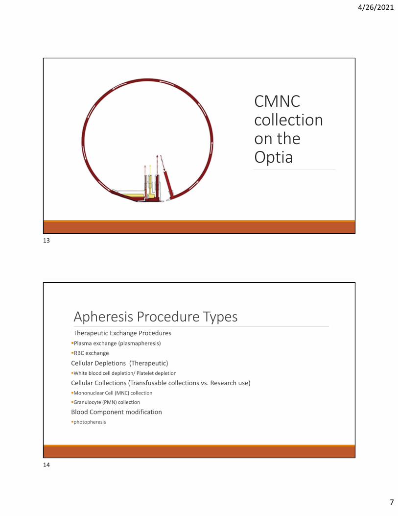
OptiaMNC: Dual‐stage Cell Collection
1.Whole blood IN 2.RBC OUT. 3.Plasma OUT.
11
12
4/26/2021
7
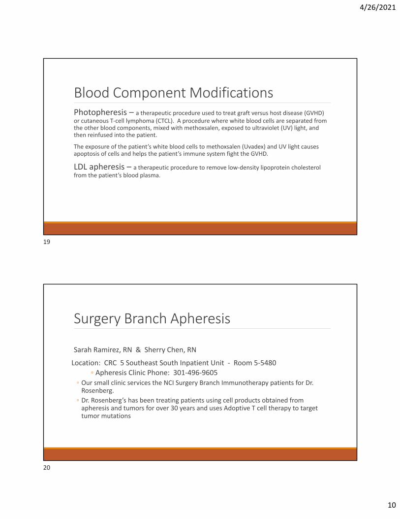
CMNC collection on the Optia
Apheresis Procedure TypesTherapeutic Exchange Procedures
Plasma exchange (plasmapheresis)
RBC exchange
Cellular Depletions (Therapeutic)
White blood cell depletion/ Platelet depletion
Cellular Collections (Transfusable collections vs. Research use)
Mononuclear Cell (MNC) collection
Granulocyte (PMN) collection
Blood Component modification
photopheresis
13
14

4/26/2021
8
TPETherapeutic plasma exchanges are used to treat a variety of autoimmune diseases. The goal of these procedures is to remove and therefore reduce the large number of disease‐causing antibodies in the patient’s plasma portion of the blood. These antibodies are attacking the body and causing problematic symptoms.
Examples:
1. TTP ‐ Thrombotic thrombocytopenic purpura
2. Myasthenia gravis
3. CIDP ‐ Chronic inflammatory demyelinating polyradiculoneuropathy
4. Antibody mediated organ rejection (kidney, heart)
5. Hyperviscosity syndrome
6. Cryoglobulinemia
RBCX Red Blood cell exchange procedures are most commonly used for the management of symptoms for patients with sickle cell disease. These procedures can lower RBCs containing hemoglobin S (HbS) and rapidly replaces them with healthy RBCs while maintaining an isovolemic fluid balance. These procedure also reduces viscosity and can increase the patient’s hematocrit.
Red Blood cell exchange is indicated for severe complications related to SCD such as: acute stroke, acute chest syndrome, and priapism.
Red Blood cell exchange can be used for other rare indications such as treatment of severe infections of malaria or other intraerythrocytic pathogens.
15
16

4/26/2021
9
Cellular DepletionsA white blood cell depletion is a therapeutic procedure used to reduce the number of white cells in the patient’s blood.
This can be used for acute leukemia when the patient’s white blood cell burden is very high and needs to be reduced prior to chemotherapy being started. A depletion can reduce the white blood cell count by 10‐70%.
Platelet depletions, another cellular depletion can be done to therapeutically remove platelet for conditions such as essential thrombocytosis.
Cellular CollectionsMNC collections
A collection of the patient’s buffy coat or white blood cell layer.
This procedure collects MNCs from whole blood based on the cell size or gravity.
These procedures can be for research use or for transfusion. Transfusion can be for autologous use or allogeneic use
.
17
18
4/26/2021
10
Blood Component Modifications Photopheresis – a therapeutic procedure used to treat graft versus host disease (GVHD) or cutaneous T‐cell lymphoma (CTCL). A procedure where white blood cells are separated from the other blood components, mixed with methoxsalen, exposed to ultraviolet (UV) light, and then reinfused into the patient.
The exposure of the patient’s white blood cells to methoxsalen (Uvadex) and UV light causes apoptosis of cells and helps the patient’s immune system fight the GVHD.
LDL apheresis – a therapeutic procedure to remove low‐density lipoprotein cholesterol from the patient’s blood plasma.
Surgery Branch Apheresis
Sarah Ramirez, RN & Sherry Chen, RN
Location: CRC 5 Southeast South Inpatient Unit ‐ Room 5‐5480◦ Apheresis Clinic Phone: 301‐496‐9605
◦ Our small clinic services the NCI Surgery Branch Immunotherapy patients for Dr. Rosenberg.
◦ Dr. Rosenberg’s has been treating patients using cell products obtained from apheresis and tumors for over 30 years and uses Adoptive T cell therapy to target tumor mutations
19
20

4/26/2021
11
Surgery Branch Protocols TIL and TCR studies
The Surgery Branch is studying how Adoptive cell therapy, a type of immunotherapy, can help cause tumor regression in common epithelial cancers.
Our patients typically have stage 4 cancer, including:
Metastatic melanoma
Gastrointestinal cancers (esophageal, gastric, duodenal, colon and rectal)
Hepatobiliary cancers (cholangiocarcinoma, pancreatic, hepatocellular)
Breast cancer
Ovarian cancer
Non‐small cell lung cancer
Metastatic kidney cancer
TIL and TCRTIL ‐ Adoptive cell therapy using TIL, tumor infiltrating lymphocytes, a therapy pioneered by the NCI Surgery Branch. This therapy uses a patient’s own autologous tumors cells which are extracted by surgically removing a tumor. Then the TIL cells are grown in wells and tested for tumor recognition. Autologous MNC apheresis cells are collected and used to stimulate the patient’s TILs to rapidly expand. They are tested again before being given to the patient.
The TCR protocols are another type of adoptive cell therapy that use genetically modified T cells to treat certain cancers. In these protocols apheresis cells are collected from the patient and genetically modified to recognize specific target proteins on the patient’s tumor. Before the patient receives their newly modified cells, they are given a lymphodepleting chemotherapy regimen.
21
22
4/26/2021
12
SB Cell ProcessingThe processing of apheresis cells by the Surgery Branch Cell processing facility occurs very soon after the apheresis procedure is complete.
Lymphocyte separation medium is used to separate red blood cells from lymphocytes in the apheresis product.
The cells are then washed a few times to further separate out red blood cells.
The cells are then frozen by cryopreserving them in liquid nitrogen
SB Cell Uses and ProcessingThe MNC cells collected during apheresis are used in a few different ways by the Surgery Branch Cell Processing Facility.
The lymphocytes are used as feeder cells in the REP, or rapid expansion phase of our protocols. Cells are thawed, counted and irradiated. The feeder cells used in the REP are irradiated so that they themselves will not expand. Cells are then combined with IL‐2 and patient lymphocytes from TIL in medium to expand for 14 days.
For Gene Therapy (TCR) protocols cells collected from apheresis are the starting material. The cells are thawed and stimulated with anti‐CD3. They are then transduced with T‐cell receptors (TCRs) that are chosen for their ability to recognize certain tumor neo‐antigens. Once the cells have grown for 10 days, they are rapidly expanded using other apheresis cells.
Apheresis cells collected are also used in SB research studies to screen for neoantigens and determine which neoantigens gene therapy treatments will target.
23
24

4/26/2021
13
SB PatientsThe first step is the patient referral process. The Surgery Branch referral nurses have the very tough job of screening these complex patients and bringing in the patients that are most likely eligible. Once they are further evaluated for eligibility at NIH, the patients will typically undergo an operation and a large volume MNC, Mononuclear cell apheresis collection where 15‐20 liters of whole blood are processed. The harvested patient cells then take between 3 weeks – 4 months to be grown and processed in the SB Cell processing labs. Some patients will return home to their oncologist to go back on standard chemotherapy regimens during this time. Once the cells are ready and the patient is ready, they will return to NIH for their lymphodepleting chemo and cell infusion. Our patients will be inpatients for about 3 weeks. After they are discharged, they will come back and have another short apheresis procedure on their first follow‐up visit.
Vascular Access Apheresis procedures require an access that can accommodate blood flow rates of up to 100 ml/minute, this is much less than hemodialysis (~400 mL/minute)◦ Examples of Access include:
◦ Central venous catheters (tunneled or non‐tunneled)
◦ AV Fistula/AV Graft
◦ Peripheral veins
◦ Implanted ports (PowerFlow Implantable Apheresis Port, Vortex port) these are special high flow ports designed for long term apheresis use only
Many factors are considered when deciding between using peripheral access versus central access.
25
26
4/26/2021
14
Factors to consider for accessSize and quality of peripheral veins
Location of peripheral veins
Hydration status
Prior chemotherapy history
Prior venipuncture issues
Prior Surgeries, especially axillary lymph node dissections/lymphedema
Frequency of apheresis procedures
Peripheral AccessPeripheral access – Peripheral access is preferred if possible due to lower risk of infection and thrombosis associated with central catheters.
Draw site (inlet) – 17 gauge Hemodialysis fistula needle set & 18 gauge Apheresis needle set –must be in an antecubital vein
Return site – 18 or 20 gauge IV catheter – can be a hand, wrist, forearm, or antecubital vein
27
28

4/26/2021
15
Apheresis needles
18 or 20 gauge IV catheters for return lines
29
30

4/26/2021
16
Vascular Access‐ Central linesIf patient does not have adequate peripheral veins, or cannot have peripheral venipunctures, a central line is needed
Femoral and IJ lines – femoral lines are preferred (IJ lines require chest X‐ray placement confirmation)
Double lumen
Able to accommodate high flow rates at low venous pressures
Preferred catheters (for Surgery Branch Apheresis patients)
Mahurkar – 10 or 11.5 French X 12 cm (non‐tunneled)
Arrow – central venous catheter
Mahurkar catheter
31
32

4/26/2021
17
Pre‐screening and Venous Assessment
During Covid, we have done many prescreening telephone calls with our scheduled apheresis patients. Under normal circumstances, patients are seen for a venous assessment in the outpatient clinic during a pre‐screen visit with the Immunotherapy team.
We explain the procedure in detail (2 arms, immobility, use of a bed pan/urinal during apheresis)
We explain the type of venous options and possible need for a central line.
We discuss the importance of having breakfast the morning of the apheresis procedure and hydrating well 24‐48 hours prior to coming for apheresis.
We also encourage the patient to limit caffeine and fluids on the morning of the apheresis to reduce the number of times the patient may need to use the bedpan/urinal.
Special Considerations for a Successful ProcedurePatient size – small patient = low blood volume = slow flow rates
Low WBC count = low cell yield in apheresis product
High platelet count (above 200), high citrate concentration needed, slower procedure rate, possible low cell yield
Low hgb/hct = difficult interface and poor efficiency (low cell yield)
Poor venous access‐slow/intermittent flow, poor efficiency
Patient anxiety, and/or pain – patient has a difficult time maintaining position
Prior treatment (chemo, radiation, Immunotherapy)‐ poor product yield
33
34

4/26/2021
18
Pre‐procedure considerationsThe patient’s overall condition is important in determining how the apheresis will go.
Things to consider:
Low hgb/hct = difficult cell collection interface and poor efficiency (low white blood cell yield)
patients need Hgb of at least 8 (Dowling prefers Hgb ≥ 9.0 g/dL, platelet ≥ 50,000, WBC greater than 2,000
Poor venous access‐slow/intermittent flow, poor collection efficiency
Patient anxiety, and/or pain‐ vasospasms the patient has trouble remaining in a good position
Prior treatment (chemo, radiation, Immunotherapy)‐ patient’s bone marrow is suppressed and does not have healthy, plentiful mononuclear cells to collect
Is the patient taking any medications that will interfere with apheresis (blood thinners, steroids, aspirin)
Pre‐procedure considerations (cont.)Patient’s hydration status – dehydration, flat veins, difficult venipunctures
Recent surgery – does the patient have a chest tube or drain that will cause them to have a difficult time remaining still in the proper “apheresis position”
Lymphedema, (number of Lymph nodes removed) can that arm be used for a return site?
Does the patient have pain or abdominal tumors that will make it hard for them to lie flat for 3‐5 hours
The patient needs to eat a good breakfast the morning of the apheresis.
35
36

4/26/2021
19
Communication with TeamsIn Surgery Branch the Research Nurse calls the Apheresis team directly to schedule a patient for apheresis
An Apheresis procedure orders must then be entered in CRIS ◦ The order should include the desired number of liters of whole blood to be processed for the procedure (for example 5, 15, or 20). For Dowling, the CRIS order should be appropriate for desired cell component (example, Mononuclear cell (MNC) Apheresis Procedure for Lymphocyte & Monocyte collections & Hematopoetic Progenitor Cell (HPC) Procedure order for a peripheral blood stem cell collection)
◦ Please list under special instructions section, any lab work that should be draw in apheresis. This can be drawn at the start of the procedure. (This may be different for Dowling)
◦ Patients need to be signed onto the Surgery Branch Cell Harvest protocol before starting apheresis.
Scheduling Patients for Apheresis –Dowling ClinicThe best way to request an appointment with Dowling Clinic is to request through CRIS Appointment Request, under “Dowling Apheresis Clinic Appointment Request”
The scheduler will check the Dowling nurses’ availability along with availability in CCE, for cell processing. If Dowling Clinic can accommodate the request, their scheduler will confirm the appointment through CRIS.
Patients/Donors also need to be seen by Dowling Clinic for a venous assessment prior to apheresis. This is to determine if they may need a central line for the procedure.
Dowling Clinic Team Lead: Monica Ford ◦ [email protected]
◦ (301) 496‐1430
37
38
4/26/2021
20
Anticoagulant for Cellular Collections
ACDA‐ Anticoagulant Citrate Dextrose Solution Formula A‐is used as the anticoagulant during most cellular collection procedures including MNC and HPC procedures
Binds to patient’s calcium, makes calcium unavailable for the clotting cascade, preventing clotting in the extracorporeal circuit
Is metabolized quickly after patient leaves apheresis
Causes hypocalcemia – circumoral paresthesia, feelings of vibration muscle cramping, can progress to tetany
Calcium replacementCalcium chloride IV is infused continually for most apheresis procedures to prevent citrate toxicity and symptoms of hypocalcemia
Oral calcium tablets (TUMS) can also be given as needed for shorter procedures and Normal volunteer Apheresis procedures, such as platelet collections.
39
40

4/26/2021
21
Side Effects /Adverse Reactions
1. Citrate Reaction /Toxicity
Causes:
Smaller patients, hypocalcemia,
Long procedure, high concentration of Citrate used,
Fast inlet flow rate / citrate infusion rate.
Signs and symptoms:
Circumoral paresthesia,
Nausea/vomiting, diarrhea, Metal taste in mouth,
Light headache,
Tetany, chest pain,
Hyper anxiety, panicked feeling, sense of impending doom.
ContinuedTreatment:
Mild to moderate citrate toxicity◦ Increase IV Calcium chloride infusions
◦ Slow the inlet flow rate
◦ pause /restart the procedure at lower flow rate
◦ Increase Inlet: AC ratio
Severe citrate toxicity (Chest Pain, Muscle Spasms, Tetany)◦ Stop the procedure immediately, notify MD.
◦ Breathe into a paper bag.
41
42

4/26/2021
22
Continued2. AnxietySymptoms:
Restlessness, perspiration, flushed face, cold clammy hands,
Tachycardia and elevated blood pressure.
To prevent or reduce anxiety:
Explain apheresis process prior to procedure.
Create a friendly atmosphere.
Reasonably explain side effects.
Continued3. Vasovagal Reaction
A temporary drop in the amount of blood that flows to the brain.
Causes:
Dehydration,
Intense emotional stress, anxiety, fear, pain.
Sign and symptom:
Pallor, cold, perspiration, dizziness, light headache,
Nausea/vomiting, incontinence of urine or stool,
Bradycardia, hypotension, seizure, feeling doom.
43
44

4/26/2021
23
ContinuedTreatment:
Pause the procedure immediately,
Place patient in a flat position or Trendelenburg position immediately.
Administer normal saline in inlet and return line to keep veins open.
Take slow, deep breaths,
Apply cold compresses to head.
Encourage fluid intake,
Monitor patient’s vital signs.
Stop the procedure, notify MD.
Continued4. Vascular access complications—hematoma/ infiltration
Signs and symptoms:
Pain, swelling at the needle site, poor venous access alarms.
Treatment:
Remove needle immediately,
Apply moderate pressure to the site until bleeding stops. Cover with sterile gauze.
Apply cold pack for 5 minutes. May repeat if swelling is worse.
45
46

4/26/2021
24
Continued5. Rare reactions:
Air embolism
◦ Signs and symptoms:
Chest pain, SOB, pallor, tachycardia, hypotension, diaphoresis, change mental status, syncope.
◦ Treatment:
Stop procedure immediately,
Clamp the return line if there is visible air,
Place patient in the Trendelenburg position,
Call code
ContinuedAllergic reaction due to ethylene oxide used in sterilization of apheresis kit.
Actions:
oStop procedure immediately,
oMaintain vascular access using saline,
oNotify MD. Oral or IV antihistamines,
oIf severe, call emergency service.
47
48

4/26/2021
25
Troubleshooting /managing /maintaining the interfacePlatelet clumping: Frequently observed during procedure and difficult to predict ,
Traveling through the collect port in the connector,
Affect collection efficiency,
May lead to clotting in the system and terminate the procedure.
Causes:
Underlying disease,
Mobilization regimen,
Medications, especially chemotherapeutic agents,
Centrifugal speed of the instrument.
ContinuedActions:
Decrease Inlet: AC ratio until the clump disappears,
Monitor the connector for clumping,
If the clumping is resolved, increase AC infusion rate gradually,
If the clumping persists, leave AC infusion rate until the clumping disappears or for remainder of the run.
Monitor patient’s Citrate reaction.
49
50

4/26/2021
26
ContinuedPoor Inlet flow rate
Causes:
Vein spasm, inappropriate needle position, blood clotting and infiltration.
Actions:
Apply warm pack, Bair Hugger blanket, squeeze ball,
Adjust needle,
Flush the line with saline,
Restart IV,
Central line: reposition patient, offer bedpan/urinal, flush lumens with saline, tPA.
Post Procedure InstructionsKeep pressure dressings for 3‐4 hours,
Avoid alcohol and caffeine for 8 hours, drinking plenty of fluid for the next 24 hours,
Restrict strenuous exercise for the next 24 hours,
Use elevator, avoid stairs for 6 hours,
Lie down and elevate feet if feeling dizzy or lightheaded.
Post central line removal: a. Lie on bed for one hour,b. Walk as little as you can for 12 hours,
c. No heavy lifting for three days, no shower in 24 hours,
d.Watch out bleeding and signs of infection,
e. Call MD or go to nearby hospital for emergency.
51
52
4/26/2021
27
Post Procedure considerations for the teamLarge volume apheresis procedures (greater than 10 liters processed) will cause hemodilution (especially 20 Liters), resulting in patient’s lowered hgb/hct‐ will the patient require a RBC transfusion?
apheresis can decrease the patient’s platelet count by 40‐60% ‐ patients need to be alerted to increase risk of bleeding. The patient’s baseline platelet count should return in 48 hours.
We will draw a post‐apheresis CBC, team should expect things to look very different from pre‐apheresis CBC
If a central line was placed for the procedure and will be removed in the apheresis clinic, the patient will need to plan to remain in apheresis for about 2 hours after the procedure. The patient will be instructed to monitor their central line dressing and notify the team for any signs of bleeding or infection.
ReferencesTherapeutic apheresis: history, clinical application, and lingering uncertaintieshttps://onlinelibrary.wiley.com/doi/full/10.1111/j.1537‐2995.2009.02505.x
Therapeutic Plama Exchange
https://utswmed.org/conditions‐treatments/therapeutic‐plasma‐exchange/
Red Blood Cell Exchange (RBCX)
https://www.terumobct.com/rbcx
Red Blood Cells: Exchange, Transfuse, or Deplete
https://www.ncbi.nlm.nih.gov/pmc/articles/PMC6944943/#:~:text=The%20most%20frequent%20indication%20for,other%20acute%20complications%20of%20SCD.
53
54
4/26/2021
28
ReferencesLeukocyte Depletion by Therapeutic Leukocytapheresis in Patients with Leukemia
https://www.ncbi.nlm.nih.gov/pmc/articles/PMC3434324/
https://www.mskcc.org/cancer‐care/patient‐education/frequently‐asked‐questions‐about‐photopheresis
55