apoptosis and cancer stem cell
TRANSCRIPT

Apoptosis And Cancer stem Cell
Ayush Kaundal
M.tech. Biotechnology (14btmt04)
Shoolini University.

Apoptosis
• Apoptosis came from Greek origin; meaning “falling off or dropping off”. (Gewies,2003) It resembles leaves falling off trees or petals dropping off flowers
• Programmed cell death
• Essential for development and survival of living organisms
• Sequentially regulated suicidal programme where cells activate certain enzymes which dissolute their own nuclear component and various protein component of nucleus and cytoplasm

Cell death Mechanisms
Death by suicide
Death by suicide Death by injuryDeath by injury

Morphological Changes in Cell Death
In necrosis changes occur in mitochondria first whereas changes in nucleus are very less.
1.Cell membrane’s Selective permeability is lost and Dissolution of cell organelles
2.So pore formation in cell membrane occur
3.Intracellular content is released (hydrolases )
4.Hydrolases causes cell degradation
5.Surrounding cells are degraded and strong inflammatory responses occurs in corresponding tissues
(Searle et al., 1982; Ramírez et al., 1999)

Biochemical mediators Execution phase
Attempts to repair the damaged cell
Structural changes happens which leades to the cell death
First nuclear changes occurs and then cell membrane changes and then changes in intracytoplasmic organelles
(Kerr et al., 1972; Chamond et al., 1999)
Morphological Changes in Cell Death
In Apoptosis cell death occurs in two stages

In the nucleus, changes in both chromatin as well as nuclear membrane happen.
Chromatin become dense clumps which shifts towards the nuclear membrane and nuclear membrane remain intact.
In mitochondria there is degradation of DNA occurs.
Endoplasmic reticulum loss its structure and there is loss of the mitochondrial transmembrane potential.
The cytoplasmic membrane of apoptotic cell become deformed and it develop blebbing.
In endoplasmic reticulum the cisterns become wide and they fuse.
(Ramírez et al., 1999)
Morphological Changes in Cell Death

The phospholipids of cell membrane change their orientation and get exposed to external environment.
The fragment of cell membrane form apoptotic bodies which is actually cytoplasmic remains surrounded by cell membrane.
When the apoptotic bodies are released in external environment they are engulfed by phagocytes. As a result there is no inflammatory reaction.
At molecular level there is the activation of proteolytic enzymes which propogate the cleavage of DNA into oligonucleosomal fragments and cleavage of a multitude of specific protein substrates
(Ramírez et al., 1999)
Morphological Changes in Cell Death

Why should a cell commit suicide?
Programmed cell death is needed for proper normal development as mitosis Examples: Death of cells (During limb formation) are essential to sculpture hollow structure and formation of reproductive organ, Mullerian duct deletion is essential for male and Wolffian duct deletion is mandatory for female (Gewies, 2003)
The resorption of the tadpole tail in frog
The formation of the fingers and toes of the fetus requires the removal by apoptosis
The formation of the proper connections (synapses) between neurons in the brain

Programmed cell death is needed to destroy cells that represent a threat to the integrity of the organism
Examples:-
Cells infected with viruses
Cells of the immune system
Cells with DNA damage
Cancer cells (Uncontrolled proliferated cells)
Why should a cell commit suicide?

Apoptosis in physiologic situations• Programmed destruction during embryogenesis
• Involution of hormone dependent tissues
• Cell loss in proliferating cell populations
• Elimination of harmful self- reactive lymphocytes
• Death of host cells
Why should a cell commit suicide?

Apoptosis in bud formation during which many interdigital cells die.
Incomplete differentiation in two toes due to lack of apoptosis

Why should a cell commit suicide?
Apoptosis in pathological conditions • DNA damage
• Accumulation of misfolded proteins
• Cell death in certain infections
• Pathological atrophy in parenchymal organs

Cancer And Apoptosis
• One of the major threats to public health at present world
• Worldwide are lung cancer, stomach cancer, colorectal cancer, liver cancer and breast cancer (World Health Organization- NMH Fact sheet, 2010) cancer of head and neck regions, specially in oral cavity
• In this disease, a normal cell transformed into a malignant one due to succession of genetic changes. On the other hand apoptosis is known to eliminate potentially malignant cells, hence reduction of apoptosis can be considered to play a key role in carcinogenesis
Commonly there are three mechanisms by which apoptosis acquire resistance or reduction.
1. Disruption in balance of proapoptotic and antiapoptotic proteins
2. Reduction in function in caspases
3. Impaired death receptors signaling

Inactivating p53 Pathway in Cancer
http://p53.free.fr/p53_info/p53_cancer.html

p53 inactivation in human cancer
• p53 mutations can be found in 50% of human cancers, but their penetrance is highly heterogeneous, as reflected by the diverse remaining transactivation activity that ranges from O to 100%.
• Various DNA viruses, such as SV40, HPV or adenoviruses, encode proteins that target p53 protein.
• mdm2 accumulation is found in numerous cancers, such as sarcoma or breast carcinomas.
• PTEN, a p53 regulated gene, is mutated in various types of cancer including glioblastoma.
• Although no mutation of AKT has been found in human cancer, constitutive activation of its kinase activity has been observed via deregulation of the upstream pathway.
• Mutations in various pathways upstream of p53 (ATM, p19ARF or Hcdk2 gene) can be observed in various types of cancer.
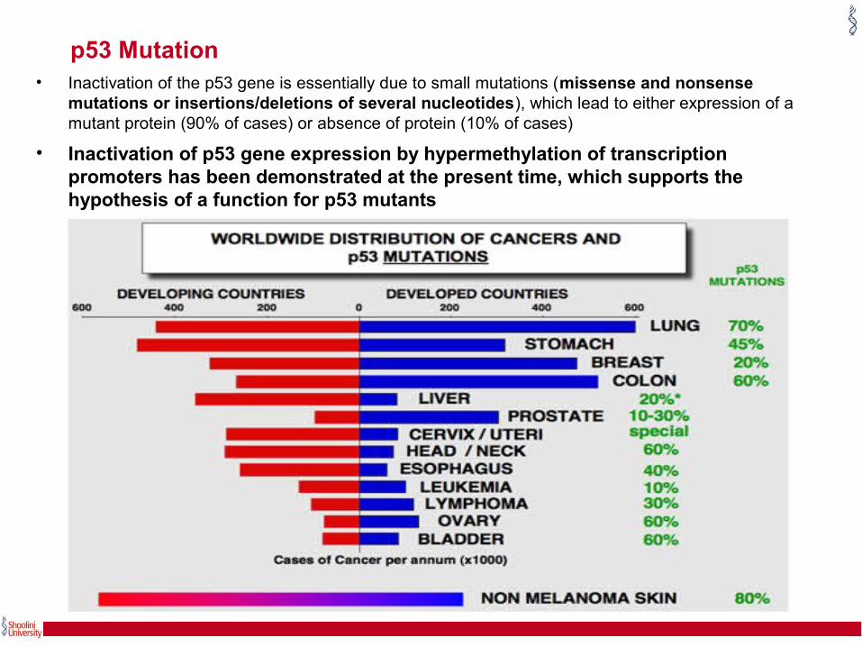
p53 Mutation• Inactivation of the p53 gene is essentially due to small mutations (missense and nonsense
mutations or insertions/deletions of several nucleotides), which lead to either expression of a mutant protein (90% of cases) or absence of protein (10% of cases)
• Inactivation of p53 gene expression by hypermethylation of transcription promoters has been demonstrated at the present time, which supports the hypothesis of a function for p53 mutants

HPV (Human papillomavirus) Infection • The E6 viral protein expressed by HPV specifically binds to the p53 protein and induces its
degradation (Scheffner et al., 1990).
• This observation explains the rarity of p53 mutations in cervical cancers (Crook et al., 1992).
• p53 inactivation by a viral protein has not been formally demonstrated in other human cancers associated with viral infection, such as HCC (Hepatocellular carcinoma) or Burkitt lymphoma.

Nuclear exclusion
In inflammatory breast cancers or neuroblastomas, molecular and immunohistochemical analyses demonstrate accumulation of wild-type p53 in the cytoplasm of tumour cells, leading to functional inactivation of p53 (Moll et al., 1995; Moll et al., 1996; Moll et al., 1992)

MMD2 (monocyte to macrophage differentiation-associated 2) Amplification
• The mdm2 protein regulates the stability of the p53 protein by transport towards the proteasome. Abnormal accumulation of the mdm2 protein is observed in many tumours, especially sarcomas.
• This accumulation can be due to amplification of the mdm2 gene, enhanced transcription of the gene or enhanced translation of its messenger RNA (Michael and Oren, 2002).
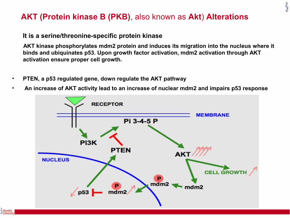
AKT (Protein kinase B (PKB), also known as Akt) Alterations
It is a serine/threonine-specific protein kinase
AKT kinase phosphorylates mdm2 protein and induces its migration into the nucleus where it binds and ubiquinates p53. Upon growth factor activation, mdm2 activation through AKT activation ensure proper cell growth.
• PTEN, a p53 regulated gene, down regulate the AKT pathway
• An increase of AKT activity lead to an increase of nuclear mdm2 and impairs p53 response

PTEN (Phosphatase and tensin homolog ) mutations
• AKT kinase phosphorylates mdm2 protein and induces its migration into the nucleus where it binds and ubiquinates p53. Upon growth factor activation, mdm2 activation through AKT activation ensure proper cell growth.
• PTEN, a p53 regulated gene, down regulate the AKT pathway.
• PTEN deletion leads to an increase of AKT activity, an increase of nuclear mdm2 and impairs p53 response
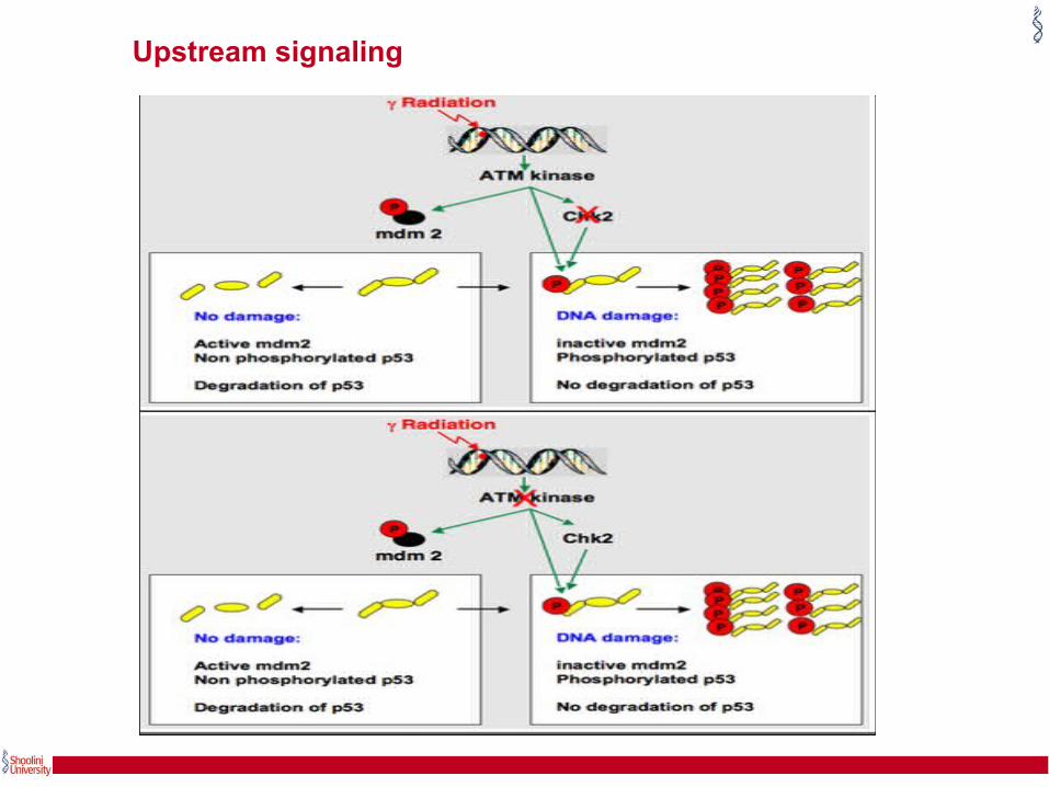
Upstream signaling

Upstream signaling
• At G1, as a consequence of DNA damage induced by gamma radiation and it activate ATM.
• Activated ATM phosphorylates p53 on ser15, CHK2 on thr68, and MDM2 on ser395.
• Subsequently, activated chk2 phosphorylates p53 on ser20.
• Together, these phosphorylations interfere with p53 binding to mdm2, leading to stabilization and activation of p53.
• Mutations of ATM in t-cell leukaemia impair the p53 response after gamma radiation.
• The function of chk2 in this pathway has been recently questioned and remains controversial.

• Bcl-2 family proteins play a vital role in regulation of apoptosis via intrinsic pathway predominantly.
• They are comprised of both pro-apoptotic and anti-apoptotic proteins. Disruption in the balance of proapoptotic and antiapoptotic protein causes disregulated apoptosis.
• p53 directly interact with members of the Bcl-2 family and influences apoptosis.
• In the cytosolic p53 apoptotic pathway, nuclear p53 induces PUMA expression, which in turn releases cytosolic p53 held inactive in the cytoplasm through binding to BclXL.
• Then, cytosolic p53 induces Bax oligomerization and mitochondrial translocation.
• In the mitochondria, p53 induces Bax and Bak oligomerization, and forms a complex with cyclophilin D in the mitochondrial inner membrane.
• These changes result in marked disruption of mitochondrial membranes and subsequent release of both soluble and insoluble apoptogenic factors.
p53 role in apoptosis

Cytosolic and mitochondrial p53 apoptotic pathways
http://www.discoverymedicine.com/Joana-D-Amaral/2010/02/20/the-role-of-p53-in-apoptosis/

PLAYERSCYTOCHROME:- Small hemoprotein found loosely associated with the inner
membrane of the mitochondrion
Bcl-2 family:- (Proapoptotic and anti apoptotic protein)
• Pro apoptotic proteins are Bax, Bak, Bad, Bcl-Xs, Bid, Bik, Bim and Hrk and anti apoptotic proteins are Bcl-2, Bcl-XL, Bcl-W, Bfl-1 and Mcl-1
• Pro apoptotic protein promote release of cytochrome-C from mitochondria whereas antiapoptotic proteins causes its blockage.
Bax:- Proapoptotic Protein
TRADD:- (TNFRSF1A-associated via death domain) It is a protein-coding gene (Disease Associated).
Fasl:- Fas ligand (FasL or CD95L) is a type-II transmembrane protein that belongs to the tumor necrosis factor (TNF) family
APAF1:- Apoptotic protease activating factor 1
Cytoplasmic protein,
Contains (from the N terminal) a caspase recruitment domain (CARD)
APOPTOSOME:- (Cytochrome c + Apaf-1) protein Complex

PLAYERS ATM :- (Ataxia telangiectasia mutated) It is a serine/threonine protein
kinase that is recruited and activated by DNA double-strand breaks.
p53
• It is upregulated modulator of apoptosis (PUMA) also known as bcl-2-binding component 3 (BBC3).
• It is a pro-apoptotic protein, member of the bcl-2 protein family.
• In humans, the bcl-2 binding component 3 protein is encoded by the bbc3 gene.
• The p53 tumor suppressor acts to integrate multiple stress signals into a series of diverse antiproliferative responses.
• One of the most important p53 functions is its ability to activate apoptosis, and disruption of this process can promote tumor progression.
(Jordan et al., 2003)

CASPASES:- Caspases are proteases that have cysteine © residues at their active site and cleave after aspartic acid (Asp) residues in their substrate proteinCaspase- 8, Caspase- 9acts as an initiator of the caspase activation cascadeCaspase-3key effector in the apoptosis pathway, amplifying the signal from initiator caspases and signifying full commitment to cellular disassembly
PLAYERS

Initiation
• Absence of stimuli - hormones, growth factors
• Activation of receptors – TNF family
• Heat ,radiation, chemicals
• Genetically programmed events

PATHWAY OF APOPTOSIS

Intrinsic Pathway

APOPTOSOME

Extrinsic Pathway
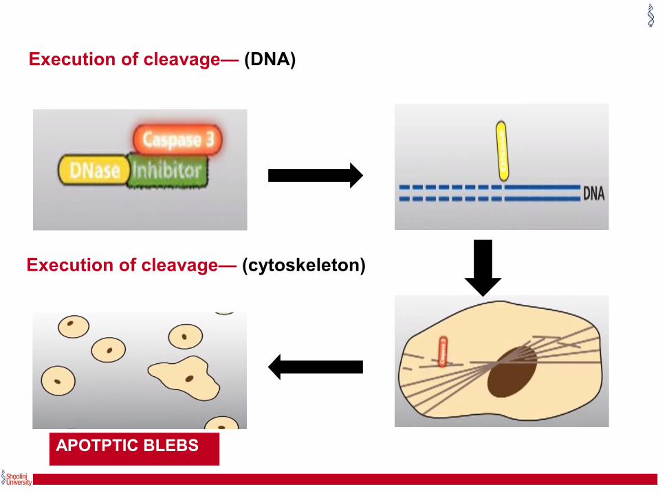
Execution of cleavage— (DNA)
Execution of cleavage— (cytoskeleton)
APOTPTIC BLEBS

Phagocytosis


Conclusions
• In the human body a homeostasis is maintained between cells produced by mitosis and cell death by apoptosis.
• Understanding apoptotic signaling mechanisms becomes important as its deregulation contributes to a wide variety of diseases.
• Apoptosis study allow us to develop effective and specific therapeutic approaches like targeted activation of proapoptotic tumor suppressors or the blockage of antiapoptotic oncogenes in the cancer and treatment of premature cell death.
(Schroder and Kaufman, 2005)

Thank you…