appendix 1 table of contents - cmaj...a1: prisma statement flow diagram - ‐ models and studies of...
TRANSCRIPT
Appendix to: Papaioannou A, Morin S, Cheung AM, et al; for the Scientific Advisory Council of Osteoporosis Canada. 2010 clinical practice guidelines for the diagnosis and management of osteoporosis in Canada: summary. CMAJ 2010. DOI 10.1503/cmaj.100771. Copyright © 2010 Canadian Medical Association or its licensors
Appendix 1 (as supplied by the authors): Background materials for 2010 Clinical Practice Guidelines for the Diagnosis and Management of Osteoporosis in Canada (revised July 2011).
APPENDIX 1
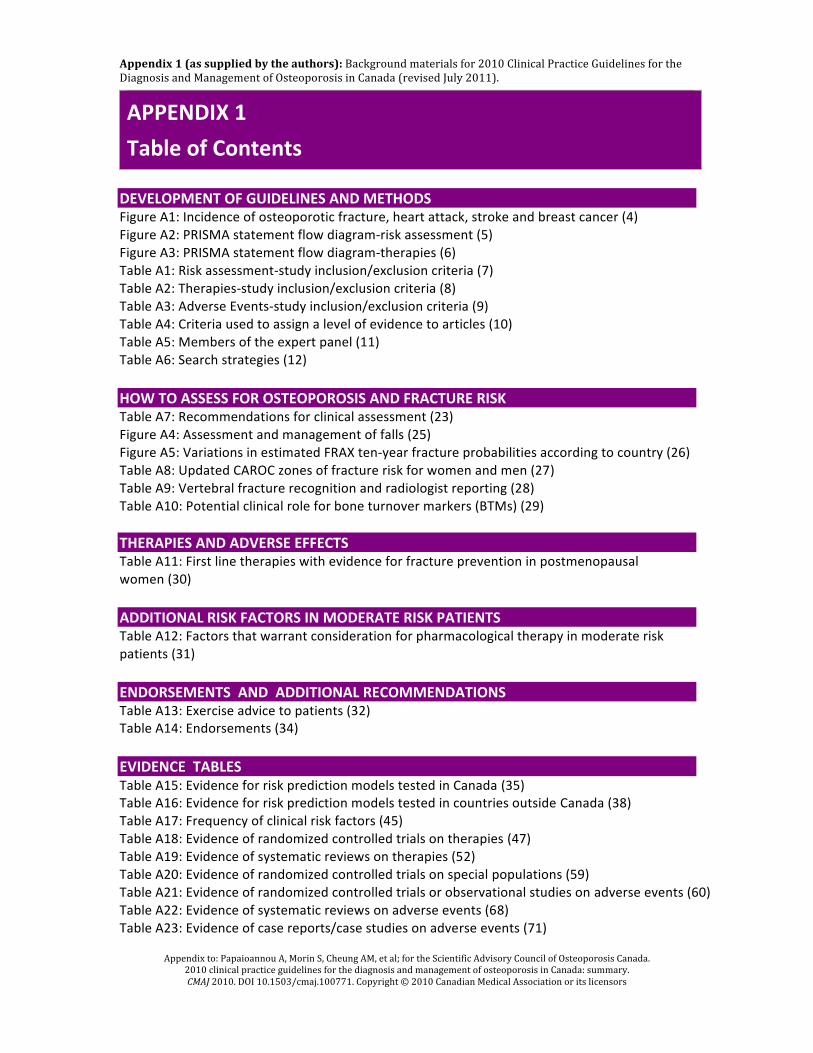
Table of Contents DEVELOPMENT OF GUIDELINES AND METHODS Figure A1: Incidence of osteoporotic fracture, heart attack, stroke and breast cancer (4) Figure A2: PRISMA statement flow diagram-‐risk assessment (5) Figure A3: PRISMA statement flow diagram-‐therapies (6) Table A1: Risk assessment-‐study inclusion/exclusion criteria (7) Table A2: Therapies-‐study inclusion/exclusion criteria (8) Table A3: Adverse Events-‐study inclusion/exclusion criteria (9) Table A4: Criteria used to assign a level of evidence to articles (10) Table A5: Members of the expert panel (11) Table A6: Search strategies (12) HOW TO ASSESS FOR OSTEOPOROSIS AND FRACTURE RISK Table A7: Recommendations for clinical assessment (23) Figure A4: Assessment and management of falls (25) Figure A5: Variations in estimated FRAX ten-‐year fracture probabilities according to country (26) Table A8: Updated CAROC zones of fracture risk for women and men (27) Table A9: Vertebral fracture recognition and radiologist reporting (28) Table A10: Potential clinical role for bone turnover markers (BTMs) (29) THERAPIES AND ADVERSE EFFECTS Table A11: First line therapies with evidence for fracture prevention in postmenopausal women (30) ADDITIONAL RISK FACTORS IN MODERATE RISK PATIENTS Table A12: Factors that warrant consideration for pharmacological therapy in moderate risk patients (31) ENDORSEMENTS AND ADDITIONAL RECOMMENDATIONS Table A13: Exercise advice to patients (32) Table A14: Endorsements (34) EVIDENCE TABLES Table A15: Evidence for risk prediction models tested in Canada (35) Table A16: Evidence for risk prediction models tested in countries outside Canada (38) Table A17: Frequency of clinical risk factors (45) Table A18: Evidence of randomized controlled trials on therapies (47) Table A19: Evidence of systematic reviews on therapies (52) Table A20: Evidence of randomized controlled trials on special populations (59) Table A21: Evidence of randomized controlled trials or observational studies on adverse events (60) Table A22: Evidence of systematic reviews on adverse events (68) Table A23: Evidence of case reports/case studies on adverse events (71)

Appendix to: Papaioannou A, Morin S, Cheung AM, et al; for the Scientific Advisory Council of Osteoporosis Canada. 2010 clinical practice guidelines for the diagnosis and management of osteoporosis in Canada: summary. CMAJ 2010. DOI 10.1503/cmaj.100771. Copyright © 2010 Canadian Medical Association or its licensors
2
DEVELOPMENT OF GUIDELINES AND METHODS
Search Strategy The systematic review of Risk Assessment Models identified and compared existing models for defining fracture risk published from January 1990 to December 2009 and examines the level of evidence that supports the use of these models in Canada. A systematic search was conducted for absolute fracture risk assessment systems or risk prediction models for people over the age of 50 with osteoporosis or low bone density following fracture. The results of the study selection and numbers of articles identified from the systematic review are presented in Figure A1: PRISMA statement flow diagram -‐ models and studies of absolute fracture risk assessment. The abstracts were screened by two authors independently (WDL and AP), who applied inclusion and exclusion criteria and selected citations to be appraised in full text. The study inclusion/exclusion criteria are listed in Table A1. Full text papers were appraised in detail and two researchers performed data abstraction independently using a pre-‐determined form. Inconsistencies or disagreements in the appraisal and data abstraction were decided by consensus of the working group and in consultation with the Chair of the working group (WDL). The systematic review of pharmacological therapies focused on the treatment of individuals over the age of 50 years at increased risk for fracture and to report on adverse events associated with these therapies as published from January 2007 to December 11, 2009. We applied the search strategy developed by MacLean and colleagues in a systematic review of treatments to prevent fractures (1). Meta-‐analyses published in the last five years for exercise, falls prevention and hip protectors were reviewed however a systematic literature search and abstraction for these topics was beyond the scope of this review.
We elected to expand our search strategy to include case series for recently reported adverse events in addition to those included in the MacLean systematic review from randomized controlled trials (Table A2). This approach allowed inclusion of reported postmarketing of adverse events. A bibliography of possible references and abstracts was generated and the abstracts were screened by two researchers independently. Each researcher applied pre-‐determined inclusion and exclusion criteria and then selected which citations were to be appraised in full text. The study inclusion/exclusion criteria are listed in Table A3. The results of the study selection and numbers of articles identified from the systematic review are presented in Figure A2: PRISMA statement flow diagram: therapies. Full text papers were appraised in detail and two researchers performed data abstraction independently using a standardized abstraction form, with separate forms for therapies and for adverse events. Inconsistencies or disagreements in the study selection and data abstraction were resolved through consensus and in consultation with the Chair of the working group (AP).

Appendix to: Papaioannou A, Morin S, Cheung AM, et al; for the Scientific Advisory Council of Osteoporosis Canada. 2010 clinical practice guidelines for the diagnosis and management of osteoporosis in Canada: summary. CMAJ 2010. DOI 10.1503/cmaj.100771. Copyright © 2010 Canadian Medical Association or its licensors
3
Methods for Developing Recommendations Each included study was assigned a level of evidence using criteria consistent with those used in previous osteoporosis guidelines (Table A4) (2)(3). Similarly, each clinical practice recommendation was graded using the same system used in previous osteoporosis guidelines by the working groups. Stakeholder Consultation This expert panel met over two days in November 2009. This expert panel consisted of experts in the field and representatives from stakeholder organizations (Table A5). The group used the RAND/UCLA method of developing consensus on the appropriateness of the guidelines (4) to ensure clinical relevance and applicability. The RAND/UCLA Appropriateness Method was developed in the 1980s. The rationale behind the method is that randomized clinical trials and other research are generally either not available or cannot provide evidence at a level of detail needed for use by clinicians in everyday practice. Although robust scientific evidence about the benefits of many procedures is lacking, physicians must nonetheless make decisions every day about when to use them (5). The RAND/UCLA method was developed to combine the best available evidence with the collective judgment of experts to yield a statement regarding the appropriateness of performing a procedure or screening test. Revisions to the guidelines were made based on the feedback provided by the panel; revised recommendations were endorsed by the panel using an electronic voting system. For more details about the database searches, refer to Table A6. REFERENCES
(1) MacLean C, Newberry S, Maglione M, McMahon M, Ranganath V, Suttorp M et al. Systematic review: comparative effectiveness of treatments to prevent fractures in men and women with low bone density or osteoporosis. Ann Intern Med 2008; 148(3):197-‐213.
(2) Brown JP, Josse RG. 2002 clinical practice guidelines for the diagnosis and management of osteoporosis in Canada. CMAJ 2002; 167(10 Suppl):S1-‐34.
(3) Meltzer S, Leiter L, Daneman D, Gerstein HC, Lau D, Ludwig S et al. 1998 clinical practice guidelines for the management of diabetes in Canada. Canadian Medical Association Journal 1998; 159(8):S1-‐S29.
(4) Brook RH, Chassin MR, Fink A. A method for the detailed assessment of the appropriateness of medical technologies. International Journal of Technology Assessment in Health Care 1986; 2:53-‐63.
(5) Fitch, S.J. Bernstein and M.D. Aguilar et al., The RAND/UCLA Appropriateness Method User's Manual: MR-‐1269-‐DG-‐XII/RE, RAND, Santa Monica, Calif (2001).

Appendix to: Papaioannou A, Morin S, Cheung AM, et al; for the Scientific Advisory Council of Osteoporosis Canada. 2010 clinical practice guidelines for the diagnosis and management of osteoporosis in Canada: summary. CMAJ 2010. DOI 10.1503/cmaj.100771. Copyright © 2010 Canadian Medical Association or its licensors
4
Figure A1:

Appendix to: Papaioannou A, Morin S, Cheung AM, et al; for the Scientific Advisory Council of Osteoporosis Canada. 2010 clinical practice guidelines for the diagnosis and management of osteoporosis in Canada: summary. CMAJ 2010. DOI 10.1503/cmaj.100771. Copyright © 2010 Canadian Medical Association or its licensors
5
Figure A2: PRISMA statement flow diagram -‐ models and studies of absolute fracture risk assessment -‐ 1990-‐January 2009
303 records remaining after duplicates removed
303 record abstracts screened 268 excluded
26 full text articles selected
360 records identified through database searching
18 excluded for the following reasons: 4=wrong study design 2=wrong population, 2=not a true clinical risk assessment system, 2=did not evaluate clinical risk factors 4=did not report absolute risk, or did not include fracture outcomes 3= wrong risk variable (such as the use of ultrasound) 1=duplicate report published.
10 records identified through expert recommendation
35 records assessed in full text
17 included in the meta-‐analysis or final report of the recommendations

Appendix to: Papaioannou A, Morin S, Cheung AM, et al; for the Scientific Advisory Council of Osteoporosis Canada. 2010 clinical practice guidelines for the diagnosis and management of osteoporosis in Canada: summary. CMAJ 2010. DOI 10.1503/cmaj.100771. Copyright © 2010 Canadian Medical Association or its licensors
6
Figure A3: PRISMA statement flow diagram: therapies Studies about benefits and adverse events of pharmacological therapies for people aged 50 and older with osteoporosis January 2007-‐December 11, 2009:
Records identified through database searching
(n = 647)
Additional records identified through other sources
(n = 4 )
Records after duplicates removed (n = 651 )
Records screened (n = 651 )
Records excluded (n = 611)
Full-‐text articles assessed for eligibility (n = 47)
Full-‐text articles excluded, (n = 13) Reasons:
wrong intervention (n=9), wrong design (n=2),
wrong population (n=1), or wrong outcome (n=1).
Studies included in evidence tables
(n = 34 )

Appendix to: Papaioannou A, Morin S, Cheung AM, et al; for the Scientific Advisory Council of Osteoporosis Canada. 2010 clinical practice guidelines for the diagnosis and management of osteoporosis in Canada: summary. CMAJ 2010. DOI 10.1503/cmaj.100771. Copyright © 2010 Canadian Medical Association or its licensors
7
Table A1: Risk assessment– study inclusion/exclusion criteria Inclusion
Population: men or women age >50 years Intervention: absolute fracture risk assessment systems or risk prediction models Comparison: not applicable Outcomes: fractures, fracture prevention Time: January 1990-‐December 14, 2009 Design: prospective and retrospective cohorts, RCTs (inactive control arm), meta-‐analysis, and systematic reviews Language: English
Exclusion
Outcomes other than fracture risk Nonclinical variables or risk factors such as ultrasound Papers that do not describe a model or system Duplicates of papers published in different journals or sources. Selection of which citation to use was based on the availability of the full text and the preference of the authors. Excluded study designs: RCTs (active arm), case series, case reports, letters, editorials, narrative reviews

Appendix to: Papaioannou A, Morin S, Cheung AM, et al; for the Scientific Advisory Council of Osteoporosis Canada. 2010 clinical practice guidelines for the diagnosis and management of osteoporosis in Canada: summary. CMAJ 2010. DOI 10.1503/cmaj.100771. Copyright © 2010 Canadian Medical Association or its licensors
8
Table A2: Therapies -‐ study inclusion/ exclusion criteria Inclusion
Population: men or women age >50 years Intervention: pharmacological therapies for osteoporosis including Bisphosphonates, Calcitonin, Estrogen, PTH, Raloxifene, Vitamin D Design: RCTs, meta-‐analysis, and systematic reviews Comparison: placebo, within class, and/or between class comparisons Outcomes: Fracture prevention: Number/% individuals with at least one vertebral/nonvertebral fracture. Time: January 2007-‐December 11, 2009 Language: English
Exclusion
Therapies other than those listed in the inclusion criteria Therapies not available in Canada Outcomes other than fracture risk Duplicates of papers published in different journals or sources. Selection of which citation to use was based on the availability of the full text and the preference of the authors. Excluded study designs: editorials, narrative reviews
Appendix to: Papaioannou A, Morin S, Cheung AM, et al; for the Scientific Advisory Council of Osteoporosis Canada. 2010 clinical practice guidelines for the diagnosis and management of osteoporosis in Canada: summary. CMAJ 2010. DOI 10.1503/cmaj.100771. Copyright © 2010 Canadian Medical Association or its licensors
9
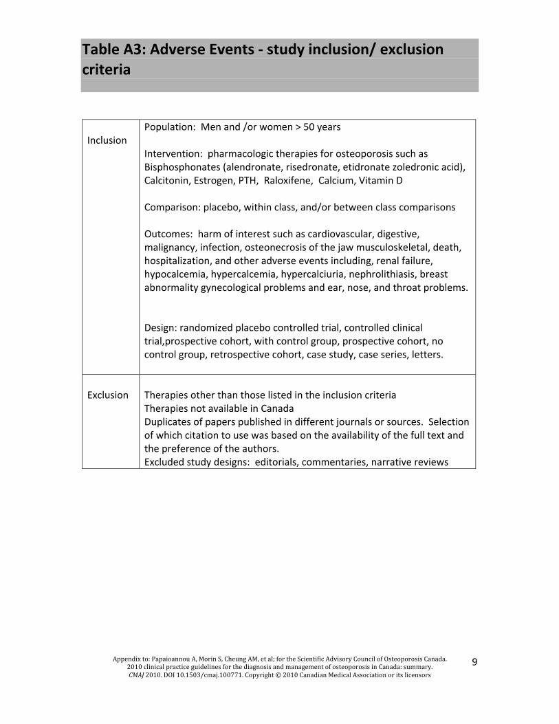
Table A3: Adverse Events -‐ study inclusion/ exclusion criteria Inclusion
Population: Men and /or women > 50 years Intervention: pharmacologic therapies for osteoporosis such as Bisphosphonates (alendronate, risedronate, etidronate zoledronic acid), Calcitonin, Estrogen, PTH, Raloxifene, Calcium, Vitamin D Comparison: placebo, within class, and/or between class comparisons Outcomes: harm of interest such as cardiovascular, digestive, malignancy, infection, osteonecrosis of the jaw musculoskeletal, death, hospitalization, and other adverse events including, renal failure, hypocalcemia, hypercalcemia, hypercalciuria, nephrolithiasis, breast abnormality gynecological problems and ear, nose, and throat problems. Design: randomized placebo controlled trial, controlled clinical trial,prospective cohort, with control group, prospective cohort, no control group, retrospective cohort, case study, case series, letters.
Exclusion
Therapies other than those listed in the inclusion criteria Therapies not available in Canada Duplicates of papers published in different journals or sources. Selection of which citation to use was based on the availability of the full text and the preference of the authors. Excluded study designs: editorials, commentaries, narrative reviews

Appendix to: Papaioannou A, Morin S, Cheung AM, et al; for the Scientific Advisory Council of Osteoporosis Canada. 2010 clinical practice guidelines for the diagnosis and management of osteoporosis in Canada: summary. CMAJ 2010. DOI 10.1503/cmaj.100771. Copyright © 2010 Canadian Medical Association or its licensors
10
Table A4: Criteria used to assign a level of evidence to articles Level Criteria
Studies of diagnosis 1 i. Independent interpretation of test results ii. Independent interpretation of the diagnostic standard iii. Selection of people suspected, but not known to have the disorder iv. Reproducible description of the test and diagnostic standard v. At least 50 people with and 50 people without the disorder 2 Meets 4 of the Level 1 criteria 3 Meets 2 of the Level 1 criteria 4 Meets 1 or 2 of the Level 1 criteria
Studies of treatment and intervention 1+ Systematic overview of meta-‐analysis of randomized controlled trials 1 1 randomized controlled trial with adequate power 2+ Systematic overview or meta-‐analysis of Level 2 randomized controlled trials 2 Randomized controlled trial that does not meet Level 1 criteria 3 Non-‐randomized controlled trial or cohort study 4 Before-‐after study, cohort study with non-‐contemporaneous controls, case-‐control study 5 Case series without controls 6 Case report or case series of < 10 patients
Studies of prognosis 1 i. Inception cohort of patients with the condition of interest, but free of the outcome of
interest ii. Reproducible inclusion and exclusion criteria iii. Follow-‐up of at least 80% of participants iv. Statistical adjustment for confounders v. Reproducible description of the outcome measures 2 Meets criterion i and 3 of the 4 of the Level 1 criteria 3 Meets criterion i and 2 of the 4 of the Level 1 criteria 4 Meets criterion i and 1 of the 4 of the Level 1 criteria
Grades of recommendation for clinical practice guidelines
Grade Criteria
A Need supportive level 1 or 1+ evidence plus consensus* B Need supportive level 2 or 2+ evidence plus consensus* C Need supportive level 3 evidence plus consensus D Any lower level of evidence supported by consensus
*As appropriate level of evidence was necessary, but not sufficient to assign a grade in recommendation; consensus was required in addition. Brown JP, Josse RG. 2002 clinical practice guidelines for the diagnosis and management of osteoporosis in Canada. CMAJ 2002; 167(10 Suppl):S1-‐34.
Appendix to: Papaioannou A, Morin S, Cheung AM, et al; for the Scientific Advisory Council of Osteoporosis Canada. 2010 clinical practice guidelines for the diagnosis and management of osteoporosis in Canada: summary. CMAJ 2010. DOI 10.1503/cmaj.100771. Copyright © 2010 Canadian Medical Association or its licensors
11
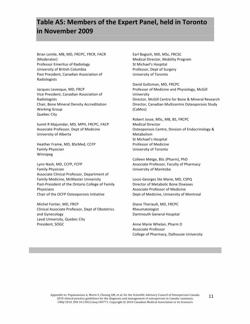
Table A5: Members of the Expert Panel, held in Toronto in November 2009
Brian Lentle, MB, MD, FRCPC, FRCR, FACR (Moderator) Professor Emeritus of Radiology University of British Columbia Past President, Canadian Association of Radiologists Jacques Levesque, MD, FRCP Vice President, Canadian Association of Radiologists Chair, Bone Mineral Density Accreditation Working Group Quebec City Sumit R Majumdar, MD, MPH, FRCPC, FACP Associate Professor, Dept of Medicine University of Alberta Heather Frame, MD, BScMed, CCFP Family Physician Winnipeg Lynn Nash, MD, CCFP, FCFP Family Physician Associate Clinical Professor, Department of Family Medicine, McMaster University Past-‐President of the Ontario College of Family Physicians Chair of the OCFP Osteoporosis Initiative Michel Fortier, MD, FRCP Clinical Associate Professor, Dept of Obstetrics and Gynecology Laval University, Quebec City President, SOGC
Earl Bogoch, MD, MSc, FRCSC Medical Director, Mobility Program St Michael’s Hospital Professor, Dept of Surgery University of Toronto David Goltzman, MD, FRCPC Professor of Medicine and Physiology, McGill University Director, McGill Centre for Bone & Mineral Research Director, Canadian Multicentre Osteoporosis Study (CaMos) Robert Josse, MSc, MB, BS, FRCPC Medical Director Osteoporosis Centre, Division of Endocrinology & Metabolism St Michael’s Hospital Professor of Medicine University of Toronto Colleen Metge, BSc (Pharm), PhD Associate Professor, Faculty of Pharmacy University of Manitoba Louis-‐Georges Ste Marie, MD, CSPQ Director of Metabolic Bone Diseases Associate Professor of Medicine Dept of Medicine, University of Montreal Diane Theriault, MD, FRCPC Rheumatologist Dartmouth General Hospital Anne Marie Whelan, Pharm D Associate Professor College of Pharmacy, Dalhousie University

Appendix to: Papaioannou A, Morin S, Cheung AM, et al; for the Scientific Advisory Council of Osteoporosis Canada. 2010 clinical practice guidelines for the diagnosis and management of osteoporosis in Canada: summary. CMAJ 2010. DOI 10.1503/cmaj.100771. Copyright © 2010 Canadian Medical Association or its licensors
12
Table A6: Search Strategies Risk Assessment Search Database: Ovid MEDLINE(R) <1990 to November Week 3 2009> Search Strategy: 1 exp "Predictive Value of Tests"/ (90230) 2 *Probability/ (2917) 3 *Logistic Models/ (1013) 4 *Models, Statistical/ (12307) 5 *Decision Support Techniques/ (3991) 6 *Computer Simulation/ (17961) 7 absolute adj3 risk ad3 prediction.tw(22) 8 Risk Assessment/mt (11087) 9 *Fractures, Bone/ (27103) 10 Osteoporosis/co [Complications] (4673) 11 *Osteoporosis, Postmenopausal/co [Complications] (437) 12 exp Prospective Studies/ (257450) 13 exp Evaluation Studies/ (113946) 14 meta-analysis.pt,sh. (20287) 15 (meta-anal: or metaanal:).tw. (28169) 16 (quantitativ: review: or quantitativ: overview:).tw. (468) 17 (methodologic: review: or methodologic: overview:).tw. (224) 18 (primary adj3 care adj3 physician).tw. (3553) 19 review.pt. and medline.tw. (21103) 20 or/14-19 (56112) 21 "randomized controlled trial".pt. (270077) 22 ("clinical trial" or "controlled clinical trial").pt. (468665) 23 (random$ or placebo$).ti,ab,sh. (673964) 24 ((singl$ or double$ or triple$ or treble$) and (blind$ or mask$)).tw,sh. (118554) 25 24 or 22 or 23 or 21 (899205) 26 13 or 12 (365325) 27 11 or 10 or 9 (31313) 28 6 or 3 or 7 or 2 or 8 or 1 or 4 or 5 (136954) 29 27 and 28 (407) 30 25 and 29 (45) 31 29 and 20 (13) 32 limit 31 to (english language and yr="1990 - 2009") (11) 33 limit 32 to (english language and yr="1990 - 2009") (11) 34 13 or 12 (365325)

Appendix to: Papaioannou A, Morin S, Cheung AM, et al; for the Scientific Advisory Council of Osteoporosis Canada. 2010 clinical practice guidelines for the diagnosis and management of osteoporosis in Canada: summary. CMAJ 2010. DOI 10.1503/cmaj.100771. Copyright © 2010 Canadian Medical Association or its licensors
13
35 34 and 29 (97) 36 limit 35 to (english language and yr="1990 - 2009") (94) 37 from 36 keep 1-94 (94) 38 from 33 keep 1-11 (11) 39 from 30 keep 1-45 (45) Database: EMBASE 1 *Probability/ (885) 2 Logistic Models.mp. or exp Statistical Model/ (20495) 3 exp Decision Support System/ (1528) 4 *Computer Simulation/ (2388) 5 *Algorithm/ (3089) 6 exp Nomogram/ (1365) 7 *Risk Assessment/ (11547) 8 (risk adj5 assessment adj5 tool).mp. [mp=title, abstract, subject headings, heading word, drug trade name, original title, device manufacturer, drug manufacturer name] (515) 9 computer model.tw. (2369) 10 absolute risk.tw. (1826) 11 absolute risk prediction.tw. (8) 12 risk of hip fracture.tw. (537) 13 bone mineral density reporting.mp. (4) 14 prognostic nomograms.tw. (13) 15 fracture probability.tw. (35) 16 assessment of fracture probability.mp. (1) 17 *Prediction/ (1489) 18 *Computer Prediction/ (115) 19 *"Prediction and Forecasting"/ (38) 20 or/1-19 (47174) 21 *Fracture/ (7044) 22 *Hip Fracture/ (3998) 23 *Vertebra Fracture/ (2069) 24 22 or 21 or 23 (12941) 25 24 and 20 (596) 26 exp meta analysis/ (34535) 27 meta?analys$.tw,sh. (35072) 28 (systematic$ adj5 review$).tw,sh. (27098) 29 (systematic$ adj5 overview$).tw,sh. (425) 30 (methodologic$ adj5 review$).tw,sh. (1532) 31 (methodologic$ adj5 overview$).tw,sh. (119) 32 ((integrative adj5 research adj5 review$) or (research adj5 integrat$)).tw. (2018) 33 (quantitativ$ adj5 synthesi$).tw,sh. (1660) 34 ((pooled or pooling) and analys$).tw,sh. (10896) 35 or/26-34 (65411) 36 exp randomized controlled trial/ (164648) 37 (random$ or placebo$).ti,ab,sh. (523013)
Appendix to: Papaioannou A, Morin S, Cheung AM, et al; for the Scientific Advisory Council of Osteoporosis Canada. 2010 clinical practice guidelines for the diagnosis and management of osteoporosis in Canada: summary. CMAJ 2010. DOI 10.1503/cmaj.100771. Copyright © 2010 Canadian Medical Association or its licensors
14
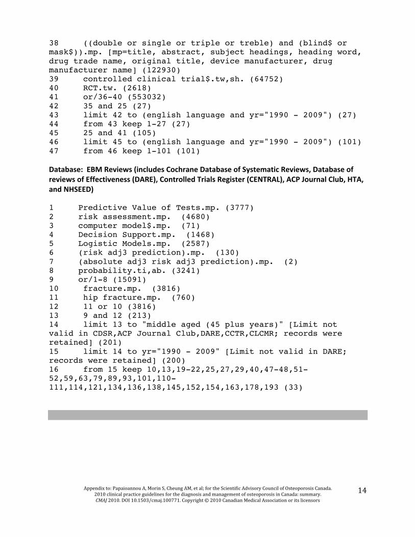
38 ((double or single or triple or treble) and (blind$ or mask$)).mp. [mp=title, abstract, subject headings, heading word, drug trade name, original title, device manufacturer, drug manufacturer name] (122930) 39 controlled clinical trial$.tw,sh. (64752) 40 RCT.tw. (2618) 41 or/36-40 (553032) 42 35 and 25 (27) 43 limit 42 to (english language and yr="1990 - 2009") (27) 44 from 43 keep 1-27 (27) 45 25 and 41 (105) 46 limit 45 to (english language and yr="1990 - 2009") (101) 47 from 46 keep 1-101 (101) Database: EBM Reviews (includes Cochrane Database of Systematic Reviews, Database of reviews of Effectiveness (DARE), Controlled Trials Register (CENTRAL), ACP Journal Club, HTA, and NHSEED) 1 Predictive Value of Tests.mp. (3777) 2 risk assessment.mp. (4680) 3 computer model$.mp. (71) 4 Decision Support.mp. (1468) 5 Logistic Models.mp. (2587) 6 (risk adj3 prediction).mp. (130) 7 (absolute adj3 risk adj3 prediction).mp. (2) 8 probability.ti,ab. (3241) 9 or/1-8 (15091) 10 fracture.mp. (3816) 11 hip fracture.mp. (760) 12 11 or 10 (3816) 13 9 and 12 (213) 14 limit 13 to "middle aged (45 plus years)" [Limit not valid in CDSR,ACP Journal Club,DARE,CCTR,CLCMR; records were retained] (201) 15 limit 14 to yr="1990 - 2009" [Limit not valid in DARE; records were retained] (200) 16 from 15 keep 10,13,19-22,25,27,29,40,47-48,51-52,59,63,79,89,93,101,110-111,114,121,134,136,138,145,152,154,163,178,193 (33)

Appendix to: Papaioannou A, Morin S, Cheung AM, et al; for the Scientific Advisory Council of Osteoporosis Canada. 2010 clinical practice guidelines for the diagnosis and management of osteoporosis in Canada: summary. CMAJ 2010. DOI 10.1503/cmaj.100771. Copyright © 2010 Canadian Medical Association or its licensors
15
Therapies Search Note: The filter for therapies is adapted from the McMaster filter: Haynes RB, Wilczynski N, McKibbon KA, Walker CJ, and Sinclair JC. Developing optimal search strategies for detecting clinically sound studies in MEDLINE. Journal of the American Medical Informatics Association 1994;1(6):447 58. and from Dickersin K, Scherer R, Lefebvre C. Identifying relevant studies for systematic reviews. BMJ 1994;309(6964):1286–91. Search terms for therapies were modified for use in OVID Medline from PubMed Searches found in Appendix A of http://effectivehealthcare.ahrq.gov/repFiles/LowBoneDensityAppendices.pdf Database: Ovid MEDLINE(R) <2005 to November Week 3 2009> Filter for Systematic reviews: 1 meta-analysis.pt,sh. (10107) 2 (meta-anal$ or metaanal$).tw. (11332) 3 (quantitative$ review$ or quantitative$ overview$).tw. (135) 4 (methodologic$ review$ or methodologic$ overview$).tw. (51) 5 review.pt. and medline.tw. (10650) 6 or/1-5 (23468) Strategy for condition: 8 osteoporosis, postmenopausal/ (2426) 9 osteoporosis/ (6922) 10 *Bone Density/ (4472) 11 *Bone Resorption/ (1139) 12 "Bone and Bones"/de, me (3462) 13 (bone adj2 densit$).tw. (7933) 14 or/8-13 (17258) Filter for RCT: 15 "randomized controlled trial".pt. (70773) 16 ("clinical trial" or "controlled clinical trial").pt. (74147) 17 (random$ or placebo$).ti,ab,sh. (176484) 18 ((singl$ or double$ or triple$ or treble$) and (blind$ or mask$)).tw,sh. (24812) 19 or/15-18 (224649) Strategy for interventions (from MacLean 2008, Appendix A – Note “mp.” is a group field indicator in OVID. The fields searched are: title, original title, abstract, name of substance word, MESH heading

Appendix to: Papaioannou A, Morin S, Cheung AM, et al; for the Scientific Advisory Council of Osteoporosis Canada. 2010 clinical practice guidelines for the diagnosis and management of osteoporosis in Canada: summary. CMAJ 2010. DOI 10.1503/cmaj.100771. Copyright © 2010 Canadian Medical Association or its licensors
16
20 (bisphosphonate* or alendronate* or etidronate* or ibandronate* or pamidronate* or risedronate*).mp. (9224) 21 (zolendronate or calcitonin or miacalcin or calcimar or cibacalcin or calcium or estrogen or estrogen$ or oestrogen or estradiol or raloxifene or teriparatide).mp. (580864) 22 (denosumab or strontium).mp. (9373) 23 (testosterone or vitamin d or glucorticoid$).mp. (102763) 24 or/20-23 (653298) Combined condition and therapies 25 24 and 14 (29849) RCT results 26 25 and 19 (5125) Limits applied: 27 limit 26 to (english language and yr="2007 - 2008" and "middle aged (45 plus years)") (365) Systematic review results 28 25 and 7 (375) Limits applied: 29 limit 28 to (english language and yr="2007 - 2008" and "middle aged (45 plus years)") (27) TOTAL RCTs 29 from 26 keep 1-291 (291) TOTAL SRs 30 from 28 keep 1-23 (23) Database: EMBASE <1980 to 2008 Week 43> Search Strategy: -‐-‐-‐-‐-‐-‐-‐-‐-‐-‐-‐-‐-‐-‐-‐-‐-‐-‐-‐-‐-‐-‐-‐-‐-‐-‐-‐-‐-‐-‐-‐-‐-‐-‐-‐-‐-‐-‐-‐-‐-‐-‐-‐-‐-‐-‐-‐-‐-‐-‐-‐-‐-‐-‐-‐-‐-‐-‐-‐-‐ Filter for systematic reviews: 1 exp meta analysis/ (34177) 2 meta?analys$.tw,sh. (34701) 3 (systematic$ adj5 review$).tw,sh. (26116) 4 (systematic$ adj5 overview$).tw,sh. (416) 5 (methodologic$ adj5 review$).tw,sh. (1505) 6 (methodologic$ adj5 overview$).tw,sh. (118) 7 ((integrative adj5 research adj5 review$) or (research adj5 integrat$)).tw. (1984) 8 (quantitativ$ adj5 synthesi$).tw,sh. (1629) 9 ((pooled or pooling) and analys$).tw,sh. (10636) 10 or/1-9 (64069)

Appendix to: Papaioannou A, Morin S, Cheung AM, et al; for the Scientific Advisory Council of Osteoporosis Canada. 2010 clinical practice guidelines for the diagnosis and management of osteoporosis in Canada: summary. CMAJ 2010. DOI 10.1503/cmaj.100771. Copyright © 2010 Canadian Medical Association or its licensors
17
Filter for RCTs: 11 exp randomized controlled trial/ (163740) 12 (random$ or placebo$).ti,ab,sh. (515765) 13 ((double or single or triple or treble) and (blind$ or mask$)).mp. (122444) 14 controlled clinical trial$.tw,sh. (61846) 15 RCT.tw. (2540) 16 or/11-15 (545307) Strategy for condition: 17 exp OSTEOPOROSIS/ (41808) 18 exp FRACTURE/ (80940) 19 (fracture adj5 prevent$).tw. (1038) 20 exp Bone Density/ (25179) 21 osteopenia.tw,sh. (7473) 22 exp Bone Demineralization/ (42807) 23 exp Bone Atrophy/ (6951) 24 exp Bone Metabolism/ (33551) 25 (osteop$ or fractur$ or BMD).ti,ab. (120443) 26 or/17-25 (179834) Strategy for interventions: 27 *Bisphosphonic Acid Derivative/ (0) 28 bisphosphonate$.ti,ab. (6112) 29 exp ZOLEDRONIC ACID/ (0) 30 exp Selective Estrogen Receptor Modulator/ (19847) 31 SERM.tw. (605) 32 exp RALOXIFENE/ (1744) 33 exp CALCITONIN/ (12507) 34 exp Parathyroid Hormone/ (21468) 35 *CALCIUM/ (97554) 36 *EXERCISE/ (31051) 37 Denosumab.mp. (87) 38 Strontium.mp. (9293) 39 *Vitamin D/ (8276) 40 or/27-39 (194695) Combined condition and therapies 39 26 and 40 (20265) RCT results: 40 16 and 40 (3755) Limits applied: 41 limit 40 to (english language and yr="2007 - 2009" and (adult <18 to 64 years> or aged <65+ years>)) (223) SR results:

Appendix to: Papaioannou A, Morin S, Cheung AM, et al; for the Scientific Advisory Council of Osteoporosis Canada. 2010 clinical practice guidelines for the diagnosis and management of osteoporosis in Canada: summary. CMAJ 2010. DOI 10.1503/cmaj.100771. Copyright © 2010 Canadian Medical Association or its licensors
18
42 39 and 10 (601) Limits applied: 43 limit 42 to (english language and yr="2007 - 2009" and (adult <18 to 64 years> or aged <65+ years>)) (3) TOTAL Embase SRs 44 from 43 keep 1-3 (3) TOTAL Embase RCTs 45 from 41 keep 1-223 (223) Database: EBM Reviews -‐ includes Cochrane Database of Systematic Reviews, Database of reviews of Effectiveness (DARE), Controlled Trials Register (CENTRAL), ACP Journal Club, Cochrane Methods Register, Health Technology Assessment, and NHS Economic Evaluation Database. -‐-‐-‐-‐-‐-‐-‐-‐-‐-‐-‐-‐-‐-‐-‐-‐-‐-‐-‐-‐-‐-‐-‐-‐-‐-‐-‐-‐-‐-‐-‐-‐-‐-‐-‐-‐-‐-‐-‐-‐-‐-‐-‐-‐-‐-‐-‐-‐-‐-‐-‐-‐-‐-‐-‐-‐-‐-‐-‐-‐ Condition: 1 (osteoporosis or osteopenia or osteopaenia or fracture$ or bone mineral).mp. (9518) Therapies: bisphosphonate$.mp. (654) 3 (alendronate$ or fosamax).mp. (533) 4 (resindronate$ or actonel).mp. (7) 5 (etidronate$ or didronel).mp. (251) 6 (pamidronate$ or aredia).mp. (358) 7 (zoledronic acid$ or zometa).mp. (160) 8 (selective estrogen receptor modulator$ or serm$).mp. (393) 9 (raloxifene or evista).mp. (468) 10 (calcitonin$ or miacalcin or calcimar or cibacalcin).mp. (823) 11 (parathyroid hormone$ or pth).mp. (1401) 12 (teriparatide or fosteo).mp. (90) 13 (exercis$ and (calcium or vitamin d)).mp. (1177) 14 Denosumab.mp. (11) 15 Strontium.mp. (172) 16 or/2-15 (5233) Combined condition and therapies: 17 1 and 16 (2036) Limits applied (where database will allow): 19 limit 18 to "middle aged (45 plus years)" (1995) 20 limit 19 to english language (328) 21 limit 20 to yr="2007 - 2008" (323) from 21 keep 1-318 (323)

Appendix to: Papaioannou A, Morin S, Cheung AM, et al; for the Scientific Advisory Council of Osteoporosis Canada. 2010 clinical practice guidelines for the diagnosis and management of osteoporosis in Canada: summary. CMAJ 2010. DOI 10.1503/cmaj.100771. Copyright © 2010 Canadian Medical Association or its licensors
19
Adverse Event Search Database: Ovid MEDLINE(R), Ovid MEDLINE(R) In-‐Process Search Strategy: ------------------------------------------------------------ Interventions: 1 (zolendronate or calcitonin or miacalcin or calcimar or cibacalcin or calcium or estrogen or estrogen$ or oestrogen or estradiol or raloxifene or teriparatide).mp. [mp=title, original title, abstract, name of substance word, subject heading word] (99354) 2 (testosterone or vitamin d).mp. (18970) 3 *Diphosphonates/ (2107) 4 (bisphosphonate or alendronate or etidronate or ibandronate or pamidronate or risedronate).mp. [mp=title, original title, abstract, name of substance word, subject heading word] (3478) 5 (denosumab or strontium).mp. [mp=title, original title, abstract, name of substance word, subject heading word] (1451) 6 (zolendronate or calcitonin or miacalcin or calcimar or cibacalcin or calcium or estrogen or estrogen$ or oestrogen or estradiol or raloxifene or teriparatide).mp. [mp=title, original title, abstract, name of substance word, subject heading word] (99354) 7 (testosterone or vitamin d or glucorticoid$).mp. [mp=title, original title, abstract, name of substance word, subject heading word] (18993) 8 *Diphosphonates/ (2107) 9 strontium.mp. [mp=title, original title, abstract, name of substance word, subject heading word] (1328) 10 bazedoxifene.mp. [mp=title, original title, abstract, name of substance word, subject heading word] (38) 11 zolendronic acid.mp. [mp=title, original title, abstract, name of substance word, subject heading word] (10) 12 (bisphosphonate or alendronate or etidronate or ibandronate or pamidronate or risedronate).mp. [mp=title, original title, abstract, name of substance word, subject heading word] (3478) 13 or/1-12 (114743) Harms filter: 14 (safe or safety).ti,ab. (101269) 15 ((adverse or undesirable or harm$ or serious or toxic) adj3 (effect$ or reaction$ or event$ or outcome$)).ti,ab. (67415) 16 *Product Surveillance, Postmarketing/ (536)

Appendix to: Papaioannou A, Morin S, Cheung AM, et al; for the Scientific Advisory Council of Osteoporosis Canada. 2010 clinical practice guidelines for the diagnosis and management of osteoporosis in Canada: summary. CMAJ 2010. DOI 10.1503/cmaj.100771. Copyright © 2010 Canadian Medical Association or its licensors
20
17 *Adverse Drug Reaction Reporting Systems/ (754) 18 *Clinical Trials, Phase IV as Topic/ (15) 19 *substance-related disorders/ (6965) 20 *drug toxicity/ (510) 21 *abnormalities, drug induced/ (588) 22 *drug monitoring/ (963) 23 *drug hypersensitivity/ (1336) 24 23 or 19 or 15 or 18 or 20 or 16 or 22 or 14 or 17 or 21 (161929) 25 24 and 13 (5679) Study Design filter: 26 observational.mp. (19304) 27 exp Cohort Studies/ (173210) 28 case reports.pt. (210794) 29 government publications.pt. (7) 30 guideline.pt. (1455) 31 Practice Guideline.pt. (4155) 32 technical report.pt. (520) 33 30 or 28 or 31 or 27 or 29 or 26 or 32 (389025) 34 24 and 13 (5679) 35 33 and 34 (793) Specific harms: 36 atrial fibrillation.mp. or *Atrial Fibrillation/ (9878) 37 exp Acute Coronary Syndrome/ (1271) 38 pulmonary embolism.mp. or *Pulmonary Embolism/ (5618) 39 Thromboembolism/ or thromboembolic event.mp. (2786) 40 cerebrovascular accident.mp. or *Stroke/ (12560) 41 *Esophageal Diseases/ or Esophageal ulcerations.mp. (527) 42 Intestinal Perforation/ or Uterine Perforation/ or Esophageal Perforation/ or Tympanic Membrane Perforation/ or Peptic Ulcer Perforation/ (1904) 43 *Gastroesophageal Reflux/ (2989) 44 *Nausea/ (482) 45 *Vomiting/ (754) 46 *Heartburn/ (130) 47 *Breast Neoplasms/ (29012) 48 *Breast Diseases/ (902) 49 *Uterine Hemorrhage/ (394) 50 *Osteosarcoma/ (1350) 51 *Osteonecrosis/ (944) 52 Cardiac death.mp. or *Death/ (3952) 53 Colon Cancer.mp. or *Colonic Neoplasms/ (9265) 54 Lung Cancer.mp. or *Lung Neoplasms/ (23752) 55 joint pain.mp. or *Arthralgia/ (1464) 56 *Arthritis/ (1424) 57 *Hypocalcemia/ (373)
Appendix to: Papaioannou A, Morin S, Cheung AM, et al; for the Scientific Advisory Council of Osteoporosis Canada. 2010 clinical practice guidelines for the diagnosis and management of osteoporosis in Canada: summary. CMAJ 2010. DOI 10.1503/cmaj.100771. Copyright © 2010 Canadian Medical Association or its licensors
21
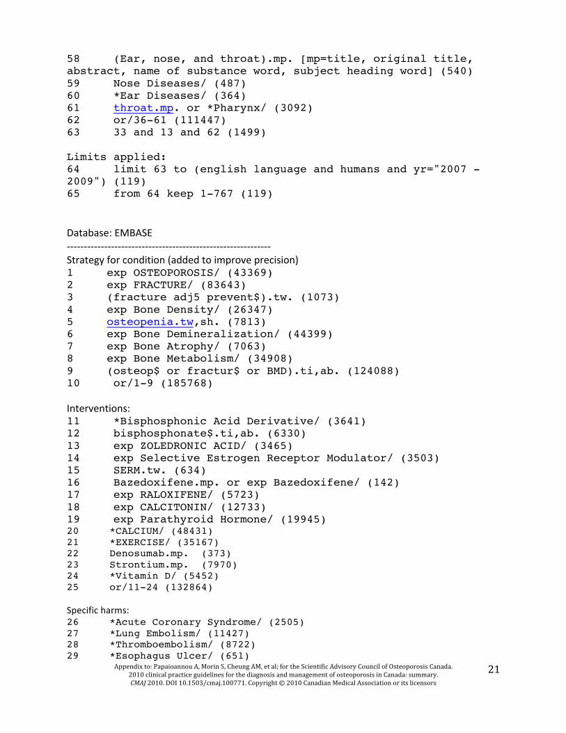
58 (Ear, nose, and throat).mp. [mp=title, original title, abstract, name of substance word, subject heading word] (540) 59 Nose Diseases/ (487) 60 *Ear Diseases/ (364) 61 throat.mp. or *Pharynx/ (3092) 62 or/36-61 (111447) 63 33 and 13 and 62 (1499) Limits applied: 64 limit 63 to (english language and humans and yr="2007 - 2009") (119) 65 from 64 keep 1-767 (119) Database: EMBASE -‐-‐-‐-‐-‐-‐-‐-‐-‐-‐-‐-‐-‐-‐-‐-‐-‐-‐-‐-‐-‐-‐-‐-‐-‐-‐-‐-‐-‐-‐-‐-‐-‐-‐-‐-‐-‐-‐-‐-‐-‐-‐-‐-‐-‐-‐-‐-‐-‐-‐-‐-‐-‐-‐-‐-‐-‐-‐-‐-‐ Strategy for condition (added to improve precision) 1 exp OSTEOPOROSIS/ (43369) 2 exp FRACTURE/ (83643) 3 (fracture adj5 prevent$).tw. (1073) 4 exp Bone Density/ (26347) 5 osteopenia.tw,sh. (7813) 6 exp Bone Demineralization/ (44399) 7 exp Bone Atrophy/ (7063) 8 exp Bone Metabolism/ (34908) 9 (osteop$ or fractur$ or BMD).ti,ab. (124088) 10 or/1-9 (185768) Interventions: 11 *Bisphosphonic Acid Derivative/ (3641) 12 bisphosphonate$.ti,ab. (6330) 13 exp ZOLEDRONIC ACID/ (3465) 14 exp Selective Estrogen Receptor Modulator/ (3503) 15 SERM.tw. (634) 16 Bazedoxifene.mp. or exp Bazedoxifene/ (142) 17 exp RALOXIFENE/ (5723) 18 exp CALCITONIN/ (12733) 19 exp Parathyroid Hormone/ (19945) 20 *CALCIUM/ (48431) 21 *EXERCISE/ (35167) 22 Denosumab.mp. (373) 23 Strontium.mp. (7970) 24 *Vitamin D/ (5452) 25 or/11-24 (132864) Specific harms: 26 *Acute Coronary Syndrome/ (2505) 27 *Lung Embolism/ (11427) 28 *Thromboembolism/ (8722) 29 *Esophagus Ulcer/ (651)

Appendix to: Papaioannou A, Morin S, Cheung AM, et al; for the Scientific Advisory Council of Osteoporosis Canada. 2010 clinical practice guidelines for the diagnosis and management of osteoporosis in Canada: summary. CMAJ 2010. DOI 10.1503/cmaj.100771. Copyright © 2010 Canadian Medical Association or its licensors
22
30 *Colon Perforation/ or *Stomach Perforation/ or *Digestive System Perforation/ or *Esophagus Perforation/ or *Intestine Perforation/ or *Small Intestine Perforation/ or *Appendix Perforation/ or *Perforation/ or *Duodenum Perforation/ or *Large Intestine Perforation/ or *Ulcer Perforation/ (6625) 31 *Ulcer/ (2197) 32 *Nausea/co, si [Complication, Side Effect] (2117) 33 *Gastroesophageal Reflux/dm, co, si, th [Disease Management, Complication, Side Effect, Therapy] (1477) 34 *Heart Atrium Fibrillation/dm, co, si, th (5107) 35 *Cerebrovascular Accident/dm, co, si, th (1739) 36 *Vomiting/co, si [Complication, Side Effect] (2723) 37 *Heartburn/ (789) 38 *Breast Cancer/co, si [Complication, Side Effect] (1199) 39 *Breast Disease/ or *Breast Malformation/ or Breast abnormality.mp. (2587) 40 *Uterus Bleeding/ (1008) 41 *Osteosarcoma/ (7052) 42 *Bone Necrosis/ (2179) 43 Cardiac death.mp. or *Heart Death/ (9037) 44 *Colon Cancer/ (12293) 45 *Lung Cancer/ (25991) 46 joint pain.mp. or *Arthralgia/ (4260) 47 muscle pain.mp. or *Myalgia/ (3802) 48 *Muscle Cramp/ (567) 49 *Hypocalcemia/ (2149) 50 *Ear Nose Throat Disease/ (470) 51 or/25-50 (245767) Study design: 52 observational.ti,ab. (32108) 53 cohort study.mp. or *Cohort Analysis/ (32730) 54 case report.ti,ab. (119611) 55 guideline.ti,ab. (11814) 56 52 or 53 or 55 or 54 (193374) 57 25 and 56 and 51 and 10 (494) Limits applied: 58 limit 57 to (human and yr="2007 -Current") (142) 59 from 58 keep 1-142 (142) 60 from 59 keep 1-142 (142)

Appendix to: Papaioannou A, Morin S, Cheung AM, et al; for the Scientific Advisory Council of Osteoporosis Canada. 2010 clinical practice guidelines for the diagnosis and management of osteoporosis in Canada: summary. CMAJ 2010. DOI 10.1503/cmaj.100771. Copyright © 2010 Canadian Medical Association or its licensors
23
HOW TO ASSESS FOR OSTEOPOROSIS AND FRACTURE RISK
Table A7: Recommendations for Clinical Assessment Assessment Recommended Elements History Identify risk factors for low BMD, future fractures and falls:
Prior fragility fractures Parental hip fracture Glucocorticoid use Current smoking High alcohol intake (≥3 units per day) Rheumatoid arthritis Inquire about falls in the previous 12 months Inquire about gait and balance
Measure weight (weight loss of >10% since age 25 is significant)
Measure height annually (prospective loss > 2cm) (historical height loss > 6 cm) Measure rib to pelvis distance ≤ 2 fingers' breadth Measure occiput-‐to-‐wall distance (forkyphosis)> 5cm
Physical Examination
Assess fall risk by using Get-‐Up-‐and-‐Go Test (ability to get out of chair without using arms, walk several steps and return) (11)
1. Brown JP, Josse RG. 2002 clinical practice guidelines for the diagnosis and management of osteoporosis in Canada. CMAJ 2002; 167(10 Suppl):S1-‐34. 2. Lindsay R, Silverman SL, Cortet B, Hanley DA, Barton IP, Broy SB et al. RIsk of new vertebral fracture in the year following a fracture. Journal of the American Medical Association 2001; 285:320-‐323. 3. Tinetti ME. Preventing falls in elderly persons. N Engl J Med 2003; 348:42-‐49. 4. Ioannidis G, Papaioannou A, Hopman WM, Akhtar-‐Danesh N, Anastassiades T, Pickard L et al. Relation between fractures and mortality: results from the Canadian Multicentre Osteoporosis study. Canadian Medical Association Journal 2009; 181(5):265-‐271. 5. Papaioannou A, Kennedy CC, Ioannidis G, Sawka AM, Hopman WM, Pickard L et al. The impact of incident fractures on health-‐related quality of life: 5 years of data from the Canadian Multicentre Osteoporosis Study. Osteoporos Int 2009; 20(5):703-‐715. 6. Papaioannou A, Kennedy CC, Cranney A, Hawker G, Brown JP, Kaiser SM et al. Risk factors for low BMD in healthy men age 50 years or older: a systematic review. Osteoporos Int 2009; 20:507-‐518. 7. Waugh EJ, Lam MA, Hawker GA, McGowan J, Papaioannou A, Cheung AM et al. Risk factors for low bone mass in healthy 40-‐60 year old women: A systematic review of the literature. Osteoporos Int 2009; 20:1-‐21. 8. Cummings SR, Nevitt MC, Browner WS, Stone K, Fox KM, Ensrud KE et al. Risk factors for hip fracture in white women. Study of Osteoporotic Fractures Research Group. N Engl J Med 1995; 332(12):767-‐773.
Screening for vertebral fractures

Appendix to: Papaioannou A, Morin S, Cheung AM, et al; for the Scientific Advisory Council of Osteoporosis Canada. 2010 clinical practice guidelines for the diagnosis and management of osteoporosis in Canada: summary. CMAJ 2010. DOI 10.1503/cmaj.100771. Copyright © 2010 Canadian Medical Association or its licensors
24
9. Papaioannou A, Joseph L, Ioannidis G, Berger C, Anastassiades T, Brown JP et al. Risk factors associated with incident clinical vertebral and nonvertebral fractures in postmenopausal women: the Canadian Multicentre Osteoporosis Study (CaMos). Osteoporos Int 2005; 16(5):568-‐578. 10. van Staa TP, Leufkens HG, Abenhaim L, Zhang B, Cooper C. Use of oral corticosteroids and risk of fractures. J Bone Miner Res 2000; 15(6):993-‐1000. 11. American Geriatrics Society, British Geriatrics Society, American Academy of Orthopaedic Surgeons Panel on Falls Prevention. Guideline for the prevention of falls in older persons. J Am Geriatr Soc 2001; 49:664-‐672. 12. Ganz DA, Bao Y, Shekelle PG, Rubenstein LZ. WIll my patient fall? Journal of the American Medical Association 2007; 297:77-‐86. 13. Bensen R, Adachi JD, Papaioannou A, Ioannidis G, Olszynski WP, Sebaldt RJ et al. Evaluation of easily measured risk factors in the prediction of osteoporotic fractures. BMC Musculoskeletal Disorders 2005; 6:47. 14. Cawthon PM, Fullman RL, Marshall L, Mackey DC, Fink HA, Cauley JA et al. Physical performance and risk of hip fractures in older men. J Bone Miner Res 2008; 23:1037-‐1044. 15. Gates S, Fisher JD, Cooke MW, Carter YH, Lamb SE. Multifactorial assessment and targeted intervention for preventing falls and injuries among older people in community and emergency care settings: systematic review and meta-‐analysis. BMJ 2008; 336(7636):130-‐133. 16. Kanis J, Johnell O, Gullberg B, Allander E, Elffors L, Ranstam J et al. Risk factors for hip fracture in men from southern Europe: the MEDOS study. Mediterranean Osteoporosis Study. Osteoporos Int 1999; 9:45-‐54. 17. Morin S, Tsang JF, Leslie WD. Weight and body mass index predict bone mineral density and fractures in women aged 40 to 59 years. Osteoporos Int 2008. 18. Green AD, Colon-‐Emeric C, Bastian L, Drake MT, Lyles KW. Does this woman have osteoporosis? Journal of the American Medical Association 2004; 292(23):2890-‐2900. 19. Siminoski K, Warshawski RS, Jen H, Lee K. The accuracy of historical height loss for the detection of vertebral fractures in postmenopausal women. Osteoporos Int 2006; 17(2):290-‐296. 20. Briot K, Legrand E, Pouchain D, Monnier S, Roux C. Accuracy of patient-‐reported height loss and risk factors for height loss among postmenopausal women. Canadian Medical Association Journal 2010; 182(6):558-‐562. 21. Moayyeri A, Luben RN, Bingham SA, Welch AA, Wareham NJ, Khaw KT. Measured height loss predicts fractures in middle-‐aged and older men and women: the EPIC-‐Norfolk prospective population study. J Bone Miner Res 2008; 23:425-‐432. 22. Siminoski K, Adachi JG, Hanley DA, Cline G, Iannidis G, Hodsman A et al. Accuracy of height loss during prospective monitoring for detection of incident vertebral fractures. Osteoporos Int 2005; 16(4):403-‐410. 23. Kaptoge S, Armbrecht G, Felsenberg D, Lunt M, O'Neill TW, Silman AJ et al. When should the doctor order a spine x-‐ray? Identifying vertebral fractures for osteoporosis care: results from the European Prospective Osteoporosis Study (EPOS). J Bone Miner Res 2004; 19:1982-‐1993.
Appendix to: Papaioannou A, Morin S, Cheung AM, et al; for the Scientific Advisory Council of Osteoporosis Canada. 2010 clinical practice guidelines for the diagnosis and management of osteoporosis in Canada: summary. CMAJ 2010. DOI 10.1503/cmaj.100771. Copyright © 2010 Canadian Medical Association or its licensors
25
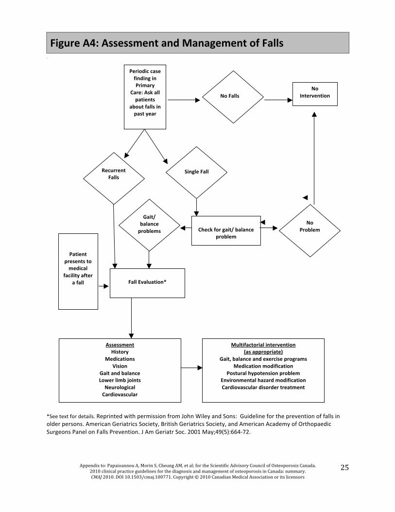
Figure A4: Assessment and Management of Falls
*See text for details. Reprinted with permission from John Wiley and Sons: Guideline for the prevention of falls in older persons. American Geriatrics Society, British Geriatrics Society, and American Academy of Orthopaedic Surgeons Panel on Falls Prevention. J Am Geriatr Soc. 2001 May;49(5):664-‐72.
Recurrent Falls
Single Fall
No Falls
No Intervention
Check for gait/ balance
problem
No Problem
Gait/ balance problems
Fall Evaluation*
Assessment History
Medications Vision
Gait and balance Lower limb joints
Neurological Cardiovascular
Patient presents to medical
facility after a fall
Multifactorial intervention (as appropriate)
Gait, balance and exercise programs Medication modification
Postural hypotension problem Environmental hazard modification Cardiovascular disorder treatment
Periodic case finding in Primary
Care: Ask all patients
about falls in past year

Appendix to: Papaioannou A, Morin S, Cheung AM, et al; for the Scientific Advisory Council of Osteoporosis Canada. 2010 clinical practice guidelines for the diagnosis and management of osteoporosis in Canada: summary.
CMAJ 2010. DOI 10.1503/cmaj.100771. Copyright © 2010 Canadian Medical Association or its licensors 26
Figure A5: Variations in estimated FRAX ten-year fracture probabilities according to country
(FRAX version 3.0, www.shef.ac.uk/FRAX) 10-Year Major Fracture Probability
0
5
10
15
20
25
30
Swed
en
Switz
erla
nd
US
Cau
casi
an
Aust
ria
Uni
ted
King
dom
CAN
ADA
Belg
ium
Japa
n
Italy
Arge
ntin
a
Hon
g Ko
ng
Finl
and
Ger
man
y
US
His
pani
c
US
Asia
n
Fran
ce
New
Zea
land
US
Blac
k
Spai
n
Leba
non
Chi
na
Turk
ey
Per
cent
frac
ture
Female Male
Age 65 years, prior fracture with femoral neck T-score -2.5
10-Year Hip Fracture Probability
0
2
4
6
8
10
12
Swed
en
Aust
ria
Belg
ium
Switz
erla
nd
Italy
Uni
ted
King
dom
Arge
ntin
a
US
Cau
casi
an
Hon
g Ko
ng
Ger
man
y
Finl
and
CAN
ADA
Fran
ce
New
Zea
land
Japa
n
Spai
n
US
His
pani
c
US
Asia
n
Leba
non
Chi
na
US
Blac
k
Turk
ey
Per
cent
frac
ture
Female Male
Age 65 years, prior fracture with femoral neck T-score -2.5

Appendix to: Papaioannou A, Morin S, Cheung AM, et al; for the Scientific Advisory Council of Osteoporosis Canada. 2010 clinical practice guidelines for the diagnosis and management of osteoporosis in Canada: summary. CMAJ 2010. DOI 10.1503/cmaj.100771. Copyright © 2010 Canadian Medical Association or its licensors
27
Table A8: Updated CAROC Zones of Fracture Risk for Women and Men using Femoral Neck T-‐Score
Women
Age Low Risk Moderate Risk High Risk
50 above -‐2.5 -‐2.5 to -‐3.8 below -‐3.8
55 above -‐2.5 -‐2.5 to -‐3.8 below -‐3.8
60 above -‐2.3 -‐2.3 to -‐3.7 below -‐3.7
65 above -‐1.9 -‐1.9 to -‐3.5 below -‐3.5
70 above -‐1.7 -‐1.7 to -‐3.2 below -‐3.2
75 above -‐1.2 -‐1.2 to -‐2.9 below -‐2.9
80 above -‐0.5 -‐0.5 to -‐2.6 below -‐2.6
85 above +0.1 +0.1 to -‐2.2 below -‐2.2
Men Age Low Risk Moderate Risk High Risk
50 above -‐2.5 -‐2.5 to -‐3.9 below -‐3.9
55 above -‐2.5 -‐2.5 to -‐3.9 below -‐3.9
60 above -‐2.5 -‐2.5 to -‐3.7 below -‐3.7
65 above -‐2.4 -‐2.4 to -‐3.7 below -‐3.7
70 above -‐2.3 -‐2.3 to -‐3.7 below -‐3.7
75 above -‐2.3 -‐2.3 to -‐3.8 below -‐3.8
80 above -‐2.1 -‐2.1 to -‐3.8 below -‐3.8
85 above -‐2.0 -‐2.0 to -‐3.8 below -‐3.8

Appendix to: Papaioannou A, Morin S, Cheung AM, et al; for the Scientific Advisory Council of Osteoporosis Canada. 2010 clinical practice guidelines for the diagnosis and management of osteoporosis in Canada: summary. CMAJ 2010. DOI 10.1503/cmaj.100771. Copyright © 2010 Canadian Medical Association or its licensors
28
Table A9: Vertebral Fracture Recognition and Radiologist Reporting
Physicians should be aware of the importance of vertebral fracture diagnosis in assessing future
osteoporotic fracture risk.
Vertebral compression fractures incidental to radiologic examinations done for other reasons should
be identified and reported.
Vertebral fractures should be assessed from lateral spinal or chest radiographs according to the
semiquantitative method of Genant and colleagues. Grade II (26-‐40%) and Grade III (>40%) fractures
as classified by this method should be given the greatest emphasis.
Semiquantitative fracture diagnosis should include the recognition of changes such as loss of
vertebral end-‐plate parallelism, cortical interruptions, and quantitative changes in the anterior,
midbody, and posterior heights of vertebral bodies.
When spine radiographs are performed to assess the presence of vertebral fractures, anteroposterior
examinations may assist in the initial evaluation.
The standard follow-‐up need only consist of single lateral views of the thoracic and lumbar spine that
include T4 to L4 vertebrae.
Dual-‐energy X-‐ray absorptiometry examinations that include lateral spinal morphological
assessments (VFA, Vertebral Fracture Assessment) may contribute to fracture recognition.
Educational material about the clinical importance of vertebral fracture recognition as a potential
indicator of future osteoporotic fracture risk with its associated morbidity and mortality should be
directed to all physicians.
Lentle BC, Brown JP, Khan A, Leslie WD, Levesque J, Lyons DJ et al. Recognizing and reporting vertebral fractures: reducing the risk of future osteoporotic fractures. Canadian Association of Radiologists Journal 2007; 58(1):27-‐36.

Appendix to: Papaioannou A, Morin S, Cheung AM, et al; for the Scientific Advisory Council of Osteoporosis Canada. 2010 clinical practice guidelines for the diagnosis and management of osteoporosis in Canada: summary. CMAJ 2010. DOI 10.1503/cmaj.100771. Copyright © 2010 Canadian Medical Association or its licensors
29
Table A10: Potential Clinical Role for Bone Turnover Markers (BTMs)
BTMs provide an estimate of bone turnover for the entire skeleton, although other organs that may
contribute to BTM levels, in addition to the proportion attributed to skeletal turnover.
Bone formation markers most commonly used are osteocalcin, PINP, and BALP and the most
commonly used bone resorption markers are NTX and CTX.
Despite their relatively high variability, the differences in BTMs between those with normal
(premenopausal) and elevated (osteoporosis) turnover are generally large. This characteristic allows
for the use of BTMs to identify those persons at high risk for bone loss and subsequent fracture.
Decreasing controllable variability is crucial, from both the analytical side within the laboratory and
the pre-‐analytical side through careful instructions to patients and standardization in sample
collection. By minimizing variability sensitivity is enhanced.
Markers of bone resorption and bone formation may help to assess and assign fracture risk and to
monitor the effects of osteoporosis therapy.
CTX = C-‐telopeptide, NTX = N-‐telopeptide, BALP = bone specific alkaline phosphatase,
PINP = procollagen type 1 N-‐terminal propeptide.
Brown JP, Albert C, Nassar BA, Adachi JD, Cole D, Davison KS et al. Bone turnover markers in the management of postmenopausal osteoporosis. Clin Biochem 2009; 42(10-‐11):929-‐942.

Appendix to: Papaioannou A, Morin S, Cheung AM, et al; for the Scientific Advisory Council of Osteoporosis Canada. 2010 clinical practice guidelines for the diagnosis and management of osteoporosis in Canada: summary. CMAJ 2010. DOI 10.1503/cmaj.100771. Copyright © 2010 Canadian Medical Association or its licensors
30
THERAPIES AND ADVERSE EFFECTS
Table A11: First Line Therapies with Evidence for Fracture Prevention in Postmenopausal Women*
Antiresorptive Therapy
Bone Formation Therapy
Bisphosphonates
Type of Fracture
Alendronate
Risedronate Zoledronic Acid
Deno sumab
Raloxifene
Hormone Therapy (Estrogen)**
Teriparatide
Vertebral
Hip -‐
-‐
Non-‐vertebral+
-‐
+In clinical trials, nonvertebral fractures are a composite endpoint including hip, femur, pelvis, tibia, humerus, radius, and clavicle. * For postmenopausal women, indicates first line therapies and Grade A recommendation. For men requiring treatment, alendronate, risedronate, and zoledronic acid can be used as first line therapies for prevention of fractures [Grade D]. ** Hormone therapy (estrogen) can be used as first line therapy in women with menopausal symptoms.

Appendix to: Papaioannou A, Morin S, Cheung AM, et al; for the Scientific Advisory Council of Osteoporosis Canada. 2010 clinical practice guidelines for the diagnosis and management of osteoporosis in Canada: summary. CMAJ 2010. DOI 10.1503/cmaj.100771. Copyright © 2010 Canadian Medical Association or its licensors
31
ADDITIONAL RISK FACTORS IN MODERATE RISK PATIENTS
Table A12: Factors that Warrant Consideration for Pharmacologic Therapy in Moderate Risk Patients Additional vertebral fracture(s) (>25% height loss with end-‐plate disruption) identified on VFA or lateral
spine X-‐ray
Previous wrist fracture in individuals older than age 65 or those with T-‐score ≤ -‐2.5
Lumbar spine T-‐score much lower than femoral neck T-‐score
Rapid bone loss
Men on androgen deprivation therapy for prostate cancer
Women on aromatase inhibitor therapy for breast cancer
Long-‐term or repeated systemic glucocorticoid use (oral or parenteral) that does not meet the conventional criteria for recent prolonged systemic glucocorticoid use (i.e., ≥ 3 months cumulative during the preceding year at a prednisone equivalent dose ≥ 7.5 mg daily)
Recurrent falls defined as falling 2 or more times in the past 12 months
Other disorders strongly associated with osteoporosis, rapid bone loss or fractures

Appendix to: Papaioannou A, Morin S, Cheung AM, et al; for the Scientific Advisory Council of Osteoporosis Canada. 2010 clinical practice guidelines for the diagnosis and management of osteoporosis in Canada: summary. CMAJ 2010. DOI 10.1503/cmaj.100771. Copyright © 2010 Canadian Medical Association or its licensors
32
EXERCISE ADVICE TO PATIENTS Safe and evidence-‐based exercise recommendations have been developed by combining Canada’s Physical Activity Guide with advice specific to individuals with osteoporosis.
Table A13: Exercise Advice
General Tips To avoid injury and excessive muscle soreness, exercise should be introduced
gradually. Start with shorter durations and/or lower intensities, and work up to the targets above.
Comfortable, properly fitting clothing and footwear should be worn.
Stretching of major muscle groups is recommended after exercise (not before). Hold each stretch for 15-‐30 seconds in a position of mild discomfort. It should not be painful.
Seek out trained professionals, such as physiotherapists or kinesiologists, to help with the design of your exercise program.
Endurance Exercise Endurance exercises are activities that are performed continuously that increase
your heart rate and breathing, such as biking, walking, dancing, climbing stairs or aerobics.
Endurance exercises should be performed 4-‐7 days per week for 20 to 60 minutes, where the time needed depends on effort. High intensity exercises like jogging, stair-‐climbing or fast dancing can be performed for 20-‐30 minutes, whereas moderate intensity exercises like walking or water aerobics should be performed for 30-‐60 minutes.
You can perform several shorter exercise bouts throughout the day if you cannot perform the required amount of time all at once.
Choose weight-‐bearing exercises, such as brisk walking, dancing, or land aerobics more often than exercises where you do not bear your weight, like swimming or biking.
Individuals with moderate or high risk of fracture should avoid high-‐impact activities, such as skipping, or activities with a high fall risk.

Appendix to: Papaioannou A, Morin S, Cheung AM, et al; for the Scientific Advisory Council of Osteoporosis Canada. 2010 clinical practice guidelines for the diagnosis and management of osteoporosis in Canada: summary. CMAJ 2010. DOI 10.1503/cmaj.100771. Copyright © 2010 Canadian Medical Association or its licensors
33
Table A13: Exercise Advice cont.
Strength Training
Strength training exercises are activities where you use your muscles against something that provides resistance, such as resistance tubing or dumbbells, or your body weight.
Strength training exercises should be performed 2-‐4 days per week.
Choose exercises for all of the major muscle groups. At minimum, include exercises for the legs (hip and knee extensors and flexors), chest, back extensors, abdominal muscles, and muscles that pull your shoulders back (scapular retractors). Exercises for the arms, shoulders and lower leg muscles can also be added. Ideally, 8-‐10 exercises should be performed.
8-‐12 repetitions of each exercise should be performed. The weight should be chosen such that the intensity of each exercise (how hard it feels at the end of 8-‐12 repetitions) should be moderate to high (5-‐8 on a scale of 0-‐10). It is best to start with one set of each exercise and progress to 2-‐3 sets.
Individuals with moderate or high risk of fracture should avoid exercises that involve bending, twisting or holding weights overhead. Since many exercises for the abdominal muscles involve bending and twisting, it may be better to choose isometric exercises (where a position is held but there is no joint movement) or pelvic tilts.
Strength training exercises can modified to standing, seated or lying positions.
Exercises for correcting posture and posture awareness training are recommended for individuals with a curved spine.
Balance Training Balance training activities are those that challenge your stability, and they should be
performed 2 or more times per week.
You can start with simple exercises and progress along to more challenging ones, depending on ability. An example progression: stand behind a chair holding on with both hands remove one hand remove both hands stand on one leg (with or without hands on chair) repeat these steps with eyes closed progress to more dynamic exercises like side stepping, walking heel to toe.

Appendix to: Papaioannou A, Morin S, Cheung AM, et al; for the Scientific Advisory Council of Osteoporosis Canada. 2010 clinical practice guidelines for the diagnosis and management of osteoporosis in Canada: summary. CMAJ 2010. DOI 10.1503/cmaj.100771. Copyright © 2010 Canadian Medical Association or its licensors
34
ENDORSEMENTS
Table A14: Endorsements Canadian Association of Physician Assistants Canadian Association of Radiologists Canadian Chiropractic Association Canadian Orthopaedic Association Canadian Osteopathic Association Canadian Panel of the International Society for Clinical Densitometry Canadian Pharmacists Association Canadian Rheumatology Association Canadian Society of Endocrinology and Metabolism Dietitians of Canada Nurse Practitioners’ Association of Ontario Society of Obstetricians and Gynaecologists of Canada

Appendix to: Papaioannou A, Morin S, Cheung AM, et al; for the Scientific Advisory Council of Osteoporosis Canada. 2010 clinical practice guidelines for the diagnosis and management of osteoporosis in Canada: summary. CMAJ 2010. DOI 10.1503/cmaj.100771. Copyright © 2010 Canadian Medical Association or its licensors
35
Table A15: Evidence for Risk Prediction Models in Canada Author, Year, Design, Country
Population Size Sex, Age,
Number fractures
Risk outcome Results: Independence Discrimination Calibration
Level of evidence(1)
Leslie 2008, Historical Cohort, Canada(3)
N=20,579 Validation: Women, ≥47.5 years (mean 64yrs, SD 10ys). Referred for clinical DXA.
N=1173*
10-‐year probability of Composite (hip, clinical spine, forearm, humerus) from Age and Femoral neck BMD.
Independence: Yes (compared with predictions for Age and Femoral neck T-‐score for Sweden from Kanis J et al: Osteoporos Int 12:989–995, 2001). Discrimination: Strong linear correlation between predicted and observed fracture rates based upon age-‐only (r = 0.95) and age plus BMD (r = 0.99). Corrected for healthy survival bias (whereby elderly women referred for BMD testing had lower mortality than expected), women had observed fracture rates no different than predicted. Calibration: Swedish 10-‐year fracture risk data generally applicable to the Canadian female population referred for clinical BMD testing, though fracture rates were underestimated in the oldest and highest risk subgroups due to healthy selection bias.
1
Leslie 2009, Historical Cohort, Canada(4)
N=16,205 Validation: Women, ≥50 years (mean 65yrs, SD 9yrs). Referred for clinical DXA.
N=757*
Rate per 1,000 person-‐years (proportional to 10-‐year probability) from Age, BMD, Prior fracture and Major corticosteroid use.
Independence: Yes (validation of CAROC v1.0 system, Siminoski K et al: Can Assoc Radiol J 2005;56:178–188). Discrimination: Significant gradient in fracture rates for risk categories (low, moderate, high). Incremental increase in fracture rates from prior fracture or major corticosteroid use. Calibration: Basal risk (i.e., for age and BMD, no additional risk factors) minimum T-‐score low (observed 4.1 vs expected <10), moderate (observed 8.4 vs expected 10-‐20), high (observed 17.1 vs expected >20). Basal risk for femoral neck T-‐score low (observed 4.8 vs expected <10), moderate (observed 9.1 vs expected 10-‐20), high (observed 21.9 vs expected >20). Basal risk for total hip T-‐score low (observed 5.2 vs expected <10), moderate (observed 10.3 vs expected 10-‐20), high (observed 27.8 vs expected >20). Prior fracture (observed 13.9 vs expected 10) or major corticosteroid use (observed 11.2 vs expected 10). Greater effect of prior fracture at major sites (hip, clinical spine, forearm, humerus) 25.9 than other sites 5.5.
1

Appendix to: Papaioannou A, Morin S, Cheung AM, et al; for the Scientific Advisory Council of Osteoporosis Canada. 2010 clinical practice guidelines for the diagnosis and management of osteoporosis in Canada: summary. CMAJ 2010. DOI 10.1503/cmaj.100771. Copyright © 2010 Canadian Medical Association or its licensors
36
* Composite (hip, clinical spine, forearm, humerus)
Leslie, 2010, Historical Cohort Canada(5)
N=39,603 (36,730 women and 2,873 men) Women (mean 66yrs, SD 10yrs) and men (mean 68 yrs SD 10yrs) with age >50yrs. Referred for clinical DXA.
N= 2,543*
10-‐year probability of osteoporotic fracture from Sex, Age, BMD (femoral neck and minimum site), Prior fracture and Major corticosteroid use. Fracture risk categorized as low (<10%), moderate (10-‐20%) or high (>20%).
Independence: Yes (validation of CAROC v1.0 system, Siminoski K et al: Can Assoc Radiol J 2005;56:178–188.. Discrimination: Significant gradient of fracture risk for risk categories (Iow, moderate, high) in both men and women. Based upon minimum T-‐score: 10-‐year fracture risk in men increased from 6.6% in low risk category to 10.7% in the moderate risk category and 19.5% in the high risk category; risk in women increased from 5.1% in low risk category to 8.2% in the moderate risk category and 20.8% in the high risk category. Based on femoral neck T-‐score: 10-‐year fracture risk in men increased from 7.2% in low risk category to 10.7% in the moderate risk category and 22.3% in the high risk category; risk in women increased from 5.6% in low risk category to 10.0% in the moderate risk category and 23.3% in the high risk category. Calibration: Observed ten year fracture risk was at the lower end of the nominal range for the moderate and high risk categories indicating overestimation in risk predictions (slightly greater for minimum than femoral neck T-‐score).,
1
Ettinger 2005, Historical Cohort (KPMCP), Prospect. Cohort, USA (SOF) and Canada (CaMos)(2)
Derivation (fx rates): Women 45-‐75 years (70% White, 7.5% African-‐Am, 8% Latino, 13.5% Asian). N=400,000. Validation: SOF Women age 65-‐79 years, White, N=~3,400. CaMos Women age 65-‐79 years, N=~8,600.
Derivation 14,528 fracture fxs incl. 3,412 hip fxs. Clinical vertebral, Composite (hip, forearm, humerus).
5-‐year absolute fracture risk (six levels <2.5% to >10%) using seven clinical with RRs from literature review (Age, BMI<21, Current smoker, Number prior fxs, Mother or Sister hip fx) and BMD (minimum spine and hip Z-‐score).
Independence: Yes. Discrimination: Strong linear relationship with the model's predicted fracture risk and observed fracture rates in SOF (nonvertebral and morphometric vertebral) and CaMos (nonvertebral and clinical vertebral). Calibration: Calculated nonvertebral fracture rates about two-‐fold higher than found in SOF and three-‐fold higher than found in CaMos. Calculated spine fractures about three-‐fold higher than found in CaMOS and similar to the morphometric spine fracture rate found in SOF.
2

Appendix to: Papaioannou A, Morin S, Cheung AM, et al; for the Scientific Advisory Council of Osteoporosis Canada. 2010 clinical practice guidelines for the diagnosis and management of osteoporosis in Canada: summary. CMAJ 2010. DOI 10.1503/cmaj.100771. Copyright © 2010 Canadian Medical Association or its licensors
37
REFERENCES (1) Durosier C, Hans D, Krieg MA, Schott AM. Prediction and discrimination of osteoporotic hip fracture in postmenopausal women. J Clin Densitom 2006; 9(4):475-‐495. (2) Ettinger B, Hillier TA, Pressman A, Che M, Hanley DA. Simple computer model for calculating and reporting 5-‐year osteoporotic fracture risk in postmenopausal women. J Womens Health (Larchmt ) 2005; 14(2):159-‐171. (3) Leslie WD, Tsang JF, Lix LM. Validation of ten-‐year fracture risk prediction: a clinical cohort study from the Manitoba Bone Density Program. Bone 2008; 43(4):667-‐671. (4) Leslie WD, Tsang JF, Lix L, Manitoba Bone Density Program. Simplified system for absolute fracture risk assessment: Clinical validation in Canadian women. J Bone Miner.Res. 24[2], 353-‐360. 2009. (5)Leslie W D, Lix LM. Simplified ten year absolute fracture risk assessment: A comparison of men and women. J Clin Densitom 2010; in press.

Appendix to: Papaioannou A, Morin S, Cheung AM, et al; for the Scientific Advisory Council of Osteoporosis Canada. 2010 clinical practice guidelines for the diagnosis and management of osteoporosis in Canada: summary. CMAJ 2010. DOI 10.1503/cmaj.100771. Copyright © 2010 Canadian Medical Association or its licensors
38
Author, Year, Design, Country
Population Size Sex, Age,
Number fractures Risk outcome Results: Independence Discrimination Calibration
Level of evidence(1)
Abrahamsen B, 2006, Modified RCT (Controls), Denmark(1)
N=872 Healthy women 45-‐58 ys (Mean 50.7, SD 2.9) and either 3-‐24 m past last menses or with perimenopausal symptoms.
Composite (clinical spine N=8, hip N=1, forearm N=64, proximal humerus N=7). Any fx N=78 women.
10-‐year fx risk (clinical spine, hip, forearm, shoulder) from Age and Total hip T-‐score.
Independence: Yes (compared with predictions for Age and Femoral neck T-‐score for Sweden from Kanis J et al: Osteoporos Int 12:989–995, 2001). Discrimination: The risk of fracture increased by 1.32 (95% CI, 1.02; 1.70) for each unit decrease in femoral neck T score and by 1.30 (95% CI, 1.06; 1.58) for each unit decrease in lumbar spine T score at baseline. Relative risk gradients were similar to those of the recent meta-‐analysis. Calibration: Absolute fracture risk higher than expected from the Kanis algorithm at all T-‐score levels: (Observed versus Expected) +1 (6.3% vs 2.4%), +0.5 (7.2% vs 3.0%), 0 (8.2% vs 3.8%), −0.5 (9.4% vs 4.7%), −1 (10.7% vs 5.9%), −1.5 (12.0% vs 7.4%). −2 (13.6% vs 9.2%), −2.5 (15.4% vs 11.3%).
1
Ahmed LA, 2006, Prospect. Cohort, Norway(2)
N=5,364 Women, Age 65-‐84 (TROST)
Hip fx (N=49 over 5 years)
Point score (Age>80, Weight or BMI, Height, Maternal hip fracture, Fracture after age 50, Self-‐reported health, Physically inactive, Long-‐acting benzodiazepines, Anticonvulsant drugs, Pulse rate, Caffeine intake, Unable to rise from chair, Hyperthyroidism).
Independence: Yes (modified version of risk score from Cummings SR: N Engl J Med 1995; 332: 767–773). Discrimination: 5-‐year hip fracture risk for score 0-‐2 2.8% (95% CI 1.6–3.9% ) vs 5+ 11% (95% CI 3.7-‐18.2%). Independent stratification using point score and forearm BMD tertile. Calibration: Not assessed.
2
Table A16: Evidence for Risk Predication Models tested in Countries outside Canada

Appendix to: Papaioannou A, Morin S, Cheung AM, et al; for the Scientific Advisory Council of Osteoporosis Canada. 2010 clinical practice guidelines for the diagnosis and management of osteoporosis in Canada: summary. CMAJ 2010. DOI 10.1503/cmaj.100771. Copyright © 2010 Canadian Medical Association or its licensors
39
Bagger YZ, 2006, Prospect. Cohort, Denmark(3)
Postmenopausal women age 45-‐70 (Mean 63.7, SD=8.1).
Incident fx (N=1,591) during mean 7.3 years, Vertebral (radiographic), Nonvertebral (wrist, hip, humeral fracture, rib, ankle, and foot), Any fracture. Trauma fractures excluded.
Fx rate per 1,000 person-‐years. Independence: Not assessed (derivation only). Discrimination: Rates of osteoporotic fracture increased with decreasing bone mass at all three skeletal sites (P<0.001). Osteoporotic BMD (T-‐score <-‐2.5) had similar predictive values of fractures regardless of the skeletal site of measurement. Absolute risk of osteoporotic fractures increased with increasing age at the same level of bone mass. Women with prior osteoporotic fractures had increased relative risk of new fracture after adjustment for age and BMD. Calibration: Not assessed (derivation only).
2
Black D, 2001, Prospect. Cohort, USA(4)
N=7,782 Caucasian women, ambulatory ≥65years (mean 73.3 years)
Hip (N=231), morphometric vertebral (N=N/A), nonvertebral (N=N/A)
5-‐year risk from FRACTURE Index (five ordinal age categories; total hip T-‐score, fracture after age 50 years, maternal hip fracture, weight less than 57 kg, smoking, use of arms to stand from chair).
Independence: Yes for hip fracture prediction (French EPIDOS cohort, 7575 women aged 75 years and older, mean 80.5 years, 261 hip fx after 4 years). Discrimination (FRACTURE Index with BMD): Derivation cohort hip fx prediction area under the ROC curve 0.766. Hip fx prediction (derivation vs validation) Index 1-‐2 (0.4%), 3-‐4 (0.9%), 5 (1.9%), 6-‐7 (3.9%), 8-‐13 (8.7%). Nonvertebral fx prediction (derivation only) Index 1-‐2 (8.6%), 3-‐4 (13.1%), 5 (16.5%), 6-‐7 (19.8%), 8-‐13 (27.5%). Vertebral fx prediction (derivation only) Index 1-‐2 (1.2%), 3-‐4 (2.5%), 5 (5.3%), 6-‐7 (7.1%), 8-‐13 (11.2%). Calibration: Not assessed (non-‐quantitative system)
2

Appendix to: Papaioannou A, Morin S, Cheung AM, et al; for the Scientific Advisory Council of Osteoporosis Canada. 2010 clinical practice guidelines for the diagnosis and management of osteoporosis in Canada: summary. CMAJ 2010. DOI 10.1503/cmaj.100771. Copyright © 2010 Canadian Medical Association or its licensors
40
Chen P, 2009, Prospect Cohort,, Canada(5)
N=2761 with complete results (of 7,753). Men and women ≥50 years (Age Mean 64.3, 71.9% female).
Incident fragility vertebral morphometric (N=343), nonvertebral (hip, forearm/wrist, ribs, pelvis, and other), N=200, Any fx
5-‐year risk of any incident fragility fx from Sex, Age, Femoral neck T-‐score and Spine fx (morphometric)
Independence: Not assessed (derivation only). Discrimination: The GR for the original WHO risk factors was 1.88 (ROC AUC 0.67) and including spine fx status (yes/no) improved the GR to 2.08 (AUC 0.70). Fracture risk increased in both men and women with increasing age, more negative T-‐score, and presence of spine fracture. A model considering these three risk factors captured almost all of the predictive information (AUC 0.69) provided by a model considering spine fracture status plus the WHO risk factors (AUC 0.70). Calibration: Not independently assessed.
2
Ensrud KE, 2008, Prospect Cohort (SOF), USA(6)
N=6701. Caucasian women ≥65 years (Mean 76.7 years, SD 4.8).
Nonspine fx (N=2,200 after 7.9 years) and Hip fx (N=707 after 9.3 years).
Hip fx rate (per 1,000 person years) and Relative risk of falls, disability, fracture, and death from SOF index (weight loss, inability to rise from a chair 5 times without using arms, and reduced energy level) versus CHS index (unintentional weight loss, poor grip strength, reduced energy level, slow walking speed, and low level of physical activity). No BMD variables.
Independence: No (internal cross-‐validation performed). Discrimination: Hip fx rate (per 1,000 person years) from CHS index robust 30.2, intermediate 43.5, frail 78.4; from SOF index robust 32.9, intermediate 44.8, frail 70.7. Frail women had a higher age-‐adjusted risk of recurrent falls (odds ratio, 2.4), disability (odds ratio, 2.2-‐2.8), nonspine fracture (hazard ratio, 1.4-‐1.5), hip fracture (hazard ratio, 1.7-‐1.8), and death (hazard ratio, 2.4-‐2.7). AUC revealed no differences between CHS index vs the SOF index in discriminating falls (AUC = 0.6; P = .66), disability (AUC = 0.64; P = .23), nonspine fracture (AUC = 0.55; P = .80), hip fracture (AUC = 0.63; P = .64), or death (AUC = 0.72; P = .10). Calibration: Not independently assessed.
2

Appendix to: Papaioannou A, Morin S, Cheung AM, et al; for the Scientific Advisory Council of Osteoporosis Canada. 2010 clinical practice guidelines for the diagnosis and management of osteoporosis in Canada: summary. CMAJ 2010. DOI 10.1503/cmaj.100771. Copyright © 2010 Canadian Medical Association or its licensors
41
Kanis JA, 2001, Retrospect. Cohort with Statist. Modelling, Sweden (Malmo)(7)
N=not reported. Men and women >45 years,
Composite (hip, clinical spine, forearm, humerus)
10-‐year fx risk (clinical spine, hip, forearm, shoulder) from Sex, Age and Femoral neck T-‐score.
Independence: Not assessed (derivation only). Discrimination: 10 year fracture probabilities increased with decreasing T-‐score and increasing age (with the exception of forearm in men), Age is an independent risk factor not captured by BMD. For a given BMD there was a 3-‐ to 7-‐fold increase in risk over 50 years depending on the T-‐score. A similar phenomenon was observed in both sexes for all fracture types. Calibration: Not assessed (derivation only).
3
Kanis JA, 2007, Multiple Prospect. Cohorts(8)
N=46,340, 9 Primary derivation cohorts (Mean age 65 years, 68% female) comprising Rotterdam, EVOS/EPOS, CaMos, Rochester, Sheffield, DOES, Hiroshima and Gothenburg I and II).
Hip (primary N=850, validation N=3,350) and Composite (hip, clinical spine, forearm, humerus; primary N=4,168, validation 18,533).
10-‐year probability of Hip and Composite (hip, clinical spine, forearm, humerus) from FRAX (Sex, Age, BMI, Prior fracture, Parental hip fracture, Corticosteroids, RA, Smoking, Alcohol intake and Femoral neck BMD).
Independence: Yes (11 Validation cohorts (N=230,486, Mean age 63 years, 100% female). Discrimination: GR (at age 70) in primary cohorts for hip fx (2.91, 95%CI 2.56–3.31, ROC AUC 0.78) and Composite fx (1.61, 95%CI 1.54–1.68, ROC AUC 0.63). GR and AUC were comparable in the validation cohorts compared with the original cohorts. For example, for hip fracture prediction with BMD, the mean AUC was 0.74 in the validation cohorts compared with 0.78 in the original cohorts. For all osteoporotic fracture prediction with BMD, the mean AUC was 0.62 in the validation cohorts and 0.63 in the original cohorts. Calibration: N/A (population specific).
1
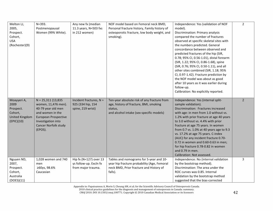
Appendix to: Papaioannou A, Morin S, Cheung AM, et al; for the Scientific Advisory Council of Osteoporosis Canada. 2010 clinical practice guidelines for the diagnosis and management of osteoporosis in Canada: summary. CMAJ 2010. DOI 10.1503/cmaj.100771. Copyright © 2010 Canadian Medical Association or its licensors
42
Melton LJ, 2005, Prospect. Cohort, USA (Rochester)(9)
N=393. Postmenopausal Women (99% White).
Any new fx (median 11.3 years, N=503 fxs in 212 women)
NOF model based on Femoral neck BMD, Personal fracture history, Family history of osteoporotic fracture, low body weight, and smoking).
Independence: Yes (validation of NOF model). Discrimination: Primary analysis compared the number of fractures observed at specific skeletal sites with the numbers predicted. General concordance between observed and predicted fractures of the hip (SIR, 0.78; 95% CI, 0.56-‐1.01), distal forearm (SIR, 1.22; 95% CI, 0.86-‐1.68), spine (SIR, 0.76; 95% CI, 0.50-‐1.11), and all other sites combined (SIR, 1.18; 95% CI, 0.97-‐1.42). Fracture prediction by the NOF model was about as good after 10 years as it was earlier during follow-‐up. Calibration: No explicitly reported.
2
Moayyeri A, 2009 Prospect. Cohort, United Kingdom (EPIC)(10)
N = 25,311 (13,835 women, 11,476 men). 40-‐79 year old men and women in the European Prospective Investigation into Cancer Norfolk study (EPOS).
Incident fractures, N = 925 (334 hip, 154 spine, 219 wrist)
Ten-‐year absolute risk of any fracture from age, history of fracture, BMI, smoking status, and alcohol intake (sex-‐specific models)
Independence: Yes (internal split-‐sample validation). Discrimination: Fractures increased with age: in men from 1.0 without vs. 1.2% with prior fracture at age 40 years to 3.0 without vs. 4.4% with prior fracture at age 75 years. In women from 0.7 vs. 1.0% at 40 years age to 9.3 vs. 17.2% at age 75 years. C-‐index (AUC) for any incident fracture 0.70-‐0.72 in women and 0.60-‐0.63 in men; for hip fracture 0.78-‐0.82 in women and 0.79 in men. Calibration: Not assessed.
2
Nguyen ND, 2007, Prospect. Cohort, Australia (DOES)(11)
1,028 women and 740 men ≥60ys, 98.6% Caucasian
Hip fx (N=127) over 13 ys follow-‐up. Excln fx from major trauma.
Tables and nomograms for 5-‐year and 10-‐year hip fracture probability (Age, Femoral neck BMD, Prior fracture and History of falls).
Independence: No (internal validation by the bootstrap method). Discrimination: The area under the ROC curves was 0.85. Internal validation by the bootstrap method suggested that the bias-‐corrected
3

Appendix to: Papaioannou A, Morin S, Cheung AM, et al; for the Scientific Advisory Council of Osteoporosis Canada. 2010 clinical practice guidelines for the diagnosis and management of osteoporosis in Canada: summary. CMAJ 2010. DOI 10.1503/cmaj.100771. Copyright © 2010 Canadian Medical Association or its licensors
43
estimate of predictive discrimination of 0.70 for women and 0.65 for men. Calibration: Not independently assessed. The maximum calibration error in predicting probability of fracture was about 2% for women and 7% for men by the bootstrap method.
Nguyen ND, 2008, Prospect. Cohort, Australia (DOES)(12)
1,358 women and 858 men ≥60ys. Women Mean 71ys, SD=8. Men Mean 70ys, SD=6.
Low trauma , non-‐pathological clinical fx (women 426, men 149 during 13 years). Excl digit, head, cervical fx.
Nomograms for 5-‐year and 10-‐year fracture risk (used Age, Fracture history, Fall history, and BMD T-‐score or Weight).
Independence: No (internal validation by the bootstrap method). Discrimination: Receiver operating characteristic curve suggested that model with BMD (AUC = 0.75 for both sexes) performed better than model with weight (AUC = 0.72 for women and 0.74 for men). Calibration: Maximum calibration error in predicting probability of fracture was 0.4% in women and 0.6–1.9% in men by the bootstrap method.
2
Robbins J, 2007, Prospect. Cohort, USA (WHI)(13)
N=93,676. Validation (Clinical Trial HRT, (low fat diet), Calc/Vit D) N>60,000 (10,750 with DXA). Women, 50-‐79 years, White 84%. Derivation (Observ. Study)
Hip fx (N=1132 derivation, N=791 validation incl. 80 with DXA).
5-‐year risk of hip fx from summed point score (eleven factors: age, self-‐reported health, weight, height, race/ethnicity, self-‐reported physical activity, history of fracture after age 54 years, parental hip fracture, current smoking, current corticosteroid use, and treated diabetes). No BMD variables.
Independence: Yes Discrimination: Receiver operating characteristic curves in the validation cohort showed AUC 80% (95% confidence interval [CI], 0.77%-‐0.82%). For DXA subgroup, DXA prediction AUC 79% (95% CI, 73%-‐85%) vs WHI algorithm 71% (95% CI, 66%-‐76%). Calibration (Observed vs Predicted in DXA subgroup): T-‐score >−2.5 (60 vs 57), T-‐score <−2.5 (20 vs 23), WHO point score <21 (65 vs 64.9), WHO point score >21 (15 vs 15.1).
1
BMD = bone mineral density, BM I = body mass index, DXA = dual energy x-‐ray densitometry, Fx = fracture, SD = standard deviation, HRT = hormone replacement therapy, ROC = receiver operating curve, AUC = area under curve, CI = confidence interval, GR = gradient of risk, SIR = standardized incidence ratio.

Appendix to: Papaioannou A, Morin S, Cheung AM, et al; for the Scientific Advisory Council of Osteoporosis Canada. 2010 clinical practice guidelines for the diagnosis and management of osteoporosis in Canada: summary. CMAJ 2010. DOI 10.1503/cmaj.100771. Copyright © 2010 Canadian Medical Association or its licensors
44
REFERENCES (1) Abrahamsen B, Vestergaard P, Rud B, Barenholdt O, Jensen JE, Nielsen SP et al. Ten-‐year absolute risk of osteoporotic fractures according to BMD T score at menopause: the Danish Osteoporosis Prevention Study. Journal of Bone & Mineral Research 2006; 21(5):796-‐800. (2) Ahmed LA, Schirmer H, Fonnebo V, Joakimsen RM, Berntsen GK, Ahmed LA et al. Validation of the Cummings' risk score; how well does it identify women with high risk of hip fracture: the Tromso Study. European Journal of Epidemiology 2006; 21(11):815-‐822. (3) Bagger YZ, Tanko LB, Alexandersen P, Hansen HB, Qin G, Christiansen C et al. The long-‐term predictive value of bone mineral density measurements for fracture risk is independent of the site of measurement and the age at diagnosis: results from the Prospective Epidemiological Risk Factors study. Osteoporos Int 2006; 17(3):471-‐477. (4) Black DM, Steinbuch M, Palermo L, Dargent-‐Molina P, Lindsay R, Hoseyni MS et al. An assessment tool for predicting fracture risk in postmenopausal women. Osteoporos Int 2001; 12(7):519-‐528. (5) Chen P, Krege JH, Adachi JD, Prior JC, Tenenhouse A, Brown JP et al. Vertebral fracture status and the World Health Organization risk factors for predicting osteoporotic fractur risk. J Bone Miner Res 2009; 24(3):495-‐502. (6) Ensrud KE, Ewing SK, Taylor BC, Fink HA, Cawthon PM, Stone KL et al. Comparison of 2 frailty indexes for prediction of falls, disability, fractures, and death in older women.[see comment]. Arch Intern Med 2008; 168(4):382-‐389. (7) Kanis JA, Johnell O, Oden A, Dawson A, De Laet C, Jonsson B. Ten year probabilities of osteoporotic fractures according to BMD and diagnostic thresholds. Osteoporos Int 2001 12(12):989-‐995. (8) Kanis JA, Oden A, Johnell O, Johansson H, De Laet C, Brown J et al. The use of clinical risk factors enhances the performance of BMD in the prediction of hip and osteoporotic fractures in men and women. Osteoporos Int 2007; 18(8):1033-‐1046. (9) Melton LJ, III, Atkinson EJ, Khosla S, Oberg AL, Riggs BL, Melton LJ et al. Evaluation of a prediction model for long-‐term fracture risk. Journal of Bone & Mineral Research 2005; 20(4):551-‐556. (10) Moayyeri A, Kaptoge S, Luben RN, Wareham NJ, Bingham S, Reeve J et al. Estimation of absolute fracture risk among middle-‐aged and older men and women: the EPIC-‐Norfo population cohort study. European Journal of Epidemiology 2009; 24(5):259-‐266. (11) Nguyen ND, Frost SA, Center JR, Eisman JA, Nguyen TV, Nguyen ND et al. Development of a nomogram for individualizing hip fracture risk in men and women. Osteoporos Int 2007; 18(8):1109-‐1117. (12) Nguyen ND, Frost SA, Center JR, Eisman JA, Nguyen TV, Nguyen ND et al. Development of prognostic nomograms for individualizing 5-‐year and 10-‐year fracture risks. Osteoporos Int 2008; 19(10):1431-‐1444. (13) Robbins J, Aragaki AK, Kooperberg C, Watts N, Wactawski-‐Wende J, Jackson RD et al. Factors associated with 5-‐year risk of hip fracture in postmenopausal women. JAMA 200 298(20):2389-‐2398.

Appendix to: Papaioannou A, Morin S, Cheung AM, et al; for the Scientific Advisory Council of Osteoporosis Canada. 2010 clinical practice guidelines for the diagnosis and management of osteoporosis in Canada: summary. CMAJ 2010. DOI 10.1503/cmaj.100771. Copyright © 2010 Canadian Medical Association or its licensors
45
Table A17: Frequency of clinical risk factors included in the risk assessment models
Risk factor References (below table)
Number of uses (studies)
Number of uses (models)
Age [1, 2, 3, 4, 5-‐18] 18 14 Sex [1, 2, 3, 4, 5-‐18] 18 14 Prior history of fracture [1, 2, 4, 5, 6, 8, 9, 11, 12, 14-‐18] 14 11 BMI or weight [2, 5, 6, 9, 11, 12, 16-‐18] 9 7 Parental history of hip fracture or osteoporosis [2, 5, 6, 9, 12, 14, 16, 18] 8 6 Smoking history [2, 5, 6, 9, 11, 12, 16, 18] 8 6 Glucocorticoid use [1, 2, 5, 8, 12, 16, 18] 7 4 Rheumatoid arthritis [2, 5, 16, 18] 4 2 Excessive alcohol intake [2, 5, 16, 18] 4 2 Inability to rise from chair without arms [9, 10, 14] 3 3 Level of physical activity [10, 12, 14] 3 3 Weight loss [10, 14] 2 2 Height or height loss [12, 14] 2 2 Fall history [4, 17] 2 2 Self-‐reported health [12, 14] 2 2 Number of prior fractures [5, 6] 2 2 Slow walking speed [10] 1 1 Hip fracture in sister [6] 1 1 Long-‐acting benzodiazepines [14] 1 1 Pulse rate [14] 1 1 Caffeine intake [14] 1 1 Anticonvulsant use [14] 1 1 Hyperthyroidism [14] 1 1 Depth perception [14] 1 1 Visual contrast sensitivity [14] 1 1 Vertebral fracture severity [5] 1 1 Reduced energy [10] 1 1 Poor grip strength [10] 1 1 Diabetes [12] 1 1 Race [12] 1 1
Appendix to: Papaioannou A, Morin S, Cheung AM, et al; for the Scientific Advisory Council of Osteoporosis Canada. 2010 clinical practice guidelines for the diagnosis and management of osteoporosis in Canada: summary. CMAJ 2010. DOI 10.1503/cmaj.100771. Copyright © 2010 Canadian Medical Association or its licensors
46
REFERENCES (1) Siminoski K, Leslie WD, Frame H et al. (2005) Recommendations for bone mineral density reporting in Canada. Can.Assoc.Radiol.J. 56:178-‐188. (2) Fujiwara S, Nakamura T, Orimo H et al. (2008) Development and application of a Japanese model of the WHO fracture risk assessment tool (FRAX). Osteoporos.Int. 19:429-‐435. (3) Kanis JA, Johnell O, Oden A et al. (2001) Ten year probabilities of osteoporotic fractures according to BMD and diagnostic thresholds. Osteoporos.Int. 12:989-‐995. (4) Nguyen ND, Frost SA, Center JR et al. (2007) Development of a nomogram for individualizing hip fracture risk in men and women. Osteoporos.Int. 18:1109-‐1117. (5) Chen P, Krege JH, Adachi JD et al. (2009) Vertebral fracture status and the World Health Organization risk factors for predicting osteoporotic fracture risk. J.Bone Miner.Res. 24:495-‐502. (6) Ettinger B, Hillier TA, Pressman A et al. (2005) Simple computer model for calculating and reporting 5-‐year osteoporotic fracture risk in postmenopausal women. J.Womens Health (Larchmt.) 14:159-‐171. (7) Leslie WD, Tsang JF, Lix LM (2008) Validation of ten-‐year fracture risk prediction: a clinical cohort study from the Manitoba Bone Density Program. Bone 43:667-‐671. (8) Leslie WD, Tsang JF, Lix L et al. (2009) Simplified system for absolute fracture risk assessment: Clinical validation in Canadian women. J Bone Miner.Res. 24:353-‐360. (9) Black DM, Steinbuch M, Palermo L et al. (2001) An assessment tool for predicting fracture risk in postmenopausal women. Osteoporos.Int. 12:519-‐528. (10) Ensrud KE, Ewing SK, Taylor BC et al. (2008) Comparison of 2 frailty indexes for prediction of falls, disability, fractures, and death in older women.[see comment]. Arch.Intern.Med. 168:382-‐389.
(11) Melton LJ, III, Atkinson EJ, Khosla S et al. (2005) Evaluation of a prediction model for long-‐term fracture risk. Journal of Bone & Mineral Research 20:551-‐556. (12) Robbins J, Aragaki AK, Kooperberg C et al. (2007) Factors associated with 5-‐year risk of hip fracture in postmenopausal women. JAMA 298:2389-‐2398. (13) Abrahamsen B, Vestergaard P, Rud B et al. (2006) Ten-‐year absolute risk of osteoporotic fractures according to BMD T score at menopause: the Danish Osteoporosis Prevention Study. Journal of Bone & Mineral Research 21:796-‐800. (14) Ahmed LA, Schirmer H, Fonnebo V et al. (2006) Validation of the Cummings' risk score; how well does it identify women with high risk of hip fracture: the Tromso Study. European Journal of Epidemiology 21:815-‐822. (15) Bagger YZ, Tanko LB, Alexandersen P et al. (2006) The long-‐term predictive value of bone mineral density measurements for fracture risk is independent of the site of measurement and the age at diagnosis: results from the Prospective Epidemiological Risk Factors study. Osteoporos.Int. 17:471-‐477. (16) Kanis JA, Johnell O, Oden A et al. (2008) FRAX and the assessment of fracture probability in men and women from the UK. Osteoporos.Int. 19:385-‐397. (17) Nguyen ND, Frost SA, Center JR et al. (2008) Development of prognostic nomograms for individualizing 5-‐year and 10-‐year fracture risks. Osteoporos.Int. 19:1431-‐1444. (18) Kanis JA, Oden A, Johnell O et al. (2007) The use of clinical risk factors enhances the performance of BMD in the prediction of hip and osteoporotic fractures in men and women. Osteoporos.Int. 18:1033-‐1046.

Appendix to: Papaioannou A, Morin S, Cheung AM, et al; for the Scientific Advisory Council of Osteoporosis Canada. 2010 clinical practice guidelines for the diagnosis and management of osteoporosis in Canada: summary. CMAJ 2010. DOI 10.1503/cmaj.100771. Copyright © 2010 Canadian Medical Association or its licensors
47
Table A18: Evidence of Randomized Controlled Trials on Therapies Study Country of Origin Total Number of Patients
Age (yr, mean)/ % Female
Baseline mean BMD (t-‐score or g/cm2) % Fractures
Intervention (dose)
Outcome # Fractures/ total treated
# Fractures/ total control
Effect (95% CI)
NNT* ^ ARR Level of Evidence
Teriparatide: Any new vertebral fracture
NR NR RR=0.28 (0.20-‐ 0.38)
NR NR
Teriparatide: New adjacent fracture
NR NR RR= 0.25 (0.15-‐ 0.41)
NR NR
Teriparatide: New non-‐ adjacent fracture
NR NR RR= 0.30 (0.19-‐ 0.46)
NR NR
Raloxifene: Any new vertebral fracture
NR NR RR=0.46 (0.38-‐ 0.57)
NR NR
Raloxifene: New adjacent fracture
NR NR RR=0.46 (0.38-‐ 0.61)
NR NR
Bouxsein et al., 2009(1) USA N = 1226 (from 2 RCTs, n=398, n=82(6)
68.7 years 100%
Lumbar spine: -‐2.7 Mean number of vertebral fractures:
Teriparatide (20mg or 40mg) Raloxifene (60mg or 120mg)
Raloxifene: New non-‐adjacent fracture
NR NR RR=0.47 (0.36-‐ 0.62)
NR NR
Level 1
HRT: New vertebral/non-‐ vertebral fractures
0/12 3/24 (12.5%)
p=0.25 NR 4.8 (3.9-‐5.8)
Campbell et al., 2009(2) United Kingdom N = 50
<60years 100%
BMD: NR Fractures: NR
HRT: minimum 2mg oestradiol; 0.625mg conjugated estrogen; 50mcg transdermal estradiol Etidronate: 400mg
Etidronate: New vertebral/non-‐ vertebral fractures
1/24 (4.2%)
2/23 (8.7%)
p=0.97
Level 1
*Numbers needed to treat ^ Absolute risk reduction

Appendix to: Papaioannou A, Morin S, Cheung AM, et al; for the Scientific Advisory Council of Osteoporosis Canada. 2010 clinical practice guidelines for the diagnosis and management of osteoporosis in Canada: summary. CMAJ 2010. DOI 10.1503/cmaj.100771. Copyright © 2010 Canadian Medical Association or its licensors
48
New vertebral fracture
86/3702 (2.3%)
264/3691 (7.2%)
0.32 (0.26 to 0.41) p<0.001
NR 4.8 (3.9-‐5.8)
Nonvertebral fracture
238/3702 (6.4%)
293/3691 (7.9%)
0.80 (0.67 to 0.95) 0.01
NR 1.5 (0.3-‐2.7)
Hip fracture 26/3702 (.70%)
42/3691 (1.1%)
0.60 (0.37 to 0.97) 0.04
NR 0.3 (-‐0.1-‐0.7)
New clinical vertebral fracture
29/3702 (.78%)
92/3691 (2.5%)
0.31 (0.20 to 0.47) <0.001
NR 1.7 (1.1-‐2.3)
Cummings et al., 2009(3) Multi-‐center, International (USA, United Kingdom, Australia, France, Czech Republic, Italy, Argentina, Denmark N = 7808
60-‐90years 100% Intervention: 72.3years Control: 72.3years
Lumbar spine: -‐2.8 Total hip: -‐1.9 Femoral neck: -‐2.2 27% vertebral fractures
Denosumab 60mg
Multiple (≥2) new vertebral fractures
23/3702 (.62%)
59/3691 (1.6%)
0.39 (0.24 to 0.63) <0.001
NR 1.0 (0.5-‐1.5)
Level 1
Nonvertebral fracture
428/ 5044 (8.5%)
438/5057 (8.7%)
HR = 0.96 (0.84-‐1.10)
NR NR Ensrud et al., 2008(4)* United States N = 10, 101
67.5 years 100%
BMD not performed 6% history of fracture
Raloxifene (60 mg/d orally)
Clinical vertebral fracture
64/5044 (1.3%)
97/5057 (1.9%)
HR = 0.65 (0.47 -‐0.89)
NR NR
Level 1

Appendix to: Papaioannou A, Morin S, Cheung AM, et al; for the Scientific Advisory Council of Osteoporosis Canada. 2010 clinical practice guidelines for the diagnosis and management of osteoporosis in Canada: summary. CMAJ 2010. DOI 10.1503/cmaj.100771. Copyright © 2010 Canadian Medical Association or its licensors
49
Clinical fractures regardless of renal function
NR NR NR NR
Spinal fractures regardless of renal function
NR NR NR NR
Clinical Fractures
NR NR Severely reduced eGFR : Moderately reduced or normal
NR NR
55-‐80 years 100% eGFR <45ml/minute: 74.6 years eGFR≥ 45ml/minute: 68.1 years
femoral neck ≤0.68g/cm2
eGFR <45ml/minute: 0.54g/cm2
eGFR≥ 45ml/minute: 0.59g/cm2
Spinal Fractures NR NR Severely reduced eGFR : Moderately reduced or normal
NR NR
Clinical Fractures Women with osteoporosis (n = 3214) With Alendronate
NR NR Severely reduced eGFR : Moderately reduced or normal
NR NR
Spinal Fractures Women with osteoporosis (n = 3214) With Alendronate
NR NR Severely reduced eGFR : Moderately reduced or normal
NR NR
Nonvertebral fracture
79/1065 (7.6%)
107/1062 (10.7%)
HR: 0.73 (0.55-‐ 0.98) p=.03
NR NR
Hip fracture 23/1065 (2.0%)
33/1062 (8.5%)
HR: 0.70 (0.41-‐ 1.19) p=.18
NR NR
Jamal et al., 2007(5) Multi-‐center: Canada & USA N = 6458
5mg of alendronate**
Vertebral 21/1065 (1.7%)
39/1062 (3.8%)
HR: 0.54(0.32-‐ 0.92) p=.02
NR NR
Level 1

Appendix to: Papaioannou A, Morin S, Cheung AM, et al; for the Scientific Advisory Council of Osteoporosis Canada. 2010 clinical practice guidelines for the diagnosis and management of osteoporosis in Canada: summary. CMAJ 2010. DOI 10.1503/cmaj.100771. Copyright © 2010 Canadian Medical Association or its licensors
50
New non-‐vertebral fractures at 2 years
14/152 (9.2%)
35/148 (23.6%)
p= 0.0026 NR NR
New non-‐vertebral fractures at 1 year
10/158 (6.3%)
17/158 (10.8%)
p = 0.227 NR NR
Ringe et al., 2009(6) Germany N = 316
Intervention: 55.8years Control: 58.0years 0 females
Intervention: Lumbar spine -‐3.34 Femoral neck -‐2.63 Total hip -‐2.45 Control: Lumbar spine –3.29 Femoral neck –2.65 Total hip –2.59
Risedronate 5mg (plus 1000mg elementary calcium, 800IU vitamin D
New non-‐vertebral fractures at 2 years
18/152 (11.8%)
33/148 (22.3%)
p = 0.032 NR NR
Level 1
New vertebral/ nonvertebral fracture: BMD loss 0-‐4%
5/182 (2.7%)
15/149 (10.0%)
0.27 (0.10-‐0.73)
NR 7.4
New vertebral/ nonvertebral fracture: BMD loss >4%
2/82 (2.4%)
14/61 (23.0%)
0.11 (0.03-‐.45)
NR 20.6
Level 1
New vertebral/ nonvertebral fracture: BMD gain 0-‐4%
16/270 (5.9%)
19/124 (15.3%)
0.39 (0.21-‐0.73)
NR 9.4
Watts et al., 2009(7) USA N = 1216
Mean age range across groups stratified by BMD change: 68.0 – 70.0years 100%
Mean lumbar spine across groups stratified by BMD change: -‐2.27 to -‐2.68 Mean femoral neck across groups stratified by BMD change: -‐2.21 to -‐2.54
Teriparatide 20 or 40µg/d
New vertebral/ nonvertebral fracture: BMD gain >4%
14/282 (5.0%)
9/66 (13.6%)
0.36 (0.16-‐0.80) NR 8.6
HRT = Hormone Replacement Therapy *This study also provided data on fracture incidence rates per 1000 person years for nonvertebral fractures (HR, 0.96; 95% CI, 0.84–1.10), hip/femur fractures (HR, 0.85; 95% CI.64–1.13) and wrist fractures (HR, 0.95; 95% CI, 0.73–1.24). **Stratified by eGFR: <45ml/minute and ≥ 45ml/minute REFERENCES
(1) Bouxsein ML, Chen P, Glass EV, Kallmes DF, Delmas PD, Mitlak BH. Teriparatide and raloxifene reduce the risk of new adjacent vertebral fractures in
postmenopausal women with osteoporosis. Results from two randomized controlled trials. Journal of Bone and Joint Surgery 2009; 91:1329-‐1338.

Appendix to: Papaioannou A, Morin S, Cheung AM, et al; for the Scientific Advisory Council of Osteoporosis Canada. 2010 clinical practice guidelines for the diagnosis and management of osteoporosis in Canada: summary. CMAJ 2010. DOI 10.1503/cmaj.100771. Copyright © 2010 Canadian Medical Association or its licensors
51
(2) Campbell IA, Francis RM, Prescott RJ, Reid DM. Homorne replacement therapy (HRT) or etidronate for osteoporosis in postmenopausal asthmatics on glucocorticoids:A randomized factorial trial. Scottish Medical Journal 2009; 54(1):21-‐25.
(3) Cummings SR, Martin SJ, McClung MR, Siris ES, Eastell R, Reid IR et al. Denosumab for prevention of fractures in postmenopausal women with osteoporosis. N Engl J Med 2009; 361:756-‐765.
(4) Ensrud KE, Stock JL, Barrett-‐Connor E, Grady D, Mosca L, Khaw KT et al. Effects of raloxifene on fracture risk in postmenopausal women: the raloxifene
use for the heart trial. J Bone Miner Res 2008; 23(1):112-‐120.
(5) Jamal SA, Bauer DC, Ensrud KC, Cauley JA, Hochberg M, Ishani A et al. Alendronate treatment in women with normal to severely impaired renal function: an analysis of the Fracture Intervention Trial. J Bone Miner Res 2007; 22(4):503-‐508.
(6) Ringe JD, Farahmand P, Faber H, Dorst A. Sustained efficacy of risedronate in men with primary and secondary osteoporosis: results of a 2-‐year study.
Rheumatology International 2009; 29:311-‐315.
(7) Watts NB, Miller PD, Kohlmeier LA, Sebba A, Chen P, Wong M et al. Vertebral fracture risk is reduced in women who lose femoral neck BMD with
teriparatide treatment. J Bone Miner Res 2009; 24(6):1125-‐1131.

Appendix to: Papaioannou A, Morin S, Cheung AM, et al; for the Scientific Advisory Council of Osteoporosis Canada. 2010 clinical practice guidelines for the diagnosis and management of osteoporosis in Canada: summary. CMAJ 2010. DOI 10.1503/cmaj.100771. Copyright © 2010 Canadian Medical Association or its licensors
52
Table A19: Evidence of Systematic Reviews on Therapies Study (Population) Inclusion Criteria
Intervention Outcomes (# of studies, participants)
Results Risk Ratio (M-‐H, Fixed, 95% CI)
Number Needed to Treat (NNT)
Absolute Risk Reduction (ARR)
Level of Evidence
any dose of vitamin D preventing nonvertebral fracture (12, N= 41279)
RR=0.86 (0.77-‐0.96)
NR NR Bischoff-‐Ferrari et al., 2009(1) (Older individuals ≥65years) Double blinded RCTs, oral vitamin D supplementation (cholecalciferol [vitaminD3] or ergocalciferol); min 1 year follow-‐up of 1 year; > 1 fracture in each trial; age ≥ 65 years; adherence report, method of fracture confirmation.
Supplemental vitamin D, with or without calcium supplementation Placebo or calcium supplementation alone
Any dose of vitamin D preventing hip fractures (8, N = 40886)
RR=0.91 (0.78-‐1.05)
NR NR
Level 2+
Vertebral (2, N = 7802)
0.33 (0.27, 0.4) significant
13 NR
Vertebral fracture: clinical (1, N = 7736)
0.23 (0.14, 0.37) significant
50 NR
Nonvertebral Fracture (2, N = 9868)
0.75 (0.66, 0.85) Significant
50 NR
NICE 2008(2) (Men and women at risk for osteoporotic fracture: with osteoporosis or osteopenia; osteoporosis defined as t-‐score≤ -‐2.5SD; corticosteroid induced osteoporosis included)
Zoledronic Acid 5mg
Hip (2, N = 9868)
0.62 (0.47, 0.83) significant
100 NR
Level 2+ (included RCTs and quasi-‐randomized studies)
RCTs, quasi-‐randomized only in absence of other evidence, English language except those translated for Cochrane reviews, study N> 10.
Vertebral fracture (9, N = 8074)
0.55(0.46, 0.66) significant
33 NR Level 2+
Alendronic acid vs placebo/no treatment
Nonvertebral Fracture (8, N = 10429)
0.83 (0.74, 0.93) significant
50 NR
Hip Fracture (3, N = 7453)
0.62 (0.4, 0.96) Significant
100 NR
Wrist (3, N = 7453) 0.85 (0.67, 1.09) Significant
NR NR
Etidronate vs placebo/no treatment
Vertebral fracture (8, N = 1039)
0.51 (0.31, 0.83) significant
25 NR Level 2+

Appendix to: Papaioannou A, Morin S, Cheung AM, et al; for the Scientific Advisory Council of Osteoporosis Canada. 2010 clinical practice guidelines for the diagnosis and management of osteoporosis in Canada: summary. CMAJ 2010. DOI 10.1503/cmaj.100771. Copyright © 2010 Canadian Medical Association or its licensors
53
Nonvertebral fracture (4,
N = 472) 0.72 (0.29, 1.8) NS
NR NR
Hip Fracture (2, N= 246) 1.02 (0.21, 4.94) ns
NR NR
Wrist (1, N = 209) 4.95 (0.24, 101.93) ns
NR NR
All fractures (4, N = 420) 0.78 (0.42, 1.44) NR NR Risedronate vs placebo Vertebral Fracture (7, N =
2845) 0.61 (0.5, 0.74) significant
17 NR Level 2+
NR
Nonvertebral Fracture (7, N = 2845)
0.81 (0.72, 0.9) significant
50 NR
Hip fracture (7, N = 12658)
0.73 (0.58, 0.92) Significant
100 NR
Wrist Fracture (4, N = 11923)
0.68 (0.43, 1.07) NS
NR NR
Humerus (2, N = 2439) 0.46 (0.23, 0.93) Significant
100 NR
Teriparatide vs placebo Vertebral (2, N = 910) 0.36 (0.23, 0.57) Significant
11 NR Level 2+
Nonvertebral (2, N = 1383 )
0.49 (0.27, 0.87) Significant
50 NR
Hip (1, N = 1085) 0.25 (0.03, 2.24) NS
NR NR
Wrist (1, N = 1085) 0.29 (0.06, 1.38) NS
NR NR
Humerus (1, N = 1085) 1.01 (0.14, 7.11) NS
NR NR
Calcitonin vs placebo Vertebral (4, N = 11842) 0.65 (0.48, 0.88) Significant
13 NR Level 2+
Hip (3, N = 11774) 0.54 (0.18, 1.59) NS
NR NR
Arm: all fractures wrist, ulna, humerus, radius (2, N = 11745)
0.79 (0.38, 1.61) NS
NR NR
HRT vs placebo/no treatment
Vertebral (4, N = 11842) 0.67 (0.48, 0.93) Significant
100 NR Level 2+

Appendix to: Papaioannou A, Morin S, Cheung AM, et al; for the Scientific Advisory Council of Osteoporosis Canada. 2010 clinical practice guidelines for the diagnosis and management of osteoporosis in Canada: summary. CMAJ 2010. DOI 10.1503/cmaj.100771. Copyright © 2010 Canadian Medical Association or its licensors
54
Nonvertebral (3, N = 1174
0.73(0.65, 0.81) Significant
33 NR
Hip (2, N = 11745) 0.63 (0.42, 0.93) Significant
Infinity NR
All fractures (3, N = 11556)
0.7 (0.63, 0.78) significant
25 NR
Raloxifene vs placebo Vertebral (2, N = 4639) 0.64 (0.54, 0.78) significant
25 NR Level 2+
Nonvertebral (2, N = 7793)
0.91 (0.78, 1.05) NS
NR NR
Hip (2, N = 7793) 1.12 (0.64, 1.94) NS
NR NR
Wrist (1, N = 7705) 0.88 (0.68, 1.14) NS
NR NR
Hydroxylated vitamin D vs placebo
Vertebral (1, N = 246) 4 (0.45, 35.28) NS
NR NR Level 2+
Nonvertebral (1, N = 246) 0.46 (CI 0.18, 1.18) NS
NR NR
Native vitamin D‡ vs placebo/no treatment
Vertebral (3, N = 8801)
0.66 (0.4, 1.08) NS
NR NR Level 2+
Nonvertebral (8, N = 22098)
1.01 (0.94, 1.1) NS
NR NR
Hip (8, N = 22098) 1.14 (0.98, 1.32) NS
NR NR
All clinical fractures (1, N = 3314)
0.96 (0.7, 1.33) NS
NR NR Level 2+
Native vitamin D‡ + calcium vs placebo/no treatment
All clinical fractures (1, N = 5063)
0.96 (0.78, 1.18) NS
NR NR
Vertebral (7, N = 6013) 0.84 (0.66, 1.08) NS
NR NR Level 2+
Nonvertebral (5, N = 5717) 0.92 (0.79, 1.05) NS
NR NR
Calcium vs placebo
Wrist (2, N = 4160) 1.05 (0.57, 1.92) NS
NR NR
Distal forearm fracture (1, N = 1471)
0.64 (0.4, 1.02) NS NR NR
Upper Limb (2, N = 4103) 1.06 (0.75, 1.5) NS NR NR
All fractures (1, N = 5574) 0.9 (0.79, 1.03) NS NR NR
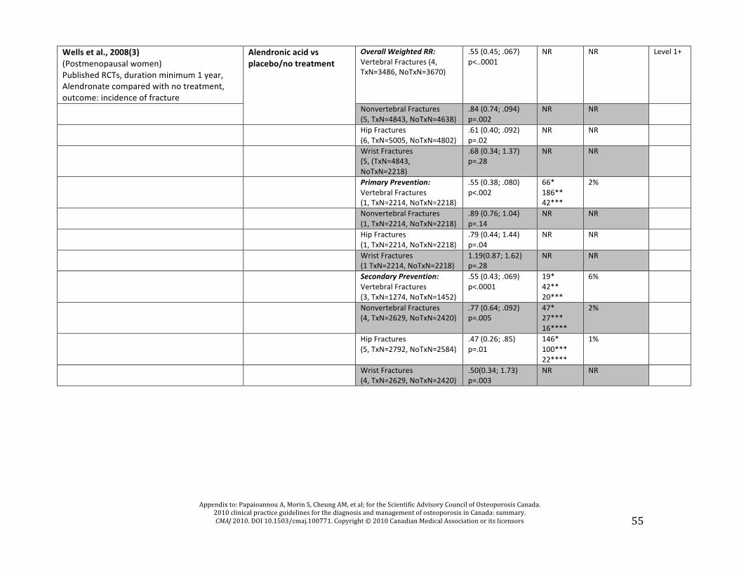
Appendix to: Papaioannou A, Morin S, Cheung AM, et al; for the Scientific Advisory Council of Osteoporosis Canada. 2010 clinical practice guidelines for the diagnosis and management of osteoporosis in Canada: summary. CMAJ 2010. DOI 10.1503/cmaj.100771. Copyright © 2010 Canadian Medical Association or its licensors
55
Wells et al., 2008(3) (Postmenopausal women) Published RCTs, duration minimum 1 year, Alendronate compared with no treatment, outcome: incidence of fracture
Overall Weighted RR: Vertebral Fractures (4, TxN=3486, NoTxN=3670)
.55 (0.45; .067) p<..0001
NR NR Level 1+
Alendronic acid vs placebo/no treatment
Nonvertebral Fractures (5, TxN=4843, NoTxN=4638)
.84 (0.74; .094) p=.002
NR NR
Hip Fractures (6, TxN=5005, NoTxN=4802)
.61 (0.40; .092) p=.02
NR NR
Wrist Fractures (5, (TxN=4843, NoTxN=2218)
.68 (0.34; 1.37) p=.28
NR NR
Primary Prevention: Vertebral Fractures (1, TxN=2214, NoTxN=2218)
.55 (0.38; .080) p<.002
66* 186** 42***
2%
Nonvertebral Fractures (1, TxN=2214, NoTxN=2218)
.89 (0.76; 1.04) p=.14
NR NR
Hip Fractures (1, TxN=2214, NoTxN=2218)
.79 (0.44; 1.44) p=.04
NR NR
Wrist Fractures (1 TxN=2214, NoTxN=2218)
1.19(0.87; 1.62) p=.28
NR NR
Secondary Prevention: Vertebral Fractures (3, TxN=1274, NoTxN=1452)
.55 (0.43; .069) p<.0001
19* 42** 20***
6%
Nonvertebral Fractures (4, TxN=2629, NoTxN=2420)
.77 (0.64; .092) p=.005
47* 27*** 16****
2%
Hip Fractures (5, TxN=2792, NoTxN=2584)
.47 (0.26; .85) p=.01
146* 100*** 22****
1%
Wrist Fractures (4, TxN=2629, NoTxN=2420)
.50(0.34; 1.73) p=.003
NR NR

Appendix to: Papaioannou A, Morin S, Cheung AM, et al; for the Scientific Advisory Council of Osteoporosis Canada. 2010 clinical practice guidelines for the diagnosis and management of osteoporosis in Canada: summary. CMAJ 2010. DOI 10.1503/cmaj.100771. Copyright © 2010 Canadian Medical Association or its licensors
56
Wells et al., 2008(4) (Postmenopausal women) RCTs duration minimum 1 year, postmenopausal women only, primary & secondary prevention trials
Etidronate vs placebo/no treatment
Overall Weighted RR 400mg: Vertebral Fractures (8, TxN=430, NoTxN=428)
.59 (0.36;
.096) p=.03 NR NR Level 1+
Nonvertebral Fractures (7, TxN=393, NoTxN=394)
.98 (0.68; 1.42) p=.9
NR NR
Hip Fractures (4, TxN=295, NoTxN=294)
1.2 (0.37; 3.88) p=.8
NR NR
Wrist Fractures (4, TxN=295, NoTxN=294)
.87 (0.32; 2.36) p=.8
NR NR
Primary Prevention 400mg: Vertebral Fractures (2, TxN=81, NoTxN=82)
3.03(0.32; 28.44) p=.3
NR NR
Nonvertebral Fractures (2, TxN=81, NoTxN=82)
.56 (0.20; 1.61) p=.3
NR NR
Hip Fractures (0, N/A)
N/A NR NR
Wrist Fractures (0, N/A)
N/A NR NR
Secondary Prevention 400mg: Vertebral Fractures (6, TxN=349, NoTxN=346)
.53 (0.32;
.087) p=.01 20* 41*** 19****
5%
Nonvertebral Fractures (5, TxN=312, NoTxN=312)
1.07(0.72; 1.60) p=.7
NR NR
Hip Fractures (4, TxN=295, NoTxN=294)
1.20(0.37; 3.88 p=.8
NR NR
Wrist Fractures (4, TxN=295, NoTxN=294)
.87(0.32; 2.36) p=.8
NR NR
Secondary Prevention 200mg: Vertebral Fractures (2, N = 172)
0.32, (0.16; 0.64) p<.05
NR NR
Hip Fractures (1, N = 132)
0.33, (0.01; 8.04) p=NS
NR NR
Wrist Fractures (1, N = 132)
0.06(.05; 5.38) p=NS
NR NR
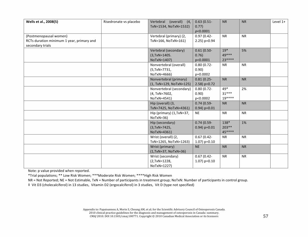
Appendix to: Papaioannou A, Morin S, Cheung AM, et al; for the Scientific Advisory Council of Osteoporosis Canada. 2010 clinical practice guidelines for the diagnosis and management of osteoporosis in Canada: summary. CMAJ 2010. DOI 10.1503/cmaj.100771. Copyright © 2010 Canadian Medical Association or its licensors
57
Wells et al., 2008(5)
Risedronate vs placebo Vertebral (overall) (4, TxN=1534, NoTxN=1532)
0.63 (0.51-‐0.77) p<0.0001
NR NR Level 1+
(Postmenopausal women) RCTs duration minimum 1 year, primary and secondary trials
Vertebral (primary) (2, TxN=166, NoTxN=161)
0.97 (0.42-‐2.25) p=0.94
NR NR
Vertebral (secondary) (3,TxN=1405. NoTxN=1407)
0.61 (0.50-‐0.76) p<0.0001
19* 49*** 23****
5%
Nonvertebral (overall) (5,TxN=7731, NoTxN=4666)
0.80 (0.72-‐0.90) p=0.0002
NR NR
Nonvertebral (primary) (1, TxN=129, NoTxN=125)
0.81 (0.25-‐2.58) p=0.72
NR NR
Nonvertebral (secondary) (4, TxN=7602, NoTxN=4541)
0.80 (0.72-‐0.90) p<0.0002
49* 31*** 19****
2%
Hip (overall) (3, TxN=7425, NoTxN=4361)
0.74 (0.59-‐0.94) p=0.01
NR NR
Hip (primary) (1,TxN=37, NoTxN=36)
NE NR NR
Hip (secondary) (3,TxN=7425, NoTxN=4361)
0.74 (0.59-‐0.94) p=0.01
138* 203** 45****
1%
Wrist (overall) (2, TxN=1265, NoTxN=1263)
0.67 (0.42-‐1.07) p=0.10
NR NR
Wrist (primary) (1,TxN=37, NoTxN=36)
NE NR NR
Wrist (secondary) (2,TxN=1228, NoTxN=1227)
0.67 (0.42-‐1.07) p=0.10
NR NR
Note: p value provided when reported. *Trial populations; ** Low Risk Women; ***Moderate Risk Women; ****High Risk Women NR = Not Reported; NE = Not Estimable, TxN = Number of participants in treatment group; NoTxN: Number of participants in control group. ‡ Vit D3 (cholecalciferol) in 13 studies, Vitamin D2 (ergocalciferol) in 3 studies, Vit D (type not specified)

Appendix to: Papaioannou A, Morin S, Cheung AM, et al; for the Scientific Advisory Council of Osteoporosis Canada. 2010 clinical practice guidelines for the diagnosis and management of osteoporosis in Canada: summary. CMAJ 2010. DOI 10.1503/cmaj.100771. Copyright © 2010 Canadian Medical Association or its licensors
58
REFERENCES
1) Bischoff-‐Ferrari HA, Willett WC, Wong JB, Stuck AE, Staehelin HB, Orav EJ et al. Prevention of nonvertebral fractures with oral vitamin D and dose dependency: a meta-‐analysis of randomized controlled trials. Arch Intern Med 2009; 169:551-‐561.
2) National Institute for Health and Clinical Excellence. Osteoporosis: assessment of fracture risk and the prevention of osteoporotic fracture in
individuals at high risk. 2008. National Collaborating Centre for Nursing and Supportive Care. Ref Type: Report
3) Wells GA, Cranney A, Peterson J, Boucher M, Shea B, Robinson V et al. Alendronate for the primary and secondary prevention of osteoporotic fractures in postmenopausal women. Cochrane Database Syst Rev 2008;(1):CD001155.
4) Wells GA, Cranney A, Peterson J, Shea B, Welch V, Coyle D et al. Etidrodronate for the primary and secondary prevention of osteoporotic fractures in postmenopausal women. Cochrane Database of Systematic Reviews 2008;(1):CD003376.
5) Wells G, Cranney A, Peterson J, Boucher M, Shea B, Robinson V et al. Risedronate for the primary and secondary prevention of osteoporotic fractures
in postmenopausal women. Cochrane Database Syst Rev 2008;(1):CD004523.

Appendix to: Papaioannou A, Morin S, Cheung AM, et al; for the Scientific Advisory Council of Osteoporosis Canada. 2010 clinical practice guidelines for the diagnosis and management of osteoporosis in Canada: summary. CMAJ 2010. DOI 10.1503/cmaj.100771. Copyright © 2010 Canadian Medical Association or its licensors
59
Table A20: Evidence of Randomized Controlled Trials on Special Populations Study Country of Origin Population Total Number of Patients
Age (yr, mean)/ % Female
Baseline mean BMD(t-‐score or g/cm2) % Fractures
Intervention (dose)
Outcome # Fractures/ total treated
# Fractures/ total control
Results Risk Ratio (95% CI)
Number Needed to Treat (NNT)
Absolute Risk Reduction (ARR)
Level of Evidence
New vertebral fractures
10/679 (1.5%)
26/673 (3.9%)
0.38 (0.19-‐0.78)
p=0.006
NR NR Smith et al.(1), 2009 Mulit-‐center, International (USA, Canada, Mexico, Finland, Czech Republic) Men receiving androgen-‐deprivation therapy for nonmetastatic prostate cancer N=1468
Intervention: 75.3years Control: 75.5years 0 female
Intervention: Lumbar spine, -‐0.3 Total hip -‐0.9 Femoral neck -‐1.4 Vertebral fracture: 21.1% History of osteoporotic fracture: 22.2% Control: Lumbar spine -‐0.4 Total hip -‐0.9 Femoral neck -‐1.4 Vertebral fracture 23.7% History of osteoporotic fracture 26.7%
Denosumab 60mg
New fracture at any site
38/734 (5.2%)
53/734 (7.2%)
0.72 (0.48-‐1.07) p=0.10
NR NR
Level 1
REFERENCE (1) Smith MR, Egerdie B, Toriz NH, Feldman R, Tammela TLJ, Saad F et al. Denosumab in men receiving androgen-‐deprivation therapy for prostrate cancer. N Engl J Med 2009; 361(8):745-‐755.
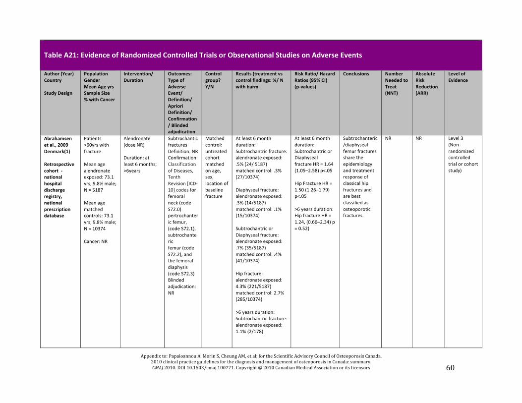
Appendix to: Papaioannou A, Morin S, Cheung AM, et al; for the Scientific Advisory Council of Osteoporosis Canada. 2010 clinical practice guidelines for the diagnosis and management of osteoporosis in Canada: summary. CMAJ 2010. DOI 10.1503/cmaj.100771. Copyright © 2010 Canadian Medical Association or its licensors
60
Table A21: Evidence of Randomized Controlled Trials or Observational Studies on Adverse Events
Author (Year) Country Study Design
Population Gender Mean Age yrs Sample Size % with Cancer
Intervention/ Duration
Outcomes: Type of Adverse Event/ Definition/ Apriori Definition/ Confirmation/ Blinded adjudication
Control group? Y/N
Results (treatment vs control findings: %/ N with harm
Risk Ratio/ Hazard Ratios (95% CI) (p-‐values)
Conclusions Number Needed to Treat (NNT)
Absolute Risk Reduction (ARR)
Level of Evidence
Abrahamsen et al., 2009 Denmark(1) Retrospective cohort -‐ national hospital discharge registry, national prescription database
Patients >60yrs with fracture Mean age alendronate exposed: 73.1 yrs; 9.8% male; N = 5187 Mean age matched controls: 73.1 yrs; 9.8% male; N = 10374 Cancer: NR
Alendronate (dose NR) Duration: at least 6 months; >6years
Subtrochantic fractures Definition: NR Confirmation: Classification of Diseases, Tenth Revision [ICD-‐10] codes for femoral neck (code S72.0) pertrochanteric femur, (code S72.1), subtrochanteric femur (code S72.2), and the femoral diaphysis (code S72.3) Blinded adjudication:NR
Matched control: untreated cohort matched on age, sex, location of baseline fracture
At least 6 month duration: Subtrochantric fracture: alendronate exposed: .5% (24/ 5187) matched control: .3% (27/10374) Diaphyseal fracture: alendronate exposed: .3% (14/5187) matched control: .1% (15/10374) Subtrochantric or Diaphyseal fracture: alendronate exposed: .7% (35/5187) matched control: .4% (41/10374) Hip fracture: alendronate exposed: 4.3% (221/5187) matched control: 2.7% (285/10374) >6 years duration: Subtrochantric fracture: alendronate exposed: 1.1% (2/178)
At least 6 month duration: Subtrochantric or Diaphyseal fracture HR = 1.64 (1.05–2.58) p<.05 Hip Fracture HR = 1.50 (1.26–1.79) p<.05 >6 years duration: Hip fracture HR = 1.24, (0.66–2.34) p = 0.52)
Subtrochanteric/diaphyseal femur fractures share the epidemiology and treatment response of classical hip fractures and are best classified as osteoporotic fractures.
NR NR Level 3 (Non-‐randomized controlled trial or cohort study)
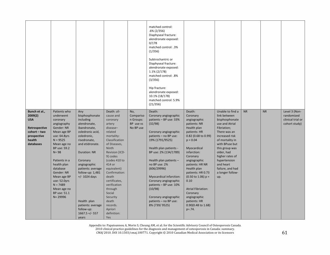
Appendix to: Papaioannou A, Morin S, Cheung AM, et al; for the Scientific Advisory Council of Osteoporosis Canada. 2010 clinical practice guidelines for the diagnosis and management of osteoporosis in Canada: summary. CMAJ 2010. DOI 10.1503/cmaj.100771. Copyright © 2010 Canadian Medical Association or its licensors
61
matched control: .6% (2/356) Diaphyseal fracture: alendronate exposed: 0/178 matched control: .3% (1/356) Subtrochantric or Diaphyseal fracture: alendronate exposed: 1.1% (2/178) matched control: .8% (3/356) Hip fracture: alendronate exposed: 10.1% (18/178) matched control: 5.9% (21/356)
Bunch et al., 2009(2) USA Retrospective cohort – two prospective health databases
Patients who underwent coronary angiography Gender: NR Mean age BP use: 64.8yrs N = 9525 Mean age no BP use: 59.2 N= 98 Patients in a health plan database Gender: NR Mean age BP use: 52.0yrs N = 7489 Mean age no BP use: 51.1 N= 29996
Any bisphosphonate including alendronate, ibandronate, zoledronic acid, zoledronic, risedronate, and etidronate. Duration: NR Coronary angiographic patients: average follow-‐up: 1,481 +/-‐ 1024 days Health plan patients: average follow-‐up: 1667.5 +/-‐ 557 years
Death: all-‐cause and coronary artery disease–related mortality: Classification of Diseases, Ninth Revision [ICD-‐9] codes (codes 410 to 414 or equivalent) Confirmation: death certificates, verification through Social Security death records. Apriori definition: Yes
No, Comparison Groups: BP use vs No BP use
Death: Coronary angiographic patients – BP use: 33% (32/98) Coronary angiographic patients – no BP use: 19% (1791/9525) Health plan patients -‐ BP use: 2% (134/1789) Health plan patients – no BP use: 2% (606/29996) Myocardical infarction: Coronary angiographic patients – BP use: 10% (10/98) Coronary angiographic patients – no BP use: 8% (739/ 9525)
Death: Coronary angiographic patients: NR Health plan patients: HR 0.82 (0.68 to 0.99) p = 0.04 Myocardical infarction: Coronary angiographic patients: HR NR Health plan patients: HR 0.73 (0.50 to 1.06) p = 0.10 Atrial Fibriation: Coronary angiographic patients: HR 0.90(0.48 to 1.68) p=.74.
Unable to find a link between bisphosphonate use and Atrial Fibriation; There was an increased risk of mortality in with BPuse but this group was older, had higher rates of hypertension and heart failure, and had a longer follow-‐up.
NR NR Level 3 (Non-‐randomized clinical trial or cohort study)

Appendix to: Papaioannou A, Morin S, Cheung AM, et al; for the Scientific Advisory Council of Osteoporosis Canada. 2010 clinical practice guidelines for the diagnosis and management of osteoporosis in Canada: summary. CMAJ 2010. DOI 10.1503/cmaj.100771. Copyright © 2010 Canadian Medical Association or its licensors
62
Blinded Adjudication: NR Myocardial infarction: NR Apriori definition: NR Blinded Adjudication: NR Atrial Fibriation: International Classification of Diseases, Ninth Revision [ICD-‐9] codes Confirmation: hospital electrocardiographic databases and physician review. Apriori definition: Yes Blinded Adjudication: NR
Health plan patients -‐ BP use: 1% (68/1789) Health plan patients – no BP use: 1% (343/ 29996) Atrial Fibriation: Coronary angiographic patients – BP use: 10% (10/98) Coronary angiographic patients – no BP use: (10% 964/ 9525) Health plan patients -‐ BP use: 3% (220/1789) Health plan patients – no BP use: 3% (792/ 29996)
Health plan patients: HR 0.82(0.66 to 1.01) p=.63. Death: Coronary angiographic patients vs Health plan patients: p<0.0001 Myocardio infarction: Coronary angiographic patients vs Health plan patients: p= n.s. Atrial fibriation: Coronary angiographic patients vs Health plan patients: p= n.s.

Appendix to: Papaioannou A, Morin S, Cheung AM, et al; for the Scientific Advisory Council of Osteoporosis Canada. 2010 clinical practice guidelines for the diagnosis and management of osteoporosis in Canada: summary. CMAJ 2010. DOI 10.1503/cmaj.100771. Copyright © 2010 Canadian Medical Association or its licensors
63
Cartsos et al. 2008(3) USA Retrospective cohort – medical claims data. Codes of diagnosis or procedure.
People with OP or cancer 89.9% female Age with OP: 59.9 N = 714,217 Cancer: 37.7% (269137/714,217) OP: 62.3% (445080/ 714,217)
Comparison (OP and Cancer) of Intravenous BP use (pamidronate and/or zoledronic acid) and PO BP use (alendronate, etidronate, ibandronate, risedronate or tiludronate) Duration: NR
Inflammatory ONJ/ exposed necrotic bone persisting at least 8 weeks in those taking BPs Apriori Definition: Yes Confirmation: None; identified by International Classification of Diseases, ninth revision (ICD-‐9) code 526.4 describing inflammatory or necrotic processes in the mandible or maxilla Adjudication: NR LIMITATION: ICD-‐9 was used to ID cases
Comparison groups: OP – no BP use Cancer – no BP us
OP – IV BP: 0.48% (9/1858) OP -‐ PO BP:0.08% (150/179870) OP – No BP use: 0.13% (339/263352) Ca – IV BP: 0.46% (39/8545) OP -‐ PO BP: 0.12% (31/25039) Ca – No BP use: 0.11% (251/235553)
OP – IV BP: Orcrude = 4.01 (2.06–7.78) p<0.05 OP-‐ PO BP: Orcrude = 0.65 (0.54–0.79) p<.05 Ca-‐ IV BP: Orcrude = 4.47 (3.19–6.27 )p<.05 Ca-‐PO BP: Orcrude = 1.18 (0.81–1.72) p=ns
Increased risk of inflammatory conditions and surgical procedures of the jaw for IV BP users, but did not see increases in PO BP users.
NR NR Level 3 (Non-‐randomized clinical trial or cohort study)

Appendix to: Papaioannou A, Morin S, Cheung AM, et al; for the Scientific Advisory Council of Osteoporosis Canada. 2010 clinical practice guidelines for the diagnosis and management of osteoporosis in Canada: summary. CMAJ 2010. DOI 10.1503/cmaj.100771. Copyright © 2010 Canadian Medical Association or its licensors
64
Grbic et al, 2008(4) USA RCT Prospective cohort, with control group -‐3 year int’l multi center, randomized, double blind, placebo controlled clinical trial
Postmenopausal women taking zoledronic acid 100% Female, Age: 73.1 N =7714 nZoledronic acid = 3875 nplacebo = 3861 Cancer: NR
5mg zoledronic acid – once yearly IV over 15 minutes Duration: Up to three years
ONJ (not a primary endpoint)/ exposed bone in the maxillofacial area persisting more than 6 weeks despite appropriate management Apriori Definition: Yes Confirmation: independent adjudication by 5 dental experts (surgeons, pathologists, peridonist) Blinded adjudication: Yes-‐ after search of trial database using 60 MedRA terms
Yes No Spontaneous reports of ONJ in either Treatment and Placebo Potential for Maxillofacial AE: Treatment = 2.6% (101/3675) Placebo = 3.3% (127/3861) ONJ (after blinded investigation by adjudication committee: number with a lesion that met the criteria for ONJ): Treatment = 0.03% (1/3675) Placebo: 0.03% (1/3861) <1 in 10,000 patient years
NR ONJ is rare in postmenopausal women and delayed healing of lesions can occur with and without BP use over 3 years.
NR NR Level 3 (Non-‐randomized clinical trial or cohort study)
Miller et al., 2007(5) Multicentre/ International (USA, Spain) 2 prospective randomized double blind placebo controlled trials
Study 1: Post-‐menopausal women; 100% Female Age: 69.5 (SD = 7.0; range = 42-‐86) M = 1637 Cancer: 0 (exclusion criteria) Study 2: 100% Male Age: 58.7 (SD = 13.0; range = 28-‐65) N = 437 Cancer: 0 (exclusion criteria)
TPTD20 – single 20μg/d sc injection; (Calcium 1000mg, vitamin D 400-‐1200IU) TPTD40-‐ single 40μg/d sc injection; (Calcium 1000mg, vitamin D 400-‐1200IU) Duration: Study 1: Median: 19 months
Hypercalciuria -‐ urinary calcium excretion >300mg/d (7.5 mmol/d); confirmed by 24-‐hr urinary collection lab testing Hypercalcemia -‐ serum calcium >10.6mg/dl (2.65mmol/liter) at 4-‐h after dose. Not defined or confirmed: Kidney calculus Urinary track calcifications Kidney pain
Yes Number with adverse event: Both hypercalciuria and hypercalcemia: Study 1: TPTD20 = 0.57% (3/527); TPTD40 = NR but not significantly different from TPTD20. Placebo NR but noted as not significantly different from TPT20 or TPT40 interventions. Study 2: NR Hypercalciuria with normal serum calcium: Study 1: TPTD20 = 9.3% (49/527); TPTD40 = NR but not significantly different from TPTD20. Placebo NR but noted
NR Urinary calcium excretion was increased with TPTD treatment for up to 12 months, compared with placebo and baseline values, but the magnitude of these changes is unlikely to be clinically relevant or warrant urinary calcium monitoring for most patients.
NR NR Level 2 (randomized controlled trial that does not meet level 1 criteria)

Appendix to: Papaioannou A, Morin S, Cheung AM, et al; for the Scientific Advisory Council of Osteoporosis Canada. 2010 clinical practice guidelines for the diagnosis and management of osteoporosis in Canada: summary. CMAJ 2010. DOI 10.1503/cmaj.100771. Copyright © 2010 Canadian Medical Association or its licensors
65
(interquartile range: 17-‐21) Study 2: Median: 11 months (interquartile range: 9-‐12)
Possible urolithiasis Hematuria Apriori Definition: Yes Adjudication: NR
as not significantly different from TPT20 or TPT40 interventions. Study 2: NR Hypercalcemia with normal urinary calcium: Study 1: TPTD20 = 0.95% (5/527); TPTD40 = NR but not significantly different from TPTD20. Placebo NR. Study 2: NR Kidney Calculus: Study 1: TPTD20 = 0.38% (2/527); TPTD40 = NR; placebo = 0.37% (2/536). Study 2: TPTD20 = 1.4% (2/145); TPTD40 = 0.76% (1/132); placebo = 0.71% (1/141). Urinary Tract calcifications: Study 1: TPTD20 = 0.19% (1/527); TPTD40 = 0.18% (1/541); Placebo NR. Study 2: NR. Kidney pain: Study 1: TPTD20 = 0.57% (3/527); TPTD40 = 0.18% (1/541); placebo = NR. Study 2: TPTD20 = NR; TPTD40= 0.76% (1/132) Possible urolithiasis: Study 1: TPTD20 = .11% (6/527); TPTD40 = .37% (2/541); Placebo NR. Study 2: 1.2% (5/418) Hematuria: Study 1: TPTD20 = 0.76% (4/527); TPTD40 =

Appendix to: Papaioannou A, Morin S, Cheung AM, et al; for the Scientific Advisory Council of Osteoporosis Canada. 2010 clinical practice guidelines for the diagnosis and management of osteoporosis in Canada: summary. CMAJ 2010. DOI 10.1503/cmaj.100771. Copyright © 2010 Canadian Medical Association or its licensors
66
0.74% (4/541), Placebo: 1.1% (6/536); Study 2: NR.
Mosca et al., 2009(6) Multicentre/ International (USA, United Kingdom, )
Post-‐ menopausal women with or at increased risk of coronary heart disease Mean age: 67.5years N = 10101 nraloxifene = 5044 nplacebo = 5057 Cancer: NR
Raloxifene, oral, 60mg per day Duration: NR
Venous thromboembolism (VTE): deep vein thrombosis, pulmonary embolism, and intracranial thrombosis (ie, retinal vein thrombosis); WHO criteria. Confirmation: Doppler study or venogram findings. Cerebrovascular accident/stroke: rapid onset of a persistent neurological deficit attributed to an obstruction in cerebral blood flow and/or cerebral hemorrhage not due to trauma, tumor, infection, or other certain etiology; lasting more than 24 hours unless death occurred or there was a demonstrable lesion compatible with an acute stroke on imaging. Confirmation: Imaging/ Stroke
Yes VTE: Raloxifene: 0.02% (103/5044) Placebo: 0.01% (71/5057) Stroke: Raloxifene: 0.05% (249/5044) Placebo: 0.04% (224/5057) Death: Raloxifene: 0.11% (554/5044) Placebo: 0.12% (595/5057)
VTE HR: 1.44 (1.06–1.95) p=.02 Stroke HR: 1.10 (0.92–1.32) p=.30 Death HR: 0.92 (0.82–1.03) p=.16
Postmenopausal women at increased risk for coronary events taking raloxifene had higher incidences of venous thromoboemboloism and fatal stroke than those in placebo group.
NR VTE: ARI=0.12 per 100 women
Level 1 (RCT with adequate power)

Appendix to: Papaioannou A, Morin S, Cheung AM, et al; for the Scientific Advisory Council of Osteoporosis Canada. 2010 clinical practice guidelines for the diagnosis and management of osteoporosis in Canada: summary. CMAJ 2010. DOI 10.1503/cmaj.100771. Copyright © 2010 Canadian Medical Association or its licensors
67
End Point Committee adjudication. Death (noncoronary cardiovascular, including cerebrovascular cause or venous thromboembolism Confirmation: available clinical information, death certificate, and/or autopsy information. Apriori Definition: Yes Blinded adjudication: Yes
BP = Bisphosphonate; ONJ = osteonecrosis of the jaw; OP = Osteoporosis; Ca = Cancer; NR = Not reported; PO = orally REFERENCES
(1) Abrahamsen B, Eiken P, Eastell R. Subtrochanteric and diaphyseal femur fractures in patients treated with alendronate: A register-‐based national cohort study. Journal of Bone & Mineral Research 2009; 24(1095):1102.
(2) Bunch TJ, Anderson JL, May HT, Muhlestein JB, Horne BD, Crandall BG et al. Relation of bisphosphonate therapies and risk of developing atrial fibrillation. American Journal of Cardiology 2009; 103:824-‐828.
(3) Cartsos VM, Zhu S, Zavras AI. Bisphosphonate use and the risk of adverse jaw outcomes: a medical claims study of 714,217 people. Journal of the American Dental Association 2008; 139:23-‐30.
(4) Grbic JT, Landesberg R, Lin SQ, Mesenbrink P, Reid IR, Leung PC et al. Health Outcomes and Reduced Incidence with Zoledronic Acid Once Yearly Pivotal Fracture Trial Research Group. Journal of the American Dental Association 2008; 139:32-‐40.
(5) Miller PD, Bilezikian JP, Diaz-‐Curiel M, Chen P, Marin F, Krege JH et al. Occurrence of hypercalciuria in patients with osteoporosis treated with teriparatide. The Journal of Clinical Endocrinology & Metabolism 2007; 92(9):3535-‐3541.
(6) Mosca L, Grady D, Barrett-‐Connor E, Collins P, Wenger N, Abramson BL et al. Effect of raloxifene on stroke and venous thromboembolism according to subgroups in postmenopausal women at increased risk of coronary heart disease. Stroke 2009; 40:147-‐155.

Appendix to: Papaioannou A, Morin S, Cheung AM, et al; for the Scientific Advisory Council of Osteoporosis Canada. 2010 clinical practice guidelines for the diagnosis and management of osteoporosis in Canada: summary. CMAJ 2010. DOI 10.1503/cmaj.100771. Copyright © 2010 Canadian Medical Association or its licensors
68
Table A22: Evidence of Systematic Reviews on Adverse Events Author Year Country
Eligibility criteria
Intervention; number of RCTs (n) or observational studies
Harm/ Definition/ Confirmation/ A priori definition
Outcomes (type of harm) N with adverse events
Risk Ratio/ Hazard Ratios (p-‐values)
Conclusions Number Needed to Treat (NNT)
Absolute Risk Reduction (ARR)
Level of Evidence
Heaney et al. 2008(1) USA
Inclusion criteria – not specified; Excluded studies of treatment agents and disease conditions that may alter kidney stone risk (e.g., teriparatide, glucocorticoid-‐induced osteoporosis) and studies in men (as risk of stones is higher in men).
Calcium supplementation (at various doses from 500-‐1000mg/d) Observational studies: N= 6 Bone active agent registration trials: N= 4 Calcium supplement trials: N= 12 Unpublished Woman’s Health Initiative data: N=3
Kidney Stones/ Not defined as stones were not a primary endpoint in of the studies reviewed. Registration trials: queried kidney stones, neprolithissis, renal calculi and similar terms. Confirmation: NR Apriori definition: NR
Registration trials: Active agent 0.50% (70/14,598); Placebo: 0.35% (37/10697) RCTs: Ca aggregate: 0.18% (10/5513); Placebo aggregate: 0.17% (8/4706) FROM WHI Studies: Calcium study: 2.5% (449/18176); Placebo: 2.1% (381/18106) WHI Observation Study: 2.5% (2327/91676) WHI Clinical trials: 2.8% (1877/68132) Observational Studies: Stone occurrence ranges from 36.0 -‐191.3 /100,000/yr across 6 studies
Not reported
Most of the studies show no increase in stone risk with high calcium intake (from either diet or supplements). Contrariwise there is a substantial body of evidence, both from controlled trials and from observational studies, indicating that there is an inverse relationship between calcium intake and stone risk.
NR NR Level 2+ but it’s not clear b/c they did not include their inclusion criteria

Appendix to: Papaioannou A, Morin S, Cheung AM, et al; for the Scientific Advisory Council of Osteoporosis Canada. 2010 clinical practice guidelines for the diagnosis and management of osteoporosis in Canada: summary. CMAJ 2010. DOI 10.1503/cmaj.100771. Copyright © 2010 Canadian Medical Association or its licensors
69
Pazianas et al. 2007(2) USA
Population: adult (age _>18 years) male and/or female patients with ONJ; patients received bisphosphonates for the treatment of OP only; reported data included the baseline characteristics of the study population (age; sex; comorbidities; concomitant medications; history of surgical procedures, dental trauma, or dental infection), the characteristics of bisphosphonate treatment (specific bisphosphonate, dose, duration of treatment,
11 studies reporting 26 cases of ONJ Treatment: 88% (23/26) patients received alendronate, and 4% (1/26) received monotherapy with risedronate or pamidronate; 4% (1/26) received a combination of alendronate and zoledronic acid. Dose: Alendronate was administered at a daily dose of 10 mg PO in 4 patients, at a weekly dose of 40 mg PO in 3 patients, and at a weekly dose of 70 mg PO in 3 patients. No cases of ONJ were observed in patients treated with a monthly or cyclic bisphosphonate regimen. Duration: Provided for 10 patients: All 10 were receiving alendronate for a mean duration of 40 months (range, 12-‐72 months).
ONJ Definition: presense of non-‐healing exposed necrotic bone in the maxillofacial region with BP use for the treatment of osteoporosis. Confirmation: NR Apriori Definition: Yes
ONJ: Retrospective study (3 studies): N = 12 Case Report (5 studies): N = 5 Case Series (3 studies): N = 9 No cases of ONJ were identified in patients prescribed ibandronate or etidronate for the treatment of OP. Among patients with a reported duration of bisphosphonate treatment, no clear time dependency was observed.
NR ONJ in patients on BP’s was low. Common characteristics of those who develop ONJ = >60 yrs, female, previous invasive dental treatment. Incomplete reporting and confounding variables makes it difficult to draw further conclusions about the relationship between ONJ and BP use.
NR NR Level 2+ included observational studies in the review, not just RCTs

Appendix to: Papaioannou A, Morin S, Cheung AM, et al; for the Scientific Advisory Council of Osteoporosis Canada. 2010 clinical practice guidelines for the diagnosis and management of osteoporosis in Canada: summary. CMAJ 2010. DOI 10.1503/cmaj.100771. Copyright © 2010 Canadian Medical Association or its licensors
70
mode of administration), clinical features of ONJ (signs, symptoms, site), treatment protocol used to manage ONJ, or the prevalence of ONJ in patients with OP treated with bisphosphonates; and the publication involved a case report, case series, or observational study.
REFERENCES
(1) Heaney RP. Calcium supplementation and incident kidney stone risk: A systematic review. Journal of the American College of Nutrition 2008; 27(5):519-‐527.
(2) Pazianas M, Miller P, Blumentals WA, Bernal M, Kothawala P. A review of the literature on osteonecrosis of the jaw in patients with osteoporosis treated with oral bisphosphonates: prevalence, risk factors, and clinical characteristics. Clinical Therapeutics 2009; 29:1548-‐1558.

Appendix to: Papaioannou A, Morin S, Cheung AM, et al; for the Scientific Advisory Council of Osteoporosis Canada. 2010 clinical practice guidelines for the diagnosis and management of osteoporosis in Canada: summary. CMAJ 2010. DOI 10.1503/cmaj.100771. Copyright © 2010 Canadian Medical Association or its licensors
71
Table A23: Evidence of Case Reports/ Case Studies on Adverse Events Author (Year) Country
Number of cases Gender Age (yrs) % with Cancer % with OP
Method of Case Identification
Intervention/ Duration Outcomes: Type of Harm/ Definition/ Confirmation
Level of Evidence
Chatziavramidis et al., 2008(1) Greece
N =2 Female, 64 and 65yrs old Cancer: NR OP: 100% (2/2)
Presentation in clinical practice
Nasal calcitonin spray Duration: 21 and 22 months (first reported at 14 and 12 months)
Intranasal lesions/ nasal septum perforation, synechiae between nasal septum and inferior nasal concha Confirmation: anterior rhinoscopy
Level 6 (Case report or series of <10 patients)
Engroff & Coletti 2008(2) USA
N = 1 Female, 74 Cancer: 0 OP:100% (1/1)
Presentation in clinical practice
PO alendronate dose not specified Duration: 5 years
Osteonecrosis of the palate/ exposure and necrosis of the palatal torus Confirmation: CT imaging
Level 6 (Case report or series of <10 patients)
Friedrich & Blake 2007(3) Germany
N = 4 Female: N = 2, 68 and 72 years Male: N = 2, 50 and 55 yrs Cancer: 75% (3/4) OP: 0
Presentation in clinical practice
Patient 1: 55 year old man prescribed pamidronate and zoledronic acid Patient 2: 68 year old woman zoledronic acid Patient 3: 72 year old woman prescribed zoledronic acid, Patient 4: 56 year old man prescribed PO clodronate, zoledronic acid 8 mg, 1x per month (doses otherwise not specified) Duration: 2, 3, 3, and 4 years respectively
Avascular mandibular osteonecrosis/ not defined (Cases described as having intraorally exposed mandible and/or incomplete healing after dental extraction). Confirmation: Physical examination and histological investigations, scintigraphy, or dental surgery (revision of extraction site and decortication of bone or mandible resection)
Level 6 (Case report or series of <10 patients)
Grana et al., 2008(4) Spain
N=1 Female, 64 Cancer: 0 OP:100% (1/1)
Presentation in clinical practice
Alendronate 70mg weekly Duration: 4 years 7 months (first reported at 2 years)
ONJ/ exposed bone in the mandible, maxilla, or palate that heals poorly or persists beyond 6-‐8 weeks. Confirmation: CT imaging
Level 6 (Case report or series of <10 patients)

Appendix to: Papaioannou A, Morin S, Cheung AM, et al; for the Scientific Advisory Council of Osteoporosis Canada. 2010 clinical practice guidelines for the diagnosis and management of osteoporosis in Canada: summary. CMAJ 2010. DOI 10.1503/cmaj.100771. Copyright © 2010 Canadian Medical Association or its licensors
72
Ing-‐Lorenzini et al., 2009(5) Switzerland
N=8 Female: N= 7 Male: N = 1 67.5yrs (range: 57-‐86)
Presentation in clinical practice
Any bisphosphonate treatment (Alendronate, Risadronate, Pamidronate); varying doses Duration 16 months to 8 years
Subtrochanteric fractures, involving a cortical thickening at the lateral subtrochanteric cortex with a horizontal fx line originating at the precise level and eventually extending medially. Confirmation: radiographs
Level 6 (Case report or series of <10 patients)
Kilickap et al., 2008(6) Turkey
N=1 Female, 48 Cancer: 100% ( 1/1) OP: 0
Presentation in clinical practice
Zoledronic acid 4 mg IV over 15 minutes Duration: Symptoms evident within 24 hours after the first dose of zoledronic acid
anterior uveitis/ not defined (Case was admitted with pain, visual loss, hyperemia, periorbital swelling and described as having corneal keratic precipitates, ciliary injection and moderate amount of cells in the anterior chamber. Confirmation: Ocular examination, biomicroscopic anterior segment examination, dilated retinal examination, intraocular pressures, laboratory evaluation.
Level 6 (Case report or series of <10 patients)
Kumar et al., 2008(7) USA
N = 13 76.9% (10) Female 72.3 (range: 63-‐80) Cancer: 30.8% (4/13); OP: 69.2% (9/13)
Presentation in clinical practice (between October 2005-‐2007) – not clear if this was retrospective or prospective
69.2% (9) Alendronate 70mg PO once/ week 23.1% (3) Zoledronic Acid 4 mg IV once/ month 7.7% (1) Pamidronate 90mg IV once/month Duration: Alendronate: 12 – 120 months Zoledronic Acid: 8 -‐ 48 months Pamidronate: 36 Months
ONJ/ not defined Confirmation: radiographs and CT imaging (selected cases only) ONJ – only mandibular involvement: 69.2% (9/13) ONJ – only maxillary involvement: 23.1% (3/13) ONJ – both mandibular and maxillary involvement: 7.7% (1/13)
Level 5 (Case series without controls)
Kwek et al., 2008(8) Singapore
N = 17 Female, Mean age: 66 yrs (range = 53-‐82yrs) Cancer: NR OP: 58.8% (10/17) (6/17 – ostopenia)
Retrospective review of all patients admitted to hospital between May 1, 2005 – January 31, 2007 with a low energy subtrochanteric femur fracture while taking alendronate
Alendronate with calcium supplementation (N = 16); Risedronate for 6 yrs after 4 yrs of alendronate (N = 1) Dosages not specified. Duration: Average = 4.4 years (range = 2 -‐8yrs).
Low energy subtrochanteric fracture (N = 17) – fracture within the region of the femur 5 cm distal to the lesser trochanter (low energy not defined, expect as related to the absence of high energy trauma). Confirmation: Radiography (Roentgenograms)
Level 5 (Case series without controls)

Appendix to: Papaioannou A, Morin S, Cheung AM, et al; for the Scientific Advisory Council of Osteoporosis Canada. 2010 clinical practice guidelines for the diagnosis and management of osteoporosis in Canada: summary. CMAJ 2010. DOI 10.1503/cmaj.100771. Copyright © 2010 Canadian Medical Association or its licensors
73
Meek & Nix 2007(9) USA
N = 1 Male, 79 Cancer: 0 OP:100% (1)
Presentation in clinical practice
Alendronate – 10mg PO daily Duration: NR
Hypocalcemia, subsequently developed celiac sprue/ an abnormal immune-‐mediated response to gluten and other related peptides. Confirmation: Upper endoscopy and small bowel biopsy.
Level 6 (Case report or series of <10 patients)
Neviaser et al., 2008(10) USA
N = 70 84.3% (59) Female Mean age: 74.7yrs Cancer: NR OP: 44.3% (31/70)
Retrospective review of all patients admitted to a level 1 trauma center between January 2002 and March 2007 with a low energy subtrochanteric and midshaft femur fracture; identified via ICD-‐9* codes
Alendronate – dosage not specified Duration: Mean (N=16): 6.2 yrs; range = 1 -‐ 10yrs 62.5% (10/16) showed the fracture pattern and BP duration was significantly longer than those who did not exhibit the pattern but were taking alendronate (n=6): 6.9 years versus 2.5 years of use, respectively (P = 0.002).
Low energy femoral shaft fractures – subtrochanteric and midshaft femur fractures caused by the equivalent to a fall from a standing height or less. Confirmation: Radiographs Simple, transverse or short oblique pattern in areas of thickened cortices with a unicortical beak: Fracture in those taking alendronate: 76% (19/ 25); Fracture in those not taking alendronate: 2.2% (1/45). 95% (19/20) patients identified as having the fracture pattern were taking alendronate (95%). (95% CI [19.0–939.4], P < 0.0001).
Level 5 (Case series without controls)
Rinchuse et al. 2007(11)
N = 2 Female, 35 Male, 77 Cancer: 50% (1/2) OP: 50% (1/2)
Presentation in clinical practice
Patient #1: Alendronate sodium 70mg PO once per week Patient #2: IV zoledronic acid, 500mg once per month Duration: Patient #1: 4 yrs, 11months Patient #2: for 11 months prior to orthodontic treatment and throughout orthodontic treatment (13months)
Impeded tooth movement (due to osteoclast destruction and decreased microcirculation limiting bone turnover and remodeling). Confirmation: Radiographs Osteonecrosis of the mandible at the site of a bleeding ulceration of the buccal mucosa of the lower right jaw (It was noted that Patient #2 was predisposed to ONJ because of age, metastatic cancer, prior chemotherapy, history of steroid use, and periodontal disease.) Confirmation: Consultation with oral surgeon
Level 6 (Case report or series of <10 patients)

Appendix to: Papaioannou A, Morin S, Cheung AM, et al; for the Scientific Advisory Council of Osteoporosis Canada. 2010 clinical practice guidelines for the diagnosis and management of osteoporosis in Canada: summary. CMAJ 2010. DOI 10.1503/cmaj.100771. Copyright © 2010 Canadian Medical Association or its licensors
74
Vieillard et al., 2008(12) France
N = 13 92.3% female Age: 62.6 Cancer: 92.3% (12/13) OP: 1/13
Recruited through physicians likely to see ONJ (oncologists, hematologists, rheumatologists, urologists, radiotherapists, dental oral, maxillofacial surgeons)
At time of diagnosis: IV BP: 92.3% (12/13) Zoledronic Acid IV: 76.9% (10/13) Pamidronate IV: 15.4% (2/13) PO alendronate (10 mg/day then 70 mg/week): 7.7% (1/13) Duration: Mean = 24 months Clodronate (N = 4): M = 15.75mos Pamidronate (N = 6): M = 25.8 months Zolendronic Acid (N=10): M = 22.6 mos Alenondrate N = 1: 60 months
ONJ/ lesion exposing the bone that developed either spontaneously or after a tooth extraction in a non-‐irradiated region, failure to heal despite appropriate management, bisphosphonate therapy, and absence of local metastasis or myeloma tumor. Confirmation: Orthopantomogram and CT imaging.
Level 5 (Case series without controls)
Wong & Cheng, 2008(13) China
N = 2 N = 2 100% Female, 73 & 74 Cancer: 50% (1/2) OP: 50% (1/2)
Presentation in clinical practice
Case 1: Zoledronic acid IV once per month (dose not specified) Case 2: PO alendronate (dose not specified) Duration: Case 1: NR; Case 2: 10 years
ONJ/ not defined but described as exposure of necrotic bone (Maxillary = 1; Mandible = 1 Confirmation: CT imaging
Level 6 (Case report or series of <10 patients)
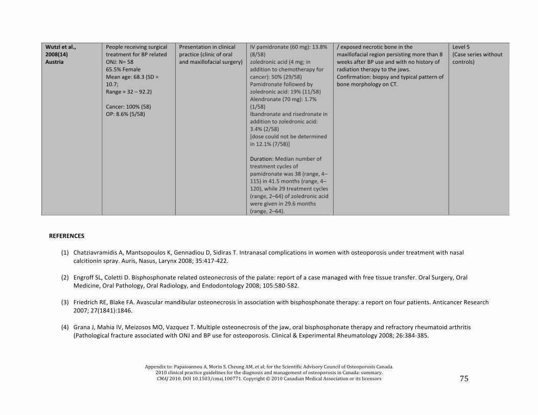
Appendix to: Papaioannou A, Morin S, Cheung AM, et al; for the Scientific Advisory Council of Osteoporosis Canada. 2010 clinical practice guidelines for the diagnosis and management of osteoporosis in Canada: summary. CMAJ 2010. DOI 10.1503/cmaj.100771. Copyright © 2010 Canadian Medical Association or its licensors
75
Wutzl et al., 2008(14) Austria
People receiving surgical treatment for BP related ONJ: N= 58 65.5% Female Mean age: 68.3 (SD = 10.7; Range = 32 – 92.2) Cancer: 100% (58) OP: 8.6% (5/58)
Presentation in clinical practice (clinic of oral and maxillofacial surgery)
IV pamidronate (60 mg): 13.8% (8/58) zoledronic acid (4 mg; in addition to chemotherapy for cancer): 50% (29/58) Pamidronate followed by zoledronic acid: 19% (11/58) Alendronate (70 mg): 1.7% (1/58) Ibandronate and risedronate in addition to zoledronic acid: 3.4% (2/58) [dose could not be determined in 12.1% (7/58)] Duration: Median number of treatment cycles of pamidronate was 38 (range, 4–115) in 41.5 months (range, 4–120), while 29 treatment cycles (range, 2–64) of zoledronic acid were given in 29.6 months (range, 2–64).
/ exposed necrotic bone in the maxillofacial region persisting more than 8 weeks after BP use and with no history of radiation therapy to the jaws. Confirmation: biopsy and typical pattern of bone morphology on CT.
Level 5 (Case series without controls)
REFERENCES
(1) Chatziavramidis A, Mantsopoulos K, Gennadiou D, Sidiras T. Intranasal complications in women with osteoporosis under treatment with nasal calcitionin spray. Auris, Nasus, Larynx 2008; 35:417-‐422.
(2) Engroff SL, Coletti D. Bisphosphonate related osteonecrosis of the palate: report of a case managed with free tissue transfer. Oral Surgery, Oral Medicine, Oral Pathology, Oral Radiology, and Endodontology 2008; 105:580-‐582.
(3) Friedrich RE, Blake FA. Avascular mandibular osteonecrosis in association with bisphosphonate therapy: a report on four patients. Anticancer Research 2007; 27(1841):1846.
(4) Grana J, Mahia IV, Meizosos MO, Vazquez T. Multiple osteonecrosis of the jaw, oral bisphosphonate therapy and refractory rheumatoid arthritis (Pathological fracture associated with ONJ and BP use for osteoporosis. Clinical & Experimental Rheumatology 2008; 26:384-‐385.

Appendix to: Papaioannou A, Morin S, Cheung AM, et al; for the Scientific Advisory Council of Osteoporosis Canada. 2010 clinical practice guidelines for the diagnosis and management of osteoporosis in Canada: summary. CMAJ 2010. DOI 10.1503/cmaj.100771. Copyright © 2010 Canadian Medical Association or its licensors
76
(5) Ing-‐Lorenzini K, Desmeules J, Plachta O, Suva D, Dayer P, Peter R. Low energy femoral fractures associated with the long-‐term use of bisphosphonates. A case series from a Swiss University Hospital. Drug Safety 2009; 32(9):775-‐785.
(6) Kilickap S, Ozdamar Y, Altundag MK, Dizdar O. A case report: zoledronic acid-‐induced anterior uveitis. Medical Oncology 2008; 25:238-‐240.
(7) Kumar SKS, Meru MC, Sedghizadeh PP. Osteonecrosis of the jaws secondary to bisphosphonate therapy: a case series. The Journal of Contemporary Dental Practice 2008; 9(1):63-‐69.
(8) Kwek EBK, Goh SK, Koh JSB, Png MA, Howe TS. An emerging pattern of subtrochanteric stress fractures: A long-‐term complication of alendronate therapy? Injury 2008; 39:224-‐231.
(9) Meek SE, Nix K. Hypocalcemia after alendronate therapy in a patient with celiac disease. Endrocrine Practice 2007; 13(4):403-‐407.
(10) Neviaser AS, Lane JM, Lenart BA, Edobor-‐Osula F, Lorich DG. Low-‐energy femoral shaft fracture associated with alendronate use. Journal of Orthopedic Trauma 2008; 22(5):346-‐350.
(11) Rinchuse DJ, Rinchuse DJ, Sozovicka MF, Robison JM, Pendleton R. Orthodontic treatment of patients using bisphosphonates: A report of 2 cases. American Journal of Orthodontics and Dentofacial Orthopedics 2007; 131:321-‐326.
(12) Vieillard MH, Maes JM, Penel G, Facon T, Magro L, Bonneterre J et al. Thirteen cases of jaw osteonecrosis in patients on bisphosphonate therapy. Joint Bone Spine 2008; 75:34-‐40.
(13) Wong YK, Cheung JC. Bisphosphonate-‐related osteonecrosis of the jaws -‐ Report of 2 cases and strategies on prevention and management. Quintessence International 2008; 39:195-‐201.
(14) Wutzl A, Biedermann E, Wanschitz F, Seemann R, Klug C, Baumann A et al. Treatment results of bisphosphonate-‐related osteonecrosis of the jaws. Head & Neck 2008; 30:1224-‐1230.