applicability of tableside flat panel detector ct … · · 2012-01-11ct parenchymal cerebral...
TRANSCRIPT
ORIGINALRESEARCH
Applicability of Tableside Flat Panel DetectorCT Parenchymal Cerebral Blood VolumeMeasurement in Neurovascular Interventions:Preliminary Clinical Experience
P. MordasiniM. El-KoussyC. Brekenfeld
G. SchrothU. Fischer
J. BeckJ. Gralla
BACKGROUND AND PURPOSE: CBV is a vital perfusion parameter in estimating the viability of brainparenchyma (eg, in cases of ischemic stroke or after interventional vessel occlusion). Recent techno-logic advances allow parenchymal CBV imaging tableside in the angiography suite just before, during,or after an interventional procedure. The aim of this work was to analyze our preliminary clinicalexperience with this new imaging tool in different neurovascular interventions.
MATERIALS AND METHODS: FPD-CBV measurement was performed on a biplane FPD angiographicsystem. Eighteen patients (11 women, 7 men) were examined (age range, 18–86 years; median, 58.7years). In the 10 patients with stroke, the extent of intracranial hypoperfusion was evaluated. Theremaining 8 patients had an intracranial hemorrhage; periprocedural CBV was evaluated during thecourse of interventional treatment.
RESULTS: In the 18 cases studied, 23 CBV measurements were performed. Twenty acquisitions wereof sufficient diagnostic quality. The remaining 3 acquisitions failed technically, 1 due to motion artifactsand 2 due to injection technique and/or hardware failure.
CONCLUSIONS: FPD-CBV measurement in the angiography suite provides a feasible and helpful toolfor peri-interventional neuroimaging. It extends the intraprocedural imaging modalities to the level oftissue perfusion. However, the technique has technical limitations and shows room for improvementin the future.
ABBREVIATIONS: ACA � anterior cerebral artery; CBF � cerebral blood flow; CTP � CT perfusion;FPD � flat panel detector; MRP � MR perfusion imaging; TIMI � Thrombolysis in MyocardialInfarction
Today, FPD-mounted C-arm angiographic systems havebeen widely introduced into neurointerventional suites.
FPD imaging techniques not only allow the acquisition ofhigh-quality 3D vascular imaging (3D rotational angiogra-phy) but also obtain CT-like cross-sectional soft-tissue imag-ing (FPD-CT). Applications include assessment of the extentof SAH or intracerebral hemorrhage and the width of the ven-tricles,1-4 and immediate evaluation of coil and stent place-ment in the treatment of intracranial aneurysms.5,6 FPD-CThas also been shown to be capable of visualizing the struts of astent during intracranial placement6-8 and extracranial carotidartery stent placement.9 FPD technology allows fast imagingwithout the need for time-consuming transfer to a CT facilityand, therefore, has become a helpful tool for immediate, “onthe table” evaluation of treatment results and intraproceduralcomplications.
CBV is an important parameter for assessing the viabilityof brain tissue. A significant drop of CBV in the clinical setting
of acute ischemic stroke signifies failure of local parenchymalcompensatory mechanisms and the irreversibility of the isch-emic damage. The CBF and the temporal perfusion maps helpto delineate the potentially reversible ischemic penumbra. Re-cently, the possibility of performing whole-brain parenchymalCBV measurements has been added to the spectrum ofFPD-CT imaging. Feasibility studies in canines10,11 and pre-liminary clinical studies12,13 have shown that CBV mappingby using FPD-CT after intravenous contrast media injectionis feasible, safe, and capable of depicting CBV values compa-rable with standard multisection CT perfusion. Further-more, FPD-CBV imaging offers the advantage of whole-braincoverage not always available by conventional CT perfusionmeasurements.
However, despite the current limitation in temporal reso-lution being unable to provide additional dynamic perfusionmaps like CBF or TTP measurements, tableside FPD-CBVmay offer a further adjunctive on-line tool to evaluate andmonitor cerebral perfusion as a physiologic parameter toguide treatment decisions and to evaluate treatment effect.Particularly, patients undergoing revascularization proce-dures for acute or chronic stroke may benefit from advancedimage-guided management. Only a few clinical data on theimpact on clinical decision-making and peri-interventionalmonitoring of neurovascular procedures by using FPD-CBVhave been published so far, to our knowledge.
The purpose of this study was to review our preliminaryclinical experience evaluating the applicability and usability of
Received January 24, 2011; accepted after revision May 1.
From the Departments of Interventional and Diagnostic Neuroradiology (P.M., M.E.-K., C.B.,G.S., J.G.), Neurology (U.F.), and Neurosurgery (J.B.), Inselspital, University of Bern,Switzerland.
Please address correspondence to Jan Gralla, MD, MSc, Department of Interventional andDiagnostic Neuroradiology, University of Bern, Inselspital, Freiburgstr 4, CH-3010 Bern,Switzerland; e-mail: [email protected]
Indicates article with a supplemental on-line table.
http://dx.doi.org/10.3174/ajnr.A2715
154 Mordasini � AJNR 33 � Jan 2012 � www.ajnr.org
tableside peri-interventional FPD-CBV mapping as an addi-tional imaging tool in different neurovascular interventions.
Materials and Methods
Patient CharacteristicsBetween August and November 2010, 18 patients (11 women, 7 men;
age range, 18 – 86 years; median, 58.7 years) with different cere-
brovascular pathologies referred for cerebral angiography were
examined. This Health Insurance Portability and Accountability Act–
compliant study was performed according to the guidelines of our
institution, and informed consent was waived. Presenting symptoms
included acute stroke or stroke-like symptoms in 6 patients, subacute
stroke in 4 patients, headache and nausea in 6 patients, and impair-
ment/loss of consciousness in 2 patients. In the 10 patients with
stroke, the causative arterial stenosis or occlusion was confirmed. The
remaining 8 patients had an intracranial hemorrhage, 3 had an SAH
due to aneurysmal rupture and 1, due to a posttraumatic dissecting
aneurysm. The FPD-CBV acquisitions were performed in these cases
to exclude hypoperfusion complicating the endovascular interven-
tion (eg, stent placement, coiling, and/or embolization). Patient clin-
ical data and characteristics are summarized in the On-line Table.
FPD-CBV MeasurementFPD-CBV measurement was performed on a biplane FPD angiographic
system (Axiom Artis Zee; Siemens, Erlangen, Germany). As previously
described,12,13 data acquisition consisted of 2 rotations: an initial mask
run followed by a second fill run. Data acquisition was performed by
using the following imaging parameters: acquisition time, 8 seconds; 70
kV; 616 � 480 matrix; projection on a 30 � 40 cm flat panel size; 200°
total angle, 0.5°/frame; 400 frames total; dose, 1.20 �Gy/frame.
To obtain the necessary steady-state contrast medium opacifica-
tion of the brain parenchyma for CBV data acquisition during the fill
run, the technique of “bolus watching” was applied as previously de-
scribed.12,13 In brief, contrast medium injection was started simulta-
neously with the mask run. After return of the C-arm to the starting
position, conventional DSA acquisitions at a rate of 1 image per sec-
ond were obtained until opacification of the transverse sinus was seen
by the interventionalist as a surrogate marker for steady-state paren-
chymal contrast medium filling. At this point, the fill run for CBV
data acquisition was manually started.
Intravenous and intra-arterial injection protocols according to
the interventionalist’s discretion were used. Contrast media injec-
tions were performed by using 50 mL of iodinated contrast material
(iopamidol, Iopamiro 300; Bracco, Milan, Italy) either through a pe-
ripheral cubital venous access (20-ga) in 15 patients or intra-arterially
in 3 patients through a pigtail catheter (6F, Torcon NB Advantage
Catheter; Cook, Bloomington, Indiana) placed in the ascending aorta
at a rate of 5 mL/s by using a power injector (Angiomat Illumena; Liebel-
Flarsheim, Cincinnati, Ohio). However, during the study, the route of
pre- and postprocedural contrast application was consistent in all cases,
allowing a comparison of datasets within the case. The details of the
injection protocol for each patient are described in the On-line Table.
Postprocessing of FPD-CBV DataPostprocessing of FPD-CBV data was performed by using commer-
cially available imaging software (syngo Neuro PBV IR; Siemens) in-
stalled on the imaging workstation (Leonardo; Siemens). Multiplanar
reconstructions (section thickness, 5 mm) were performed covering
the whole-brain parenchyma in axial, coronal, and sagittal planes.
CBV images were stored on a workstation for analysis. Images were
visually assessed together by 2 neuroradiologists (P.M., M.E.-K.) in
consensus, unaware of patient histories and pathology for asymmetry
in parenchymal CBV.
ResultsIn the 18 cases studied, 23 CBV measurements were per-formed. Twenty acquisitions were of proper diagnostic qual-ity. The remaining 3 acquisitions had technical failure (1 due tomotion artifacts, 2 due to injection technique and/or hardwarefailure). The technical failures decreased as further patients wereexamined, probably representing a learning-curve effect.
Illustrative CasesCase 1. A 53-year-old patient (patient 1) was referred for
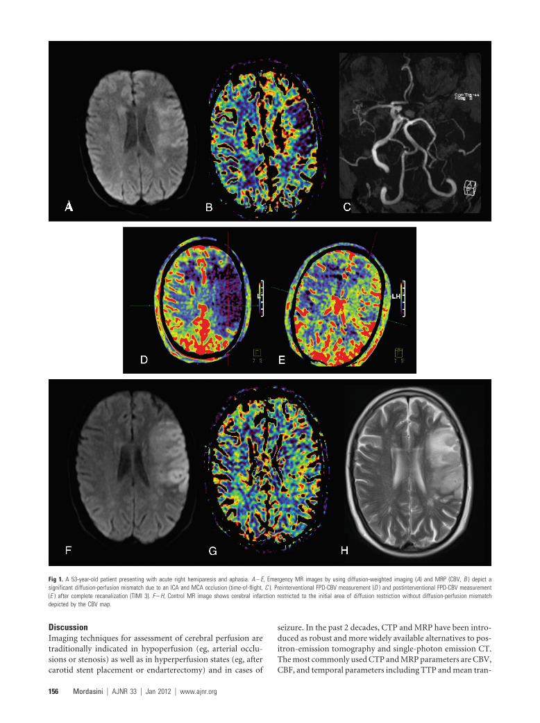
stroke with acute right hemiparesis and aphasia, 3.5 hoursafter the onset of symptoms. The National Institutes of HealthStroke Scale score at presentation was 19. Emergency MR im-aging revealed a diffusion restriction of the left MCA territoryand a wide diffusion-perfusion mismatch including the wholeleft MCA territory and partial ACA territory due to an ICA andM1 occlusion (Fig 1A�C). The patient was immediatelytransferred to the neurointerventional suite for endovascularstroke treatment. Diagnostic DSA confirmed an occlusion ofthe left ICA from the bifurcation. Preinterventional FPD-CBVby using an intra-arterial injection protocol with a pigtail cath-eter placed in the descending aorta was performed, showingdecrease in CBV values consisting of the whole MCA and partsof the ACA territory (Fig 1D). TIMI 3 recanalization of theICA and M1 could be achieved by stent placement in the ICAand intra-arterial thrombolysis by using 1 million IU of uroki-nase. Immediate postinterventional control FPD-CBV mea-surement depicted normalization of CBV values in the leftMCA and ACA territories with signs of hyperperfusion in theparietal lobe (Fig 1E). Control MR imaging 4 days after theintervention showed cerebral infarction restricted to the initialarea of diffusion restriction in the left MCA territory withoutdiffusion-perfusion mismatch (Fig 1F�H).
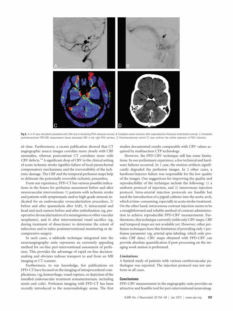
Case 2. A 47-year-old patient (patient 11) presented to theemergency department with an episode of sudden headacheand nausea. CT evaluation showed an SAH and a suspectedPICA aneurysm on the right. DSA depicted a partially saccularmultilobulated aneurysm of the PICA with fusiform dilationincorporating the adjacent vessel segment (Fig 2A). Because ofits partially fusiform appearance and the extent of the lesion, adissecting aneurysm was suspected and segment occlusion in-tended. Pre- and postinterventional FPD-CT was performedto evaluate the extent of the CBV deficit to anticipate the ne-cessity of decompressive craniotomy in this sedated and intu-bated patient. Vessel sacrifice by embolization of the PICA byusing N-butyl-2-cyanoacrylate (Histoacryl) (Tissue Seal, AnnArbor, Michigan) was performed, and complete vessel occlu-sion could be achieved (Fig 2B).
Immediately after the termination of the embolization, anFPD-CBV measurement was performed depicting a CBV de-crease in the right PICA territory (Fig 2C). A control CT scanconfirmed the postinterventional PICA infarction (Fig 2D),and decompressive craniotomy was performed.
INTERVEN
TION
AL
ORIGINAL
RESEARCH
AJNR Am J Neuroradiol 33:154 –58 � Jan 2012 � www.ajnr.org 155
DiscussionImaging techniques for assessment of cerebral perfusion aretraditionally indicated in hypoperfusion (eg, arterial occlu-sions or stenosis) as well as in hyperperfusion states (eg, aftercarotid stent placement or endarterectomy) and in cases of
seizure. In the past 2 decades, CTP and MRP have been intro-duced as robust and more widely available alternatives to pos-itron-emission tomography and single-photon emission CT.The most commonly used CTP and MRP parameters are CBV,CBF, and temporal parameters including TTP and mean tran-
Fig 1. A 53-year-old patient presenting with acute right hemiparesis and aphasia. A�E, Emergency MR images by using diffusion-weighted imaging (A) and MRP (CBV, B ) depict asignificant diffusion-perfusion mismatch due to an ICA and MCA occlusion (time-of-flight, C ). Preinterventional FPD-CBV measurement (D ) and postinterventional FPD-CBV measurement(E ) after complete recanalization (TIMI 3). F�H, Control MR image shows cerebral infarction restricted to the initial area of diffusion restriction without diffusion-perfusion mismatchdepicted by the CBV map.
156 Mordasini � AJNR 33 � Jan 2012 � www.ajnr.org
sit time. Furthermore, a recent publication showed that CTangiographic source images correlate more closely with CBFanomalies, whereas postcontrast CT correlates more withCBV defects.14 A significant drop of CBV in the clinical settingof acute ischemic stroke signifies failure of local parenchymalcompensatory mechanisms and the irreversibility of the isch-emic damage. The CBF and the temporal perfusion maps helpto delineate the potentially reversible ischemic penumbra.
From our experience, FPD-CT has various possible indica-tions in the future for perfusion assessment before and afterneurovascular interventions: 1) patients with ischemic strokeand patients with symptomatic and/or high-grade stenosis in-dicated for an endovascular revascularization procedure, 2)before and after spasmolysis after SAH, 3) intracranial andhead and neck tumors before and after embolization (eg, pre-operative devascularization of a meningioma or other vascularneoplasms), and 4) after interventional vessel sacrifice (eg,during treatment of dissections) to determine the extent ofinfarction and to tailor postinterventional monitoring or de-compressive surgery.
In such cases, a tableside technique integrated into theneuroangiography suite represents an extremely appealingmethod for on-line peri-interventional assessment of perfu-sion. This provides the advantage of rapid on-line decision-making and obviates tedious transport to and from an MRimaging or CT scanner.
Furthermore, to our knowledge, few publications onFPD-CT have focused on the imaging of intraprocedural com-plications, (eg, hemorrhage, vessel rupture, or depiction of theinstalled endovascular treatment armamentarium, includingstents and coils). Perfusion imaging with FPD-CT has beenrecently introduced to the neuroradiologic arena. The first
studies documented results comparable with CBV values ac-quired by multisection CTP technology.
However, the FPD-CBV technique still has some limita-tions. In our preliminary experience, a few technical and hard-ware failures occurred. In 1 case, the motion artifacts signifi-cantly degraded the perfusion images. In 2 other cases, ahardware/injector failure was responsible for the low qualityof the images. Our suggestions for improving the quality andreproducibility of the technique include the following: 1) auniform protocol of injection, and 2) intravenous injectionprotocol. Intra-arterial injection protocols are feasible butneed the introduction of a pigtail catheter into the aortic arch,which is time-consuming, especially in acute stroke treatment.On the other hand, intravenous contrast injection seems to bea straightforward and reliable method of contrast administra-tion to achieve reproducible FPD-CBV measurements. Fur-thermore, this technique currently yields only CBV maps. CBFand temporal maps are not available yet. However, other per-fusion techniques have this limitation of providing only 1 per-fusion parameter (eg, arterial spin-labeling, which only pro-vides CBF data). CBU maps obtained with FPD-CBV canprovide absolute quantification if post-processing on the im-aging work station is performed.
LimitationsA limited study of patients with various cerebrovascular pa-thologies was reported. The injection protocol was not uni-form in all cases.
ConclusionsFPD-CBV measurement in the angiography suite provides anattractive and feasible tool for peri-interventional neuroimag-
Fig 2. A, A 47-year-old patient presented with SAH due to dissecting PICA aneurysm (arrow). B, Complete vessel occlusion after superselective Histoacryl embolization (arrow). C, Immediatepostinterventional FPD-CBV measurement shows decreased CBV in the right PICA territory. D, Postinterventional control CT scan confirms the similar extension of PICA infarction.
AJNR Am J Neuroradiol 33:154 –58 � Jan 2012 � www.ajnr.org 157
ing. It extends the periprocedural imaging modalities to thelevel of tissue perfusion. However, the technique still has sometechnical limitations and shows room for improvement.
Disclosures: Jan Gralla, Consultant: ev3, Details: principal investigator of the Star Trial(Solitaire FR in Stroke); no connection to this study.
References1. Doelken M, Struffert T, Richter G, et al. Flat-panel detector volumetric CT for
visualization of subarachnoid hemorrhage and ventricles: preliminary re-sults compared to conventional CT. Neuroradiology 2008;50:517–23
2. Struffert T, Richter G, Engelhorn T, et al. Visualisation of intracerebral haem-orrhage with flat-detector CT compared to multislice CT: results in 44 cases.Eur Radiol 2009;19:619 –25
3. Struffert T, Eyupoglu IY, Huttner HB, et al. Clinical evaluation of flat-paneldetector compared with multislice computed tomography in 65 patients withacute intracranial hemorrhage: initial results: clinical article. J Neurosurg2010;113:901– 07
4. Engelhorn T, Struffert T, Richter G, et al. Flat panel detector angiographic CTin the management of aneurysmal rupture during coil embolization. AJNRAm J Neuroradiol 2008;29:1581– 84
5. White PM, Gilmour JN, Weir NW, et al. AngioCT in the management of neu-rointerventional patients: a prospective, consecutive series with associateddosimetry and resolution data. Neuroradiology 2008;50:321–30
6. Richter G, Engelhorn T, Struffert T, et al. Flat panel detector angiographic CT
for stent-assisted coil embolization of broad-based cerebral aneurysms. AJNRAm J Neuroradiol 2007;28:1902– 08
7. Benndorf G, Claus B, Strother CM, et al. Increased cell opening and prolapse ofstruts of a Neuroform stent in curved vasculature: value of angiographic com-puted tomography—technical case report. Neurosurgery 2006;58(suppl 2):ONS-E380
8. Psychogios MN, Schramm P, Buhk JH, et al. Angiographic CT after intrave-nous contrast agent application: a noninvasive follow-up tool after intracra-nial angioplasty and stenting. AJNR Am J Neuroradiol 2010;31:1886 –91. Epub2010 Jul 15
9. Mordasini P, Al Senani F, Gralla J, et al. The use of flat panel angioCT (DynaCT)for navigation through a deformed and fractured carotid stent. Neuroradiol-ogy 2010;52:629 –32
10. Ahmed AS, Zellerhoff M, Strother CM, et al. C-arm CT measurement of cere-bral blood volume: an experimental study in canines. AJNR Am J Neuroradiol2009;30:917–22
11. Bley T, Strother CM, Pulfer K, et al. C-arm CT measurement of cerebral bloodvolume in ischemic stroke: an experimental study in canines. AJNR Am J Neu-roradiol 2010;31:536 – 40
12. Struffert T, Deuerling-Zheng Y, Kloska S, et al. Flat detector CT in the evalua-tion of brain parenchyma, intracranial vasculature, and cerebral bloodvolume: a pilot study in patients with acute symptoms of cerebral ischemia.AJNR Am J Neuroradiol 2010;31:1462– 69
13. Struffert T, Deuerling-Zheng Y, Kloska S, et al. Cerebral blood volume imagingby flat detector computed tomography in comparison to conventionalmultislice perfusion CT. Eur Radiol 2010;21:882– 89. Epub 2010 Sep 21
14. Sharma M, Fox AJ, Symons S, et al. CT angiographic source images: flow- orvolume-weighted? AJNR Am J Neuroradiol 2011;32:359 – 64
158 Mordasini � AJNR 33 � Jan 2012 � www.ajnr.org