application guide - csbia51969aa april 2009 beckman coulter, inc. 4300 n. harbor blvd. fullerton, ca...
TRANSCRIPT

Application Guide
PA 800 plus Pharmaceutical Analysis System
Carbohydrate Labeling and Analysis
April 2009
Beckman Coulter, Inc.4300 N. Harbor Blvd.Fullerton, CA 92835
A51969AA

Application GuidePA 800 plus Pharmaceutical Analysis SystemCarbohydrate Labeling and AnalysisA51969AA (April 2009)
Copyright © 2009 Beckman Coulter, Inc.
Beckman Coulter, Inc. grants a limited non-exclusive license to the owner or operator of a PA 800 plus instrument to make a copy, solely for laboratory use, of any portion or all of the online help and electronic documents shipped with the PA 800 plus instrument.
Trademarks:Following is a list of Beckman Coulter trademarks:
• Beckman Coulter®
• 32 Karat™
All other trademarks are the property of their respective owners.
Find us on the World Wide Web at: www.beckmancoulter.com and www.CELeader.com
Beckman Coulter Ireland Inc.Mervue Business Park,Mervue, Galway,Ireland (353 91 774068)
Beckman Coulter do Brasil Com e Imp de Prod de Lab LtdaEstr dos Romeiros, 220 - Galpao G3 - Km 38.5zip code 06501-001 - Sao Paulo - SP - BrasilCNPJ: 42.160.812/0001-44
製造販売元 : ベックマン・コールター株式会社東京都江東区有明二丁目 5番 7号
生产商:贝克曼库尔特有限公司,美国加利福尼亚州富勒顿市,邮编:92835,电话:(001)714-871-4848

Revision History
Initial Issue, A51969AA, April 200932 Karat Software version 9.1PA 800 plus Software version 1.1PA 800 plus Firmware version 9.0
A51969AA iii

Revision History
A51969AAiv

Safety Notices
Symbols and Labels
Introduction
The following is a description of symbols and labels used on the Beckman Coulter PA 800 plus Pharmaceutical Analysis System or shown in this manual.
WARNING
If the equipment is used in a manner not specified by Beckman Coulter, Inc., the protection provided by the instrument may be impaired.
General Biohazard Symbol
This caution symbol indicates a possible biohazard risk from patient specimen contamination.
Caution, Biohazard Label
This caution symbol indicates a caution to operate only with all covers in position to decrease risk of personal injury or biohazard.
A51969AA v

Safety NoticesSymbols and Labels
Caution, Moving Parts Label
This caution symbol warns the user of moving parts that can pinch or crush.
High Voltage Electric Shock Risk Symbol
This symbol indicates that there is high voltage and there is a risk of electric shock when the user works in this area.
Class 1 Laser Caution Label
A label reading “THIS PRODUCT CONFORMS TO APPLICABLE REQUIREMENTS OF 21 CFR 1040 AT THE DATE OF MANUFACTURE” is found near the Name Rating tag. The laser light beam is not visible.
Sharp Object Label
A label reading “CAUTION SHARP OBJECTS” is found on the PA 800 plus.
CAUTIONPARTS MOVEAUTOMATICALLY
A015081L.EPS
144557-AB
A016352L.EPS
A016350L.EPS
CLASS 1 LASER PRODUCTTHIS PRODUCT CONFORMS TO APPLICABLE REQUIREMENTS OF 21 CFR 1040 AT THE DATE OF MANUFACTURE.
MANUFACTURED:
726024-C
CAUTIONSHARP OBJECTS
A16558-AA
A016351L.EPS
A51969AAvi

Safety NoticesSymbols and Labels
Recycling Label
This symbol is required in accordance with the Waste Electrical and Electronic Equipment (WEEE) Directive of the European Union. The presence of this marking on the product indicates:
1. The device was put on the European Market after August 13, 2005.
2. The device is not to be disposed of via the municipal waste collection system of any member state of the European Union.
It is very important that customers understand and follow all laws regarding the proper decontamination and safe disposal of electrical equipment. For Beckman Coulter products bearing this label, please contact your dealer or local Beckman Coulter office for details on the take back program that facilitates the proper collection, treatment, recovery, recycling, and safe disposal of this device.
Disposal of Devices Containing Mercury Components
This product contains a mercury-added part. Recycle or dispose of according to local, state, or federal laws. It is very important that you understand and comply with the safe and proper disposal of devices containing mercury components (switch, lamp, battery, relay, or electrode). The mercury component indicator label can vary depending on the type of device.
A51969AA vii

Safety NoticesSymbols and Labels
Restriction of Hazardous Substances (RoHS) Labels
These labels and materials declaration table (the Table of Hazardous Substance's Name and Concentration) are to meet People's Republic of China Electronic Industry Standard SJ/T11364-2006 "Marking for Control of Pollution Caused by Electronic Information Products" requirements.
RoHS Caution Label
This logo indicates that this electronic information product contains certain toxic or hazardous elements, and can be used safely during its environmental protection use period. The number in the middle of the logo indicates the environmental protection use period for the product. The outer circle indicates that the product can be recycled. The logo also signifies that the product should be recycled immediately after its environmental protection use period has expired. The date on the label indicates the date of manufacture.
RoHS Environmental Label
This logo indicates that the product does not contain any toxic or hazardous substances or elements. The "e" stands for electrical, electronic and environmental electronic information products. This logo indicates that this electronic information product does not contain any toxic or hazardous substances or elements, and is green and is environmental. The outer circle indicates that the product can be recycled. The logo also signifies that the product can be recycled after being discarded, and should not be casually discarded.
A51969AAviii

Safety NoticesSymbols and Labels
Alerts for Warning, Caution, Important, and Note
WARNING
WARNING indicates a potentially hazardous situation which, if not avoided, could result in death or serious injury. The warning can be used to indicate the possibility of erroneous data that could result in an incorrect diagnosis (does not apply to all products).
CAUTION
CAUTION indicates a potentially hazardous situation, which, if not avoided, may result in minor or moderate injury. It may also be used to alert against unsafe practices. The caution can be used to indicate the possibility of erroneous data that could result in an incorrect diagnosis (does not apply to all products).
IMPORTANT IMPORTANT is used for comments that add value to the step or procedure being performed. Following the advice in the IMPORTANT notice adds benefit to the performance of a piece of equipment or to a process.
NOTE NOTE is used to call attention to notable information that should be followed during installation, use, or servicing of this equipment.
A51969AA ix

Safety NoticesSymbols and Labels
A51969AAx

Contents
Revision History, iii
Safety Notices, v
Symbols and Labels, vIntroduction, vGeneral Biohazard Symbol, vCaution, Biohazard Label, vCaution, Moving Parts Label, viHigh Voltage Electric Shock Risk Symbol, viClass 1 Laser Caution Label, viSharp Object Label, viRecycling Label, viiDisposal of Devices Containing Mercury Components, viiRestriction of Hazardous Substances (RoHS) Labels, viii
RoHS Caution Label, viiiRoHS Environmental Label, viii
Alerts for Warning, Caution, Important, and Note, ix
Carbohydrate Labeling and Analysis Kit, 1
Overview, 1Safety Information, 1General Precautions, 2
Materials and Reagents, 2Contents of this Kit, 2Materials Needed but Not Provided in Kit, 2Storage Conditions, 3
Overview of the Procedure, 3
Releasing Oligosaccharides From Glycoproteins, 3Enzymatic Release of the N-Linked Oligosaccharides, 4Working with the Supernatant, 4Principle of the Labeling Method, 4
Preparing the Labeling Reagents, 5Choosing the Proper Fluorophore, 5
xi

Contents
Preparing the Reagents, 5Preparing the Standards, 6
Performing the Labeling Reaction, 6
Preparing the PA 800 Instrument, 8
Installing and Pre-conditioning the N-CHO Capillary, 9Clean the Interface Block, 10Insert the Cartridge, 10
Preparing the Buffer Trays, 10
Sample Vial Setup, 12
Running the Samples, 13Initial Conditions, 13LIF Detector Initial Conditions, 14Time Program, 15
Evaluation of Results, 17
System Shutdown and Capillary Storage, 18Short Term Storage (<24 hours) of Capillary, 18Long Term Storage (>24 hours) of Capillary, 18
Troubleshooting Procedures, 19
Appendix, 20LIF Detector Calibration Procedure, 20
Setting the Calibration Corrector Factors (CCFs), 20Automatic Calibration, 21
CCF Troubleshooting Procedures, 24CCF <1, 24CCF >1, 24No Step Change Detected, 24
xii

Carbohydrate Labeling and Analysis Kit
Overview
Beckman Coulter’s Carbohydrate Labeling & Analysis Kit uses capillary electrophoresis to separate and quantify oligosaccharides released from glycoproteins. This kit has been developed to work with the PA 800 plus Pharmaceutical Analysis System, using the 488 nm laser and the 520 nm emission filter.
This kit contains the reagents, buffer and coated capillaries required to label, separate and quantify oligosaccharides. This kit also provides a glucose size marker for relative size determination and a maltose standard for quantitation and mobility characterization of the released oligosaccharides.
For further information, please refer to the following publication:
Quantitative Analysis of Sugar Constituents of Glycoproteins by Capillary Electrophoresis, Fu Tai, A. Chen, Thomas S. Dobashi and Ramon A. Evangelista, “Glycobiology”, Vol. 8, No. 11, pp. 1045-1052, 1998.
Safety Information
NOTE This application guide has been validated for use in the PA 800 Enhanced Protein Characterization and the PA 800 plus Pharmaceutical Analysis Systems.
IMPORTANT The Carbohydrate Labeling and Analysis Kit is for laboratory use only.
IMPORTANT In this procedure use 1 M sodium cyanoborohydride in THF, which is a very reactive compound. Please consult the MSDS before using or disposing of this reagent.
CAUTION
Refer to the Material Safety Data Sheets (MSDS) information, available at www.BeckmanCoulter.com, regarding the proper handling of materials and reagents. Always follow standard laboratory safety guidelines.
CAUTION
When installing or removing capillaries from the cartridge, use safety goggles.
A51969AA 1
Carbohydrate Labeling and Analysis KitMaterials and Reagents
General Precautions
• Prior to using the instrument, turn on the laser and let it stabilize for 15 minutes. Turn it off when not in use for longer periods of time.
• Do not interchange the capillary (i.e. if a capillary is being used for carbohydrate analysis, do not use the same capillary in another application).
• When the capillary is not in use or after finishing with all experiments, it is recommended to run a shutdown method to rinse the capillary and leave the ends immersed in water, to avoid capillary blockage and to keep the capillary from drying out.
• Prior to using the capillary for the first time, it is recommended to rinse the capillary for 10 minutes at 30 psi pressure with DDI water and then rinse for 10 minutes at 30 psi with the Carbohydrate Separation Gel Buffer.
Materials and Reagents
Contents of this Kit
Materials Needed but Not Provided in Kit
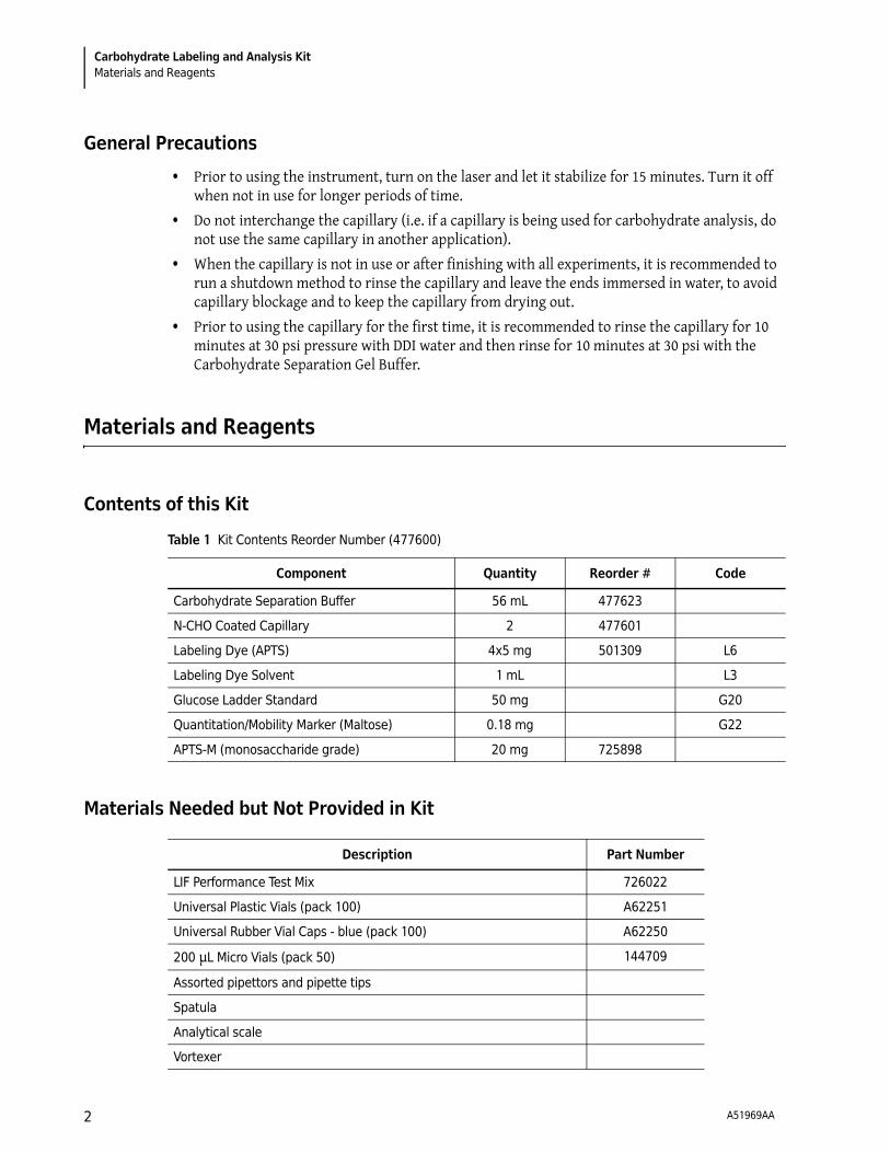
Table 1 Kit Contents Reorder Number (477600)
Component Quantity Reorder # Code
Carbohydrate Separation Buffer 56 mL 477623
N-CHO Coated Capillary 2 477601
Labeling Dye (APTS) 4x5 mg 501309 L6
Labeling Dye Solvent 1 mL L3
Glucose Ladder Standard 50 mg G20
Quantitation/Mobility Marker (Maltose) 0.18 mg G22
APTS-M (monosaccharide grade) 20 mg 725898
Description Part Number
LIF Performance Test Mix 726022
Universal Plastic Vials (pack 100) A62251
Universal Rubber Vial Caps - blue (pack 100) A62250
200 μL Micro Vials (pack 50) 144709
Assorted pipettors and pipette tips
Spatula
Analytical scale
Vortexer
A51969AA2

Carbohydrate Labeling and Analysis KitOverview of the Procedure
Storage Conditions
The kit components are stable for one year when stored under the following conditions:
• Upon receipt, the kit should be stored at 2 to 8°C.
• Reconstituted Quantitation Control (G22) and labeled glucose ladder standard (G20) should be stored at -35 to -15°C.
• Reconstituted labeling reagent (APTS) shall be stored at -35 to -15°C. It is stable for 2 weeks after reconstitution.
Overview of the Procedure
The carbohydrate analysis involves the following three tasks:
• Enzymatic release of the N-linked oligosaccharides from the glycoprotein.
• Labeling of the mixture of released oligosaccharides.
• Analysis of the flouorophore-labeled oligosaccharides by capillary electrophoresis.
Releasing Oligosaccharides From Glycoproteins
IMPORTANT This labeling kit does not contain releasing enzymes.
There are multiple enzymatic and chemical procedures for releasing oligosaccharides from proteins. In order to successfully label the released oligosaccharide, it is essential that the reducing termini of the oligosaccharide is not destroyed by the deglycosylation method.
The following is a suggested protocol for N-deglycosylation, using N-Glycosidase F (PNGase F):
Bench top micro-centrifuge
Water bath
Centrifugal vacuum evaporator
Sodium Cyanoborohydride, 1 M/THF Sigma-Aldrich 296813
2-mercaptoethanol Sigma-Aldrich M7154
Double Deionized (DDI) water filtered through 0.2 μm filter
N-glycosidase F (PNGase F) New England Biolabs P0704S
Nonidet® NP-40 non ionic detergent
Description Part Number
A51969AA 3

Carbohydrate Labeling and Analysis KitReleasing Oligosaccharides From Glycoproteins
Enzymatic Release of the N-Linked Oligosaccharides
1. Aliquot the sample volume in order to have between 25-300 μg of glycoprotein.
2. Dry the glycoprotein solution completely in a centrifugal vacuum evaporator.
3. Add 45 μL of 1X PBS buffer.
4. Add 1.0 μL of 5% SDS (or a volume that gives final concentration of 0.1% SDS).
5. Add 1.5 μL of 1:10 dilution of 2-mercaptoethanol in DDI water (or a volume that gives final concentration of 50 mM 2-mercaptoethanol).
6. Heat the sample for 5 minutes at 100°C.
IMPORTANT If the denatured protein precipitates, discard the sample and restart this process, repeating steps 1 through 5, and for step 6, denature the protein by incubating it at 37°C for 10 minutes.
7. Cool the sample to room temperature.
8. Add 5 μL of Nonidet® NP40 non-ionic detergent (or a sufficient volume that gives a final concentration of 0.75%).
9. Add PNGase F enzyme, according to its activity.
10. React for 15 hours at 37°C.
11. Add approximately 150 μL of cold ethanol (or 3 times the actual reaction mixture volume).
12. Vortex the mixture and place it on ice for complete protein precipitation.
13. Centrifuge the samples at 15,000 rpm for 5 minutes.
14. Withdraw and save the supernatant.
IMPORTANT This solution contains the RELEASED N-LINKED OLIGOSACCHARIDES.
15. Discard the solid. This is the deglycosylated protein.
Working with the Supernatant
There are two options for working with the supernatant:
For quantitative analysis, add an internal standard to the supernatant. See Preparing the Standards beginning with Step 1a.
For qualitative analysis, bring the supernatant to dryness in the centrifugal vacuum evaporator. These oligosaccharides are ready to be labeled with APTS. See Performing the Labeling Reaction in this chapter.
Principle of the Labeling Method
After release (enzymatic or chemical), the oligosaccharides can be labeled with a fluorophore called 8-aminopyrene-1,3,6=, 6-trisulfonate. The stoichiometry of labeling reaction is one APTS molecule per molecule of oligosaccharide. Figure 1 illustrates the labeling reaction of an N-linked oligosaccharide with APTS.
A51969AA4

Carbohydrate Labeling and Analysis KitPreparing the Labeling Reagents
Figure 1 Labeling Reaction of an Oligosaccharide with APTS
The efficiency of the labeling reaction is dependent on temperature and the amount of oligosaccharides. This protocol has been optimized for labeling 5 nmoles or less of total oligosaccharides.
Samples with amounts higher than 5 nmoles may give a lower reaction yield. Use maltose (G22) as an internal labeling control and/or as an internal mobility marker.
Preparing the Labeling Reagents
Choosing the Proper Fluorophore
APTS-M is a high-purity fluorophore intended for labeling monosaccharides. APTS-M contains citric acid as a catalyst. APTS (L6) can be used for oligosaccharides.
Preparing the Reagents
1 Prepare the APTS labeling reagent.
To a vial of APTS labeling dye (L6)
a. Add 48 μL of Labeling dye solvent (15% acetic acid – L3).
b. Vortex for 5 seconds until complete dissolution.
c. Store at -35 to -15°C for up to 2 weeks when not in use.
2 Prepare the APTS-M (Monosaccharide grade) Labeling Dye
a. Add 400 μL of DDI water to the APTS-M vial.
b. Vortex the mixture for 5 seconds until all of the solid is dissolved.
c. Store the reconstituted APTS-M at -35 to -15°C for up to two weeks.
A51969AA 5

Carbohydrate Labeling and Analysis KitPerforming the Labeling Reaction
Preparing the Standards
1 Prepare the Maltose Quantitative Control/Mobility Marker (G22).
a. Add 500 μL of DDI water to the Maltose quantitative control labeled G22. This makes a solution containing 500 nmoles of maltose, or concentration of 1 nmol/μL.
b. For quantitation purposes, add 5 μL of G22 reconstituted to your released oligosaccharides.
c. Dry down the sample in a centrifugal evaporator and proceed to labeling protocol, as described in the next section.
d. Store at -35 to -15°C when not in use.
NOTE When using the maltose solution for quantitation purposes, make sure that the solution is prepared fresh and used right away. Bacterial contamination can digest the sugars, leading to a miscalculation of your quantitation.
2 Prepare the Glucose Ladder Standard (G20).
a. Weigh and dissolve 5 mg of Glucose Ladder Standard (G20) in 80 μL DDI water in 1.5 mL microcentrifuge tube. Sonicate if necessary.
b. Aliquote at least ten 2 μL portions of the Glucose ladder standard solution to 0.5 mL microcentrifuge vials and dry them in a centrifugal vacuum evaporator. The dried glucose
ladder can be stored at room temperature or used immediately.
Performing the Labeling Reaction
1 Label the Sample and/or Standard (G20).
a. Add 2 μL of 1 M sodium cyanoborohydride/THF to the dried oligosaccharide sample.
b. Add 2 μL of APTS Labeling Reagent to the sample.
IMPORTANT Due to the reaction of sodium cyanoborohydride with water, avoid moisture and store this product under dry conditions. Use a dry needle to dispense this chemical. Pass dry argon into the chemical bottle through the septum, while dispensing 2 μL from the bottle.
c. Incubate the reaction mixture at 37°C for 4 hours, or at 60°C for 90 minutes, or at room temperature overnight (mildest reaction).
NOTE A mild labeling condition is necessary to avoid losing the sialic acid residues from your oligosaccharide chain. You can leave the reaction mixture at room temperature overnight and in the dark to do this. For Glucose Ladder incubation, select any of the temperature/ time options.
Figure 2 illustrates a summary of the overall labeling protocol.
A51969AA6

Carbohydrate Labeling and Analysis KitPerforming the Labeling Reaction
2 Prepare the Sample and/or Standard for capillary electrophoresis.
a. To the 4 μL reaction mixture prepared above add:
• For Glucose Ladder Standard: add 96 μL of DDI water
• For all other samples: add 46 μL DDI water
This solution can be stored at 2 to 8°C.
b. Take an aliquot of 5 μL of the solution above and add 195 μL of DDI water.
c. Mix the contents welland transfer the solution into a micro vial for analysis in the PA 800 system see Sample Vial Setup.
Figure 3 illustrates a summary of the sample preparation for injection.
Figure 2 Labeling Reaction Scheme
A51969AA 7

Carbohydrate Labeling and Analysis KitPreparing the PA 800 Instrument
Figure 3 Sample Preparation for Injection
Preparing the PA 800 Instrument
Before proceeding, review the following procedures in the PA 800 plus System Maintenance User’s Guide (A51964AA).
• Install the LIF Detector
• Calibrate the LIF Detector
• Capillary Cartridge Procedures
• Cleaning the Interface Block and the Opening Levers
A51969AA8

Carbohydrate Labeling and Analysis KitInstalling and Pre-conditioning the N-CHO Capillary
Installing and Pre-conditioning the N-CHO Capillary
• Prior to using the PA 800 plus system for carbohydrate analysis, the N-CHO capillary must be installed in a capillary cartridge. To properly install the capillary, refer to the Capillary Cartridge sections in the PA 800 plus System Maintenance User’s Guide (A51964AA).
IMPORTANT Be sure to pre-rinse a new capillary for 10 minutes at 30 psi pressure with DDI water and then rinse for 10 minutes at 30 psi with Carbohydrate Separation Buffer prior to the first run.
For this analysis, you will be using a 50 μm I.D. N-CHO capillary. The length from injection inlet to detector should be 40 cm, with the total length being 50.2 cm. Be sure to measure the capillary dimensions accurately and record the dimensions in the capillary/performance tab of the Advanced Method Options, see Figure 4. This will be necessary for accurate mobility determinations.
Figure 4 Capillary Performance Screen
IMPORTANT To get good reproducibility from capillary to capillary and accurate mobility assignments, it is important to adhere to the capillary pre-measurement procedure.
IMPORTANT The cut ends of capillaries should be inspected carefully under magnification. The cut must be clean (not jagged) and perpendicular to the capillary length (not angled). Poor cuts will result in poor resolution and poor sample loading.
NOTE When not in use, submerge the capillary ends in DDI water to prevent the capillary from drying out.
• Turn off the PA 800 system and then install the LIF detection components.
• Turn on the system and permit the laser to warm up for at least 30 minutes, prior to calibration.
A51969AA 9

Carbohydrate Labeling and Analysis KitPreparing the Buffer Trays
Clean the Interface Block
Clean the electrodes, opening levers, capillary tips, and interface block carefully using a Kimwipe soaked with DDI water. Do this on a weekly basis or when changing chemistries on the PA 800 system. Accumulation on the interface block can lead to current leakage errors.
Insert the Cartridge
Insert the cartridge into the system. Close the cartridge and sample covers.
Preparing the Buffer Trays
1 Fill the vials with the appropriate volumes of each reagent:
• 1.5 mL of DDI water per H20 vial (4 vials)
• 1.5 mL of Carbohydrate Separation Buffer per Gel-R vial (1 vial)
• 1.3 mL of Carbohydrate Separation Buffer per Gel-S vial (2 vials)
• 0.8 mL of DDI water per waste vial (1 vial)
A51969AA10

Carbohydrate Labeling and Analysis KitPreparing the Buffer Trays
Figure 5 Universal Vials and Caps
2 Cover each vial with a cap as shown in Figure 5. Caps are not intended for re-use and should be disposed of after the buffer used is exhausted. Re-using vial caps can result in pressure leakage and capillary breakage.
3 Place the reagent vials on the inlet and outlet buffer trays as indicated in Figure 6.
NOTE During the separation, salts migrate from one vial of gel buffer to the other, changing the composition of the buffer. Therefore, it is recommended that the vials of buffer be replaced with fresh buffer after 20 runs. For unattended operation of more than 20 runs, simply program the increment function to increment the water and buffer vials after 20 runs. Vials will increment in a forward direction. Ensure that the tray is filled with the appropriate number of vials to manage the number of runs that are to be performed.
NOTE The H20 vials are used to clean the capillary tips after the sample introduction step to prevent sample carryover. Replace the vials with fresh water if rinse water becomes contaminated.
A schematic of the buffer tray setup is illustrated in Figure 6.
1. Universal Vial Cap2. Maximum Fill Level3. Universal Vials
A51969AA 11

Carbohydrate Labeling and Analysis KitSample Vial Setup
Figure 6 Buffer Tray Configuration
Sample Vial Setup
Before placing the 200 microliter sample vials into the universal vials, ensure that no bubbles are at the bottom of the sample vials. If bubbles exist, centrifuge the sample vials for two minutes at 1,000 g and repeat if necessary. Place a cap on the universal vial and ensure a good seal, see Figure 7.
Place the universal vials into the 48-position inlet sample tray positions A1 through C8. For the best quantitative results, perform one injection per vial, introducing replicates in separate vials.
A51969AA12

Carbohydrate Labeling and Analysis KitRunning the Samples
Figure 7 Micro Vial Inside a Universal Vial
Running the Samples
Initial Conditions
1. Universal Cap2. Micro Vial
3. Universal Vial4. Micro Vial inside Universal Vial
Table 2 Test Run: Initial Conditions
Capillary Temperature: 20°
Sample Storage Temperature: 10°
Auxiliary Data Channel: Current
A51969AA 13

Carbohydrate Labeling and Analysis KitRunning the Samples
Figure 8 Initial Condition Tab
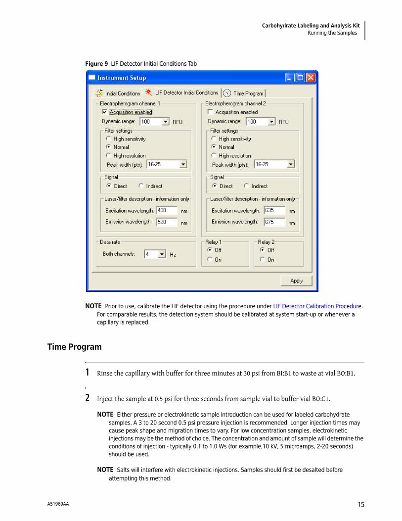
LIF Detector Initial Conditions
Table 3 Test Run: LIF Detector Initial Conditions
Detection Laser Induced Fluorescence
Wavelength Excitation - 488 nm, Emission - 520 nm
Data Rate 4 Hz
Dynamic Range 100 RFU (relative fluorescence units)
Filter Setting Normal
Peak Width 16 - 25
A51969AA14
Carbohydrate Labeling and Analysis KitRunning the Samples
Figure 9 LIF Detector Initial Conditions Tab
NOTE Prior to use, calibrate the LIF detector using the procedure under LIF Detector Calibration Procedure. For comparable results, the detection system should be calibrated at system start-up or whenever a capillary is replaced.
Time Program
1 Rinse the capillary with buffer for three minutes at 30 psi from BI:B1 to waste at vial BO:B1.
2 Inject the sample at 0.5 psi for three seconds from sample vial to buffer vial BO:C1.
NOTE Either pressure or electrokinetic sample introduction can be used for labeled carbohydrate samples. A 3 to 20 second 0.5 psi pressure injection is recommended. Longer injection times may cause peak shape and migration times to vary. For low concentration samples, electrokinetic injections may be the method of choice. The concentration and amount of sample will determine the conditions of injection - typically 0.1 to 1.0 Ws (for example,10 kV, 5 microamps, 2-20 seconds) should be used.
NOTE Salts will interfere with electrokinetic injections. Samples should first be desalted before attempting this method.
A51969AA 15

Carbohydrate Labeling and Analysis KitRunning the Samples
3 Wait 0.2 minutes with vials BI:A4 and BO:A4, see Figure 10. This step dips the capillary in water to protect against sample carry over. Change the rinse water vials if they are contaminated.
4 Separate Step - 20 minutes from vial BI:C1 to vial BO:C1 (buffer), see Figure 10. The constant voltage should be at 30 kV, with reverse polarity and a 0.17 ramp time.
5 Autozero at 1.0 minute.
6 End at 20.0 minutes.
Figure 10 Time Program Tab
A51969AA16

Carbohydrate Labeling and Analysis KitEvaluation of Results
Figure 11 Typical Current Profile
NOTE Prior to use, calibrate the LIF detector using the procedure under LIF Detector Calibration Procedure. For comparable results, the detection system should be calibrated at system start-up or whenever a capillary is replaced.
Evaluation of Results
The test mixture contains the APTS-labeled glucose oligomers consisting of at least 20 individual oligomers. An example of this test mix is highlighted in Figure 12.
Field Strength Generated 598 V/cm
Typical Current, see Figure 11 under 20 μA
A51969AA 17

Carbohydrate Labeling and Analysis KitSystem Shutdown and Capillary Storage
Figure 12 Electropherogram - Glucose Ladder
System Shutdown and Capillary Storage
Short Term Storage (<24 hours) of Capillary
Perform a three minute, 30 psi rinse with water. The capillary may be stored on the instrument with the capillary ends immersed in water.
Whenever the capillary has not been used for three hours or longer, rinse the capillary by performing a three minute, 30 psi rinse with water before performing a separation.
Long Term Storage (>24 hours) of Capillary
1 Perform a 3 minute, 30 psi rinse with water and then with Carbohydrate Separation Buffer for 3 minutes.
2 Remove the capillary from the instrument and place in a cassette box with the capillary ends placed in vials of DDI water.
3 Store the cartridge box at 2°C to 8°C in an upright position.
A51969AA18
Carbohydrate Labeling and Analysis KitTroubleshooting Procedures
Whenever the capillary has not been used for three hours or longer, rinse the capillary by performing a 3 minute, 30 psi rinse with water and then a 10 minute rinse with Carbohydrate Separation Buffer before performing a separation.
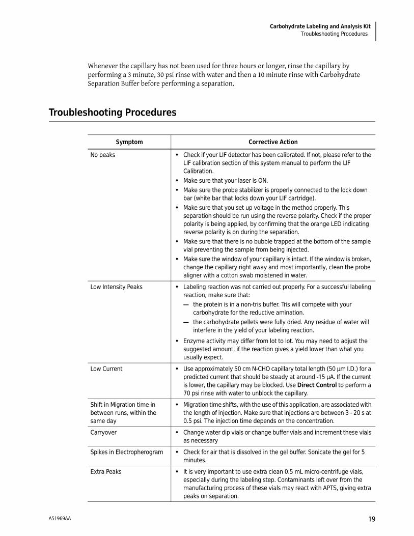
Troubleshooting Procedures
Symptom Corrective Action
No peaks • Check if your LIF detector has been calibrated. If not, please refer to the LIF calibration section of this system manual to perform the LIF Calibration.
• Make sure that your laser is ON. • Make sure the probe stabilizer is properly connected to the lock down
bar (white bar that locks down your LIF cartridge). • Make sure that you set up voltage in the method properly. This
separation should be run using the reverse polarity. Check if the proper polarity is being applied, by confirming that the orange LED indicating reverse polarity is on during the separation.
• Make sure that there is no bubble trapped at the bottom of the sample vial preventing the sample from being injected.
• Make sure the window of your capillary is intact. If the window is broken, change the capillary right away and most importantly, clean the probe aligner with a cotton swab moistened in water.
Low Intensity Peaks • Labeling reaction was not carried out properly. For a successful labeling reaction, make sure that:— the protein is in a non-tris buffer. Tris will compete with your
carbohydrate for the reductive amination.— the carbohydrate pellets were fully dried. Any residue of water will
interfere in the yield of your labeling reaction.
• Enzyme activity may differ from lot to lot. You may need to adjust the suggested amount, if the reaction gives a yield lower than what you usually expect.
Low Current • Use approximately 50 cm N-CHO capillary total length (50 μm I.D.) for a predicted current that should be steady at around -15 μA. If the current is lower, the capillary may be blocked. Use Direct Control to perform a 70 psi rinse with water to unblock the capillary.
Shift in Migration time in between runs, within the same day
• Migration time shifts, with the use of this application, are associated with the length of injection. Make sure that injections are between 3 - 20 s at 0.5 psi. The injection time depends on the concentration.
Carryover • Change water dip vials or change buffer vials and increment these vials as necessary
Spikes in Electropherogram • Check for air that is dissolved in the gel buffer. Sonicate the gel for 5 minutes.
Extra Peaks • It is very important to use extra clean 0.5 mL micro-centrifuge vials, especially during the labeling step. Contaminants left over from the manufacturing process of these vials may react with APTS, giving extra peaks on separation.
A51969AA 19

Carbohydrate Labeling and Analysis KitAppendix
Appendix
LIF Detector Calibration Procedure
The PA 800 LIF detector calibration kit procedure is used to calibrate the detection system for carbohydrate analysis. This will ensure consistent results from day to day and from capillary to capillary, as detection is measured in relative fluorescence units (RFU). This calibration procedure allows you to normalize the signal generated from your analyte, relative to a known concentration of sodium fluorescein. Perform the calibration procedure when installing a new capillary or after detector and cartridge changes.
Setting the Calibration Corrector Factors (CCFs)
IMPORTANT The user must have instrument administration privileges.
1 Launch 32 Karat.
2 Go to Tools | Enterprise Login.
3 Enter the user name PA 800 and password plus and select Log in.
4 Select the CHO instrument icon and then Configure Instrument.
5 Select Configure on the instrument configuration window.
6 Select the LIF Detector icon in the right pane of the PA 800 System Configuration window.
7 The 32 Karat Software Configuration dialog box appears as shown in Figure 13.
Table 4 Reagents Needed to Perform the Procedure
Materials Part Number
LIF Performance Test Mix 726022
DDI water N/A
A51969AA20

Carbohydrate Labeling and Analysis KitAppendix
Figure 13 32 Karat Software Configuration dialog
8 Select LIF Calibration Wizard from the Instrument Configuration window to display the Calibration Wizard. See Figure 14.
Automatic Calibration
1 Choose Auto as the calibration option, as shown in Figure 14 and click Next.
A51969AA 21

Carbohydrate Labeling and Analysis KitAppendix
Figure 14 Calibration Wizard Screen - Auto Select
2 Set up the target RFU value. The target value for N-CHO-coated capillary is 7, as shown in Figure 15 and click Next.
Figure 15 Calibration Wizard Screen - Step 2
A51969AA22

Carbohydrate Labeling and Analysis KitAppendix
3 Fill one buffer vial with 1.5 mL DDI water and another with 200 μL of diluted LIF Performance Test Mix (1:1 with DDI water) in a micro vial. Place them in the buffer vial positions as indicated in Figure 16.
4 Place an empty vial in the waste position.
IMPORTANT The system performance test kit (713360) contains instructions for performing detector noise tests on a bare-fused silica capillary, which uses borate buffer. Do not use the borate buffer for calibration of the N-CHO capillary. Use a 50% LIF Performance Test mix solution (100 μL of LIF Test Mix with 100 μL of DDI water) in a micro vial.
5 Click Next to perform the automatic calibration, as shown below.
Figure 16 Calibration Wizard Screen - Step 3
6 After the calibration is completed, a dialog box displays as shown in Figure 17.
A number will show in the Calibration Correction Factor field.
If it is below 10, click Accept to complete the calibration procedure.
If it is above 10, see CCF Troubleshooting Procedures.
A51969AA 23

Carbohydrate Labeling and Analysis KitAppendix
Figure 17 Calibration Wizard Screen - Step 4
CCF Troubleshooting Procedures
CCF <1
CCF >1
No Step Change Detected
The LIF Calibration method is basically a comparison of detector signals between a non-fluorescent solution to a known fluorescent solution. When one rinses with the non-fluorescent solution
Table 5 CCF <1
Symptom Corrective Action
CCF is still below 1, but not below 0.1 • There is no problem with the system. Run a standard and verify acceptable performance of the system.
System performance is not acceptable or the CCF is below 0.1
• Verify that the capillary for the carbohydrate analysis was used, and it is not broken.
• Verify that the laser provided with the PA 800 was used. • Verify that the correct filters are installed in the LIF detector. • Contact a Beckman Coulter service representative.
Table 6 CCF >1
Symptom Corrective Action
CCF is more than 1.0 but less than 10 • Run a standard to determine if the system performance is still acceptable.
CCF is more than 10 or if the system performance is not acceptable.
• Verify that the laser provided with the PA 800 is being used for calibration.
• Verify that the correct filters are installed in the LIF detector. • Replace the chemistries and capillary and repeat the
calibration. • Contact a Beckman Coulter service representative.
A51969AA24

Carbohydrate Labeling and Analysis KitAppendix
followed by a fluorescent solution rinse, one should see the first part of the detector signal near zero and the second part of the detector signal near the target fluorescent value. This detector output is step shaped in appearance.
If no step change is observed, the appropriate solutions are not passing the detector or the detector is not able to detect them.
1 Verify that the solution is rinsing through the capillary by performing a direct control 5 minute 20 psi pressure rinse of water, from buffer inlet A1 to an empty buffer vial in outlet B1.
2 Once the rinse has begun, lift the sample cover.
3 Observe the outlet end of the capillary in B1. Droplets should be formed on the outlet end of the capillary.
• If no droplets are formed, either the capillary is plugged or the instrument has a pressure failure.
• Replace the capillary and repeat the rinse. If there is still no droplet observed, contact a Beckman Coulter service representative.
Once flow through the capillary is verified, the detection system is the only other possible cause, when no step is detected.
4 Verify that the correct filters are installed in the LIF detector.
5 Verify that the laser provided with the PA 800 system is connected and on.
6 Run the calibration test with a new capillary and chemistries.
If there is still no step change detected, contact a Beckman Coulter service representative.
A51969AA 25

© 2009 Beckman Coulter, Inc.All Rights Reserved
www.beckmancoulter.com andwww.CELeader.com