application of denaturing high-performance liquid ...aem.asm.org/content/79/17/5186.full.pdf ·...
TRANSCRIPT

Application of Denaturing High-Performance Liquid Chromatographyfor Monitoring Sulfate-Reducing Bacteria in Oil Fields
Outi Priha,a Mari Nyyssönen,a Malin Bomberg,a Arja Laitila,a Jaakko Simell,b Anu Kapanen,a Riikka Juvonena
VTT Technical Research Centre of Finland, Espoo, Finlanda; Kemira, Espoo, Finlandb
Sulfate-reducing bacteria (SRB) participate in microbially induced corrosion (MIC) of equipment and H2S-driven reservoirsouring in oil field sites. Successful management of industrial processes requires methods that allow robust monitoring of mi-crobial communities. This study investigated the applicability of denaturing high-performance liquid chromatography(DHPLC) targeting the dissimilatory sulfite reductase ß-subunit (dsrB) gene for monitoring SRB communities in oil field sam-ples from the North Sea, the United States, and Brazil. Fifteen of the 28 screened samples gave a positive result in real-time PCRassays, containing 9 � 101 to 6 � 105 dsrB gene copies ml�1. DHPLC and denaturing gradient gel electrophoresis (DGGE) com-munity profiles of the PCR-positive samples shared an overall similarity; both methods revealed the same samples to have thelowest and highest diversity. The SRB communities were diverse, and different dsrB compositions were detected at different geo-graphical locations. The identified dsrB gene sequences belonged to several phylogenetic groups, such as Desulfovibrio, Desulfo-coccus, Desulfomicrobium, Desulfobulbus, Desulfotignum, Desulfonatronovibrio, and Desulfonauticus. DHPLC showed an advan-tage over DGGE in that the community profiles were very reproducible from run to run, and the resolved gene fragments couldbe collected using an automated fraction collector and sequenced without a further purification step. DGGE, on the other hand,included casting of gradient gels, and several rounds of rerunning, excising, and reamplification of bands were needed for suc-cessful sequencing. In summary, DHPLC proved to be a suitable tool for routine monitoring of the diversity of SRB communitiesin oil field samples.
Uncontrolled growth of microbes in oil field production sys-tems may have severe negative impacts on productivity of the
systems. Microbial activity may lead to increased frequency ofequipment failure because of corrosion, elevated hydrogen sulfide(H2S) concentrations, reservoir souring, formation of metal sul-fide scales, filter plugging, loss of injectivity, and inefficient heatexchange (1).
Many of the microbes are indigenous to the oil-bearing deepsubsurface environments, but oil production operations also in-troduce microbes to the system (2, 3). One of the most commonand problematic groups of bacteria present in oil and gas fieldsystems is sulfate-reducing bacteria (SRB) (4, 5). The activity ofSRB is one of the most significant sources of H2S in reservoirsouring. The most frequently isolated mesophilic SRB from oilproduction waters belong to the genus Desulfovibrio, but mem-bers of the genera Desulfacinum, Desulfobacter, Desulfobacterium,Desulfomicrobium, and Desulfotomaculum have also been isolated(3). Thermophilic groups frequently detected from oil productionwaters include the bacterial genera Thermodesulfobacterium andThermodesulforhabdus and the archaeal genus Archaeoglobus (6).
In order to effectively manage microbe-induced reservoir foul-ing, methods that allow robust monitoring of detrimental micro-bial communities are required. Currently, SRB in the oil fieldindustry are mainly detected and quantified by using the culture-dependent most probable number (MPN) method which requires28 days to obtain results (7). Furthermore, only a limited numberof bacteria can be assessed by a culture-dependent approach (8).Direct culture-independent detection of SRB from communityDNA using denaturing gradient gel electrophoresis (DGGE) orterminal restriction length polymorphism (TRFLP) analysis cansignificantly speed up microbial community profiling and processmonitoring and simultaneously provide information about spa-tial and temporal changes in microbial community composition
(9–12). However, these methods are labor-intensive, and gel-based analysis allows only a limited number of samples to be an-alyzed during the same assay under consistent conditions. As aresult, new methods that allow automated and high-throughputanalysis of process samples would be advantageous.
Denaturing high-performance liquid chromatography (DHPLC)is a method that has traditionally been used for DNA mutationalanalysis, especially for detection of single nucleotide polymor-phisms, in medical applications. In recent years, the technique hasbeen successfully applied to the study of bacterial diversity in var-ious ecosystems, such as seawater (13), the gut (14), dairy prod-ucts (15), soil, fermenter sludge and compost (16), sediments(17), and human infections (18–20), and to monitor fungal com-munities present in air (21), wood decay (22), cheese (23), andmilk (24). Similar to DGGE, DHPLC can theoretically resolveDNA fragments with sequence and/or size differences. However,DHPLC is based on separation of partially heat-denatured DNAfragments by ion pair reverse-phase liquid chromatography in-stead of a denaturing chemical gradient. The DNA fragments areeluted from the column by an increasing gradient of acetonitrile,are detected by UV or fluorescence detector, and can be collectedusing an automated fraction collector for sequence analysis.
The aim of this study was to characterize SRB communitiesfrom oil field samples and to develop a DHPLC method for pro-filing SRB communities from oil field reservoirs.
Received 8 April 2013 Accepted 17 June 2013
Published ahead of print 21 June 2013
Address correspondence to Outi Priha, [email protected].
Copyright © 2013, American Society for Microbiology. All Rights Reserved.
doi:10.1128/AEM.01015-13
5186 aem.asm.org Applied and Environmental Microbiology p. 5186–5196 September 2013 Volume 79 Number 17
on April 7, 2019 by guest
http://aem.asm
.org/D
ownloaded from
MATERIALS AND METHODSStrains and environmental samples. Bacterial and archaeal strains ortheir DNA were obtained from the VTT Culture Collection(culturecollection.vtt.fi) or from the German Resource Centre for Biolog-ical Material (www.dsmz.de) (Table 1). In addition to these strains, Des-ulfomicrobium macestii DSM 4194 (VTT E-001444) was used in the DGGEladder and quantitative PCR (qPCR) standards. The strains were chosento represent microbial diversity detected at oil field sites or in marineenvironments. The strains were grown essentially as recommended by theculture collections. Injection water and samples of produced water (waterrecovered with the oil) (n � 28) were obtained from different oil field sitesin the North Sea, the United States, and Brazil (Table 2). The water sam-ples were filtered onto Sterivex-GP 0.22-�m-pore-size filter units (Milli-pore, Billerica, MA, USA) and shipped frozen to the laboratory.
DNA isolation. For the DNA analysis, Sterivex filter units containingthe oil field samples were aseptically broken with a hammer, and the filters
were cut into pieces (approximately 2.5 cm2) with a sterile scalpel andplaced with sterile forceps into the lysing tube of a DNA extraction kit.Total DNA from pure cultures and environmental samples was isolatedwith a FastDNA Spin kit for soil (MP Biomedicals, Santa Ana, CA, USA)according to the manufacturer’s instructions, except that the cells werelysed for 2 min in a FastPrep-24 instrument (MP Biomedicals). ForDesulfobacter hydrogenophilus (DSM 3380), Desulfotomaculum geothermi-cum (DSM 3669), and Archaeoglobus fulgidus (DSM 4304), a PowerSoilDNA extraction kit (MoBio Laboratories Inc., Carlsbad, CA, USA) wasused according to the manufacturer’s instructions, with the exception thatthe cells were lysed by bead beating with a Ribolyser (Hybaid) device for30 s at 6 m s�1.
Real-time quantitative PCR. Quantification of the dsrB copy numberin the extracted DNA was performed using a LightCycler 480 instrument(Roche Diagnostics, Basel, Switzerland) and SYBR green-based detectionfor double-stranded DNA. An approximately 350-bp fragment of the dsrB
TABLE 1 Bacterial and archaeal strains used for method optimization and as reference strains
Phylogenetic cluster and species StrainIsolation source(type and location)
GenBankaccession no.
GCcontent(%)
Fragmentlength(bp)
DHPLCmigration(%)
DGGEmigration(%)
Deltaproteobacteria/Desulfobacterales/Desulfobacteraceae
Desulfobacter hydrogenophilus DSM 3380 Marine mud, Italy NAa NA NA 30 10Desulfatiferula olefinivorans DSM 18843/VTT
E-103143Oil-polluted sediment,
FranceDQ826725 61 381 61 72
Desulfobacter curvatus DSM 3379T/VTTE-001657T
Marine mud, Italy AF418199 52 350 NA 57
Desulfobacter vibrioformis DSM 8776/VTTE-103147
Water-oil from oilplatform, Norway
AJ250472 52 381 36 28
Desulfosarcina variabilis DSM 2060T/VTTE-001656T
Marine black mud, France AF191907 58 378 58 57
Deltaproteobacteria/Desulfovibrionales/Desulfovibrionaceae
Desulfatibacillum alkenivorans DSM 16219 Oil-polluted sediment,France
AY504426 59 377 54 57
Desulfovibrio vulgaris DSM 644T/VTTE-001447T
Soil, UK U16723 62 378 NA 57
Desulfovibrio alaskensis DSM 16109/VTTE-103145
Gravel material fromsoured oil reservoir, USA
CP000112 61 353 NA 54
Desulfovibrio desulfuricans DSM 17464/VTTE-103144
Oil well corrosion site, USA AJ249777 61 378 57 54
Deltaproteobacteria/Desulfovibrionales/Desulfohalobiaceae
Desulfonauticus autotrophicus DSM 4206 Oil production water,Germany
NA 45 314 22 9
Deltaproteobacteria/Desulfovibrionales/Desulfomicrobiaceae
Desulfomicrobium apsheronum DSM 5918/VTTE-103146
Water, oil-bearing deposits,Russia
AF418188 59 393 58 59
Clostridia/Clostridiales/PeptococcaceaeDesulfotomaculum geothermicum DSM 3669/VTT
E-061476T
Anoxic geothermal groundwater, France
AF273029 57b NA 52, 53c 44
Archaea/Euryarchaeota/Archaeoglobi/Archaeoglobales/Archaeoglobaceae
Archaeoglobus fulgidus DSM 4304 Submarine hot spring, Italy NC_000917 51 360 31 39a NA, not available.b The GC content is an estimate as the GenBank sequence entry does not cover the complete amplicon used in this study.c The two values represent the two peaks in the chromatogram.
Application of DHPLC for Monitoring SRB in Oil Fields
September 2013 Volume 79 Number 17 aem.asm.org 5187
on April 7, 2019 by guest
http://aem.asm
.org/D
ownloaded from

gene was amplified with primers dsr4R (5=-GTGTAGCAGTTACCGCA-3=) and DSRp2060F (5=-CAACATCGTYCAYACCCAGGG-3=) (11, 25).The amplification was done in a 20-�l reaction volume containing Light-Cycler 480 SYBR green I master mix (Roche Diagnostics), 0.5 �M eachprimer, and 2 �l of sample DNA. The amplification reaction programconsisted of initial denaturation at 95°C for 5 min and 35 cycles with 10 sat 95°C (denaturation), 20 s at 57°C (annealing), and 20 s at 72°C (elon-gation). At the end of the run a melting curve analysis was performed from65 to 97°C.
For preparation of standards for real-time PCR, the dsrB gene frag-ments from Desulfovibrio desulfuricans DSM 17464, Desulfobacter vibrio-formis DSM 8776, and Desulfomicrobium macestii DSM 4194 were clonedinto the pGEM-T vector (Promega, Madison, WI, USA) and transformedinto Escherichia coli JM109 cells according to the manufacturer’s instruc-tions. The plasmids were purified with a QIAprep Spin Miniprep kit (Qia-gen, Hilden, Germany), and the dsrB copy number of the plasmid prepa-rations was calculated based on their DNA concentration, measured by aNanoDrop 2000 spectrophotometer (Thermo Scientific, Wilmington,DE, USA). In every run, a dilution series of a mixture of these circularplasmids was incorporated as a standard curve. DNAs from Pyrobaculumislandicum DSM 4184 and Allochromatium vinosum DSM 180 were in-cluded as negative controls in each run (11). Each standard and samplewere run as at least three replicates.
PCR amplification of dsrB gene fragments for DHPLC and DGGE.The same dsrB gene fragment as targeted in qPCR was amplified for com-munity profiling by DHPLC and DGGE with primers dsr4R and
DSRp2060F. In order to separate the dsrB gene fragments in DGGE, a40-bp GC clamp was attached to the forward primer (5=-CGCCCGCCGCGCGCGGCGGGCGGGGCGGGGGCACGGGGGG-3=) (26). In theDHPLC analysis, gene fragments amplified both with and without the GCclamp were evaluated. The PCR amplification was performed in 50-�lreaction mixtures containing 1� DynaZyme II buffer (10 mM Tris-HCl,pH 8.8, 1.5 mM MgCl2, 50 mM KCl, and 1% Triton X-100), 0.2 mM eachdeoxynucleoside triphosphate, 50 pmol of each primer, 1 U of DynazymeII DNA polymerase (Finnzymes, Espoo, Finland), and 2 �l of templateDNA. The PCR was performed in a MasterCycler thermal cycler (Eppen-dorf, Germany). The PCR program consisted of a 5-min initial denatur-ation at 94°C followed by 35 cycles of 1 min at 94°C, 1 min at 55°C, and 1min at 72°C, with a final elongation step at 72°C for 10 min. The amplifi-cation products were run in a 1.5% agarose gel and purified with aQIAquick PCR purification kit (Qiagen) according to the instructions ofthe manufacturer. The concentration of the purified PCR products wasdetermined with a Nanodrop ND-1000 spectrophotometer (Thermo Sci-entific).
DHPLC. The DHPLC analysis was performed with a WAVE 4500microbial analysis system composed of a DNASep cartridge, high-preci-sion Peltier oven, quaternary gradient solvent delivery system, WAVEautosampler model 7200, and UV/visible light (UV/VIS) and fluorescencedetectors (Transgenomic, Inc., Omaha, NE, USA). DHPLC analysis wasoptimized with individual and mixed dsrB gene products from pure cul-tures (Table 1) adjusted to a final concentration of 10 ng �l�1 by dilutionin PCR-grade water. The separation of gene fragments was optimized by
TABLE 2 Description of oilfield samples and SRB quantification results from qPCR of dsrB gene fragment
Sample location and code Sample type and sourceSampling date(mo/yr)
Filtered vol(ml)
No. of dsrB genecopies/mla
Sample selected foridentification
North SeaMOB1 Injection water, well 1 01/2010 100 5 � 105 YesMOB2 Injection water, well 2 02/2010 100 4 � 104 YesMOB3 Injection water, well 2 04/2010 100 7 � 104
MOB4 Injection water, well 3 05/2010 100 NDMOB5 Injection water, well 4 05/2010 100 NDMOB6-1 Produced water, well 5 01/2010 40 NDMOB6-2 Produced water, well 6 01/2010 40 NDMOB6-3 Produced water, well 7 01/2010 40 NDMOB6-4 Produced water, well 8 01/2010 40 NDMOB6-5 Produced water, well 9 01/2010 40 5 � 103 YesMOB6-6 Produced water, well 10 01/2010 40 NDMOB6-7 Produced water, well 11 01/2010 40 NDMOB6-8 Produced water, well 12 01/2010 40 NDMOB7 Injection water, well 13 11/2010 100 5 � 102
MOB8 Produced water, well 13 12/2010 50 3 � 103
MOB9 Produced water, well 13 12/2010 50 9 � 102 YesMOB10 Produced water, well 13 12/2010 50 1 � 103
BrazilMOB11 Produced water, well 14 03/2011 1000 6 � 103 YesMOB12 Produced water, well 15 03/2011 1000 1 � 104 Yes
USAMOB13A Produced water, well 16 07/2010 20 2 � 104 YesMOB13B Produced water, well 16 07/2010 20 6 � 105
MOB14 Produced water, well 17 11/2010 150 4 � 102 YesMOB15 Produced water, well 18 03/2011 100 NDMOB16 Produced water, well 19 03/2011 100 NDMOB20 Produced water, well 19 04/2011 20 NDMOB17 Produced water, well 20 03/2011 500 9 � 101* YesMOB19 Produced water, well 20 04/2011 500 NDMOB18 Produced water, well 21 04/2011 500 8 � 102 Yes
a ND, not detected; *, extrapolated.
Priha et al.
5188 aem.asm.org Applied and Environmental Microbiology
on April 7, 2019 by guest
http://aem.asm
.org/D
ownloaded from

varying the temperature (from 62°C to 70°C), acetonitrile gradient com-position (from 46 to 70%), gradient rate (from 0.5% min�1 to 2%min�1), and flow rate (from 0.3 ml min�1 to 1.2 ml min�1). For purecultures, a constant amount of 50 ng of each PCR amplification productwas injected into the column, and for oil field samples 5 �l of undilutedamplification product was used. The amplification products were stainedwith WAVE Optimized HS staining solution during separation, and theelution of dsrB gene fragments was recorded with a fluorescence detectorand visualized as chromatograms using Navigator software, version 3.0.0(build 31) (Transgenomic, Inc.). All buffers and dyes were obtained fromTransgenomic, Inc. Buffer A contained 0.1 M triethylammonium acetate(TEAA), and buffer B contained 0.1 M TEAA and 25% acetonitrile.
In the DHPLC analysis, the dsrB gene fragments were collected tochilled 96-well plates for sequencing by using an FCW 180 WAVE frag-ment collector (Transgenomic, Inc.) and the automated threshold func-tion of the Navigator software (Transgenomic, Inc.). The unpurified frac-tions containing the collected peaks were then directly reamplified fromthe fractions with the corresponding primers under the same conditionsas used in the original PCRs.
DGGE. DGGE analysis was performed with a DCode universal muta-tion detection system (Bio-Rad Laboratories GmbH, Germany). ThePCR-amplified dsrB gene fragments containing the GC clamp were sepa-rated in a DGGE gel (160 mm by 160 mm by 1 mm) containing 8%acrylamide and 35 to 70% denaturing gradient (100% denaturing gradi-ent contains 7 M urea and 40% formamide) in 0.5� TAE buffer (20 mMTris, 10 mM acetate, 0.5 mM EDTA, pH 8.0) at 85 V and 60°C for 16 h. TheDGGE gels were stained with SYBR green II (New England BioLabs,Ipswich, MA, USA) and imaged with a GelDoc imager (Bio-Rad, Hercu-les, CA, USA) under UV light. A sequence ladder consisting of PCR prod-ucts from selected reference strains was run in parallel with the samples.The SRB ladder was a mixture of PCR products from Desulfobacter hy-drogenophilus DSM 3380, Desulfonauticus autotrophicus DSM 4206, Des-ulfobacter vibrioformis DSM 8776, Archaeoglobus fulgidus DSM 4304, Des-ulfotomaculum geothermicum DSM 3669, Desulfatiferula olefinivoransDSM 18843, Desulfovibrio desulfuricans DSM 17464, Desulfatibacillumalkenivorans DSM 16219, Desulfomicrobium macestii DSM 4194, andDesulfomicrobium apsheronum DSM 5918 (Table 1, Fig. 1).
The bands corresponding to different dsrB gene fragments were ex-cised from the DGGE gel using a Pasteur pipette and kept in 20 �l ofPCR-grade water overnight at �4°C. The obtained gene fragments werethen reamplified for sequencing with the corresponding primers underthe same conditions as used in the original PCR. This procedure wasrepeated until a single band was obtained in the DGGE gel. The reampli-fication products were subsequently purified with a QIAquick PCR puri-fication kit (Qiagen).
Sequencing. Sequencing of DHPLC fragments and DGGE bands wasperformed from both ends of the PCR amplicon with a BigDye Termina-tor, version 3.1, cycle sequencing kit (Applied Biosystems, California,USA) in an ABI Prism 310 genetic analyzer (Applied Biosystems);alterna-tively, samples were sent to Macrogen, Inc., for custom DNA sequencingusing the EZ-purification service (Amsterdam, The Netherlands).
Community profiles and sequence analysis. For comparisons of thesimilarity of community profiles generated by DHPLC and DGGE, thechromatogram and gel data were transferred into BioNumerics, version5.10, software (Applied Maths, Sint-Martens-Latem, Belgium). Pearson’scurve-based correlations were calculated, and clustering was done withthe unweighted pair group method with arithmetic mean (UPGMA). TheShannon-Wiener diversity index was calculated to compare DHPLC andDGGE profiles (27), and they were compared by one-way analysis of vari-ance (ANOVA) using SPSS, version 19 (IBM, USA). The dsrB gene se-quences were checked, assembled, and manually edited using the Ge-neious Pro software package (Biomatters, Inc., Auckland, New Zealand)or Kodon, version 3.61 (Applied Maths). The edited dsrB sequences (138to 357 bp) were aligned to reference sequences (357 bp) using theMUSCLE (multiple sequence comparison by log expectation) alignment
feature in Geneious Pro. The alignment was edited manually, and a max-imum-likelihood phylogenetic tree was calculated on the nucleic acid se-quence alignment using PhyLM (28) and a Jukes-Cantor substitutionmodel (29). Bootstrap support for the nodes was calculated with 1,000random repeats.
Nucleotide sequence accession numbers. The dsrB gene fragmentnucleotide sequences of �200 bp were deposited in GenBank under the ac-cession numbers KF269027 to KF269070.
RESULTSSRB detection with quantitative real-time PCR. The SRB weredetected with qPCR targeting the dsrB gene from 15 of the 28water samples obtained from oil fields in the North Sea, the UnitedStates, and Brazil (Table 2). The qPCR assay was linear in the rangeof 2 � 102 to 2 � 107 dsrB copies per PCR. The dsrB gene copynumbers in the samples ranged from 102 to 105 ml�1. Based on theqPCR results, 15 positive samples were selected for communityanalysis.
Optimization of the DHPLC protocol. Optimal conditionsfor the analysis of PCR-amplified dsrB gene fragments by DHPLCwere determined with PCR amplicons from 12 sulfate-reducingbacterial strains and one archaeal strain (Table 1). Varying oventemperature, buffer flow rate, and strength and duration of theacetonitrile gradient showed that the best separation of dsrB genefragments could be achieved by using an oven temperature of65°C, a 54 to 70% acetonitrile gradient that changed 1.5% min�1,and a 0.6-ml min�1 flow rate. The best peak separation was ob-tained with PCR amplicons containing the GC-rich clamp, whilevery poor separation was observed with gene fragments amplifiedwithout the GC clamp (data not shown).
Under the final optimized conditions, 10 strains out of the 13tested yielded one or two major peaks in the chromatogram (Fig.1). The first dsrB gene fragments to elute from the column 3 to 6min after injection belonged to Desulfobacter and Desulfonauticusspecies as well as to Archaeoglobus fulgidus. These species had rel-atively low GC contents (45 to 52%) (Table 1). The gene frag-ments derived from Desulfotomaculum, Desulfomicrobium, Desul-fovibrio, Desulfatibacillum, Desulfatiferula, and Desulfosarcinastrains eluted 8 to 10 min after injection and were characterized byhigher (57 to 61%) GC contents (Table 1). A significant (P � 0.01)positive correlation was observed between percent GC and reten-tion time (r � 0.97). In general, the strains were distinguishablefrom each other based on the retention time of their correspond-ing dsrB amplicons. However, some strains had overlapping re-tention times. These included Archaeoglobus fulgidus DSM 4304and Desulfobacter hydrogenophilus DSM 3380, Desulfotomaculumgeothermicum DSM 3669 and Desulfatibacillum alkenivorans DSM16219, and Desulfosarcina variabilis DSM 2060 and Desulfomicro-bium apsheronum DSM 5918. The results were consistent acrossdifferent runs, independent of whether the PCR-amplified genefragments were injected to the column separately or as part of amixture (data not shown). Three strains, Desulfobacter curvatusDSM 3379, Desulfovibrio alaskensis DSM 16109, and Desulfovibriovulgaris DSM 644, could not be successfully separated by DHPLC.They produced smeared peaks in the chromatograms indepen-dent of different running parameters.
The separation of dsrB gene fragments in DHPLC was consis-tent with DGGE analysis (Fig. 1). The dsrB gene fragments show-ing low retention times on DHPLC were the ones to denature firstin the DGGE analysis, whereas the ones possessing higher GCcontents and retention times also migrated further in the denatur-
Application of DHPLC for Monitoring SRB in Oil Fields
September 2013 Volume 79 Number 17 aem.asm.org 5189
on April 7, 2019 by guest
http://aem.asm
.org/D
ownloaded from

ing gradient of the DGGE. However, among the dsrB gene frag-ments having lower GC contents, there were some discrepan-cies between the two methods in the order in which the dsrBfragments were denatured/eluted. For instance, the dsrB genefragment PCR amplified from Archaeoglobus fulgidus DSM4304 and Desulfobacter hydrogenophilus DSM 3380 eluted si-multaneously in the DHPLC analysis but migrated far apart inthe denaturing gel. In DGGE, strains Desulfatibacillum alken-ivorans DSM 16219, Desulfobacter curvatus DSM 3379, Desul-fosarcina variabilis DSM 2060, Desulfovibrio vulgaris DSM 644,and Desulfomicrobium macestii DSM 4194 could not be sepa-rated from each other, whereas Desulfatibacillum alkenivoransDSM 16219 and Desulfosarcina variabilis DSM 2060 wereclearly distinguishable by DHPLC. Thus, the overall resolutionwas similar with both community profiling methods.
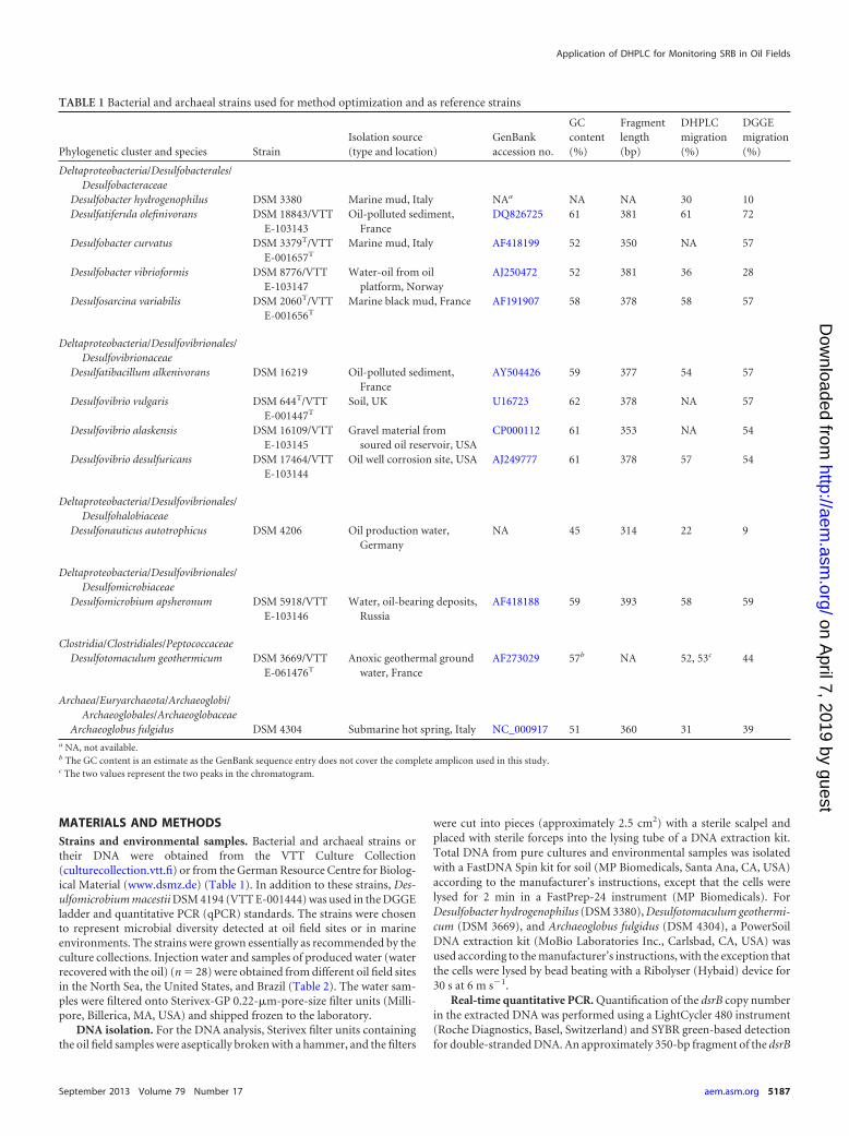
Community profiles of oil field samples. As shown in Fig. 2,DHPLC analysis of the dsrB gene profiles of the oil field sampleswas reproducible between different runs. Even though peak inten-sities varied across different runs, this had no effect on peak reten-tion times. DHPLC and DGGE community profiles shared anoverall similarity; samples with the lowest and highest diversitywere detected by both methods (Fig. 3). The mean Shannon-Wie-ner diversity indexes were 1.17 (standard deviation [SD], 0.60)and 1.27 (SD, 0.36) for DHPLC and DGGE, respectively, and didnot differ statistically significantly (P � 0.1) from each other. Theclustering of SRB community profiles determined with DHPLCand DGGE shared an overall similarity with some exceptions (Fig.4). The grouping followed the geographical location: samplesMOB7 to MOB10 collected from the same North Sea site groupedtogether with both methods, as well as MOB2 and MOB3 from
FIG 1 Separation of dsrB gene fragments PCR amplified from pure culture bacterial and archaeal cultures in a DHPLC chromatogram (a) and DGGE gel (b).DSM numbers are identified in Table 1.
Priha et al.
5190 aem.asm.org Applied and Environmental Microbiology
on April 7, 2019 by guest
http://aem.asm
.org/D
ownloaded from

another North Sea site and MOB13A and -B from the same pro-duced water in the United States.
One sample from each of these sample sets having similar pro-files was selected for further analysis; i.e., altogether 10 sampleswere selected for identification of DHPLC fractions and DGGEbands by sequencing (Table 2). Sequencing of a total of 37 DHPLCfragments was attempted, and 19 of them were successfully se-quenced without purification steps (51%) (Fig. 3). From DGGEgels 54 bands were excised, out of which 27 (50%) produced read-able sequences after several reamplification and purificationrounds.
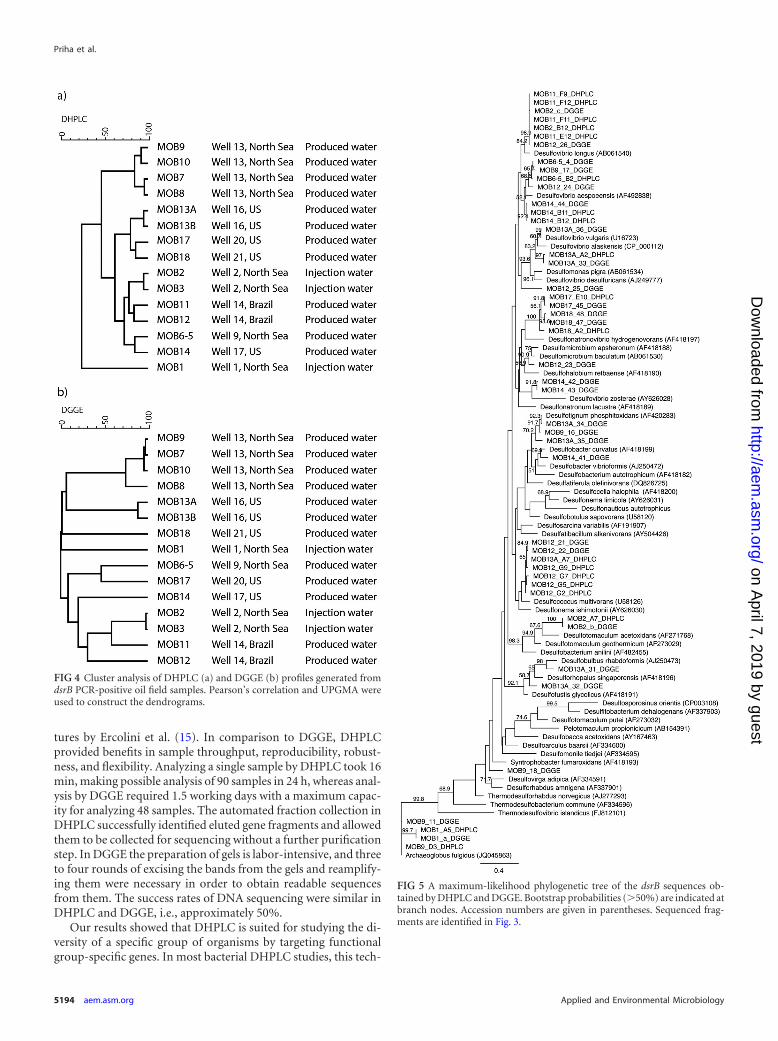
Based on the phylogenetic analysis, the SRB found from the oilfield water samples fell within several taxa (Fig. 5). The dsrB frag-ments amplified from North Sea water samples and detected byboth DHPLC and DGGE were most closely related to Archaeoglo-bus (MOB1), Desulfovibrio longus (MOB2), and Desulfotomacu-lum acetoxidans (MOB2). In North Sea produced-water samplesthe fragments from DGGE and DHPLC were most similar toDesulfovibrio aespoeensis (MOB6-5) and Archaeoglobus fulgidus(MOB9). In addition, three other fragments closest to Desulfotig-num phosphitoxidans, Desulfovibrio aespoeensis, and Syntropho-bacteraceae were identified with DGGE. In the produced waterfrom Brazil (MOB11) no fragments from DGGE could be identi-fied, and all four fragments from DHPLC fell close to Desulfovibriolongus in the phylogenetic analysis. In the other sample from Bra-zil (MOB12), the four fragments identified with DHPLC and twoidentified by DGGE fell within the same cluster close to Desulfo-coccus multivorans. The DGGE analysis of the MOB12 sampleshowed three additional fragments belonging to Desulfomicro-bium sp., Desulfovibrio aespoeensis, and Desulfovibrio desulfuri-cans. Produced-water sample MOB13 from the United States hadthe highest SRB diversity. Both methods identified dsrB fragmentsclose to Desulfovibrio sp. In addition, a DHPLC fragment close toDesulfococcus multivorans, as well DGGE fragments close toDesulfobulbus rhabdoformis, Desulfotignum phosphitoxidans, andDesulforhopalus singaporensis, was identified. In another pro-duced-water sample from the United States (MOB14), a fragmentclose to Desulfovibrio aespoeensis was detected by both methods.DGGE also showed additional fragments close to Desulfobactercurvatus and Desulfovibrio zosterae. The fragments identified fromthe well water samples MOB17 and MOB18 detected by both DH-PLC and DGGE all fell within the same cluster close to Desulfon-atronovibrio hydrogenovorans.
DISCUSSION
Uncontrolled microbial growth can have detrimental effects onproduction efficiency in oil production systems. SRB are one ofthe most common and problematic group of bacteria found inoil field systems (4, 5). These organisms are notoriously diffi-cult to cultivate. MPN-based methods routinely applied in theoil and gas industry mainly reveal the easily cultivable species.Culture-independent methods for detecting and monitoringthe presence and diversity of detrimental microbes are neededfor enhanced process control, as well as for increasing knowl-edge about SRB ecology in oil fields. In this study, we evaluatedthe applicability of DHPLC to monitoring SRB in injection andproduced waters from oil production sites in comparison withDGGE. To our knowledge, this is the first study in whichDHPLC has been applied to SRB community profiling. TheSRB were first detected by quantitative real-time PCR (qPCR).The analysis was based on the dissimilatory sulfite reductase(dsr) gene that encodes the enzyme catalyzing the conversion ofsulfite to sulfide during sulfate reduction. Because this gene isrequired by all sulfate reducers, it has frequently been used as afunctional marker both in qPCR and in community profilingby DGGE (9, 11, 30). Twenty-eight water samples were ob-tained from oil fields from very distinct geographic locations,15 of which contained SRB based on qPCR quantification ofdsrB gene fragments. These samples were analyzed by DHPLCand DGGE targeting the same functional gene (Table 2).
The qPCR assay targeting the dsrB fragment was shown to beapplicable for the detection and quantification of SRB in environ-mental samples. The linear range of the assay, 2 � 102 to 2 � 107
dsrB copies per PCR, was well in the range found in other studiesin which dsrB gene fragments were quantified (9, 30, 31). WhenqPCR is used, it should be borne in mind that the result is influ-enced by copy number, which may vary among species (Ribo-somal RNA Operon Copy Number Database [http://rrndb.mmg.msu.edu/]). Bacteria exhibit great variation in 16S rRNA genecopy numbers, but the variation in the dsrAB copy number seemsto be more restricted, which may make it a better candidate forquantitative applications. Notably Desulfobulbus rhabdoformis,Desulfovibrio vulgaris, Desulfitobacterium hafniense, and Archaeo-globus fulgidus have only a single copy of the dsr gene althoughmore than one copy of dsr has been detected in some Desulfovibriospecies (32).
FIG 2 Replicate measurements of sample MOB3 in DHPLC. Samples are identified by date and time on the right side of the graph.
Application of DHPLC for Monitoring SRB in Oil Fields
September 2013 Volume 79 Number 17 aem.asm.org 5191
on April 7, 2019 by guest
http://aem.asm
.org/D
ownloaded from

DHPLC and DGGE showed similar powers of discriminationwith pure cultures of SRB when the dsrB gene fragment was tar-geted. The addition of the GC clamp was needed for discriminat-ing genetic differences, as found also by Barlaan et al. (13). Elutionof low-GC-content (45 to 52%) dsrB gene fragments before gene
fragments with high GC contents (57 to 61%) in DHPLC showedthat the separation was dependent on GC content, as in DGGE. Ageneral agreement between the GC content and the behavior ofgene fragments in DHPLC has also been reported in previousstudies (13, 16, 33). Three SRB, however, produced irresolvable
FIG 3 Community profiling of oil field samples. (a) Samples MOB1 to MOB11; (b) samples MOB12 to MOB18. DHPLC chromatograms are shown on the left,and DGGE gel lanes are on the right. Fragments collected from DHPLC or cut from DGGE gels and sequenced are marked with letters and numbers. �,sequencing was not successful; not included in the phylogenetic tree.
Priha et al.
5192 aem.asm.org Applied and Environmental Microbiology
on April 7, 2019 by guest
http://aem.asm
.org/D
ownloaded from

smears in DHPLC which could not be explained by the GC con-tents of the amplicons (52 to 62%). Troedsson et al. (33) reportedthat the elution behavior of DNA fragments in DHPLC is corre-lated with their DNA helicity at the assay temperature.
DHPLC and DGGE gave, in general, similar results for SRBdiversity in the oil field samples (Fig. 3 and 4). Similar DHPLC andDGGE profiles have also been obtained for intestinal bacterialcommunities by Goldenberg et al. (14) and for natural whey cul-
FIG 3 continued
Application of DHPLC for Monitoring SRB in Oil Fields
September 2013 Volume 79 Number 17 aem.asm.org 5193
on April 7, 2019 by guest
http://aem.asm
.org/D
ownloaded from
tures by Ercolini et al. (15). In comparison to DGGE, DHPLCprovided benefits in sample throughput, reproducibility, robust-ness, and flexibility. Analyzing a single sample by DHPLC took 16min, making possible analysis of 90 samples in 24 h, whereas anal-ysis by DGGE required 1.5 working days with a maximum capac-ity for analyzing 48 samples. The automated fraction collection inDHPLC successfully identified eluted gene fragments and allowedthem to be collected for sequencing without a further purificationstep. In DGGE the preparation of gels is labor-intensive, and threeto four rounds of excising the bands from the gels and reamplify-ing them were necessary in order to obtain readable sequencesfrom them. The success rates of DNA sequencing were similar inDHPLC and DGGE, i.e., approximately 50%.
Our results showed that DHPLC is suited for studying the di-versity of a specific group of organisms by targeting functionalgroup-specific genes. In most bacterial DHPLC studies, this tech-
FIG 5 A maximum-likelihood phylogenetic tree of the dsrB sequences ob-tained by DHPLC and DGGE. Bootstrap probabilities (�50%) are indicated atbranch nodes. Accession numbers are given in parentheses. Sequenced frag-ments are identified in Fig. 3.
FIG 4 Cluster analysis of DHPLC (a) and DGGE (b) profiles generated fromdsrB PCR-positive oil field samples. Pearson’s correlation and UPGMA wereused to construct the dendrograms.
Priha et al.
5194 aem.asm.org Applied and Environmental Microbiology
on April 7, 2019 by guest
http://aem.asm
.org/D
ownloaded from

nique has been used for 16S rRNA gene analysis (13–15). ForSRBs, no 16S rRNA gene-based primer is available to detect allknown SRB species. Wagner et al. (25) amplified the 1.9-kb dsrABgene fragment with DSR1F and DSR4 primers, but later a shorterdsrB sequence was shown to be adequate for distinguishing be-tween different species of SRB (11, 30). It is obvious that somedsrB fragments, despite their sequence divergence, may comigratein both DHPLC and DGGE. Therefore, the DHPLC or DGGEprofiles do not necessarily entirely reflect the true diversity in thefield.
The SRB communities in the oil field samples were diverse, andthe sequences identified belonged to several dsrB gene clusters(Fig. 5). Desulfovibrio-related sequences were the most commonand were found from 7 of the 10 identified samples. Sequencesclose to Desulfococcus, Desulfomicrobium, Desulfobulbus, Desulfo-tignum, Desulfonatronovibrio, and Desulfonauticus were also de-tected. A wide range of Desulfovibrionaceae, Desulfobacteraceae,and Desulfotomaculum-related sequences have previously beenfound from oil field samples (34–38). Archaeoglobus fulgidus-likesequences were found from samples MOB1 and MOB9, whichwere both from the North Sea. Archaeoglobus sp. has previouslybeen selectively enriched and immunomagnetically capturedfrom three different platforms in the North Sea by Beeder et al. (6).Sample MOB7 was injection seawater, and samples MOB8 toMOB10 were produced waters from the same site, obtained 1month after injection. The similarity of SRB profiles of these sam-ples shows that the SRB injected into the well come back up, indi-cating that a continuous flow of SRB in the injection water to thereservoir may increase the risk of microbiological H2S production,resulting in reservoir souring.
Care must be taken, however, in the interpretation of whichspecies are indigenous in the oil field sites and which are intro-duced during reservoir development or sampling procedures. Theaim of this study was not a systematic screening of oil field bacte-rial communities, for which the current sample set is not suitable.The sample set in this study included only water samples withplanktonic bacteria even though the majority of bacteria in natureare attached to surfaces and form biofilms. For better understand-ing of the microbiology of oil reservoirs, improved sampling pro-cedures of both planktonic and biofilm SRB communities wouldbe needed (2, 39).
In this study, the DHPLC method was optimized and success-fully applied for the profiling of SRB communities in oil fieldsamples. Amplified dsrB fragments could be separated and col-lected by DHPLC. The results were consistent with DGGE analy-sis, which showed the applicability of the technique for studyingthe diversity of SRB based on dsrB gene sequence divergence. Theadvantage of DHPLC was that it provided a reproducible andautomated method of analysis with a high sample throughputcapability and flexibility, which are important for routine processmonitoring in the oil sector. It is anticipated that the applicationdescribed here also has broader applicability in the environmentaldiversity analysis of SRB.
ACKNOWLEDGMENTS
We thank Merja Salmijärvi and Tarja Nordenstedt for skillful technicalassistance. We are grateful to Kemira Oyj for providing the oil field sam-ples.
The research was funded by Kemira Oyj, VTT Technical ResearchCentre of Finland, the Finnish Funding Agency for Technology and In-
novation, and the Academy of Finland. The support of these organiza-tions is gratefully acknowledged.
REFERENCES1. Sanders P, Sturman P. 2005. Biofouling in the oil industry, p 171–198. In
Ollivier B, Magot M (ed), Petroleum microbiology. ASM Press, Washing-ton, DC.
2. Dahle H, Garshol F, Madsen M, Birkeland N-K. 2008. Microbial com-munity structure analysis of produced water from a high-temperatureNorth Sea oil-field. Antonie Van Leeuwenhoek 93:37– 49.
3. Magot M, Ollivier B, Patel BKC. 2000. Microbiology of petroleumreservoirs. Antonie Van Leeuwenhoek 77:103–116.
4. Bass C, Lappin-Scott H. 1997. The bad guys and the good guys in petro-leum microbiology. Oilfield Rev. 9:17–25.
5. Birkeland N-K. 2005. Sulfate-reducing bacteria and Archaea, p 35–54. InOllivier B, Magot M (ed), Petroleum microbiology. ASM Press, Washing-ton, DC.
6. Beeder J, Nilsen RK, Rosnes JT, Torsvik T, Lien T. 1994. Archaeoglobusfulgidus isolated from hot North Sea oil field waters. Appl. Environ. Mi-crobiol. 60:1227–1231.
7. NACE International. 2004. Field monitoring of bacterial growth in oiland gas systems. Standard TM0194-2004. NACE International, Hous-ton, TX.
8. Amann RI, Ludwig W, Schleifer K-H. 1995. Phylogenetic identificationand in situ detection of individual microbial cells without cultivation.Microbiol. Rev. 59:143–169.
9. Foti M, Sorokin DY, Lomans B, Mussman M, Zacharaova EE, PimenovNV, Kuenen JG, Muyzer G. 2007. Diversity, activity, and abundance ofsulfate-reducing bacteria in saline and hypersaline soda lakes. Appl. Envi-ron. Microbiol. 73:2093–2100.
10. Fourcans A, Ranchou-Peyruse A, Caumette P, Duran R. 2008. Molec-ular analysis of the spatio-temporal distribution of sulfate-reducing bac-teria (SRB) in Camargue (France) hypersaline microbial mat. Microb.Ecol. 56:90 –100.
11. Geets J, Borremans B, Diels L, Springael D, Vangronsveld J, van derLelie D, Vanbroekhoven K. 2006. DsrB gene-based DGGE for commu-nity and diversity surveys of sulfate-reducing bacteria. J. Microbiol. Meth-ods 66:194 –205.
12. Pérez-Jiménez JR, Kerkhof LJ. 2005. Phylogeography of sulfate-reducingbacteria among disturbed sediments, disclosed by analysis of the dissimi-latory sulfite reductase genes (dsrAB). Appl. Environ. Microbiol. 71:1004 –1011.
13. Barlaan EA, Sugimori M, Furukawa S, Takeuchi K. 2005. Profiling andmonitoring microbial populations by denaturing high-performance liq-uid chromatography. J. Microbiol. Methods 61:399 – 412.
14. Goldenberg O, Herrmann S, Marjoram G, Noyer-Weidner M, Hong G,Bereswill S, Gobel UB. 2007. Molecular monitoring of the intestinal floraby denaturing high performance liquid chromatography. J. Microbiol.Methods 68:94 –105.
15. Ercolini D, Frisso G, Mauriello G, Salvatore F, Coppola S. 2008.Microbial diversity in natural whey cultures used for the production ofCaciocavallo Silano PDO cheese. Int. J. Food Microbiol. 124:164 –170.
16. Wagner AO, Malin C, Illmer P. 2009. Application of denaturing high-performance liquid chromatography in microbial ecology: fermentorsludge, compost, and soil community profiling. Appl. Environ. Microbiol.75:956 –964.
17. Kjellerup BV, Sun X, Ghosh U, May HD, Sowers KR. 2008. Site-specificmicrobial communities in three PCB-impacted sediments are associatedwith different in situ dechlorinating activities. Environ. Microbiol. 10:1296 –1309.
18. Domann E, Hong G, Imirzalioglu C, Turschner S, Kühle J, Watzel C,Hain T, Hossain T, Chakraborty T. 2003. Culture-independent identi-fication of pathogenic bacteria and polymicrobial infections in the geni-tourinary tracts of renal transplant recipients. J. Clin. Microbiol. 41:5500 –5510.
19. Imirzalioglu C, Hain T, Chakraborty T, Domann E. 2008. Hiddenpathogens uncovered: metagenomic analysis of urinary tract infections.Andrologia 40:66 –71.
20. Jacinto RC, Gomes BP, Desai M, Rajenfram D, Shah HN. 2007. Bacte-rial examination of endodontic infections by clonal analysis in concertwith denaturing high-performance liquid chromatography. Oral Micro-biol. Immunol. 22:403– 410.
Application of DHPLC for Monitoring SRB in Oil Fields
September 2013 Volume 79 Number 17 aem.asm.org 5195
on April 7, 2019 by guest
http://aem.asm
.org/D
ownloaded from

21. Nieguitsila A, Goldenberg O, Deville M, Arne P, Benoit-Valiergue H,Chermette R, Latouche-Cottenot S, Pissard S, Guillot J. 2010. Molecularmonitoring of fungal communities in air samples by denaturing high-performance liquid chromatography (D-HPLC). J. Appl. Microbiol. 109:910 –917.
22. Maurice S, Le Floch G, Bras-Quéré M, Barbier G. 2011. Improvedmolecular methods to characterise Serpula lacrymans and other Basidio-mycetes involved in wood decay. J. Microbiol. Methods 84:208 –215.
23. Mounier J, Le Blay G, Vasseur V, Le Floch G, Jany JL, Barbier G. 2010.Application of denaturing high-performance liquid chromatography(DHPLC) for yeasts identification in red smear cheese surfaces. Lett. Appl.Microbiol. 51:18 –23.
24. Delavenne E, Mounier J, Asmani K, Jany J-L, Barbier G, Le Blay G.2011. Fungal diversity in cow, goat and ewe milk. Int. J. Food Microbiol.151:247–251.
25. Wagner M, Roger AJ, Flax JL, Brusseau GA, Stahl DA. 1998. Phylogenyof dissimilatory sulfite reductases supports an early origin of sulfate respi-ration. J. Bacteriol. 180:2975–2982.
26. Muyzer G, De Waal EC, Uitterlinden AG. 1993. Profiling of complexmicrobial populations by denaturing gradient gel electrophoresis analysisof polymerase chain reaction-amplified genes encoding for 16S rRNA.Appl. Environ. Microbiol. 59:695–700.
27. Boon N, De Windt W, Verstraete W, Top EM. 2002. Evaluation ofnested PCR-DGGE (denaturing gradient gel electrophoresis) with group-specific 16S rRNA primers for the analysis of bacterial communities fromdifferent wastewater treatment plants. FEMS Microbiol. Ecol. 39:101–112.
28. Guindon S, Gascuel O. 2003. A simple, fast, and accurate algorithm toestimate large phylogenies by maximum likelihood. Syst. Biol. 52:696 –704.
29. Jukes TH, Cantor CR. 1969. Evolution of protein molecules, p 21–123. InMunro HN (ed), Mammalian protein metabolism. Academic Press, NewYork, NY.
30. Agrawal A, Lal B. 2009. Rapid detection and quantification of bisulfitereductase genes in oil field samples using real-time PCR. FEMS Microbiol.Ecol. 69:301–312.
31. Ben-Dov E, Brenner A, Kushmaro A. 2007. Quantification of sulfate-reducing bacteria in industrial wastewater, by real-time polymerasechain reaction (PCR) using dsrA and apsA genes. Microb. Ecol. 54:439 – 451.
32. Kondo R, Nedwell DB, Purdy KJ, de Queiroz Silva S. 2004. Detectionand enumeration of sulphate-reducing bacteria in estuarine sediments bycompetitive PCR. Geomicrobiol. J. 21:145–157.
33. Troedsson C, Lee RF, Stokes V, Walters TL, Simonelli P, Frischer ME.2008. Development of a denaturing high-performance liquid chromatog-raphy method for detection of protest parasites of metazoans. Appl. En-viron. Microbiol. 74:4336 – 4345.
34. Leu J-Y, McGovern-Traa CP, Porter AJR, Harris WJ, Hamilton WA.1998. Identification and phylogenetic analysis of thermophilic sulfate-reducing bacteria in oil field samples by 16S rDNA gene cloning and se-quencing. Anaerobe 4:165–174.
35. Magot M, Caumette P, Desperrier JM, Matheron R, Dauga C, GrimontF, Carreau L. 1992. Desulfovibrio longus sp. nov., a sulphate-reducingbacterium isolated from an oil-producing well. Int. J. Syst. Bacteriol. 42:398 – 403.
36. Ommedal H, Torsvik T. 2007. Desulfotignum toluenicum sp. nov., a noveltoluene-degrading, sulphate-reducing bacterium isolated from an oil-reservoir model column. Int. J. Syst. Evol. Microbiol. 57:2865–2869.
37. Tardy-Jacquenod C, Magot M, Patel BKC, Matheron R, Caumette P.1998. Desulfotomaculum halophilum sp. nov., a halophilic sulfate-reducing bacterium isolated from oil production facilities. Int. J. Syst.Bacteriol. 48:333–338.
38. Voordouw G, Armstrong SM, Reimer MF, Fouts B, Telang AJ, Shen Y,Gevertz D. 1996. Characterization of 16S rRNA genes from oil field mi-crobial communities indicates the presence of a variety of sulfate-reducing, fermentative, and sulfide-oxidizing bacteria. Appl. Environ. Mi-crobiol. 62:1623–1629.
39. Magot M. 2005. Indigenous microbial communities in oil fields, p 21–33.In Ollivier B, Magot M (ed), Petroleum microbiology. ASM Press, Wash-ington, DC.
Priha et al.
5196 aem.asm.org Applied and Environmental Microbiology
on April 7, 2019 by guest
http://aem.asm
.org/D
ownloaded from