application of therapeutic hypothermia in the intensive
TRANSCRIPT

Intensive Care Medicine© Springer-Verlag 2004DOI 10.1007/s00134-003-2151-y
Review
Application of therapeutic hypothermia in theintensive care unitOpportunities and pitfalls of a promising treatment modality
Kees H. Polderman (✉)
K. H. PoldermanDepartment of Intensive Care, VU University Medical Center, PO Box 7057, 1007 MB Amsterdam,The Netherlands
✉ K. H. PoldermanPhone: +31-20-4443912Fax: +31-20-6392125E-mail: [email protected]
Received: 6 May 2003 / Accepted: 18 December 2003
Abstract Induced hypothermia can be used to protect the brain from post-ischemic and traumatic
neurological injury. Potential clinical applications and the available evidence are discussed in a
separate paper. This review focuses on the practical aspects of cooling and physiological changes
induced by hypothermia, as well as the potential side effects that may develop. These side effects
can be serious and, if not properly dealt with, may negate some or all of hypothermia’s potential
benefits. However, many of these side effects can be prevented or modified by high-quality intensive
care treatment, which should include careful monitoring of fluid balance, tight control of metabolic
aspects such as glucose and electrolyte levels, prevention of infectious complications and various
other interventions. The speed and duration of cooling and rate of re-warming are key factors in
determining whether hypothermia will be effective; however, the risk of side effects also increases
with longer duration. Realizing hypothermia’s full therapeutic potential will therefore require
meticulous attention to the prevention and/or early treatment of side effects, as well as a basic
knowledge and understanding of the underlying physiological and pathophysiological mechanisms.
These and other, related issues are dealt with in this review.
1
Keywords Review · Induced hypothermia · Neurological injury · Neuroprotection · Side effects
· Physiological changes · Cardiopulmonary resuscitation (CPR) · Arrhythmias · Coagulation ·
Shivering · Electrolyte disorders · Intracranial pressure · Ischemic brain injury · Reperfusion injury
IntroductionInduced hypothermia is being used with increasing frequency to provide protection for the brain,
spinal cord and perhaps other organs, such as the heart, against post-ischemic and post-traumatic
injury. A large body of evidence from animal experiments suggests that hypothermia may be
effective in various clinical situations if applied appropriately and quickly enough. These
observations have been confirmed by an increasing number of clinical studies showing that
hypothermia can be successfully used clinically for indications such as post-hypoxic injury following
cardiopulmonary resuscitation (CPR). These issues and the evidence supporting various clinical
applications of induced hypothermia are discussed in a separate review.
The expanding use of hypothermia in medicine means that most intensivists and others working
in the ICU are likely to be confronted with patients who are treated with artificial cooling. Therefore,
it is important that those employing hypothermia as a medical tool obtain a basic understanding
of the underlying mechanisms, the physiology of temperature regulation and the many physiological
changes taking place when a patient is cooled. Moreover, hypothermia can be a two-edged sword;
although significant benefits can be achieved, there are many potential side effects that, if left
untreated, can diminish or even negate the potential benefits. These side effects, as well as
physiological changes associated with cooling and various practical aspects in inducing hypothermia,
are the topic of this review.
Physiology and mechanisms
Physiology of temperature regulation and induction of
hypothermia
The human body can be roughly divided into two thermal compartments: a “core” compartment,
consisting of the trunk and head, excluding the skin, and a “peripheral” compartment, consisting
of the skin and extremities. Under normal circumstances core body temperature is strictly regulated
around a set point of 36.60±0.38°C. Slight variations in this set point occur in the course of a day;
2

usually body temperature is highest at ±18:00 h. The temperature of the peripheral compartment
is less strictly controlled and, under normal circumstances, is 2–4°C lower than the core temperature.
This difference increases in cold environments and decreases in warm environments. The core
temperature is regulated by limiting or increasing heat transfer to the periphery through vasodilation
or vasoconstriction; in turn; heat loss from the peripheral compartment is regulated through changes
in skin perfusion (again through vasodilation or vasoconstriction) and by increasing or decreasing
the production of sweat. When warm blood flows from the core to the periphery, heat is transferred
from the blood to the surrounding tissues and to the cooler tissue near the skin. The rate of
conduction from peripheral blood vessels to the outside depends on the diffusion coefficient, which
is determined by tissue characteristics. For example, fat insulates about three times as well as
muscle, so that obese patients will lose heat more slowly than those who are lean [1, 2]. In
experiments in healthy volunteers, the increase in metabolic rate due to shivering is attenuated by
the square root of percent body fat [2]. In addition, there are differences between different muscle
groups in regard to the intensity of shivering and the amount of heat that can be generated. Muscles
of the trunk region began to shiver sooner, and at a higher intensity, than those of the limbs [2].
Apart from sweat production (evaporation), heat loss can occur via convection, conduction and
radiation (Table 1). The amount of heat loss depends on the temperature gradient, exposed surface
and thermal conductivity. At rest and under normal circumstances 50–70% of the heat loss in
awake patients occurs through radiation [1, 3]. In sedated patients in the ICU most heat loss will
occur via radiation and convection. When patients are actively cooled this is often accomplished
by facilitating convection and/or conduction, as well as by facilitating the transfer of heat from
the core to the peripheral compartment (see below).
[Table 1 will appear here. See end of document.]
Induction of hypothermia
If hypothermia develops (either accidentally or intentionally induced), the body will immediately
try to counteract this disturbance in homeostasis. The initial response will be to decrease heat loss,
mainly through increasing sympathetic tone and through vasoconstriction in the skin. This response
complicates attempts to induce therapeutic hypothermia by external cooling (see below). In addition,
heat production will be increased through shivering and, in later phases, through the increased
metabolism of fats, carbohydrates and proteins. Shivering can lead to increases in oxygen
consumption of between 40% and 100% [4, 5, 6], an undesirable effect particularly in patients
with neurological and/or posthypoxic injury. These responses can be counteracted by the
3
administration of sedatives, anesthetics, opiates and/or paralyzing drugs (see below). Sedation and
anesthesia also increase peripheral blood flow, thereby increasing the transfer of heat from the
core to the periphery. As explained above, the rate of heat loss is determined by the temperature
gradient, body composition and the conductive properties of the environment. For example, water
is a much better conductor of heat than air and, thus, wet skin will transfer heat much more easily
than dry skin. Heat loss is further increased by the use of alcohol-based, rather than water-based,
solutions.
It should be noted that the capacity and effectiveness of the mechanisms to control body
temperature decrease with age. Younger patients will therefore react earlier and with greater
intensity and effectiveness to changes in body temperature than older patients. In addition, older
patients have a lower rate of metabolism, often a lower body mass index (BMI) and less effective
vascular response (i.e., less vasoconstriction). Thus, in general, the induction of hypothermia in
younger patients will be significantly more difficult than in older patients. Induction of hypothermia
in younger patients often requires high doses of sedatives to counteract the above-mentioned
counter-regulatory mechanisms. Similarly, achieving hypothermia through surface cooling in
obese patients will take more time due to the insulating properties of fat. This implies that the
surface cooling of obese patients will be more difficult and require significantly more time to
achieve target temperatures.
Metabolic and cellular effects of hypothermia
Hypothermia affects many intracellular processes. Some of these are directly related to its protective
effects; these aspects are discussed in more detail in Part 1 of this review. Here we will focus on
those features that are relevant to physiological and pathophysiological changes induced by cooling.
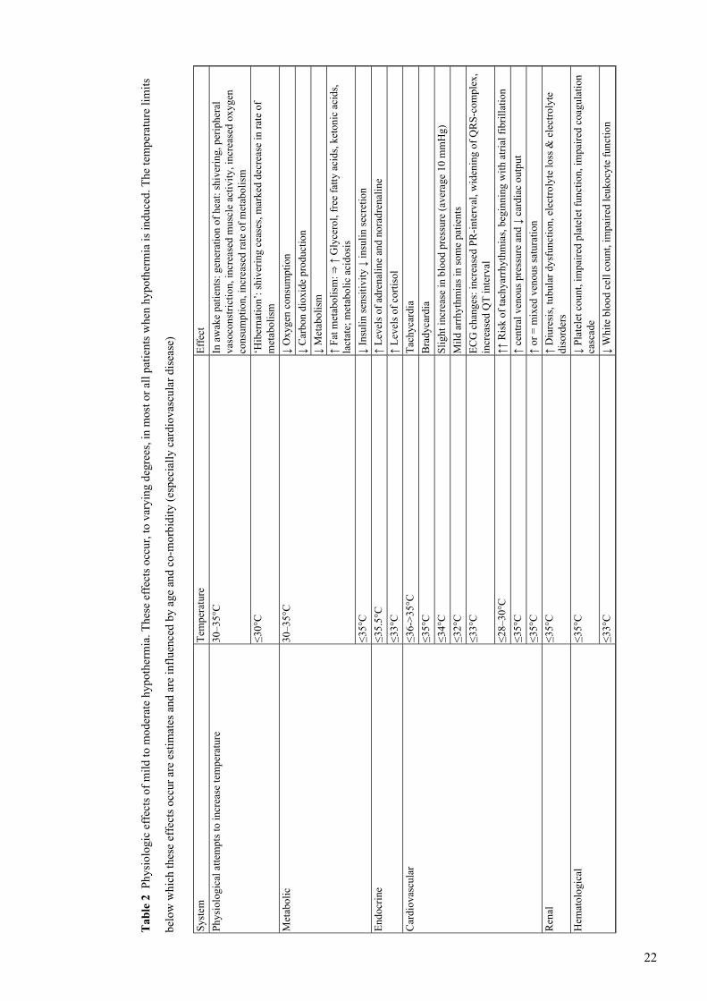
These changes are listed in Tables 2, 3 and 4.
[Table 2 will appear here. See end of document.]
[Table 3 will appear here. See end of document.]
[Table 4 will appear here. See end of document.]
Hypothermia leads to a lowering of the metabolic rate. Indeed, in the past it was assumed that
the protective effects of hypothermia were due solely to the slowing of cerebral metabolism, with
associated decreases in consumption of glucose and oxygen. It has since become clear that other
mechanisms are involved, which probably play a much greater role than the changes in metabolic
rate. These issues are discussed in Part 1 of this review. Nevertheless, the effects on metabolism
are significant and probably do play a part in providing neuroprotection. In addition, these changes
4
in metabolism occur in all organ systems; this means, for example, that there will be a decrease
in oxygen consumption and carbon dioxide production (which implies that ventilator settings
should be adjusted), a reduction in feeding requirements, etc. Metabolism is reduced by between
5% and 7% per Celsius degree reduction in body temperature [7, 8, 9]. Cerebral blood flow is also
decreased, but, when corrected for the decrease in metabolism, the net result is a relative increase.
Many hypothermia-induced metabolic changes occur relatively quickly, within the first few
hours. These include changes in energy metabolism and decreases in adenosine tri-phosphate
(ATP) demand. Other changes, such as a rise in lactate levels, occur over a longer period of time
(>3 h). Induction of hypothermia also leads to an increase in membrane stability, with decreased
permeability of cellular membranes, the blood-brain barrier and blood vessel walls [10, 11, 12,
13, 14]. One of the consequences of this is a decrease in edema formation, that appears to be one
of the ways in which hypothermia can protect against neurological injury. In addition, hypothermia
can prevent or mitigate the excessive influx of Ca2+ into the cell, as well as decrease accumulation
of the excitatory neurotransmitter glutamate in the extracellular space [15]. Calcium influx and
glutamate accumulation are key elements in the destructive cascade that can follow a period of
ischemia; calcium influx into the cell can lead to mitochondrial dysfunction and the activation of
various enzymes which can cause additional cell injury and death [15]. Hypothermia also leads
to a decrease in intracellular acidosis (although the extracellular pH usually decreases slightly
during cooling, due to increased levels of lactic acid, glycerol, free fatty acids and ketonic acids;
see below).
Hypothermia also influences the immune system, with an inhibition of neutrophil and macrophage
function, suppression of inflammatory reactions and inhibition of the release of pro-inflammatory
cytokines [16, 17, 18]. This effect on immune response may contribute to hypothermia’s
neuroprotective effects, but, of course, increases the risk of infections (see below). Other
anti-inflammatory mechanisms include the prevention or mitigation of reperfusion-related DNA
injury, lipid peroxidation and leukotriene production as well as a decrease in the production of
nitric oxide [19, 20]. In addition, hypothermia decreases reperfusion injury and free radical
production [19].
Practical aspects and side effectsInduction of hypothermia induces a large number of physiological changes in the circulatory and
respiratory systems, coagulation system, drug metabolism, etc. (listed in Table 2). For the successful
5

use of hypothermia, awareness of these physiological effects and pathophysiological mechanisms
is of key importance. The failure to demonstrate positive effects of hypothermia in some clinical
trials may be partly due to insufficient regard for side effects causing the (partial) negation of
protective effects. In addition, unawareness of hypothermia’s physiological consequences may
lead to over-treatment. For example, even mild hypothermia induces decreases in cardiac output,
mild acidosis, a rise in lactate levels and a moderate increase in levels of amylase. These changes
are normal, do not signify any deterioration in the patients’ condition and do not require treatment.
Naturally, such changes can sometimes be unwanted, such as shivering with its associated rise in
oxygen consumption and patient discomfort. Many of these physiological effects can be counteracted
by appropriate medication, such as sedatives, analgesics or paralyzers. The use of therapeutic
hypothermia will usually require ICU admission and monitoring and often (but not always) sedation
and intubation.
The physiological and pathophysiological effects of cooling largely depend on the degree of
hypothermia. For example, a significant risk for severe arrhythmias occurs only at temperatures
below 28–30°C. Such low temperatures are now rarely employed in induced hypothermia, although
they are used more frequently in specific surgical procedures, such as major vascular surgery. This
review will focus on the effects of mild to moderate hypothermia (31–35°C).
The physiological adaptations to hypothermia, changes in laboratory values and potential side
effects are listed in Tables 2, 3 and 4. These changes depend to varying degrees on the patients’
age, underlying disease, co-morbidity etc. Some of these changes can be suppressed or prevented
by medication, appropriate sedation or other factors.
Cardiovascular and hemodynamic effects
Hypothermia is initially associated with sinus tachycardia, after which bradycardia develops. This
is partly due to decreases in metabolism and partly to the direct effects of hypothermia on the
heart. Various ECG changes may occur (listed in Table 2). The risk of arrhythmias during mild
or moderate hypothermia is very low, but increases significantly when the temperature drops below
30°C. The initial arrhythmia is usually atrial fibrillation, which can be followed (at temperatures
≤28°C) by the risk of ventricular flutter or fibrillation. An additional problem is that arrhythmias
in deeply hypothermic patients are difficult to treat, as the myocardium becomes less responsive
to defibrillation and anti-arrhythmic drugs. When therapeutic hypothermia is applied, therefore,
great care should be taken to keep temperatures at 30°C or more, as the risk of clinically significant
arrhythmias increases exponentially below this temperature level.
6

Initially, the induction of mild hypothermia increases myocardial oxygen demand relative to
supply; the mechanism is probably a hypothermia-induced increase in plasma levels of adrenaline
and noradrenaline leading to an increase in cardiac output and oxygen demand [21]. With further
reductions in temperature, decreases in heart rate and the slowing of metabolism will reduce cardiac
afterload and oxygen demand. Mild hypothermia decreases cardiac output by about 25% and leads
to increased vascular resistance and a rise in central venous pressure. During severe hypothermia
(≤30°C) left ventricular contractility itself may decrease, inducing systolic and diastolic dysfunction.
In healthy subjects mild hypothermia (35.5°C) has been shown to increase coronary perfusion [21,
22]. However, one study reported that, in patients with pre-existent coronary artery disease, coronary
vasoconstriction may occur during hypothermia [22]. This difference is presumed to be caused
by endothelial dysfunction associated with atherosclerosis [21]. This would imply that there is a
theoretical risk of myocardial injury during the induction of mild hypothermia in patients with
cardiovascular disease, especially in the phase when cooling is initiated and the heart rate
temporarily increases.
On the other hand, there is strong evidence from animal studies that the induction of hypothermia
during or following myocardial infarction can decrease the infarct size [23, 24, 25, 26, 27, 28, 29,
30]. Hypothermia has been used in one clinical study in 42 patients with acute myocardial infarction
undergoing emergency percutaneous coronary intervention [31]. Twenty-one patients were treated
with hypothermia for 3 h after reperfusion, the other patients served as controls. The hypothermia
group had a trend to smaller infarct sizes and fewer major adverse cardiac events, though these
differences did not reach statistical significance in this small number of patients. Although firm
conclusions regarding the benefits for cases of myocardial infarctions cannot yet be drawn, these
data do at least suggest that hypothermia did not adversely effect outcome in these patients with
coronary artery disease.
Coagulation
Hypothermia induces a mild bleeding diathesis, with increased bleeding time due to its effect on
platelet count [32, 33], platelet function [32, 33, 34], the kinetics of clotting enzymes and
plasminogen activator inhibitors [35, 36] and other steps in the coagulation cascade [36, 37, 38].
It should be pointed out that the laboratory results of standard coagulation tests such as prothrombin
time and partial thromboplastin times will remain normal, because these tests are usually performed
at 37°C in the lab. Tests will be prolonged only if they are performed at the patient’s actual core
temperature [39]. However, in spite of the above-mentioned abnormalities, the risk of significant
7

bleeding is very low, even in patients with traumatic brain injury (TBI) [40]. None of the clinical
trials in patients with TBI, subarachnoid hemorrhage, stroke or post-anoxic coma have reported
increased intracranial bleeding associated with cooling. These observations are confirmed by data
from animal experiments showing decreased extravasation of hemoglobin during hypothermia
[12]. Overall, few bleeding complications were seen in any of the major clinical trials using
hypothermia, and risks of bleeding should therefore not preclude the use of hypothermia if deemed
appropriate. Platelets and/or fresh frozen plasma can be administered to improve coagulation if
necessary.
Coagulation disorders may be a greater problem in trauma patients. Here the use of therapeutic
hypothermia is somewhat controversial and a potential conflict between ‘protecting the brain and
protecting the body’ may arise. Various studies have reported an association between hypothermia
and adverse outcome in trauma patients [41, 42, 43]; this link has given hypothermia an ominous
reputation among trauma surgeons and has led to recommendations of the aggressive re-warming
of trauma patients. However, its should be pointed out that most of these studies were uncontrolled
and retrospective, and in most cases no multivariate analysis was performed to correct for potential
confounders [review of this issue: 44]. One study that did perform multivariate analysis, correcting
for factors such as presence of shock (associated with both hypothermia and adverse outcome,
and therefore a potential confounder) concluded that hypothermia is a marker, but not a cause, of
adverse outcome [45]. Thus, although hypothermia does induce a degree of coagulopathy, its
reputation in trauma patients may be partly undeserved; the use of therapeutic hypothermia in
trauma patients should, therefore, not be automatically excluded. This view is underscored by
observations that active re-warming of hypothermic patents with TBI may adversely affect outcome
[46]. We therefore recommend that the use of hypothermia be considered in trauma patients who
meet inclusion criteria as set out in Part 1 of this review (for example, patients who have undergone
CPR with unclear neurological outcome) provided they do not have active bleeding and are
hemodynamically stable.
Infection
Evidence from clinical and in vitro studies shows that hypothermia can impair immune function.
Indeed, (as discussed above) inhibition of inflammatory responses may be one of the mechanisms
through which hypothermia exerts neuroprotective effects. Hypothermia inhibits the release of
various pro-inflammatory cytokines [16, 17] and suppresses chemotactic migration of leukocytes
and phagocytosis [47]. Hypothermia-induced insulin resistance and hyperglycemia may further
8

increase infection risks (see below). Thus, there are plausible mechanisms for an immunosuppressive
effect of hypothermia.
A number of studies, mostly in patients with stroke or TBI, have indeed reported higher risks
of pneumonia when therapeutic hypothermia is used over longer periods of time (≥48–72 h) [48,
49]. However, other studies using hypothermia for prolonged periods in patients with TBI reported
no increase in infection rates [50, 51]. This may be attributable to antibiotic prophylaxis or selective
decontamination of the digestive tract (SDD), which were used in some of these studies [51].
Short-term cooling (≤24 h) does not appear to increase the risk of infection [50, 52, 53]. Overall,
the risk of respiratory tract infections appears to increase when patients are cooled for 48 h or
more; this problem appears manageable with rigorous surveillance and, perhaps, prophylactic
measures.
Some studies have also reported a higher risk of wound infections associated with hypothermia
[54, 55]. This may be related to both diminished leukocyte function and hypothermia-induced
vasoconstriction. Animal studies have shown that the establishment of infection probably occurs
within the first 3 h of bacterial inoculation [55, 56, 57] and is facilitated by local vasoconstriction
and hypoperfusion. This may be important in patients requiring surgery during treatment with
hypothermia. Moreover, other wounds, including bed sores and catheter insertion sites, are more
likely to show progression and/or impaired healing during cooling.
Hypovolemia, fluid balance and electrolytes
The induction of hypothermia can lead to the loss of significant amounts of fluids, due to so-called
hypothermia-induced diuresis [58, 59, 60, 61]. This may be especially pronounced in patients with
TBI, in whom diabetes insipidus, induced by cranial trauma, and administration of medication
such as mannitol may exacerbate fluid losses [51, 59, 60]. The impact of this may be significant,
especially in patients with TBI or subarachnoidal hemorrhage (SAH) where even very brief episodes
of hypovolemia or hypotension can significantly, and adversely, affect outcome [62, 63, 64].
Indeed, any beneficial effects of hypothermia may be lost due to side effects if these are not treated
proactively and vigorously [58]. Therefore, close attention should be paid to the patients’ diuresis
and fluid balance especially during induction of hypothermia (i.e., the phase when the patients’
body temperature is decreasing, which is the phase when excessive diuresis and hypovolemia are
most likely to occur [51, 58, 59]). In our center we infuse 500–1000 ml of saline and electrolytes
(see below) in TBI patients upon initiation of cooling (provided the patients are young and have
no significant counter-indications) and supplement fluid losses that occur during cooling [51, 58,
9

59]. However, these problems are much less evident in other categories of patients, such as those
with post-anoxic coma following CPR [52, 53, 65]. The reason for this difference is probably that
the risk of excessive fluid loss in TBI patients is caused by a combination of hypothermia and
other factors, such as the administration of mannitol to decrease intracranial pressure.
Another important problem is induction of electrolyte disorders. We and others have observed
severe electrolyte disorders (i.e., low levels of Mg, K, P and Ca) during cooling of patients with
TBI [59, 66]. Such electrolyte disorders can cause cardiac arrhythmias as well as hypotensive
episodes with decreases in cerebral blood flow. Magnesium may be especially important in this
regard, because of its specific role in mitigating neurological injuries [67, 68, 69, 70, 71].
Intracellular free magnesium in the brain declines by up to 60% following moderate traumatic
brain injury in rats [72]. Numerous animal studies have shown that magnesium depletion leads to
significantly worse outcomes in experimental TBI; administration of magnesium before or even
after trauma substantially mitigates secondary injury and reduces the loss of cortical cells [67, 68,
69, 73, 74, 75, 76]. Magnesium may also play a role in the prevention of reperfusion injury, which
is one of the key mechanisms underlying secondary neurological injury [76]. In addition, loss of
magnesium is associated with vasoconstriction of cerebral and coronary arteries [77, 78, 79].
Clinical studies in ICU patients have shown that hypomagnesemia is associated with adverse
outcome [80]. Severe head injury itself is associated with significant loss of electrolytes including
magnesium [71]; thus many patients with TBI have hypomagnesemia at admission [71], which
subsequently can be significantly exacerbated by the induction of hypothermia [59]. Electrolyte
disorders are easily treated or prevented; physicians utilizing induced hypothermia should be aware
of these risks. It should be noted that serum levels do not always accurately reflect magnesium
status [79], and in our opinion magnesium levels should be maintained in the high or high-normal
range in all patients with neurological injury [70]. Other electrolytes, such as phosphorus and
potassium, should also be monitored closely and maintained in the high-normal range [70].
Other metabolic effects
Hypothermia decreases insulin sensitivity and insulin secretion, which can lead to hyperglycemia.
Hyperglycemia is associated with increased infection rates, increased incidence of renal failure
and critical illness neuropathy and various other complications, while prevention of hyperglycemia
and tight control of glucose levels may decrease morbidity and mortality in ICU patients [81].
These protective effects are due to the prevention of hyperglycemia per se rather than to the direct
effects of insulin [82, 83]. This underscores the importance of tight glucose regulation, especially
10

in patients treated with hypothermia in whom hyperglycemia is more likely to develop. The amounts
of insulin required to maintain glucose levels within the normal range are likely to increase during
the induction of hypothermia and physicians applying hypothermia should be aware of this
phenomenon.
Hypothermia also induces mild acidosis through various mechanisms including increased
synthesis of glycerol, free fatty acids, ketonic acids and lactate. These changes are normal metabolic
consequences of hypothermia and should not be attributed to complications such as bowel ischemia.
Shivering
As discussed in “Physiology and mechanisms”, the body will employ various mechanisms to
generate heat, including shivering which may increase oxygen consumption and patient discomfort.
In ventilated patients this can be counteracted by the administration of sedatives and analgesics
or, if deemed appropriate, the administration of muscle paralyzers. Shivering can be attenuated
by relatively small doses of opiates; meperidine (pethidine) appears to be somewhat more effective
in this regard due to a higher activity at the kappa receptor [15]. This means that lower doses
(12.5–25 mg) can be used, which may be especially important if hypothermia is used in awake
patients. When using paralyzers and/or opiates is deemed undesirable, alternatives with which to
treat shivering include the administration of clonidine, neostigmine and ketanserine. However,
care should be taken to avoid adverse effects; for example, clonidine may aggravate
hypothermia-induced bradycardia.
Miscellaneous
Another important issue is the effect of hypothermia on drug metabolism and pharmacokinetics.
The enzymes that metabolize most drugs are highly temperature-sensitive and, thus, drug
metabolism is significantly affected by hypothermia. Clearance of various drugs is decreased and
in most patients doses should be lowered during hypothermia. Unfortunately, few data are available
regarding the effects of hypothermia on the metabolism of specific drugs. However, the studies
that have been carried out confirm the expectation that plasma levels increase and the effects of
drugs are prolonged. For example, plasma levels of propofol increase by approximately 30% and
of fentanyl by 15% when individuals are 3°C hypothermic [55]. A number of the medications for
which data are available are listed in Table 2.
11

Cooling methods and practical guidelines
Methods to induce hypothermia
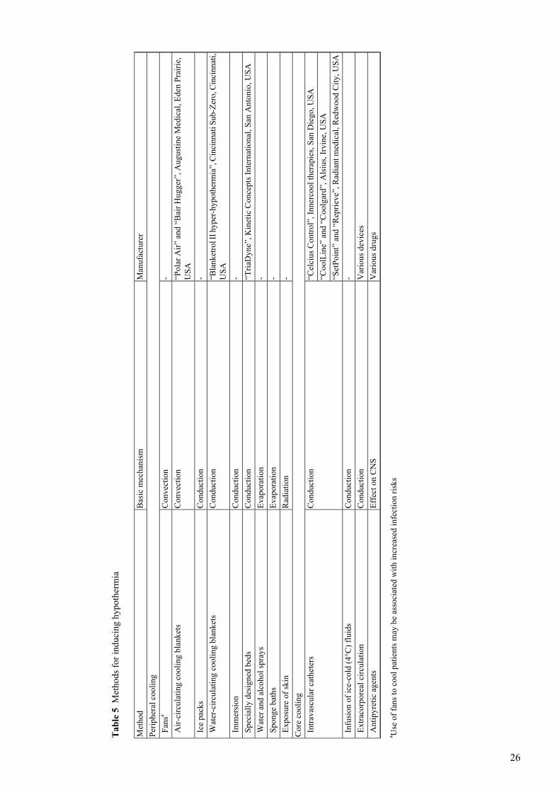
There are numerous strategies to cool patients, based on the four basic mechanisms for heat loss:
convection, conduction, evaporation and radiation. In addition, heat generation in patients with
hyperthermia can sometimes be reduced by antipyretic agents. However, in patients with elevated
temperature caused by impaired thermoregulation (such as central fever or heat stroke) these agents
are often ineffective. Various cooling techniques have been used in in vitro and clinical studies,
including ice-water circulating blankets, ice bags, air mattresses, cooling catheters, intravenous
infusion of cooled fluids (4°C) followed by cooling through other methods, the infusion of
extracorporeally cooled blood via the carotid artery, helmets and cooling caps with cold fluids or
chemical cooling capabilities, ice-water nasal lavage, cold peritoneal lavage and cardiopulmonary
bypass [31, 84, 85, 86, 87, 88, 89, 90, 91, 92, 93]. The methods most commonly employed in the
clinical setting are summarized in Table 5. Cooling caps and coils wrapped around the head have
been used mainly in infants and neonates, but have also been tried in adults.
[Table 5 will appear here. See end of document.]
Most large clinical trials published so far have used either water-cooling or air-cooling blankets.
Air-cooling blankets have also been used in general wards in awake patients [94, 95]. Water-cooling
blankets are much more efficient for cooling than for warming patients, because the temperature
difference can be set much higher; during warming a set temperature above 40–42°C can cause
burns, whereas the skin is much more tolerant of lower temperatures. As explained in “Physiology
and mechanisms”, the speed of inducing hypothermia may be important in achieving optimum
effects. The times required to achieve target temperatures have varied considerably in the clinical
trials published so far, ranging from approximately 2 h [51, 52] to around 8 h [46, 53]. These time
periods depend on patient factors (nature of the underlying disease or injury, age, sex, BMI),
countermeasures to prevent shivering and heat generation, and on technical aspects such as the
cooled surface, temperature of the blankets or air, cooling capacity, etc. In a recently published
study in patients with TBI we were able to induce temperatures of 34°C or below in 95% of our
patients within 2 h, by using two cooling blankets (above and below the patient) with the water
temperature set at 4°C until the core temperature was 33°C or less (the target temperature being
32°C), and by using water and alcohol sprays and exposing the areas of skin that were not directly
12

cooled [51]. Heat transfer from core to periphery may also be facilitated by the use of vasodilatory
medication.
An even quicker method was described in a preliminary report by Bernard et al. [96], who used
large volumes (30 ml/kg) of ice-cold (4°C) intravenous fluid (lactated Ringer’s solution) to cool
22 comatose survivors of out-of-hospital cardiac arrest quickly. These authors were able to decrease
core temperature from 35.5 to 33.8°C within 30 min with no adverse consequences, and concluded
that this was an inexpensive and effective method of initially inducing mild hypothermia. We have
used this method in selected cases in our own clinic with good results and no adverse effects.
A new development is the availability of intravascular cooling catheters such as the CoolLine,
SetPoint and others (Table 5). These are central lines with two or three balloons filled with
temperature-controlled saline, allowing direct intravascular (and, therefore, core) cooling via the
subclavian, superior caval or femoral vein. Experience with these catheters has been relatively
limited so far, although they are rapidly gaining in popularity. Initially, two small feasibility trials
in six [90] and eight patients [91] reported that it was an effective, relatively quick method to
induce and maintain hypothermia, and that it was less labor-intensive than the ‘conventional
methods’ listed above. Of note, no thrombus formation on these catheters was observed upon
removal in these studies. Endovascular cooling has also been used to induce brief periods of
hypothermia in 20 non-sedated patients undergoing percutaneous coronary intervention with the
aim of reducing infarct size [31]. A larger trial using this device to cool 51 patients with
hyperthermia in a neurosurgical ICU has recently been published; the authors reported that the
device was safe and effective in inducing hypothermia [92]. However, no studies have yet been
published in which these devices have been used for longer-term (>24 h) cooling.
Yet more novel approaches include the use of selective brain cooling [86], peritoneal cooling
[88] and ice-water nose cooling [93]. Experience with these methods is limited to animal studies
and/or small case series [93]. Treatment with paracetamol or acetaminophen may serve as an
accessory method to lower temperature, especially in patients with hyperthermia, although their
effectiveness in patients with neurological injury is limited [94].
Practical guidelines
Therapeutic hypothermia can be used in various types of neurological injury and perhaps for other
indications, such as the prevention of reperfusion injury. The use of hypothermia will often require
intubation, mechanical ventilation, sedation and, at times, pharmacologic paralysis to prevent
shivering. A major problem induced by these measures is that they may significantly hamper
13
neurological assessment of the patient; thus patients should be carefully monitored for, for example,
the development of seizures, which may present much less clearly. As outlined in “Practical aspects
and side effects”, the induction of hypothermia can cause a number of side effects; however, many
of these can be prevented or attenuated. Fluid balance should be carefully monitored and
hypovolemia and hypotension avoided, especially in patients with TBI or SAH. Electrolyte disorders
(especially hypomagnesemia) and arrhythmias should be prevented, if necessary by the early or
prophylactic administration of anti-arrhythmic agents. This also applies to hyperglycemia, which
should be combated through intensive insulin therapy and frequent monitoring.
Infections should be prevented by early or even prophylactic treatment with antibiotics. Bleeding
complications can be avoided by the timely administration of platelets or fresh frozen plasma,
particularly if surgical interventions or invasive procedures are performed. From this perspective,
realizing hypothermia’s full therapeutic potential presents a great challenge to ICU physicians,
requiring first-rate quality in many aspects of intensive care. This applies not only to intensivists
but, in equal measure, to ICU nurses and others caring for critically ill patients in the ICU.
Hypothermia-induced vasoconstriction of the skin and increased risk of wound infections will
increase the risk of bed sores, as will the sedation and paralysis often required in these patients.
The risk of respiratory tract infections may increase, requiring extra vigilance and interventions
by the nursing staff, physiotherapist and others. Infections should be treated promptly and
aggressively. The use of selective decontamination of the digestive tract should be considered in
these patients.
The insertion sites of central venous catheters will require close monitoring and extra care. The
patient may become relatively unstable, especially in the cooling phase while the core temperature
is decreasing. Polyuresis, electrolyte disorders and hypotension may develop, especially in patients
with TBI; ventilator settings will need to be changed as oxygen demand and production of carbon
dioxide decrease due to the slowing of metabolism. Blood samples must be frequently drawn and
analyzed on site or sent for analysis; the patient must be closely monitored for any signs of shivering,
seizures etc. All this will, at least temporarily, increase the nursing work load. This is something
to keep in mind as various studies have demonstrated that the lack of nursing and medical staff
due to increased workload can adversely affect outcome [97].
The successful application of hypothermia thus requires a concerted team effort and the
difficulties involved, as well as potential side effects and risks, should not be underestimated.
Using this therapy requires vigilance, attentiveness and experience. ICUs considering the use of
therapeutic hypothermia should adopt strict guidelines and protocols, and provide training for ICU
14

physicians, nursing staff and other members of the ICU team. When hypothermia has been used
it is important that patients should not be re-warmed too quickly, as this may have adverse effects
especially in patients with TBI.
Summary and conclusionsA large body of evidence suggests that hypothermia can be used to prevent or limit damage to the
injured brain and spinal cord, and perhaps the heart, in selected categories of patients. It is important
to induce hypothermia as quickly as possible, as protection appears to be greater when cooling is
initiated early (although benefits have been reported even when cooling was initiated many hours
after injury). As shown in this review, the induction of hypothermia will affect every organ in the
body and it is important that ICU staff members are aware of this and are able to distinguish
physiological changes from pathophysiological side effects.
The successful application of hypothermia requires the use of strict protocols, vigilance by the
medical and nursing staff, and close attention to the prevention of side effects. The volume status
of patients treated with cooling should be monitored carefully, to prevent hypovolemia and
hypotension. Other measures should include the frequent monitoring of electrolyte levels to avoid
electrolyte disorders (especially hypomagnesemia) and prevention or early treatment of arrhythmias,
if necessary by early or prophylactic administration of anti-arrhythmic agents. In addition,
hyperglycemia should be avoided through intensive insulin therapy and frequent monitoring of
glucose levels, and infections should be prevented by early or prophylactic treatment. Bleeding
complications can be avoided by the administration of platelets or plasma if surgical interventions
or invasive procedures are required. Patients should be re-warmed slowly, as too rapid re-warming
can adversely affect outcome.
References1. Sessler DI (2000) Perioperative heat balance. Anesthesiology 92:578–594
2. Tikuisis P, Bell DG, Jacobs I (1991) Shivering onset, metabolic response and convective heat transferduring cold air exposure. J Appl Physiol 70:1996–2002
3. English MJM (2001) Physical principles of heat transfer. Curr Anaesth Crit Care 12:66–71. DOI10.1054/cacc.2001.0331
4. Matsukawa T, Sessler DI, Sessler AM, Schroeder M, Ozaki M, Kurz A, Cheng C (1995) Heat flow anddistribution during induction of general anesthesia. Anesthesiology 82:662–673
5. Frank SM, Fleisher LA, Olson KF, Gorman RB, Higgins MS, Breslow MJ, Sitzmann JV, Beattie C(1995) Multivariate determinates of early postoperative oxygen consumption: the effects of shivering,core temperature and gender. Anesthesiology 83:241–249
15

6. Horvath SM, Spurr GB, Hutt BK, Hamilton LH (1956) Metabolic cost of shivering. J Appl Physiol8:595–602
7. Milde LN (1992) Clinical use of mild hypothermia for brain protection: a dream revisited. J NeurosurgAnesthesiol 4:211–215
8. Small DL, Morley P, Buchan AM (1999). Biology of ischemic cerebral cell death. Prog CardiovascDis 42:185–207
9. Schaller B, Graf R (2003) Hypothermia and stroke: the pathophysiological background. Pathophysiology10:7–35
10. Fischer S, Renz D, Wiesnet M, Schaper W, Karliczek GF (1999) Hypothermia abolishes hypoxia-inducedhyperpermeability in brain microvessel endothelial cells. Brain Res Mol Brain Res 74:135–144
11. Chopp M, Knight R, Tidwell CD, Helpern JA, Brown E, Welch KM (1989) The metabolic effects ofmild hypothermia on global cerebral ischemia and recirculation in the cat: comparison to normothermiaand hyperthermia. J Cereb Blood Flow Metab 9:141–148
12. Kinoshita K, Chatzipanteli K, Alonso OF, Howard M, Dietrich WD (2002) The effect of braintemperature on hemoglobin extravasation after traumatic brain injury. J Neurosurg 97:945–953
13. Chi OZ, Liu X, Weiss HR (2001) Effects of mild hypothermia on blood-brain barrier disruption duringisoflurane or pentobarbital anesthesia. Anesthesiology 95:933–938
14. Huang ZG, Xue D, Preston E, Karbalai H, Buchan AM (1999) Biphasic opening of the blood-brainbarrier following transient focal ischemia: effects of hypothermia. Can J Neurol Sci 26:298–304
15. Siesjo BK, Bengtsson F, Grampp W, Theander S (1989) Calcium, excitotoxins and neuronal death inbrain. Ann NY Acad Sci 568:234–251
16. Kimura A, Sakurada S, Ohkuni H, Todome Y, Kurata K (2002) Moderate hypothermia delaysproinflammatory cytokine production of human peripheral blood mononuclear cells. Crit Care Med30:1499–1502
17. Aibiki M, Maekawa S, Ogura S, Kinoshita Y, Kawai N, Yokono S (1999) Effect of moderate hypothermiaon systemic and internal jugular plasma IL-6 levels after traumatic brain injury in humans. J Neurotraum16:225–232
18. Frank SM (2001) Consequences of hypothermia. Curr Anaesth Crit Care 12:79–86
19. Busto R, Globus MY, Dietrich WD, Martinez E, Valdés I, Ginsberg MD (1989) Effect of mildhypothermia on ischemia-induced release of neurotransmitters and free fatty acids in rat brain. Stroke20:904–910
20. Dempsey RJ, Combs DJ, Maley ME, Cowen DE, Roy MW, Donaldson DL (1987) Moderate hypothermiareduces postischemic edema development and leukotriene production. Neurosurgery 21:177–181
21. Frank SM, Satitpunwaycha P, Bruce SR, Herscovitch P, Goldstein DS (2003) Increased myocardialperfusion and sympathoadrenal activation during mild core hypothermia in awake humans. Clin Sci(Lond) 104:503–508
22. Nabel EG, Ganz P, Gordon JB, Alexander RW, Selwyn AP (1988) Dilation of normal and constrictionof atherosclerotic coronary arteries caused by the cold pressor test. Circulation 77:43–52
23. Hale SL, Dae MW, Kloner RA (2003) Marked reduction in no-reflow with late initiation of hypothermiain a rabbit myocardial infarct model. J Am Coll Cardiol 41 (6 Suppl B):381–382
24. Hale SL, Dae MW, Kloner RA (2003) Hypothermia during reperfusion limits ‘no-reflow’ injury in arabbit model of acute myocardial infarction. Cardiovasc Res 59:715–722
25. Hale SL, Kloner RA (2002) Elevated body temperature during myocardial ischemia/reperfusionexacerbates necrosis and worsens no-reflow. Coron Artery Dis 13:177–181
26. Hale SL, Kloner RA (1998) Myocardial temperature reduction attenuates necrosis after prolongedischemia in rabbits. Cardiovasc Res 40:502–507
27. Miki T, Liu GS, Cohen MV, Downey JM (1998) Mild hypothermia reduces infarct size in the beatingrabbit heart: a practical intervention for acute myocardial infarction? Basic Res Cardiol 93:372–383
28. Hale SL, Dave RH, Kloner RA (1997) Regional hypothermia reduces myocardial necrosis even wheninstituted after the onset of ischemia. Basic Res Cardiol 92:351–357
16
29. Hale SL, Kloner RA (1997) Myocardial temperature in acute myocardial infarction: protection withmild regional hypothermia. Am J Physiol 273(1 Pt 2):H220–227
30. Dae MW, Gao DW, Sessler DI, Chair K, Stillson CA (2002) Effect of endovascular cooling onmyocardial temperature, infarct size and cardiac output in human-sized pigs. Am J Physiol Heart CircPhysiol 282:H1584–1591
31. Dixon SR, Whitbourn RJ, Dae MW, Grube E, Sherman W, Schaer GL, Jenkins JS, Baim DS, GibbonsRJ, Kuntz RE, Popma JJ, Nguyen TT, O’Neill WW (2002) Induction of mild systemic hypothermiawith endovascular cooling during primary percutaneous coronary intervention for acute myocardialinfarction. J Am Coll Cardiol 40:1928–1934
32. Valeri CR, Feingold H, Cassidy G, Ragno G, Khuri S, Altschule MD (1987) Hypothermia-inducedreversible platelet dysfunction. Ann Surg 205:175–181
33. Michelson AD, MacGregor H, Barnard MR, Kestin AS, Rohrer MJ, Valeri CR (1994)Hypothermia-induced reversible platelet dysfunction. Thromb Haemost 71:633–640
34. Watts DD, Trask A, Soeken K, Perdue P, Dols S, Kaufmann C (1998) Hypothermic coagulopathy intrauma: effect of varying levels of hypothermia on enzyme speed, platelet function and fibrinolyticactivity. J Trauma 44:846–854
35. Valeri CR, MacGregor H, Cassidy G, Tinney R, Pompei F (1995) Effects of temperature on bleedingtime and clotting time in normal male and female volunteers. Crit Care Med 23:698–704
36. Patt A, McCroskey B, Moore E (1988) Hypothermia-induced coagulopathies in trauma (Review). SurgClin North Am 68:775–785
37. Ferrara A, MacArthur JD, Wright HK, Modlin IM, McMillen MA (1990) Hypothermia and acidosisworsen coagulopathy in the patients requiring massive transfusion. Am J Surg 160:515–518
38. Reed RL, Bracey AW, Hudson JD, Miller TA, Fischer RP (1990) Hypothermia and blood coagulation:dissociation between enzyme activity and clotting factor levels. Circ Shock 32:141–152
39. Rohrer MJ, Natale AM (1992) Effect of hypothermia on the coagulation cascade. Crit Care Med20:1402–1405
40. Resnick DK, Marion DW, Darby JM (1994) The effect of hypothermia on the incidence of delayedtraumatic intracerebral hemorrhage. Neurosurgery 34:352–356
41. Jurkovich GJ, Greiser WB, Luterman A, Curreri PW (1987) Hypothermia in trauma victims: an ominouspredictor of survival. J Trauma 27:1019–1024
42. Luna GK, Maier RV, Pavlin EG, Anardi D, Copass MK, Oreskovich MR (1987) Incidence and effectof hypothermia in seriously injured patients. J Trauma 27:1014–1018
43. Gentilello LM, Jurkovich GJ, Stark MS, Hassantash SA, O’Keefe GE (1997) Is hypothermia in thevictim of major trauma protective or harmful? Ann Surg 226:439–447
44. Tisherman SA, Rodriguez A, Safar P (1999) Trauma care in the new millennium. Therapeutichypothermia in traumatology. Surg Clin North Am 79:1269–1289
45. Steinemann S, Shackford SR, Davis JW (1990) Implications of admission hypothermia in traumapatients. J Trauma 30:200–202
46. Clifton GL, Miller ER, Choi SC, Levin HS, McCauley S, Smith KR, Muizelaar JP, Wagner FC, MarionDW, Luerssen TG, Chesnut RM, Schwartz M (2001) Lack of effect of induction of hypothermia afteracute brain injury. N Engl J Med 344:556–563
47. Salman H, Bergman M, Bessler H, Alexandrova S, Beilin B, Djaldetti M (2000) Hypothermia affectsthe phagocytic activity of rat peritoneal macrophages. Acta Physiol Scand 168:431–436
48. Shiozaki T, Hayakata T, Taneda M, Nakajima Y, Hashiguchi N, Fujimi S, Nakamori Y, Tanaka H,Shimazu T, Sugimoto H (2001) A multicenter prospective randomized controlled trial of the efficacyof mild hypothermia for severely head injured patients with low intracranial pressure. Mild HypothermiaStudy Group in Japan. J Neurosurg 94:50–54
49. Schwab S, Georgiadis D, Berrouschot J, Schellinger PD, Graffagnino C, Mayer SA (2001) Feasibilityand safety of moderate hypothermia after massive hemispheric infarction. Stroke 32:2033–2035
50. Marion DW, Penrod LE, Kelsey SF, Obrist WD, Kochanek PM, Palmer AM, Wisniewski SR, DeKoskyST (1997) Treatment of traumatic brain injury with moderate hypothermia. N Engl J Med 336:540–546
17

51. Polderman KH, Tjong Tjin Joe R, Peerdeman SM, Vandertop WP, Girbes ARJ (2002) Effects ofartificially induced hypothermia on intracranial pressure and outcome in patients with severe traumatichead injury. Intensive Care Med 28:1563-1567
52. Bernard SA, Gray TW, Buist MD, Jones BM, Silvester W, Gutteridge G, Smith K (2002) Treatmentof comatose survivors of out-of-hospital cardiac arrest with induced hypothermia. N Engl J Med346:557–563
53. The Hypothermia after Cardiac Arrest Study Group (2002) Mild therapeutic hypothermia to improvethe neurologic outcome after cardiac arrest. N Engl J Med 346:549–556
54. Kurz A, Sessler DI, Lenhardt R and the Study of Wound Infection and Temperature Group (1996)Perioperative normothermia to reduce the incidence of surgical-wound infection and shortenhospitalization. New Engl J Med 334:1209–1215
55. Sessler DI (2001) Complications and treatment of mild hypothermia. Anesthesiology 95:531–543
56. Sheffield CW, Sessler DI, Hunt TK (1994) Mild hypothermia during isoflurane anesthesia decreasesresistance to E. Coli dermal infection in guinea pigs. Acta Anesthesiol Scand 38:201–205
57. Sheffield CW, Sessler DI, Hunt TK, Scheuenstuhl H (1994) Mild hypothermia during halothaneanesthesia decreases resistance to S. Aureus dermal infection in guinea pigs. Wound Rep Reg 2:48–56
58. Polderman KH, Girbes ARJ, Peerdeman SM, Vandertop WP (2001) Hypothermia (review/comment).J Neurosurgery 94:853–855
59. Polderman KH, Peerdeman SM, Girbes ARJ (2001) Hypophosphatemia and hypomagnesemia inducedby cooling in patients with severe head injury. Journal of Neurosurg 94:697–705
60. Kaufman HH, Timberlake G, Voelker J, Pait TG (1993) Medical complications of head injury. MedClin North Am 77:43–60
61. Weinberg AD (1993) Hypothermia. Ann Emerg Med 22:370–377
62. The Brain Trauma Foundation. The American Association of Neurological Surgeons (2000) The JointSection on Neurotrauma and Critical Care. Guidelines for cerebral perfusion pressure. J Neurotrauma17:507–511
63. Fearnside MR, Cook RJ, McDougall P, McNeil RJ (1993) The Westmead Head Injury Project outcomein severe head injury. A comparative analysis of pre-hospital, clinical and CT variables. Br J Neurosurg7:267–279
64. Chesnut RM, Marshall SB, Piek J, Blunt BA, Klauber MR, Marshall LF (1993) Early and late systemichypotension as a frequent and fundamental source of cerebral ischemia following severe brain injuryin the Traumatic Coma Data Bank. Acta Neurochir Suppl (Wien) 59:121–125
65. Polderman KH, Sterz F, van Zanten ARH, Uray T, Losert H, de Waal R, Girbes ARJ, Holzer M (2003)Induced hypothermia improves neurological outcome in asystolic patients with out-of hospital cardiacarrest (abstract). Circulation 108:IV-581 [abstract 2646]
66. Aibiki M, Kawaguchi S, Maekawa N (2001) Reversible hypophosphatemia during moderate hypothermiatherapy for brain-injured patients. Crit Care Med 29:1726–1730
67. McIntosh TK Vink R, Yamakami I, Faden AI (1989) Magnesium protects against neurological deficitafter brain injury. Brain Res 482:252–260
68. Vink R (1988) Decline in intracellular free Mg2+ is associated with irreversible tissue injury after braintrauma. J Biol Chem 263:757–761
69. Vink R, Cernak I (2000) Regulation of intracellular free magnesium in central nervous system injury.Front Biosci 5:656–665
70. Polderman KH, Zanten ARH van, Girbes ARJ (2003) The importance of magnesium in critically illpatients: a role in mitigating neurological injury and in the prevention of vasospasms. Intensive CareMed 29:1202–1203
71. Polderman KH, Bloemers F, Peerdeman SM, Girbes ARJ (2000) Hypomagnesemia andhypophosphatemia at admission in patients with severe head injury. Crit Care Med 28:2022–2025
72. Dietrich WD, Alonso O, Busto R, Globus MYT, Ginsberg MD (1994) Posttraumatic brain hypothermiareduces histopathological damage following concussive brain injury in the rat. Acta Neuropathol87:250–258
18

73. Vacanti FX, Ames AA (1983) Mild hypothermia and Mg++ protect against irreversible damage duringCNS ischemia. Stroke 15:695–698
74. Saatman KE, Bareyre FM, Grady MS, McIntosh TK (2001) Acute cytoskeletal alterations and cell deathinduced by experimental brain injury are attenuated by magnesium treatment and exacerbated bymagnesium deficiency. J Neuropathol Exp Neurol 60:183–194
75. Bareyre FM, Saatman KE, Raghupathi R, McIntosh TH (2000) Postinjury treatment with magnesiumchloride attenuates cortical damage after traumatic brain injury in rats. J. Neurotrauma 17:1029–1039
76. Garcia LA, Dejong SC, Martin SM, Smith RS, Buettner GR, Kerber RE (1998) Magnesium reducesfree radicals in an in vivo coronary occlusion-reperfusion model. J Am Coll Cardiol 32:536–539
77. Pyne GJ, Cadoux-Hudson TA, Clark JF (2001) Magnesium protection against in vitro cerebral vasospasmafter subarachnoid haemorrhage. Br J Neurosurg 15:409–415
78. Teragawa H, Kato M, Yamagata T, Matsuura H, Kajiyama G (2000)The preventive effect of magnesiumon coronary spasm in patients with vasospastic angina. Chest 118:1690–1695
79. Weisinger JR, Bellorín-Font E (1998) Magnesium and phosphorus. Lancet 352:391–396
80. Rubeiz GJ, Thill-Baharozian M, Hardie D, Carlson RW (1993) Association of hypomagnesemia andmortality in acutely ill medical patients. Crit Care Med 21:203–209
81. Van den Berghe G, Wouters P, Weekers F, Verwaest C, Bruyninckx F, Schetz M, Vlasselaers D,Ferdinande P, Lauwers P, Bouillon R (2001) Intensive insulin therapy in critically ill patients. NewEngl J Med 345:1359–1367
82. Van den Berghe G, Wouters PJ, Bouillon R, Weekers F, Verwaest C, Schetz M, Vlasselaers D,Ferdinande P, Lauwers P (2003) Outcome benefit of intensive insulin therapy in the critically ill: insulindose versus glycemic control. Crit Care Med 31:359–366
83. Finney SJ, Zekveld C, Elia A, Evans TW (2003) Glucose control and mortality in critically ill patients.JAMA 290:2041–2047
84. Connolly J, Boyd R, Calvin J (1962) The protective effect of hypothermia in cerebral ischemia:experimental and clinical application by selective brain cooling in the human. Surgery 52:15–24
85. Schwartz AE, Stone JG, Finck AD, Sandhu AA, Mongero LB, Adams DC, Jonassen AE, Young WL,Michler RE (1996) Isolated cerebral hypothermia by single carotid artery perfusion of extracorporeallycooled blood in baboons. Neurosurgery 39:577–581
86. Gelman B, Schleien CL, Lohe A, Kuluz JW (1996) Selective brain cooling in infant piglets after cardiacarrest and resuscitation. Crit Care Med 24:1009–1017
87. Natale JA, D’Alecy LG (1989) Protection from cerebral ischemia by brain cooling without reducedlactate accumulation in dogs. Stroke 20:770–777
88. Xiao F, Safar P, Alexander H (1995) Peritoneal cooling for mild cerebral hypothermia after cardiacarrest in dogs. Resuscitation 30:51–59
89. O’Donnel J, Axelrod P, Fisher C, Lorber B (1997) Use and effectiveness of hypothermia blankets forfebrile patients in the intensive care unit. Clin Infect Dis 24:1208–1213
90. Georgiadis D, Schwarz S, Kollmar R, Schwab S (2001) Endovascular cooling for moderate hypothermiain patients with acute stroke. Stroke 32:2550–2553
91. Doufas AG, Akca O, Barry A, petrusca DA, Suleman MI, Morioka N, Guarnaschelli JJ, Sessler DI(2002) Initial experience with a novel heat-exchanging catheter in neurosurgical patients. Anesth Analg95:1752–1756
92. Schmutzhard E, Engelhardt K, Beer R, Brössner G, Pfausler B, Spiss H, Unterberger I, Kampfl A (2002)Safety and efficacy of a novel intravascular cooling device to control body temperature in neurologicintensive care patients: a prospective pilot study. Crit Care Med 30:2481–2488
93. Andrews PJD, Harris BA (2002) The rationale for selective brain cooling. Year book of intensive careand emergency medicine. Vincent JL (ed) Springer Verlag, Berlin Heidelbrg New York, pp 738–747
94. Mayer SA, Commichau C, Scarmeas N, Presciutti M, Bates J, Copeland D (2000) Clinical trial of anair-circulating cooling blanket for fever control in critically ill neurologic patients. Neurology 56:292–298
19

95. Kammersgaard LP, Rasmussen BH, Jørgensen HS, Reith J, Weber U, Olsen TS (2000) Feasibility andsafety of inducing modest hypothermia in awake patients with acute stroke through surface cooling: acase-control study: the Copenhagen Stroke Study. Stroke 31:2251–2256
96. Bernard S, Buist M, Monteiro O, Smith K (2003) Induced hypothermia using large volume, ice-coldintravenous fluid in comatose survivors of out-of-hospital cardiac arrest: a preliminary report.Resuscitation 56:9–13
97. Tarnow-Mordi WO, Hau C, Warden A, Shearer AJ (2000) Hospital mortality in relation to staff workload:a 4-year study in an adult intensive-care unit. Lancet 356:185–189
20

Tab
le 1 Mechanisms of heat loss
Influencing factors
Definition
Mechanism
Independent of temperature of surrounding air; depends on
temperature and emissivity of surrounding objects
Transfer of heat between the separated surfaces of two objects with
different temperatures via electromagnetic (infrared) radiation,
without direct contact between the objects and without a heat transfer
medium. Accounts for 50–70% of heat loss in awake patients
Radiation
Difference in surface temperatures; insulation between these surfaces
Direct transfer of heat from one surface to a second, adjacent surface.
Amount of heat loss is closely related to contact surface; in standing
Conduction
patients heat loss through conduction is negligible, but this increases
in the sitting or lying positions
Difference in temperature between surface and air; (speed of)
movement of air (“wind chill factor”)
Transfer of heat from a surface to the surrounding air. Accounts for
20–30% of heat loss at room temperature in the absence of wind
Convection
Influenced by saturated vapor pressure at skin, temperature & the
air
Heat loss derived from the evaporation of water from skin & lungs.
Accounts for ±15% of heat loss (5% from the skin, 10% from the
lungs) under non-sweating circumstances
Evaporation
21
Tab
le 2 Physiologic effects of mild to moderate hypothermia. These effects occur, to varying degrees, in most or all patients when hypothermia is induced. The temperature limits
below which these effects occur are estimates and are influenced by age and co-morbidity (especially cardiovascular disease)
Effect
Temperature
System
In awake patients: generation of heat: shivering, peripheral
vasoconstriction, increased muscle activity, increased oxygen
consumption, increased rate of metabolism
30–35°C
Physiological attempts to increase temperature
‘Hibernation’: shivering ceases, marked decrease in rate of
metabolism
≤30°C
↓ Oxygen consumption
30–35°C
Metabolic
↓ Carbon dioxide production
↓ Metabolism
↑ Fat metabolism: ⇒ ↑ Glycerol, free fatty acids, ketonic acids,
lactate; metabolic acidosis
↓ Insulin sensitivity ↓ insulin secretion
≤35°C
↑ Levels of adrenaline and noradrenaline
≤35.5°C
Endocrine
↑ Levels of cortisol
≤33°C
Tachycardia
≤36->35°C
Cardiovascular
Bradycardia
≤35°C
Slight increase in blood pressure (average 10 mmHg)
≤34°C
Mild arrhythmias in some patients
≤32°C
ECG changes: increased PR-interval, widening of QRS-complex,
increased QT interval
≤33°C
↑↑ Risk of tachyarrhythmias, beginning with atrial fibrillation
≤28–30°C
↑ central venous pressure and ↓ cardiac output
≤35°C
↑ or = mixed venous saturation
≤35°C
↑ Diuresis, tubular dysfunction, electrolyte loss & electrolyte
disorders
≤35°C
Renal
↓ Platelet count, impaired platelet function, impaired coagulation
cascade
≤35°C
Hematological
↓ White blood cell count, impaired leukocyte function
≤33°C
22

Impaired bowel function/impaired intestinal motility/ileus, mild
pancreatitis (occurs very frequently!) ↑ liver enzymes
≤35°C
Gastrointestinal
Impaired neutrophil and macrophage function; suppression of
pro-inflammatory mediator release; ⇒ increased risk of infection
(mainly pneumonia & wound infections)
≤35°C
Immune suppression
↓ Consciousness, lethargy, coma
≤30–31°C
Neurological
Altered clearance of various medications (data available for muscle
paralyzers, propofol, fentanyl, phenytoin, pentobarbital, verapamil,
≤35°C
Pharmacokinetics
propanol and volatile anesthetics (reduced clearance), but in all
likelihood applies to many other types of medication)
No effect on gentamycin clearance in animal experiment
No effect on neostigmine effect or clearance in healthy volunteers
23

Tab
le 3 Frequently occurring changes in laboratory measurements induced by hypothermia. The extent of these changes depends on the degree of hypothermia; the lower body
temperature, the more pronounced the changes in laboratory values will be
Effect
Frequency
Mild to moderate increase in serum amylase levels (300–600 µ/l)
Almost always
Mild thrombocytopenia (platelet count 100–150x1012 )
Increase in serum lactate levels (2.5–5 mmol/l)
Moderate to severe thrombocytopenia (platelet count 30–100x1012)
Frequent
Rise in serum glucose levels (due to decreased insulin sensitivity and decreased insulin secretion)
High serum amylase levels (600–1200 µ/l)
High serum lactate levels (5–7 mmol/l)
Decrease in levels of potassium (K), magnesium (Mg), phosphate (P), calcium (Ca)
Leukocytopenia (WBC (2–3x10⁹/l)
Mild increase in liver enzymes (particularly SGOT and SGPT)
Occurring regularly
Metabolic acidosis (due to increase in lactate levels and increased production of free fatty acids, ketones
and glycerol)
Slightly increased APTT and APTT
Manifest acidosis, lactate levels ≥7 mmol/l
Occurring occasionally
Severe leukocytopenia (WBC <2x10⁹ )
Increase in serum amylase ≥1200 µ/l
Severe thrombocytopenia (platelet count ≤30x1012 )
Manifest coagulation disorders with marked increase in APTT and PTT
24

Tab
le 4 Potential side effects of hypothermia. These are effects that occur relatively frequently during induction of hypothermia and that are often unwelcome. Many of these effects
can be prevented or counteracted, or the effects mitigated. Physicians applying therapeutic hypothermia should be aware of these potential side effects
Effect
Frequency/ degree of risk
Coagulopathy: increased bleeding time,increased APTT/CT,thrombocytopenia, thrombocytopathia
High risk
Impaired coagulation cascade
Electrolyte disordersa (loss of K, Mg, P, Ca)
Hypovolemia (due to increased diuresis/hypothermia-induced diuresis)b
Rise in serum amylase
Changes in drug effects & drug metabolism
Insulin resistance
Manifest bleeding, severe coagulation disorders (possibly higher risk in trauma patients and/or patients
who already have bleeding problems for other reasons; hypothermia-induced coagulopathy may
Low risk
increase extent and severity of bleeding in these cases. See discussion in “Practical aspects and side
effects”)
Airway infections
Wound infections and healing
Myocardial ischemia
Manifest pancreatitis
Rare
Intracerebral bleeding
a Depends on category of patients; higher risk in TBI and SAH, lower risk in post-anoxia/CPR
b Risk appears to be significantly higher in patients with TBI than in patients following CPR
25
Tab
le 5 Methods for inducing hypothermia
Manufacturer
Basic mechanism
Method
Peripheral cooling
-Convection
Fansa
“Polar Air” and “Bair Hugger”, Augustine Medical, Eden Prairie,
USA
Convection
Air-circulating cooling blankets
-Conduction
Ice packs
“Blanketrol II hyper-hypothermia”, Cincinnati Sub-Zero, Cincinnati,
USA
Conduction
Water-circulating cooling blankets
-Conduction
Immersion
“TriaDyne”, Kinetic Concepts International, San Antonio, USA
Conduction
Specially designed beds
-Evaporation
Water and alcohol sprays
-Evaporation
Sponge baths
-Radiation
Exposure of skin
Core cooling
“Celcius Control”, Innercool therapies, San Diego, USA
Conduction
Intravascular catheters
“CoolLine” and “Coolgard”, Alsius, Irvine, USA
“SetPoint” and “Reprieve”, Radiant medical, Redwood City, USA
-Conduction
Infusion of ice-cold (4°C) fluids
Various devices
Conduction
Extracorporeal circulation
Various drugs
Effect on CNS
Antipyretic agents
a Use of fans to cool patients may be associated with increased infection risks
26