application of ultrasound multimodal score in the ... · scale ultrasound, two-dimensional color...
TRANSCRIPT

ORIGINAL ARTICLE Open Access
Application of ultrasound multimodal scorein the assessment of endometrialreceptivity in patients with artificialabortionYan Jiao1,2, Nianyu Xue3, Xujuan Shui2, Caicha Yu2 and Chunhong Hu1*
Abstract
Background: This study aimed to evaluate the value and feasibility of ultrasound multimodal score in theevaluation of endometrial receptivity in patients with artificial abortion (AA).
Methods: Sixty-eight patients with AA (AA group) and 70 women of the childbearing age without any history ofabortion (control group) were recruited between January 2018 and December 2018. All subjects received theexamination of endometrium in the middle luteum phase (7–9 days after ovulation) with two-dimensional gray-scale ultrasound, two-dimensional color Doppler ultrasound, and three-dimensional ultrasound, and the quantitativescores were obtained and compared between two groups.
Results: The quantitative score of endometrial receptivity was 10.46 ± 2.99 in the AA group and 13.49 ± 2.21 in thecontrol group showing significant difference (p < 0.05).
Conclusions: Ultrasound multimodal quantitative scores can be used to evaluate the endometrial receptivity ofpatients with AA.
Keywords: Endometrial receptivity, Artificial abortion, Ultrasonic multimode, Quantitative scores
Key points
� Multimodal ultrasound can effectively evaluate someendometrium-related factors such as endometrialmorphology, blood flow, and peristaltic waves.
� The ultrasound multimodal scoring is based onsome features of the endometrium and may providea more comprehensive reflection of endometrialreceptivity.
� In the future, this scoring system has the potentialto assist the reproductive physician to moreaccurately predict the outcome of pregnancy andplan the treatment.
BackgroundIn recent years, the prevalence of artificial abortion is in-creasing and has become an important public healthproblem. Abortion can cause mechanical damages to theendometrium, which may lead to secondary infertility,seriously affecting the physical and mental health andquality of life [1]. Studies have shown that secondary in-fertility may be related to the alteration of endometrialreceptivity [2], and thus, it is important to evaluate theendometrial receptivity after artificial abortion. A varietyof studies have suggested that ultrasound can be used toassess the endometrial receptivity [3]. However, fewstudies were conducted to evaluate the endometrial re-ceptivity in patients with induced abortion by usingultrasound multimodal score [4]. This study aimed toevaluate the clinical value of ultrasound multimodalscore in the evaluation of endometrial receptivity in pa-tients with induced abortion.
© The Author(s). 2020 Open Access This article is distributed under the terms of the Creative Commons Attribution 4.0International License (http://creativecommons.org/licenses/by/4.0/), which permits unrestricted use, distribution, andreproduction in any medium, provided you give appropriate credit to the original author(s) and the source, provide a link tothe Creative Commons license, and indicate if changes were made.
* Correspondence: [email protected] of Radiology, The First Affiliated Hospital of SoochowUniversity, No. 188 Shizi Street, Suzhou 215006, ChinaFull list of author information is available at the end of the article
Insights into ImagingJiao et al. Insights into Imaging (2020) 11:29 https://doi.org/10.1186/s13244-020-0840-5

Materials and methodsGeneral characteristicsPatients who had not given birth but received artificialabortion of one to three times were recruited as the artifi-cial abortion (AA) group (n=68) between January 2018and December 2018. In addition, 70 subjects of the child-bearing age who had not given birth and no history of arti-ficial abortion were recruited as the control group in thesame period. The inclusion criteria were as follows: Pa-tients were ≥ 23 years, but ≤ 35 years; the menstrual cyclewas regular within 1 year (28–35 days); the thyroid func-tion was normal; patients were negative for abortion-related immune antibodies; the periodicity of sexual hor-mones was normal; the follicular hormone level was nor-mal in the early follicular phase; the chromosomes of thefather, the mother, and the infant were normal; ultrason-ography of the uterus, uterine appendages, and pelvisshowed normal; organic diseases and infectious diseaseswere excluded; there were no gynecological surgerieswithin prior 2months; there were no medications affect-ing the hormones; and the semen analysis showed normal.The signed informed consent was obtained from eachsubject. The height and weight were measured for the cal-culation of body mass index (BMI). On the day of ultra-sound examination, blood was collected to detect theestradiol, progesterone, testosterone, luteinizing hormone,prolactin, and follicle-stimulating hormone.
UltrasonographyDetermination of implantation window phaseThe follicles were examined by ultrasonography sincethe 10th day of the menstrual cycle. The size of follicleswas observed in the peri-ovulation phase until ovulation(follicle rupture or disappearance). If ovulation was notobserved, the examination was abandoned. Implantationwindow phase was defined as 7–9 days after ovulationwhen ultrasonography was performed. In AA group,ultrasonography was performed at 3 months after thelast artificial abortion.
Ultrasound examinationColor Doppler ultrasound system (GE Voluson E8) withvaginal probe (RIC6-12-D) at the frequency of 5–8MHzwas used for ultrasound examination. Patients lied in alithotomy position. The vaginal probe was wrapped witha condom, smeared with coupling agents, and thenplaced into the vagina. Measurement was done at the va-ginal fornix.
Two-dimensional gray-scale ultrasound modeMeasurement of endometrial thickness: The distance be-tween the interface of anterior wall muscle and the in-tima and the interface of posterior wall muscle and theintima was measured at 2 cm from the bottom of theuterine in the middle long axis view of the uterus. Meas-urement was done thrice and the mean was calculated.< 7 mm or > 14mm was given 1 point, 7–8 mm wasgiven 2 points, and 9–14mm was given 3 points.Observation of endometrial morphology: The echoes
of the endometrium was observed. Then, the endomet-rial patterns were divided into three types based on theGonen system: type A, trilaminar pattern (endometrialthree-layer pattern), consisting of hyperechoic outer andmiddle layers, hypoechoic inner layers, and evident echoat the intrauterine midline; type B, relatively homoge-neous hyperechoic endometrium, with unclear endomet-rial layers, obscure intrauterine midline echo, but clearinterface between endometrial and muscular layers; andtype C, homogeneous hyperechoic endometrium withoutintrauterine midline echo [5] (Fig. 1). Type C was given1 point, type B was given 2 points, and type A was given3 points.Observation of endometrial movement: In the middle
long axis view of the uterus, the position of the probe wasfixed, and the patient was asked to calmly breathe. Thetype and frequency (times/min) of endometrial wave peri-stalsis were recorded for 3min. Ljland et al. divided theendometrial peristalsis into 5 types: (1) positive wave, theperistaltic wave from the cervix to fundus; (2) negative
Fig. 1 Endometrial patterns based on the Gonen system. Type A: trilaminar pattern (endometrial three-layer pattern) consisting of hyperechoicouter and middle layers, hypoechoic inner layers, and evident echo at the intrauterine midline; type B: relatively homogeneous hyperechoicendometrium, with unclear endometrial layers, obscure intrauterine midline echo, but clear interface between endometrial and muscular layers;type C: homogeneous hyperechoic endometrium without intrauterine midline echo
Jiao et al. Insights into Imaging (2020) 11:29 Page 2 of 7

wave, the peristaltic wave from the fundus to the cervix;(3) static wave, the endometrium in a static state; (4) bidir-ectional wave, the endometrium at the fundus and the cer-vix contract simultaneously; and (5) local peristaltic wave,the wave with low amplitude, no obvious directionalityand rhythm [6]. Negative wave, static wave, bidirectionalwave, and local peristaltic wave were given 1 point, posi-tive wave < 1/min or > 3/min were given 2 points, andpositive wave 1–3/min were given 3 points.
Two-dimensional color Doppler ultrasound examinationObservation of blood flow distribution in the intima andsubintima: In the meddle long axis view of the uterus,the subintimal area was defined as the area 3 mm awayfrom the intimal edge, and the endometrial and suben-dometrial perfusion was detected with CDFI mode andclassified as 3 types: type I, blood flow is present in theendometrium and subendometrium; type II, blood flowis absent in the endometrium, but present in the suben-dometrium; and type III, blood flow is not observed inboth endometrium and subendometrium [7] (Fig. 2).Type III was given 1 point, type II was given 2 points,and type I was given 3 points.
Three-dimensional ultrasound examinationMeasurement of endometrial volume and vascularizationflow index: The energy Doppler blood flow imaging modewas used for measurement at a sensitive state. The branch-ing of endometrial blood flow was assessed. Then, the 3Dmode was set, VOCAL software was used for analysis, theendometrium was outlined, and then the endometrial
volume (EV) and vascularization flow index (VFI) were de-termined [3, 8–10]. EV < 2 cm3 was given 1 point, EV of2–4 cm3 was given 2 points, and EV > 4 cm3 was given 3points. Unmeasurable VFI was given 1 point, VFI < 0.24was given 2 points, and VFI > 0.24 was given 3 points.
Endometrial ultrasound scoreAs shown in Table 1, the endometrial thickness, endomet-rial type, endometrial peristalsis, endometrial and suben-docardial blood flow distribution, EV, and VFI werescored in each subject, and the sum of these scores in eachsubject was used as the ultrasound multimodal score.
Statistical analysisStatistical analysis was performed with SAS 9.4 (SAS Insti-tute Inc., Cary, NC, USA). Normal distribution of datawas tested with one-sample Kolmogorov-Smirnov test,and normal distribution was defined once p > 0.05 was ob-served. Data with normal distribution were compared withTukey test between two groups. Data with abnormal dis-tribution were compared with Kruskal-Wallis rank sumtest, followed by Dwass-Steel-Critchlow-Fligner (DSCF)test between two groups. A value of p < 0.05 was consid-ered statistically significant.
ResultsAll the data acquired in two groups showed normaldistribution.General characteristics: There were no significant dif-
ferences in the age, BMI, and other demographics be-tween two groups (p > 0.05) (Table 2).
Fig. 2 Endometrial blood flow patterns. Red lines indicate blood flow. Type I: blood flow is present in the endometrium and subendometrium;type II: blood flow is absent in the endometrium, but present in the subendometrium; type III, blood flow is not observed in both endometriumand subendometrium
Table 1 Criteria for endometrial ultrasound scoring
Parameter 1 point 2 points 3 points
Endometrial thickness (mm) < 7 or > 14 7–8 9–14
Endometrial type C B A
Endometrial peristalsis (per min) Negative wave or other types Positive < 1 or > 3 Positive 1–3
Endocardial and subendocardial blood flow distribution III II I
EV (cm3) < 2 2–4 > 4
VFI Immeasurable < 0.24 > 0.24
Jiao et al. Insights into Imaging (2020) 11:29 Page 3 of 7
Hormone levels in two groups: Significant differencesin the levels of progesterone, estradiol, testosterone,luteinizing hormone, prolactin, and follicle-stimulatinghormone were not observed between two groups (p >0.05) (Table 2).Endometrial ultrasound multimodal score: The ultra-
sound multimodal score was 10.46 ± 2.99 in the AAgroup and 13.49 ± 2.21 in the control group. The ultra-sound multimodal score in the AA group was markedlylower than in the control group (p < 0.05) (Table 2)(Figs. 3 and 4).
DiscussionArtificial abortion may inevitably damage the endometriumand other organs in the reproductive system [11, 12]. Inaddition, artificial abortion also has the potential to causesome complications such as secondary abortion, abnormalplacenta, premature delivery, and ectopic pregnancy [13].Therefore, endometrial repair is the most important forthe post-abortion rehabilitation. At present, the clinicalmanifestations such as time of postoperative bleeding,blood loss, and time to recovery of menstrual cycle as wellas the endometrial thickness are monitored for the
Table 2 Different parameters in two groups
Group n Age(years)
BMI(kg/m2)
Estradiol(ng/mL)
Progesterone(pg/mL)
Testosterone(nmol/L)
Luteinizing hormone(mIU/mL)
Prolactin(nmol/L)
Follicle-stimulatinghormone (mIU/mL)
Endometrial ultrasoundmultimodal score
AA 68 24.79 ± 4.47 21.30 ± 4.21 300.88 ± 41.06 8.81 ± 2.19 2.31 ± 0.55 68.17 ± 13.41 0.58 ± 0.15 14.59 ± 3.86 10.46 ± 2.99
Control 70 25.38 ± 3.48 21.72 ± 3.47 300.17 ± 32.00 8.69 ± 1.88 2.26 ± 0.47 69.16 ± 10.41 0.57 ± 0.11 15.69 ± 3.78 13.49 ± 2.21*
tP
0.8630.389
0.6390.524
0.1130.910
0.3450.731
0.5730.567
0.4830.630
0.4460.657
1.6910.093
6.7540.000
Note: *p < 0.05 vs AA group
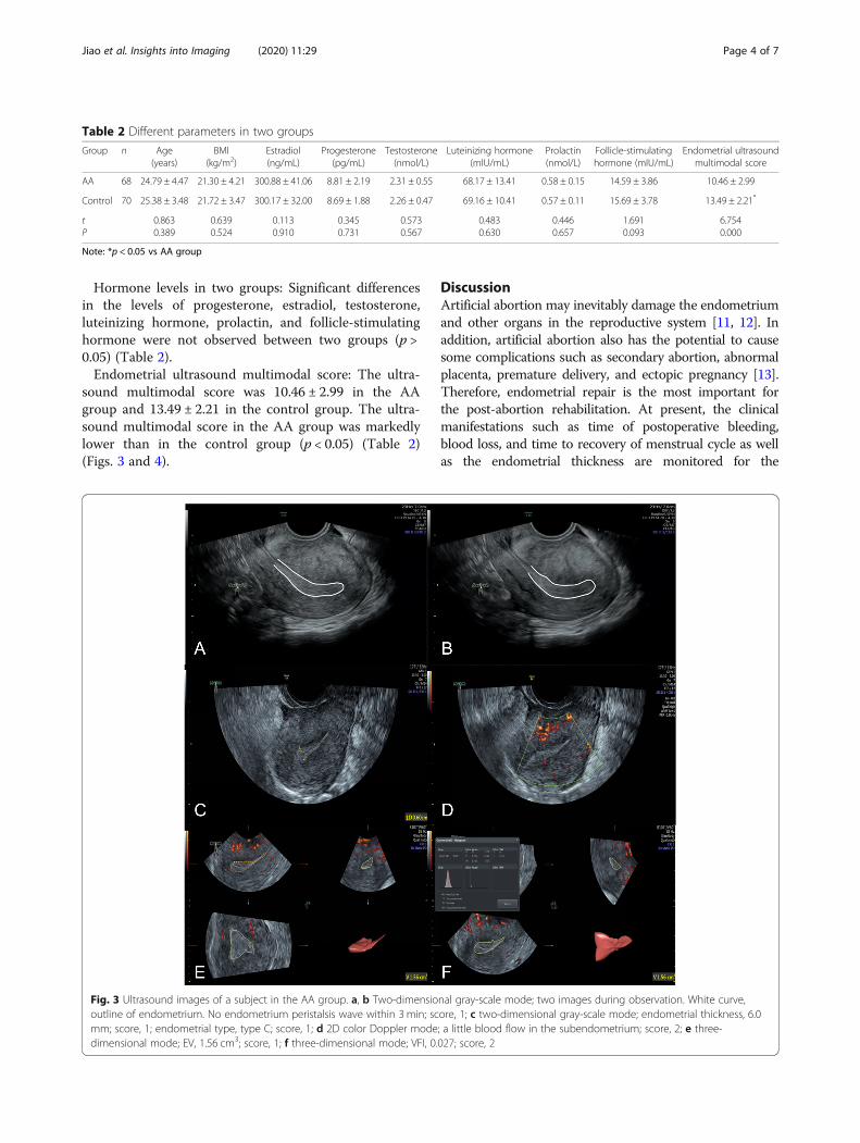
Fig. 3 Ultrasound images of a subject in the AA group. a, b Two-dimensional gray-scale mode; two images during observation. White curve,outline of endometrium. No endometrium peristalsis wave within 3 min; score, 1; c two-dimensional gray-scale mode; endometrial thickness, 6.0mm; score, 1; endometrial type, type C; score, 1; d 2D color Doppler mode; a little blood flow in the subendometrium; score, 2; e three-dimensional mode; EV, 1.56 cm3; score, 1; f three-dimensional mode; VFI, 0.027; score, 2
Jiao et al. Insights into Imaging (2020) 11:29 Page 4 of 7
assessment of endometrial repair, but these features havelimitations in the monitoring of disease condition and thediagnosis and treatment of diseases. In recent years, in-creasing attention has been paid to the role of recovery ofendometrial receptivity in the endometrial repair [4, 14–16]. Although many studies have been conducted to inves-tigate the endometrial receptivity, the methods in thesestudies are often different and there are no widely acceptedcriteria for the assessment of endometrial receptivity. Inthis study, a new ultrasound multimodal endometrial scor-ing system was developed based on the findings in previousstudies. The total score was 18 in our scoring system. Thehigher the score, the better the endometrial receptivity,and vice versa [17, 18]. Ultrasound multimodal score usedin this study included endometrial thickness, endometrialtype, endometrial peristaltic wave, blood flow, volume, andVFI, which can be easily acquired with present ultrasoundtechnology and universally accepted in clinical practices.Besides, every indicator was acquired with a universalmeasuring method, and the scoring of each indicator
adopted a universal standard. What is more, there werestill requirements for the operators that they have to beskilled to acquire the above indicators. Meanwhile, themeasuring method and scoring standard must be con-ducted according to the rules set in this study. This wasnot hard for experienced sonographer of gynecology.Above measures will reduce the influence of subjectivejudgement of sonographer on ultrasound multimodal scorefurthest.In the present study, results showed the endometrial
ultrasound multimodal score was 10.46 ± 2.99 in the AAgroup and 13.49 ± 2.21 in the control group. The endomet-rial ultrasound multimodal score in the AA group was sig-nificantly lower than in the control group. This indicatesthat, compared with normal controls, the endometrium ofpatients with artificial abortion have one or more abnor-malities (morphology, blood flow, and peristaltic wave),which reduces the total ultrasound multimodal score. Thismay be related to the pathological responses of the endo-metrium (significant reduction in the endometrial
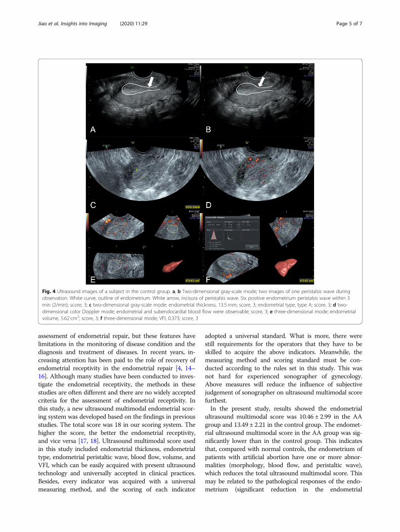
Fig. 4 Ultrasound images of a subject in the control group. a, b Two-dimensional gray-scale mode; two images of one peristalsis wave duringobservation. White curve, outline of endometrium. White arrow, incisura of peristalsis wave. Six positive endometrium peristalsis wave within 3min (2/min); score, 3; c two-dimensional gray-scale mode; endometrial thickness, 13.5 mm; score, 3; endometrial type, type A; score, 3; d two-dimensional color Doppler mode; endometrial and subendocardial blood flow were observable; score, 3; e three-dimensional mode; endometrialvolume, 5.62 cm3; score, 3; f three-dimensional mode; VFI, 0.373; score, 3
Jiao et al. Insights into Imaging (2020) 11:29 Page 5 of 7

glandular epithelium and marked increase in collagen fi-bers) after the mechanical injury to the endometrium [19–22]. These pathological responses may dramatically in-crease the proportion of collagen fibers in the endomet-rium and, as a result, the blood flow in the endometriumreduces, the endometrium becomes thinner, and the EVreduces. Studies have shown that the endometrial thick-ness, EV, VFI, and other parameters in abortion patientsare significantly lower than in normal pregnant subjects[23–25]. Most previous studies about endometrial receptiv-ity evaluated one or some of the indicators including endo-metrial thickness, endometrial type, endometrial peristalticwave, blood flow, volume, and VFI. Actually, each indicatorcould reflect different characteristics of endometrium, re-spectively. That is to say more indicators could evaluatethe endometrium more comprehensively. However, thereis still a need of considering feasibility in clinics. This studyadopted six typical indicators, not only fully reflecting theendometrium situation, but also not bringing difficulties toclinical practices. These indicators still had a relatively bigfeasibility and popularization possibility for not only ultra-sonic technique but also operators at present. In thepresent study, two-dimensional gray-scale ultrasound, two-dimensional color Doppler ultrasound, and three-dimensional ultrasound were employed simultaneously toassess the endometrial morphological, structure, and bloodflow, which is helpful for the comprehensive evaluation ofendometrial receptivity. This can effectively compensatefor the disadvantage of individual factor in the assessmentof endometrial receptivity. In addition, studies have indi-cated that endometrial receptivity is affected by the sexualhormones and age [26, 27]. In the present study, therewere no significant differences in the age and BMI betweenAA group and control group. In addition, marked differ-ences were also not observed in other hormones such asprogesterone, estradiol, testosterone, luteinizing hormone,prolactin, and follicle-stimulating hormone. This indicatesthat the difference in the endometrial receptivity betweenthe AA group and the control group is mainly ascribed tothe endometrial state, but not the age and hormones. Theultrasound modes used in this study have been well popu-larized in clinical practice. Therefore, the method used inthe present study is a simple, safe, non-invasive, and repro-ducible tool for the assessment of endometrial receptivityand deserves clinical properties [2].There were still limitations in this study. There are no
widely accepted criteria or consensuses on the multi-modal ultrasound assessment of endometrial receptivity.In this study, a new system was developed for the assess-ment of endometrial receptivity on the basis of previousfindings and it might be subjective in the developmentof this system. Therefore, it is necessary to further inves-tigate the clinical value and practicality and to improvethis scoring system.
ConclusionsIn summary, multimodal ultrasound can effectivelyevaluate some endometrium related factors such asendometrial morphology, blood flow, and peristalticwaves. The ultrasound multimodal scoring is based onsome features of the endometrium and may provide amore comprehensive reflection of endometrial receptiv-ity. In the future, this scoring system has the potential toassist the reproductive physician to more accurately pre-dict the outcome of pregnancy and plan the treatment.However, the relationship and interaction of various fac-tors are still unclear, and more studies are needed toconfirm our findings.
AbbreviationsAA: Artificial abortion; BMI: Body mass index; DSCF: Dwass-Steel-Critchlow-Fligner; EV: Endometrial volume; VFI: Vascularization flow index
Authors’ contributionsYJ and CH designed this study. YJ, NX, XS, CY, and CH collected andinterpreted the patient data. YJ analyzed the data. YJ was the majorcontributor in writing the manuscript. All authors read and approved thefinal manuscript.
FundingThis study was supported by the Basic and Public Studies of ZhejiangProvince (No. LGF18H180003) and the Health and Family PlanningCommission of Zhejiang Province, China (No. 2018ZD043, 2019KY665).
Availability of data and materialsThe datasets used and/or analyzed during the current study are availablefrom the corresponding author on reasonable request.
Ethics approval and consent to participateThis study was approved by the Ethics Committee of The First AffiliatedHospital of Soochow University.
Consent for publicationThe signed informed consent was obtained from each subject.
Competing interestsThe authors declare that they have no competing interests.
Author details1Department of Radiology, The First Affiliated Hospital of SoochowUniversity, No. 188 Shizi Street, Suzhou 215006, China. 2Obstetrics andGynecology Ultrasonic Department, Wenzhou Peoples’ Hospital, Wenzhou325000, China. 3Department of Diagnostic Ultrasonography, Ningbo FirstHospital, Ningbo 315010, China.
Received: 19 September 2019 Accepted: 30 January 2020
References1. Attali L (2016) Psychological aspects of abortion. J Gynecol Obstet Biol
Reprod (Paris) 45:1552–15672. Xu X, Li Z, Liu J, Yu S, Wei Z (2017) MicroRNA expression profiling in
endometriosis-associated infertility and its relationship with endometrialreceptivity evaluated by ultrasound. J Xray Sci Technol 25:523–532
3. Arya S, Kupesic Plavsic S (2017) Preimplantation 3D ultrasound: current usesand challenges. J Perinat Med 45:745–758
4. Gao Y, Hong X, Wang Z, Zhu Y (2018) Endometrial receptivity andconception outcome among women with light menstrual bleeding ofunidentified etiology. Int J Gynaecol Obstet 140:37–41
5. Yadav P, Singla A, Sidana A, Suneja A, Vaid NB (2017) Evaluation ofsonographic endometrial patterns and endometrial thickness as predictorsof ectopic pregnancy. Int J Gynaecol Obstet 136:70–75
Jiao et al. Insights into Imaging (2020) 11:29 Page 6 of 7

6. Kuijsters NP, Methorst WG, Kortenhorst MS, Rabotti C, Mischi M, Schoot BC(2017) Uterine peristalsis and fertility: current knowledge and futureperspectives: a review and meta-analysis. Reprod Biomed Online 35:50–71
7. Takahashi S, Komatsu S, Ohara T et al (2018) Detecting intimal tear andsubintimal blood flow of thrombosed acute aortic dissection with ulcer-likeprojections using non-obstructive angioscopy. J Cardiol Cases 18:164–167
8. Pandey H, Guruvare S, Kadavigere R, Rao CR (2018) Utility of threedimensional (3-D) ultrasound and power Doppler in identification of highrisk endometrial cancer at a tertiary care hospital in southern India: apreliminary study. Taiwan J Obstet Gynecol 57:522–527
9. Shuai Z, Lian F, Li P, Yang W (2015) Effect of transcutaneous electricalacupuncture point stimulation on endometrial receptivity in womenundergoing frozen-thawed embryo transfer: a single-blind prospectiverandomised controlled trial. Acupunct Med 33:9–15
10. Kim A, Jung H, Choi WJ, Hong SN, Kim HY (2014) Detection of endometrialand subendometrial vasculature on the day of embryo transfer andprediction of pregnancy during fresh in vitro fertilization cycles. Taiwan JObstet Gynecol 53:360–365
11. Gerdts C, Hudaya I (2016) Quality of care in a Safe-Abortion Hotline inIndonesia: Beyond Harm Reduction. Am J Public Health 106:2071–2075
12. Maged AM, Al-Inany H, Salama KM, Souidan II, Abo Ragab HM, Elnassery N(2016) Endometrial scratch injury induces higher pregnancy rate for womenwith unexplained infertility undergoing IUI with ovarian stimulation: arandomized controlled trial. Reprod Sci 23:239–243
13. Chae S, Desai S, Crowell M, Sedgh G (2017) Reasons why women haveinduced abortions: a synthesis of findings from 14 countries. Contraception96:233–241
14. Kojo T, Ae R, Tsuboi S, Nakamura Y, Kitamura K (2017) Differentials invariables associated with past history of artificial abortion and currentcontraception by age: results of a randomized national survey in Japan. JObstet Gynaecol Res 43:516–522
15. Miravet-Valenciano J, Ruiz-Alonso M, Gómez E, Garcia-Velasco JA (2017)Endometrial receptivity in eutopic endometrium in patients with endometriosis:it is not affected, and let me show you why. Fertil Steril 108:28–31
16. Altmae S, Koel M, Vosa U et al (2017) Meta-signature of human endometrialreceptivity: a meta-analysis and validation study of transcriptomicbiomarkers. Sci Rep 7:10077
17. Lessey BA, Kim JJ (2017) Endometrial receptivity in the eutopicendometrium of women with endometriosis: it is affected, and let me showyou why. Fertil Steril 108:19–27
18. Elsokkary M, Eldin AB, Abdelhafez M et al (2019) The reproducibility of thenovel utilization of five-dimensional ultrasound and power Doppler in theprediction of endometrial receptivity in intracytoplasmic sperm-injectedwomen: a pilot prospective clinical study. Arch Gynecol Obstet 299:551–558
19. Dueholm M, Hjorth IM, Dahl K, Hansen ES, Ørtoft G (2019) Ultrasoundscoring of endometrial pattern for fast-track identification or exclusion ofendometrial cancer in women with postmenopausal bleeding. J MinimInvasive Gynecol 26:516–525
20. Zhang XH, Liu ZZ, Tang MX, Zhang YH, Hu L, Liao AH (2015) Morphologicalchanges and expression of cytokine after local endometrial injury in amouse model. Reprod Sci 22:1377–1386
21. Wei A, Feng H, Jia XM, Tang H, Liao YY, Li BR (2018) Ozone therapyameliorates inflammation and endometrial injury in rats with pelvicinflammatory disease. Biomed Pharmacother 107:1418–1425
22. Yang JH, Chen CD, Chou CH et al (2019) Intentional endometrial injuryincreases embryo implantation potentials through enhanced endometrialangiogenesis. Biol Reprod 100:381–389
23. Tan SY, Hang F, Purvarshi G, Li MQ, Meng DH, Huang LL (2015) Decreasedendometrial vascularity and receptivity in unexplained recurrent miscarriagepatients during midluteal and early pregnancy phases. Taiwan J ObstetGynecol 54:522–526
24. Azumaguchi A, Henmi H, Ohnishi H, Endo T, Saito T (2017) Role of dilatationand curettage performed for spontaneous or induced abortion in theetiology of endometrial thinning. J Obstet Gynaecol Res 43:523–529
25. Baradwan S, Baradwan A, Al-Jaroudi D (2018) The association betweenmenstrual cycle pattern and hysteroscopic march classification withendometrial thickness among infertile women with Asherman syndrome.Medicine (Baltimore) 97:e11314
26. Check JH, Cohen R (2011) Live fetus following embryo transfer in a womanwith diminished egg reserve whose maximal endometrial thickness was lessthan 4 mm. Clin Exp Obstet Gynecol 38:330–332
27. Kasius A, Smit JG, Torrance HL et al (2014) Endometrial thickness andpregnancy rates after IVF: a systematic review and meta-analysis. HumReprod Update 20:530–541
Publisher’s NoteSpringer Nature remains neutral with regard to jurisdictional claims inpublished maps and institutional affiliations.
Jiao et al. Insights into Imaging (2020) 11:29 Page 7 of 7