applied surface science - university of · pdf filelasemi et al. / applied surface science 433...
TRANSCRIPT

F
Pl
Na
b
c
d
a
ARRAA
KLIMF
1
pcciptgFanopcb
tc
h0
Applied Surface Science 433 (2018) 772–779
Contents lists available at ScienceDirect
Applied Surface Science
journa l homepage: www.e lsev ier .com/ locate /apsusc
ull Length Article
ulsed laser ablation and incubation of nickel, iron and tungsten iniquids and air
. Lasemi a, U. Pacher a, L.V. Zhigilei a,b, O. Bomatí-Miguel a,c, R. Lahoz d, W. Kautek a,∗
University of Vienna, Department of Physical Chemistry, Währinger Strasse 42, A-1090 Vienna, AustriaUniversity of Virginia, Department of Materials Science & Engineering, PO Box 400745, Charlottesville, VA 22904-4745, USAAutonomous University of Madrid, Department of Applied Physics, Calle Francisco Tomás y Valiente 7, E-28049 Madrid, SpainCentro de Química y Materiales de Aragón, University of Zaragoza – CSIC, María de Luna 3, E-50018 Zaragoza, Spain
r t i c l e i n f o
rticle history:eceived 21 April 2017eceived in revised form 10 October 2017ccepted 11 October 2017vailable online 12 October 2017
eywords:
a b s t r a c t
Incubation effects in the nanosecond laser ablation of metals exhibit a strong dependence on the thermaland mechanical properties of both the target material and the background gas or liquid. The incubationin air is controlled mainly by thermal properties such as the heat of vaporization. In liquid, the correlationof the incubation and the ultimate tensile stress of the metals suggests that incubation may be related tothe mechanical impact on the solid material by the cavitation bubble collapse, causing accumulation ofvoids and cracks in the subsurface region of the ablation craters. At high ultimate tensile stress, however,
aser ablationncubation
etalsluid interfaces
the low sensitivity to the environment suggests that the mechanical impact is likely to play a negligiblerole in the incubation. Finally, the correlation between the incubation and the carbon content of alco-holic liquids may be explained by an absorptivity increase of the cavity surfaces due to carbonaceousdeposits generated by laser-induced pyrolysis, or by the mechanical impact of long-living bubbles athigher dynamic viscosity of liquids.
© 2017 Elsevier B.V. All rights reserved.
. Introduction
Laser processing in liquids has attracted attention because ofotential applications in surface cleaning, etching, welding, drilling,utting, and micromachining of metals, alloys, polymers, semi-onductors, glasses and ceramics [1,2]. Laser ablation of metalsn liquids enables the in-situ study of corrosion and repassivationrocesses [3,4] as well as the production of biocompatible nanopar-icles for medical and catalytic applications [5–7]. The pulsed lasereneration of colloidal metal nanoparticles (e.g., Ag, Au, Pt, Pd, Cu,e, Ni, W) in distilled water and organic solvents attracted muchttention in the past two decades [7–17]. The complex mecha-ism of laser ablation in liquids involves a series of steps extendedver many orders of magnitude in time and involving ablation,lasma expansion inside a cavitation bubble, the penetration ofondensed nano-sized phases into the liquid, as well as secondaryeam-colloid interaction [18–20].
The dependence of ablation rates and threshold fluences onhe number of laser pulses and the background medium, the so-alled incubation, has not been understood for metals in contrast
∗ Corresponding author.E-mail address: [email protected] (W. Kautek).
ttps://doi.org/10.1016/j.apsusc.2017.10.082169-4332/© 2017 Elsevier B.V. All rights reserved.
to dielectric materials. Therefore, this phenomenon needs furtherinvestigations as has been endeavoured in this work.
In order to evaluate ablation and incubation phenomena it isnecessary to correlate quantitative ablation and incubation datawith the physico-chemical properties of the target materials. In theregime of nanosecond-pulse laser interactions, the behaviour of theablation is generally controlled by optical properties and/or ther-mal conductivity of the target material. In this case, the thresholdfluence can be roughly estimated as follows:
F th ≈ �Uleff (1-R)−1, (1)
where �U is the energy density needed to heat, melt, and vaporizethe target material, R the reflectivity, and leff is the effective depthof energy deposition [21–23]. In the nanosecond laser interactionwith metals, leff is close to the heat diffusion length lT ≈ (��)0.5,where � is the laser pulse duration, � = �/(�cp) the thermal diffu-sivity of the material, � the thermal conductivity, � the density,and cp the heat capacity. The energy density �U can be approxi-mated by the heat of vaporization, Lv × � (Table 1), which accountsfor more than 95% of �U for any material considered in this work.
Nanosecond pulses show higher threshold values than sub-picosecond pulses [24–28]. Long pulse durations (≥ns) induceplasma shielding and scattering so that radiation is lost for theabsorption process [27], and/or heat diffusion into the bulk materi-

N. Lasemi et al. / Applied Surface Science 433 (2018) 772–779 773
Table 1Thermal and physical constants of various metals [65]. Tv: boiling point; Tm: melting point; cp: heat capacity; �: thermal conductivity; �: density; Lv: heat of vaporization;Lm: heat of melting; �: heat diffusivity; R: reflectivity.
Tv/K Tm/K cp/JK−1 kg−1 �/g cm−3 Lv/Jg−1 Lm/Jg−1 �/Wm−1 K−1 �/cm2 s−1 R532nm
637609400
a(bzab
D
Wflr
F
wtIiItaiicdbompet(
tranbh(e
aoqb
2
5dlwAeab
Ni 3005 1726 444 8.90
Fe 3023 1808 444 7.87
W 5933 3683 133 19.30
ls increases, and, thus, the threshold increases as well (acc. to Eq.1)). The experimental determination of the ablation threshold cane based on the evaluation of the squared diameter of the ablatedone, D2, related to the Gaussian beam radius (w0), the fluence F,nd the threshold fluence Fth with the assumption of a Gaussianeam profile [22,29]:
2 = 2w02ln(F/F th). (2)
hen the ablation exhibits incubation behaviour, the thresholduence as a function of the number of pulses, Fth(N), can be rep-esented as [22,29]
th(N) = F th(1)N�−1, (3)
here � is a material-dependent incubation coefficient, Fth(1) ishe single shot ablation threshold, and N is the number of pulses.n this simple phenomenological model, it was suggested that thencubation is related to the accumulation of mechanical damage.f � = 1, incubation is absent, whereas for values between 0 and 1he material is weakened by defect accumulation [22,29]. For met-ls, the factor � is typically between 0 and 1. The origin of thencubation in metals is still under debate. It was suggested thatncubation might be related to fatigue damages [29,30]. A theoreti-al expression for the incubation coefficient is not existent, nor is aescription of which materials should exhibit a larger incubationalehaviour. There have been few attempts to quantify the influencef the target material conversion and structural modification in aulti-pulse experiment by phenomenological [22,29,31–33] and
hysical models based on reflectivity changes due to roughnessvolution at low pulse numbers [34] and bulk defects [35,36]. Inhe later model, incubation was attributed to high-density defectsHDDs) in the absorption volume of the solid.
The finite incubation of metals in air may be correlated withhe formation of voids and/or cracks in the resolidifying layers thatemain after the ablation process [7,37,38]. The subsurface regionsffected by the void/crack formation are expected to exhibit sig-ificantly lower heat diffusion lengths, lT, than the homogeneousulk material due to strongly reduced heat diffusivities �. Thus, theeat dissipation is reduced and the ablation threshold decreasesEq. (1)) with the void accumulation. No incubation investigationsxist so far for laser ablation in liquids.
In this work, models considering various materials propertiesnd structural modifications [7,35–38] – beyond the well describedptical changes (comp. dielectrics) – are discussed and semi-uantitatively correlated with the observed contrasting incubationehaviour of Ni, Fe, and W in various media.
. Experimental
The target materials were nickel (Alfa Aesar; purity ≥99.5%,0 × 50 × 2 mm), iron (Goodfellow; purity 99.5%, tempered, roundisk, diameter 25 mm, thickness 0.5 mm), and tungsten (Goodfel-
ow; 99.95%, tempered, as rolled, 20 × 20 × 0.5 mm). The liquidsere distilled water, ethanol, butanol, and isopropanol (Sigma-
ldrich; p.a.). The metal platelets were cleaned by ultrasonication inthanol, and were positioned in a glass cell with an optical windowllowing the horizontal access of the laser beam. This was focusedy a plano-convex lens with a focal length of 92 mm yielding a8 292 91 0.23 0.605 272 80 0.23 0.569 192 173 0.67 0.49
depth of focus of 1.5 mm. The cell was positioned on a motorizedXY-scanning stage. The energy attenuation was performed by apolarizer with a half-wave plate. A power meter (OPHIR Photon-ics) could be positioned after the polarizer (THORLABS). The focusposition in air and various liquid media were experimentally eval-uated by microscopically measuring the ablation area on a silicontarget (Zeiss AxioVision software) as a function of the distance ofthe focusing plano-convex lens. A Q-switched Nd:YAG laser sys-tem was employed emitting at a wavelength of 532 nm (SpectraPhysics GCR-130, ≤1.2 W, pulse duration of 5 ns, repetition rate of20 Hz, beam diameter of about 5 mm). The images of the modifiedtarget regions were recorded by a CCD camera connected to an opti-cal microscope (OLYMPUS, STM-MJS). The recorded ablated areaswere evaluated by Zeiss AxioVision software. From this, the averagediameter D and D2 were calculated in order to evaluate the (D2-lnF)-relationship [22,29]. In order to evaluate the Gaussian beam radius,the so-called cutting edge technique was applied [39,40]. Imagingof various morphologies of the laser-ablated craters was performedby scanning electron microscopy (SEM; Zeiss Supra 55 VP).
3. Results and discussion
Laser ablation in various fluids in comparison to air environ-ment was performed at various F- and N-values. The phenomenonof incubation relies on laser-induced conversion, modification oftarget microstructure, and phase changes. Therefore, scanningelectron microscopy with secondary electron emission (SE-SEM)detection was performed on ablation craters of Ni (Fig. 1) and Fe(Fig. 2) in both water and ethanol, and on W in water (Fig. 3) incomparison to air environment. SE-SEM was chosen to achievesurface-sensitive information (secondary electron escape depth<20 nm) on the morphology and possibly also on conversion prod-ucts. Charging effects and/or varying secondary electron emissionrates can support this analysis [41,42].
The SE-SEM image of a nickel crater generated in air showsstrong charging in contrast to the pristine surface of the sample(Fig. 1A). This is indicative of the conversion of Ni to a much thickerNiOx film inside the crater as compared to the untreated surface.Actually, NiOx exhibits an extremely low conductivity at room tem-perature only comparable to fully oxidized iron oxide, Fe2O3 [43].When the ablation takes place under water, less formation of NiOx
can be expected. Only few thicker isolated oxide regions appearedas “flakes” in the crater bottom (Fig. 1B). This suggests that themixture of water and Ni vapour during the bubble lifetime doesnot provide as much reactive oxygen for a thick coverage of thecrater with this low-conductivity NiOx in contrast to air (Fig. 1A).Actually, gas chromatography showed that during laser ablationin water, water splitting led to both hydrogen and oxygen in thebubbles [44]. Both the presence of hydrogen and the relative short-age of oxygen in the bubble result in the formation of less NiOx
(Fig. 1B) as compared to the air case (Fig. 1A). The ablation craterunder ethanol also shows much less charging than in the case of air(Fig. 1A) but still exhibits a pronounced morphology with increased
SE emission that is not restricted to the edges (Fig. 1C). That mayindicate the formation of conductive non-oxidic conversion phases,such as nickel carbide due to the carbonization of ethanol [44–48].Further investigations on this issue are under way.
774 N. Lasemi et al. / Applied Surface Science 433 (2018) 772–779
Fig. 1. SEM images of Ni ablated craters in various media; A: air (N = 500, F = 310 Jcm−2); B: Water (N = 500, F = 400 Jcm−2); C: Ethanol (N = 500, F = 400 Jcm−2).
Fig. 2. SEM images of Fe ablated craters in various media; A: air (N = 500, F = 310 Jcm−2); B: Water (N = 500, F = 400 Jcm−2); C: Ethanol (N = 500, F = 400 Jcm−2).
A: air
eciamapvlctTscrlte
Fig. 3. SEM images of W ablated craters in various media;
An SE image of an iron ablation crater in air (Fig. 2A) does notxhibit the type of charging as observed with Ni (comp. Fig. 1A). Feertainly converted to an oxide after laser treatment. Oxidation ofron leads to different iron oxides (FeOx) with various phases suchs hematite (�-Fe2O3), maghemite (�-Fe2O3), wüstite (FeO) andagnetite (Fe3O4). Hematite is the most stable phase of FeOx with
n optical band gap of ∼2.2 eV [49–51]. However, the presence of alasma containing a finite concentration of free electrons may pro-ide a reducing function [52]. Thus, the resulting Fe oxides containower oxidation states, e.g. in FeO or Fe3O4, which show metalliconductivity [43], and therefore no charging in SEM. Apparently,he FeO or Fe3O4 mixture typical for iron in air has been formed.he exception of this finding is observed at the crater edges, wheretrongly charged “flakes” appeared (Fig. 2A). Actually, air oxygenould access the heated iron target freely outside the plasma region
esulting in completely oxidized iron, i.e. Fe2O3, which has an ateast 20 orders of magnitude lower conductivity than the Fe2+ con-aining oxide phases [43]. The craters under water (Fig. 2B) andthanol (Fig. 2C) showed no charging suggesting the absence of(N = 100, F = 310 Jcm−2); B: Water (N = 100, F = 700 Jcm−2).
Fe2O3, because the fluid confined plasma protected the crater sur-faces from complete oxidation, leading only to highly conductivespecies FeO and Fe3O4.
Tungsten ablation in air showed no charging of oxide phases(Fig. 3A) both at the edges and in the crater. The crater walls exhib-ited a smoother morphology than the original target surface dueto melting and resolidification followed by oxidation. It should beconsidered that WO3 under atmospheric conditions is intrinsically‘self-doped’ by native oxygen vacancy point defects resulting incoloured WO3−x films with transparencies depending on the levelof oxygen vacancies [53–57]. The plasma containment under waterled to surface modification by the strong cavitation impact extend-ing outside the irradiated area (Fig. 3B).
The evaluation of the ablation thresholds and the incubationbehaviour is based on the measurement of the crater diameters D
by optical microscopy and fitting the data points to Eq. (2). In orderto document this procedure, but also the collateral modificationfeatures around the craters, representative images of the respec-
N. Lasemi et al. / Applied Surface Science 433 (2018) 772–779 775
Fig. 4. Representative optical micrographs of laser-treated nickel. N = 500. a: in water. b: in ethanol. A: F = 100 Jcm−2, B: F = 200 Jcm−2, C: F = 400 Jcm−2, D: F = 700 Jcm−2, E:F = 1100 Jcm−2.
F ethanF
te
i5
ig. 5. Representative optical micrographs of laser-treated iron. a: in water. b: in = 1100 Jcm−2.
ive ablation craters of Ni (Fig. 4) and Fe (Fig. 5) in both water andthanol, and of W in water (Fig. 6) are presented.
In water, Ni shows a modification zone with reduced reflectiv-ty next to the crater edges increasing with fluence, i.e. from ca.0 m at 100 Jcm−2 up to ca. 300 m at 1100 cm−2 (Fig. 4a). A
ol. N = 500. A: F = 100 Jcm−2, B: F = 200 Jcm−2, C: F = 400 Jcm−2, D: F = 700 Jcm−2, E:
closer inspection of the respective SE-SEM image (Fig. 1B) showsno major NiOx growth within the cavity zone. The origin of this
phenomenon is likely to be related to the cavitation bubble dynam-ics, although the mechanistic details are not yet established. The
776 N. Lasemi et al. / Applied Surface Science 433 (2018) 772–779
Fig. 6. Representative optical micrographs of laser-treated tungsten in water. N = 100. A: F = 100 Jcm−2, B: F = 200 Jcm−2, C: F = 400 Jcm−2, D: F = 700 Jcm−2, E: F = 1100 Jcm−2.
FF
zi
tihscr
mitFunwfiaa
taierpc
ig. 7. Squared diameter of the ablation area versus pulse fluence (D2 vs F; Eq. (2)).e in ethanol. ♦: N = 50, �: N = 100, ©: N = 200, �: N = 500, �: N = 1000.
one of reduced reflectance is much smaller in ethanol (Fig. 4b),.e. <10 m at 100 Jcm−2 up to ca. 50 m at 1100 Jcm−2.
The optical images of craters on Fe in water (Fig. 5a) showedhe roughened region outside the craters similarly to the SE-SEMmages (Fig. 2B). This kind of cavitation regions in ethanol exhibitedigher optical reflectivity (Fig. 5b). Possibly carbonaceous conver-ion products due to the pyrolysis of ethanol [45–48] may be theause. The optical images of W craters in water (Fig. 6) show againoughening outside the cavities.
The multi-pulse incubation behaviour quantified by theaterial-dependent incubation coefficient � was evaluated accord-
ng to Eq. (3) based on a D2-logF evaluation of the values of thehreshold fluence (Eq. (2)). Representative results are depicted fore in ethanol (Figs. 7 and 8). Due to the difficulties with the eval-ation of diameters for shallow craters generated at low pulseumbers N and low fluences, only the data points at higher N and Fere considered in fitting to Eq. (2). All evaluated incubation coef-cients � are presented in Fig. 9. The incubation of the substrates inir depends strongly on the metal type, with Ni exhibiting negligiblend W the highest incubation.
All investigated metals exhibit a moderate, but finite incuba-ion in air, i.e. � < 1.0 (Fig. 9). The incubation in metals may bettributed to the formation and accumulation of voids and/or cracksn the subsurface layer within the laser spot. A porous region gen-
rated due to the void formation is expected to exhibit a drasticallyeduced heat diffusion length lT and thermal diffusivities � as com-ared to the homogeneous bulk material. Thus, the heat dissipationan be reduced and the ablation threshold may decrease (comp.Fig. 8. Incubation behaviour of iron in ethanol (acc. Eq. (3)).
Eq. (1)) due to the sub-surface void accumulation [3,7,37,38]. Thisqualitative hypothetical model is correlated to a recently developedconcept for sub-picosecond laser interactions with metals, wherenanovoids are observed to form due to rarefaction processes in thenear-surface region of the target [37,58]. Further research on theapplicability of this model to the nanosecond irradiation regime isunder way.
Ni in air exhibited negligible incubation (� ≈ 1, Fig. 9) suggestingthat the before-mentioned qualitative void/crack formation mech-anism in the surface-near bulk is of minor importance. In water,however, a substantially higher incubation (� = 0.52) with respectto air environment is observed. Possible reaction products, such asoxides, should not have a strong effect on the irradiation absorp-tion. Therefore, the possibility of an accumulation of defects, suchas voids and cracks (comp. [37,38]), below the cavities due to thecavitation impact under water may be considered as cause for theincreased thermal containment with increasing N. A contribution ofthe reduction of the thermal diffusivity by the admixture of oxidephases into the metal matrix may also contribute to the thermalcontainment.
The comparison of incubation coefficients � in water andalcohols, which exhibit strongly varying surface tension �(22–25 mN m−1 for alcohols [59,60] and 72 mN m−1 for water),shows a lack of clear correlation between � and �. In contrastto acoustically generated cavitation bubble dynamics [61], laser
induced bubbles may be driven by overpressure with minimal con-tribution from surface tension [62]. However, the estimations of thevapour pressure at the bubble wall based on the values of surfacetension and dynamic viscosity of water and alcohols and the equa-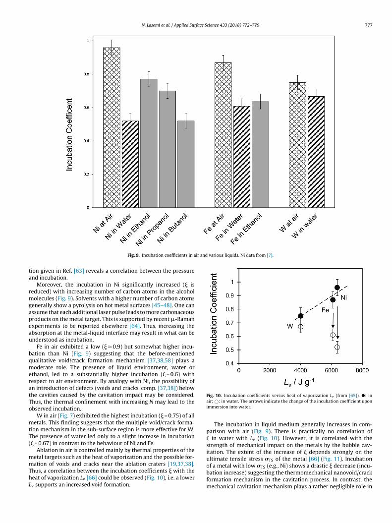
N. Lasemi et al. / Applied Surface Science 433 (2018) 772–779 777
ir and various liquids. Ni data from [7].
ta
rmgapeau
bqmeratTo
mtT(
mmThL
Fig. 9. Incubation coefficients in a
ion given in Ref. [63] reveals a correlation between the pressurend incubation.
Moreover, the incubation in Ni significantly increased (� iseduced) with increasing number of carbon atoms in the alcohol
olecules (Fig. 9). Solvents with a higher number of carbon atomsenerally show a pyrolysis on hot metal surfaces [45–48]. One canssume that each additional laser pulse leads to more carbonaceousroducts on the metal target. This is supported by recent -Ramanxperiments to be reported elsewhere [64]. Thus, increasing thebsorption at the metal-liquid interface may result in what can benderstood as incubation.
Fe in air exhibited a low (� ≈ 0.9) but somewhat higher incu-ation than Ni (Fig. 9) suggesting that the before-mentionedualitative void/crack formation mechanism [37,38,58] plays aoderate role. The presence of liquid environment, water or
thanol, led to a substantially higher incubation (� = 0.6) withespect to air environment. By analogy with Ni, the possibility ofn introduction of defects (voids and cracks, comp. [37,38]) belowhe cavities caused by the cavitation impact may be considered.hus, the thermal confinement with increasing N may lead to thebserved incubation.
W in air (Fig. 7) exhibited the highest incubation (� = 0.75) of alletals. This finding suggests that the multiple void/crack forma-
ion mechanism in the sub-surface region is more effective for W.he presence of water led only to a slight increase in incubation� = 0.67) in contrast to the behaviour of Ni and Fe.
Ablation in air is controlled mainly by thermal properties of theetal targets such as the heat of vaporization and the possible for-
ation of voids and cracks near the ablation craters [19,37,38].hus, a correlation between the incubation coefficients � with theeat of vaporization Lv [66] could be observed (Fig. 10), i.e. a lowerv supports an increased void formation.
Fig. 10. Incubation coefficients versus heat of vaporization Lv (from [65]). �: inair; ©: in water. The arrows indicate the change of the incubation coefficient uponimmersion into water.
The incubation in liquid medium generally increases in com-parison with air (Fig. 9). There is practically no correlation of� in water with Lv (Fig. 10). However, it is correlated with thestrength of mechanical impact on the metals by the bubble cav-itation. The extent of the increase of � depends strongly on theultimate tensile stress TS of the metal [66] (Fig. 11). Incubation
of a metal with low TS (e.g., Ni) shows a drastic � decrease (incu-bation increase) suggesting the thermomechanical nanovoid/crackformation mechanism in the cavitation process. In contrast, themechanical cavitation mechanism plays a rather negligible role in
778 N. Lasemi et al. / Applied Surface S
Fig. 11. Incubation coefficients versus ultimate tensile stress (from [66]). �: inai
tt
aApch
4
traltgsmaaiohcl
A
tAdp(
R
[
[
[
[
[
[
[
[
[
[
[
[
[
[
[
[
[
[
[
[
[survey of the state of the art for homogeneous materials, Int. J. Fatigue 20
TS
ir; ©: in water. The arrows indicate the change of the incubation coefficient uponmmersion into water.
he incubation in a metal with high TS (e.g. W) because � is almosthe same as in air.
A third, optical absorption mechanism may come into play whenlcoholic fluids with varying carbon numbers are applied (Fig. 9).n influence of this number on the incubation may suggest aossible absorptivity increase of the cavity surfaces due to carbona-eous deposits generated by laser-induced pyrolysis, particularly atigher carbon contents like in butanol.
. Conclusions
The ablation and incubation of metals in air is correlated withhermal properties such as the heat of vaporization, and may beelated to the accumulation of high-density defects such as voidsnd cracks below the ablation crater surface. The introduction of aiquid medium changes the cause for high-density defects from ahermal to a mechanical due to bubble cavitation impact, as sug-ested by the correlation of the incubation and the ultimate tensiletress of the metal. In particular, nickel exhibits relatively low ulti-ate tensile stress and shows the highest incubation increase in
liquid medium. This is in strong contrast to tungsten that has much higher ultimate tensile stress than nickel and shows anncubation behaviour barely affected by a liquid environment. Anptical absorption mechanism may become important when alco-ols with varying carbon numbers are applied: the carbon contentould affect the tendency to generate carbonaceous deposits byaser-induced pyrolysis thus increasing the incubation.
cknowledgements
Partial financial support by the Austrian Science Fund (FWF)hrough the Lise Meitner Programme (project M 1984), the H2020ction MSCA-IF 656908-NIMBLIS-ESR, the National Science Foun-ation through Grant CMMI-1301298, and the MAT2015-67354-Rroject of the Spanish Ministry of Economy and CompetitivenessMINECO) is gratefully acknowledged.
eferences
[1] A. Kruusing, Underwater and water-assisted laser processing: Part 2—Etching,
cutting and rarely used methods, Opt. Laser. Eng. 41 (2004) 329–352.[2] J. Lv, X. Dong, K. Wang, W. Duan, Z. Fan, X. Mei, Study on process andmechanism of laser drilling in water and air, Int. J. Adv. Manuf. Technol. 86(2016) 1443–1451.
[
cience 433 (2018) 772–779
[3] T.O. Nagy, U. Pacher, H. Pöhl, W. Kautek, Atomic emission stratigraphy bylaser-induced plasma spectroscopy: quantitative depth profiling of metal thinfilm systems, Appl. Surf. Sci. 302 (2014) 189–193.
[4] T.O. Nagy, M.J.J. Weimerskirch, U. Pacher, W. Kautek, Repassivationinvestigations on aluminium: physical chemistry of the passive state,Zeitschrift für Physikalische Chemie 230 (2016) 1303–1327.
[5] S. Barcikowski, G. Compagnini, Advanced nanoparticle generation andexcitation by lasers in liquids, Phys. Chem. Chem. Phys. 15 (2013) 3022–3026.
[6] S. Barcikowski, V. Amendola, G. Marzun, C. Rehbock, S. Reichenberger, D.Zhang, B. Gökce, Handbook of Laser Synthesis of Colloids, 2016.
[7] N. Lasemi, U. Pacher, C. Rentenberger, O. Bomatí-Miguel, W. Kautek,Laser-assisted synthesis of colloidal Ni/NiOx core/shell nanoparticles in waterand alcoholic solvents, ChemPhysChem 18 (2017) 1118–1124.
[8] F. Mafune, Y. Kohno, T. Kondow, H. Sawabe, Formation and size control ofsilver nanoparticles by laser ablation in aqueous solution, J. Phys. Chem. B 104(2000) 9111–9117.
[9] V.K. Pavel, V.V. Valerii, V.S. Aleksandr, A.S. Georgii, Production of copper andbrass nanoparticles upon laser ablation in liquids, Quantum. Electron. 34(2004) 951.
10] M.S.F. Lima, F.P. Ladário, R. Riva, Microstructural analyses of the nanoparticlesobtained after laser irradiation of Ti and W in ethanol, Appl. Surf. Sci. 252(2006) 4420–4424.
11] W.T. Nichols, T. Sasaki, N. Koshizaki, Laser ablation of a platinum target inwater. I. Ablation mechanisms, J. Appl. Phys. 100 (2006) 114911.
12] V. Amendola, P. Riello, S. Polizzi, S. Fiameni, C. Innocenti, C. Sangregorio, M.Meneghetti, Magnetic iron oxide nanoparticles with tunable size and freesurface obtained via a green approach based on laser irradiation in water, J.Mater. Chem. 21 (2011) 18665–18673.
13] C. Rehbock, V. Merk, L. Gamrad, R. Streubel, S. Barcikowski, Size control oflaser-fabricated surfactant-free gold nanoparticles with highly dilutedelectrolytes and their subsequent bioconjugation, Phys. Chem. Chem. Phys. 15(2013) 3057–3067.
14] N. Haram, N. Ahmad, Effect of laser fluence on the size of copper oxidenanoparticles produced by the ablation of Cu target in double distilled water,Appl. Phys. A 111 (2013) 1131–1137.
15] H.J. Jung, M.Y. Choi, Specific solvent produces specific phase Ni nanoparticles:a pulsed laser ablation in solvents, J. Phys. Chem. C 118 (2014) 14647–14654.
16] M. Boutinguiza, M. Meixus, J. del Val, A. Riveiro, R. Comesana, F. Lusquinos, J.Pou, Synthesis and characterization of Pd nanoparticles by laser ablation inwater using nanosecond laser, Phys. Procedia 83 (2016) 36–45.
17] C. Gellini, F.L. Deepak, M. Muniz-Miranda, S. Caporali, F. Muniz-Miranda, A.Pedone, C. Innocenti, C. Sangregorio, Magneto-plasmonic colloidalnanoparticles obtained by laser ablation of nickel and silver targets in water, J.Phys. Chem. C 121 (2017) 3597–3606.
18] J. Lam, D. Amans, F. Chaput, M. Diouf, G. Ledoux, N. Mary, K. Masenelli-Varlot,V. Motto-Ros, C. Dujardin, �-Al2O3 nanoparticles synthesised by pulsed laserablation in liquids: a plasma analysis, Phys. Chem.Chem. Phys. 16 (2014)963–973.
19] C.-Y. Shih, C. Wu, M.V. Shugaev, L.V. Zhigilei, Atomistic modeling ofnanoparticle generation in short pulse laser ablation of thin metal films inwater, J. Colloid Interface Sci. 489 (2017) 3–17.
20] S. Reich, P. Schönfeld, P. Wagener, A. Letzel, S. Ibrahimkutty, B. Gökce, S.Barcikowski, A. Menzel, T. dos Santos Rolo, A. Plech, Pulsed laser ablation inliquids: impact of the bubble dynamics on particle formation, J. ColloidInterface Sci. 489 (2017) 106–113.
21] E. Matthias, M. Reichling, J. Siegel, O.W. Käding, S. Petzoldt, H. Skurk, P.Bizenberger, E. Neske, The influence of thermal diffusion on laser ablation ofmetal films, Appl. Phys. A 58 (1994) 129–136.
22] J. Krüger, W. Kautek, Ultrashort pulse laser interaction with dielectrics andpolymers, Adv. Polym. Sci. 168 (2004) 247–289.
23] M. Forster, L. Égerházi, C. Haselberger, C. Huber, W. Kautek, Femtosecondlaser interaction with pulsed-laser deposited carbon thin films of nanoscalethickness, Appl. Phys. A 102 (2011) 27–33.
24] P.K. Kennedy, S.A. Boppart, D.X. Hammer, B.A. Rockwell, G.D. Noojin, W.P.Roach, A first-order model for computation of laser-induced breakdownthresholds in ocular and aqueous media. II. Comparison to experiment, IEEE J.Quantum Electron. 31 (1995) 2250–2257.
25] J. Noack, A. Vogel, Laser-induced plasma formation in water at nanosecond tofemtosecond time scales: calculation of thresholds, absorption coefficients,and energy density, IEEE J. Quantum Electron. 35 (1999) 1156–1167.
26] A. Vogel, J. Noack, K. Nahen, D. Theisen, S. Busch, U. Parlitz, D.X. Hammer, G.D.Noojin, B.A. Rockwell, R. Birngruber, Energy balance of optical breakdown inwater at nanosecond to femtosecond time scales, Appl. Phys. B 68 (1999)271–280.
27] D. Bäuerle, Laser Processing and Chemistry, 4 ed., Springer-Verlag, BerlinHeidelberg, 2011.
28] K.C. Phillips, H.H. Gandhi, E. Mazur, S.K. Sundaram, Ultrafast laser processingof materials: a review, Adv. Opt. Photonics 7 (2015) 684–712.
29] Y. Jee, M.F. Becker, R.M. Walser, Laser-induced damage on single-crystalmetal surfaces, J. Opt. Soc. Am. B 5 (1988) 648–659.
30] A. Fatemi, L. Yang, Cumulative fatigue damage and life prediction theories: a
(1998) 9–34.31] J. Byskov-Nielsen, J.-M. Savolainen, M.S. Christensen, P. Balling, Ultra-short
pulse laser ablation of metals: threshold fluence, incubation coefficient andablation rates, Appl. Phys. A 101 (2010) 97–101.

face S
[
[
[
[
[
[
[
[
[
[
[
[
[
[
[
[
[
[
[
[
[
[
[
[
[
[
[
[
[[
[
[
[
N. Lasemi et al. / Applied Sur
32] F. Di Niso, C. Gaudiuso, T. Sibillano, F.P. Mezzapesa, A. Ancona, P.M. Lugarà,Role of heat accumulation on the incubation effect in multi-shot laserablation of stainless steel at high repetition rates, Opt. Express 22 (2014)12200–12210.
33] B. Neuenschwander, B. Jaeggi, M. Schmid, G. Hennig, Surface structuring withultra-short laser pulses: basics, limitations and needs for high throughput,Phys. Procedia 56 (2014) 1047–1058.
34] T. Häfner, J. Heberle, M. Dobler, M. Schmidt, Influences on incubation in pslaser micromachining of steel alloys, J. Laser Appl. 28 (2016) 022605.
35] A. Naghilou, O. Armbruster, M. Kitzler, W. Kautek, Merging spot size and pulsenumber dependence of femtosecond laser ablation thresholds: modeling anddemonstration with high impact polystyrene, J. Phys. Chem. C 119 (2015)22992–22998.
36] A. Naghilou, O. Armbruster, W. Kautek, Femto- and nanosecond pulse laserablation dependence on irradiation area: the role of defects in metals andsemiconductors, Appl. Surf. Sci. 418 (2017) 487–490.
37] C. Wu, M.S. Christensen, J.-M. Savolainen, P. Balling, L.V. Zhigilei, Generationof subsurface voids and a nanocrystalline surface layer in femtosecond laserirradiation of a single-crystal Ag target, Phys. Rev. B 91 (2015) 035413.
38] E.T. Karim, M.V. Shugaev, C. Wu, Z. Lin, H. Matsumoto, M. Conneran, J.Kleinert, R.F. Hainsey, L.V. Zhigilei, Experimental characterization andatomistic modeling of interfacial void formation and detachment in shortpulse laser processing of metal surfaces covered by solid transparentoverlayers, Appl. Phys. A 122 (2016) 407.
39] R. Diaz-Uribe, M. Rosete-Aguilar, R. Ortega-Martinez, Position sensing of aGaussian beam with a power meter and a knife edge, Rev. Mex. Fis. (1993)484–492.
40] M. González-Cardel, P. Arguijo, R. Díaz-Uribe, Gaussian beam radiusmeasurement with a knife-edge: a polynomial approximation to the inverseerror function, Appl. Opt. 52 (2013) 3849–3855.
41] K.H. Kim, Z. Akase, T. Suzuki, D. Shindo, Charging effects on SEM/SIM contrastof metal/insulator system in various metallic coating conditions, Mater. Trans.51 (2010) 1080–1083.
42] P. Li, S.X. Bao, D.Z. Zhang, L.B. Zhuang, L.L. Ma, Application of secondaryelectron composition contrast imaging method in microstructure studies oncathode materials of TWT, Mater. Sci. Forum 689 (2011) 255–259.
43] R. Waser, in: H. Schaumburg (Ed.), Lineare und nicht-lineare Widerstände,Keramik, Vieweg + Teubner Verlag, Wiesbaden, 1994, pp. 129–218.
44] M.-R. Kalus, N. Barsch, R. Streubel, E. Gokce, S. Barcikowski, B. Gokce, Howpersistent microbubbles shield nanoparticle productivity in laser synthesis ofcolloids – quantification of their volume, dwell dynamics, and gascomposition, Phys. Chem. Chem. Phys. 19 (2017) 7112–7123.
45] V. Amendola, G.A. Rizzi, S. Polizzi, M. Meneghetti, Synthesis of gold
nanoparticles by laser ablation in toluene: quenching and recovery of thesurface plasmon absorption, J. Phys. Chem. B 109 (2005) 23125–23128.46] H.Y. Kwong, M.H. Wong, C.W. Leung, Y.W. Wong, K.H. Wong, Formation ofcore/shell structured cobalt/carbon nanoparticles by pulsed laser ablation intoluene, J. Appl. Phys. 108 (2010) 034304.
[
[
cience 433 (2018) 772–779 779
47] V. Amendola, P. Riello, M. Meneghetti, Magnetic nanoparticles of iron carbide,iron oxide, iron@iron oxide, and metal iron synthesized by laser ablation inorganic solvents, J. Phys. Chem. C 115 (2011) 5140–5146.
48] V. Amendola, M. Meneghetti, What controls the composition and thestructure of nanomaterials generated by laser ablation in liquid solution?Phys. Chem. Chem. Phys. 15 (2013) 3027–3046.
49] M.A. Aegerter, M. Mennig, Sol-Gel Technologies for Glass Producers andUsers, Springer, New York, 2004.
50] Z.N. Kayani, E.S. Khan, F. Saleemi, S. Riaz, S. Naseem, Optical and magneticproperties of iron oxide thin films, Mater. Today Proc. 2 (2015) 5568–5571.
51] S. Phokha, S. Pinitsoontorn, S. Rujirawat, S. Maensiri, Polymer pyrolysissynthesis and magnetic properties of LaFeO3 nanoparticles, Physica B 476(2015) 55–60.
52] P. Pouli, D.C. Emmony, C.E. Madden, I. Sutherland, Analysis of thelaser-induced reduction mechanisms of medieval pigments, Appl. Surf. Sci.173 (2001) 252–261.
53] T. He, J. Yao, Photochromic materials based on tungsten oxide, J. Mater. Chem.17 (2007) 4547–4557.
54] M. Acosta, D. González, I. Riech, Optical properties of tungsten oxide thinfilms by non-reactive sputtering, Thin Solid Films 517 (2009) 5442–5445.
55] D.B. Migas, V.L. Shaposhnikov, V.N. Rodin, V.E. Borisenko, Tungsten oxides. I.Effects of oxygen vacancies and doping on electronic and optical properties ofdifferent phases of WO3, J. Appl. Phys. 108 (2010) 093713.
56] X. He, Y. Yin, J. Guo, H. Yuan, Y. Peng, Y. Zhou, D. Zhao, K. Hai, W. Zhou, D.Tang, Memristive properties of hexagonal WO3 nanowires induced by oxygenvacancy migration, Nanoscale Res. Lett. 8 (2013) 50.
57] K. Aguir, C. Lemire, D.B.B. Lollman, Electrical properties of reactively sputteredWO3 thin films as ozone gas sensor, Sens. Actuators B: Chem. 84 (2002) 1–5.
58] J.-M. Savolainen, M.S. Christensen, P. Balling, Material swelling as the firststep in the ablation of metals by ultrashort laser pulses, Phys. Rev. B 84 (2011)193410.
59] R.E. Bolz, G.L. Tuve, CRC Handbook of Tables for Applied Engineering Science,2 ed., 1973.
60] R.C. Weast, Handbook of Chemistry and Physics, 64 ed., CRC Press, 1984.61] C.E. Brennen, Cavitation and Bubble Dynamics, Oxford University Press,
Oxford, 1995.62] J. Lam, J. Lombard, C. Dujardin, G. Ledoux, S. Merabia, D. Amans, Dynamical
study of bubble expansion following laser ablation in liquids, Appl. Phys. Lett.108 (2016) 074104.
63] I. Akhatov, O. Lindau, A. Topolnikov, R. Mettin, N. Vakhitova, W. Lauterborn,Collapse and rebound of a laser-induced cavitation bubble, Phys. Fluids 13(2001) 2805–2819.
64] R. Lahoz, A. Mayoral, G.F. de la Fuente, C. Rentenberger, D. Díaz-Fernández, L.
Soriano, W. Kautek, O. Bomati-Miguel, (in publication).65] D.R. Lide, H.P.R. Frederikse, Handbook of Chemistry and Physics, 76 ed., CRCPress Boca Raton, FL, 1995–1996.
66] A.M. Howatson, P.G. Lund, J.D. Todd, Properties of matter, Engineering Tablesand Data, Springer, Netherlands, Dordrecht, 1972, pp. 41.