applying the new irecist guidelines · clin oncol 33: 31, 3531-3543. criteria publication over time...
TRANSCRIPT

Author: K. Luby, Mint Medical
Applying the New iRECIST GuidelinesRadiologic and Clinical Trial Considerations
www.iRECIST.com

2
iRECIST1: Guidelines for response criteria for use in trials testing immunotherapeutics
The RECIST (Response Evaluation Criteria in Solid Tumors) guidelines (RECIST 1.0 and RECIST 1.1) have been the standard for response evaluation in clinical trials and have supported myriad regulatory approvals in solid tumors. RECIST first published in 20002 (v1.0) and later revised in 20093 as RECIST 1.1 provided a methodology for the consistent and reproducible evaluation of imaging and clinical data as a surrogate endpoint for survival and quality of life based endpoints.
The hallmark of RECIST is capturing a patient’s disease extent beginning with a ‘Baseline’ tumor burden quan-tification and following a patient radiographically from one imaging exam to the next over time to evaluate the change in that tumor burden both quantitatively and qualitatively. This same concept has been widely applied to other standardized oncologic response criteria and a therapeutics’ efficacy has been largely measured against the principle that a decrease from baseline indicates a response to treatment while an increase from nadir (lowest tumor burden value met since baseline) is consistent with a worsening of disease.
RECIST (both 1.0 and 1.1 variants) has been widely adopted for use in clinical trials for evaluating efficacy and has served as a defining methodology used by oncologists to guide clinical decision making for patient care in continuing, stopping or switching therapy. In immuno-therapy treatments, however, RECIST may not consistently provide the most accurate correlation of overall survival with response evaluation by evaluation of tumor burden with an increase in tumor burden being a definitive indicator of progression.
Immuno-therapies include monoclonal antibodies, check-point inhibitors, immunomodulatory agents, cytokines and antibody-drug conjugates. Patients on these therapies may demonstrate what has been termed ‘pseudo-pro-gression’, with an initial increase in size of existing baseline lesions and/or the appearance of new disease with patients subsequently demonstrating a response, albeit delayed, to therapy. A confounding aspect also noted is the relative decrease in size of existing baseline disease with the appearance of new disease – a common pattern observed with immuno-therapies. This can occur as immune cells infiltrate disease sites simulating apparent growth. In other cases, as the body mounts an immune response following treatment, disease can initially worsen until an adaptive immune response can be achieved. Disease sites which are likely present at baseline but not vi-sible on imaging appear as new disease on initial follow-up evaluations only to subsequently resolve. Even though worsening is transient, traditional response assessment (i.e., RECIST) would consider this evidence of treatment failure.
1 Seymour, L, et al; iRECIST: guidelines for response criteria for use in trials testing immunotherapeutics.
Lancet Oncol.18(3):e143-e152. (iRECIST).
2 Therasse P, Arbuck SG, Eisenhauer EA, et al. New guidelines to evaluate the response to treatment in solid tumors.
European Organization for Research and Treatment of Cancer, National Cancer Institute of the United States,
National Cancer Institute of Canada. J Natl Cancer Inst 2000; 92:205-16. (RECIST 1.0).
3 Eisenhauer EA, Therasse P, Bogaerts J, et al. New response evaluation criteria in solid tumours:
revised RECIST guideline (version 1.1). Eur J Cancer 2009; 45:228-47. (RECIST 1.1).

3
2000 RECIST 1.0
Multiple irRECISTimplementations
2009 - irRC
2017 - iRECIST
2009 - RECIST 1.1
A well-known example was seen in patients receiving Ipilimumab for melanoma where nearly 10% of subjects with clinical responses would have met progression by traditional WHO4 criteria (WHO being an earlier mea-sure of tumor response than RECIST 1.0)5. Further reports of Ipilimumab in melanoma have also shown similar findings6. Other immuno-therapy agents as well as other indications aside from melanoma (e.g., breast, bladder, colorectal, gastric, lung, sarcoma) have reported few relative incidents of this immune-related response, with some reporting only a few more subjects who would be considered progressions by RECIST7.
4 WHO handbook for reporting results of cancer treatment. Geneva (Switzerland):
World Health Organization Offset Publication No. 48, 1979. (WHO criteria)
5 Wolchok JD, et al: Guidelines for the evaluation of immune therapy activity in solid tumors:
Immune-related response criteria. Clin Cancer Res 15:7412-7420, 2009. (irRC)
6 Hodi FS, et al: Evaluation of immune-related response criteria (irRC) in patients (pts) with advanced melanoma (MEL)
treated with the anti-PD-1 monoclonal antibody MK-3475. J Clin Oncol 32, 2014 (suppl 15s; abstr 3006) 10.
7 Chiou L, Burotto M; Pseudoprogression and Immune-Related Response in Solid Tumors. J. Clin Oncol 33: 31, 3531-3543.
Criteria publication over time with focus on criteria for Immuno-Therapy Agents

4
Immune-Related Response Criteria (irRC)
In irRC, new lesions that meet the criteria for
measurability are added to the Sum of the Pro-
ducts of Perpendicular Diameters (SPPD) of Tar-
get Lesions.
If Target Lesion + New Measurable lesion SPPD
meets the threshold for progression, the subject
is considered progressed. Any new lesions which
do not meet the minimum measurable lesion size
at baseline do not trigger progression but do pre-
vent a complete response.
Criteria for Immuno-Therapy Agents
Even with the somewhat lower incidence of pseudo-progression overall related to immuno-therapies, it remains a known possibility that must be accounted for in clinical trials and when evaluating patient care decisions with these agents. A first approach to evaluating response with immuno-therapy agents evolved in 2009 from Ipili-mumab treatment with the Immune-Related Response Criteria (irRC) which has been widely applied. Melanoma patients treated with Ipilimumab who were followed with traditional WHO criteria appeared to progress but later went on to demonstrate a response to therapy. A different approach was explored since these tradi-tional response criteria failed to accurately correlate with treatment response observations.
Based on the original WHO guidelines which utilized bi-dimensional measurements of tumors and with slightly different response and progression thresholds from RECIST 1.0, irRC incorporated a new concept in clinical trials: New or worsening disease observed on imaging does not always correlate with a lack of response to treatment.
In 2009, RECIST 1.1 was published bringing further refinement to the widely-utilized criteria for solid tumors. As RECIST 1.1 and irRC emerged around the same time, other iterations of response criteria natu-rally developed merging the principles of RECIST 1.1 with irRC. However, while multiple interpretations and iterations of ‘immune-RECIST’ were used in clinical trials, no singular publication was referenced. Rather the approach was subject to the user’s interpretation. Some approaches simply used irRC with unidimensional measurements and the response/progression thresholds of RECIST 1.1. Other implementations utilized a con-firmatory progression evaluation which required further worsening of disease to establish a progression event.
Since the publication of irRC in 2009, there has been a growing approach to standardization of the criteria with RECIST 1.1 based concepts. The 2017 publication of iRECIST provides an opportunity for this standardization of response assessment and an approach to utilizing the principles established by RECIST 1.1 while considering the evolving therapeutic effects of immuno-therapy agents.
“ New or worsening disease observed on imaging does not always correlate with a lack of response to treatment.

5
iCR immune Complete Response
iPR immune Partial Response
iSD immune Stable Disease
iUPD immune unconfirmed Progression
iCPD immune confirmed Progression
iNN immune Non-iCR/Non-iUPD
NLT New Lesion Target
NLNT New Lesion Non-Target
NE Not Evaluable
TPR Time Point Response
iBOR immune Best Overall Response
iPFS Immune Progression Free Survival
PSPD Pseudo-progression
SOM Sum of Measures
New Immuno-Therapy Response Criteria – iRECIST
iRECIST is intended to provide a standard approach to the evaluation of solid tumors with measurements and assessment of the disease burden in trials where an immunotherapy is used. iRECIST also strives to collect data for future trials so that a data warehouse can be subsequently utilized to validate iRECIST.
iRECIST follows similar recommendations provided by RECIST 1.1 on a lesion level in terms of methods of measure-ment, size criteria, disease selection and categorization. The methodology of determining response is also comparable between iRECIST and RECIST 1.1.
A primary difference between iRECIST and RECIST 1.1 is the concept of ‘status reset’ if RECIST 1.1 progression is fol-lowed at the next assessment by tumor shrinkage or disappearance, thus confirmation of progression is a requirement in iRECIST. Additionally, new lesions are categorized differently from RECIST 1.1 as Target or Non-Target New Lesions in iRECIST. Perhaps the most distinguishing aspect of iRECIST is that the Overall Response is designated with a prefix of “i” (i.e., iCR, iPR, iSD, iUPD, iCPD). Furthermore, the time point response and best overall response is to be recorded separately from RECIST 1.1.
Concepts and LexiconiRECIST is based on RECIST 1.1. To differentiate responses by iRECIST from RECIST 1.1, a prefix of “i” (for immune res-ponse) is appended to the front of the time point designati-on (iCR, iPR, iSD). The progression category is additionally sub-divided into unconfirmed and confirmed progression (iUPD and iCPD respectively). Another variation is the treatment of new lesions which are sub-divided into Tar-get New Lesions (New Lesion Target (NLT)) and Non-Tar-get New Lesions (New Lesion Non-Target (NLNT)).
Table 1 further demonstrates these differences. (on the next page)

6
RECIST 1.1 iRECIST
Definitions of measurable and non-measurable disease; numbers and site of target disease
Measurable lesions are 10 mm or more in long diame-ter (15 mm for nodal lesions); Maximum of 5 lesions (2 per organ);All other disease considered non-target (must be 10 mm or longer in short axis for nodal disease)
No change; however, NEW lesions are evaluated as per RECIST 1.1 but are recorded separately on the case report form (CRF) (but not included in the sum of lesions for target lesions identified at baseline)
CR, PR or SD
Cannot have met criteria for PD prior to CR, PR or SD May have had iUPD (1 or more instances), but not iCPD, prior to iCR, iPR or iSD
Confirmation of CR, PR
Only required for non-randomized trials
Confirmation of SD
Not Required
New Lesions (unequivocal)
Results in PD.Recorded but not measured.
Results in iUPD but iCPD is only assigned based on this category if at next assessment:• Additional NL appear or• Increase in size of NLs (≥5 mm for sum of NLT or any increase in NLNT)The appearance of New Lesions can also confirm pro-gression first observed at the prior timepoint in the Target or Non-Target Category.
Independent blinded review and central collection of scans
Recommended in some circumstances Collection of scans (but not independent review) re-commended for all trials
Confirmation of PD
Not required (unless equivocal) Required
Consideration of clinical status
Not included in assessment Clinical stability is considered in whether treatment is continued after iUPD
iCR - iPR - iSD - iUPD - iCPD
Table 1. RECIST 1.1 compared to iRECIST8
8 Seymour, L, et al; iRECIST: guidelines for response criteria for use in trials testing immunotherapeutics.
Lancet Oncol.18(3):e143-e152. (iRECIST).

7
iRECIST Principles
iRECIST considers the categories of Target Lesions, Non-Target Lesions and New Lesions. Each category has a categorical response assessment that drives the overall response assessment.
Category 1: Target LesionsiCR, iPR and iSD can be assigned after iUPD has been documented provided iCPD was not confirmed. iUPD is defined by RECIST 1.1 criteria for PD and can be reported multiple times as long as iCPD is not met.
PD is confirmed in the target lesion category if the next imaging assessment (4 weeks but no more than 8 weeks later after iUPD) confirms further increase in the sum of measures (SOM) of target disease from iUPD, with an increase of at least 5 mm.
iCPD is not met if iCR, iPR or iSD criteria (compared to baseline and as defined by RECIST 1.1) are met at the next assessment after iUPD. The status is ‘reset’ and iUPD must be met again followed by a iCPD to confirm progression.
Category 2: Non-Target LesionsiUPD (not iCPD) may have been documented prior to iCR or Non-iCR/Non-iUPD and may be assigned multiple times provided iCPD is not met.
Progression in the non-target lesion category is confirmed if subsequent imaging, (conducted at least 4 weeks but no more than 8 weeks after iUPD) shows further increase from iUPD.
If iCR or non-iCR/non-iUPD criteria is met after iUPD, the status is ‘reset’ and iUPD must be met again followed by iCPD to confirm progression.
Category 3: New LesionsNew lesions in iRECIST are categorized as Target (measurable) or Non-Target (non-measurable) in accordance with RECIST 1.1 principles. Five new lesions, no more than two per organ, should be measured and recorded as New Lesions Target (NLT), but are NOT to be included in SOM of the original target lesions identified at base-line. Other new lesions are recorded as New Lesion Non-Target (NLNT).
NLT and NLNT can drive a iUPD and iCPD. Progression is confirmed (iCPD) in the New Lesion category if the next imaging assessment (conducted at least 4 weeks but no more than 8 weeks after iUPD), confirms additional new lesions or further increase in new lesion size from iUPD (SOM increase in NLT ≥ 5 mm, any increase for NLNT).

8
Achieving iCPD
At the confirmatory time point, an overall assessment of iCPD can be met by: iUPD in a lesion category followed by iCPD in the same lesion category AND/OR iUPD in a different lesion category
Target Lesion iUPD
Non-Target Lesion iUPD
New Lesion iCPD
Target Lesion iUPD
Non-Target Lesion iCPD
New Lesion iUPD
Target Lesion iCPD
Non-Target Lesion iUPD
New Lesion iUPD
Target Lesion iUPDFollowed at next
assessment byat least one
Non-Target Lesion iUPDFollowed at next
assessment byat least one
New Lesion iUPDFollowed at next
assessment byat least one
iCPD

9
Incorporating iRECIST with RECIST 1.1
iRECIST recommends that clinical trials which allow continued treatment following a RECIST 1.1 based progres-sion event should only do so provided the patient is clinically stable. Following progression by RECIST 1.1, the next imaging assessment to evaluate tumor burden status should not be longer than 8 weeks later unless evi-dence exist that pseudo-progression is a known occurrence. It is always the decision of the patient and treating physician to continue or discontinue therapy. As with RECIST 1.1, the protocol must define how iRECIST will be incorporated into a trial, in parti-cular, how the study endpoints will be supported (RECIST 1.1 or iRECIST). For the purpose of evalua-ting potential pseudo-progression events, conducting a trial where both RECIST 1.1 and iRECIST response assessment is collected may provide useful data on the efficacy of the therapeutic under investigation. Furthermore, when defining criteria for a given protocol using RECIST 1.1 and iRECIST, the following parameters should also be considered:
Confirmation of Response• Is confirmation of a iCR or iPR required? (Confirmation of response by RECIST 1.1 is typically only a require-
ment in non-randomized trials.)
• What is the minimum time in days between two responses to consider confirmation met (i.e., 28 days)? Is there a window (i.e., + or - 5 days)?
Missing and Not Evaluable Assessments• How will missing response assessments be handled?
• How many gaps are allowed between responses and what assessments are allowed in order to consider a response confirmed?
• Are multiple Not Evaluable assessments between responses allowed in order to consider a response confir-med? For example, is iPR-NE-iPR a confirmed iPR? iCR-NE-iCR? iPR-NE-NE-iPR?
• Is an assessment of iSD allowed between two responses for confirmation to be met? For example, is iPR-iSD-iPR a confirmed iPR? How does iUPD factor into the confirmation of response? For example, iPR-iUPD-iPR?
Confirmation of Progression Time• What is the minimum time for confirmation to report iCPD (e.g., 28 days)?
When to apply iRECIST
iRECIST recommends that RECIST 1.1 should
continue to be used as the primary criteria for re-
sponse based endpoints in randomized trials in-
tended for registration with iRECIST considered
exploratory.

10
Baseline Follow-Up 1 Follow-Up 2
Target LesionSOD = 100 mm SOD = 130 mm
(30% Increase - Nadir)SOD = 147 mm(≥ 5 mm Increase – from iUPD)
Non-Target Lesions None None None
New Lesions None None
Overall Respon-se by iRECIST
iUPD iCPD
Explanation: At Follow-up 2, iCPD confirms the iUPD at Follow-up 1 with a ≥ 5 mm Increase from iUPD and the threshold for progression (≥ 20 % increase from nadir) is still met.
Baseline Follow-Up 1 Follow-Up 2
Target LesionSOD = 100 mm SOD = 130 mm
(30% Increase - Nadir)SOD = 131 mm(< 5 mm Increase – from iUPD)
Non-Target Lesions None None None
New Lesions None None
Overall Respon-se by iRECIST
iUPD iUPD
Explanation: At Follow-up 2, there is NOT a ≥ 5 mm Increase from iUPD even though the threshold for progression (≥ 20 % increase from nadir) is still met; iUPD is reported again for Follow-up 2. Confirmed Progression is not met.
Example Case 1
Example Case 2

11
Baseline Follow-Up 1 Follow-Up 2
Target LesionSOD = 100 mm SOD = 130 mm
(30% Increase - Nadir)SOD = 70 mm(30% Decrease - Baseline)
Non-Target Lesions None None None
New Lesions None None
Overall Respon-se by iRECIST
iUPD iPR
Explanation: At Follow-up 2, progression is not confirmed. The SOD decreases and meets criteria for Partial Response (≥ 30 % decrease from baseline). iUPD must again be met before iCPD can be met and the case considered confirmed progression.
Baseline Follow-Up 1 Follow-Up 2
Target LesionSOD = 100 mm SOD = 130 mm
(30% Increase - Nadir)SOD = 120 mm(20% increase – Nadir; 5 mm minimum not met)
Non-Target Lesions Non-iCR/Non-iUPD iUPD
New Lesions None None
Overall Respon-se by iRECIST
iUPD iCPD
Explanation: At Follow-up 1, iUPD is met by Target Lesions. Target lesions remain iUPD at Follow-up 2 but do not meet minimum 5 mm absolute increase. However, Non-Target Lesions have unequivocally progressed meeting iUPD. Confirmed Progression is met since Non-Target iUPD confirms Target Lesion iUPD at Follow-up 1.
Example Case 3
Example Case 4

1212
Baseline Follow-Up 1 Follow-Up 2
Target LesionSOD = 100 mm SOD = 120 mm
(20% Increase - Nadir)SOD = 122 mm(20% increase – Nadir; 5 mm minimum not met)
Non-Target Lesions Non-iCR/Non-iUPD Non-iCR/Non-iUPD
New LesionsNone New Lung Lesion - present
(New Lesion Non-Target)
Overall Respon-se by iRECIST
iUPD iCPD
Explanation: At Follow-up 1, iUPD is met by Target Lesions. Target lesions remain iUPD at Follow-up 2 but do not meet minimum 5 mm absolute increase. However, a new Non-Target lung lesion appears at Follow-up 2 meeting iUPD in the New Lesion category. Confirmed Progression is met since the New Lesion iUPD confirms iUPD by Target Lesions at Follow-up 1 even though Target lesions did not meet confirmed progression (iCPD) independently.
Baseline Follow-Up 1 Follow-Up 2
Target LesionSOD = 100 mm SOD = 100 mm
(No Change)SOD = 100 mm(No Change)
Non-Target Lesions Non-iCR/Non-iUPD Non-iCR/Non-iUPD
New LesionsNew Liver Lesion - 17 mm(New Lesion Target)
New Liver Lesion - 19 mm (from Follow-up 1)
Overall Respon-se by iRECIST
iUPD iSD
Explanation: At Follow-up 1, iUPD is met by the New Target Liver Lesion. This new lesion remains present but does not increase by the 5mm minimum needed for a iCPD thus progression is not confirmed and response is based on other disease findings which meet criteria for iSD.
Example Case 6
Example Case 5

13
About mintLesion™
mintLesion™ image analysis software streamlines imaging and clinical data evaluation for clinical trials and research in accordance with standardized response criteria comparing medical images from multiple modalities across time points. Multiple workfl ow options are available in mintLesion™ including Single and Double Reads, Adjudication, Eligibility, and Consensus Review.
mintLesion™ is designed to facilitate research in clinical trials with imaging endpoints. This includes the trans-fer and storage of clinical trial DICOM data, a workfl ow optimized for consistent interpretation and reviewing of image fi ndings, the review of image meta-data, and the simplifi ed provision of results to physicians and other clinical systems, e.g. PACS and Clinical Trial Management Systems.
The mint Lesion™ software is a 510(k) – cleared, Class II medical device which conforms to the European Medical Devices Act and bears the CE mark. mint Lesion utilizes strict user authorization, passwords, user permissions,clinical trial creation and assignment, data entry logging, audit trail management, electronic signature, and post-approval revision tracking and identifi cation.
Software screenshot of mintLesion™

14
mintLesion™ facilitates radiological assessment with automatic response classification. Standardized response criteria are inherent to the mintLesion™ software with conformance by criteria-based edit-checks. Novel criteria configurations are also supported based on trial and therapeutic specific needs.Radiologists and other physician specialists utilize mintLesion™ to access subject images, catalog disease findings, annotate findings, for qualitative and quantitative analysis, evaluating clinical data, and storing measurement values.
mintLesion™ offers configurability with flexibility to meet trial specific requirements. The oncologic res-ponse assessment analysis facilitated by mint Lesion is further enhanced by the structured reporting templates that provide an organized, consistent, and reproducible display of the analysis criteria evaluation required for clinical trials.
• Multi-modality analysis including CT, MRI, PET, X-ray, Bone Scan, DCE-MRI and other DICOM series.
• Quantitative measurement tools including density, texture and volume with image correlation and lesion matching.
• Structured reporting with diagrams, graphs, and lesion measurement snapshots.
• Multi-criteria analysis on the same imaging data.
• Real-time reporting of results.
• Data change functionality with original read data preservation.
• Trial Dashboard for read monitoring and data mining.
Sample report - multiple visualisations from one trial
15
PCWG2
RANO
Cheson / Lugano
irRECIST
iRECISTChoi
RECIST 1.1
mRECIST HCC
WHO
irRC
mRECIST Mesothelioma
RECIST 1.0
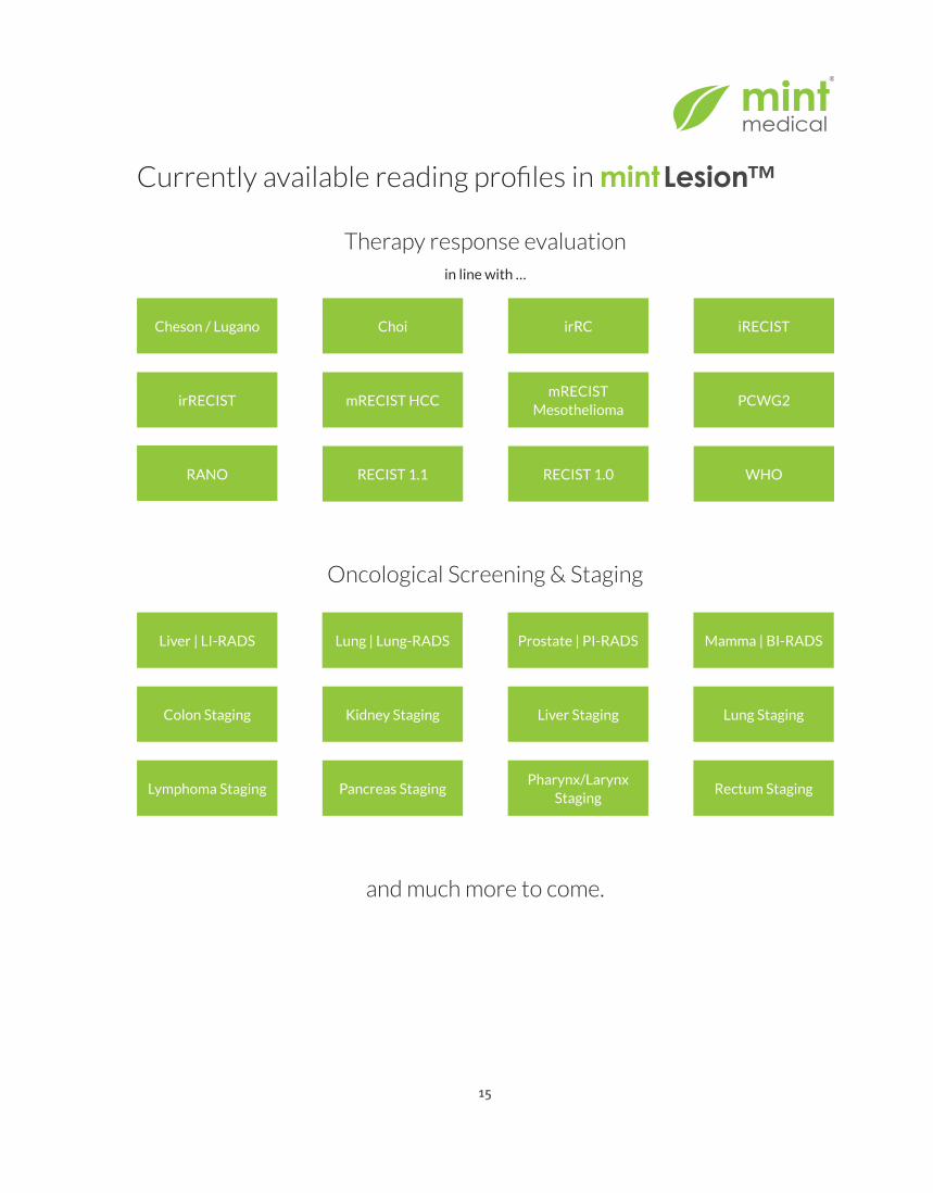
Currently available reading profiles in mintLesion™
Therapy response evaluationin line with …
Oncological Screening & Staging
Prostate | PI-RADS
Rectum StagingPharynx/Larynx
Staging
Mamma | BI-RADS
Colon Staging
Pancreas Staging
Lung | Lung-RADS
Lung Staging
Lymphoma Staging
Liver | LI-RADS
Liver StagingKidney Staging
and much more to come.

16
Mint Medical was founded by researchers of the German Cancer Research Center (DKFZ).
It is based on the innovation and experience from more than 20 years of research work in the area of
medical imaging.
Mint Medical aims to provide a new quality in clinical trial imaging and improves reproducibility and objectiveness in day-to-day clinical routine.
For further information or to schedule a test drive:
+49 6221 6479760Mint Medical GmbHFriedrich-Ebert-Straße 269221 Dossenheim
In the [email protected]
+1 844 200 MINTMint Medical Inc100 Horizon Center BlvdHamilton, NJ 08691
