approach to lymphadenopathy in children - wikispaces localized response from lymphocyte and...
TRANSCRIPT

Approach to lymphadenopathy in children
Components of the Lymphatic System
Lymph
Lymphatic Vessels Lymphatic Capillaries Lymphatic Vessels Lymphatic Trunks Lymphatic Ducts
Lymphatic Organs Thymus Lymph Nodes Spleen Tonsils
Lymphatic cells

Components of the Lymphatic System
Lymph: a fluid similar to plasma–does not have plasma proteins
Lymphatic vessels : network that carries lymph from peripheral tissues to the venous system
Lymphatic cells: Lymphocytes, phagocytes, and other immune system cells
Lymphoid tissues and lymphoid organs: found throughout the body
Function of the Lymphatic System
Reabsorbs excess interstitial fluid: returns it to the venous circulation maintain blood volume levels
Absorption & transport of fatty acids and fats drain into larger lymphatic vessels eventually into the bloodstream.
lymphocyte development, and the immune response.
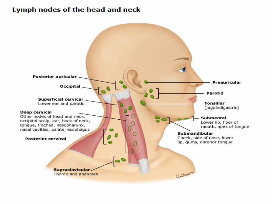
Lymph nodes
Lymph nodes are
bean shaped organs
found in clusters.
over 800 lymph nodes
300 are located in the head and neck
Lymphadenopathy
refers to enlargement of the lymph nodes.
Lymphadenitis refers to enlarged lymph nodes that are inflamed
Lymphadenopathy
is common
is an early indicator of some diseases.
Early recognition of disease improves
prognosis for recovery.
Lymphadenopathy
Normal lymph nodes are discrete, non tender, and mobile without fixation to underlying tissues.
Significant enlarged:
>1 cm in cervical and axillary,
>1.5cm in inguinal nodes
Pathophysiology
Localized response from lymphocyte and macrophage – viral/ bacterial infection
Localized infiltration by inflammatory cells in response to infection of nodes- lymphadenitis
Proliferation of neoplastic lymphocyte or macrophages- neoplasm

Lymphadenopathy
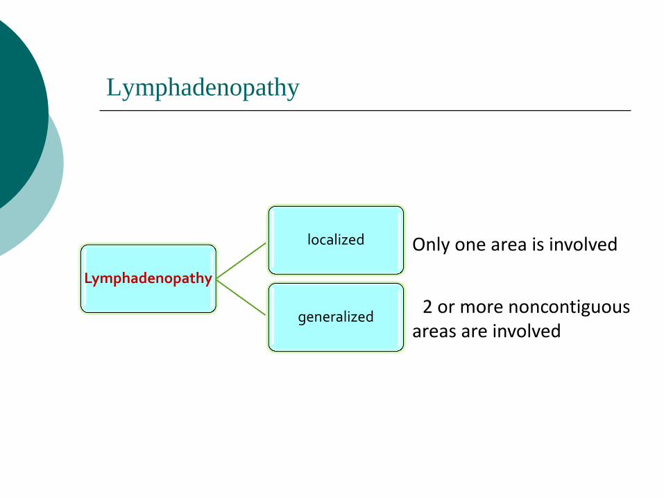
Lymphadenopathy
localized
generalized
Only one area is involved rea is involved
22 or more noncontiguous areas are involved or more noncontiguous areas are involved
Lymphadenopathy
Generalized lymphadenopathy (enlargement of >2 noncontiguous node regions) is caused by systemic disease
Regional lymphadenopathy is most frequently the result of infection in the involved node and/or its drainage area
Lymphadenopathy
Distinguishing between localized and generalized LAP is
important. In primary care patients with unexplained LAP 75%localized lymphadenopathy 25%generalized lymphadenopathy.
Presentation of lymphadenopathy
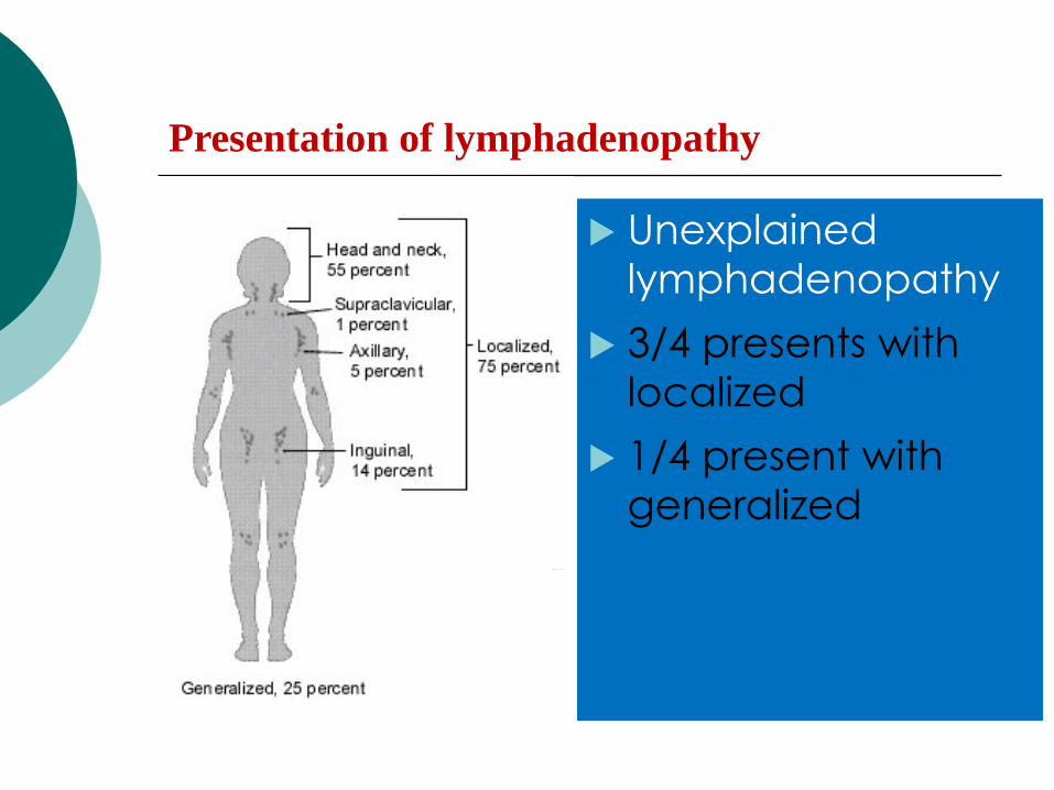
Unexplained
lymphadenopathy
3/4 presents with
localized
1/4 present with
generalized
INITIAL EVALUATION
History
1. lymph node enlargement
2. associated symptoms (local and systemic)
3. potential exposures
4. past medical history
associated symptoms
o Local symptoms of infection
Cough: pneumonia (bacterial, viral, fungal)
Sore throat: GAS, adenovirus, diphtheria
Skin lesions Staphylococcus aureus, HSV, cat scratch disease
Constitutional symptoms
May indicate malignancy, Mycobacterium tuberculosis, rheumatologic disease
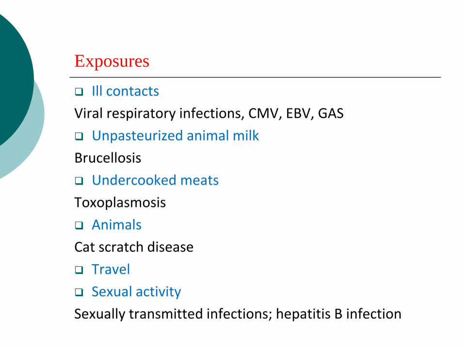
Exposures
Ill contacts
Viral respiratory infections, CMV, EBV, GAS
Unpasteurized animal milk
Brucellosis
Undercooked meats
Toxoplasmosis
Animals
Cat scratch disease
Travel
Sexual activity
Sexually transmitted infections; hepatitis B infection
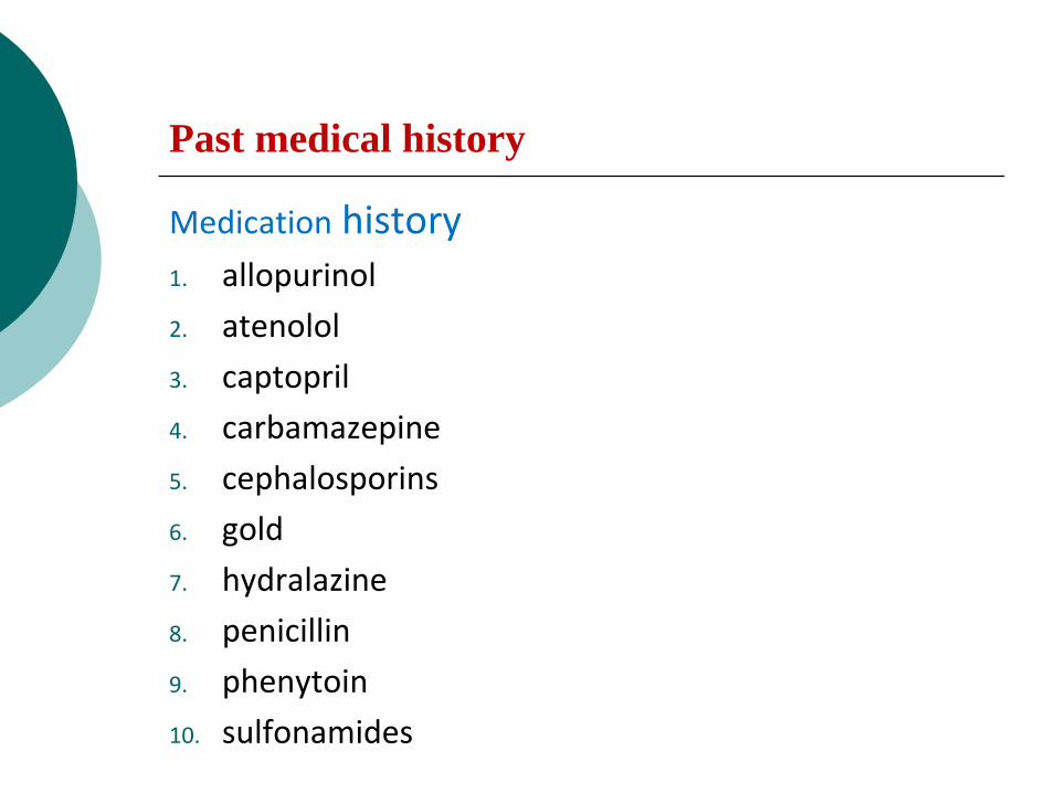
Past medical history
Medication history 1. allopurinol
2. atenolol
3. captopril
4. carbamazepine
5. cephalosporins
6. gold
7. hydralazine
8. penicillin
9. phenytoin
10. sulfonamides
Past medical history
recurrent infections, skin abscesses, suppurative adenitis
Chronic granulomatous disease
Autoimmune disease
Autoimmune lymphoproliferative syndrome
Immunization status
Diphtheria, measles, rubella
Physical examination
General examination
Systemic examination
Lymph nodes
Physical examination
General examination
Vital signs
Weight loss of >10 percent of body weight may be indicative of malignancy.
Fever indicative of infection .
Head, eyes, ears, nose, throat
Conjunctival injection – Kawasaki disease
Oropharynx – Dental problems, pharyngitis, herpangina
Systemic examination
Chest and CVS
Abdomen
Hepatosplenomegaly [EBV], [CMV], brucellosis, [HIV]
Skin
Localized lesions (cat scratch disease)
generalized rash (viral illness)
Lymph nodes
Remember:
Normal lymph nodes are not palpable
Examine the draining lymph nodes area of any lesion
Examine the area drained by affected lymph nodes
Lymph nodes
An examination of the lymph nodes forms part of the routine for most body systems.
no need to percuss or auscultate, examination involves inspection followed by palpation
Lymph nodes
The following groups of lymph nodes are to be examined:
1- Cervical groups
2- Axillary groups
3- Inguinal groups
4- Epitrochlear lymph nodes.
5- Remember that the liver and spleen are parts of the lymphoid tissue
Lymph nodes
SSSSS (5S):
1- Site.
2- Shape.
3- Size.
4- Surface: Smooth, nodular, irregular.
5- Skin overlying the swelling (scars, colour…).
Lymph nodes
Confirm your inspection
Size
Temprature
Tenderness
Consistency
Mobility
Draining area
Lymph nodes Location
Localized lymphadenopathy
present in only one region generally suggests local causes
search for pathology in the area of node drainage
Generalized adenopathy
present in two or more noncontiguous regions
usually is a manifestation of systemic disease
Lymph nodes Size
normal lymph nodes are <1 cm in diameter.
epitrochlear region usually are less than 0.5 cm in diameter
normal lymph nodes in the inguinal region usually are less than 1.5 cm in diameter.
The risk of malignancy is increased in lymph nodes >2 cm in diameter
Consistency
Fluctuance usually indicates infection usually S. aureus or group A Streptococcus
Hard nodes generally are due to cancer or previous inflammation.
Firm, rubbery nodes may indicate lymphoma or chronic leukemia
Matting
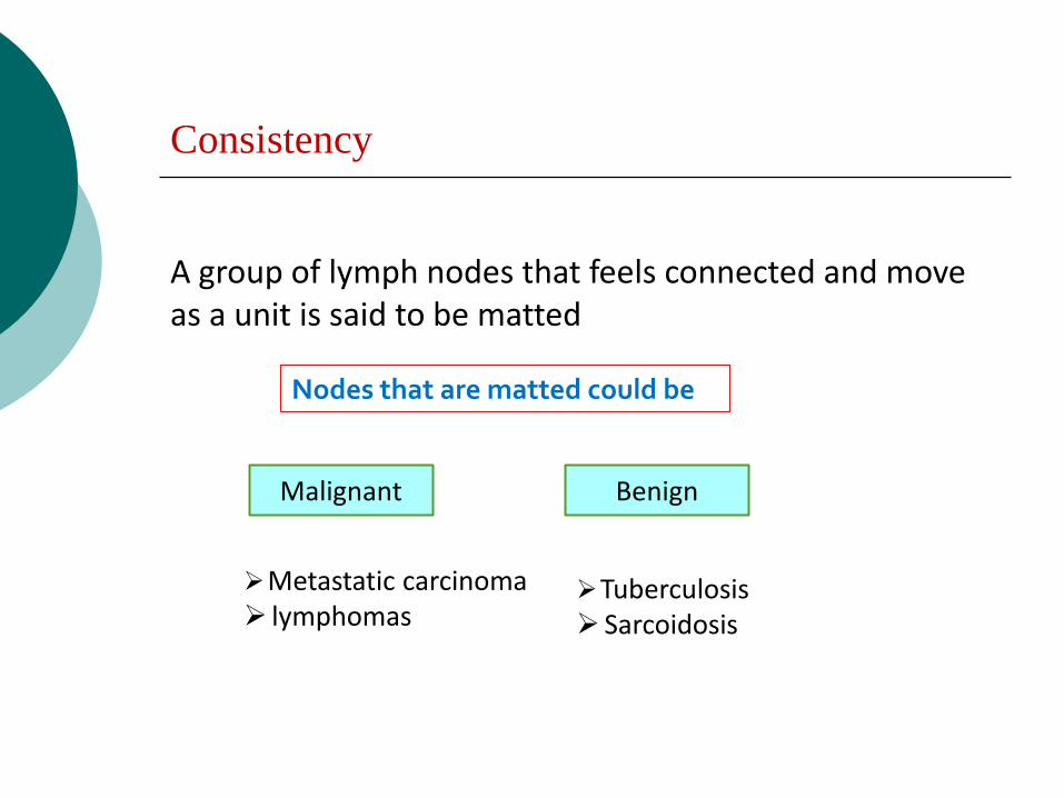
Consistency
A group of lymph nodes that feels connected and move as a unit is said to be matted
Nodes that are matted could be
Malignant Benign
Metastatic carcinoma lymphomas
Tuberculosis Sarcoidosis
Fixation
Normal lymph nodes are freely movable
fixed to adjacent tissues (eg, deep fascia)
invading cancers
inflammation in tissue surrounding the nodes.
Tenderness
Indication of rapid increase in size: stretch of capsular shell
NOT useful in determining benign vs malignant state
Inflammation, suppuration, hemorrhage
GENERAL PRINCIPLES
Urgency and extent of evaluation
Step-wise approach
Worrisome features
glucocorticoids
Urgency and extent of evaluation
generally is benign and self-limited.
It is not necessary to identify the underlying etiology in every patient
is determined by how ill the patient appears
the lymphadenopathy resolves without explanation before invasive diagnostic testing is undertaken.
the evaluation of LAP in children occurs in stages over approximately four weeks
Step-wise approach
The first stage
treat conditions that appear obvious based upon the history and examination eg, throat culture for group A streptococcal pharyngitis.
the second stage
provide a two-week trial of antibiotic therapy or a two- to three week period of observation.
Step-wise approach
If the cause remains uncertain, less common causes and causes that require specific treatment (eg, tuberculosis) are evaluated.
after four weeks, the diagnosis remains uncertain and the lymph node has not regressed in size, biopsy may
be warranted.

Worrisome features
Systemic symptoms
fever >1 week
night sweats
weight loss [>10 percent of body weight]
Supraclavicular nodes
Fixed, nontender nodes
Lymph nodes >2 cm in diameter that have increased in size
Worrisome features
Abnormal chest radiograph
particularly mediastinal mass or hilar adenopathy
Abnormal CBC and differential
lymphoblasts, cytopenias in more than one cell line
Persistently elevated ESR/CRP
rising ESR/CRP despite antibiotic therapy
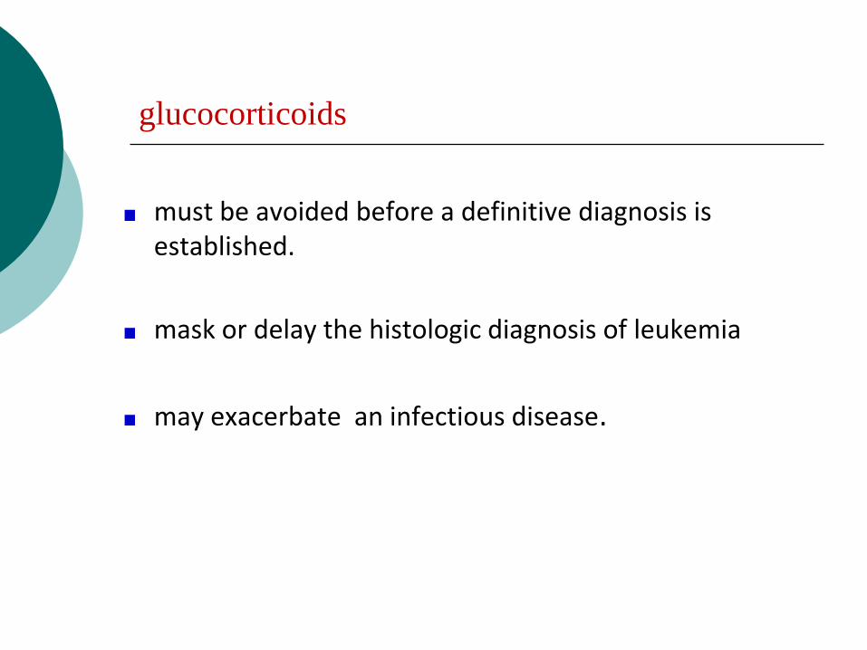
glucocorticoids
must be avoided before a definitive diagnosis is established.
mask or delay the histologic diagnosis of leukemia
may exacerbate an infectious disease.
Generalized lymphadenopathy
differential diagnosis
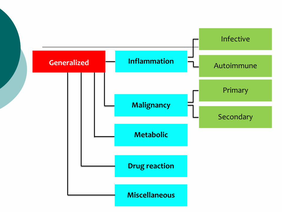
Generalized Inflammation
Infective
Autoimmune
Malignancy
Primary
Secondary
Metabolic
Drug reaction
Miscellaneous
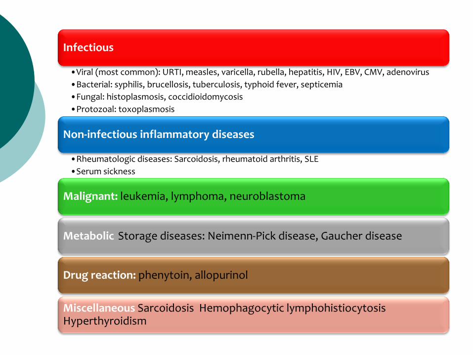
Infectious
•Viral (most common): URTI, measles, varicella, rubella, hepatitis, HIV, EBV, CMV, adenovirus
•Bacterial: syphilis, brucellosis, tuberculosis, typhoid fever, septicemia
•Fungal: histoplasmosis, coccidioidomycosis
•Protozoal: toxoplasmosis
Non-infectious inflammatory diseases
•Rheumatologic diseases: Sarcoidosis, rheumatoid arthritis, SLE
•Serum sickness
Malignant: leukemia, lymphoma, neuroblastoma
Metabolic Storage diseases: Neimenn-Pick disease, Gaucher disease
Drug reaction: phenytoin, allopurinol
Miscellaneous Sarcoidosis Hemophagocytic lymphohistiocytosis Hyperthyroidism
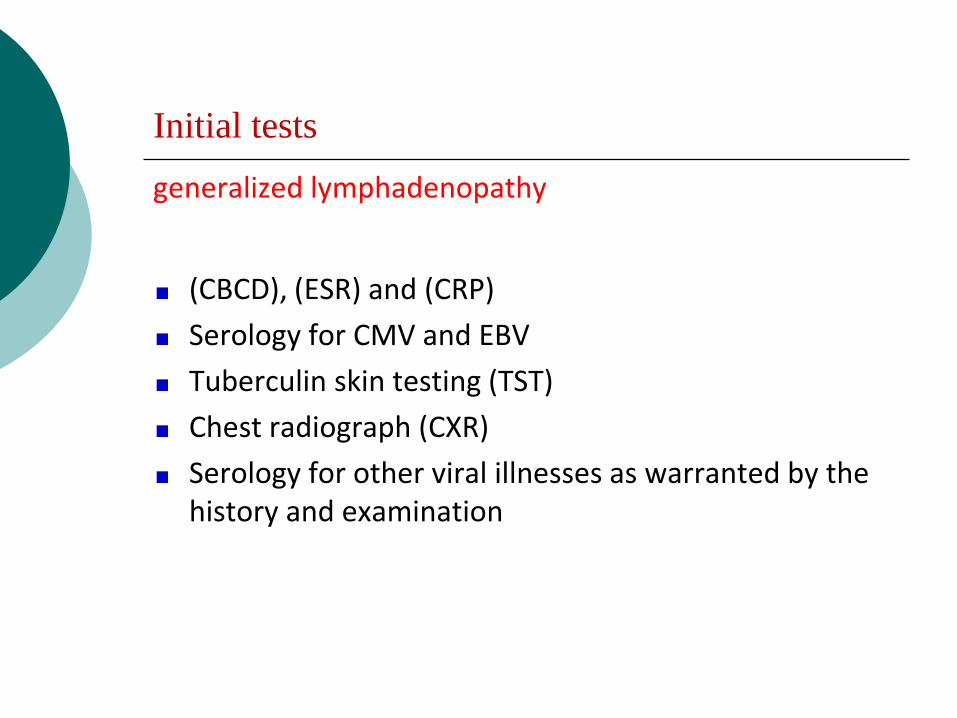
Initial tests
generalized lymphadenopathy
(CBCD), (ESR) and (CRP)
Serology for CMV and EBV
Tuberculin skin testing (TST)
Chest radiograph (CXR)
Serology for other viral illnesses as warranted by the history and examination
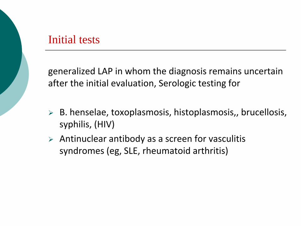
Initial tests
generalized LAP in whom the diagnosis remains uncertain after the initial evaluation, Serologic testing for
B. henselae, toxoplasmosis, histoplasmosis,, brucellosis, syphilis, (HIV)
Antinuclear antibody as a screen for vasculitis syndromes (eg, SLE, rheumatoid arthritis)
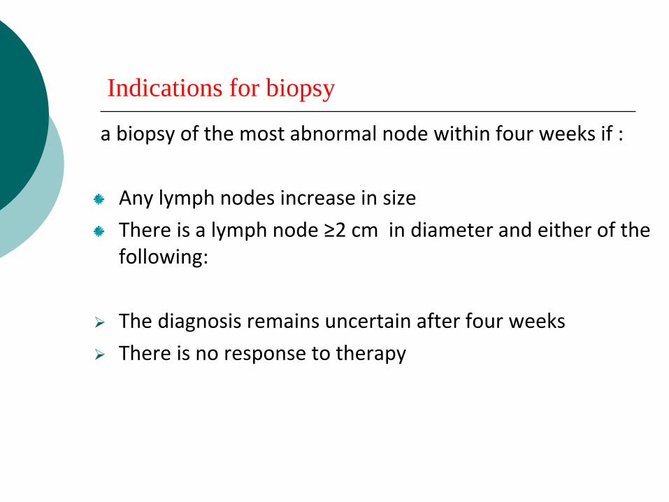
Indications for biopsy
a biopsy of the most abnormal node within four weeks if :
Any lymph nodes increase in size
There is a lymph node ≥2 cm in diameter and either of the following:
The diagnosis remains uncertain after four weeks
There is no response to therapy
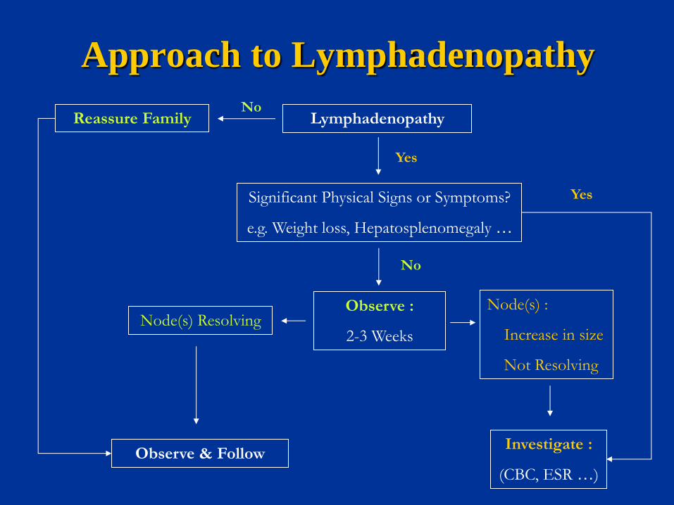
Approach to Lymphadenopathy
Lymphadenopathy No
Reassure Family
Yes
Significant Physical Signs or Symptoms?
e.g. Weight loss, Hepatosplenomegaly …
Investigate :
(CBC, ESR …)
Observe :
2-3 Weeks
Node(s) :
Increase in size
Not Resolving
Node(s) Resolving
Observe & Follow
Yes
No
Localized adenopathy
The causes of localized adenopathy vary with the lymph
node region
Localized adenopathy
benign clinical history
an unremarkable physical examination
no constitutional symptoms
should be reexamined in three to four weeks

Localized adenopathy
constitutional symptoms or signs
risk factors for malignancy
lymphadenopathy that persists for three to four weeks
should undergo a biopsy.
Cervical lymphadenopathy
DEFINITIONS
Cervical lymphadenopathy –Enlarged lymph node(s) of the neck, including preauricular, parotid,
Acute lymphadenitis – Develops over a few days
Subacute/chronic lymphadenitis Develops over weeks to months.
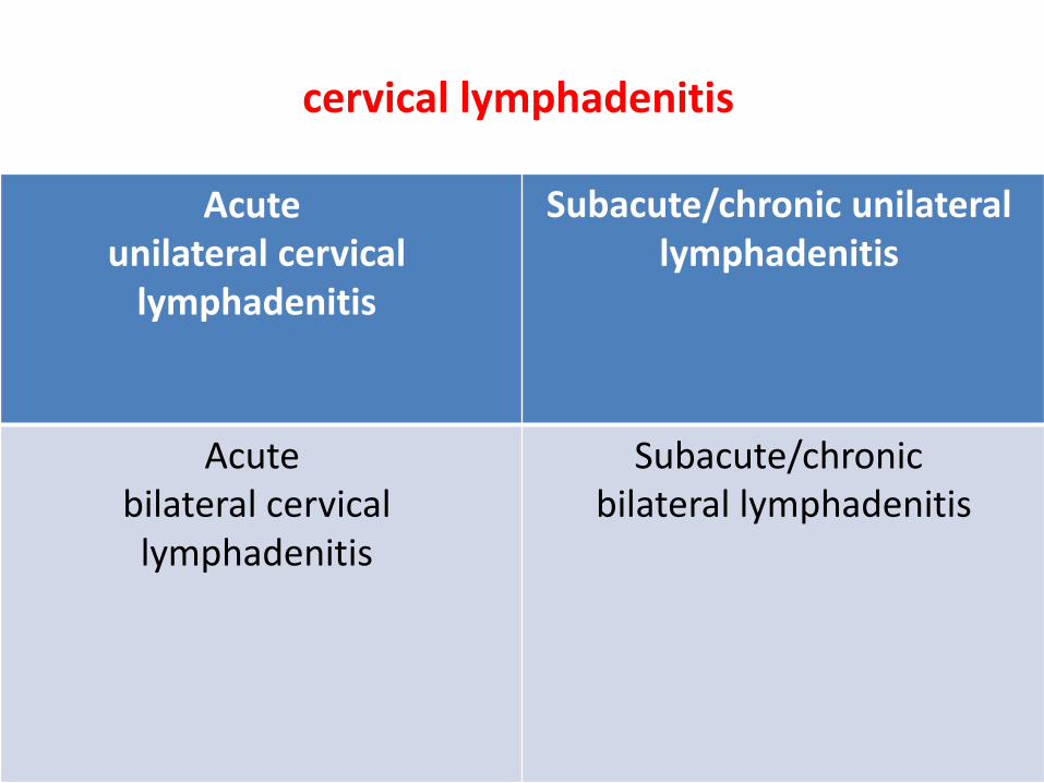
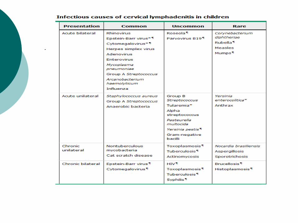
cervical lymphadenitis
Subacute/chronic unilateral lymphadenitis
Acute
unilateral cervical lymphadenitis
Subacute/chronic
bilateral lymphadenitis
Acute
bilateral cervical lymphadenitis
Acute bilateral cervical lymphadenitis
Caused by self-limited viral upper respiratory infection
enterovirus
adenovirus
influenza virus
The LN(reactive LN) typically are
small, rubbery
mobile, discrete
minimally tender
No erythema or warmth
Acute unilateral cervical lymphadenitis
is usually caused by bacteria
S. Aureus
GAS
S. aureus and GAS
-occur in children younger than 5 years of age
-history of a recent URI or impetigo
-Submandibular nodes are affected in more than 50 %
-The lymph node usually is
3 to 6 cm in diameter
tender
warm
erythematous
poorly mobile
-One-fourth to one-third of infected nodes suppurate and become fluctuant
Subacute/chronic bilateral cervical lymphadenitis
Most often caused by EBV or CMV infection
EBV causes infectious mononucleosis
manifest as
Fever,
exudative pharyngitis
lymphadenopathy
hepatosplenomegaly
Subacute/chronic unilateral cervical lymphadenitis
Nontuberculous mycobacteria (NTM) infections
Bartonella henselae-cat scratch disease (CSD)
TB
Toxoplasmosis
Axillary lymphadenopathy
• Local infection
• Cat scratch disease
• Brucellosis
• Reactions to immunizations
• Non Hodgkin lymphoma
• Juvenile rheumatoid arthritis
Axillary
Inguinal lymphadenopathy
Local infection Diaper dermatitis Syphilis Genital herpes
Inguinal
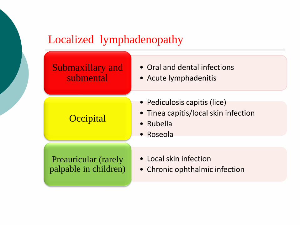
Localized lymphadenopathy
• Oral and dental infections
• Acute lymphadenitis Submaxillary and
submental
• Pediculosis capitis (lice)
• Tinea capitis/local skin infection
• Rubella
• Roseola
Occipital
• Local skin infection
• Chronic ophthalmic infection Preauricular (rarely palpable in children)
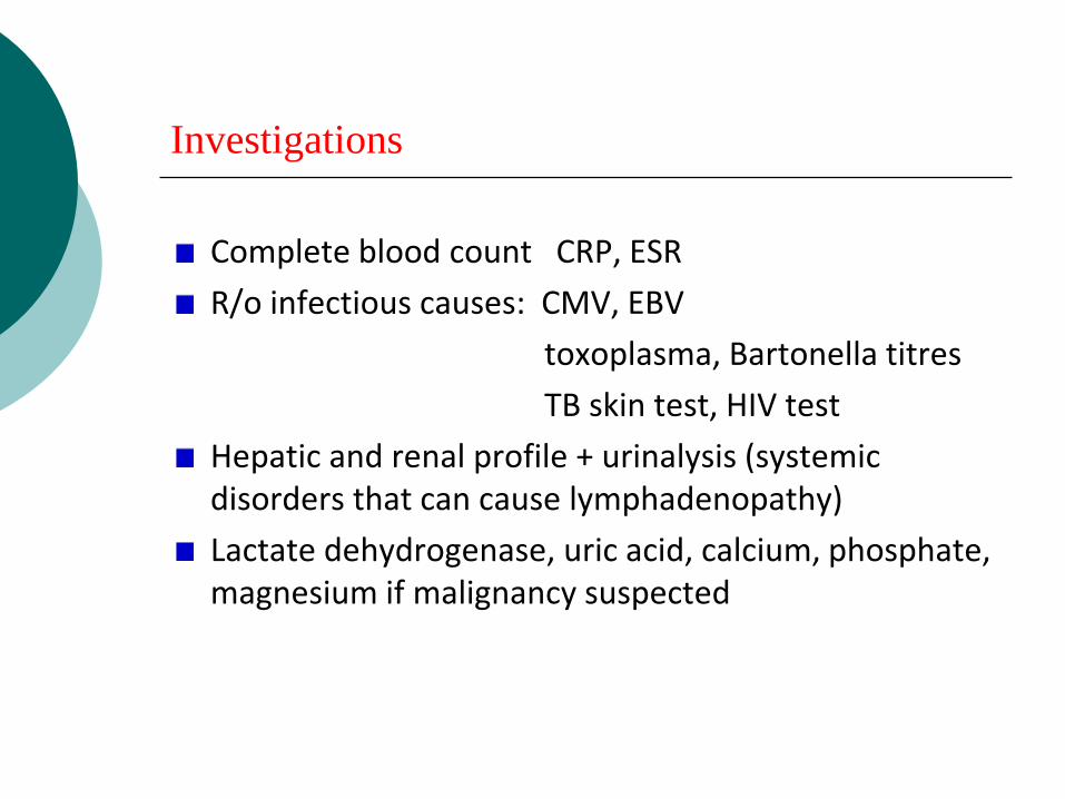
Investigations
Complete blood count CRP, ESR
R/o infectious causes: CMV, EBV
toxoplasma, Bartonella titres
TB skin test, HIV test
Hepatic and renal profile + urinalysis (systemic disorders that can cause lymphadenopathy)
Lactate dehydrogenase, uric acid, calcium, phosphate, magnesium if malignancy suspected
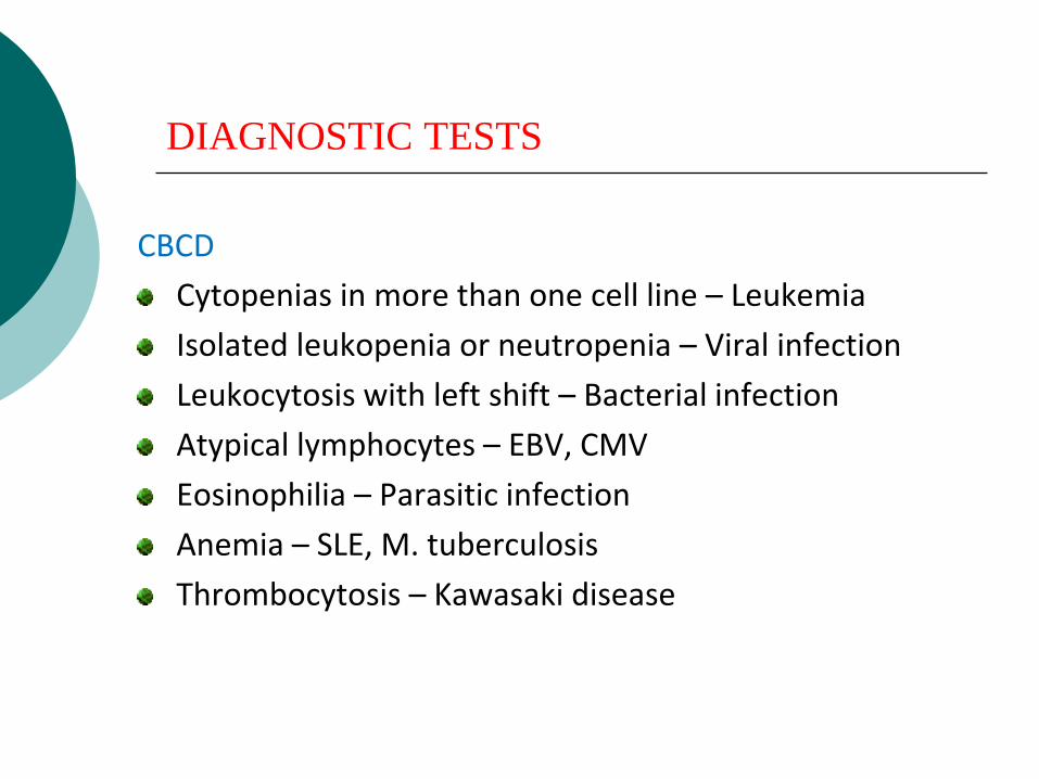
DIAGNOSTIC TESTS
CBCD
Cytopenias in more than one cell line – Leukemia
Isolated leukopenia or neutropenia – Viral infection
Leukocytosis with left shift – Bacterial infection
Atypical lymphocytes – EBV, CMV
Eosinophilia – Parasitic infection
Anemia – SLE, M. tuberculosis
Thrombocytosis – Kawasaki disease
DIAGNOSTIC TESTS
ESR and CRP
acute phase reactants
a nonspecific marker of inflammation
Persistent or increasing elevation may warrant biopsy
DIAGNOSTIC TESTS
Tuberculin skin test
TST is indicated to screen for M. tuberculosis infection.
Chest radiograph
(CXRs) in children with peripheral LAP are made on a case-by-case basis.
DIAGNOSTIC TESTS
generally obtain chest radiographs in children with:
Generalized LAP at the time of presentation
Supraclavicular LAP at the time of presentation
Cervical or inguinal adenopathy ≥2 cm in diameter who do not have signs or symptoms of infection
DIAGNOSTIC TESTS
Ultrasonography
the lymph node may be helpful in the presence and extent of an abscess
Abdominal ultrasonography may be warranted in children with unexplained inguinal adenopathy
Lymph node biopsy
Lymph node cultures material may be obtained via
excisional biopsy
needle aspiration
incision and drainage
abscess fluid should be sent for
1. Gram stain
2. bacterial culture (aerobic and anaerobic), mycobacterial stain and culture
3. fungal stain and culture.

Lymph node biopsy
indicated in patients with worrisome features
prefer open biopsy to fine needle aspiration
Largest node
Avoid inguinal & axillary
Supra clavicular-highest diagnostic yield
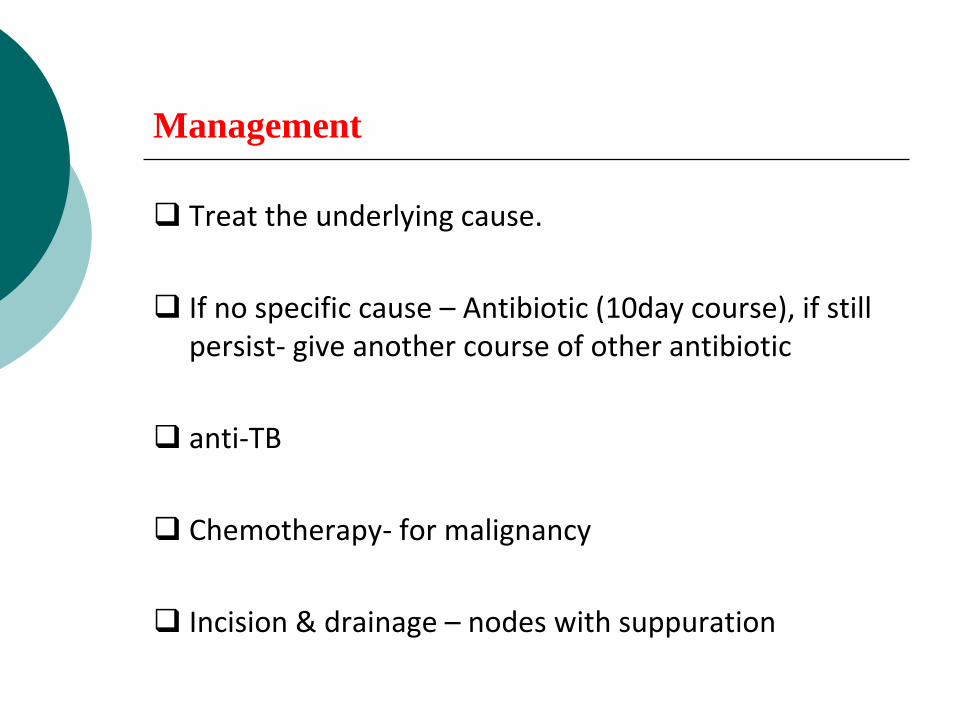
Management
Treat the underlying cause.
If no specific cause – Antibiotic (10day course), if still persist- give another course of other antibiotic
anti-TB
Chemotherapy- for malignancy
Incision & drainage – nodes with suppuration
Acute bilateral LN
self-limited viral illness.
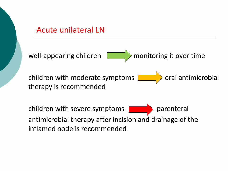
Acute unilateral LN
well-appearing children monitoring it over time
children with moderate symptoms oral antimicrobial therapy is recommended
children with severe symptoms parenteral
antimicrobial therapy after incision and drainage of the inflamed node is recommended
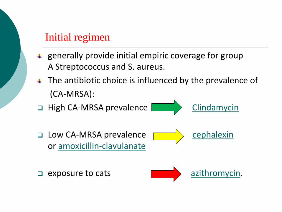
Initial regimen
generally provide initial empiric coverage for group A Streptococcus and S. aureus.
The antibiotic choice is influenced by the prevalence of
(CA-MRSA):
High CA-MRSA prevalence Clindamycin
Low CA-MRSA prevalence cephalexin or amoxicillin-clavulanate
exposure to cats azithromycin.
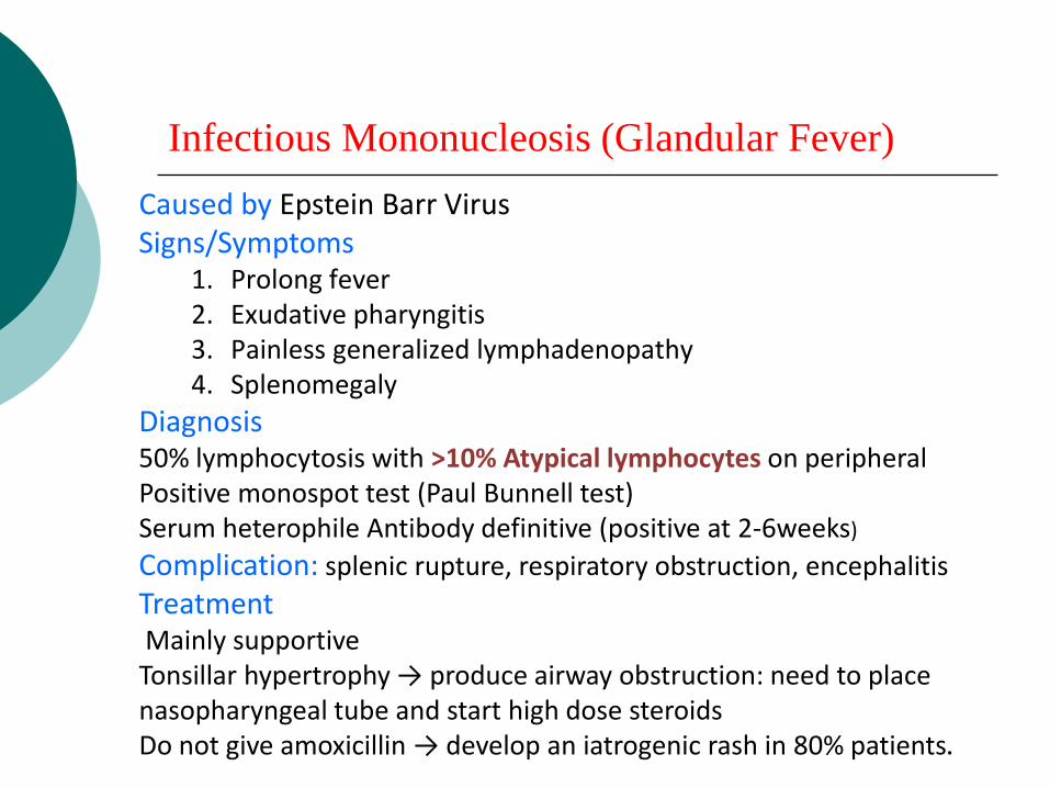
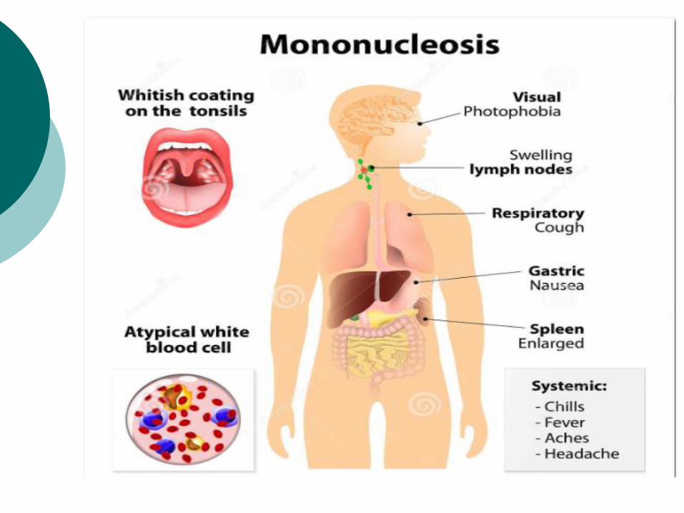
Infectious Mononucleosis (Glandular Fever)
Caused by Epstein Barr Virus Signs/Symptoms
1. Prolong fever 2. Exudative pharyngitis 3. Painless generalized lymphadenopathy 4. Splenomegaly
Diagnosis 50% lymphocytosis with >10% Atypical lymphocytes on peripheral Positive monospot test (Paul Bunnell test) Serum heterophile Antibody definitive (positive at 2-6weeks)
Complication: splenic rupture, respiratory obstruction, encephalitis
Treatment Mainly supportive Tonsillar hypertrophy → produce airway obstruction: need to place nasopharyngeal tube and start high dose steroids Do not give amoxicillin → develop an iatrogenic rash in 80% patients.
Kawasaki Disease
Five Characteristics of Disease
Fever >5 days
Cervical lymphadenopathy (usually unilateral)
Erythema and edema of palms and soles with desquamation of skin
Nonpurulent Bilateral Conjunctivitis
Strawberry Tongue
Complications
Coronary artery aneurysms
Coronary artery thromboses
Myocardial infarction
Treatment
IVIG and Aspirin

Kawasaki Disease