aquaporin-1 expression in glial tumors february 1, 2005
TRANSCRIPT

EXPERIMENTAL STUDIES
EXPRESSION OF THE AQUAPORIN-1 WATER CHANNEL
IN HUMAN GLIAL TUMORS
Kotaro Oshio, M.D., Ph.D.Department of NeurologicalSurgery, University of California,San Francisco,San Francisco, California
Devin K. Binder, M.D.,Ph.D.Department of NeurologicalSurgery, University of California,San Francisco,San Francisco, California
Yu Liang, Ph.D.Brain Tumor Research Center, andDepartment of NeurologicalSurgery, University of California,San Francisco,San Francisco, California
Andrew Bollen, M.D.Brain Tumor Research Center, andDepartment of Pathology,University of California,San Francisco,San Francisco, California
Burt Feuerstein, M.D.,Ph.D.Brain Tumor Research Center, andDepartment of NeurologicalSurgery, University of California,San Francisco,San Francisco, California
Mitchel S. Berger, M.D.Brain Tumor Research Center, andDepartment of NeurologicalSurgery, University of California,San Francisco,San Francisco, California
Geoffrey T. Manley, M.D.,Ph.D.Department of NeurologicalSurgery, University of California,San Francisco,San Francisco, California
Reprint requests:Geoffrey T. Manley, M.D., Ph.D.,Department of NeurologicalSurgery, University of California,San Francisco,1001 Potrero Avenue, Building 1,Room 101,San Francisco, CA 94110.Email: [email protected]
Received, January 14, 2004.
Accepted, August 11, 2004.
OBJECTIVE: Malignant glial tumors are associated with cerebral edema. The aqua-porins (AQPs) are a family of membrane proteins that provide a major pathway forwater transport in mammals. In the central nervous system, AQP1 is selectivelyexpressed in the choroid plexus and thought to participate in cerebrospinal fluidproduction. Prior studies have suggested that AQP1 may be up-regulated in glialtumors, potentially contributing to tumor-associated edema. The objective of this studywas to investigate the expression of AQP1 in a large series of human glial tumors.METHODS: Thirty-six human glial tumors were obtained from the University ofCalifornia, San Francisco Neurosurgery Tissue Bank. AQP1 expression was evaluatedby reverse transcriptase polymerase chain reaction, complementary deoxyribonucleicacid gene array, Western blot analysis, and immunohistochemical analyses.RESULTS: AQP1, normally restricted to choroid epithelia, was highly expressed inglioblastomas. Complementary deoxyribonucleic acid array, Western blot analysis,and immunohistochemical analysis revealed intense up-regulation of AQP1 expres-sion in all glioblastomas studied.CONCLUSION: The abnormal up-regulation of AQP1 in glial tumors suggests apotential pathological role for this membrane water channel and raises the possibilitythat selective AQP1 inhibition might offer a new therapeutic target for treatment oftumor-associated edema.
KEY WORDS: Aquaporins, Brain tumor, Edema, Gene array, Reverse transcriptase-polymerase chainreaction
Neurosurgery 56:375-381, 2005 DOI: 10.1227/01.NEU.0000148904.57841.6B www.neurosurgery-online.com
Afamily of molecular water channelscalled aquaporins (AQPs) has beenidentified in mammals. AQPs are small
integral membrane proteins (molecularweight, �30,000) that provide a major path-way for water transport in the kidney, brain,secretory epithelia, and other organs (16). Sev-eral AQPs have been identified in regions ofthe central nervous system that are thought toparticipate in the production and absorptionof brain fluid (2, 6, 17). AQP1, first identifiedin red blood cells and renal proximal tubularepithelium (3), is expressed in the ventricular-facing membrane of the choroid plexus epi-thelium. In the kidney, AQP1 functions pri-marily as a water pore to facilitatetransmembrane transport of water driven byosmotic gradients. The specific localization ofAQP1 in the choroid plexus suggests that itplays an important role in facilitating water
transport across the choroid plexus epithe-lium apical membrane during secretion of ce-rebrospinal fluid (5, 8, 13). AQP4 is expressedin astrocytic end-feet at the blood-brain bar-rier and plays an important role in cerebralwater balance and development of brainedema (6). AQP9 is also expressed in astro-cytes and is up-regulated after ischemia (1).
Expression of AQPs has been documentedin a small group of human brain tumors. Endoet al. (4) demonstrated AQP1 immunoreactiv-ity on glioblastoma cell lines implanted in ro-dents. Differential gene expression analysisidentified up-regulation of AQP1 in four pri-mary human glioblastomas multiforme (7).Saadoun et al. (11) confirmed AQP1 up-regulation by immunohistochemistry in fivehigh-grade astrocytomas. AQP4, which nor-mally is expressed on the foot processes ofastrocytes, also seems to be up-regulated in
NEUROSURGERY VOLUME 56 | NUMBER 2 | FEBRUARY 2005 | 375

glioblastomas (12). The purpose of this study was to charac-terize AQP1 expression in larger number of human glial tu-mors by reverse transcriptase-polymerase chain reaction (RT-PCR), Western blot, immunohistochemistry, and differentialgene expression analysis.
PATIENTS AND METHODS
RT-PCR Analysis
Human brain tumors were immediately homogenized inTrizol reagent (Invitrogen Corp., Carlsbad, CA) for messengerribonucleic acid (mRNA) isolation by use of the OligotexmRNA mini kit (Qiagen, Studio City, CA). The complemen-tary deoxyribonucleic acids (cDNAs) were generated frommRNA by SuperScriptII (Invitrogen Corp.) ribonuclease Hreverse transcriptase primed with oligo(dT)18 and randomhexamers. After reverse transcription, PCR products were am-plified with gene-specific primers designed to amplify a por-tion of the coding sequences of each of the nine known humanAQPs: AQP1 through AQP9. The reactions contained 1 �l ofthe RT reaction as template cDNA and were performed for 30cycles, in a 30-second, 94°C denaturation step; a 30-second,60°C annealing step; and a 1-minute, 72°C extension step. PCRproducts were separated on 2% agarose gels and visualizedwith ethidium bromide staining.
Immunohistochemical Analysis
Fresh brain tumor tissue was fixed overnight in 10% forma-lin (Fisher Scientific, Pittsburgh, PA). The next day, the tissuewas dehydrated in ethyl alcohol and xylene, paraffin-mounted, and sectioned at 8-�m thickness. Sections were em-bedded on slides and stored at room temperature. For immu-nohistochemical analysis, slides were warmed to 60°C for 10minutes. Tissue was rehydrated and rinsed in 1� phosphate-buffered saline (PBS) (pH 7.4). Endogenous peroxidase activ-ity was quenched by immersion in 3% H2O2 in methanol for10 minutes. A nonspecific blocking procedure with 5% normalgoat serum (Vector Laboratories, Inc., Burlingame, CA) wasperformed before application of primary antibodies. Affinity-purified rabbit polyclonal antirat AQP1 antibody (ChemiconInternational, Temecula, CA) was used. This antibody israised against a 19-amino acid synthetic peptide (251–269)from the cytosolic carboxy-terminal domain of AQP1. Theslides were incubated overnight in anti-AQP1 antibody (1:1000) at 4°C in PBS containing 5% normal goat serum, pH 7.4.The next day, sections were incubated in a 1:500 biotinylatedgoat antirabbit immunoglobulin G secondary antibody (Vec-tor Laboratories, Inc.) for 1 hour at room temperature in PBScontaining 5% normal goat serum, pH 7.4, and then incubatedin 1:100 Vectastain ABC reagent (Vector Laboratories, Inc.) for1 hour at room temperature in PBS containing 5% normal goatserum, pH 7.4. Diaminobenzidine (Sigma Chemical Co., St.Louis, MO) was used to develop the reaction product. Aftermultiple rinses in PBS and a final rinse in distilled water,
sections were counterstained with hematoxylin (Biomeda,Foster City, CA), dehydrated, cleared, and coverslipped.
Western Blot Analysis
Western blot analysis was performed on fresh frozen braintumor tissue or on control brain (obtained from resection forepilepsy). Samples were homogenized in buffer (250 mmol/Lsucrose; 10 mmol/L Tris·HCl, pH 7.4; and 20 �g/ml phenyl-methylsulfonyl fluoride) and centrifuged at 1000 rpm for 10minutes at 4°C. The soluble fraction was dissolved in sodiumdodecyl sulfate (Pierce, Rockford, IL) and centrifuged at 10000rpm for 10 minutes at 4°C. Protein concentration was deter-mined by Bradford assay (Bio-Rad, Hercules, CA). Ten micro-grams of each sample were loaded onto 12% Tris-glycine gels(Invitrogen Novex, Carlsbad, CA) in Tris-glycine sodium do-decyl sulfate buffer (Invitrogen Novex), and run at 90 V for 2hours at room temperature. The gel was transferred to apolyvinylidene fluoride membrane overnight at 4°C. Themembrane was rinsed with Tris-buffered saline (TBS)-T buffer(0.02 mol/L Tris, 0.225 mol/L NaCl, 0.1% Triton, and distilledwater), and then placed in blocking solution of TBS-T with 3%nonfat milk (Sigma Chemical Co.) for 30 minutes before ap-plication of primary antibodies at room temperature. Anti-AQP1 primary antibody (1:1000) was incubated overnight at4°C in TBS-T buffer containing 1% bovine serum albumin(Sigma Chemical Co.), pH 7.6. After three rinses in TBS-Tbuffer, the membrane was incubated in horseradishperoxidase-linked donkey anti-rabbit immunoglobulin G(Amersham Pharmacia, Piscataway, NJ) diluted 1:5000 inTBS-T buffer containing 1% bovine serum albumin, pH 7.6, for30 minutes at room temperature. After rinsing in TBS-T buffer,the membrane was incubated with ECL Plus chemilumines-cent detection reagents (Amersham Pharmacia Biotech, Pisca-taway, NJ) for 5 minutes and exposed to film (Hyperfilm ELC;Amersham Pharmacia Biotech).
Gene Array Analysis
Total RNA was extracted from freshly resected and snap-frozen tumor samples stored at �80°C with Trizol reagent.Both primary (n � 26) and recurrent (n � 4) glioblastomaswere analyzed in duplicate. In some cases, more than oneindependent sample from each tumor was processed to exam-ine the variability of gene expression within the tumor. Threeseparate samples of brain tissue (cerebellum, left motor cortex,and right motor cortex) were used as controls. A sample ofgliotic tissue without tumor also was included as a control.mRNA was isolated from the total RNA samples in accor-dance with the manufacturer’s protocol for the Fastrack kit(Invitrogen Corp.). Double-stranded cDNA was synthesizedwith a Life Technologies Superscript cDNA Synthesis System(Invitrogen Corp.) by use of oligo(dT)24 primers with a T7RNA polymerase promoter site added to its 3' end (Genset, LaJolla, CA). The isolated cDNA was used for in vitro transcrip-tion with the T7 Megascript system (Ambion, Austin, TX) inthe presence of biotin-11-cytidine 5'-triphosphate and biotin-
OSHIO ET AL.
376 | VOLUME 56 | NUMBER 2 | FEBRUARY 2005 www.neurosurgery-online.com

16-uridine 5'-triphosphate (Enzo Diagnostics, New York, NY).Next, 25 to 50 �g of the cRNA product in buffer (40 mmol/LTris-acetate, pH 8.1/100 mmol/L potassium acetate, 30mmol/L magnesium acetate) was fragmented at 94°C for 35minutes. It was then used as a hybridization mix with 0.1mg/ml herring sperm DNA (Sigma Chemical Co.), plus fourcontrol bacterial and phage cRNA (1.5 pmol/L BioB, 5 pmol/LBioC, 25 pmol/L BioD, and 100 pmol/L Cre) samples to serveas internal controls for hybridization efficiency as directed bythe manufacturer (Affymetrix, Santa Clara, CA). Aliquots ofthe hybridization cRNA mixtures (10 �g cRNA in 200 �lhybridization mix) were hybridized to each Genechip array(Affymetrix) and washed and scanned with the GeneArrayscanner G2500A (Hewlett Packard) according to proceduresdeveloped by the manufacturer (Affymetrix). Scanned outputfiles were visually inspected for hybridization artifacts andthen analyzed with Genechip 3.1 software. Arrays were scaledto an average intensity of 100 and analyzed independently.The expression level of the AQP1 transcript was calculated bydividing the level of AQP1 expression in the tumor sample bythe average level of AQP1 expression from normal brain (threeindependent samples) to provide the amount of fold increasein gene expression.
Magnetic Resonance Imaging Edema Index
Tumor volume was estimated on preoperative T1-weightedmagnetic resonance imaging scans (1 mm slice thickness) withgadolinium enhancement. Peritumoral brain edema was eval-uated on T2-weighted fast spin echo scans. Tumor and peri-tumoral brain edema volumes were approximated similar tothe method described by Tamiya et al. (14). Maximum per-pendicular diameters of tumor and peritumoral brain edemawere measured on axial scans, and the maximum extent in thecoronal plane was measured by integrating the number ofaxial 1-mm slices. Volumes were approximated by use of theformula for an ellipsoid (V � 4⁄3�abc), where a, b, and c areradii (half diameter of maximum extent). Edema index wascalculated as Vedema � Vtumor/Vtumor and as such was alwaysat least 1 (yields a value of 1 when no edema is present).
RESULTS
RT-PCR Analysis
RT-PCR was performed to identify transcripts encodinghuman AQPs in brain tumor specimens (Fig. 1). Reverse-transcribed cDNA prepared from human glioma specimenswas PCR-amplified with specific primers for human AQP1 toAQP9. As expected, the glial AQP water channel AQP4 wasexpressed in all glial tumor samples (data not shown). AQP1,which is normally restricted to choroid plexus epithelia, alsowas highly expressed in glioblastomas. Figure 1 shows a rep-resentative RT-PCR analysis of AQP1 expression in 4 of the 36tumors analyzed.
Immunohistochemical Analysis
Immunohistochemical analysis was performed on all tu-mors; representative data from two Grade IV glioblastomamultiforme tumors are shown (Fig. 2). Figure 2A shows typicalGrade IV histological characteristics with anaplastic glial cellsand foci of microvascular proliferation. The correspondingAQP1 immunoreactivity from the same tumor is shown inFigure 2B. Strong AQP1 immunoreactivity is observed on themembranes of the neoplastic astrocytes. It is interesting thatAQP1 immunoreactivity seemed to be localized to astrocytemembranes and was not evident in areas of microvascularproliferation. Figure 2C shows a different Grade IV tumor withneoplastic astrocytes and multiple mitoses. Again, the corre-
FIGURE 1. Representative RT-PCR analysis of human glioma specimens.RT-PCR analysis with AQP1-specific primers demonstrated the expressionof AQP1 (300-base pair PCR product) in glioma samples (Lanes 1–4).Human �-actin (661-base pair PCR product) also was amplified as aninternal control. Kidney mRNA was positive for both primer pairs (�). Asa negative control, a glioma sample was analyzed without reverse tran-scriptase (�). M, marker.
FIGURE 2. Photomicrographs showing representative immunohistochemi-cal analyses of AQP1 protein expression in human glioma specimens. A,glioblastoma multiforme showing multiple foci of microvascular prolifera-tion (hematoxylin and eosin). B, immunoperoxidase AQP1 stain showingmultiple negative foci of microvascular proliferation but strongly positiveimmunoreactivity on the membranes of the neoplastic astrocytes. C, glio-blastoma multiforme composed of neoplastic astrocytes with multiple mito-ses (hematoxylin and eosin). D, immunoperoxidase AQP1 stain showingstrong membrane immunoreactivity. Scale bars, 50 �m.
AQUAPORIN-1 EXPRESSION IN GLIOMAS
NEUROSURGERY VOLUME 56 | NUMBER 2 | FEBRUARY 2005 | 377

sponding AQP1 tumor immunoreactivity is localized primar-ily to glial membranes (Fig. 2D). AQP1 immunoreactivity incontrol brain was localized to the choroid plexus as describedpreviously (8) (data not shown).
Western Blot Analysis
Western blot analysis confirmed robust AQP1 protein ex-pression in gliomas (Fig. 3). In glial tumors of various grades(Grade II, n � 3; Grade III, n � 3; Grade IV, n � 4), there wasa dramatic up-regulation of AQP1 protein expression (Fig. 3).In addition, it seemed qualitatively that higher-grade astrocy-tomas (Grades III and IV) expressed more AQP1 protein thanlow-grade astrocytomas (Grade II) (Fig. 3). In nontumorousbrain (obtained from epilepsy resection), there was a faintband at 28 kDa, presumably reflecting low basal levels ofexpression of AQP1 or possible inclusion of a small amount ofchoroid plexus with the medial temporal lobe surgical speci-men (N, Fig. 3). Protein from cerebral cortex demonstrated noAQP1 band (�, Fig. 3). Protein from mouse and rat cortex alsofailed to demonstrate any AQP1 expression (data not shown).
Gene Array Analysis
To further study the expression of AQP1 in high-gradeastrocytomas, we studied data from an extensive cDNA mi-croarray analysis that included approximately 7000 humangenes and expressed sequence tags (as described in Patientsand Methods). We compared the expression levels of AQP1 in26 primary and 4 recurrent glioblastomas with normal brain(Table 1). For the primary tumors there were 13 men and 13women with a mean age of 63 years (range, 28–82 yr). Theaverage expression of AQP1 in these tumors was increased 4.1� 0.8-fold higher than that in normal brain. The mean age ofpatients with recurrent tumors was 37 years (range, 13–69 yr),and the patient group comprised two men and two women.Similar to the primary tumors, the expression of AQP1 inrecurrent tumors was increased an average of 3.9 � 1.3-fold. Inall samples, the increased expression of AQP1 was confirmed
by immunohistochemical and Western blot analysis (Figs. 2and 3). A control sample of gliotic tissue demonstrated noincrease in AQP1 expression.
Magnetic Resonance Imaging Edema Index
Of the tumors studied by Western blot analysis (four GradeIV, three Grade III, and three Grade II), we were able to obtainpreoperative magnetic resonance images to calculate edema
FIGURE 3. Western blot analysis of ACP1 protein in human gliomaspecimens. The dense band at approximately 28 kDa is visible in lanes cor-responding to glial tumors. �, positive control (kidney); �, negative con-trol (cortex); N, human temporal lobe (patient with epilepsy); Grade II,astrocytoma; Grade III, anaplastic astrocytoma; Grade IV, glioblastomamultiforme.
TABLE 1. Gene array analysis of aquaporin-1 expression inhuman glioblastomasa
Tumor type and patient sex/age (yr)Fold increase
in AQP1expressionb
PrimaryM/28 4.4F/38 3.0F/67 3.8F/69 3.9M/78 5.7M/66 4.6M/50 3.8M/82 4.4F/61 4.6F/71 3.2M/73 3.8F/61 4.1F/73 2.9M/77 4.9F/55 2.5M/62 4.1M/62 3.6F/73 4.0F/73 5.4M/56 4.2M/79 5.7F/58 3.9F/34 3.6M/67 4.1F/71 4.9M/62 4.5
SecondaryM/13 5.5F/69 3.4M/51 4.6F/54 2.4
No tumor (peritumoral gliosis)M/51 1
a AQP1, aquaporin-1.b Mean increase � standard error of the mean: primary tumors, 4.1 � 0.8(n � 26); secondary tumors, 3.9 � 1.3 (n � 4); no tumor, 1.
OSHIO ET AL.
378 | VOLUME 56 | NUMBER 2 | FEBRUARY 2005 www.neurosurgery-online.com

index in 70% (7 of 10). Edema index was estimated as de-scribed in Patients and Methods. Edema index was higher forhigh-grade (Grades III and IV) lesions (edema index, 7.4 � 2.9;n � 4) than for low-grade (Grade II) tumors (edema index, 1.7� 0.5; n � 3).
DISCUSSION
By use of RT-PCR, immunohistochemistry, and Westernblotting, we demonstrated dramatic up-regulation of themembrane water channel AQP1 in human glial tumors. TheDNA microarray analysis confirmed these results and dem-onstrated approximately a fourfold increase in AQP1 expres-sion in glioblastomas as compared with normal brain.
This study confirms and extends prior analyses of AQPexpression in glial tumors. Endo et al. (4) demonstrated AQP1immunoreactivity in glioblastoma cell lines implanted in ro-dents, and Markert et al. (7) identified up-regulation of AQP1in four primary human glioblastomas multiforme. A priorimmunohistochemical study confirmed AQP1 up-regulationin astrocytic tumors (five low-grade and five high-grade as-trocytomas) (11). In that study, there seemed to be a gradedincrease in AQP1 from low- to high-grade tumors. Our West-ern blot analysis of Grade II, III, and IV tumors also suggestsqualitatively that low-grade glial tumors may up-regulateAQP1 to a lesser extent than high-grade tumors but still dra-matically more than control brain. However, a larger numberof astrocytomas, in particular the low-grade lesions, will needto be analyzed to determine whether there are significantquantitative differences among different grades of tumors.
The immunohistochemical data suggest that the increase inAQP1 protein is localized primarily if not exclusively to mem-branes of neoplastic astrocytes. On the basis of cellular morphol-ogy, no AQP1 expression was observed in cerebral endothelium.The de novo expression of AQP1 in glial tumors could havesignificant functional significance for these cells. The exact mech-anism for the up-regulation of AQP1 in cells that do not normallyexpress this protein is unknown. Possible mechanisms includededifferentiation or loss of cell type-specific transcriptional reg-ulation. In addition, concomitant corticosteroid use may affectendogenous or tumor-related AQP1 expression. However, thisfactor is difficult to assess because the majority of these patientsare treated with high-dose corticosteroids. Corticosteroid regu-lation of AQP1 should be studied in appropriate animal modelsto address this issue.
We demonstrated recently that AQP4, the glial water channelnormally expressed in astrocytes, provides the principal route forwater transport in astrocytes (16). When AQP4 was deleted fromastrocytes, water permeability decreased by sevenfold. Thus,expression of AQP1 in addition to AQP4 in glial tumor cellscould significantly increase water permeability in these cells.However, further experiments with primary glial tumor cells,using our new calcein-quenching method for measurement ofcell membrane water permeability, will be required.
The demonstrated role of AQPs in transmembrane watertransport and their marked up-regulation in neoplastic glial
membranes suggests a potential role in formation of tumor-associated edema. A common property of malignant glialtumors is marked edema in surrounding brain tissue (peritu-moral edema), but the mechanism of this is uncertain (10). It ispossible that AQP1 up-regulation allows excess water to flowinto cells and out into surrounding brain interstitium, perhapsin combination with blood-brain barrier breakdown observedin glioma microvessels. We demonstrated recently that AQP1can increase water permeability fivefold in choroid plexusepithelial cells (9). Thus, it may be speculated that abnormallyhigh AQP1 expression in glial tumors may “re-set” the waterbalance in local areas of brain around the tumor or preventclearance of existing extracellular fluid. The clinical relevanceof these observations remains to be established. Tumor-associated edema is associated with significant clinical mor-bidity such as focal neurological dysfunction, mass effect, andseizures. Inhibition of AQP expression or function may offer anew therapeutic option for tumor-associated cerebral edema.The only known inhibitor of AQP1 is mercury chloride. Stud-ies are under way to identify nontoxic, potent, and selectiveAQP1 inhibitors with high-throughput screening (15). Futurestudies are required to determine whether inhibition of AQP1up-regulation by genetic modification or direct inhibition ofAQP1 protein via selective inhibitors will abrogate tumor-associated edema and improve clinical outcome.
REFERENCES
1. Badaut J, Hirt L, Granziera C, Bogousslavsky J, Magistretti PJ, Regli L:Astrocyte-specific expression of aquaporin-9 in mouse brain is increasedafter transient focal cerebral ischemia. J Cereb Blood Flow Metab 21:477–482, 2001.
2. Badaut J, Lasbennes F, Magistretti PJ, Regli L: Aquaporins in brain: Distri-bution, physiology, and pathophysiology. J Cereb Blood Flow Metab 22:367–378, 2002.
3. Denker BM, Smith BL, Kuhajda FP, Agre P: Identification, purification, andpartial characterization of a novel Mr 28,000 integral membrane protein fromerythrocytes and renal tubules. J Biol Chem 263:15634–15642, 1988.
4. Endo M, Jain RK, Witwer B, Brown D: Water channel (aquaporin 1) expres-sion and distribution in mammary carcinomas and glioblastomas.Microvasc Res 58:89–98, 1999.
5. Hasegawa H, Zhang R, Dohrman A, Verkman AS: Tissue-specific expres-sion of mRNA encoding rat kidney water channel CHIP28k by in situhybridization. Am J Physiol 264:C237–C245, 1993.
6. Manley GT, Fujimura M, Ma T, Noshita N, Filiz F, Bollen AW, Chan P,Verkman AS: Aquaporin-4 deletion in mice reduces brain edema after acutewater intoxication and ischemic stroke. Nat Med 6:159–163, 2000.
7. Markert JM, Fuller CM, Gillespie GY, Bubien JK, McLean LA, Hong RL, LeeK, Gullans SR, Mapstone TB, Benos DJ: Differential gene expression profil-ing in human brain tumors. Physiol Genomics 5:21–33, 2001.
8. Nielsen S, Smith BL, Christensen EI, Agre P: Distribution of the aquaporinCHIP in secretory and resorptive epithelia and capillary endothelia. ProcNatl Acad Sci U S A 90:7275–7279, 1993.
9. Oshio K, Song Y, Verkman AS, Manley GT: Aquaporin-1 deletion reducesosmotic water permeability and cerebrospinal fluid production. ActaNeurochir Suppl (Wien) 86:525–528, 2003.
10. Papadopoulos MC, Saadoun S, Davies DC, Bell BA: Emerging molecularmechanisms of brain tumour oedema. Br J Neurosurg 15:101–108, 2001.
11. Saadoun S, Papadopoulos MC, Davies DC, Bell BA, Krishna S: Increasedaquaporin 1 water channel expression in human brain tumours. Br J Cancer87:621–623, 2002.
AQUAPORIN-1 EXPRESSION IN GLIOMAS
NEUROSURGERY VOLUME 56 | NUMBER 2 | FEBRUARY 2005 | 379

12. Saadoun S, Papadopoulos MC, Davies DC, Krishna S, Bell BA: Aquaporin-4expression is increased in oedematous human brain tumours. J NeurolNeurosurg Psychiatry 72:262–265, 2002.
13. Speake T, Whitwell C, Kajita H, Majid A, Brown PD: Mechanisms of CSFsecretion by the choroid plexus. Microsc Res Tech 52:49–59, 2001.
14. Tamiya T, Ono Y, Matsumoto K, Ohmoto T. 2001. Peritumoral brain edemain intracranial meningiomas: Effects of radiological and histological factors.Neurosurgery 49:1046–1052
15. Verkman AS: Drug discovery in academia. Am J Physiol Cell Physiol286:C465–C474, 2004.
16. Verkman AS, Mitra AK: Structure and function of aquaporin water chan-nels. Am J Physiol Renal Physiol 278:F13–F28, 2000.
17. Yamamoto N, Yoneda K, Asai K, Sobue K, Tada T, Fujita Y, Katsuya H,Fujita M, Aihara N, Mase M, Yamada K, Miura Y, Kato T: Alterations in theexpression of the AQP family in cultured rat astrocytes during hypoxia andreoxygenation. Brain Res Mol Brain Res 90:26–38, 2001.
COMMENTS
Water channels or aquaporins (AQPs) have received at-tention recently, and data are emerging that detail the
distribution and localization of these proteins in various tis-sues. Thus far, 10 mammalian AQP homologs have been iden-tified, and results of several studies have underscored theimportance of these proteins to basic physiology in a variety oforgan systems (1, 2). The distribution of these channels can besummarized as follows: AQP0, lens major intrinsic protein/lens fiber cells; AQP1, nonpigmented epithelial cells of theanterior chamber of the eye, bronchial vascular lining, choroidplexus, and the proximal tubules of the kidney; AQP2, renalcollecting ducts; AQP4, brain glia and retina; AQP5, cornealepithelial cells of the eye and apical membranes of lacrimaland salivary glands; and AQP6, renal collecting ducts. Withgene deletion in animal models, there is mounting evidence ofthe role of AQP in the pathogenesis of a variety of diseases,including nephrogenic diabetes insipidus (AQP1 and AQP2),congenital cataracts (AQP0), cerebral edema associated withtraumatic brain injury, cerebral ischemia and brain tumors(AQP4), and Sjögren’s syndrome (AQP5).
By use of reverse transcriptase polymerase chain reaction,complementary deoxyribonucleic acid gene array, Westernblot, and immunohistochemical analyses, Oshio et al. demon-strated a massive upregulation of AQP1 in 36 human glialtumors. Although the authors failed to demonstrate a quanti-tative relationship between AQP1 upregulation and gliomagrade, the data suggest a quantitative relationship. This articlecomplements the findings reported previously that also dem-onstrated a massive upregulation of AQP1 (5) as well as AQP4(3, 6) in human astrocytoma and metastatic adenocarcinoma.However, two important questions arise. First, although it iseasier to understand the upregulation of AQP4 in brain tu-mors (most abundantly expressed in the normal brain), rea-sons for upregulation of AQP1 (expressed in the choroidplexus in normal brain) are intriguing and more complex. Isthis abnormal upregulation of AQP1 a reflection of the factthat it is a more primitive channel phylogenetically? Little isknown regarding the distribution and evolution of AQP1 andother AQP in brain development, and it is plausible that there
is upregulation of this channel in brain tumors representingprimitive cell lines. Second, what is the effect of corticosteroidson AQP expression in the brain in general and tumors inparticular? This question is difficult to address because themajority of patients with gliomas are treated with corticoste-roids. However, it should be noted that the AQP1 promoterregion in other malignancies (erythroleukemia) has beenshown to possess steroid-influencing domains (4). Experimen-tal studies with mixed neuronal and glial cell cultures and inanimal models of brain tumor may provide unique insightsinto the evolution of tumor-associated cerebral edema. Fur-thermore, specific pharmacological inhibitors, lacking atpresent, will provide new insights into the role of this familyof proteins in health and disease and unravel important ther-apeutic targets in the future.
Anish BhardwajHenry BremBaltimore, Maryland
1. Agre P, King LS, Yasui M, Guggino WB, Ottersen OP, Fujiyoshi Y, Engel A,Nielsen S: Aquaporin water channels: From atomic structure to clinicalmedicine. J Physiol 542(Pt 1):3–16, 2002.
2. Kozono D, Yasui M, King LS, Agre P: Aquaporin water channels: Atomicstructure molecular dynamics meet clinical medicine. J Clin Invest 109:1395–1399, 2002.
3. Markert JM, Fuller CM, Gillespie GY, Bubien JK, McLean LA, Hong RL, LeeK, Gullans SR, Mapstone TB, Benos DJ: Differential gene expression profilingin human brain tumors. Physiol Genomics 5:21–33, 2001.
4. Moon C, King LS, Agre P: Aqp1 expression in erythroleukemia cells: Geneticregulation of glucocorticoid and chemical induction. Am J Physiol 273:C1562–C1570, 1997.
5. Saadoun S, Papadopoulos MC, Davies DC, Bell BA, Krishna S: Increasedaquaporin 1 water channel expression in human brain tumours. Br J Cancer87:621–523, 2002.
6. Saadoun S, Papadopoulos MC, Davies DC, Krishna S, Bell BA: Aquaporin-4expression is increased in oedematous human brain tumours. J NeurolNeurosurg Psychiatry 72:262–265, 2002.
The authors have demonstrated with convincing evidencethat AQP is increased in glial tumors. The role of AQP in
water regulation and transport raises interesting speculationthat this upregulation may relate to tumor-induced edema.This remains entirely speculative, as the majority of edemaseems to arise from a defective blood-brain barrier. The issueregarding whether corticosteroid use can influence AQP levelsor activity cannot be answered by this study, but this meritsfurther investigation.
Joseph M. PeipmeierNew Haven, Connecticut
The authors combined gene array, reverse transcriptasepolymerase chain reaction, Western blotting, and immu-
nohistochemistry to compare AQP1 expression with tumorgrade in a series of human glial tumors. They also make acomparison between magnetic resonance imaging indices ofedema and AQP1 expression.
OSHIO ET AL.
380 | VOLUME 56 | NUMBER 2 | FEBRUARY 2005 www.neurosurgery-online.com
They demonstrate that whereas AQP1 is normally restrictedto choroid epithelia, it is highly expressed in glioblastoma,suggesting a potential role for this membrane water channel intumor-induced edema. The corollary is that selective inhibi-tion of AQP1 might be of therapeutic benefit for patients inresolving symptoms. Such promise awaits further studies.
Nelson M. OyesikuAtlanta, Georgia
AQP1 is a cellular membrane protein associated with watertransport pathways. Because of the previously reported
expression of AQP1 in a small group of gliomas, the authors
hypothesized a role in tumor-associated brain edema andanalyzed its expression in a larger number of gliomas. Theyfound AQP1 to be up-regulated in high-grade gliomas and toa lesser extent in low-grade gliomas, although the data wereinsufficient to correlate with tumor grade to statistical signif-icance. As a preliminary investigation, this article confirms theassociation of AQP1 with gliomas; however, any clinical cor-relation with edema and histological findings or any potentialtherapeutic application will require further studies.
Jeffrey N. BruceNew York, New York
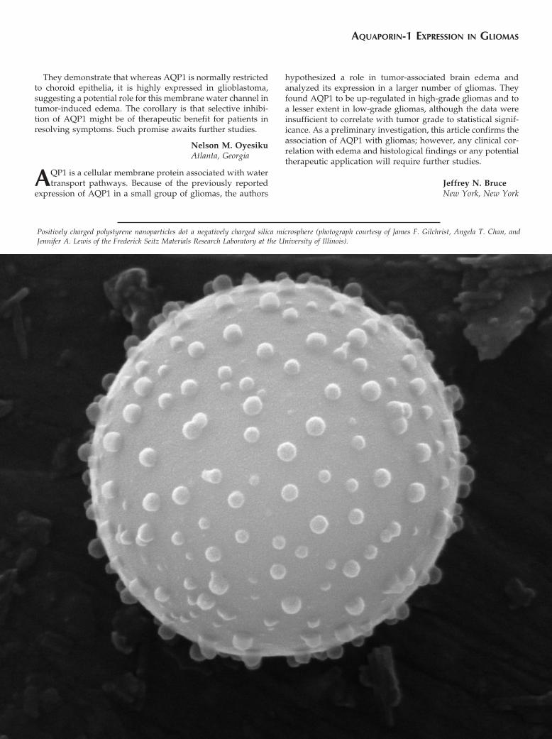
Positively charged polystyrene nanoparticles dot a negatively charged silica microsphere (photograph courtesy of James F. Gilchrist, Angela T. Chan, andJennifer A. Lewis of the Frederick Seitz Materials Research Laboratory at the University of Illinois).
AQUAPORIN-1 EXPRESSION IN GLIOMAS