architectures of multisubunit complexes revealed by a...
TRANSCRIPT

RESEARCH ARTICLE
Architectures of multisubunit complexes revealed by a visibleimmunoprecipitation assay using fluorescent fusion proteinsYohei Katoh*, Shohei Nozaki*, David Hartanto, Rie Miyano and Kazuhisa Nakayama‡
ABSTRACTIn this study, we elucidated the architectures of two multisubunitcomplexes, the BBSome and exocyst, through a novel application offluorescent fusion proteins. By processing lysates from cellsco-expressing GFP and RFP fusion proteins for immunoprecipitationwith anti-GFP nanobody, protein–protein interactions could bereproducibly visualized by directly observing the immunoprecipitatesunder a microscope, and evaluated using a microplate reader, withoutrequiring immunoblotting. Using this ‘visible’ immunoprecipitation (VIP)assay, wemappedbinarysubunit interactionsof theBBSomecomplex,and determined the hierarchies of up to four subunit interactions. Wealso demonstrated the assembly sequence of the BBSome around thecentrosome, and showed that BBS18 (also known as BBIP1 andBBIP10) serves as a linker between BBS4 and BBS8 (also known asTTC8). We also applied the VIP assay to mapping subunit interactionsof the exocyst tethering complex. By individually subtracting theeight exocyst subunits from multisubunit interaction assays, weunequivocally demonstrated one-to-many subunit interactions (Exo70with Sec10+Sec15, and Exo84 with Sec10+Sec15+Exo70). Thesimple, versatile VIP assay described here will pave the way tounderstanding the architectures and functions of multisubunitcomplexes involved in a variety of cellular processes.
KEYWORDS: Fluorescent fusion protein, Membrane traffic, Exocyst,BBSome, Protein–protein interaction, Anti-GFP nanobody
INTRODUCTIONFollowing the complete sequencing of the genomes of humans andother organisms, studies of the interactions between known proteinshave improved our understanding of protein function. Protein–protein interactions can be assessed by a variety of methods. Forexample, pulldown assays using glutathione S-transferase (GST)-fusion proteins to precipitate cellular proteins, which are oftenexogenously expressed as epitope-tagged proteins, have often beenemployed to study protein–protein interactions in vitro; likewise,co-immunoprecipitation assays using antibodies to short epitopesequences to precipitate exogenously expressed epitope-taggedproteins have been used for a similar purpose in cells. In both typesof assays, proteins interacting with the ‘bait’ proteins are usuallydetected by immunoblotting, which entails electrophoresis andsubsequent electroblotting onto membranes, followed by detectionwith primary and secondary antibodies and chemiluminescencereagents. In addition, genetic methods, such as the yeast two-hybridsystem, have also been frequently employed to reveal protein–
protein interactions. However, two-hybrid assays have often yieldedfalse-positive results and missed significant interactions (forexample, see supplementary material Fig. S1), although theyare higher in sensitivity and throughput than pulldown andco-immunoprecipitation assays.
Our group has been studying the regulation of intracellularprotein trafficking by the membrane trafficking machineries.Various multisubunit complexes participate in membranetrafficking processes, including tethering and fusion of carriervesicles with target membranes (Hong and Lev, 2014). Forexample, we have recently shown that the exocyst complexregulates tethering of transferrin-receptor-containing recyclingvesicles to the plasma membrane, downstream of the Rab11small GTPase (Takahashi et al., 2012). The exocyst is composedof eight subunits (Sec3, Sec5, Sec6, Sec8, Sec10, Sec15, Exo70,and Exo84 in yeast, and also known as EXOC1–EXOC8 inmammals) (Hsu et al., 1998; Terbush et al., 2001). However, thearchitecture of the complex is poorly understood (Liu and Guo,2012; Munson and Novick, 2006).
Protein trafficking to and/or within cilia is also mediated bymultisubunit complexes, including the BBSome, and intraflagellartransport (IFT)-A and IFT-B complexes (Sung and Leroux, 2013).The BBSome is composed of seven or eight subunits [BBS1, BBS2,BBS4,BBS5,BBS7,BBS8 (also known asTTC8),BBS9andBBS18(also known as BBIP1 and BBIP10)] and has been implicated inprotein transport to and/or within the cilia (Jin and Nachury, 2009;Loktev et al., 2008; Sung and Leroux, 2013). All the BBS proteinswere identified by mutations of their respective genes in patients withBardet–Biedl syndrome (BBS), a genetically heterogeneous disease(ciliopathy) characterized by a wide spectrum of clinical features,including rod–cone dystrophy, morbid obesity, polydactyly, genitalanomalies, learning difficulties, and renal anomalies (M’Hamdi et al.,2014; Madhivanan and Aguilar, 2014).
In order to obtain insight into the cellular functions of theBBSome, we attempted to map detailed interactions amongBBSome subunits. However, the subunit interaction dataobtained using the yeast two-hybrid system were unreliable (seesupplementary material Fig. S1), and BBS proteins expressed inEscherichia coli were largely insoluble. By contrast, BBSomesubunits expressed as fluorescent fusion proteins in HEK293Tcells were soluble. Hence, we analyzed binary interactions betweenBBSome subunits expressed as fluorescent fusion proteins inHEK293T cells. During the course of these analyses, we sought toestablish a novel and versatile assay to visualize protein–proteininteractions based on immunoprecipitation, without requiringimmunoblotting. Using this versatile assay system, which wenamed the visible immunoprecipitation (VIP) assay, we mappedthe detailed interactions of the BBSome and exocyst subunits. Thesimple VIP assay established here will contribute to understandingof the architectures of various multisubunit complexes involved ina variety of cellular processes, and drive functional studies byReceived 12 January 2015; Accepted 30 April 2015
Graduate School of Pharmaceutical Sciences, Kyoto University, Kyoto 606-8501,Japan.*These authors contributed equally to this work
‡Author for correspondence ([email protected])
2351
© 2015. Published by The Company of Biologists Ltd | Journal of Cell Science (2015) 128, 2351-2362 doi:10.1242/jcs.168740
Journal
ofCe
llScience

validating protein networks predicted from unbiased globalanalyses of protein–protein interactions.
RESULTSUnreliable BBSome subunit interaction data obtained usingthe yeast two-hybrid systemOur initial attempts to reveal interactions between BBSome subunitsusing GST pulldown assays were unsuccessful, because none of theexamined BBSome subunits fused to GST were soluble in E. coli.Next, we attempted to determine BBSome subunit interactions usingthe yeast two-hybrid system, but failed to obtain reliable interactiondata (supplementary material Fig. S1): (1) an interaction betweenBBS8 and BBS9, which have been reported to form a stoichiometriccomplex in a co-immunoprecipitation assay (Nachury et al., 2007),was detected when they were expressed as fusion proteins with theGal4 DNA-binding domain and activation domain, respectively, butnot when the two proteins were expressed in a reverse bait–preyconfiguration; (2) the interaction between BBS2 and BBS7, whichhave also been reported to form a stoichiometric complex (Nachuryet al., 2007),was not detected in either of the bait–prey configurations;and (3) BBS5 elicited self-activation when expressed as a fusion withthe DNA-binding domain.
Outline of a novel method for visualizing protein–proteininteractionsAn outline of a novel method to determine protein–proteininteractions without performing immunoblotting is summarized inFig. 1. Expression vectors for two proteins (protein X and protein Y)
fused to EGFP and tagRFP (tRFP), respectively, were transfectedinto cultured cells (e.g. HEK293T cells). Expression of these fusionproteins can be confirmed in living cells by observing thetransfectants under a fluorescence microscope (Fig. 2A). At thisstep and/or the following immunoprecipitation step with anti-GFPnanobody (see Fig. 2B), considerable variability in the expressionlevels of fluorescent fusion proteins can be reproducibly observed.In such cases, changing the promoter of the expression vector to astronger one (e.g. the CAG promoter) often improves the proteinexpression level. However, in our experience, the expression levelof one protein sometimes varies when co-expressed protein isdifferent.
Lysates prepared from the transfected cells were then processed forimmunoprecipitation. As an alternative to conventional anti-GFPantibodies, we exploited anti-GFP nanobody, which is aGFP-bindingprotein derived from a llama single-heavy chain antibody, and whoseprimary and three-dimensional structures are open (Kubala et al.,2010; Saerens et al., 2005); we expressed and purified anti-GFPnanobody fused to GST in E. coli (see Materials and Methods). Celllysates containing EGFP and tRFP fusion proteins were pulled downwith GST-tagged anti-GFP nanobody pre-bound to glutathione–Sepharose-4B beads. Instead of performing SDS–polyacrylamide gelelectrophoresis and subsequent immunoblotting (Fig. 1C),we directlyobserved beads bearing immunoprecipitates under a fluorescencemicroscope (Fig. 1A; for example, see Fig. 2B) or a confocal laser-scanning microscope (for an example, see Fig. 4). If protein Xinteracts with protein Y, not only the EGFP signal but also the tRFPsignal is detectable on the surface or perimeter of the beads. By
Fig. 1. Outline of the ‘visible’ immunoprecipitation assay. HEK293T cells were transfected with expression vectors for proteins X and Y, fused to GFP andRFP, respectively. After expression of the fluorescent fusion proteins was confirmed under a fluorescence microscope, cell lysates were prepared and processedfor immunoprecipitation with GST-tagged anti-GFP nanobody pre-bound to glutathione–Sepharose-4B beads. (A) Beads bearing immunoprecipitates weredirectly observed with a fluorescent microscope. If protein X interacted with protein Y, both the green and red signals were detected on the surface or perimeterof the beads. If protein X did not interact with protein Y, only the green signal was detected. (B) Beads bearing immunoprecipitates were placed in a 96-well plate,and the fluorescence intensity in each well was measured using a fluorescence microplate reader. (C) Proteins bound to the precipitated beads were processedfor conventional immunoblotting: SDS-PAGE, electroblotting onto a membrane and detection with an anti-RFP antibody.
2352
RESEARCH ARTICLE Journal of Cell Science (2015) 128, 2351-2362 doi:10.1242/jcs.168740
Journal
ofCe
llScience

contrast, if protein X does not interact with protein Y, only the EGFPsignal is detected. The relative intensities of the interactions can beroughly estimated by quantifying the fluorescence signals in theacquired bead images (Fig. 2C) or by subjecting the beads tomeasurement of fluorescence intensities with a microplate reader(Fig. 1B; Fig. 2D).
Application of theVIPassay to determine binary interactionsof BBSome subunitsWe first verified the effectiveness and reproducibility of the VIPassay by examining interactions of BBS9 with other BBSomesubunits. An earlier study characterizing the BBSome has suggestedthat BBS9 is the central organizing subunit of the BBSome (Nachuryet al., 2007). Expression vectors for BBS9 N-terminally tagged withtRFP and each of the eight BBSome subunits N-terminally tagged
with EGFP were co-transfected into HEK293T cells. After proteinexpressionwas confirmed in living cells by fluorescencemicroscopy(Fig. 2A), lysates prepared from the transfected cells were subjectedto immunoprecipitation using GST-tagged anti-GFP nanobody pre-bound to glutathione–Sepharose beads. Images of beads bearingimmunoprecipitates were then acquired using the same microscopeand exposure times. As shown in Fig. 2B, the tRFP–BBS9 signal onthe precipitated beads was detectable at varying intensities whentRFP–BBS9 was co-expressed with EGFP-tagged BBS1, BBS2,BBS5 and BBS8. The rank order of the intensities of the BBS9interactions, roughly determined by quantitation of red signalintensities of the acquired images, was BBS2>BBS8>BBS1≥BBS5(Fig. 2C). When beads bearing immunoprecipitates were subjectedto direct measurement of the fluorescence intensities with amicroplate reader, the same rank order was obtained (Fig. 2D).
Fig. 2. Interactions of BBS9 with other BBSome subunits revealed by the VIP assay. HEK293T cells cultured in six-well plates were transfected withexpression vectors for tRFP–BBS9 and either EGFP or each BBSome subunit fused to EGFP, as indicated. (A) At 24 h after transfection, expression of the greenand red fluorescent fusion proteins were confirmed using a BZ-8000 all-in-one–type fluorescence microscope. Lysates prepared from the transfected cells wereprecipitated with GST-tagged anti-GFP nanobody pre-bound to glutathione–Sepharose-4B beads. (B) Beads bearing immunoprecipitates were observed, andthe beads images were acquired, using a BZ-8000 microscope under fixed conditions (for green fluorescence, sensitivity ISO 400, exposure 1/30 s; and for redfluorescence, sensitivity ISO 800, exposure 1/10 s). (C) Red fluorescence intensities in the acquired images were measured using Image J and is expressed asbar graphs. From each value, the value of fluorescence intensity obtained from cells expressing EGFP and tRFP–BBS9 was subtracted as background.(D) Fluorescence intensities on the precipitate beads were measured directly, using a microplate reader. The values were expressed as bar graphs as in C. In CandD, the values aremeans±s.d. of three independent experiments. (E) Proteins bound to the precipitated beads (upper two panels) or input proteins (10%; lowertwo panels) were processed for immunoblotting with antibodies to tRFP (top and third panels) or GFP (second and bottom panels). (F) The band intensities in thetop panel in E were quantified using ImageJ and expressed as bar graphs.
2353
RESEARCH ARTICLE Journal of Cell Science (2015) 128, 2351-2362 doi:10.1242/jcs.168740
Journal
ofCe
llScience

We then confirmed the tRFP–BBS9 interactions with otherBBSome subunits tagged with EGFP by co-immunoprecipitationfollowed by conventional immunoblotting. As shown in Fig. 2E,the tRFP–BBS9 band was detected when lysates of cells co-expressing EGFP–BBS1, –BBS2, –BBS5 or –BBS8 weresubjected to immunoprecipitation with GST-tagged anti-GFPnanobody (top panel). Although relative expression levels(Fig. 2E, bottom panel) and amounts in the precipitates (second
panel) of the EGFP–BBS proteins varied from protein to protein,the rank order of the relative band intensities (Fig. 2F) was roughlyparallel with that determined by the VIP assay (Fig. 2C,D). Takentogether, these results support the idea that the VIP assay is aconvenient, reproducible, and qualitative or semi-quantitativealternative to conventional co-immunoprecipitation followed byimmunoblotting. Because the most important point is that thisassay is handy and convenient, we did not further investigate
Fig. 3. All-by-all VIP assays of BBSome subunits.HEK293Tcells cultured in six-well plates were transfected withexpression vectors for tRFP- and EGFP-tagged BBSomesubunits, as indicated. After expression of the green and redfluorescent fusion proteins was confirmed, lysates wereprepared from the transfected cells and precipitated withGST-tagged anti-GFP nanobody pre-bound to glutathione–Sepharose beads. The green (A) and red (B) fluorescencesignals on the precipitated beads were observed, and the beadimages were acquired, using a BZ-8000 microscope. Theexperiments were repeated twice, and essentially the sameresults were obtained. (C) The BBSome subunit interactionmap predicted from the data shown in B.
2354
RESEARCH ARTICLE Journal of Cell Science (2015) 128, 2351-2362 doi:10.1242/jcs.168740
Journal
ofCe
llScience

whether there is a linear relationship between the strength of theinteractions (the affinity of the proteins) and the fluorescenceintensity of the precipitated beads. More quantitative methods,such as surface plasmon resonance and/or isothermal titrationcalorimetry, will be needed to measure precise affinity,stoichiometry, and the kinetics of the interactions between baitand prey proteins.To obtain insight into the detailed architecture of the BBSome,
we examined the 64 possible combinations of BBSome subunitsusing the VIP assay (Fig. 3A,B). In striking contrast to the resultsobtained using the yeast two-hybrid system (supplementary
material Fig. S1), all of the detectable binary interactionsbetween EGFP- and tRFP-tagged BBSome subunits were alsodetected using reverse combinations of the fluorescent protein tags(Fig. 3B). Except for a weak interaction between EGFP–BBS7 andtRFP–BBS7, none of the BBSome subunits exhibited a homophilicinteraction. Because the expression levels of fluorescent fusionproteins sometimes vary depending on co-expressed proteins, weroutinely qualify binary interactions as ‘positive’ when redfluorescent signals are visible on the perimeter of precipitatedbeads in reciprocal combinations of bait and prey fusion proteinsunder fixed conditions (see Materials and Methods, and the legend
Fig. 4. Visible three- and four-hybrid assays of BBSome subunits. (A) HEK293T cells grown on 6-cm dishes were transfected with expression vectorsfor EGFP–BBS4, tRFP–BBS8 and either tBFP–BBS18 (upper panels) or tBFP (lower panels). After expression of the green and red fluorescent fusion proteinswas confirmed, lysates were prepared from the transfected cells and precipitated with GST-tagged anti-GFP nanobody pre-bound to glutathione–Sepharosebeads. The green (left), blue (middle) and red (right) fluorescence signals on the precipitated beads were observed, and the bead images were acquired using anA1R-MP confocal laser-scanning microscope. (B) HEK293T cells grown in 6-cm dishes were transfected with expression vectors for EGFP–BBS4, mCherry–BBS9, either tBFP–BBS18 or tBFP, and either iRFP–BBS8 or iRFP, as indicated, and then processed as described in A. The signals for EGFP (left), tBFP(second column), iRFP (third column), and mCherry (right) on the precipitated beads were acquired using a confocal microscope. (C) HEK293T cells grown in6-cm dishes were transfected with expression vectors for EGFP–BBS2, tRFP–BBS1, either tBFP–BBS7 or tBFP, and either iRFP–BBS9 or iRFP, as indicated,and then processed as described in A. The signals for EGFP (left), tBFP (second column), iRFP (third column) and tRFP (right) on the precipitated beads wereacquired using a confocal microscope. These experiments were repeated twice, and essentially the same results were obtained.
2355
RESEARCH ARTICLE Journal of Cell Science (2015) 128, 2351-2362 doi:10.1242/jcs.168740
Journal
ofCe
llScience

for Fig. 2B). Therefore, interactions that are not qualified aspositive do not always mean that the two proteins do not interactwith each other.A model of the BBSome architecture predicted from the
interaction data is schematically shown in Fig. 3C. The followingfeatures are evident: (1) BBS9 is the hub subunit of the BBSome,as suggested by the initial BBSome study (Nachury et al., 2007);
(2) the core subcomplex consists of BBS1, BBS2, BBS7, andBBS9, all of which share some structural features with subunits ofthe clathrin adaptor complexes and the COPI coat complex (Jinet al., 2010); (3) BBS18 and BBS8 serve as connectors betweenBBS4 and BBS9; and (4) BBS5, which has a phosphoinositide-binding pleckstrin homology (PH) domain (Nachury et al., 2007), islocated at the periphery of the core subcomplex.
Fig. 5. Construction of the BBSome on the basis of centrosome-localized BBS4. (A) HEK293T cells transfected with an expression vector for each BBSomesubunit fused to EGFP were observed under a fluorescence microscope. (B) HEK293T cells transfected with expression vectors for tRFP–BBS4 and Arl13b–EGFP. (C) HEK293T cells transfected with expression vectors for tRFP–BBS4 and EGFP–BBS18. (D) HEK293T cells transfected with expression vectors fortRFP–BBS4 and EGFP–BBS8 (upper panels), or those two proteins together with tRFP–BBS18 (lower panels). (E) HEK293T cells transfected with expressionvectors for EGFP–BBS9 and the indicated BBSome subunit(s) fused to tRFP. (F) HEK293T cells transfected with expression vectors for EGFP–BBS1 and theindicated BBSome subunit(s) fused to tRFP. (G) HEK293T cells transfected with expression vectors for EGFP–BBS2 and the indicated BBSome subunit(s) fusedto tRFP. (H) HEK293T cells transfected with expression vectors for EGFP–BBS5 and the indicated BBSome subunit(s) fused to tRFP. (I) HEK293T cellstransfected with expression vectors for the indicated EGFP–BBS7 and BBSome subunit(s) fused to tRFP. (J) For the experiments shown in panels B–I, cells withand without centrosomal signals for the EGFP-tagged BBS proteins were counted, and the percentage of positive cells is expressed as a bar graph. (K) HEK293Tcells cultured in six-well plates were transfected with expression vectors for either EGFP or EGFP–PCM1C and either tRFP–BBS4 or –BBS8, and processed asdescribed in the legend for Fig. 3A,B. (L) Architecture of the BBSome predicted from the data shown in Figs 2–5.
2356
RESEARCH ARTICLE Journal of Cell Science (2015) 128, 2351-2362 doi:10.1242/jcs.168740
Journal
ofCe
llScience

ApplicationofVIPassay todetermine ternaryandquaternaryinteractions of BBSome subunitsOne notable feature of the BBSome model delineated above is thatBBS18 is a component of the BBSome that serves as a linkerbetween BBS4 and BBS8, both of which are almost entirelycomposed of tetratricopeptide repeats. BBS18, which was originallyreferred to as BBIP10 (for BBSome-interacting protein of 10 kDa),was missed in the initial BBSome isolation (Nachury et al., 2007),but was identified as a component of the BBSome in a subsequentstudy (Loktev et al., 2008) and has been recently found to bemutated in BBS patients (Scheidecker et al., 2014). However,Loktev et al. also reported that BBS18 associates with the BBSomeinside the cilium, but not at centriolar satellites (Loktev et al., 2008).To confirm that BBS18 is an integral component of the BBSome,
we then applied the VIP assay to predicted ternary interactions ofBBS4, BBS18 andBBS8. To this end, we co-expressed these proteinsas EGFP, tagBFP (tBFP) and tRFP fusions in HEK293T cells, andsubjected the cell lysates to precipitation with GST–anti-GFPnanobody. As shown in Fig. 4A, tRFP–BBS8 was co-precipitatedwith EGFP–BBS4 when tBFP–BBS18 was co-expressed (upperpanels). In striking contrast, tRFP–BBS8 was not precipitated at allwhen co-expressed with tBFP in place of tBFP–BBS18 (lowerpanels). Thus, our results unequivocally show that BBS18 is essentialfor connecting BBS4 and BBS8.We then applied the VIP assay to the quaternary interactions of
BBSome subunits in order to delineate the BBSome architecture. Tothis end, each of four BBS subunits was fused to EGFP, tBFP,infrared-RFP (iRFP), or tRFP or mCherry. We first examined thepredicted linear interactions of BBS4, BBS18, BBS8 and BBS9.As shown in Fig. 4B, mCherry–BBS9 was co-precipitated withEGFP–BBS4 in the presence of co-expressed tBFP–BBS18 andiRFP–BBS8 (top panels). In marked contrast, mCherry–BBS9 wasnot co-precipitated at all in the absence of either tBFP–BBS18(second row) or iRFP–BBS8 (third row), or both (bottom panels).Thus, these results confirm the linear interactions, BBS4–BBS18–BBS8–BBS9, predicted from the binary interaction data.We next examined the predicted circular interactions of BBS9,
BBS2, BBS7 and BBS1. As shown in Fig. 4C, tRFP–BBS1co-precipitated with EGFP–BBS2 in the presence of co-expressedtBFP–BBS7 and iRFP–BBS9 (top panels). Unlike the case of thelinear interactions shown in Fig. 4B, tRFP–BBS1was co-precipitatedwith EGFP–BBS2 in the absence of either iRFP–BBS9 (second row)or tBFP–BBS7 (third row). However, tRFP–BBS1 was not co-precipitated in the absence of both iRFP–BBS9 and tBFP–BBS7(bottom panels). These results confirm the predicted circularinteractions: BBS1 can interact indirectly with BBS2 in two ways,namely, through BBS7 and BBS9.Thus, the VIP assay is a powerful tool, not only for revealing
binary protein–protein interactions but also for determining theorder and/or hierarchy of those interactions. The assays using threeor four distinct fluorescent fusion proteins can thereby be referred toas visible ‘three-hybrid’ or ‘four-hybrid’ assays, respectively.
Construction of the BBSome on the basis of centrosome-localized BBS4The VIP assay data presented so far successfully delineated thearchitecture of the BBSome. We next investigated whether thehierarchy of the interactions thus predicted holds true underintracellular conditions. For this purpose, we exploited the fact thatexogenously expressed BBS4 is localized at centriolar satellitesaround the centrosome (Kim et al., 2004; Zhang et al., 2012).When exogenously expressed in HEK293T cells, EGFP–BBS4
was localized around the centrosome, whereas none of otherBBSome subunits expressed as EGFP fusions exhibited a typicalpericentrosomal localization (Fig. 5A). tRFP–BBS4 was alsoobserved around the basal body (Fig. 5B).
When co-expressed with tRFP–BBS4, a significant fraction ofEGFP–BBS18 was localized around the centrosome (Fig. 5C,J);likewise, a significant fraction of EGFP–BBS8 became localizedaround the centrosome when co-expressed with tRFP–BBS4 and–BBS18, but not when co-expressed with tRFP–BBS4 alone(Fig. 5D,J). A significant fraction of EGFP–BBS9 was localizedaround the centrosome when co-expressed with tRFP–BBS4,–BBS18, and –BBS8, but not when co-expressed with tRFP–BBS4and –BBS18 (Fig. 5E,J). Less efficiently, however, fractions ofEGFP–BBS1, –BBS2 and –BBS5 (Fig. 5F–H, respectively, andFig. 5J) were localized around the centrosome when co-expressedwith tRFP–BBS4, –BBS18, –BBS8 and –BBS9, but not whenco-expressed with tRFP–BBS4, –BBS18 and –BBS8. Finally,although much less efficiently, EGFP–BBS7 was detectablearound the centrosome when co-expressed with tRFP–BBS4,–BBS18, –BBS8, –BBS9, –BBS1 and –BBS2, but not whentRFP–BBS1 and –BBS2 were omitted (Fig. 5I,J).
Previous studies have shown that BBS4 interacts with theC-terminal region of pericentriolar material 1 (PCM1) and localizesat centriolar satellites around the centrosome and basal body (Kimet al., 2004; Zhang et al., 2012). By contrast, another study hasreported that the same C-terminal region of PCM1 interacts withBBS8 (Ansley et al., 2003). Therefore, we examined whether thePCM1 C-terminal region interacts with BBS4 or BBS8, or both. Asshown in Fig. 5K, the VIP assay showed that EGFP–PCM1C caninteract with BBS4, but not with BBS8. Taken together with the datashown in Fig. 5A–I, we conclude that the BBSome can localizearound the centrosome on the basis of centrosome-localizing BBS4(Fig. 5L), although our data cannot discriminate between twopossibilities: (1) the BBSome is constructed around the centrosomeand/or basal body, or (2) the pre-assembled BBSome is recruited enbloc to the centrosome and/or basal body. We favor the formerpossibility, because components of the BBS–chaperonin complex,which is required for BBSome assembly (Seo et al., 2010; Zhanget al., 2012), are localized around the basal body (Marion et al., 2009).
Application of theVIPassay to determine binary interactionsof exocyst subunitsWe next applied the VIP assay to reveal the architecture of anothermultisubunit complex, the exocyst. The exocyst complex, which iscomposedof eight subunits, is involved in tetheringof exocytic vesiclesto the plasma membrane for secretion (Heider andMunson, 2012; Liuand Guo, 2012). Although the interactions among exocyst subunitshave been studied using the yeast two-hybrid system and biochemicalbinding assays (Matern et al., 2001; Munson and Novick, 2006; Vegaand Hsu, 2001), the current subunit interaction map remains highlyspeculative (Liu and Guo, 2012; Munson and Novick, 2006).
We first examined 64 possible combinations of exocyst subunitsfused to EGFP and either tRFP or mCherry. As shown in Fig. 6, theVIP assay revealed various binary interactions; some, such asSec8–Sec10 and Sec10–Sec15, have been suggested in previousstudies (Matern et al., 2001; Vega and Hsu, 2001), whereas others,such as Sec3–Sec6, have not been previously reported. It is alsonotable that, unlike the case of the BBSome, some exocyst subunits(Sec3, Sec8, Sec10 and Exo70) exhibited robust homophilicinteractions. These results are compatible with a previouscrystallographic study showing that the N-terminal region of yeastSec3 dimerizes through domain swapping (Baek et al., 2010), and
2357
RESEARCH ARTICLE Journal of Cell Science (2015) 128, 2351-2362 doi:10.1242/jcs.168740
Journal
ofCe
llScience

with identification of Sec10–Sec10 interaction by a recent high-quality interactome (Rolland et al., 2014).In addition to strong interactions detected unequivocally by the
VIP assay, there were alsoweakly detected interactions, such as thoseof Sec3–Sec10 and Sec5–Sec6. In the context of these weakinteractions, the perimeters of precipitated beads were faintlydecorated in red (Fig. 6B). We cannot exclude the possibility thatthese weak interactions were indirect and mediated by endogenousexocyst subunits present in non-transfected cells, given that the Sec3–Sec8 and Sec8–Sec10 interactions, as well as the Sec5–Sec3 andSec3–Sec6 interactions, appeared to be very strong. We did notfurther pursue this issue in this study. By contrast, some binaryinteractions, such as Sec5–Sec10 and Sec5–Exo70 detected ina previous study using in vitro translated yeast exocyst subunits (Guoet al., 1999) were not detected by the VIP assay. Although we do not
know the exact reason for the discrepancy, one possible explanation isthat yeast andmammalian exocyst complexes may differ in the detailsof their architecture, since overall amino acid identities ofcorresponding yeast and human subunits are very limited; forexample, 20.2% for Sec5 (see supplementary material Table S1).
One versus multi-subunit interactions revealed bysubtraction analysisOne important point to be noted in the binary subunit interactionanalysis is that Exo70 or Exo84 did not exhibit any obviousinteraction with other exocyst subunits, although Exo70 didundergo a homophilic interaction (Fig. 6B). This was somewhatunexpected, given that the exocyst complex contains both theExo70 and Exo84 subunits (Hsu et al., 1998; Terbush et al., 2001).However, when Exo70–EGFP was co-expressed with all other
Fig. 6. All-by-all VIP assays of exocyst subunits.HEK293T cells cultured in six-well plates were transfectedwith expression vectors for exocyst subunits fused toEGFP and either mCherry or tRFP, as indicated, andprocessed as described in the legend for Fig. 3A,B. Theexperiments were repeated twice, and essentially thesame results were obtained.
2358
RESEARCH ARTICLE Journal of Cell Science (2015) 128, 2351-2362 doi:10.1242/jcs.168740
Journal
ofCe
llScience

exocyst subunits fused to tRFP or mCherry in HEK293T cells, andsubjected to immunoprecipitation with GST-tagged anti-GFPnanobody pre-bound to glutathione–Sepharose beads, distinct redfluorescent signals were observed on the precipitated beads(Fig. 7A, top panels). This result proves that Exo70 is indeedincluded in the exocyst complex. A similar experiment showed thatExo84 is also included in the exocyst complex (Fig. 7B, toppanels).To obtain insight into the interaction modes of Exo70 and Exo84
in the exocyst complex, we then performed the VIP assay whenomitting one of the other subunits. As shown in Fig. 7A, redfluorescent signals were not observed on beads precipitated withExo70–EGFP when either Sec10 (third row from the bottom) orSec15 (second row from the bottom) was omitted. Similarly, redfluorescent signals on beads precipitated with Exo84–EGFP wereextremely lowwhen Sec10, Sec15 or Exo70was omitted (Fig. 7B).These results raise the possibility that Exo70 interacts with acomplex of Sec10 and Sec15, and that Exo84 interacts witha complex composed of Sec10, Sec15 and Exo70.
To determine whether Exo70 can interact with a complex of Sec10and Sec15, we co-expressed Exo70–EGFP with mCherry-fusedSec10, mCherry-fused Sec15 or both proteins in HEK293T cells, andthen subjected these cells to the VIP assay. As expected from thebinary interaction assay (Fig. 6) and the subtraction analysis(Fig. 7A), Exo70–EGFP co-precipitated mCherry-fused proteinsonly when both mCherry–Sec10 and –Sec15 were co-expressed(Fig. 7C). Similar experiments were performed for Exo84. As shownin Fig. 7D, Exo84–EGFP co-precipitated red fluorescent fusionproteins when mCherry–Sec10, mCherry–Sec15 and Exo70–tRFPwere simultaneously co-expressed, but did not co-precipitate whenany one of the three red fluorescent exocyst subunits was omitted.
We then confirmed the VIP data by co-immunoprecipitation,followed by conventional immunoblotting. As shown in Fig. 7E,neither mCherry–Sec10 (lane 1) nor mCherry–Sec15 (lane 2) wasco-immunoprecipitated by GST-tagged anti-GFP nanobody whenco-expressed alone with Exo70–EGFP. In striking contrast, bandsfor both mCherry–Sec10 and –Sec15 were detected in theimmunoprecipitate when both fusion proteins were co-expressed
Fig. 7. One-to-many subunit interactions of exocyst subunits revealed by VIP assay. (A,B) HEK293T cells cultured in six-well clustered plates weretransfected with expression vectors for either Exo70–EGFP (A) or Exo84–EGFP (B) and all but one (as indicated) of the other exocyst subunits fused to a redfluorescent protein (mCherry or tRFP as shown in Fig. 6), and then processed as described in the legend for Fig. 3A,B. (C–E) HEK293T cells cultured in six-wellplates were transfected with expression vectors for either Exo70–EGFP (C,E, lanes 1–3) or Exo84–EGFP (D,E, lanes 4–7) and other exocyst subunit(s) (asindicated) fused to red fluorescent protein (mCherry or tRFP). At 24 h after transfection, lysates were prepared from the cells and precipitated with GST-taggedanti-GFP nanobody pre-bound to glutathione–Sepharose beads. (C,D) The green (left panels) and red (right panels) fluorescence signals on the precipitatedbeads were acquired using a BZ-8000microscope. (E) Proteins bound to the precipitated beads (upper panel) or input proteins (5%; lower panel) were processedfor immunoblotting with anti-tRFP antibody. Note that, for an unknown reason, Exo70–tRFP gave rise to two closely apposed bands, and the lower band wassuperimposed on the mCherry–Sec10 band. (F) The exocyst subunit interaction map predicted from the data shown in Fig. 6 and Fig. 7A–E. Exo70 can interactwith the Sec10–Sec15 complex (shown in a light-blue box), and Exo84 with the Sec10–Sec15–Exo70 complex (shown in a yellow box). Sec3, Sec8, Sec10 andExo70 exhibit homophilic interactions (surrounded by red lines), in addition to their heterophilic interactions with other subunits. The thickness of the blue line andred frame indicate the strengths of interactions and homophilic interactions, respectively. (G) HEK293T cells cultured in six-well plates were transfected withexpression vectors for Sec5–EGFP and other exocyst subunit(s) (as indicated) fused to a red fluorescent protein (mCherry or tRFP), and then processed asdescribed in E. In A–E and G, the experiments were repeated at least twice, and essentially the same results were obtained.
2359
RESEARCH ARTICLE Journal of Cell Science (2015) 128, 2351-2362 doi:10.1242/jcs.168740
Journal
ofCe
llScience
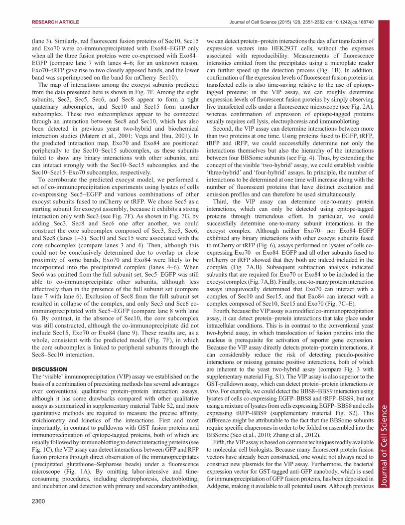
(lane 3). Similarly, red fluorescent fusion proteins of Sec10, Sec15and Exo70 were co-immunoprecipitated with Exo84–EGFP onlywhen all the three fusion proteins were co-expressed with Exo84–EGFP (compare lane 7 with lanes 4–6; for an unknown reason,Exo70–tRFP gave rise to two closely apposed bands, and the lowerband was superimposed on the band for mCherry–Sec10).The map of interactions among the exocyst subunits predicted
from the data presented here is shown in Fig. 7F. Among the eightsubunits, Sec3, Sec5, Sec6, and Sec8 appear to form a tightquaternary subcomplex, and Sec10 and Sec15 form anothersubcomplex. These two subcomplexes appear to be connectedthrough an interaction between Sec8 and Sec10, which has alsobeen detected in previous yeast two-hybrid and biochemicalinteraction studies (Matern et al., 2001; Vega and Hsu, 2001). Inthe predicted interaction map, Exo70 and Exo84 are positionedperipherally to the Sec10–Sec15 subcomplex, as these subunitsfailed to show any binary interactions with other subunits, andcan interact strongly with the Sec10–Sec15 subcomplex and theSec10–Sec15–Exo70 subcomplex, respectively.To corroborate the predicted exocyst model, we performed a
set of co-immunoprecipitation experiments using lysates of cellsco-expressing Sec5–EGFP and various combinations of otherexocyst subunits fused to mCherry or tRFP. We chose Sec5 as astarting subunit for exocyst assembly, because it exhibits a stronginteraction only with Sec3 (see Fig. 7F). As shown in Fig. 7G, byadding Sec3, Sec8 and Sec6 one after another, we couldconstruct the core subcomplex composed of Sec3, Sec5, Sec6,and Sec8 (lanes 1–3). Sec10 and Sec15 were associated with thecore subcomplex (compare lanes 3 and 4). Then, although thiscould not be conclusively determined due to overlap or closeproximity of some bands, Exo70 and Exo84 were likely to beincorporated into the precipitated complex (lanes 4–6). WhenSec6 was omitted from the full subunit set, Sec5–EGFP was stillable to co-immunoprecipitate other subunits, although lesseffectively than in the presence of the full subunit set (comparelane 7 with lane 6). Exclusion of Sec8 from the full subunit setresulted in collapse of the complex, and only Sec3 and Sec6 co-immunoprecipitated with Sec5–EGFP (compare lane 8 with lane6). By contrast, in the absence of Sec10, the core subcomplexwas still constructed, although the co-immunoprecipitate did notinclude Sec15, Exo70 or Exo84 (lane 9). These results are, as awhole, consistent with the predicted model (Fig. 7F), in whichthe core subcomplex is linked to peripheral subunits through theSec8–Sec10 interaction.
DISCUSSIONThe ‘visible’ immunoprecipitation (VIP) assay we established on thebasis of a combination of preexisting methods has several advantagesover conventional qualitative protein–protein interaction assays,although it has some drawbacks compared with other qualitativeassays as summarized in supplementary material Table S2, and morequantitative methods are required to measure the precise affinity,stoichiometry and kinetics of the interactions. First and mostimportantly, in contrast to pulldowns with GST fusion proteins andimmunoprecipitation of epitope-tagged proteins, both of which areusually followed by immunoblotting to detect interacting proteins (seeFig. 1C), the VIP assay can detect interactions between GFP and RFPfusion proteins through direct observation of the immunoprecipitates(precipitated glutathione–Sepharose beads) under a fluorescencemicroscope (Fig. 1A). By omitting labor-intensive and time-consuming procedures, including electrophoresis, electroblotting,and incubation and detection with primary and secondary antibodies,
we can detect protein–protein interactions the day after transfection ofexpression vectors into HEK293T cells, without the expensesassociated with reproducibility. Measurements of fluorescenceintensities emitted from the precipitates using a microplate readercan further speed up the detection process (Fig. 1B). In addition,confirmation of the expression levels of fluorescent fusion proteins intransfected cells is also time-saving relative to the use of epitope-tagged proteins: in the VIP assay, we can roughly determineexpression levels of fluorescent fusion proteins by simply observinglive transfected cells under a fluorescence microscope (see Fig. 2A),whereas confirmation of expression of epitope-tagged proteinsusually requires cell lysis, electrophoresis and immunoblotting.
Second, the VIP assay can determine interactions between morethan two proteins at one time. Using proteins fused to EGFP, tRFP,tBFP and iRFP, we could successfully determine not only theinteractions themselves but also the hierarchy of the interactionsbetween four BBSome subunits (see Fig. 4). Thus, by extending theconcept of the visible ‘two-hybrid’ assay, we could establish visible‘three-hybrid’ and ‘four-hybrid’ assays. In principle, the number ofinteractions to be determined at one timewill increase along with thenumber of fluorescent proteins that have distinct excitation andemission profiles and can therefore be used simultaneously.
Third, the VIP assay can determine one-to-many proteininteractions, which can only be detected using epitope-taggedproteins through tremendous effort. In particular, we couldsuccessfully determine one-to-many subunit interactions in theexocyst complex. Although neither Exo70– nor Exo84–EGFPexhibited any binary interactions with other exocyst subunits fusedto mCherry or tRFP (Fig. 6), assays performed on lysates of cells co-expressing Exo70– or Exo84–EGFP and all other subunits fused tomCherry or tRFP showed that they both are indeed included in thecomplex (Fig. 7A,B). Subsequent subtraction analysis indicatedsubunits that are required for Exo70 or Exo84 to be included in theexocyst complex (Fig. 7A,B). Finally, one-to-manyprotein interactionassays unequivocally determined that Exo70 can interact with acomplex of Sec10 and Sec15, and that Exo84 can interact with acomplex composed of Sec10, Sec15 and Exo70 (Fig. 7C–E).
Fourth, because theVIPassay is amodified co-immunoprecipitationassay, it can detect protein–protein interactions that take place underintracellular conditions. This is in contrast to the conventional yeasttwo-hybrid assay, in which translocation of fusion proteins into thenucleus is prerequisite for activation of reporter gene expression.Because the VIP assay directly detects protein–protein interactions, itcan considerably reduce the risk of detecting pseudo-positiveinteractions or missing genuine positive interactions, both of whichare inherent to the yeast two-hybrid assay (compare Fig. 3 withsupplementary material Fig. S1). The VIP assay is also superior to theGST-pulldown assay, which can detect protein–protein interactions invitro. For example, we could detect the BBS8–BBS9 interaction usinglysates of cells co-expressing EGFP–BBS8 and tRFP–BBS9, but notusing amixture of lysates from cells expressing EGFP–BBS8 and cellsexpressing tRFP–BBS9 (supplementary material Fig. S2). Thisdifference might be attributable to the fact that the BBSome subunitsrequire specific chaperones in order to be folded or assembled into theBBSome (Seo et al., 2010; Zhang et al., 2012).
Fifth, theVIPassay is basedon common techniques readilyavailableto molecular cell biologists. Because many fluorescent protein fusionvectors have already been constructed, one would not always need toconstruct new plasmids for the VIP assay. Furthermore, the bacterialexpression vector for GST-tagged anti-GFP nanobody, which is usedfor immunoprecipitation of GFP fusion proteins, has been deposited inAddgene, making it available to all potential users. Although previous
2360
RESEARCH ARTICLE Journal of Cell Science (2015) 128, 2351-2362 doi:10.1242/jcs.168740
Journal
ofCe
llScience

studies have reported similar qualitative protein–protein interactionassaysbasedondirect imagingof beads (Patel et al., 2007;Schulte et al.,2008; Zhou et al., 2013), bait proteins to detect fluorescence-taggedprey proteins were limited in these visual assays because bait proteinswere first immobilized on the beads. By contrast, by using anti-GFPnanobody immobilized on the beads, a wide variety of GFP fusionproteins are available as bait in the VIP assay. Recently, more powerfulsingle-molecule pulldown and co-immunoprecipitation analyses havebeen reported (Aggarwal and Ha, 2014; Lee et al., 2013). In theseanalyses, after immobilization of conventional anti-GFP or anti-mCherry antibody on the surface of quartz slide, cell lysates containingthe bait protein tagged with YFP or mCherry and the prey proteintagged with mCherry or GFP were added, and proteins trapped by theimmobilized antibody were visualized by total internal reflectionfluorescence microscopy. Thus, unlike the VIP assay, these serve assingle-molecule biochemical tools to enable absolute quantification ofprotein complexes. However, users of these single-molecule analysesare limited due to the use of total-internal reflection fluorescencemicroscopy, whereas the VIP assay does not require any specialequipment, except for a conventional fluorescence microscope.In addition to the aforementioned advantages, we have developed
some techniques that save time and reduce the unit cost of the assay(for details, see Materials and Methods). For example, in place ofnormal anti-GFP antibody and protein A or G agarose beads, we usedbacterially expressed GST-tagged anti-GFP nanobody pre-bound toglutathione–Sepharose beads. Nanobodies are camelid-derivedsingle-domain immunoglobulins with a small size (∼15 kDa) thatare composed of heavy chain homodimers devoid of light chains. Thevirtually inexhaustible source of the anti-GFP antibody makes itpossible to perform interaction assays on a large scale. Althoughprevious studies have made use of anti-GFP nanobody to performvarious interaction assays with low or medium throughput (Pichleret al., 2012; Rothbauer et al., 2008; Zolghadr et al., 2008), thosestudies were all limited to binary interactions. In this study, we madeuse of anti-GFP nanobody to co-immunoprecipitate up to eightproteins fused to up to four distinct fluorescent proteins, therebyelucidating the architectures of the BBSome and exocyst complexes.Thus, the simple, versatile VIP assay established here will not onlypave the way to understanding the architectures and functions ofvarious multisubunit complexes involved in a variety of cellularprocesses, but also drive functional studies by validating proteinnetworks predicted from unbiased global protein–protein interactionanalyses (e.g. see Rolland et al., 2014).
MATERIALS AND METHODSPlasmidsThe whole coding sequences of BBSome and exocyst subunits, listed insupplementary material Table S3, were amplified by PCR from human brain,kidney, or liver cDNA library and cloned into various types of fluorescentprotein vectors: pEGFP-C1, pEGFP-N1 (Clontech), pcDNA3-EGFP-C,pTagRFP-T-C, pTagRFP-T-N (kind gifts from Hideki Shibata, NagoyaUniversity, Japan) (Shibata et al., 2010), pCAG-mCherry-C (a kind gift fromRoger Tsien, University of California - San Diego, CA) (Shaner et al., 2005),pTagBFP2-C (Evrogen), and pcDNA3-iRFP-C (a kind gift from MichiyukiMatsuda, Kyoto University, Japan). Plasmids used in this study are listed insupplementary material Table S4. For yeast two-hybrid assays, cDNAs ofBBSome subunits were subcloned into pGBKT7 and pGADT7 (Clontech).
AntibodiesThe following antibodies were obtained from the indicated vendors:monoclonal mouse anti-GFP (JL-8), BD Biosciences; polyclonal rabbitanti-tRFP antibody, Evrogen; and horseradish-peroxidase-conjugatedsecondary antibodies, Jackson ImmunoResearch Laboratories.
Preparation of GST-tagged anti-GFP nanobody beadsA DNA fragment encoding anti-GFP nanobody, synthesized based on thesequence previously (Kubala et al., 2010), was subcloned into pGEX-6P-1(GE Healthcare). We have deposited the plasmid encoding GST-taggedanti-GFP nanobody to Addgene (ID number 61838). E. coli BL21(DE3)cells transformed with the GST-fused anti-GFP nanobody vector weretreated with 0.1 mM IPTG for 4 h at 30°C to induce protein expression,lysed, and used to purify the recombinant protein with glutathione–Sepharose 4B beads (GE Healthcare). The yield of purified GST–anti-GFPnanobody was ∼5 mg/l of bacterial culture. The protein concentration wasadjusted to ∼200 µg/ml for immunoprecipitation assays.
VIP assaysHEK293T cells, cultured in Dulbecco’s modified Eagle’s medium (DMEM)with high glucose (Nacalai Tesque) supplemented with 5% fetal bovineserum, were plated in six-well plates. Approximately 1.6×10⁶ cells weretransfected with EGFP (2 µg) and tRFP or mCherry (2 µg) fusion constructsusing Polyethylenimine Max (20 µg) (Polysciences), and then cultured for24 h. Before the assay, expression of fluorescent fusion proteins wasconfirmed under a fluorescence microscope. The cells were lysed in 250 µl oflysis buffer (20 mM HEPES-KOH pH 7.4, 150 mM NaCl, 0.1% TritonX-100 and 10% glycerol) containing protease inhibitor cocktail (NacalaiTesque). After 15 min on ice, the cell lysates were centrifuged at 16,100 g for15 min at 4°C in a microcentrifuge. The supernatants (200 µl) were incubatedwith 5 µl of GST-tagged anti-GFP nanobody pre-bound to glutathione–Sepharose 4B beads in 0.2 ml 8-Tube Strips (Greiner) for 1 h at 4°C. The tubestrips were centrifuged at 2000 g for 30 s at room temperature. Theprecipitated beads were washed three times with 180 µl of lysis buffer, andthen transferred into a 96-well plate for observation. Fluorescence on thebeads was observed using an all-in-one type fluorescence microscope(Biozero BZ-8000, Keyence) using a 20×/0.75 NA objective lens under fixedconditions (for green fluorescence, sensitivity ISO 400, exposure 1/30 s; andfor red fluorescence, sensitivity ISO 800, exposure 1/10 s). Image acquisitionwas performed under fixed conditions. The quantification of fluorescenceintensity was performed using the ImageJ software (National Institutes ofHealth). Fluorescence was also measured with a microplate reader (EnVision,PerkinElmer) equipped with filter sets appropriate for detecting fluorescence.After fluorescence measurement, the materials bound to the beads weresubjected to immunoblot analysis using anti-tRFP or anti-GFP antibody.
For expression of combinations of EGFP, tRFP or mCherry, tBFP andiRFP fusion constructs, approximately 3.2×10⁶ HEK293T cells grownon 6-cm dishes were transfected with the expression vectors (12 µg)using Polyethylenimine Max (60 µg), and then cultured for 24 h.Immunoprecipitation was performed as described above. Fluorescenceon the beads was measured with a confocal laser-scanning microscope(A1R-MP, Nikon) equipped with four lasers (405, 488, 561 and 638 nmwavelength) and using 20×/0.75 NA objective lens.
Forexpression of combinations of up toeight ofEGFPand tRFPormCherryfusion constructs,∼1.6×10⁶ cells grown in 6-well plates were transfected withthe expression vectors (8 µg) using Polyethylenimine Max (40 µg), and thencultured for 24 h. Immunoprecipitation was performed as described above.
Immunofluorescence microscopyDNA transfection and immunofluorescence analysis of HEK293T cellswere performed as described previously (Takahashi et al., 2012).
ImmunoblottingProteins in cell lysates, prepared as described above, were separated by SDS-PAGEand electroblotted onto an Immobilon-P transfer membrane (Millipore).The membrane was blocked in 5% skimmed milk and incubated sequentiallywith primary and horseradish-peroxidase-conjugated secondary antibodies.Detection was carried out using a Chemi-Lumi One L kit (Nacalai Tesque).
Yeast two-hybrid assayYeast Y2H-Gold cells (Clontech) transformed with a pGBKT7-based baitvector were plated on synthetic defined medium lacking tryptophan(SD –W). Yeast Y187 cells transformed with pGADT7-based prey vectorwere plated on synthetic medium lacking leucine (SD –L). Colonies on
2361
RESEARCH ARTICLE Journal of Cell Science (2015) 128, 2351-2362 doi:10.1242/jcs.168740
Journal
ofCe
llScience

these plates were picked up, and bait and prey were mixed together in YPADmedium. The mated diploid cells were streaked on synthetic medium lackingtryptophan and leucine (SD –WL). The cells were replicated and grownon synthetic medium lacking tryptophan, leucine, histidine (SD –WLH),and adenine (SD –WLHA). Growth of cells on the selection plates wasassessed after 2 or 3 days of incubation.
AcknowledgementsWewould like to thank Michiyuki Matsuda, Hideki Shibata, and Roger Tsien for kindlyprovidingmaterials, andMichiyukiMatsudaandHye-WonShinoncritical commentsonthe manuscript. We also thank Yohei Hagiya, Minako Kobayashi, Takashi Matsumoto,Tomoki Naito, Minako Ohgi and Noriko Takahashi for technical assistance.
Competing interestsThe authors declare no competing or financial interests.
Author contributionsY.K. designed and performed experiments, and prepared the manuscript; S.N., D.H.and R.M. designed and performed experiments; and K.N. designed experimentsand prepared the manuscript.
FundingThis work was supported in part by a Grant-in-Aid for Scientific Research onInnovative Areas ‘Cilia and Centrosome’ from the Ministry of Education, Culture,Sports, Science and Technology, Japan [grant number 25113514 to K.N.]; a grantfrom the Japan Society for Promotion of Science [grant number 22390013 to K.N.];and a grant from the Uehara Memorial Foundation. This work was also indirectlysupported by a Grant-in-Aid for Scientific Research on Innovative Areas‘Fluorescence Live imaging’ from the Ministry of Education, Culture, Sports, Scienceand Technology of Japan.
Supplementary materialSupplementary material available online athttp://jcs.biologists.org/lookup/suppl/doi:10.1242/jcs.168740/-/DC1
ReferencesAggarwal, V. and Ha, T. (2014). Single-molecule pull-down (SiMPull) for new-agebiochemistry. Bioessays 36, 1109-1119.
Ansley, S. J., Badano, J. L., Blacque, O. E., Hill, J., Hoskins, B. E., Leitch, C. C.,Kim, J. C., Ross, A. J., Eichers, E. R., Teslovich, T. M. et al. (2003). Basal bodydysfunction is a likely cause of pleiotropic Bardet–Biedl syndrome. Nature 425,628-633.
Baek, K., Knodler, A., Lee, S. H., Zhang, X., Orlando, K., Zhang, J., Foskett, T. J.,Guo, W. and Dominguez, R. (2010). Structure-function study of the N-terminaldomain of exocyst subunit Sec3. J. Biol. Chem. 285, 10424-10433.
Guo, W., Roth, D., Walch-Solimena, C. and Novick, P. (1999). The exocyst is aneffector for Sec4p, targeting secretory vesicles to sites of exocytosis. EMBO J. 18,1071-1080.
Heider, M. R. and Munson, M. (2012). Exorcising the exocyst complex. Traffic 13,898-907.
Hong,W. and Lev, S. (2014). Tethering the assembly of SNARE complexes.TrendsCell Biol. 24, 35-43.
Hsu, S.-C., Hazuka, C. D., Roth, R., Foletti, D. L., Heuser, J. and Scheller, R. H.(1998). Subunit composition, protein interactions, and structures of themammalian brain sec6/8 complex and septin filaments. Neuron 20, 1111-1122.
Jin, H. and Nachury, M. V. (2009). The BBSome. Curr. Biol. 19, R472-R473.Jin, H., White, S. R., Shida, T., Schulz, S., Aguilar, M., Gygi, S. P., Bazan, J. F.and Nachury, M. V. (2010). The conserved Bardet-Biedl syndrome proteinsassemble a coat that traffics membrane proteins to cilia. Cell 141, 1208-1219.
Kim, J. C., Badano, J. L., Sibold, S., Esmail, M. A., Hill, J., Hoskins, B. E., Leitch,C. C., Venner, K., Ansley, S. J., Ross, A. J. et al. (2004). The Bardet-Biedlprotein BBS4 targets cargo to the pericentriolar region and is required formicrotubule anchoring and cell cycle progression. Nat. Genet. 36, 462-470.
Kubala, M. H., Kovtun, O., Alexandrov, K. and Collins, B. M. (2010). Structuraland thermodynamic analysis of the GFP:GFP-nanobody complex. Protein Sci.19, 2389-2401.
Lee, H.-W., Kyong, T., Yoo, J., Kim, T., Chung, C., Ryu, J. Y., Lee, H., Park, K., Lee,S., Jones, W. D. et al. (2013). Real-time single-molecule co-immunoprecipitationanalyses reveal cancer-specific Ras signalling dynamics. Nat. Commun. 4, 1505.
Liu, J. and Guo, W. (2012). The exocyst complex in exocytosis and cell migration.Protoplasma 249, 587-597.
Loktev, A. V., Zhang, Q., Beck, J. S., Searby, C. C., Scheetz, T. E., Bazan, J. F.,Slusarski, D. C., Sheffield, V. C., Jackson, P. K. and Nachury, M. V. (2008). ABBSome subunit links ciliogenesis, microtubule stability, and acetylation. Dev.Cell 15, 854-865.
Madhivanan, K. and Aguilar, R. C. (2014). Ciliopathies: the trafficking connection.Traffic 15, 1031-1056.
Marion, V., Stoetzel, C., Schlicht, D., Messaddeq, N., Koch, M., Flori, E., Danse,J. M., Mandel, J.-L. and Dollfus, H. (2009). Transient ciliogenesis involvingBardet-Biedl syndrome proteins is a fundamental characteristic of adipogenicdifferentiation. Proc. Natl. Acad. Sci. USA 106, 1820-1825.
Matern, H. T., Yeaman, C., Nelson, W. J. and Scheller, R. H. (2001). The Sec6/8complex in mammalian cells: characterization of mammalian Sec3, subunitinteractions. Proc. Natl. Acad. Sci. USA 98, 9648-9653.
M’Hamdi, O., Ouertani, I. and Chaabouni-Bouhamed, H. (2014). Update on thegenetics of Bardet-Biedl syndrome. Mol. Syndromol. 5, 51-56.
Munson, M. and Novick, P. (2006). The exocyst defrocked, a framework of rodsrevealed. Nat. Struct. Mol. Biol. 13, 577-581.
Nachury,M. V., Loktev, A. V., Zhang,Q.,Westlake, C. J., Peranen, J., Merdes, A.,Slusarski, D. C., Scheller, R. H., Bazan, J. F., Sheffield, V. C. et al. (2007). Acore complex of BBS proteins cooperates with the GTPase Rab8 to promoteciliary membrane biogenesis. Cell 129, 1201-1213.
Patel, S. S., Belmont, B. J., Sante, J. M. and Rexach,M. F. (2007). Natively unfoldednucleoporins gate protein diffusion across the nuclear pore complex.Cell 129, 83-96.
Pichler, G., Jack, A., Wolf, P. and Hake, S. B. (2012). Versatile toolbox for highthroughput biochemical and functional studies with fluorescent fusion proteins.PLoS ONE 7, e36967.
Rolland, T., Tasan,M., Charloteaux,B., Pevzner, S. J., Zhong,Q., Sahni, N., Yi, S.,Lemmens, I., Fontanillo, C., Mosca, R. et al. (2014). A proteome-scale map of thehuman interactome network. Cell 159, 1212-1226.
Rothbauer, U., Zolghadr, K., Muyldermans, S., Schepers, A., Cardoso, M. C.and Leonhardt, H. (2008). A versatile Nanotrap for biochemical and functionalstudies with fluorescent fusion proteins. Mol. Cell. Proteomics 7, 282-289.
Saerens, D., Pellis, M., Loris, R., Pardon, E., Dumoulin, M., Matagne, A., Wyns,L., Muyldermans, S. and Conrath, K. (2005). Identification of a universal VHHframework to graft non-canonical antigen-binding loops of camel single-domainantibodies. J. Mol. Biol. 352, 597-607.
Scheidecker, S., Etard, C., Pierce, N. W., Geoffroy, V., Schaefer, E., Muller, J.,Chennen, K., Flori, E., Pelletier, V., Poch, O. et al. (2014). Exome sequencing ofbardet-Biedl syndrome patient identifies a null mutation in the BBSome subunitBBIP1 (BBS18). J. Med. Genet. 51, 132-136.
Schulte, R., Talamas, J., Doucet, C. and Hetzer, M. W. (2008). Single bead affinitydetection (SINBAD) for the analysis of protein-protein interactions. PLoS ONE 3,e2061.
Seo, S., Baye, L. M., Schulz, N. P., Beck, J. S., Zhang, Q., Slusarski, D. C. andSheffield, V. C. (2010). BBS6, BBS10, and BBS12 form a complex with CCT/TRiC family chaperonins and mediate BBSome assembly. Proc. Natl. Acad. Sci.USA 107, 1488-1493.
Shaner, N. C., Steinbach, P. A. and Tsien, R. Y. (2005). A guide to choosingfluorescent proteins. Nat. Methods 2, 905-909.
Shibata, H., Inuzuka, T., Yoshida, H., Sugiura, H., Wada, I. and Maki, M. (2010).The ALG-2 binding site in Sec31A influences the retention kinetics of Sec31A atthe endoplasmic reticulum exit sites as revealed by live-cell time-lapse imaging.Biosci. Biotech. Biochem. 74, 1819-1826.
Sung, C.-H. and Leroux, M. R. (2013). The roles of evolutionarily conservedfunctional modules in cilia-related trafficking. Nat. Cell Biol. 15, 1387-1397.
Takahashi, S., Kubo, K., Waguri, S., Yabashi, A., Shin, H.-W., Katoh, Y. andNakayama, K. (2012). Rab11 regulates exocytosis of recycling vesicles at theplasma membrane. J. Cell Sci. 125, 4049-4057.
Terbush, D. R., Guo, W., Dunkelbarger, S. and Novick, P. (2001). Purification andcharacterization of yeast exocyst complex. Meth. Enzymol. 329, 100-110.
Vega, I. E. and Hsu, S.-C. (2001). The exocyst complex associates withmicrotubules to mediate vesicle targeting and neurite outgrowth. J. Neurosci.21, 3839-3848.
Zhang, Q., Yu, D., Seo, S., Stone, E. M. and Sheffield, V. C. (2012). Intrinsicprotein-protein interaction-mediated and chaperonin-assisted sequentialassembly of stable Bardet-Biedl syndrome protein complex, the BBSome.J. Biol. Chem. 287, 20625-20635.
Zhou, Y., Hong, W. and Lu, L. (2013). Imaging beads-retained prey assay for rapidand quantitative protein-protein interaction. PLoS ONE 8, e59727.
Zolghadr, K., Mortusewicz, O., Rothbauer, U., Kleinhans, R., Goehler, H.,Wanker, E. E., Cardoso, M. C. and Leonhardt, H. (2008). A fluorescent two-hybrid assay for direct visualization of protein interactions in living cells.Mol. Cell.Proteomics 7, 2279-2287.
2362
RESEARCH ARTICLE Journal of Cell Science (2015) 128, 2351-2362 doi:10.1242/jcs.168740
Journal
ofCe
llScience