are small molecules or proteins that are produced in one
TRANSCRIPT

Hormones are small molecules or proteins that are produced in one tissue,released into the bloodstream, and carried to other tissues, where they actthrough specific receptors to bring about changes in cellular activities.
We also include in this discussion short-lived signals such as NO, which acts locally,on neighboring cells.
Hormones serve to coordinate the metabolic activities of several tissues or organs.
Virtually every process in a complex organism is regulated by one or morehormones: maintenance of blood pressure, blood volume, and electrolytebalance; embryogenesis; sexual differentiation, development, and reproduction;hunger, eating behavior, digestion, and fuel allocation—to name but a few.
We examine here the methods for detecting and measuring hormones and theirinteraction with receptors, and we consider a representative selection of hormonetypes.

The coordination of metabolism in mammals is achieved by the neuroendocrinesystem.
Individual cells in one tissue sense a change in the organism’s circumstances andrespond by secreting a chemical messenger that passes to another cell in thesame or different tissue, where the messenger binds to a receptor molecule andtriggers a change in this target cell.
These chemical messengers may relay information over very short or very longdistances.
In neuronal signaling, the chemical messenger is a neurotransmitter(acetylcholine, for example) and may travel only a fraction of a micrometer, acrossa synaptic cleft to the next neuron in a network.
In hormonal signaling, the messengers—hormones—are carried in thebloodstream to neighboring cells or to distant organs and tissues; they may travela meter or more before encountering their target cell.


Except for this anatomic difference, these two chemical signaling mechanisms areremarkably similar, and the same molecule can sometimes act as bothneurotransmitter and hormone.
Epinephrine and norepinephrine, for example, serve as neurotransmitters atcertain synapses of the brain and at neuromuscular junctions of smooth muscleand as hormones that regulate fuel metabolism in liver and muscle.
The following discussion of cellular signaling emphasizes hormone action, drawingon discussions of fuel metabolism in earlier chapters, but most of the fundamentalmechanisms described here also occur in neurotransmitter action.

How is a hormone detected and isolated?
First, researchers find that a physiological process in one tissue depends on asignal that originates in another tissue.
Insulin, for example, was first recognized as a substance that is produced in thepancreas and affects the concentration of glucose in blood and urine .
Once a physiological effect of the putative hormone is discovered, a quantitativebioassay for the hormone can be developed.
In the case of insulin, the assay consisted of injecting extracts of pancreas (a crudesource of insulin) into experimental animals deficient in insulin, then quantifyingthe resulting changes in glucose concentration in blood and urine.

To isolate a hormone, the biochemist fractionates extracts containing the putativehormone, with the same techniques used to purify other biomolecules (solventfractionation, chromatography, and electrophoresis), and then assays eachfraction for hormone activity.
Once the chemical has been purified, its composition and structure can bedetermined.
This protocol for hormone characterization is deceptively simple.
Hormones are extremely potent and are produced in very small amounts.
Obtaining sufficient quantities of a hormone to allow its chemical characterizationoften requires biochemical isolations on a heroic scale.

When Andrew Schally and Roger Guillemin independently purified andcharacterized thyrotropin-releasing hormone (TRH) from the hypothalamus,Schally’s group processed about 20 tons of hypothalamus from nearly two millionsheep, and Guillemin’s group extracted the hypothalamus from about a millionpigs.
TRH proved to be a simple derivative of the tripeptide Glu–His–Pro).
Once the structure of the hormone was known, it could be chemically synthesizedin large quantities for use in physiological and biochemical studies.
For their work on hypothalamic hormones, Schally and Guillemin shared the NobelPrize in Physiology or Medicine in 1977, along with Rosalyn Yalow, who (withSolomon A. Berson) developed the extraordinarily sensitive radioimmunoassay(RIA) for peptide hormones and used it to study hormone action.

This technique revolutionized hormonere search by making possible the rapid,quantitative, and specific measurement of hormones in minute amounts.

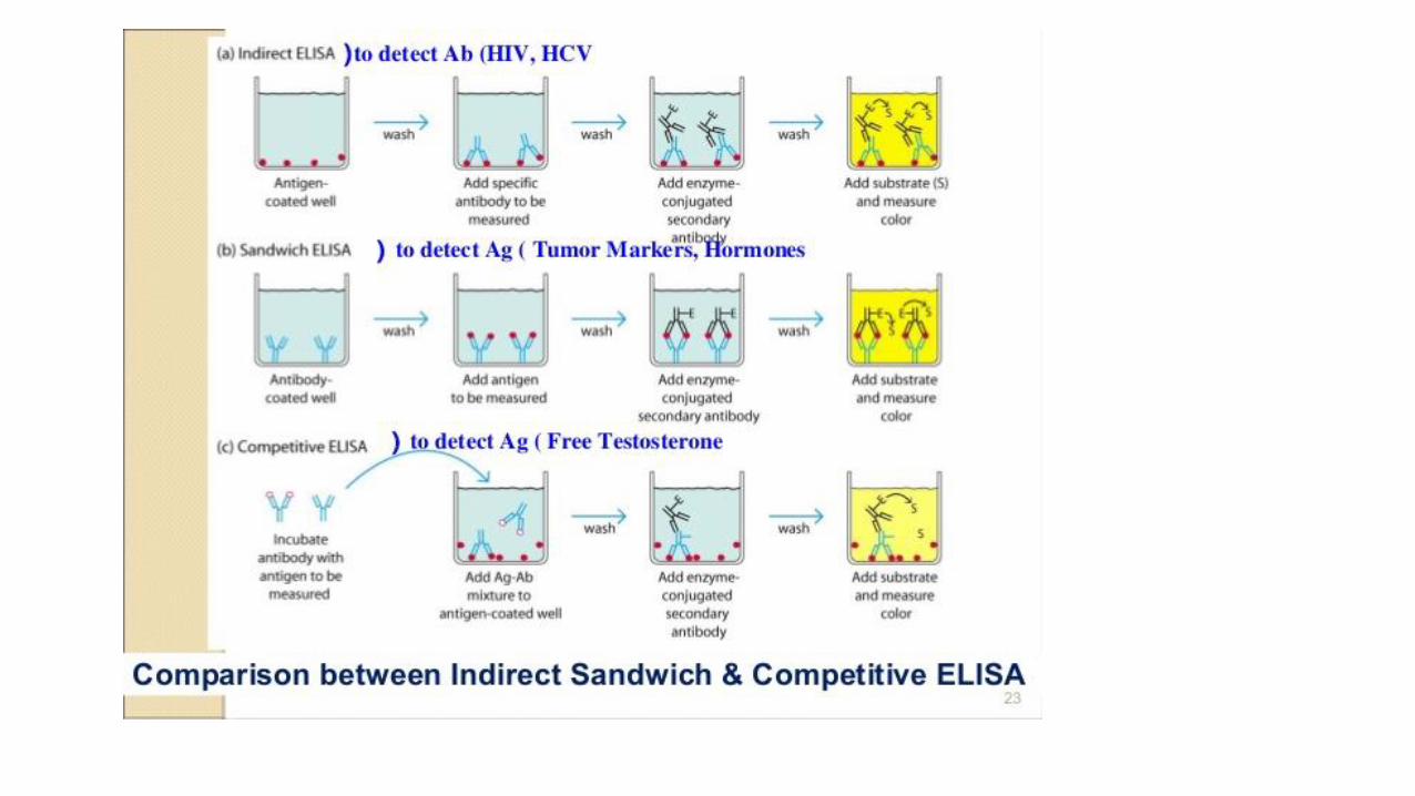
Hormone-specific antibodies are the key to RIA and its modern equivalent, theenzyme-linked immunosorbent assay (ELISA).
Purified hormone, injected into rabbits, mice, or chickens, elicits antibodies thatbind to the hormone with very high affinity and specificity.
These antibodies may be purified and either radioisotopically labeled (for RIA) orconjugated with an enzyme that produces a colored product (for ELISA).
The tagged antibodies are then allowed to interact with extracts containing thehormone.
The fraction of antibody bound by the hormone in the extract is quantified byradiation detection or photometry.
Because of the high affinity of the antibody for the hormone, such assays can bemade sensitive to picograms of hormone in a sample.


All hormones act through highly specific receptors in hormone-sensitive targetcells, to which the hormones bind with high affinity.
Each cell type has its own combination of hormone receptors, which define therange of its hormone responsiveness.
Moreover, two cell types with the same type of receptor may have differentintracellular targets of hormone action and thus may respond differently to thesame hormone.
The specificity of hormone action results from structural complementaritybetween the hormone and its receptor; this interaction is extremely selective, soeven structurally similar hormones can have different effects if they preferentiallybind to different receptors.
The high affinity of the interaction allows cells to respond to very lowconcentrations of hormone.

In the design of drugs intended to intervene in hormonal regulation, we need toknow the relative specificity and affinity of the drug and the natural hormone.
The intracellular consequences of ligand-receptor interaction are of at least fourgeneral types:
(1) a second messenger, such as cAMP, cGMP, or inositol trisphosphate, generatedinside the cell acts as an allosteric regulator of one or more enzymes;
(2) a receptor tyrosine kinase is activated by the extracellular hormone;
(3) a change in membrane potential results from the opening or closing of ahormone-gated ion channel; and
(4) a steroid or steroid-like molecule causes a change in the level of expression(transcription of DNA into mRNA) of one or more genes, mediated by a nuclearhormone receptor protein.

Water-soluble peptide and amine hormones, such as insulin and epinephrine, actextracellularly by binding to cell surface receptors that span the plasmamembrane.
When the hormone binds to its extracellular domain, the receptor undergoes aconformational change analogous to that produced in an allosteric enzyme bybinding of an effector molecule.
The conformational change triggers the effect of the hormone.
With metabotropic receptors, the change activates or inhibits an enzymedownstream from the receptor; with ionotropic receptors, an ion channel in theplasma membrane opens or closes, resulting in a change in membrane potentialor in the concentration of an ion such as Ca2+.


A single hormone molecule, in forming a hormone-receptor complex, activates acatalyst that produces many molecules of second messenger, so the receptorserves as both signal transducer and signal amplifier.
The signal may be further amplified by a signaling cascade, a series of steps inwhich a catalyst (such as a protein kinase) activates another catalyst (anotherprotein kinase), resulting in very large amplifications of the original signal.
A cascade of this type occurs in the regulation of glycogen synthesis andbreakdown by epinephrine.
Epinephrine activates (through its receptor) adenylyl cyclase, which producesmany molecules of cAMP for each molecule of receptor-bound hormone.
Cyclic AMP in turn activates cAMP-dependent protein kinase (protein kinase A),which activates glycogen phosphorylase b kinase, which activates glycogenphosphorylase b.

The result is signal amplification: one epinephrine molecule causes the productionof many thousands or millions of molecules of glucose 1-phosphate fromglycogen.
Water-insoluble hormones, including the steroid, retinoid, and thyroid hormones,readily pass through the plasma membrane of their target cells to reach theirreceptor proteins in the nucleus.
The hormone-receptor complex itself carries the message: it interacts with DNA toalter the expression of specific genes, changing the enzyme complement of thecell and thereby changing cellular metabolism.
Hormones that act through plasma membrane receptors generally trigger veryrapid physiological or biochemical responses.
Just seconds after the adrenal medulla secretes epinephrine into the bloodstream,skeletal muscle responds by accelerating the breakdown of glycogen.

By contrast, the thyroid hormones and the sex (steroid) hormones promotemaximal responses in their target tissues only after hours or even days.
These differences in response time correspond to different modes of action. Ingeneral, the fast-acting hormones lead to a change in the activity of one or morepre-existing enzymes in the cell, by allosteric mechanisms or covalentmodification.
The slower-acting hormones generally alter gene expression, resulting in thesynthesis of more (upregulation) or less (downregulation) of the regulatedprotein(s).
Mammals have several classes of hormones, distinguishable by their chemicalstructures and their modes of action.
Peptide, catecholamine, and eicosanoid hormones act from outside the target cellvia cell surface receptors.

Steroid, vitamin D, retinoid, and thyroid hormones enter the cell and act throughnuclear receptors.
Nitric oxide (a gas) also enters the cell, but activates a cytosolic enzyme, guanylylcyclase.
Hormones can also be classified by the way they get from their point of release totheir target tissue.
Endocrine hormones are released into the blood and carried to target cellsthroughout the body (insulin and glucagon are examples).
Paracrine hormones are released into the extracellular space and diffuse toneighboring target cells (the eicosanoid hormones are of this type).
Autocrine hormones affect the same cell that releases them, binding to receptorson the cell surface.

Mammals are hardly unique in possessing hormonal signaling systems.
Insects and nematode worms have highly developed systems for hormonalregulation, with fundamental mechanisms similar to those in mammals.
Plants, too, use hormonal signals to coordinate the activities of their tissues.

The peptide hormones vary in size, from 3 to more than 200 amino acid residues.
They include the pancreatic hormones insulin, glucagon, and somatostatin; theparathyroid hormone calcitonin; and all the hormones of the hypothalamus andpituitary.
These hormones are synthesized on ribosomes in the form of longer, precursorproteins (prohormones), then packaged into secretory vesicles and proteolyticallycleaved to form the active peptides.
In many peptide hormones the terminal residues are modified, as in TRH.
Insulin is a small protein (Mr 5,800) with two polypeptide chains, A and B, joinedby two disulfide bonds.
It is synthesized in the pancreas as an inactive single-chain precursor,preproinsulin, with an aminoterminal “signal sequence” that directs its passageinto secretory vesicles.

Proteolytic removal of the signal sequence andformation of three disulfide bonds producesproinsulin, which is stored in secretory granules(membrane vesicles filled with protein synthesizedin the ER) in pancreatic β cells.
When blood glucose is elevated sufficiently totrigger insulin secretion, proinsulin is converted toactive insulin by specific proteases, which cleavetwo peptide bonds to form the mature insulinmolecule and C peptide, which are released intothe blood by exocytosis.
All three fragments have physiological effects:insulin stimulates glucose uptake and fat synthesis,and C peptide acts through G protein–coupledreceptors (GPCRs) in various tissues to mitigateeffects of reduced insulin synthesis, such asdiabetic nerve pain.

There are other cases in whichprohormone proteins undergospecific cleavage to produceseveral active hormones.
Pro-opiomelanocortin (POMC)gene encodes a large polypeptidethat is progressively carved up intoat least nine biologically activepeptides.

The concentration of peptide hormones in secretory granules is so high that thevesicle contents are virtually crystalline; when the contents are released byexocytosis, a large amount of hormone is released suddenly.
The capillaries that serve peptide-producing endocrine glands are fenestrated(punctuated with tiny holes or “windows”), so the hormone molecules readilyenter the bloodstream for transport to target cells elsewhere.
As noted earlier, all peptide hormones act by binding to receptors in the plasmamembrane.
They cause the generation of a second messenger in the cytosol, which changesthe activity of an intracellular enzyme, thereby altering the cell’smetabolism.

The water-soluble compounds epinephrine (adrenaline) andnorepinephrine (noradrenaline) are catecholamines, namedfor the structurally related compound catechol.
They are synthesized from tyrosine.
Catecholamines produced in the brain and in other neuraltissues function as neurotransmitters, but epinephrine andnorepinephrine are also hormones, synthesized and secretedby the adrenal glands (adrenals).
Like the peptide hormones, catecholamines are highlyconcentrated in secretory granules and released byexocytosis, and they act through surface receptors togenerate intracellular second messengers.
They mediate a wide variety of physiological responses toacute stress

The eicosanoid hormones (prostaglandins, thromboxanes, leukotrienes, andlipoxins) are derived from the 20-carbon polyunsaturated fatty acids arachidonate(20:4(Δ5,8,11,14) and eicosapentaenoic acid (EPA; 20:5(Δ5,8,11,14,17).
Unlike the hormones described above, they are not synthesized in advance andstored; they are produced when needed.
The enzymes of the pathways leading to prostaglandins and thromboxanes arevery widely distributed in mammalian tissues; most cells can produce thesehormone signals, and cells of many tissues can respond to them through specificplasma membrane receptors.

The eicosanoid hormones are paracrine hormones, secreted into the interstitialfluid (not primarily into the blood) and acting on nearby cells.
Some prostaglandins promote the contraction of smooth muscle, including thatof the intestine and uterus (and can therefore be used medically to induce labor).
They also mediate pain and inflammation in some tissues.
Many antiinflammatory drugs act by inhibiting steps in prostaglandin syntheticpathways.
Thromboxanes regulate platelet function and therefore blood clotting.
Leukotrienes LTC4 and LTD4 act through plasma membrane receptors to stimulatecontraction of smooth muscle in the intestine, pulmonary airways, and trachea.
They are mediators of anaphylaxis, an immune overresponse that can includeairway constriction, altered heartbeat, shock, and sometimes death.

The steroid hormones—corticosteroid (adrenocortical) hormones and sexhormones—are synthesized from cholesterol in several endocrine tissues.
They travel to their target cells through the bloodstream, bound to carrierproteins.
More than 50 corticosteroid hormones are produced in the adrenal cortex byreactions that remove the side chain from the D ring of cholesterol and introduceoxygen to form keto and hydroxyl groups.

The corticosteroids are of two general types, defined by their actions.
Glucocorticoids, such as cortisol, primarily affect the metabolism ofcarbohydrates; mineralocorticoids, such as aldosterone, regulate theconcentrations of electrolytes in the blood.
Two types of sex hormones, androgens (including testosterone) and estrogens(including estradiol), are synthesized in the testes and ovaries.
They affect sexual development, sexual behavior, and a variety of otherreproductive and nonreproductive functions.
Their synthesis also requires cytochrome P-450 enzymes that cleave the sidechain of cholesterol and introduce oxygen atoms.
All steroid hormones act through nuclear receptors to change the level ofexpression of specific genes.

They can also have more rapid effects, mediated by receptors in the plasmamembrane.
Humans and other animals are exposed to many exogenous chemicals broadlyreferred to as “endocrine disruptors,” ranging from environmental pollutants suchas PCBs (polychlorinated biphenyls), pesticides, and pharmaceuticals to naturallyoccurring estrogens in plants, such as in soy products.
Some endocrine disruptors bind to nuclear steroid receptors and stimulatehormone-like effects; others block the receptors, preventing stimulation byendogenous hormones, or interfere with the normal metabolism of steroidhormones in the liver.

Calcitriol (1α,25-dihydroxycalcitriol) is produced from vitamin D by enzyme-catalyzed hydroxylation in the liver and kidneys.
Vitamin D is obtained in the diet or by photolysis of 7-dehydrocholesterol in skinexposed to sunlight.
Calcitriol works in concert with parathyroid hormone in Ca2+ homeostasis,regulating [Ca2+] in the blood and the balance between Ca2+deposition and Ca2+
mobilization from bone.
Acting through nuclear receptors, calcitriol activates the synthesis of an intestinalCa2+binding protein essential for uptake of dietary Ca2+.
Inadequate dietary vitamin D or defects in the biosynthesis of calcitriol result inserious diseases such as rickets, in which bones are weak and malformed.

The retinoid hormones are potent hormones that regulate the growth, survival,and differentiation of cells via nuclear retinoid receptors.
The prohormone retinol is synthesized from β-carotene, primarily in liver, andmany tissues convert retinol to the hormone retinoic acid (RA).
RA binds its specific receptor (RAR) in the nucleus, forms a dimer with anothernuclear protein, retinoid X receptor (RXR), and alters the rate of expression ofgenes responsive to RA.
All tissues are retinoid targets, as all cell types have at least one form of nuclearretinoid receptor.
In adults, the most significant targets include cornea, skin, epithelia of the lungsand trachea, and the immune system, all of which undergo constant replacementof cells.
RA regulates the synthesis of proteins essential for growth or differentiation.

The thyroid hormones T4 (thyroxine) and T3 (triiodothyronine) are synthesizedfrom the precursor protein thyroglobulin (Mr 660,000).
Up to 20 Tyr residues in thyroglobulin are enzymatically iodinated in the thyroidgland, then two iodotyrosine residues condense to form the precursor tothyroxine.
When needed, thyroxine is released by proteolysis.
Condensation of monoiodotyrosine with diiodothyronine produces T3, which isalso an active hormone released by proteolysis.
The thyroid hormones act through nuclear receptors to stimulate energy yieldingmetabolism, especially in liver and muscle, by increasing the expression of genes
encoding key catabolic enzymes.
Underproduction of thyroxine slows metabolism and can be the cause ofdepression.

When underproduction is the result of too little iodine in the diet, the thyroidgland enlarges in a futile attempt to produce more thyroxine.
This condition, called goiter, was once common in regions far from oceans (whichprovide iodine in the form of fresh seafood) and areas with low-iodine soil(yielding plants with low iodine).
Goiter has been almost eliminated in areas where iodine is routinely added totable salt.

Nitric oxide is a relatively stable free radical synthesized from molecular oxygenand the guanidinium nitrogen of arginine, in a reaction catalyzed by NO synthase.
This enzyme is found in many tissues and cell types: neurons, macrophages,hepatocytes, myocytes of smooth muscle, endothelial cells of the blood vessels,and epithelial cells of the kidney.
NO acts near its point of release, entering the target cell and activating thecytosolic enzyme guanylyl cyclase, which catalyzes the formation of the secondmessenger.
A cGMP-dependent protein kinase mediates the effects of NO by phosphorylatingkey proteins and altering their activities.
For example, phosphorylation of contractile proteins in the smooth musclesurrounding blood vessels relaxes the muscle, thereby lowering blood pressure.

The changing levels of specific hormones regulate specific cellular processes, butwhat regulates the level of each hormone?
The brief answer is that the central nervous system receives input from manyinternal and external sensors—signals about danger, hunger, dietary intake, bloodcomposition and pressure, for example—and orchestrates the production ofappropriate hormonal signals by the endocrine tissues.
For a more complete answer, we must look at the hormone-producing systems ofthe human body and some of their functional interrelationships.
The hypothalamus, a small region of the brain, is the coordination center of theendocrine system; it receives and integrates messages from the central nervoussystem.
In response to these messages, the hypothalamus produces regulatory hormones(releasing factors) that pass directly to the nearby pituitary gland through specialblood vessels and neurons that connect the two glands.


The pituitary gland has two functionally distinct parts.
The posterior pituitary contains the axonal endings of many neurons thatoriginate in the hypothalamus.
These neurons produce the short peptide hormones oxytocin and vasopressin,which move down the axon to the nerve endings in the pituitary, where they arestored in secretory granules to await the signal for their release.
The anterior pituitary responds to hypothalamic hormones carried in the blood,producing tropic hormones, or tropins.
These relatively long polypeptides activate the next rank of endocrine glands,which includes the adrenal cortex, thyroid gland, ovaries, and testes.
These glands in turn secrete their specific hormones, which are carried in thebloodstream to target tissues.

For example, corticotropin-releasing hormone secreted from the hypothalamusstimulates the anterior pituitary to release corticotropin (ACTH), which travelsthrough the blood to the zona fasciculata of the adrenal cortex and triggers therelease of cortisol.
Cortisol, the ultimate hormone in this cascade, acts through its receptor in manytypes of target cells to alter their metabolism. In hepatocytes, one effect of cortisol isto increase the rate of gluconeogenesis.
Hormonal cascades such as those responsible for the release of cortisol andepinephrine result in large amplifications of the initial signal and allow fine-tuning ofthe output of the ultimate hormone.
For example, the initial electrical signal to the hypothalamus results in the release ofa few nanograms of corticotropin-releasing hormone, which elicits the release of afew micrograms of corticotropin.
Corticotropin acts on the adrenal cortex to cause the release of milligrams of cortisol,for an overall amplification of at least a millionfold.

At each level of a hormonal cascade,feedback inhibition of earlier steps in thecascade is possible; an unnecessarilyelevated level of the ultimate hormone orof an intermediate hormone inhibits therelease of earlier hormones in the cascade.
These feedback mechanisms accomplishthe same end as those that limit theoutput of a biosynthetic pathway: aproduct is synthesized (or released) onlyuntil the necessary concentration isreached.

In addition to the top-down hierarchy ofhormonal signaling, some hormones areproduced in the digestive tract, muscle, andadipose tissue and communicate the currentmetabolic state to the hypothalamus.
These signals are integrated in thehypothalamus, and an appropriate neuronalor hormonal response is elicited.
The action of the enzyme AMP-activatedprotein kinase (AMPK) in the hypothalamus isone such integrating mechanism; it sumsvarious inputs and passes on the informationby phosphorylating key proteins in thehypothalamus.

Adipokines, for example, are peptide hormones, produced in adipose tissue, thatsignal the adequacy of fat reserves.
Leptin, released when adipose tissue is well-filled with triacylglycerols, acts in thebrain to inhibit feeding behavior, whereas adiponectin signals depletion of fatreserves and stimulates feeding.
Ghrelin is produced in the gastrointestinal tract when the stomach is empty and actsin the hypothalamus to stimulate feeding behavior; when the stomach fills, ghrelinrelease ceases.
Incretins are peptide hormones produced in the gut after ingestion of a meal; theyincrease secretion of insulin and decrease secretion of glucagon from the pancreas.
The best-studied of the incretins are glucagon-like peptide-1 (GLP-1) and glucose-dependent insulinotropic polypeptide (GIP), also referred to as gastric inhibitorypolypeptide.

Each tissue of the human body has a specialized function, reflected in its anatomyand metabolic activity.
Skeletal muscle allows directed motion; adipose tissue stores and distributes energyin the form of fats, which serve as fuel throughout the body and as thermalinsulation; in the brain, cells pump ions across their plasma membranes to produceelectrical signals.
The liver plays a central processing and distribution role in metabolism and furnishesall other organs and tissues with an appropriate mix of nutrients via thebloodstream.
The functional centrality of the liver is indicated by the common reference to allother tissues and organs as “extrahepatic.”
We begin our discussion of the division of metabolic labor by considering thetransformations of carbohydrates, amino acids, and fats in the mammalian liver.


During digestion in mammals, the three main classes of nutrients (carbohydrates,proteins, and fats) undergo enzymatic hydrolysis into their simple constituents.
This breakdown is necessary because the epithelial cells lining the intestinal lumenabsorb only relatively small molecules.
Many of the fatty acids and monoacylglycerols released by digestion of fats in theintestine are reassembled within these epithelial cells into triacylglycerols (TAGs).
After being absorbed, most sugars and amino acids and some reconstituted TAGspass from intestinal epithelial cells into blood capillaries and travel in thebloodstream to the liver; the remaining TAGs enter adipose tissue via the lymphaticsystem.
The portal vein is a direct route from the digestive organs to the liver, and the livertherefore has first access to ingested nutrients.

The liver has two main cell types.
Kupffer cells are phagocytes, important in immune function.
Hepatocytes, of primary interest here, transform dietary nutrients into the fuels andprecursors required by other tissues and export them via the blood.
The kinds and amounts of nutrients supplied to the liver are determined by diet, thetime between meals, and several other factors.
The demand of extrahepatic tissues for fuels and precursors varies from one organ toanother, and with the level of activity and overall nutritional state of the individual.
To meet these changing circumstances, the liver has remarkable metabolic flexibility.
For example, when the diet is rich in protein, hepatocytes supply themselves withhigh levels of enzymes for amino acid catabolism and gluconeogenesis.

Within hours after a shift to a high carbohydrate diet, the levels of these enzymesbegin to drop and the hepatocytes increase their synthesis of enzymes essential tocarbohydrate metabolism and fat synthesis.
Liver enzymes turn over (that is, are synthesized and degraded) at 5 to 10 times therate of enzyme turnover in other tissues, such as muscle.
Extrahepatic tissues also can adjust their metabolism to prevailing conditions, butnone of these tissue are as adaptable as the liver, and none so central to theorganism’s overall metabolism.
What follows is a survey of the possible fates of sugars, amino acids, and lipids thatenter the liver from the bloodstream.

Carbohydrates; The glucose transporter of hepatocytes (GLUT2) allows rapid, passivediffusion of glucose, so that the concentration of glucose in a hepatocyte isessentially the same as that in the blood.
Glucose entering hepatocytes is phosphorylated by glucokinase (hexokinase IV) toyield glucose 6-phosphate.
Glucokinase has a much higher Km for glucose (10 mM) than do the hexokinaseisozymes in other cells and, unlike these other isozymes, it is not inhibited by itsproduct, glucose 6-phosphate.
The presence of glucokinase allows hepatocytes to continue phosphorylating glucosewhen the glucose concentration rises well above levels that would overwhelm otherhexokinases.
The high Km of glucokinase also ensures that the phosphorylation of glucose inhepatocytes is minimal when the glucose concentration is low, preventing the liverfrom consuming glucose as fuel via glycolysis.

This spares glucose for other tissues.
Fructose, galactose, and mannose, all absorbed from the small intestine, are alsoconverted to glucose 6-phosphate.
Glucose 6- phosphate is at the crossroads of carbohydrate metabolism in the liver. Itmay take any of several major metabolic routes, depending on the current metabolicneeds of the organism.
By the action of various allosterically regulated enzymes, and through hormonalregulation of enzyme synthesis and activity, the liver directs the flow of glucose intoone or more of these pathways.
1 Glucose 6-phosphate is dephosphorylated by glucose 6-phosphatase to yield freeglucose, which is exported to replenish blood glucose.
Export is the predominant pathway when glucose 6-phosphate is in limited supply,because the blood glucose concentration must be kept sufficiently high (4 to 5 mM)to provide adequate energy for the brain and other tissues.


2 Glucose 6-phosphate not immediately needed to form blood glucose is convertedto liver glycogen, or has one of several other fates.
Following glycolysis and the pyruvate dehydrogenase reaction, 3 the acetyl-CoA soformed can be oxidized for ATP production by the citric acid cycle, with ensuingelectron transfer and oxidative phosphorylation yielding ATP.
(Normally, however, fatty acids are the preferred fuel for ATP production inhepatocytes.)
4 Acetyl-CoA can also serve as the precursor of fatty acids, which are incorporatedinto TAGs and phospholipids, and of cholesterol.
Much of the lipid synthesized in the liver is transported to other tissues by bloodlipoproteins.

5 Alternatively, glucose 6-phosphate can enter the pentose phosphate pathway,yielding both reducing power (NADPH), needed for the biosynthesis of fatty acidsand cholesterol, and D-ribose 5-phosphate, a precursor for nucleotide biosynthesis.
NADPH is also an essential cofactor in the detoxification and elimination of manydrugs and other xenobiotics metabolized in the liver.
Amino Acids; Amino acids that enter the liver follow several important metabolicroutes.
1 They are precursors for protein synthesis.
The liver constantly renews its own proteins, which have a relatively high turnoverrate (average half-life of hours to days), and is also the site of biosynthesis of mostplasma proteins.
2 Alternatively, amino acids pass in the bloodstream to other organs to be used inthe synthesis of tissue proteins.
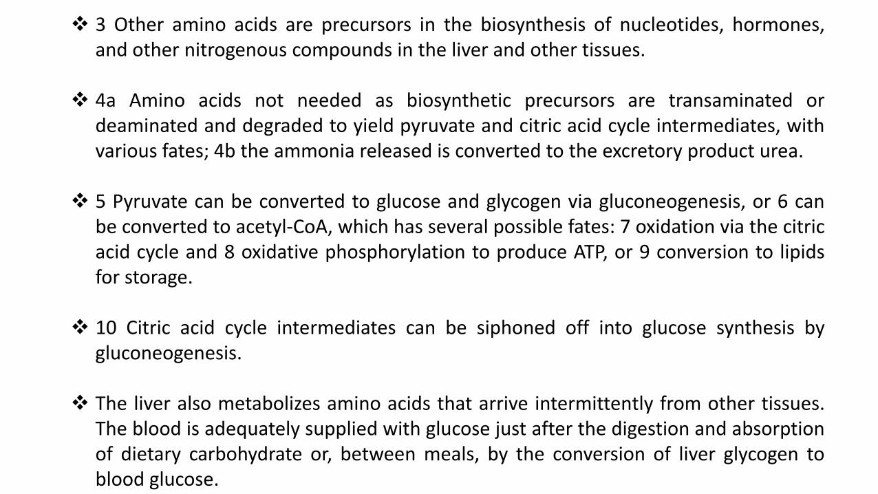
3 Other amino acids are precursors in the biosynthesis of nucleotides, hormones,and other nitrogenous compounds in the liver and other tissues.
4a Amino acids not needed as biosynthetic precursors are transaminated ordeaminated and degraded to yield pyruvate and citric acid cycle intermediates, withvarious fates; 4b the ammonia released is converted to the excretory product urea.
5 Pyruvate can be converted to glucose and glycogen via gluconeogenesis, or 6 canbe converted to acetyl-CoA, which has several possible fates: 7 oxidation via the citricacid cycle and 8 oxidative phosphorylation to produce ATP, or 9 conversion to lipidsfor storage.
10 Citric acid cycle intermediates can be siphoned off into glucose synthesis bygluconeogenesis.
The liver also metabolizes amino acids that arrive intermittently from other tissues.The blood is adequately supplied with glucose just after the digestion and absorptionof dietary carbohydrate or, between meals, by the conversion of liver glycogen toblood glucose.

During the interval between meals, especially if prolonged, some muscle protein isdegraded to amino acids.
These amino acids donate their amino groups (by transamination) to pyruvate, theproduct of glycolysis, to yield alanine, which 11 is transported to the liver anddeaminated.
Hepatocytes convert the resulting pyruvate to blood glucose via gluconeogenesis 5 ,and the ammonia to urea for excretion 4b.
One benefit of this glucose-alanine cycle is the smoothing out of fluctuations inblood glucose between meals.
The amino acid deficit incurred in muscles is made up after the next meal byincoming dietary amino acids.


Lipids; The fatty acid components of lipids entering hepatocytes also have severaldifferent fates.
1 Some are converted to liver lipids.
2 Under most circumstances, fatty acids are the primary oxidative fuel in the liver.
Free fatty acids may be activated and oxidized to yield acetyl-CoA and NADH.
3 The acetyl-CoA is further oxidized via the citric acid cycle, and 4 oxidations in thecycle drive the synthesis of ATP by oxidative phosphorylation.
5 Excess acetyl-CoA, not required by the liver, is converted to acetoacetate and β-hydroxybutyrate; these ketone bodies circulate in the blood to other tissues to beused as fuel for the citric acid cycle.
Ketone bodies, unlike fatty acids, can cross the blood-brain barrier, providing thebrain with a source of acetyl-CoA for energy-yielding oxidation.


Ketone bodies can supply a significant fraction of the energy in some extrahepatictissues—up to one-third in the heart and as much as 60% to 70% in the brain duringprolonged fasting.
6 Some of the acetyl-CoA derived from fatty acids (and from glucose) is used for thebiosynthesis of cholesterol, which is required for membrane synthesis.
Cholesterol is also the precursor of all steroid hormones and of the bile salts, whichare essential for the digestion and absorption of lipids.
The other two metabolic fates of lipids require specialized mechanisms for thetransport of insoluble lipids in blood.
7 Fatty acids are converted to the phospholipids and TAGs of plasma lipoproteins,which carry lipids to adipose tissue for storage.
8 Some free fatty acids are bound to serum albumin and carried to the heart andskeletal muscles, which take up and oxidize free fatty acids as a major fuel.

The liver thus serves as the body’s distribution center, exporting nutrients in thecorrect proportions to other organs, smoothing out fluctuations in metabolismcaused by intermittent food intake, and processing excess amino groups into ureaand other products to be disposed of by the kidneys.
Certain nutrients are stored in the liver, including iron ions and vitamin A.
The liver also detoxifies foreign organic compounds, such as drugs, food additives,preservatives, and other possibly harmful agents with no food value.
Detoxification often includes the cytochrome P-450–dependent hydroxylation ofrelatively insoluble organic compounds, making them sufficiently soluble for furtherbreakdown and excretion

There are two types of adipose tissue, white and brown, with different roles, and wefocus first on the more abundant of the two.
White adipose tissue (WAT) is amorphous and widely distributed in the body: underthe skin, around deep blood vessels, and in the abdominal cavity.
The adipocytes of WAT are large (diameter 30 to 70 μm), spherical cells, completelyfilled with a single large lipid droplet that constitutes about 65% of the cell mass andsqueezes the mitochondria and nucleus into a thin layer against the plasmamembrane.
The lipid droplet contains TAGs and sterol esters and is coated with a monolayer ofphospholipids, oriented with their head groups facing the cytosol.
Specific proteins are associated with the surface of the droplets, including perilipinand the enzymes for synthesis and breakdown of TAGs.
WAT typically makes up about 15% of the mass of a healthy young adult human.

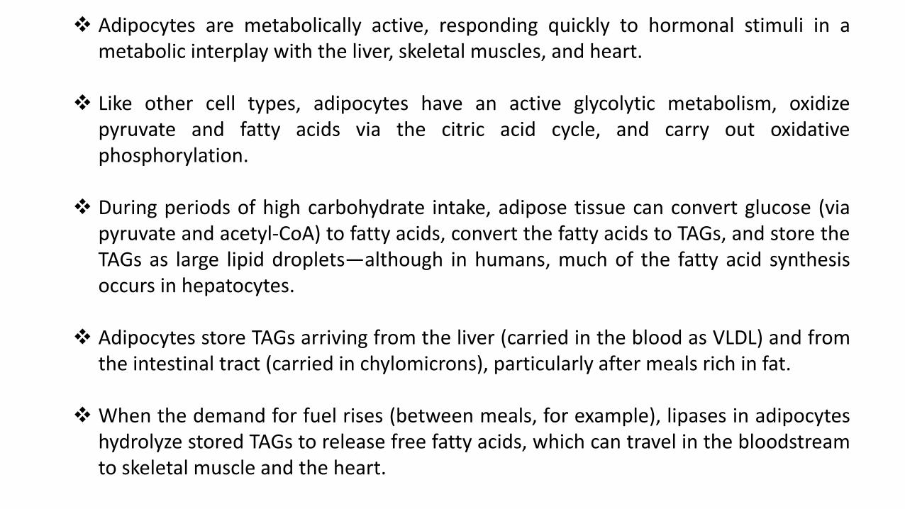
Adipocytes are metabolically active, responding quickly to hormonal stimuli in ametabolic interplay with the liver, skeletal muscles, and heart.
Like other cell types, adipocytes have an active glycolytic metabolism, oxidizepyruvate and fatty acids via the citric acid cycle, and carry out oxidativephosphorylation.
During periods of high carbohydrate intake, adipose tissue can convert glucose (viapyruvate and acetyl-CoA) to fatty acids, convert the fatty acids to TAGs, and store theTAGs as large lipid droplets—although in humans, much of the fatty acid synthesisoccurs in hepatocytes.
Adipocytes store TAGs arriving from the liver (carried in the blood as VLDL) and fromthe intestinal tract (carried in chylomicrons), particularly after meals rich in fat.
When the demand for fuel rises (between meals, for example), lipases in adipocyteshydrolyze stored TAGs to release free fatty acids, which can travel in the bloodstreamto skeletal muscle and the heart.
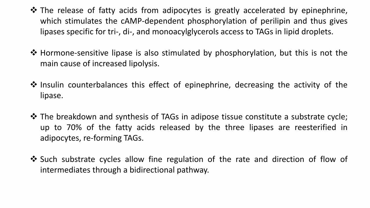
The release of fatty acids from adipocytes is greatly accelerated by epinephrine,which stimulates the cAMP-dependent phosphorylation of perilipin and thus giveslipases specific for tri-, di-, and monoacylglycerols access to TAGs in lipid droplets.
Hormone-sensitive lipase is also stimulated by phosphorylation, but this is not themain cause of increased lipolysis.
Insulin counterbalances this effect of epinephrine, decreasing the activity of thelipase.
The breakdown and synthesis of TAGs in adipose tissue constitute a substrate cycle;up to 70% of the fatty acids released by the three lipases are reesterified inadipocytes, re-forming TAGs.
Such substrate cycles allow fine regulation of the rate and direction of flow ofintermediates through a bidirectional pathway.

In adipose tissue, glycerol liberated by adipocyte lipases cannot be reused in thesynthesis of TAGs, because adipocytes lack glycerol kinase.
Instead, the glycerol phosphate required for TAG synthesis is made from pyruvate byglyceroneogenesis, requiring the action of the cytosolic PEP carboxykinase.
In addition to its central function as a fuel depot, adipose tissue plays an importantrole as an endocrine organ, producing and releasing hormones that signal the stateof energy reserves and coordinate metabolism of fats and carbohydrates throughoutthe body.
In small vertebrates and hibernating animals, a significant proportion of the adiposetissue is brown adipose tissue (BAT), distinguished from WAT by its smaller,differently shaped adipocytes.
Like white adipocytes, brown adipocytes store TAGs, but in several smaller lipiddroplets per cell rather than as a single central droplet.

BAT cells have more mitochondria and a richer supply of capillaries and innervationthan WAT cells, and it is the cytochromes of mitochondria and the hemoglobin incapillaries that give BAT its characteristic brown color.
A unique feature of brown adipocytes is their production of uncoupling protein 1(UCP1), also called thermogenin.
This protein is responsible for one of the principal functions of BAT: thermogenesis.
In brown adipocytes, fatty acids stored in lipid droplets are released, entermitochondria, and undergo complete conversion to CO2 by β oxidation and the citricacid cycle.
The reduced FADH2 and NADH so generated pass their electrons through therespiratory chain to molecular oxygen.
In WAT, protons pumped out of the mitochondria during electron transfer re-enterthe matrix through ATP synthase, with the energy of electron transfer conserved inATP synthesis.

In BAT, UCP1 provides an alternative route for the reentry of protons that bypassesATP synthase.
The energy of the proton gradient is thus dissipated as heat, which can maintain thebody (especially the nervous system and viscera) at its optimal temperature whenthe ambient temperature is relatively low.
In the human fetus, differentiation of fibroblast pre-adipocytes into BAT begins at thetwentieth week of gestation, and at the time of birth, BAT represents 1% to 5% oftotal body mass.
The brown fat deposits are located where the heat generated by thermogenesis canensure that vital tissues— blood vessels to the head, major abdominal blood vessels,and the viscera, including the pancreas, adrenal glands, and kidneys—are not chilledas the newborn enters a world of lower ambient temperature.
At birth, WAT development begins and BAT begins to disappear.

Young adult humans have much-diminished deposits of BAT, ranging from 3% of alladipose tissue in males to 7% in females, making up less than 0.1% of body mass.
However, adults have, distributed among their WAT cells, significant numbers ofadipocytes that can be converted by cold exposure or by β-adrenergic stimulationinto cells very similar to brown adipocytes.
These beige adipocytes have multiple lipid droplets, are richer in mitochondria thanwhite adipocytes, and produce UCP1, so they function effectively as heat generators.
Brown and beige adipocytes produce heat by oxidation of their own fatty acids, butthey also take up and oxidize both fatty acids and glucose from the blood at rates outof proportion to their mass.
In adaptation to warm or cold surroundings, and in the normal differentiation ofWAT, BAT, and beige adipose tissue, the nuclear transcription factor PPARγ plays acentral role.

The peptide hormone irisin, produced in muscle by exercise, triggers thedevelopment of beige adipose tissue that continues to burn fuel long after theexercise ends.
Metabolism in skeletal muscle cells—myocytes—is specialized to generate ATP as theimmediate source of energy for contraction.
Moreover, skeletal muscle is adapted to do its mechanical work intermittently, ondemand.
Sometimes skeletal muscles must work at their maximum capacity for a short time,as in a 100 m sprint; at other times more prolonged work is required, as in running amarathon or in prolonged physical labor.
There are two general classes of muscle tissue, which differ in physiological role andfuel utilization.
Slow-twitch muscle, also called red muscle, provides relatively low tension but ishighly resistant to fatigue.

It produces ATP by the relatively slow but steady process of oxidativephosphorylation.
Red muscle is very rich in mitochondria and is served by dense networks of bloodvessels, which bring the oxygen essential to ATP production.
Fast-twitch muscle, or white muscle, has fewer mitochondria than red muscle and isless well supplied with blood vessels, but it can develop greater tension and do sofaster.
White muscle is quicker to fatigue because, when active, it uses ATP faster than it canreplace it.
There is a genetic component to the proportion of red and white muscle in anyindividual, but with training, the endurance of fast-twitch muscle can be improved.
Skeletal muscle can use free fatty acids, ketone bodies, or glucose as fuel, dependingon the degree of muscular activity.

In resting muscle, the primary fuels arefree fatty acids from adipose tissue andketone bodies from the liver.
These are oxidized and degraded to yieldacetyl-CoA, which enters the citric acidcycle, ultimately yielding the energy forATP synthesis by oxidativephosphorylation.
Moderately active muscle uses bloodglucose in addition to fatty acids andketone bodies.
The glucose is phosphorylated, thendegraded by glycolysis to pyruvate, whichis converted to acetyl-CoA and oxidized viathe citric acid cycle and oxidativephosphorylation.

In resting muscle, the primary fuels are free fatty acids from adipose tissue andketone bodies from the liver.
These are oxidized and degraded to yield acetyl-CoA, which enters the citric acidcycle, ultimately yielding the energy for ATP synthesis by oxidative phosphorylation.
Moderately active muscle uses blood glucose in addition to fatty acids and ketonebodies.
The glucose is phosphorylated, then degraded by glycolysis to pyruvate, which isconverted to acetyl-CoA and oxidized via the citric acid cycle and oxidativephosphorylation.
In maximally active fast-twitch muscles, the demand for ATP is so great that theblood flow cannot provide O2 and fuels fast enough to supply sufficient ATP byaerobic respiration alone.
Under these conditions, stored muscle glycogen is broken down to lactate byfermentation.

Each glucose unit degraded yields three ATP, because phosphorolysis of glycogenproduces glucose 6-phosphate (via glucose 1-phosphate), sparing the ATP normallyconsumed in the hexokinase reaction.
Lactic acid fermentation thus responds more quickly than oxidative phosphorylationto an increased need for ATP, supplementing basal ATP production by aerobicoxidation of other fuels via the citric acid cycle and respiratory chain.
The use of blood glucose and muscle glycogen as fuels for muscular activity is greatlyenhanced by the secretion of epinephrine, which stimulates both the release ofglucose from liver glycogen and the breakdown of glycogen in muscle tissue.
Epinephrine mediates the so-called fight-or-flight response.
The relatively small amount of glycogen (about 1% of the total weight of skeletalmuscle) limits the glycolytic energy available during all-out exertion.
Moreover, the accumulation of lactate and consequent decrease in pH in maximallyactive muscles reduces their efficiency.

Skeletal muscle, however, contains another source of ATP, phosphocreatine (10 to 30mM), which can rapidly regenerate ATP from ADP by the creatine kinase reaction.
During periods of active contraction and glycolysis, this reaction proceedspredominantly in the direction of ATP synthesis; during recovery from exertion, thesame enzyme resynthesizes phosphocreatine from creatine and ATP.
Creatine serves to shuttle ATP equivalents from the mitochondrion to sites of ATPconsumption and can be the limiting factor in the development of new muscletissue.

After a period of intense muscular activity, the individual continues breathing heavilyfor some time, using much of the extra O2 for oxidative phosphorylation in the liver.
The ATP produced is used for gluconeogenesis (in the liver) from lactate that hasbeen carried in the blood from the muscles.
The glucose thus formed returns to the muscles to replenish their glycogen,completing the Cori cycle.
Actively contracting skeletal muscle generates heat as a byproduct of imperfectcoupling of the chemical energy of ATP with the mechanical work of contraction.
This heat production can be put to good use when ambient temperature is low:skeletal muscle carries out shivering thermogenesis, rapidly repeated musclecontraction that produces heat but little motion, helping to maintain the body at itspreferred temperature of 37 °C.

Heart muscle differs from skeletal muscle in that it is continuously active in a regularrhythm of contraction and relaxation, and has a completely aerobic metabolism at alltimes.
Mitochondria are much more abundant in heart muscle than in skeletal muscle,making up almost half the volume of the cells.
The heart uses mainly free fatty acids as a source of energy, but also some glucoseand ketone bodies taken up from the blood; these fuels are oxidized aerobically togenerate ATP.
Like skeletal muscle, heart muscle does not store lipids or glycogen in large amounts.
It does have small amounts of reserve energy in the form of phosphocreatine,enough for a few seconds of contraction.

Because the heart is normally aerobic and obtains its energy from oxidativephosphorylation, the failure of O2 to reach part of the heart muscle when the bloodvessels are blocked by lipid deposits (atherosclerosis) or blood clots (coronarythrombosis) can cause that region of the heart muscle to die.
This is what happens in myocardial infarction, more commonly known as a heartattack.
The metabolism of the brain is remarkable in several respects.
The neurons of the adult mammalian brain normally use only glucose as.
Astrocytes, the other major cell type in the brain, can oxidize fatty acids.
The brain, which constitutes about 2% of total body mass, has a very activerespiratory metabolism; more than 90% of the ATP produced in the neurons comesfrom oxidative phosphorylation.

The brain uses O2 at a fairly constant rate, accounting for almost 20% of the total O2consumed by the body at rest.
Because the brain contains very little glycogen, it is constantly dependent onincoming glucose in the blood.
Should blood glucose fall significantly below a critical level for even a short time,severe and sometimes irreversible changes in brain function may result.
Although the neurons of the brain cannot directly use free fatty acids or lipids fromthe blood as fuels, they can, when necessary, get up to 60% of their energyrequirement from the oxidation of β-hydroxybutyrate (a ketone body), formed in theliver from fatty acids.
The capacity of the brain to oxidize β-hydroxybutyrate via acetyl-CoA becomesimportant during prolonged fasting or starvation, after liver glycogen has beendepleted, because it allows the brain to use body fat as an energy source.

This spares muscle proteins—until they become the brain’s ultimate source ofglucose, via gluconeogenesis in the liver, during severe starvation.
In neurons, energy is required to create and maintain an electrical potential acrossthe plasma membrane.
The membrane contains an electrogenic ATP-driven antiporter, the Na+K+ ATPase,which simultaneously pumps 2 K+ ions into and 3 Na+ ions out of the neuron.
The resulting transmembrane potential changes transiently as an electrical signal, anaction potential, sweeps from one end of a neuron to the other.
Action potentials are the chief mechanism of information transfer in the nervoussystem, so depletion of ATP in neurons would have disastrous effects on all activitiescoordinated by neuronal signaling.

Bloodmediates the metabolic interactions among all tissues.
It transports nutrients from the small intestine to the liver and from the liver andadipose tissue to other organs; it also transports waste products from extrahepatictissues to the liver for processing and to the kidneys for excretion.
Oxygen moves in the bloodstream from the lungs to the tissues, and CO2 generatedby tissue respiration returns via the bloodstream to the lungs for exhalation.
Blood also carries hormonal signals from one tissue to another.
In its role as signal carrier, the circulatory system resembles the nervous system: bothregulate and integrate the activities of different organs.

The average adult human has 5 to 6 L of blood.
Almost half of this volume is occupied by three types of blood cells:
erythrocytes (red cells), filled with hemoglobin and specialized for carrying O2 andCO2;
much smaller numbers of leukocytes (white cells) of several types (includinglymphocytes, also found in lymphatic tissue), which are central to the immunesystem to defend against infections;
and platelets (cell fragments), which help to mediate blood clotting.
The liquid portion is the blood plasma, which is 90% water and 10% solutes.
Dissolved or suspended in the plasma are many proteins, lipoproteins, nutrients,metabolites, waste products, inorganic ions, and hormones.

More than 70% of the plasma solids are plasma proteins, primarily immunoglobulins(circulating antibodies), serum albumin, apolipoproteins (for lipid transport),transferrin (for iron transport), and blood-clotting proteins such as fibrinogen andprothrombin.
The ions and low molecular weight solutes in blood plasma are not fixedcomponents; they are in constant flux between blood and various tissues.
Dietary uptake of the inorganic ions that are the predominant electrolytes of bloodand cytosol (Na+, K+, and Ca2+) is, in general, counterbalanced by their excretion inthe urine.
For many blood components, something near a dynamic steady state is achieved: theconcentration of a component changes little, although a continuous flux occursbetween the digestive tract, blood, and urine.
The plasma levels of Na+, K+, and Ca2+ remain close to 140, 5, and 2.5 mM,respectively, with little change in response to dietary intake.

Any significant departure from these values can result in serious illness or death.
The kidneys play an especially important role in maintaining ion balance byselectively filtering waste products and excess ions out of the blood while preventingthe loss of essential nutrients and ions.
The human erythrocyte loses its nucleus and mitochondria during differentiation. Ittherefore relies on glycolysis alone for its supply of ATP.
The lactate produced by glycolysis returns to the liver, where gluconeogenesisconverts it to glucose, to be stored as glycogen or recirculated to peripheral tissues.
The erythrocyte has constant access to glucose in the bloodstream.

The minute-by-minute adjustments that keep the blood glucose level near 4.5 mMinvolve the combined actions of insulin, glucagon, epinephrine, and cortisol onmetabolic processes in many body tissues, but especially in liver, muscle, and adiposetissue.
Insulin signals these tissues that blood glucose is higher than necessary; as a result,cells take up excess glucose from the blood and convert it to glycogen andtriacylglycerols for storage.
Glucagon signals that blood glucose is too low, and tissues respond by producingglucose through glycogen breakdown and (in the liver) gluconeogenesis and byoxidizing fats to reduce the need for glucose.
Epinephrine is released into the blood to prepare the muscles, lungs, and heart for aburst of activity.
Cortisol mediates the body’s response to longer-term stresses.

Acting through plasma membrane receptors, insulin stimulates glucose uptake bymuscle and adipose tissue, where the glucose is converted to glucose 6-phosphate.
In the liver, insulin also activates glycogen synthase and inactivates glycogenphosphorylase, so that much of the glucose 6-phosphate is channelled into glycogen.
Insulin also stimulates the storage of excess fuel as fat in adipose tissue.
In the liver, insulin activates both the oxidation of glucose 6-phosphate to pyruvatevia glycolysis and the oxidation of pyruvate to acetyl-CoA.
The excess acetyl-CoA not needed for energy production is used for fatty acidsynthesis, and the fatty acids are exported from the liver to adipose tissue as theTAGs of plasma lipoproteins.
Insulin stimulates the synthesis of TAGs in adipocytes, from fatty acids released fromthe TAGs of VLDL.

These fatty acids are ultimately derived from the excess glucose taken up from bloodby the liver.
In summary, the effect of insulin is to favor the conversion of excess blood glucose totwo storage forms: glycogen (in the liver and muscle) and TAGs (in adipose tissue).


When glucose enters the bloodstream from the intestine after a carbohydrate-rich meal, the resulting increase in blood glucose causes increased secretion ofinsulin (and decreased secretion of glucagon) by the pancreas.
Insulin release is largely regulated by the level of glucose in the blood supplyingthe pancreas.
The peptide hormones insulin, glucagon, and somatostatin are produced byclusters of specialized pancreatic cells, the islets of Langerhans.
Each cell type of the islets produces a single hormone: α cells produce glucagon; βcells, insulin; and δ cells, somatostatin.
Several hours after the intake of dietary carbohydrate, blood glucose levels fallslightly because of the ongoing oxidation of glucose by the brain and othertissues.
Lowered blood glucose triggers secretion of glucagon and decreases insulinrelease.
Glucagon causes an increase in blood glucose concentration in several ways.
Like epinephrine, it stimulates the net breakdown of liver glycogen by activatingglycogen phosphorylase and inactivating glycogen synthase; both effects are theresult of phosphorylation of the regulated enzymes, triggered by cAMP.
Glucagon inhibits glucose breakdown by glycolysis in the liver and stimulatesglucose synthesis by gluconeogenesis.


The fuel reserves of a healthy adult human are of three types: glycogen stored in theliver and, in smaller quantities, in muscles; large quantities of TAG in adipose tissues;and tissue proteins, which can be degraded when necessary to provide fuel.
Two hours after a meal, the blood glucose level is diminished slightly, and tissuesreceive glucose released from liver glycogen.
There is little or no synthesis of TAGs.
By four hours after a meal, blood glucose has fallen further, insulin secretion hasslowed, and glucagon secretion has increased.
These hormonal signals mobilize TAGs from adipose tissue, which now become theprimary fuel for muscle and liver.


When an animal is confronted with a stressful situation that requires increasedactivity—fighting or fleeing, in the extreme case—neuronal signals from the braintrigger the release of epinephrine and norepinephrine from the adrenal medulla.
Both hormones dilate the respiratory passages to facilitate the uptake of O2, increasethe rate and strength of the heartbeat, and raise the blood pressure, therebypromoting the flow of O2 and fuels to the tissues.
This is the “fight-or-flight” response.
Epinephrine acts primarily on muscle, adipose, and liver tissues.
It activates glycogen phosphorylase and inactivates glycogen synthase by cAMP-dependent phosphorylation of the enzymes, thus stimulating the conversion of liverglycogen to blood glucose, the fuel for anaerobic muscular work.
Epinephrine also promotes the anaerobic breakdown of muscle glycogen by lacticacid fermentation, stimulating glycolytic ATP formation.

The stimulation of glycolysis is accomplished by raising the concentration of fructose2,6-bisphosphate, a potent allosteric activator of the key glycolytic enzymephosphofructokinase-1.
Epinephrine also stimulates fat mobilization in adipose tissue, by activating hormone-sensitive lipase and moving aside perilipin.
Finally, epinephrine stimulates glucagon secretion and inhibits insulin secretion,reinforcing its effect of mobilizing fuels and inhibiting fuel storage.
A variety of stressors (anxiety, fear, pain, hemorrhage, infection, low blood glucose,starvation) stimulate release of the glucocorticoid cortisol from the adrenal cortex.
Cortisol acts on muscle, liver, and adipose tissue to supply the organism with fuel towithstand the stress.
Cortisol is a relatively slow-acting hormone that alters metabolism by changing thekinds and amounts of certain enzymes synthesized in its target cells, rather than byregulating the activity of existing enzyme molecules.

In adipose tissue, cortisol leads to an increased release of fatty acids from storedTAGs.
The exported fatty acids serve as fuel for other tissues, and the glycerol is used forgluconeogenesis in the liver.
Cortisol stimulates the breakdown of nonessential muscle proteins and the export ofamino acids to the liver, where they serve as precursors for gluconeogenesis.
In the liver, cortisol promotes gluconeogenesis by stimulating synthesis of PEPcarboxykinase; glucagon has the same effect, whereas insulin has the oppositeeffect.
Glucose produced in this way is stored in the liver as glycogen or exportedimmediately to tissues that need glucose for fuel.
The net effect of these metabolic changes is to restore blood glucose to its normallevel and to increase glycogen stores, ready to support the fight-or-flight responsecommonly associated with stress.

The effects of cortisol therefore counterbalance those of insulin.
During extended periods of stress, the continued release of cortisol loses its positiveadaptive value and begins to cause damage to muscle and bone and to impairendocrine and immune function.