arteriography in ponies with strongylus vulgaris arteritis
TRANSCRIPT
Arteriography in Ponies with Strongylusvulgaris Arteritis
J. 0. D. Slocombe, V. T. Rendano, R. ap R. Owen,P. W. Pennock and B. M. McCraw*
ABSTRACT RESUME
Radiographs of the aorta and abdominal ar-teries were obtained from a normal anes-thetized pony following catheterization of afemoral artery for nonselective, semiselectiveor selective arteriography. The arteries hadsmooth borders and regular diameters andthe branches of the cranial mesenteric arterycould be followed distally on the angiogramthrough to the smaller branches proximal tothe bowel wall. Following arteriography, thepony walked normally and there were minimalalterations of the levels of serum muscle en-zymes and blood lactate. The procedures forarteriography were repeated in three days. Atthat time the femoral artery was patent andsatisfactory angiograms were obtained.
Similarly, radiographs were obtained fromtwo ponies artificially infected with Strongylusvulgaris. The cranial msenteric artery andsome of its branches, the right renal arteryand segments of the aorta had irregularborders and were enlarged. Branches of thecranial mesenteric artery could not be followeddistally because the flow of the contrast ma-terial was blocked.
Following the above procedures, euthanasiaof all ponies was expedited and the findingsof arteritis, thrombosis and dilatation ofarteries at necropsy compared favorably withinterpretations from the radiographs. At leastin the pony, arteriography can be a valuableresearch and diagnostic tool for the demon-stration of lesions associated with verminousarteritis.
*Department of Pathology (Slocombe and McCraw) andDepartment of Clinical Studies (Rendano, Owen andPennock), Ontario Veterinary College, University ofGuelph, Guelph, Ontario NlG 2W1.
Submitted June 23, 1976.
Les auteurs ont realise des radiographies del'aorte et des arteres abdominales d'un poneysain et anesthesie, apres lui avoir introduit uncatheter dans l'artere femorale, afin d'effec-tuer des arteriographies non selectives, semi-selectives et selectives. Les arteres presen-taient un contour interne lisse et un diametreregulier; l'angiogramme permit de suivre lesbranches de l'artere mesenterique craniale,jusque dans ses ramifications adjacentes 'a laparoi intestinale. Apries l'arteriographie, leponey pouvait marcher normalement; le tauxdes enzymes musculaires seriques et du lac-tate sanguin ne manifesta que tres peu d'alt&ration. Au bout de trois jours, les auteurs pro-cede'rent a une autre arteriographie; l'arterefemorale etait alors Wante et ils purent obte-nir des angiogrammes satisfaisants.
Les auteurs procederent aussi a des radio-graphies similaires, chez deux poneys preala-blement infectes avec Strongylus vulgaris.L'artere mesenterique craniale et certaines deses branches, l'artere renale droite et des seg-ments de l'aorte posterieure presentaient del'hypertrophie et un contour interne irregulier.I1 s'avera impossible de suivre les branches del'artere mesent6rique craniale, a cause d'unobstacle a la progression de la substance decontraste.A la fin de leurs exp6riences, les auteurs sa-
crifierent les poneys; les lesions arteriellesd'inflammation, de thrombose et de dilatation,retrouvees lors de la necropsie, correspondaientbien "a ce qu'avaient revele les radiographies.Par consequent, du moins chez le poney, l'arte-riographie peut constituer une technique vala-ble de recherche et de diagnostic, en ce quiconcerne la demonstration des lesions d'art6-rite vermineuse.
Volume 41 - April, 1977 137
INTRODUCTION
Strongylus vulgaris is one of the mostpathogenic parasites of equids in which itcan induce an acute arteritis. The patho-genesis of this condition has been docu-mented (4, 5). The purpose of this studywas to explore the feasibility and useful-ness of arteriography for revealing vascularlesions in the living animal.
MATERIALS AND METHODS
Three Shetland-cross ponies were used:one pony (A) for demonstrating the ap-
pearance of normal arteries and the feasi-bility of repeated catheterization for arte-riography, two ponies (B and C) with
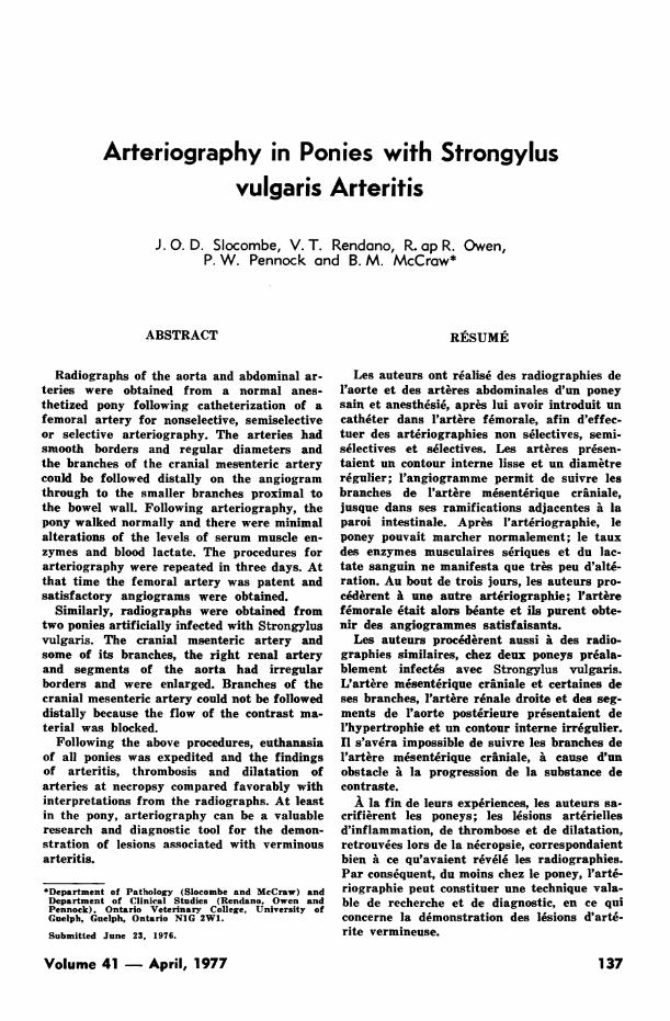
Fig. 1. Pony A, uni,fected. Nonselective arteriographywith positioning of the tip of the catheter immediatelycraniad of the coeliac artery (arrow). Note smoothborders and regular diameters of arteries.
138
induced arterial changes. Necropsies werecarried out immediately following arterio-graphy.
REARING AND INFECTION
Pony foals were obtained within 24 to 36hours after birth and reared in isolation.Foals were bottle fed milk replacer' untilthree months of age. Solid pelleted feed2was offered at about two weeks of age andformed the only ration after weaning.
Infective S. vulgaris larvae were obtainedby first retrieving adult female S. vulgarisfrom the cecum and colon of horses slaught-ered at an abattoir. The worms were choppedfinely with a scalpel and mixed thoroughlywith sterile sheep feces. This mixture wasincubated at 26°C for nine to 12 days. In-fective larvae were harvested from this cul-ture.
'Foal-lac, Borden Chemical, Borden Inc., Norfolk, Vir-ginia.215% Horse Feed, Shur-Gain Division, Canada PackersLtd., Toronto, Ontario.
Fig. 2. Line tracing of Fig. 1. Abbreviations: CA - co-eliac artery, CBA - colic branch of the ileocolic artery(= ventral colic artery), CEA - lateral or medial cecalartery, CT - tip of catheter, ICA - ileocolic artery(= ileo-ceco-colic artery), JA - jejunal arteries (=intestinal arteries), RA - right renal artery, RCA -
right colic artery (= dorsal colic artery).
Can. J. comp. Med.

was continued between the sartorius cra-nially and the gracilis caudally to exposethe femoral artery. About 3 cm of arterywas dissected free being careful not totear the anterior femoral artery and veinfrom the pedicle. Using No. 3 silk, ligatureswere placed around the dissected portionof the artery proximally and distally butonly the distal ligature was tightened. Theends of each ligature were left long formanipulating and occluding the artery bytension.An angiographic catheter was inserted
aseptically into the femoral artery in oneof two ways. In the first method, the arterywas punctured with a 16 gauge hypodermicneedle through which a guide wire was in-serted and after removal of the needle a No.7 or 8 French catheter 100 cm, long withopen end and multiple side holes4 waspassed over the guide wire. In the secondmethod a longitudinal incision was made
4Cook Incorporated, Bloomington, Indiana.
Fig. 3. Pony A, uninfected. Selective arteriography withpositioning of the tip of the catheter in the cranial me-senteric artery (arrow).
Pony A was seven and one-half monthsof age and uninfected at the time of theinvestigation. Ponies B and C were infectedvia a silicone-coated stomach tube. Pony Bwas infected at six months of age with 2000infective larvae and examined at 36 dayspostinfection (PI). Pony C was infected atsix weeks of age with 750 larvae and exam-ined at 51 days PI.
SURGERY AND CATHETERIZATION
Anesthesia was induced in each ponywith sodium thiamylal' (1 gm/300 lbs) andmaintained with oxygen and halothane mix-ture administered with a semiclosed re-breathing system with fresh gas flow of1.5 litres/min. With the pony in lateralrecumbency the skin on the medial aspectof a hind leg was prepared for surgery. A6 to 7 cm skin incision was made cranialand parallel to the pectineus muscle whichwas palpated as a tense band. The incision
3Surital, Parke Davis, Brockville, Ontario.
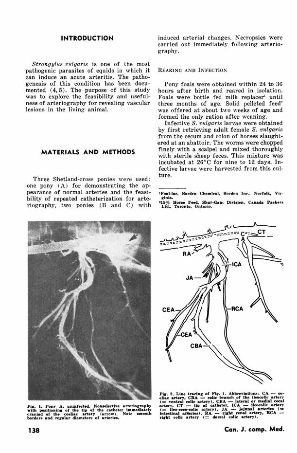
|~~~~~~~~~~~~~~~~~~~~~~~~~~~~~~~~~~~~~~~...D. ..... ........Fig. 4. Pony A, uninfected. Normal arteries with smoothintimal surfaces and regular diameters. Abbreviations:A - aorta, CBA - colic branch of the ileocolic artery(r= ventral colic artery), CMA - cranial mesentericartery, MCA - medial cecal artery, RA - right renalartery.
Volume 41 - April, 1977 139

TABLE I. Serum Muscle Enzymes and Blood Lactate Levels in International Units for Pony A(Uninfected) Pre and Post First Surgery
LactateTime Aspartatea Creatine Dehydro-Taken Transaminase Phosphokinase genase Lactate
Presurgery ................. 144 130 187Postsurgery ................ 135 127 168 245 hours postsurgery 167 178 246 132 days postsurgery .......... 161 243 204 16Reference valuesb ......... 85-550 13-180 74-206 4-11
,Formerly SGOTbFrom Lumsden, J.H. and R.C. Rowe Jr. Hematological and biochemical normal ranges for fifty standard-bred horses in training (Personal communication)
in the artery and a No. 8 or 9 French cathe-ter 125 cm long with closed end and multi-ple side holes' was inserted. The lattermethod was preferred. As the catheter wasinserted into the artery the proximal liga-ture was slackened allowing the catheterto pass by and be advanced up the femoralartery and into the aorta. The proximalligature was tightened sufficiently to con-trol hemorrhage and yet still allow move-ment of the catheter which was then filledwith heparin solution (1 in 10,000).
In survival operations (pony A) therewere some modified and additional proce-dures. A serrefine clamp was used in placeof the distal ligature and after the catheterwas removed the defect in the artery waselosed using No. 5-0 silk. The muscle layerswere sutured with No. 0 chromic gut andthe skih was closed with No. 0 nylon. Bloodsamples were taken prior to surgery andat several intervals postoperatively to de-termine changes which could occur fromocclusion of the artery and the resultingdysfunction of muscle in a hind leg. Bloodlactate levels and the following serum en-zymes were monitored: aspartate transami-nase (formerly known as SGOT), creatinephosphokinase and lactate dehydrogenase.The procedures for catheterization and an-giography were repeated on pony A threedays after the initial series.
RADIOLOGY
With the anesthetized ponies in lateralrecumbency the angiographic catheter waspositioned for nonselective, semiselectiveor selective arteriography by monitoring itunder the fluoroscopic image intensifier.
SUnited States Catheter & Instruments Co., Glen Falls,New York.
In nonselective arteriography (Fig. 1 anda line tracing in Fig. 2), the tip and sideholes of the catheter were positioned cra-niad to the origin of the coeliac artery. Insemiselective arteriography (Fig. 6) theside holes of the catheter were positionedat the origin of the coeliac or cranial me-senteric artery. In selective arteriography(Fig. 3) the tip of the catheter was placedin the vessel of primalry interest. Proper
Fig. 5. Pony A, uninfected. Selective arteriography withpositioning of the tip of the catheter (arrow CT) in thecoeliac artery. Note the constriction (due to arterialspasm) of one of the branches of the coeliac artery,possibly the hepatic artery arrows AS).
Can. J. comp. Med.140

N
B,I
Fig. 6. Pony B, S. vulgaris, 36 days postinfection. Semi-selective arteriography with positioning of the tip ofthe catheter at the origin of the coeliac artery (arrow).Note the irregular borders of enlarged arteries.
positioning of the catheter was ensured byinjecting and observing small doses of con-trast material fluoroscopically. The con-trast medium used was Hypaque-M75%6and it was injected by means of a power in-jector at 130 and 150 PSI of pressure. De-pending on the size of the animal and cathe-ter location, 30 to 50 ml of contrast mediumwere injected and radiographs were takennear the completion of the injection.
Radiographs were obtained using aPhilips7 300 Ma, 120 KV mobile X-ray ma-chine at 50 MaS and variable KV dependingon the thickness of the animal at 100 cm fo-cal film distance. Par speed screens8 andKodak RP/R9 film were used. A fine linegrid'" was used to decrease scattered radia-
6Sodium & Meglumine Diatrizoates, Winthrop Lab.Ltd.. Aurora, Ontario.
7Medio 30, Philips Electronics Industries Ltd., MedicalX-ray Division, Toronto, Ontario.8DuPont Cronex Par Speed Intensifying Screens, Du-Pont of Cnnada Ltd., Toronto, Ontario.9Kodak RP/R-14 X-omat Medical X-ray Film, KodakCanada Ltd., Toronto, Ontario.
"°Fine Line Stationary Grid, Liebel Flarsheim Co., Cin-cinnati, Ohio.
-MCA
Fig. 7. Line tracing of Fig. 6. Abbreviations: B - blad-der, CBA - colic branch of the ileocolic artery (=ventral colic artery), CT - tip of catheter, ICA -ileocolic artery (= ileo-ceco-colic artery), MCA - me-dial cecal artery, in line with the colic branch of theileocolic artery, RCA - right colic artery (= dorsalcolic artery), RK - right kidney.
tion. All films were developed in an auto-matic processor."1
RESULTS
Following the initial femoral catheteriza-tion pony A showed no sign of discomfortand three days later recatheterization ofthe femoral artery was uneventful. Dataon serum muscle enzymes and blood lactatelevels are found in Table I.
In the radiographs of pony A, uninfectedwith S. nulgairis, several branches of theabdominal aorta can be seen with smoothborders and regular diameters (Figs. 1 and3 and a line tracing in Fig. 2). The rightrenal artery and the following branches ofthe cranial mesenteric artery are clearly
"Profexray Series A71665, Litton Medical ProductsInd., Des Plaines, Illinois.
Volume 41 - April, 1977 141
visible: the right colic artery (= dorsal colicof older terminology), jejunal arteries ( =arteries of the left branch of the cranialmesenteric), the ileocolic artery (= ileo-ceco-colic), the lateral and medial cecalarteries and the colic branch of the ileocolicartery (= ventral colic) (6, 13). These ar-teries could be followed distally on the an-giogram through to the bowel wall. In Figs.1 and 5, the principal branches of the co-eliac artery are shown. Constriction, dueto arterial spasm of one of these branches,possibly the hepatic artery, is shown in Fig.5. At necropsy, the gross appearance of allthe arteries mentioned above and the vis-cera of pony A were normal. Several of theprincipal branches of the cranial mesentericartery are shown dissected in Fig. 4 andall arteries had regular diameters andsmooth intimal surfaces.The radiograph of pony B (36 days PI)
contains an enlarged and irregular cranialmesenteric artery and some of its branches(Fig. 6 with line tracing Fig. 7). Several
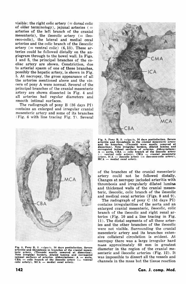
Fig. 9. Pony B, S. vulgaris, 36 days postinfection. Severearteritis and thrombosis in the cranial mesenteric nrteryand its branches. (Thrombi were mostly removed atdissection). Note irregular borders, dilated lumina andcorrugated intimal surfaces of arteries. Abbreviations:A - aorta, CBA - colic branch of the ileocolic artery(= ventral colic artery), CMA - cranial mesentericartery, ICA - ileocolic artery (= ileo-ceco-colic artery),MCA - medial cecal artery.
- :; ~MCA
Fig. 8. Pony B, 5. vulgais, 36 days postinfection. Severearteritis and thrombosis in branches of the cranial mesen-teric artery. (Thrombi mostly removed at dissection).Note irregular borders, dilated lumina and corrugatedintimal surfaces of arteries. Abbreviations: A - aorta,CBA - colic branch of the ileocolic artery (-- ventralcolic artery), MCA - medial cecal artery.
of the branches of the cranial mesentericartery could not be followed distally.Changes at necropsy included arteritis withthrombosis and irregularly dilated luminaand thickened walls of the cranial mesen-teric, ileocolic, colic branch of the ileocolicand medical cecal arteries (Figs. 8 and 9).The radiograph of pony C (51 days PI)
contains irregularities of the aorta and anenlarged cranial mesenteric, ileocolic, colicbranch of the ileocolic and right renal ar-teries (Fig. 10 and a line tracing in Fig.11). The distal segments of all these arter-ies and the other branches of the ileocolicwere not visible. Surrounding the cranialmesenteric artery and its branches exten-sive collateral circulation is evident. Atnecropsy there was a large irregular hardmass approximately 60 mm in greatestdiameter in the region of the cranial me-senteric and ileocolic arteries (Fig. 12). Itwas impossible to dissect all the vessels andchannels in the mass but the tissue reaction
Can. J. comp. Med.142

*TL
<.:..;! ^ -~~~~~~A
:pll
.7
41 r - AV
:-
Fig. 10. Pony C, S. vulgaris, 51 days postinfection. Se-lective arteriography with positioning of the tip of thecatheter in the caudal mesenteric artery (arrow). Notethe irregular borders of enlarged arteries.
of the aorta, the cranial mesenteric and(right renal arteries was extensive.
DISCUSSION
There are only a few reports on angio-graphy of the major vessels in the thoraxand abdomen of domestic animals (12,13,14, 16). The inadequacy of most X-raygenerators, the mass of the large animalsand the volume of ingesta are some of thereasons why radiographs of sufficient con-
trast for diagnostic purposes cannot beproduced. However, with the increasing use
of sophisticated and high powered X-rayequipment arteriography in the equid willbe feasible. Certainly the branches of theabdominal aorta in the angiograms fromthese ponies are clearly defined and re-
semble the anatomical descriptions forsimilar vessels in the horse (7).From necropsies of 130 horses with ar-
Volume 41 April, 1977
Fig. 12. Pony C. S. vulgaris 51 days postinfection. Severearteritis and thrombosis of the aorta, cranial mesenteric
and right renal arteries. (Thrombi were mostly removed
at dissection). The cranial mesenteric artery and its
branches were encased in an irregular hard mass from
which these vessels were dissected. Abbreviations: A -
aorta, CMA cranial mesenteric artery. RA - rightrenal artery.
terial lesions produced by S. vuulgaris, Otta-way and Bingham (10) and Poynter (11)found that 86 to 95% of the horses had le-
sions in the cranial mesenteric arteries and
50 to 65% had lesions in the cecal and colic
arteries. In the experimental studies of
143
Fig. 11. Line tracing of Fig. 10. Abbreviations: A -
aorta, CMA - cranial mesenteric artery, CT - catheter.

Drudge et al (4) and Duncan and Pinie(5), the colic branch of the ileocolic andcecal arteries were found to be part of thenormal pathway in the migration of larvaeto the cranial mesenteric artery. In Poyn-ter's (11) review of postmortem data otherarteries became affected but the incidencewas much lower (the coeliac artery up to32%,, the renal artery up to 23%, the aortaup to 20%) and rarely were the caudal me-senteric artery or jejunal arteries involved.
Arteriograms of all the vessels mentionedwere possible using nonselective, semiselec-tive or selective angiography. The nonse-lective approach with the tip of the cathe-ter just craniad to the coeliac artery wasconsidered most advantageous for threereasons. First, the time required to positionthe catheter was greatly reduced when com-pared with semiselective or selective an-giography. Secondly, the coeliac, cranialmesenteric and renal arteries could bevisualized simultaneously. Thirdly, arti-facts which can be created by the manipu-lation of a catheter inside smaller vessels(6, 17) were eliminatedPony A showed no discomfort following
femoral catheterization and angiography.The pony walked normally and there wasminimal alteration of the blood biochem-ical variables monitored. Elevations inlevels of serum muscle enzymes would haveindicated cell damage or necrosis of musclecells resulting from occlusion of the femor-al artery. Elevations in levels of blood lac-tate would have indicated increased glyco-lysis and these levels were elevated for ashort period following surgery. However,temporary elevations of lactate levels areusual following anesthesia (3). Recatheter-ization three days after the initial studywas uneventful and satisfactory radiographswere obtained. It would appear, therefore,that repeated catheterization of a femoralartery in a pony is possible and practical.The arteriographic studies indicated
that the cranial mesenteric artery and itsbranches were the principal vessels affectedby S. vulgaris and this was confirmed atnecropsy. The enlarged and irregular ap-pearance of the cranial mesenteric and ileo-colic arteries observed in the radiographfrom pony B were consistent with thechanges observed at necropsy. The exten-sive alteration of blood flow, the enlargedcranial mesenteric, ileocolic and right renalarteries and the irregularities of the aortasuggested by the arteriograph from ponyC mirrored the pathological disturbance.
found at necropsy. In the angiograms fromponies B and C, the branches of the cranialmesenteric artery could not be followeddistally because the flow of the contrastmaterial was obviously blocked by the le-sions.
In equids, definitive diagnosis of ver-minous arteritis due to S. vulgaris is diffi-cult. Rectal palpation of the enlarged cra-nial mesenteric artery and major branchesis possible only if the animal is not too largeor too small (1, 2). Where the size of the ani-mal is no hindrance a thorough rectal exam-ination may still be impossible because ofthe extensive adhesions from concurrent in-fections of Strongylus edentatus (8). Inaddition, S. edentatus can cause the lymphnodes in the region of the cranial mesen-teric artery to become quite enlarged andhard (9) and precise diagnosis of enlarge-ments due to S. vulgaris may be difficult.There is no definitive laboratory diagnostictest for recognizing the effects of migrat-ing S. vutgaris. The results presented hereindicate that, at least in the pony, arterio-graphy can be a valuable research and diag-nostic tool for the demonstration of thechanges associated with verminous arteritisand for visualizing the serial developmentof lesions.
ACKNOWLEDGMENTS
This study was supported by the OntarioMinistry of Agriculture and Food. The fi-nancial support from the Ontario RacingCommission is gratefully acknowledged.The authors wish to thank Mr. E. W. Eatonfor his valuable assistance in photography.
REFERENCES
1. BLOOD, D. C. and J. A. HENDERSON. VeterinaryMedicine. 4th Edition. London: Bailliere Tindall.1975.
2. CURTIS, R. A. Mesenteric aneurism in a horse. Can.vet. J. 5: '16-38. 1964.
3. DONAWICK, W. J. and M. A. HIZA. Metabolic careof the horse with acute intestinal obstruction.Tiidschr. Diergeneesk. 98: 980-982. 1973.
4. DRUDGE, J. H., E. T. LYONS and J. SZJANTO.Pathogenesis of migrating stages of helminths. withspecial reference to Strongylus vulgaris. In Biologyof Parasites. E. J. L. Soulsby, Editor. pp. 199-214.New York and London: Academic Press. 1966.
5. DUNCAN, J. L. and H. M. PIRIE. The pathogeneqiqof single experimental infections xX ;th Strongylusvulgaris in foals. Res. vet. Sci. 18: 82-93. 1975.
144 Can. J. comp. Med.

6. LINDBON, A. Arterial spasm caused by punctureand catheterization. Acta Radiologica 47: 449-459.1967.
7. GETTY, R. Sisson and Grossman's Anatomy of theDomestic Animals. Vol. 1. Philadelphia and London:W. B. Saunders Company. 1975.
8. McCRAW, B. M. and J. 0. D. SLOCOMBE. Earlydevelopment and pathology associated with Strongy-Ins edentatus. Can. J. comp. Med. 38: 124-138. 1974.
9. McCRAW, B. M. and J. 0. D. SLOCOMBE. Strongylusvulgaris in the horse: A review. Can. vet. J. 17:150-157. 1976.
10. OTTAWAY, C. W. and M. L. BINGHAM. Furtherobservations on the incidence of parasitic aneurysmin the horse. Vet. Rec. 58: 155-159. 1946.
11. POYNTER, D. The arterial lesions produced byStrongylus vulgaris and their relationship to themigratory route of the parasite in its host. Res.vet. Sci. 1: 205-217. 1960.
12. REID, C. F. Radiography of the alimentary canalof the horse. J. S. Afr. vet. Ass. 46: 69-72. 1975.
13. ROOT, C. R. and R. J. TASHJIAN. Thoracic andabdominal arteriography in calves. Am. J. vet. Res.32: 1193-1205. 1971.
14. SCOTT, E. A., S. K. KNELLER and D. M. WITHER-SPOON. Closure of ductus arteriosus determined bycardiac catheterization and angiography in newbornfoals. Am. J. vet. Res. 36: 1021-1023. 1975.
15. SISSON, S. and J. A. GROSSMAN. The Anatomyof the Domestic Animals. 4th Edition. Philadelphiaand London: W. B. Saunders Company. 1964.
16. TASHJIA.N, R. L., L. SI-KWANG, D. A. YARNS,K. M. DAS and H. L. STEIN. Angiocardiography incanine heartworm disease. Am. J. vet. Res. 31:415-436. 1970.
17. THEANDER, G. Arteriographic demonstration ofstationary arterial waves. Acta Radiologica 53: 417-425. 1960.
Volume 41 - April, 1977 145