artificial - orthotics · funds that would have gone into production of artificial limbs to the...
TRANSCRIPT


Artificial Limbs
V O L U M E 16 SPRING 1 9 7 2 NUMBER 1
C O N T E N T S
TRANSITION
A. Bennett Wilson, Jr iii
BODY SEGMENT PARAMETERS, PART II
Renato Contini 1
THE CHILD WITH TERMINAL TRANSVERSE PARTIAL HEMIMELIA
Barbara L. Sypniewski 20
TECHNICAL NOTES 51
COMMITTEE ON PROSTHETICS RESEARCH AND DEVELOPMENT DIVISION OF ENGINEERING
and
COMMITTEE ON PROSTHETIC-ORTHOTIC EDUCATION DIVISION OF MEDICAL SCIENCES
of the
NATIONAL RESEARCH COUNCIL
N A T I O N A L A C A D E M Y O F S C I E N C E S
2101 Constitution Avenue Washington, D. C. 20418

ArtHieial Limbs is a publication of the Committee on Prosthetics Research and Development and the Committee on Prosthetic-Orthotic Education, National Research Council, issued in the spring and autumn of each year in partial fulfillment of Veterans Administration Contract V101(134)P-74 and Social and Rehabilitation Service Contract SRS-72-6. Copyright © 1972 by the National Academy of Sciences. Quoting and reprinting are freely permitted, provided that appropriate credit is given. The opinions expressed by contributors are their own and are not necessarily those of either of the committees. Library of Congress Catalog Card No. 55-7710.
Editorial Board: Eugene F. Murphy, Ph.D., Prosthetic and Sensory Aids Service, Veterans Administration, New York, N.Y.; Herbert Elftman, Ph.D., College of Physicians and Surgeons, Columbia University, New York, N.Y., and Frank W. Clippinger, M.D., Duke University Medical Center, Durham, N.C.

Artificial Limbs, Vol. 16, No. 1, p. iii Spring 1972
TRANSITION
A. Bennett Wilson, Jr.
The National Academy of Sciences began publication of Artificial Limbs in 1954 because, at that time, there was available no periodical for the systematic dissemination of the results of research in limb prosthetics. Since that time 33 issues have been published and made available without charge to an average of 5,000 individuals concerned with the management of amputees.
Also, since the initiation of Artificial Limbs, the Veterans Administration Prosthetic and Sensory Aids Service has introduced the Bulletin of Prosthetic Research, and the American Orthotic and Prosthetic Association and the American Academy of Orthotists and Prosthetists publish the journal Orthotics and Prosthetics which is now devoted entirely to technical and professional topics.
Thus, in keeping with the philosophy that the National Academy of Sciences should not undertake programs that can be carried out by others, publication of Artificial Limbs will be discontinued with this issue. This decision will permit the Committee on Prosthetics Research and Development and the Committee on Prosthetic-Orthotic Education to devote the time and funds that would have gone into production of Artificial Limbs to the production of monographs and other publications that are not apt to be made available otherwise.
Information that would have appeared in Artificial Limbs will now be found in the Bulletin of Prosthetics Research and Orthotics and Prosthetics.
The reception given to Artificial Limbs through the years has been very rewarding to the staff, and even though it is with regret that we must advise discontinuance of its publication, we feel that the move is in the best interest of the Prosthetics and Orthotics Program.
iii

Body Segment Parameters, Part I I 1
RENATO CONTINI2
1 The investigations on which this article is based were supported principally by two research special projects grants—one from the Office of Vocational Rehabilitation and the other from the Social and Rehabilitation Service, Dept. of Health, Education, and Welfare.
2 Prosthetic-Orthotic Education Program, UCLA, Los Angeles, Calif.
The performance of human (animal) activity requires the expenditure of energy. During the contraction of the muscles involved in this activity, chemical energy is converted first into mechanical energy, then into work and heat. Some of this chemical energy is required for maintenance of body functions. In movement, however, much of the mechanical energy is required to overcome friction and tissue displacement at the joints, gravity, inertial forces, air and water resistance— all of which oppose the action desired.
Biomechanics is the science that is concerned with such effects. In order to understand better the biomechanics of movement, it is necessary to know certain characteristics of the segments involved. Among these characteristics are the mass of the segments, their centers of mass, and their mass moments of inertia. The characteristics (body parameters) themselves are not readily obtained on living subjects.
It was the purpose of two studies conducted at the New York University School of Engineering and Science to obtain some of these body parameters. The first of these studies (6), completed in 1966, was conducted on normal, healthy American males in the age range of 20-40 years. The second study (3), completed in 1970, was conducted on a random selection of adults, young males and females 20-30 years of age, some females in the 40-50 age
bracket, and a number of amputees and hemiplegics, male and female, in all age ranges.
A history, survey of measurement techniques, and data developed over the years was given in "Body Segment Parameters: A Survey of Measurement Techniques," which appeared in Artificial Limbs, Spring 1964 (7). Also, a condensation of four of the most important monographs in this field ("Center of Gravity of the Human Body" by W. Braune and O. Fischer; "Theoretical Fundamentals for a Mechanics of Living Bodies" by O. Fischer; "The Human Motor" by J. Amar; and "Space Requirements of the Seated Operator" by W. T. Dempster) has been prepared by Krogman and Johnston (10) under the sponsorship of the United States Air Force.
METHODS
Most studies undertaken previously used cadavers, but in a few studies, including those at New York University, living subjects were used. Although some available measuring techniques for compiling the data are similar for live subjects and for cadavers, other techniques must obviously differ. In general, the techniques covered here are for living subjects; thus, all techniques used on dissected cadavers are not included. When living subjects are used, particularly the elderly and those suffering with some affliction or disability, any technique utilized must be at the convenience of the subject. Some subjects cannot comfortably assume the necessary postures during the measurement processes, while for some others the procedures are physically impossible. As a result, not all measurements can be taken on all subjects, but, because of the various tech-
Artificial Limbs, Vol. 16, No. 1, pp. 1-19 Spring 1972
1

niques available, most of the desired data can be obtained.
The techniques are only briefly presented here because more adequate descriptions are available in other references.
VOLUME DETERMINATION
The body and all of its segments are irregular solids. The volume of an irregular solid may be obtained or approximated in a number of ways: by mensuration, immersion, or photogrammetry. Only the first two were used in both studies.
Mensuration
A relatively good approximation of body-segment volume can be obtained by using circumferential measurements at certain selected stations on the segment and the linear dimensions between any two consecutive circumferential measurements. If all these measurements are known for the full length of the segment, then an approximate volume can be determined. Accuracy will increase with the increased number of such measurements. This technique assumes that any two successive cross sections of the member are parallel and essentially similar geometrically. In that event, the volume contained within the two cross sections may be expressed as:
It is obviously impossible to obtain cross-sectional areas on the body segments of living subjects. If it is assumed, however, that the cross sections of the limbs are elliptical, it is possible to establish a relationship between the cross-sectional area and the perimeter at any chosen level. For any segmental portion between two levels, the volume may now be expressed as:
For a total limb divided into n segments, each h distance apart:
The derivation of this equation is given in reference 3.
Immersion
In this method, the segment whose volume is to be determined is immersed in water. Incremental volumes are taken of the segment whose total volume then is the sum of these increments. For these studies, four tanks were specially designed: an arm tank, a hand tank, a leg tank, and a foot tank. Each tank was constructed of Plexiglas, the first three cylindrical in cross section, and the last, rectangular.
The limb or body segment was completely immersed in the tank. Water was permitted to drain off in controlled increments, each representing a known change in cylinder height. Drained water was collected and measured. The difference in volume between that collected and that obtainable without the body segment in place (the actual volume of the tank for that increment) represents the volume of the body segment contained within the height increment. Whenever possible, these increments were 2.0 cm apart, but, if subjects with limited physical tolerance had minimal cross-sectional variation, the increments were increased to every 4.0 cm apart.
Photogrammetry
Two types of photogrammetric techniques are available—mono and stereo. In
2 CONTINI

the former, lines or colored shadows are projected on the subject in such fashion as to produce a contour map on the particular segment of interest. The areas contained within these contours may be measured with a planimeter, and the same general equation for determining the volume as given previously may be used. Again, the sum of all the incremental volumes of the segment represents its total volume.
In stereophotogrammetry, two cameras are used side by side to create an illusion of depth when the two photographs are juxtaposed. The resulting picture is treated as an aerial photograph of terrain upon which contour levels are applied. These then are treated as in monophoto-grammetry.
Fig. l.
Fig. 2.
DENSITY DETERMINATION
To obtain the overall body density of living subjects is extremely difficult. To obtain the density of individual segments on living subjects is virtually impossible. There are ways, however, to obtain fairly accurate values. The problems involved will not be discussed here; some of them are described in the two referenced reports (3, 6).
Empirical (Whole Body)
Whole-body volume may be approximated in several ways. The mass may be obtained by weighing accurately. The density is the ratio of mass to volume. For lean bodies, the density is higher than for fat bodies. One provisional formula for determining density, developed by Dupertuis in 1950 (8), makes use of Sheldon's somatotyping system (12) and introduces the first component (x) of the system into the equation:
d(ensity) = 1.094 - 0.0119x
A second equation developed by the Biomechanics Group at NYU, using data developed by Behnke (1), is based on the height (H) in inches, and weight (W) in pounds of the individual (figs. 1 and 2):
Anthropometric (Whole Body)
Many studies have established the reasonably close relationship between body fat and certain skin-fold thicknesses (2). The equations used for the NYU study were those developed by Pascale (11).
BODY SEGMENT PARAMETERS, PART II 3

The first depends on the measurement of the skin-fold thickness at the triceps:
d(ensity) = 1.0923 - 0.0202(St) x 0.1
The second depends on the measurement of the skin-fold thickness at the scapula:
d = 1.0896 - 0.0179(Ss) x .1
Mensuration (Whole Body)
Skerlj in 1954 (13) developed a method for determining whole-body volume. He measured 10 circumferential dimensions and 6 linear dimensions (fig. 3). From these he developed a formula that gives an approximate value for whole-body volume.
The NYU group presented (3) a modified equation using the Skerlj notation and included some correction factors derived by applying the equation to five subjects for whom the volume of the various body
segments was known. The modified formula is:
With the volume so determined, the mass may be obtained by direct weighing and the overall (whole body) density may be obtained:
d(ensity) = M(ass) /V(olume)
Empirical (Body Segments)
Until recently, very little work has been done to establish segment densities. Harless (9) conducted some studies with cadavers, as did Dempster (4, 5). At NYU, in the first of the two studies, the mass of certain body segments was established by the reaction-board method, which is described below.
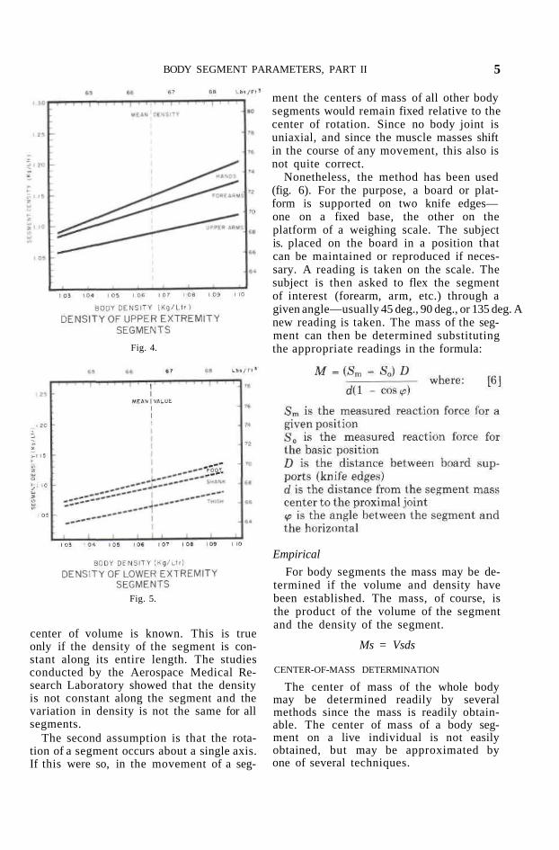
Based on these studies, two graphs were developed that relate whole-body density to body-segment density (figs. 4 and 5). These are approximations only, since no exact data are available.
Fig. 3. Linear measurements: measurements for body-volume determination (after Skerlj).
MASS DETERMINATION
In studies conducted with cadavers, weight and eventually mass are obtained directly by accurate weighing techniques applied to the total segment or to its increments. In studies with live subjects, this cannot be done. The reaction-board method may be used.
Reaction-Board Method
This method is dependent on the validity of two assumptions. The first is that the center of mass can be established if the
CONTIN] 4
center of volume is known. This is true only if the density of the segment is constant along its entire length. The studies conducted by the Aerospace Medical Research Laboratory showed that the density is not constant along the segment and the variation in density is not the same for all segments.
The second assumption is that the rotation of a segment occurs about a single axis. If this were so, in the movement of a seg
ment the centers of mass of all other body segments would remain fixed relative to the center of rotation. Since no body joint is uniaxial, and since the muscle masses shift in the course of any movement, this also is not quite correct.
Nonetheless, the method has been used (fig. 6). For the purpose, a board or platform is supported on two knife edges— one on a fixed base, the other on the platform of a weighing scale. The subject is. placed on the board in a position that can be maintained or reproduced if necessary. A reading is taken on the scale. The subject is then asked to flex the segment of interest (forearm, arm, etc.) through a given angle—usually 45 deg., 90 deg., or 135 deg. A new reading is taken. The mass of the segment can then be determined substituting the appropriate readings in the formula:
Empirical
For body segments the mass may be determined if the volume and density have been established. The mass, of course, is the product of the volume of the segment and the density of the segment.
Ms = Vsds
Fig. 4.
Fig. 5.
CENTER-OF-MASS DETERMINATION
The center of mass of the whole body may be determined readily by several methods since the mass is readily obtainable. The center of mass of a body segment on a live individual is not easily obtained, but may be approximated by one of several techniques.
BODY SEGMENT PARAMETERS, PART II 5

Volumetric Approximation A number of researchers, the NYU group
included, have assumed that the density along the segment is constant and thus have concluded that the center of mass is coincident with the center of volume. Under this assumption, the center of volume, hence the center of mass, is found in the following way:
A base line is established, usually the proximal joint of the segment. This segment is divided into a number of increments for which the volume is obtained by one of several methods (V1, V2, V3,..., Vn). The distance to the center of volume is measured from the base line (d1, d2, d3, . . ., dn).
The center of volume is determined by dividing the sum of the products of each volume times its distance from the base line, by the sum of the volumes.
Reaction-Board Method
With cadavers, segments, or with plaster models of body segments, the center of mass may be obtained by use of the reaction board, previously described.
Of these techniques, the one using the cadaver segment and the reaction board is the most accurate; the true center will vary in this technique only by the change that has occurred in the body tissues after death. Use of the plaster-of-paris cast creates the same error as that obtained by use of the volumetric technique; i.e., the error is introduced because it is assumed that the density along the segment is constant, whereas the density in any segment usually increases from the proximal to the distal end. This occurs because the ratio of bone to muscle and fat increases distally.
Fig. 6. Determination of the arm mass (reaction-board method).
SEGMENT MASS MOMENT OF INERTIA
The motions of body segments are essentially rotatory, and linear movement is the result of a number of coordinated rotatory motions. The motion is assumed to
C0NTIN1 6

occur about a fixed axis that is perpendicular to the plane in which the motion occurs. It is assumed that frictional and inertial forces occur in the plane of rotation. Rotation can be caused by a force at some distance from the axis of rotation, or by a force couple. In rotation, an inertial force resists angular acceleration which acts at the center of mass resulting in an inertial moment. This mass moment of inertia depends on the size, shape, and mass distribution of the body.
The mass moment of inertia may be determined in several ways.
Empirical
The mass moment of inertia of a body with respect to a given axis of rotation is the sum of the products of the mass increments mi (into which the total mass may be divided) by the square of their respective distances from the particular axis of rotation:
Quick Release
If a force (F) is applied to a segment at some distance (d) from the axis of rotation of the segment, it will be imparted at an angular acceleration (a) in accordance with the equation:
Fd = Ia
Because of this relationship, it is possible to determine the mass moment of inertia (I) experimentally by this quick-release method.
In this method, the body segment of interest is arranged so that it may be free to swing about the proximal joint, which in turn is restrained from motion. At some distance (d) from the axis of rotation, a cable is attached to the segment such that it will prevent rotation in one direction. The other end of the cable is attached to a spring restraint, which in turn is attached to a force-measuring device. The subject is instructed to pull against the spring with a force (F), which is recorded.
The cable is cut suddenly and the segment accelerates with an acceleration (a) that is appropriately recorded. By substitution of the known values F, d, a, the mass moment of inertia (I) can be obtained.
I = Fd/a
Pendulum
The period of a pendulum is related to the mass moment of inertia of the pendulum. For a simple pendulum, i.e., one where the mass is concentrated at some distance from the center of oscillation, the relationship is expressed by the equation:
This method utilizes plaster casts of body segments or the severed cadaver segments. The segment or its counterpart is suspended at one point near the end of the segment. It is permitted to swing through an arc of limited magnitude. The period of oscillation is obtained by some appropriate instrumentation. The values that are obtained are substituted in the above equation.
RESULTS
Results are given for tests conducted both in the first and second series of experiments. In the first series of tests, data were collected on 12 male subjects in the age range of 20-40 years. In the second series of tests, data were collected on 9 male subjects in the age range of 20-30 years, 5 female subjects ages 17-20 years, and 3 female subjects ages 40-50 years, all without disabilities. Data were also recorded on 19 additional subjects with either hemiplegia or an amputation. In the second series of tests, not all data were recorded for every subject. The following tables contain the most valid data acquired.
BODY SEGMENT PARAMETERS, PART II 7

VOLUMES
Table 1 contains the volume of body segments recorded during the first series of tests. There is only one major difference between the two series on males. In the first series, the value for volume of the upper arm—and hence the value for the whole arm—included the shoulder cap, i.e., the volume from the axilla to the acromion process. In the second series (table 2), the values of volumes for the upper and whole arm are only up to the axilla. On the basis of the mean values for the upper arm in the two series, the volume of the shoulder cap is approximately 36% of the whole upper arm.
In the second series of tests, a limited number of shoulder caps were cut off from the plaster-of-paris arms at the level of the axilla. Their dimensions, circumference at the axilla (c), and height to the acromion process (h) were taken. The volumes were obtained by immersion.
An approximate equation for determining the volume of the shoulder cap was then established:
Volume (shoulder cap) = 0.0526 hcc
This equation is approximate to ± 20% of the true value.
In all other respects, the two series of tests give comparable results. The differ-
8 CONTINI

ences in mean values are of the order of 1%-10%. Considering the limited numbers of subjects, 12 and 8 in the respective samples, the differences are not serious, and the mean values are useful in general computations. Of interest in the second series of tests is the close relationship between mean values for right-hand and left-hand volumes. The variation between means in most instances is less than the variation between the volume of right and left segments in any subject.
Table 3 indicates similar values for female subjects. There was greater inter-subject variation in this population than in that for the males. In view of this, and because there was such a limited number of
subjects both in the younger and older age groups, the values for the two groups were combined. Even so, these mean values may be less accurate than those for the male population. They are presented, however, because few other similar data are available.
The body-segment volume may be expressed as a ratio or percentage of the whole-body volume. If it is desired to estimate body-segment volume, it is better to do so on the basis of the segment volume as a percentage of whole-body volume. This probably will give a more accurate result than using an average value for the volume of body segment.
Table 4 gives such values for the first
BODY SEGMENT PARAMETERS, PART II 9

series of males. Table 5 gives similar values for the second series of males, and table 6 gives these values for females.
DENSITIES
As mentioned previously, it is very difficult to determine densities accurately. In table 7, the densities have been determined by the equations shown in the section III-B for males first series. The densities for both males and females, second
series, have been determined by dividing the mass (weight) by the volumes derived by using the NYU and Skerlj formulas and by using Pascale's equations A and B and skin-fold thicknesses.
CENTER OF VOLUME
In the absence of satisfactory techniques for determining the center of mass, it has been assumed to be coincident with the center of volume. Table 8 shows the loca-
CONTINI 10
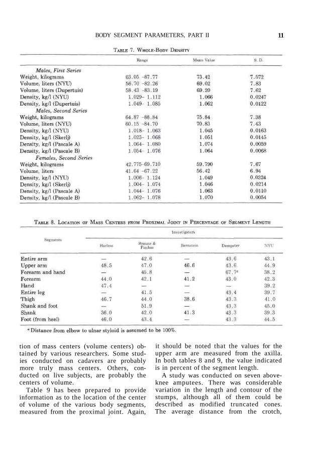
tion of mass centers (volume centers) obtained by various researchers. Some studies conducted on cadavers are probably more truly mass centers. Others, conducted on live subjects, are probably the centers of volume.
Table 9 has been prepared to provide information as to the location of the center of volume of the various body segments, measured from the proximal joint. Again,
it should be noted that the values for the upper arm are measured from the axilla. In both tables 8 and 9, the value indicated is in percent of the segment length.
A study was conducted on seven above-knee amputees. There was considerable variation in the length and contour of the stumps, although all of them could be described as modified truncated cones. The average distance from the crotch,
BODY SEGMENT PARAMETERS, PART II 11

measured downward and expressed as a percentage of the total stump length, was 32.1%, with an upper limit of 44.0% and a lower limit of 23.0%. The standard deviation was ± 6.4%.
RADIUS OF GYRATION
The radius of gyration (p) is a distance measured from the true center of mass to a point within the mass at which, if all the mass were concentrated, its effect in rotatory movements would be similar to the effect of the mass as it is actually distributed. For geometrically similar shapes, the radius of gyration along a particular axis may be expressed as a percentage of
the length of that shape along that axis. It has been assumed that every body
segment—arm, leg, upper arm, forearm— for one subject is geometrically similar to that of any other subject. If it were so, then the radius of gyration expressed in percentage of the length (p/L) should be relatively constant. It was found to be so, with minor variations. The values of p/L for the various body segments obtained by previous researchers and in the first NYU study are given in table 10. Values for the second NYU study are given in table 11.
Table 12 has been included as a guide against which the computed values of p may be compared. This table indicates the average values of p (the radius of gyration) for the populations included in the second series of NYU studies; not all values were determined for each category, and the table reflects this. The results were computed on the basis of tests and measurements were made as previously described.
DISCUSSION
The data may be used in a number of ways. Consideration must be given to the nature of the problem for which a solution is sought and the accuracy desired. If a situation exists where a prosthesis or orthosis is desired for a specified individual, it would be best to obtain data directly on the individual. In such a case, judgment should be made as to which of the various techniques available would be adapted best to the set of conditions present, i.e., the condition of the subject, the skills of
CONTINI 12

available personnel, and the facilities available.
When extreme accuracy is not required, or in cases when the problem is confined to a class of individuals, or the solution may have a general application, the data may be used in various ways, with differing degrees of accuracy. In successively decreasing order of accuracy, the following maybe done:
1. Obtain the weight and height of the subject and the length and circumferences
of the segments under consideration; use tables and graphs judiciously and, where several sets of data are available, use the most appropriate.
2. Obtain weight and height of the subject only and use tables as suggested.
3. Obtain weight and height of subject and use average data only. Data may be used for determining the length of a segment, its volume, mass, center of volume, center of mass, radius of gyration, and moment of inertia.
BODY SEGMENT PARAMETERS, PART II 13

Fig. 7.
CONTINI 14

Fig. 8.
BODY SEGMENT PARAMETERS, PART II 15

Fig. 9.
Fig. 10.
Fig. 11.
CONTINI 16

Fig. 12.
SAMPLE COMPUTATION
To determine the mass moment of inertia of the upper arm, forearm, and hand for a male patient (possibly for application of an externally powered orthosis), only the height and weight of the subject need be known.
Procedure If the subject weights 190 pounds and is
73 inches in height: 1. On graph (fig. 1), join the weight in
pounds (190) to the height in inches (73) by a straight line. At the intercept of this line with line c a value for c, approximately 12.8, is obtained.
2. On graph (fig. 2), locate c = 12.8, proceed vertically upward to intersect solid black line, then proceed horizontally from this point to determine the value of whole-body density d:
d = 66.8 pounds per cubic foot
3. On graph (fig. 4), proceed as in (2), from d = 66.8 vertically downward to intersect lines of segment densities:
d, upper arm = 68.1 lb/ft^3 d, forearm = 70.7 lb/ft^3 d, hand = 72.2 lb/ft^3
4. Given the weight of 190 pounds and whole-body density of 66.8 pounds per cubic foot, we may compute whole-body volume:
190/66.8 = 2.85 cubic feet
5. Table 4 gives values of volume for body segments in percentage of whole-body volume:
volume, upper arm = 3.495 x 0.01 x 2.85 = 0.0995 ft^3
BODY SEGMENT PARAMETERS, PART II 17

volume, forearm = 1.70 x 0.01 x 2.85 = 0.0485 ft3
volume, hand = 0.566 x 0.01 x 2.85 = 0.0161 ft3
6. Multiplying the volumes of the segments by their respective densities, the mass (or weights) of the segments are obtained:
m (w), upper arm = 0.0995 x 68.1 = 6.78 lb m (w), forearm = 0.0485 x 70.7 = 3.43 lb m (w), hand = 0.0161 x 72.2 = 1.16 lb
7. To obtain the approximate lengths of the body segments when they have not been measured, figures 7 and 8 may be used. The mean lengths expressed in terms of body height are 0.189H, 0.145H and 0.128H for the upper arm, forearm, and hand respectively. The lengths then are:
Lv = 0.189 x 73 = 13.8 in. Lf = 0.145 x 73 = 10.6 in. Lh = 0.128 x 73 = 9.35 in.
Fig. 13.
8. Having obtained the lengths of the segments, the location of the center of
CONTINI 18

volume (mass) can be determined using values given in tables 8 or 9:
c, upper arm = 0.461 x 13.8 = 6.37 in. c, forearm and hand = 0.420 (10.6 + 9.35) = 8.38 in.
9. The radius of gyration (p) for the segments may be obtained using the values in tables 10 or 11:
p, upper arm = 0.268 x 13.8 = 3.70 in. p, forearm and hand - 0.263 x (10.6 + 9.35) = 5.25 in.
10. The moment of inertia about its proximal axis of rotation is expressed by the equation:
Ij = m(pp + cc)
The moment of inertia of the upper arm about the shoulder:
The moment of inertia of the forearm about the elbow:
If the moment of inertia of the forearm and hand about the shoulder joint is desired, then the equation is:
Figures 9 through 13 have been included to facilitate any computations, to ease conversion from metric to British systems of measurement, and for graphically determining the moments of inertia.
ACKNOWLEDGMENT
Appreciation is expressed to Dr. Rudolfs Drillis and Messrs. Darrell Hill, Howard Gage, Maurice Bluestein, Albert
Yatkauskas, and George Vadell for their contributions to this research project, and to Mrs. Mary Klaus for the preparation of the reports.
REFERENCES
1. Behnke, A. R., Jr., B. G. Feen, and W. C. Welham, The specific gravity of healthy men; body weight devided by volume as index of obesity, J.A.M.A. 118:495-498, Feb. 14, 1942.
2. Brozek, J., J. K. Kihlberg, H. L. Taylor, et al., Skinfold distributions in middle-aged American men: a contribution to norms of leanness-fatness, Ann. N.Y. Acad. Sci. 110:492-502, Sept. 26, 1963.
3. Contini, Renato, Body Segment Parameters (Pathological), Tech. Rept. 1584.03, New York Univ. School of Engineering and Science, June 1970.
4. Dempster, W. T., Space Requirements of the Seated Operator, U.S. Air Force WADC Tech. Rept. 55-159, Wright-Patterson Air Force Base, Ohio, July 1955.
5. Dempster, W. T, The anthropometry of body action, Ann. N.Y. Acad. Sci. 63:559-585, 1956.
6. Drillis, Rudolfs, and Renato Contini, Body Segment Parameters, Tech. Rept. 116.03, New York Univ. School of Engineering and Science, Sept. 1966.
7. Drillis, Rudolfs, Renato Contini, and Maurice Bluestein, Body segment parameters—a survey of measurement techniques, Artif. Limbs 8:1: 44-66, Spring 1964.
8. Dupertuis, C. W., and J. M. Tanner, The pose of the subject for photogrammetric anthropometry, with especial reference to somatotyping, Amer. J. Phys. Anthrop. 8:1:27-47, March 1950.
9. Harless, E., The static moments of human limbs, Treatises of the Math.-Phys. Class of the Royal Academy of Science of Bavaria 8:69-96, 257-294, 1860.
10. Krogman, Wilton Marion, and Francis E. Johnston, Human Mechanics: Four Monographs Abridged, U.S. Air Force Systems Command Tech. Rept. AMRL-TDR-63-123, Univ. Pennsylvania Graduate School of Medicine, Philadelphia, December 1963.
11. Pascale, L. R., M. I. Grossman, H. S. Sloane, and T. Frankel, Correlations between thickness of skinfolds and body density in 88 soldiers, Hum. Biol. 28:165-176, May 1956.
12. Sheldon, W. H., C. W. Dupertuis, and C. McDermott, Atlas of Men: A Guide for Somatotyping the Adult Male at All Ages, Harper, New York, 1954.
13. Skerlj, B., Volume, density and mass distribution of the human body by means of simple an-thropometrical means, Bulletin Scient., Conseil Acad. RPFV, hub. 2:11, 1954.
BODY SEGMENT PARAMETERS, PART II 19

The Child with Terminal Transverse Partial Hemimelia: A Review of the Literature on Prosthetic Management
Barbara L. Sypniewski1
1 This article was prepared as part of an honors project at Russell Sage College—Albany Medical College School of Physical Therapy, Troy, N.Y.
INTRODUCTION
This independent-study honors project dealt with congenital skeletal limb deficiencies. This paper discusses and reviews the literature concerning the prosthetic management of the individual with unilateral terminal transverse partial hemimelia of the upper extremity. Specific topics considered are: a general description of the entity, including etiology and incidence; psychological factors affecting the limb-deficient child and his parents; normal and abnormal biomechanics of the upper extremity; components of the prosthesis (terminal devices, wrist units, elbow hinges, cuffs, harnessing, and sockets); prosthetic prescription and fitting; the trend toward early fitting; preprosthetic therapy; and prosthetic training. One section discusses the information elicited from a survey conducted by letters and questionnaires that were sent to the 28 clinics participating in the Child Prosthetics Research Program, conducted under the auspices of the Subcommittee on Child Prosthetics Problems of the Committee on Prosthetics Research and Development to ascertain the age of the congenitally skeletally limb-deficient child at the time of his initial fitting for a prosthesis. An analysis of the data from the 12
clinics replying is presented, along with the developmental criteria for fitting.
The scope of this paper is limited to the unilateral upper-extremity, below-elbow congenital amputee. Bilateral amputees, cineplasty, surgical conversion, or externally powered prostheses are not considered. The literature review was limited by time to the books and journals published in 1960 or later, with selected earlier articles. Articles published before 1960, as well as those not available at the Albany Medical College Library or through the inter-library loan system, are listed in the "Bibliography." Both reference lists were compiled from Index Medicus; Amputees, Amputations, and Artificial Limbs (published by the Committee on Pros-thetic-Orthotic Education of the National Academy of Sciences—National Research Council, Washington, D.C.); and the bibliographies of articles I reviewed.
Terminal transverse hemimelia indicates congenital absence of the entire distal part of the limb below the elbow. The term is part of the modified Frantz-O'Rahilly (10,11,38) classification nomenclature. Hemimelia is the absence of a large part of a limb, from the Greek melos meaning limb and hemi, half. Partial hemimelia indicates that less than half the limb is missing. The defect we are considering is transverse rather than longitudinal, presenting a short or very short stump similar
Artificial Limbs, Vol. 16, No. 1, pp. 20-50 Spring 1972
20

to that of an acquired below-elbow amputation.
The etiology of skeletal limb deficiencies is largely unknown, except for the well-documented teratogenic effects of thalidomide. The thalidomide tragedy has led to an increased interest in, and awareness of, what can be done for the congenital amputee (45).
The list of proposed etiological factors includes environmental conditions such as drugs, maternal health and nutrition, genetic factors or predisposition, and chromosomal aberrations (5,53,82). Most congenital defects have their origin during the first eight weeks of embryonic life (53,64).
Glessner (48) indicates that there are two distinct groups of congenital absence of limbs: (1) spontaneous intrauterine amputation after limb formation, caused by focal deficiencies, and (2) limb-bud arrests or agenesis of the terminal part of the limb. Amniotic bands wrapped tightly around part of an extremity may lead to necrosis and eventual intrauterine amputation (104). Terminal deficiencies due to limb-bud arrests are by far the most common type of congenital absence (35,48,70). The terms congenital amputation and congenital skeletal limb deficiency are used interchangeably in the literature.
Terminal transverse partial hemimelia is the most common type of congenital limb deficiency. There is unexplained preponderance of left-sided absence (2 or 3 to 1), and females are involved more frequently than males. Studies by Bergholtz (4), Davies, Friz, and Clippinger (23), Munson and Dolan (83), and Gehant (42) failed to show the greater incidence in females exhibited in Kay and Fishman's report (62).
The measures of prosthetic management in habilitation of a congenital amputee are somewhat different than those employed in the rehabilitation of an "acquired" amputee. The child must learn functional skills that he never possessed, rather than relearning substitute functional activities. The fact that the juvenile
amputee is neither skeletally nor emotionally mature is an important consideration in the prosthetic management. The growth and development of the limb-deficient child is essentially the same as that of the normal child; the environmental stimuli to motor development are not decreased significantly by unilateral deficiency. Ideally, prosthetic management should extend from birth through vocational training.
Function of the upper extremity is extremely complex and relatively independent of the contralateral extremity. With unilateral absence, there is an increased use of the remaining extremity, since the ability of a prosthesis to compensate for the loss of an arm is significantly less than is possible in the lower extremities. Below-elbow amputees are least in need of externally powered prostheses (119,120). They can effectively use body power to activate the prosthesis and receive the benefits of sensory feedback through the socket and harness. The prosthesis should be considered as an assistive device in bimanual activity. Because absence of one extremity can be easily compensated for, getting the unilateral amputee to use his prosthesis presents a great challenge. Fitting and training should be started as early as possible, before these compensations can develop.
It is generally believed that a team approach is most successful in the management of the limb-deficient child. The foremost members are the mother, who spends the most time with her child and influences him the most (97,114), and the child. Other possible members of this interdisciplinary team are the physician, orthopedist, prosthetist, occupational therapist, physical therapist, psychologist, social worker, and biomedical engineer. Each child presents unique problems to be met. Epps and Brennecke (28) outlined a sequence of treatment that includes referral, history and medical examination, intake evaluation, preprosthetic physical and occupational therapy, prescription, fabrication, thorough check-out by the
TERMINAL TRANSVERSE PARTIAL HEMIMELIA 21

team, training, and regular recheck every three or four months.
Factors influencing the cost of the prosthesis are: age at initial fitting, regular maintenance, frequency of harness adjustment, wearing pattern, operating skill, acceptance, and components prescribed {13). Average service for a prosthesis ranges from two to three years, but a child fitted during infancy may require three to five prostheses before school age {72). The additional cost of early fitting is compensated for over the years {13), especially in regard to the benefits of skill and acceptance.
PSYCHOLOGICAL ASPECTS
The importance of parental attitudes towards the child, his disability, and the idea of a prosthesis, and their effect on the eventual acceptance or rejection of a prosthesis, has been emphasized throughout the literature. There is no direct correlation between the degree of the child's deficiency and the mother's perception of the child's abnormality, her feelings toward him and the way she handles him (52). The way in which parents deal with the birth of a limb-deficient child depends to a great degree on how they have coped with previous crises. Replacement of a missing extremity with a well-functioning artificial one is valuable only if the parents can accept the idea of a prosthesis. Often, children have rejected prostheses because the parents, consciously or unconsciously, could not accept the fact that it was necessary {31,119,120).
The way in which the parents are informed of the child's deficiency may influence their later reactions. If he desires to do so, the father should be allowed to inform the mother, in the presence of a physician {106). Mothers can be profoundly influenced by the reactions of the delivery-room staff {5,115). The training of the limb-deficient child can best begin by providing the parents with a detailed, factual, realistic, and sympathetic appraisal of their baby and his prospects for future educational, vocational, and social
rehabilitation {53). Unrealistic claims that modern prosthetics and engineering can provide artificial devices as natural-looking and as efficient as the human hand can seriously hinder the habilitation program. The first few hours after the birth of the child are crucial; it is during this period that parents form attitudes and defenses that can have tremendously far-reaching effects.
With the birth of a deformed child, the parents suffer a severe psychological shock, for which they are totally unprepared. Certain emotions have been commonly expressed by parents of congenital amputees: guilt, hopelessness, death wishes, fear, anger, rejection, despair, shame, repulsion, grief, shock, hostility, and abandonment {5,8,9,20,106). The need for prompt, professional assistance is crucial. Parents are extremely sensitive to the reactions and attitudes of others, and they need help to know that they and their child are accepted. In addition to individual counseling by a psychologist, social worker, or other qualified persons, group sessions have been established {58,100,115). Parents benefit from the opportunity to verbalize their feelings and receive support and help in handling their emotions and in developing constructive attitudes. Wallace (115) noted the impact of these group-therapy sessions on the fathers, citing fewer absences, less hesitation about expressing their feelings, and awareness that their attitudes affect the child's adjustment and help to mold his self-image.
If, instead of realistic acceptance, strong defense mechanisms are built up by the parents during this early period, they will not be able to communicate with their child when he becomes aware of and questions his deficiency. One indication of the mother's acceptance of the child is the way she handles the baby. Some important factors to look for in observing parental behavior are: avoidance of direct contact with the baby, ritualistic organization and emphasis on cleanliness, barriers to communication, aggression toward
SYPNIEWSKI 22

professionals, and subconscious refusal to accept the existence of the child's abnormality (5).
The mother will eventually become the child's best therapist, and the early months must provide a basis for her later role. Parents must be aware of the importance of their love in the future rehabilitation of their child. Hall (53) and Mongeau and others (81) advocate that children become an integral part of the family immediately. Mongeau found that children taken home directly from the hospital after birth have shown greater capacity for adaptation than those who were institutionalized. A strong family basis can be of great help to the child when he may later face repeated hospitalizations for prosthetic training or other reasons. According to Gesell and Amatruda (43), a child's basic behavior traits are fairly well established by the time he is a year old. Some of these traits are hereditary and some are absorbed from the attitudes of the family.
Crisis intervention, as described by Brooks and others (9), is the awareness of impending crises in the development of the limb-deficient child and the intervention by qualified professional personnel to aid in making those transitory periods as easy as possible. One such crisis is that of homecoming. The curiosity and concern of relatives and friends must be faced. The effect of the birth of a limb-deficient child naturally has a great impact on his siblings (115). They too must be aided in adjusting to this stress situation. Other potential crisis periods are prosthetic fitting, entering school, and adolescence (49).
During the child's period of growth and development, he has the same needs for independence and self-sufficiency that normal children have. Dependence and overprotection must be avoided. Discipline must be consistent and realistic, neither extremely permissive nor extremely restrictive. The profound effects of the parents on the child cannot be overemphasized.
The manner and degree to which the
child is influenced by his deficiency is determined before he reaches conscious awareness of his condition. If he has been provided with a sense of security, acceptance, and love, he will have a strong basis from which he can develop a positive self-image and achieve independence. The limb-deficient child faces the same problems and sequence in emotional and social development as normal children, but each crisis is likely to be of greater intensity and magnitude (5,73,102). The child who has received encouragement and support from his family will expect the same type of relationship from outsiders and will approach social contacts spontaneously, rather than attempting to avoid them. The child will attain a balance between the dominance of his parents' influence and the satisfaction he gains from his independence (9). He should be encouraged to enter into social relationships with a minimum of special attention.
Taylor (108) has discussed at length the psychological needs of handicapped children. In addition to the fundamental needs of love and acceptance, she cites the needs for adventure and exploration, rebellion to release pent-up frustration, limitation of freedom, friends and social experience, privacy, achievement as a basis of self-esteem, and the need for awareness of the child as a person. These needs are the same as those operating in all nonhandi-capped individuals.
Gouin-Decarie (52) recognized that a pertinent problem in studying the psychology of a limb-deficient child relates to his conception of space, which is closely associated with the formation of the body image. She found that these children made use of a visual, rather than a tactile, image in recognizing familiar objects. Several authors have discussed the concept of body image, or schema, in child amputees (16,47,55,57,65). All have indicated the absence of marked distortion of body image in most of these individuals. Alteration of body image is, however, a significant problem in noncongenital amputees. Centers and Centers (16) analyzed the results of
TERMINAL TRANSVERSE PARTIAL HEMIMELIA 23

a draw-a-person test administered to congenital amputees. The majority of amputees represented themselves realistically, either leaving out the missing limb or including the prosthesis. They concluded that, while body images differed in a matter-of-fact way, they did not differ markedly in signs of greater conflict, anxiety, or defensiveness. The study did not support the authors' hypothesis that amputee children will have more conflict and defensiveness about their bodies than will nonamputee children.
The body image is critical in relation to the acceptance or rejection of a prosthesis. Congenital amputees experience the same processes in the formation of body image as normal children. The earlier the child is trained to wear a prosthesis, the easier it will become a part of his body image (47). One factor in the ready incorporation of the prosthesis is that modern prostheses are functionally adequate for many of the activities engaged in by young children {16). A prosthetic device is never really useful until it is integrated into the body schema. Acceptance and rejection of the prosthesis is more extensively considered in the section on early fitting.
The question of the possibility of the phenomenon of phantom sensation in congenital amputees is an interesting one. A discussion of the theories concerning the cause of this phenomenon is beyond the scope of this paper. Hoover (57), Lambert (70), and Simmel (96) believe that neither phantom-limb sensation nor pain exists in this group of individuals. Lambert bases his belief on the principle that nerve endings going to the distal limb have never developed. Simmel attributes the impossibility of phantom sensation to the fact that the absent part has never been represented in the body schema. In their census of the juvenile-amputee population, Kay and Fishman (62) reported three instances of phantoms in congenital amputees, but these could not be substantiated by further interrogation. Weinstein and Sersen (117) reported phantoms in 5 out of 30 children with congenital deficiencies.
If the presence of a phantom reflects the "need" of the child to experience a missing part, it should have functional properties. The phantoms reported in this study were usually shrunken, telescoped parts with gaps and missing appendages.
Certain other psychological aspects can best be discussed as they relate to the chronological age groups of the congenital amputee. The significant divisions are: preschool, entry into school, latency, and adolescence.
In the preschool category, a period of negativism and resistance occurs around two years of age. This is a normal reaction; the child is trying to establish his personality and achieve a little independence (9). This period of negativism often conflicts with prosthetic-training procedures, especially terminal-device activation.
Entry into school is an important milestone for any child. He moves from the security of his home environment into a competitive social society. The limb-deficient child needs a reliable basis for dealing with this new group of people. This is provided by his parents and family during the early childhood years. In his group experience, the child will test and validate ways of dealing with people outside his family (5). Adjustment is facilitated if the teacher and class are prepared and informed in advance. Healthy curiosity is the most frequent reaction of classmates, and a factual explanation of the prosthesis and its use should lead to acceptance by the classmates and increased self-confidence of the limb-deficient child. Wilson (119,120) expresses the belief that it is preferable for the limb-deficient child to attend regular school. Unnecessary special consideration should be avoided. The handicapped child may experience feelings of social devaluation, which any member of a minority group feels (5,65). Centers and Centers (17) discuss the results of a social-discrimination questionnaire. The hypothesis that peer-group children express more covert rejecting attitudes toward amputees than toward nonamputee children was supported. They attribute
SYPNIEWSKI 24

this finding to the fact that one of the most significant variables operating in social interaction is personal appearance. Centers and Centers conducted their study almost ten years ago. It would be interesting to retest this hypothesis in light of recent social trends toward greater acceptance of minority groups and increased emphasis on individual merit as opposed to sterotyped generalizations.
The preadolescent latency period is relatively calm, with no major crisis periods. The normal child experiences many conflicts during adolescence, many of which are associated with appearance. These conflicts are all compounded in the limb-deficient child. During this period, a cosmetic hand is often prescribed for the adolescent amputee to replace the functional hook for social occasions. Vocational guidance becomes increasingly important during this period of adolescence.
NORMAL AND ABNORMAL BIOMECHANICS
The arm enables the hand to be placed in position for skilled functional activities. The most commonly recognized forms of prehension include tip, palmar, three-jawed-chuck, lateral, hook grasp, cylindrical grasp, and spherical grasp. Palmar prehension employing opposition of the thumb predominates in picking up objects and holding them for use. Long tendons with muscles at a distance permit the great variety of motion characteristic of the human hand. In addition to skill, the hand frequently functions in support postures. Sensation is another major function of the hand. The hand is richly supplied with sensory-nerve endings mediating touch, temperature, pain, and position. Large areas of the cerebral cortex represent the complex sensory and motor function of the hand. Boivin (6) advocates investigation into the prehension patterns and sequences commonly used in activities of daily living. Stabilization of the wrist in various positions aids prehension. For example, the wrist assumes an angle of 145° when very strong prehension is required (703). Finley, Wirta, and Cody (30) studied
the synergic action of muscles of the upper extremity resulting in a better understanding of the relationship between central and peripheral control of movement. The three major components of the response phenomenon that they noted were: cognitive, ballistic-type physical displacement, and apparent sensing to compare, confirm, or adjust to assure successful accomplishment of the desired act. The information regarding time sequences is useful as reference material in studying pathomechan-ics.
Finger and hand movement, wrist flexion and extension, and varying degrees of pronation and supination are lacking in the congenital below-elbow amputee. Prosthetic replacement of the wrist and hand is poor, only crude prehension and positioning are possible, and there is no substitution for the lack of sensory feedback. Maximum utilization of the residual biomechanics is essential in prosthetic replacement (107). The biggest challenge is to design an upper-extremity prosthesis that (1) can be powered by and controlled with little effort, (2) can perform through the almost spherical range of a normal arm, (3) has a terminal device that can achieve prehension, (4) will respond to sensation, and (5) is cosmetically acceptable (82). Upper-extremity prosthetics are significantly deficient in all of these areas. Because of the fixed prehension pattern of the terminal device and the fixed wrist, nearly all fine orientation movements must be made at levels higher than the forearm by compensatory motions of the elbow, hand, and shoulder (103). Prosthetic controls permit only the simplest motions decomposed into their basic elements and executed slowly, in series, one at a time.
Stoner (103) notes that no prosthesis accomplishes any of the wrist-flexion movements. The reasons for this neglect of wrist replacement are: (1) usually no controls from the harness are available to furnish the power, (2) wrist motions are used in fine movement of the hand and are not essential to bring the hand into the major spheres of action about the body, and (3)
TERMINAL TRANSVERSE PARTIAL HEMIMELIA 25

loss of wrist flexion can be compensated for grossly by other arm motions. Preposition flexion devices are available and are useful for activity close to the body.
Pronation and supination are functions of forearm length. Wrist joints allow passive positioning for the most advantageous angle of terminal-device operation. With shorter forearm stumps, the mechanical advantage of flexion is decreased, in addition to the loss of pronation and supination.
Joint motions in congenital amputees are often bizarre (5). Kruger and Breyan (67) report that, in an X-ray evaluation of 16 extremities with terminal transverse partial hemimelia, 13 showed dislocation of the head of the radius. Of these, 77% showed dislocation before prescription of the initial prosthesis. It is therefore concluded that the phenomenon is inherent in the disability itself. The dislocation is asymptomatic. The authors offer two possible explanations for the phenomenon: deficiency of the ligamentous structures, or unopposed action of the biceps brach-ialis muscle. They consider the latter explanation the more likely. In short stumps, the pronator teres muscle is absent, and the biceps in flexing and supinating meets no opposition, thereby dislocating the radial head.
HARNESSING
Harnessing techniques for upper-extremity prostheses must be based on bio-mechanical analyses of the remaining movements. Successful use of the prosthesis requires a harness that allows the most efficient use of those movements that are available. The socket limits some of the residual motion of the stump itself, and the harness limits the motion of the sound extremity to some extent. The harness should distribute the weight of the prosthesis evenly over a wide area and be functional in as many positions of normal use as possible. It should transmit power with a minimum of interference and be operable by relatively inconspicuous body motions. Power is provided by the stump itself (elbow flexion) or by the rela
tive motion between two body parts (glenohumeral flexion and/or scapular abduction). Control-cable systems transmit this power from the amputee's body to the prosthesis. The suspension system may use a figure-of-eight, figure-of-nine, or shoulder-saddle chest-strap type of harness. The most common suspension is a figure-of-eight harness with a Northwestern ring-type cross (40). The Northwestern ring allows adjustment of individual harness straps. The figure-of-nine harness is often used for power transmission with Miinster-type sockets, which do not require a great deal of additional suspension. The chest strap is useful in spreading the load in heavy work (119,120) and maintaining the prosthesis in the proper position in the presence of baby fat. The harness provides some degree of feedback from the environment. O'Shea (86) has described a shoulder-saddle chest-strap harness with the primary advantage of increased comfort. Hile (56) described the adaptation and reinforcement of a brassiere to replace the chest-strap harness when breast development occurred.
Requirements for suspension and harnessing vary from individual to individual, and skillful use of the available power sources is essential to good prosthetic use. Rapid rate of growth and limited power are critical factors in designing harnesses for congenital amputees (5). Frequent adjustment by the prosthetist assures optimum harness and prosthetic function.
COMPONENTS OF THE PROSTHESIS
TERMINAL DEVICES
Two major considerations in the design of a prosthesis for a child are the continual neuromuscular and skeletal changes due to growth and the child's limited sources for power and control. Linear growth is more rapid than circumferential growth. The prosthesis can be fabricated to allow for later adjustments for growth, thus extending the functional life of the device. The components must be sturdy enough to withstand vigorous use, yet must be
SYPNIEWSKI 26

light enough to be controlled by the child. Some of the problems involved in the prosthetic replacement of human body parts are control, feedback, reliability, size, and appearance (84). Upper-extremity prostheses for children are essentially scaled-down models of adult types. However, Hall (53) and Wilson (121) note that recent advances in children's prosthetics include improved design and function of terminal devices, lightweight plastic sockets and shells, and more efficient harnessing methods. There are a large number of mechanical components available that can be combined to best meet the needs of the individual child. Split mechanical hooks stress the restoration of function at the expense of abnormal appearance, while artificial hands with cosmetic gloves attempt to combine modest levels of function with near-normal static appearance. Both hooks and artificial hands should be given the same care as the normal hand; since sensation is absent, they are more prone to damage.
There are two mechanisms of terminal-device operation: voluntary opening and voluntary closing. In the voluntary-opening type, tension on the control cable opens against a variable spring force, while in the voluntary-closing type, control-cable tension closes against the spring force. Hooks and hands are available with either mechanism. Voluntary opening is the simplest form of prehension mechanism: the prehension force is provided by special heavy rubber bands. Among the disadvantages of this type are the inability to handle delicate or heavy objects, and the fact that this mechanism is opposite to the prehension of the normal hand. An advantage of the voluntary-closing terminal device is that it more accurately simulates normal prehension, and pressure can more easily be graded to the object to be grasped. Formerly, manually controlled locks were employed, but now automatic locking is available. The fact that, to release the lock, the cable pull must be greater than the pull that closes the terminal device may be a disadvantage.
Neither mechanism has been proved superior in a wide range of activities (119, 120), but research to improve both types for juvenile amputees is continuing.
Ritter and Sammons (91) have elaborated on the advantages of voluntary-closing devices for children's prostheses. The fact that normal prehension is simulated is especially relevant in bilateral grasping. Performing different hand patterns simultaneously, as is necessary with voluntary-opening devices, is particularly difficult for the preschool child to learn, since he is still developing refinement of prehension. A description of the Army Prosthetics Research Laboratories (APRL) voluntary-closing hand, which provides palmar prehension of the three-jaw-chuck type, has been presented by Stoner (103). Teska and Swinyard (109) have described a test to evaluate its functional capacity, versatility, and durability. Research is also being conducted concerning the Robins-Aid voluntary-opening hand (88).
The concept of cosmesis, or the appearance of the prosthesis, is difficult to define, but is very important. It is a very individualized concept, having varying importance for different people. Function, cosmesis, and acceptance are almost inextricably allied (18). The area of compromise between function and cosmesis is a delicate and crucial one. Those professionals vitally concerned with function must be careful not to look down on the parents who may seem to be overly concerned with cosmesis. Several new plastics have been reported (18) that, while not identical to the color and texture of the human skin, do convey an idea of softness and warmth. These new terminal-device designs represent an attempt to combine improved function with an aesthetically satisfactory appearance, but without trying to imitate representationally the characteristics of the missing part.
It was formerly common practice to provide the congenital amputee with a plastic mitt or wafer as the initial terminal device. Dean (25), Lineberger (72), and Watkins and Ford (116) have presented arguments
TERMINAL TRANSVERSE PARTIAL HEMIMELIA 27

supporting this practice. Among the major reasons given are: cosmetic appeal, flexibility, support without slipping in creeping, avoidance of injury to the child himself or others during play, and other factors supporting early fitting in general.
The infant passive hook is now considered the better choice as an initial terminal device. Some of the reasons for its preferred function are listed by Blakeslee (5): (1) it provides for gross palmar prehension and body-support activities with skill equal to the mitt, (2) it allows the infant to hook over objects for support in pulling to a standing position, (3) it provides a holder for small objects that are placed in it, (4) it helps the infant to develop bilateral prehensile awareness, being recognized as a device to hold objects, and (5) parents who were willing to accept a prosthesis for their child readily accepted the passive hook. Shaperman (95) reported the results of an evaluation of the passive mitt and the passive hook with similar results. She also noted improved skill and increased speed of learning when the control cable was added to the passive hook. Initially, the hook presented a slightly greater safety hazard, but the injuries that did occur were minor. Shaperman noted that the hook was one ounce heavier than the mitt, but it appeared to be well within the limits of the infant's ability to lift and manipulate it easily.
Hooks are available in a variety of sizes, shapes, and weights. The Dorrance 12P or 10P hook are commonly provided for the unilateral juvenile amputee. They are canted and plastic-covered. Proponents of prescribing hooks cite the advantages of greater prehensile function, with greater visibility and facility available. Numerous authors (5,26,40,72,76,105,119,120) have expressed a preference for the use of the hook rather than the hand. Edelstein maintains that the cosmetic appeal of a skillfully used hook is greater than that of a cadaverous-looking glove. The idea that the hook can only be accepted as a tool, and that therefore it is hard to see the need for a more cosmetic socket, has been expressed by Boivin (7).
Research toward improved hook design and function is being carried out. The literature reveals progress reports in the development of the Sumida hook (87,112, 113), the Northwestern University Center control hook (87), the Steeper split hook no. 65 (101), and other more recent advances in prosthetics (21).
Carroll (14) conducted a study to analyze the prehension force needed by child amputees. The test items were related to function and varied with the age of the child. Most items tested static prehension only; the individual could either hold the object, or it slipped out of the hook because of insufficient prehension force. Dynamic prehension, or the child's ability to control the prehension force, was tested by the ability to hold a paper cup with water in it. The results of this study showed that more children were fitted adequately in regard to the size of the terminal device than in relation to the prehension force. None of the children were found to be wearing an excessive number of rubber bands. With the exception of the toddler group, the prehension force was found to be inadequate for performance of one or more of the test items. One result of this study was a set of suggested pinch forces for below-elbow amputees:
Age (years)
2-4 3-9 5-9 8-17
15-20
Pounds of force
2.25 3.5 4 5 6
Greater consideration needs to be given to the adequacy of prehension forces for the functional activities of congenital amputees.
Cosmetic hands are often prescribed when the juvenile amputee reaches adolescence. Interlocking wrist-unit mechanisms are available that permit the use of a hook for functional activities and a more cosmetic hand for social occasions. These hands usually provide a modified three-jaw-chuck prehension between movable index and middle fingers and a thumb that can lock in position. Hands
SYPNIEWSKI 28

available for children include the Dorrance no. 2 hand (50) and the APRL-Sierra child-size no. 1 hand (32,34,109). One disadvantage that must be considered is the greater weight of the hand as compared to the hook. The APRL-Sierra no. 1 hand weighs 170 grams, while the Dorrance 10x hook weighs 60 grams (111). This is especially important, considering that this additional weight has the mechanical advantage of a long forearm lever and the congenital amputee does not possess a great deal of muscle power.
The APRL-Sierra no. 1 hand was developed to meet the need for a functional and cosmetically acceptable hand for juvenile amputees. It is a voluntary-opening mechanism with a hand shell of cast aluminum, articulated index and middle fingers, a two-position thumb, and nonarticulated but flexible ring and little fingers (32). In this field study, only 7 of 77 children rejected the hand completely. The remaining participants fell into four groups: those that used the hand exclusively, those that used the hand predominantly, those that used both equally, and those that used the hook predominantly. The authors suggest that the age of the child is a major factor regarding hook or hand preference. Younger children may experience difficulty with hand weight and opening forces, may be more careless in their use of the hand, and may be less subject to social pressures toward cosme-sis. Sex appeared to be an even greater consideration than age. Girls of all ages appear to be potentially the best candidates for the Sierra-APRL no. 1 hand, while younger boys would seem least likely to accept the device. Fishman and Kay (34) performed a study to delineate the relative usefulness of the hook and the hand. The results were at variance with previous clinical impressions, which indicate that a hand is a significantly less functional terminal device than a hook. In an extensive evaluation of the Dorrance no. 2 hand in 72 bimanual activities, Gorton (50) found that no definite trends emerged to indicate that the hook was measurably more functional than the hand
or that the hand was significantly more functional. The test employed by Fish-man and Kay analyzed general and specific patterns of grasp by means of functional activities. The rating scale for performance of activities was somewhat subjective, but the detailed analysis of the results was excellent. From this study, the authors concluded that: (1) the APRL-Sierra no. 1 hand was heavier and, in most cases, more difficult to operate than the previously used hook, but these were not serious drawbacks for the majority of subjects; and (2) the hand provided somewhat less pinch force than most of the hooks and a less precise grasp. While the majority of children reported that they could perform more activities better with the hook, they also were able to specify a number of activities that were performed better with the hand, such as picking up a pencil, grasping paper, and holding silverware for eating.
Constant research and re-evaluation of prostheses is essential (77,80). Boivin (6) has written an excellent article criticizing present artificial-hand design. He maintains that an inherent belief exists that the refinement of the normal hand cannot presently be reproduced, leading to the assumption that it can never be reproduced. He cites the apparent lack of coordination and integration in biomedical engineering research, and proposes that a reason for this is that the goal is providing normal hand function, but that this is being attempted without sufficient consideration for the actual anatomical and physiological functions of the hand according to the kinesiological data presently available. One example is the fact that artificial hands flex only at the metacarpophalangeal joint, while the flexor digitorum profundus, the most active finger flexor, flexes at the interphalangeal joints as well. Boivin presents two suggestions for modification of artificial-hand design: first, that the normal transverse arch be reproduced in artificial hands, adding to cosmesis and function; and second, that artificial hands be made smaller and covered with a soft subcutaneous tissue-
TERMINAL TRANSVERSE PARTIAL HEMIMELIA 29

like material under the glove. Besides improved cosmesis, this would improve grasp by allowing better molding of the fingers over the object to be grasped. This second approach is presently being used by the Otto Bock Orthopedic Industry, Incorporated, in their new modular arm. The catalogues illustrate an above-elbow arm, but it is quite possible to employ this system for below-elbow amputees by fabricating the socket, attaching the proper length tube and the terminal device. This "System Arm" can be used for every level of upper-extremity amputation except wrist disarticulation and extremely long below-elbow amputations. Child-size systems are available. (This information was received from personal communication with Otto Bock Orthopedic Industry, Incorporated.)
WRIST UNITS, ELBOW HINGES, AND SOCKETS
Wrist units perform the dual function of attaching the terminal device to the prosthetic forearm and providing terminal-device rotation for manual preposi-tioning. There are manual-friction, manual-lock, and active-rotation units. Manual-friction is the most commonly used type. A rubber washer and a metal washer are compressed as the terminal device is screwed into place. Behavior of the unit is unpredictable because of the uneven compression and the easy accumulation of dirt, but it has the advantages of simplicity and easy maintenance. Manual-lock units allow rotation and locking of the terminal device by separate steps through the use of cylindrical inserts that have index teeth around their circumference (92). The inserts are threaded to fit the terminal-device stud. Active-rotation devices use stump rotation to produce rotation of the terminal device and are able to amplify residual stump rotation (92).
Wrist-flexion units that provide partial replacement for lost palmar and dorsal flexion of the wrist are available. By adding the extra degree of freedom, they can minimize the need for compensatory mo
tions at higher levels. These units are presently only suitable for light duty (103). Clarke, Kral, and Shaperman (19) evaluated wrist-flexion units for children. The advantages of the addition of a wrist-flexion unit to an upper-extremity prosthesis include: (1) the ability to bring the arms close to the body for self-care activities, (2) the ability to bring the arms together in the midline for bimanual activities, and (3) less need for body exertion and bending to accomplish these activities. The authors found that one angle of flexion or flexion and radial deviation is sufficient for all activities. Wrist flexion of 25° or less is comfortable and useful, and there is no advantage above 25°. They advocate that the conventional wrist unit be laminated into the forearm unit in a flexed position, after careful evaluation to determine the most advantageous angle. This overcomes the disadvantages of wrist-flexion units for children, such as added weight of the terminal device, an additional component to preposition, and mechanical unreliability. It would seem that the need for dorsiflexion at the wrist for functional activities should be further evaluated, since this study only considered variable degrees of palmar flexion.
Flexion of below-elbow prostheses is provided by hinges of various types; the main classes are "rigid," "semirigid," and "flexible." They can be made of metal, leather, or metal cable. Some elbow hinges are polycentric and have a step-up ratio to provide a greater range of motion for a short below-elbow amputation. This is useful if adequate power is available, since flexion strength is lost through this mechanism. When both power and range are insufficient, it is possible to utilize the stump power to activate a locking hinge. Flexion of the forearm is then provided by humeral flexion.
Most below-elbow prostheses require an upper-arm cuff made of leather to help to stabilize the connection between the amputee and the prosthesis necessary to adequate control (75). The most common types are the very light triceps pad and
SYPNIEWSKI 30

the open cuff. These would be the most useful for congenital amputees; the heavy-duty closed cuff would not usually be necessary.
The socket is the foundation of all upper-extremity prostheses. The standard socket designs are used for juvenile amputees, but they may fit poorly because of the large amount of soft tissues in the child and the lack of well-developed bony prominences. It is through the socket that power and control are transmitted from the stump to the prosthesis and some degree of feedback is received. Double-wall construction allows a stump-fitted inner wall with an outer wall designed for structural uniformity and cosmesis. Retention of pronation and supination in short and very short below-elbow amputees is usually not a consideration, since pronation and supination are factors of forearm length. Another important matter is stability in flexion. In short and very short stumps, a single-axis hinge helps to provide this stability.
Among the types of sockets available are single-socket, split-socket, preflexed socket, and Munster-socket designs. Single sockets are often lacking in the necessary flexion stability for congenital amputees. Because of limited range of motion, a short or very short stump may require a split socket with a step-up hinge. One degree of stump movement gives 2° or 3° of prosthesis movement, thereby increasing the range of motion, but two or three times normal force is needed to accomplish this. VanDer-werker and Rosenberger (111) described the mechanism and installation of a flexor assist for use with the step-up split socket. Pellicore (89) noted the unfavorable cosmesis of the split socket, which was later largely replaced by the use of a preflexed forearm. This improved the cosmesis somewhat and increased the functional forearm power, but the range of motion was limited to 100°-110° instead of the normal 135°.
A great deal of the recent literature is devoted to a description and discussion
of the Munster-type socket. The technique, involving intimate encapsulation of the stump, was developed by Dr. O. Hepp and Dr. G. G. Kuhn of Munster, Germany, and introduced into the United States in 1958. Short below-elbow stumps present a small attachment area, poor leverage, and a decreased useful range of motion. Some of the characteristics of the Miinster technique that help to overcome these deficiencies are: (1) the elbow is set in a preflexed position yielding the most useful range of motion, usually about 35 deg., (2) a channel is provided at the antecubital space for the biceps tendon to avoid interference between the socket and biceps tendon during flexion, and (3) the posterior aspect of the socket is fitted high around the olecranon and the epicondyles, taking advantage of these bony prominences to provide attachment and stability to the socket (33,63). These characteristics eliminate the need for split sockets with step-up hinges, giving improved prosthetic control and feedback, and often eliminate the need for a harness for suspension purposes. Younger congenital amputees may require more harnessing to maintain the prosthesis in place.
Epps and Hile (29) described the fabrication techniques and evaluated the Miinster prosthesis. Among the favorable points they found were: simplified harnessing, light weight, no perspiration problem, and excellent stability under axial-load testing. They also noted the elbow hyperextension characteristic of the individual with terminal transverse partial hemimelia. They concluded that the Munster-type prosthesis is the fitting choice for the child with a unilateral short or very short below-elbow amputation. In their investigation of the applicability of Munster-type fittings, Fishman and Kay (33) found that all of the subjects were definitely in favor of this type of prosthesis. The decrease in flexion range had no appreciable effect on prosthetic function for unilateral amputees. (Some modifications, such as lowering the anterior trim line and provision of a wrist-flexion device, may be necessary for the
TERMINAL TRANSVERSE PARTIAL HEMIMELIA 31

bilateral amputee.) Among the advantages cited are the facts that the stump does not slip out while performing overhead activities, and that less energy is required in operation of the prosthesis. They suggest that this type of fitting is functionally advantageous for amputees with very short to medium below-elbow stumps. Two factors limit the applicability of this technique for stumps of longer lengths: (1) the pronation and supination in these stumps cannot be harnessed with a Mun-ster prosthesis, and (2) the proximal socket opening at a sharp angle to the shaft presents increasing difficulty in donning and doffing the prosthesis as stump length increases.
Gazeley, Ey, and Sampson (41) reviewed four cases of fitting children with Munster sockets and concluded that the technique is not satisfactory for bilateral amputees, because of the limited flexion. Except for that, they were very pleased with its use. Gorton (51), Kay and Fishman (62), and Pellicore (89) have all cited the usefulness of the Munster-type prostheses in fitting short and very short below-elbow stumps. Gorton found the positive factors to be: increased stability and socket retention, socket comfort with minimal stump motion within the socket, harness comfort with the elimination of the triceps pad and front support strap, and improved cosme-sis due to the minimization of the harnessing system. The negative features listed were: decreased range of motion (limited to about 70°), limited elbow flexion, and harness discomfort due to the control strap riding low across the back. The other authors discovered similar findings. With the limited range of motion, it is necessary to make this the most functional range. Partial flexion is necessary to keep the prosthesis on the stump. Complete extension is not as essential to functional activity as an adequate flexion range.
The use of sockets that do not completely enclose the stump is more extensive in Germany than in the United States. With this type of prosthetic fitting, the end of the stump remains free for gripping
and touching. According to Fletcher (35, 36) and an article in the British Medical Journal (2), in congenital limb deficiency the end of the limb has a tactile sensation equivalent to that of a normal fingertip, even when the distal two-thirds of the forearm is missing. He attributes the prosthetic rejection by many children to the fact that standard prostheses rob them of this important sense of touch. He feels that fitting such an individual with an artificial limb is, in effect, performing a physiological amputation. Kuhn (68) and Jentschura, Marquardt, and Rudel (61) have described an open-end socket that enables the patient to use the sensory surface of his stump as well as the terminal device. The socket is provided with a friction joint on the dorsum of the prosthesis so that the terminal device can be bent away from the end of the stump. The economic advantage of an increased "life span" of the prosthesis, as well as the functional advantages of the open socket, have been presented by Jaramillo and Lehneis (60). The preservation of tactile sensation is an important consideration in upper-extremity prosthetic design. Increased research on open sockets is indicated, since they seem to provide a critical advantage over the standard prostheses, especially for the bilateral amputee.
PROSTHETIC PRESCRIPTION AND FITTING
The prescription of a prosthesis for a congenital amputee, as for any amputee, is best achieved by a team approach. The child's functional needs and developmental status must be ascertained in order to provide the optimum combination of components. Actual fabrication is followed by a final check-out of the compatibility of the amputee and the prosthesis.
The physician, prosthetist, and physical and occupational therapists are the main members of the prosthetic-clinic team (100). The physician, in writing the prescription, must combine his knowledge of the individual with the results of evaluations performed by other members of the team. The prosthetist advises about pos-
SYPNIEWSKI 32

sible solutions to the case, measures the patient, fabricates the prosthesis and harness, and evaluates the functional results of fitting. The physical and occupational therapists evaluate motor development, range of motion, and muscle strength, advise the physician and pros-thetist of available body power for control, suggest possible solutions to fitting problems, and perform the final checkout evaluation.
As a functional replacement for the missing limb, the prosthesis must be a simple, lightweight device that will enable the child to perform certain tasks, but not necessarily all tasks. Stamp, Mahon, and Morgan (99) found that, with the unilateral below-elbow amputee, the use of a prosthesis improves the function of the opposite, normal extremity. The combination of a normal extremity and a prosthesis is much more functionally efficient than is the combination of a normal extremity and a stump.
The functional needs of the child must be determined in order to provide a prosthesis that will fill these needs. Self-care needs are an important part of the functional evaluation. Observing the compensatory patterns that the child has naturally developed for holding or reaching yield an indication of his specific functional needs. One approach to functional evaluation (5) has been to observe which parts are missing and to formulate a prescription on the theory that these are the parts that need to be replaced prosthetically. This theory assumes that, once these are provided, the child will meet all of his activity needs. It is important that the total effect of the prosthesis is a significant gain in function. The advantages and disadvantages for each individual must be carefully considered.
It is necessary in the early examination to determine the developmental status of the child (74). This evaluation bears a significant relationship to the timing and type of prosthetic fitting. In much of the literature, the achievement of a secure
sitting balance is designated as an important criterion to upper-extremity prosthetic fitting. (The criteria for fitting are discussed more completely in the section on the trend toward early fitting.) An important part of the evaluation is the observation of the infant's prehension patterns. The infant's ability to control and relate his various arm, hand, and body movements predicts his pattern of prosthesis operation and use (5). The development of compensatory prehension patterns is one of the positive indications for fitting the child with a cable-operated hook. The child's interest, attention span, and coordination must also be determined. All of this information aids in prescribing a prosthesis and planning a training program.
In addition to this evaluation of neuromuscular development, the therapist must also determine muscle strength and range of motion. The prosthetist needs to know which structures are present and which are absent, and what sources of power are available. Muscle defects may accompany skeletal defects, as pectoral agenesis occasionally accompanies below-elbow deficiency (5). Some of the abnormalities of neuromuscular-system function to notice are: involuntary motion, deviations in the speed of motion, resistance to passive movement, atrophy, fatigue, and static or dynamic postural deviations (27). Functional muscle testing as described by Daniels, Williams, and Worthingham (22) provides valuable information. Range-of-motion tests are useful in noting any contractures or other factors limiting the range and in determining the scapular movement available to operate the devices prescribed. Sequential testing and accurate recording are necessary in functional, motor-developmental, muscle-strength, and range-of-motion evaluations.
Exact body measurements, both longitudinal and circumferential, are often made by the prosthetist at the time of fitting. In the unilateral amputee, the epicondyle-to-thumb length is important
TERMINAL TRANSVERSE PARTIAL HEMIMELIA 33

as a sizing reference for the total length of the finished prosthesis.
The choice of the components for the prosthesis is based on a thorough knowledge of the functional needs and the potentials of the individual. It was formerly accepted practice to prescribe a passive mitt, but this practice has been replaced by the use of a passive, plastic-covered hook. The hook gives the child the opportunity to incorporate the concept of a prehensile device from the start. The manual-friction wrist unit is often useful for congenital amputees. At first it can be positioned by the parents, and later by the child himself. Sockets that permit rotation are not usually indicated in short below-elbow stumps, since residual pronation and supination is minimal. The Miinster-type socket, or modifications of it, as well as conventional below-elbow double-walled laminated sockets, seem to be successful in fitting the individual with terminal transverse partial hemimelia. Harnessing and suspension are highly individualized and can make the difference between successful and unsuccessful prosthetic prescription. Some of the greatest problems in prescribing and fitting the congenital amputee arise from his rapid, uneven rate of growth, the presence of baby fat, the lack of well-defined bony prominences, and the almost constant mobility of all young children. It must be emphasized that good prescription of prosthetic components must be based on a thorough knowledge of the individual. The prosthesis should allow him to function at his highest level in his environment. For the congenital amputee, this may mean providing him with the opportunity to assume a normal pattern of development of bimanual activity. In unilateral amputees, the prosthesis functions as a helper, not as the dominant hand.
Fabrication and interim fittings are performed by the prosthetist. After careful initial measurements, a plaster cast of the stump is made. This is used to make a mold of the stump. A full description of the techniques for fabricating the pros
thesis is beyond the scope of this paper; however, a step-by-step account of fabrication is given in the Manual of Upper Extremity Prosthetics (92).
There is no universally acceptable check-out procedure for the child amputee. The standardized adult forms are not useful, because child prosthetics is a relatively new field in which improvements in techniques are constantly being made (5). Additional contraindications to a standardized form are the varied ages and developmental levels of the children, philosophies of case management and prescription which may vary from clinic to clinic, and the fact that so many modifications of the prostheses for congenital amputees are needed. The standard check-out forms must be adapted if they are to be used for child amputees. The clinic team must evaluate the fit and function. The pros-thetist's primary interest is the mechanical aspects, the therapist's is the child's functional benefit. The physician must coordinate the efforts of all of the paramedical personnel. Blakeslee (5) has presented some of the important considerations regarding check-out for the juvenile amputee.
Prosthesis fit
1. Is the prosthesis cosmetically acceptable? Is it well made, and does the workmanship follow all of the specifications of the prescription?
2. Is the prosthesis of the proper length, and is the socket fit satisfactory? Do bony prominences have sufficient space? Do the component controls appear to be within reach of the amputee?
3. In the upper-extremity prosthesis, is the harness adjusted properly and is it comfortable?
4. When the prosthesis is removed, are there any excessive pressure points in the socket area?
Functional considerations
All components must be checked to make certain they are in good working order, and must be adjusted for efficient operation by the child and/or adult. Some of the primary functional considerations are: 1. Is the prosthesis properly aligned? 2. If it is an upper-extremity device, is the control
system appropriate for this child? Will he be able to control the arm and operate the controls in the desired range of motion? Is the terminal device in good condition and does it operate smoothly? Does the harness appear to be correctly positioned and in balance?
SYPNIEWSKI 34

3. Can the prosthesis be applied with ease? Is the amputee comfortable in the standing, sitting, and walking positions and while performing functional activities?
These check-out procedures emphasize the points to consider in preprosthetic evaluations, prescription of components, and fabrication. The prosthesis must be made to fit the needs of the child; the child should not be expected to adapt to the prosthesis.
THE TREND TOWARD EARLY FITTING
A great deal has been written concerning the advantages of early fitting, and a variety of developmental criteria for fitting have been described. This section deals with the advantages of early prosthetic fitting for the upper-extremity juvenile amputee, a brief discussion of normal motor development, and a discussion of fitting at various ages. The age levels can be roughly grouped as follows: before school age, nine to twelve months, six to eight months, four to six months, and three months or younger. This grouping is the distribution that occurred naturally in the literature. The concept of prosthetic acceptance or rejection is also discussed in this section.
The philosophy of early fitting is the dominant theme of much of the literature. The difference exists in the definition of the term early. Before this concept was accepted, prescription of an artificial limb was not advised until the patient reached the middle or late teens (116), in order to avoid the expense of purchasing a device that soon would be outgrown. More recently, the child was fitted just prior to school age (26, 119), but still after the child had become oriented to one-handed function. Frantz (37) has presented a brief history of the management of the juvenile amputee during the past twenty years.
Mongeau and others (81) recommend that the habilitation of congenitally deformed children be initiated at an early
age. Many other authors have proposed reasons for early fitting. Friedmann (40) lists the following advantages: (1) to stimulate bilateral function, (2) to help the child and parents to accept the prosthesis for function or cosmesis, (3) to incorporate the prosthesis into the child's body image, (4) to improve balance, (5) to get the child accustomed to the normal length of the limb, (6) to prevent scoliosis and other skeletal abnormalities due to asymmetry, (7) to make the child aware of prehensile function, and (8) to promote eye/hand control. In addition to the advantage of greater acceptance, Blakeslee (5) cites the fact that early fitting leads to a more normal development of the residual parts and diminishes atrophy caused by disuse and hypogenesis. The prosthesis encourages physical activity, which increases growth and strength. The avoidance of substitute patterns of grasp, such as holding objects in the axilla or elbow-bend and working in an awkward or energy-consuming position, was noted as an advantage by Blakeslee (5), Brooks and others (8), Gillis (44), and Klopsteg, and Wilson et al. (65). More of the movement patterns of the upper extremity are acquired than in the lower extremity, thus increasing the importance of early fitting. Gillis maintains that the movement patterns necessary to control the prosthesis are most perfectly developed at the same time as those for the natural limb. The possibilities of atrophy through disuse and the development of contractures are greater with later prosthetic fitting (25, 79). As the result of a study conducted at the Rehabilitation Institute of Montreal, Gingras and others (46) found that in a majority of cases there was hypotrophy of the deficient limb. They found an average difference of one centimeter between the lengths of the humeri. The hypotrophy was attributed to disuse because it had been observed that patients who had early prosthetic training were enabled to put their muscles to greater use and therefore they showed less limb-length inequality. An additional advantage of early fitting mentioned by
TERMINAL TRANSVERSE PARTIAL HEMIMELIA 35

Edelstein (26) is that it aids the limb-deficient child in crawling. Children learn to use the upper-extremity prosthesis as well as, if not better than, adults (65). The advantages of skill in prosthetic use resulting from early fitting have been cited by Brooks and others (8), Dean (25), and Mayo (79). Some of the favorable results of early prosthetic fitting for the unilateral below-elbow amputee presented by Brooks and Shaperman (7) include: (1) full-time wearing of the prosthesis, (2) skillful operation of the prosthesis, (3) natural and spontaneous patterns using the prosthesis and including it in normal activities, (4) good habits of prosthesis maintenance, and (5) good acceptance of the prosthesis by the child, family, and community.
In reviewing the literature, the author noted that earlier fitting was advocated more often for children with bilateral and multiple limb deficiencies than for those with unilateral deficiencies. One possible explanation for this may be the comparatively greater need for sensory input for development and function by the former group. The supposition of earlier fitting was substantiated in a census study by Kay and Fishman (62). They suggested that this may be related to the greater need by multiple limb-deficient individuals for prosthetic assistance.
The developmental norms of Gesell and Amatruda (43) form the basis of much developmental evaluation. They are especially relevant to the unilateral congenital amputee. For instance, he may first be aware of his missing limb at about three months of age, when he attempts two-handed grasp. Vitali (114) cautions that a limb-deficient child should not be expected to achieve standards of developmental performance before others in his age group.
In an analysis of data collected over a two-year period ending on June 30, 1967, Davies, Friz, and Clippinger (24) noted that a relatively high percentage (32%) of congenital amputees were not fitted until after their eleventh birthday. Since the current philosophy is to fit congenital
amputees at a very early age, it would be interesting to know the reason for this delay. The authors could not determine whether the fault lay with the amputee clinics or with parents who were reluctant to take their children to clinics or ignorant of the prosthetic opportunities available to them.
In discussing the advantages of early fitting, there is variability in the definition of early. Brooks and Shaperman (8), Kay and Fishman (62), and Watkins and Ford (116) support the idea of fitting the unilateral below-elbow amputee before school age, at the latest. Of those authors advocating fitting when sitting balance has been achieved, some are referring to independent sitting without support (about ten months of age) and others to sitting with support (about six months). In either case, this leaves the upper extremities free in a functional position. The group of proponents includes Aitken (1), Brooks and others (8), Caine and Reeder (12), Catto and MacNaughtan (15), Jansen (59), Shaperman (95), and Wilson (119).
Several authors indicate a preference for fitting at six to eight months of age. Among these are Blakeslee (5), Gillis (44), Hall (53), Kempner (64), Lineberger (72), and Vitali (114). Lineberger and Gillis have cited the benefit of having a prosthesis to aid in crawling and pulling to a standing position.
Encouraging bilateral movement patterns and establishing familiarity with and tolerance for the limb are advantages of prosthetic fitting between four and six months of age. This is considered the best age for fitting by Edelstein (26), MacNaughtan (76), Martin (78), and Mayo (79).
Lambert and others (71) maintain that the congenital amputee should be fitted with a prosthesis as soon as he needs it. For the unilateral upper-extremity amputee, this may be as early as three months. According to Gingras and others (46), fitting this early is based not only on considerations of function, but also on the
SYPNIEWSKI 36

idea of helping the child incorporate the presence of an artificial arm into his body image and to accept it better. Tolerance and adaptation to the prosthesis as well as aid in developing sitting balance has been stressed by Nichols and others (85).
Prosthetic acceptance or rejection is a very complex concept. It is an accepted psychological principle that an individual is better able to achieve adequate adjustment to a total loss of function than to a partial one, yet prosthetic devices restore partial function. The relationship of the amputee to his prosthesis is that of man to machine. It is an intimate and long-term contact between a human being and a mechanical device. The gadget tolerance of the individual is of great importance, especially as the child grows older and develops greater skill in using the prosthesis. Both the visual consideration of cosmesis and the auditory factors of a mechanical device, such as the sound of a terminal device closing on an object, play major roles in the formation of the individual's attitude toward his prosthesis. If the prosthesis is regarded as a tool that makes him less different and gives him a better opportunity for integration into his peer group, then the child is more likely to wear and use his prosthesis. If he believes that the prosthesis accentuates the difference between himself and others, it is likely that he will reject it (17).
Throughout the literature, it has been emphasized that children usually accept a prosthesis without too much difficulty (9,72,105,119). It helps if the individual can gain immediate satisfaction from its use, rather than feeling that it is a deterrent to his activity. A child can be helped to appreciate the usefulness of the prosthesis by providing him with toys and chores that require two hands. Both a full-time wearing pattern and the ability to talk freely and openly about the prosthesis are good indicators of acceptance.
Several authors have emphasized the positive relationship between early fitting and good prosthetic acceptance. A pa
tient most easily accepts a prosthesis if he obtains it before becoming accustomed to one-handed activity (66). Kempner (64), Mongeau and others (81), and Wilson (119) believe that early fittings lead to complete patient and family acceptance. In evaluations by Brooks and Shaperman (7), children with short below-elbow stumps fitted before two years of age received the best scores for "acceptance." Gingras and others (46) found that rejection is a common occurrence if prosthetic fitting takes place after adolescence, while Blakeslee (5) found excellent acceptance and utilization if the child was fitted before four years of age, and increased rejection after that age.
Congenital amputees experience the same structuring process in regard to body image as do normal children. If a child is presented with a prosthesis during the critical stage when his body image is forming,, he will incorporate the limb into his pattern of activity and self-image (47,69). Centers and Centers (16) note that modern prostheses are functionally adequate for many of the activities engaged in by children. This may be a factor in the incorporation of the prostheses into their body images. Personality factors are directly related to acceptance of a prosthesis.
In the case of the congenital amputee, his parents' attitudes affect his personality and his acceptance or rejection of a prosthesis. Parental influence cannot be overemphasized. It is within the family structure that all of the child's attitudes are developed. A clear view of parental influence is presented by Brooks and Shaperman (7) in their discussion of a group of children who had rejected their prostheses. The group was characterized by a lack of parental support and guidance in the child's general behavior. There was a great deal of emphasis on the child's accomplishments without the prosthesis. These parents expected less of their children than their potential, openly expressed dislike for the appearance of the prosthesis, and had a limited ability to com-
TERMINAL TRANSVERSE PARTIAL HEMIMELIA 37

municate feelings and problems. One review (85) indicated that the better-educated middle-class families are most likely to help their children accept prosthetic appliances.
All of these considerations regarding acceptance and rejection are interrelated.
QUESTIONNAIRE SURVEY CONCERNING AGE AT INITIAL FITTING
The questionnaire survey sought to document a trend toward earlier initial fitting of upper-extremity prostheses in the congenital amputee. As the most frequently occurring limb deficiency, unilateral terminal transverse partial hemimelia was selected as the focus of consideration. An extensive review of the literature had seemed to indicate a trend toward earlier fitting. While children were formerly fitted just prior to school age or even during the middle or late teens, the achievement of independent sitting balance is now a widely accepted criterion for prosthetic prescription. According to Gesell and Amatruda's studies of motor development (43), the norm for the achievement of this maturational level is nine months (36 weeks).
It was the belief of the author that (1) even earlier fittings are being performed in significant numbers, (2) a passive hook is most frequently prescribed, and (3) the development of the Miinster-type socket has played a role in the trend toward earlier fitting.
Questionnaires were mailed to the 28 clinics participating in the Child Prosthetics Research Program, a cooperative endeavor conducted under the auspices of the Subcommittee on Child Prosthetics Problems of the Committee on Prosthetics Research and Development. The information requested was of three types: age at time of initial fitting, type of socket and terminal device most frequently prescribed, and basic developmental levels considered essential for fitting the prosthesis.
The sample consisted of 40 new patients with upper-extremity terminal transverse
partial hemimelia who were initially fitted between March 1, 1969, and approximately March 1, 1971. The frequency of fittings is indicated in table 1.
One clinic whose data arrived too late to be included in the chart reported fitting more than 200 cases. A relatively small number (between 15 and 20) were fitted between the ages of 6 and 9 months, and a much larger number (50 or 60) were fitted after the age of 12 months. Two other clinics indicated that the information needed to complete the questionnaire was not readily available. (One of these stated that all of their children were fitted after the age of 12 months.)
In requesting the data, no upper limit was set on the last interval (later than 12 months). For this reason, no statistical analysis of the central tendency (mean or median) was possible. The return on this survey was 43%, the low response level being partly attributable to the fact that no date was designated for the return of the questionnaire.
The frequency distribution indicated that 65% of the children were fitted under one year of age. Using nine months as the age for reaching the developmental level of independent sitting, the data indicates that 37.5% were fitted before that age. It is also interesting to note that 20% of the sample was fitted before six months and 7.5% before three months. This information indicates a trend toward fitting earlier than the widely accepted criterion of independent sitting balance. The very important concept of parental attitudes and other intangible factors were not considered, nor was the age when the child was first seen at the clinic taken into ac-
SYPNIEWSKI 38

count in this study. If it were, perhaps an even stronger trend toward earlier fitting would be noticed.
Regarding the type of terminal device, seven clinics prescribed a Dorrance 10P or 12P passive hook most frequently. One fitted a nonfunctioning hand (mitten) initially and changed to a hook at about two years of age. The other clinic listed both the passive hook and the passive hand in their response. Five of the clinics prescribed a conventional double-walled plastic-laminate socket most frequently, and four clinics most often prescribed a Munster or modified Miinster socket.
An interesting outcome of this survey was the compilation of the developmental criteria for fitting employed by the various clinics. In the following chart, the list of criteria is paired with the developmental norms described by Gesell and Amatruda (43).
Developmental Criteria
Beginning to prop on elbows Readiness for bimanual activity Head control Object transfer Beginning sitting Independent sitting balance Controlled voluntary grasp and release
G & A Norms (mo)
3 4 5 7 8 9 9-12
One clinic responded that they did not adhere to any developmental criteria, but felt that as soon as the child was three or four months old, a prosthesis could be fabricated with adequate socket fit. It was their belief that the earlier the socket was fitted, the better.
The data collected on this sample did not establish a relationship between the development of the Munster-type socket and the trend toward earlier fitting.
It is hoped that persons responsible for prescribing prostheses might consider the criteria proposed by other clinics for fitting of prostheses for congenital upper-limb amputees. The advantages that prompted the change from pre-school-age fitting to fitting at the developmental level of independent sitting continue to exert
an influence toward still earlier fitting. The greatest advantage claimed is that of acceptance of the prosthesis. Logically, if the artificial limb is provided before a one-handed activity pattern is developed, changes for acceptance are increased. It would further seem logical that, when the capacity for two-handed grasp in the midline develops (at approximately four months), a prosthetic limb should be there to oppose the normal limb. The proximal stability necessary for control is developed previously in the on-elbows position. Many factors interact to affect the age of initial fitting. The age at which the limb-deficient child is referred to the clinic is certainly a significant one. Parental attitudes are closely associated with this consideration. The development of prosthetic parts specifically designed for children is important, as is the increase in knowledge in the entire field of prosthetic management of the juvenile amputee. Dissemination of this knowledge to the related health fields, especially to those individuals in contact with the mother of the newborn child with limb deficiencies, may promote earlier referral to the appropriate prosthetic team.
It is believed that the trend toward earlier fitting is advantageous. A difference in the practice of various clinics has been noted. A polarity exists with a tendency for some clinics to fit predominantly at a very early age range and others only later. Three of the clinics indicated fitting only after 12 months. It would be useful for all the clinics that participate in the management of congenital amputees to carefully evaluate their criteria for prosthetic fitting and training.
PREPROSTHETIC THERAPY
Preprosthetic care should begin as early as possible. Hall (53) believes that physical and occupational therapy should be started as soon as the child begins to take part in his environment. A highly individualized treatment program to correct the deficiencies in range of motion, posture, and muscle strength is an important goal of preprosthetic therapy. The evalu-
TERMINAL TRANSVERSE PARTIAL HEMIMELIA 39

ations described earlier as prerequisites to prescription are also a part of the preprosthetic therapy program. Jaramillo and Lehneis {60) suggest that the child's poor attention span or negativism may be due to the lack of preprosthetic training by means of a good exercise program, rather than to poor family cooperation.
Several authors have emphasized the important role the mother plays as the therapist {5,15,60,114). She can be the best therapist for her child, since she spends more time with him than anyone else. She must understand the purposes of the therapy program and carry out the program at home. A good home program will facilitate prosthetic training. A well-informed mother can help to prevent contractures and postural deviations and to correct existing problems. It is a significant psychological asset for the mother to be an active member of the prosthetic team. An additional consideration that is the mother's responsibility in the early stages of habilitation is stump hygiene. The stump should be washed, rinsed, and dried thoroughly and inspected daily for any minor irritation or abrasion. The limb-deficient child perspires more than normal because of reduced body area (5). He should be dressed in light, unrestrictive clothing for cooling and to allow freedom of movement.
Limitations of range of motion do not occur as often in the upper extremity, and when they do occur, they do not as markedly affect its use {5,15). The best treatment is prevention. This can be accomplished by instructing the parents in positioning and active exercises to prevent contractures and build strength and endurance. Extreme caution should be used in stretching any joints in the congenital limb-deficient child. The elbow is especially vulnerable, and passive stretching is contraindicated. (The tendency for radial-head dislocation has already been discussed.) The best techniques for increasing range of motion are those that achieve relaxation of the shortened group by heavy resistance to the antagonist
muscle group. The PNF techniques of repeated contractions, slow reversal, slow reversal—hold, rhythmic stabilization, hold—relax, or slow reversal—hold—relax, as described by Knott and Voss would be appropriate. Since the young child is more flexible in his muscular structure, it is easier to reverse the adaptive shortening of the muscles than it would be in adults. Blakeslee (5) also notes the use of passive stretching, casts, and braces for the correction of flexion contractures.
The delay in the early neuromuscular development of children with congenital skeletal limb deficiencies has been noted by Blakeslee (5), Hall (53), Jaramillo and Lehneis {60), and Steele {102). The child may be delayed in the development of head and neck control, rolling over, creeping, and sitting. He may need assistance in achieving developmental tasks. For example, if the child lacks head and neck stability, placing a small pillow under his chest allows development of the trunk and neck extensors. During this early period, assistance may be needed to help strengthen the neck and trunk flexors, extensors, and rotators. Later, it may be necessary to stimulate bimanual activity, especially gross grasp, by providing large objects for the child to hold. The upper-extremity amputee may need help in pulling to a standing position so that he can adequately develop his lower-extremity musculature.
Essential muscle groups are exercised to maintain mobility and increase strength. Specific muscle groups must be strengthened in order to provide sufficient power to operate the prosthesis. Bates and Honet (3) and Montero {82) advocate the use of isometric exercises for this purpose. Exercises for neck and back extensors, flexors, and rotators are best accomplished through play activity. Catto and MacNaughtan {15) suggest using mirrors to stimulate the desired movement. The sound side should be included in the exercise program. Emphasis on strengthening the shoulder-girdle musculature for elevation, depression, scapular
SYPNIEWSKI 40

abduction and adduction, and general chest expansion (respiratory exercises) is important, since these muscles are needed to operate the prosthesis (15,28, 60,82). For the below-elbow amputee, strengthening elbow flexion and extension and any available pronation and supination is of prime importance (5,15).
Blakeslee (5) has emphasized the importance of general conditioning. Limb-deficient children tend to have a low energy output. This was observed even in below-elbow amputees who were otherwise normal in appearance and physiognomy. Greater than average endurance and output are required to operate a prosthesis. He also mentions that individual and group sports and other group activities have been successful in increasing energy output and improving general physical condition. Swimming has been found particularly advantageous.
A preprosthetic therapy program provides a good foundation for later training of the child in the use of the prosthesis.
PROSTHETIC TRAINING
Prosthetic training begins when the congenital amputee receives his prosthesis and continues periodically through vocational training. The initial training and orientation with a passive terminal device is essentially the same as that with an active terminal device, so both are considered together in this section.
Training is one of the most difficult and important phases in the management of the congenital amputee. It is essential that the child is enabled to handle his environment rather than adapting the environment to his needs. Training a congenital amputee is very different than training a traumatic juvenile or an adult amputee who once had a functional extremity. The functional level of a normal child of the same age should be the basis of achievement goals (25,53). The program progresses naturally from gross bimanual grasp to skilled functional activity. Factors affecting training are the child's neuromuscular development, attention
span, functional requirements, and parental cooperation.
The parents play an important role in the training of the juvenile amputee. The care and function of the prosthesis must be carefully explained to the parents, and they must be very aware of what it can and cannot do. The importance of the parents in prosthetic training has been emphasized by many authors (5,39,40,76,79,81, 93,116).
Unless contraindicated by medical or other reasons, full-time wearing of the prosthesis from the first application should be the aim. According to Blakeslee (5), one advantage to achieving a full-time wearing pattern as early as possible is the avoidance of the habit of removing the prosthesis for little or no reason. Later in childhood, the wearing pattern will be interrupted for repairs and refitting, so a stable pattern is desirable. Infants accept prosthesis-wearing easily, unless there is discomfort or the parents do not allow the prosthesis to be worn all day. Mac-Naughtan (76), Shaperman (93), Steele (102), and Watkins and Ford (116) advise a gradual increase in tolerance leading to full-time wear except for sleeping, bathing, and rough contact sports. This seems to be a more logical approach than to expect immediate full-time wearing after the child has become accustomed to complete freedom of movement. During the period when the child has a passive prosthesis, he should be encouraged to use it as a "helper" in bimanual grasp, crawling, and pulling to a standing position. Toys are an excellent medium for encouraging bimanual activity. The infant amputee who receives his prosthesis during the first year of life shows remarkably early proficiency in gross arm movements; he develops habits of including the arm as a total unit rather than any specific part of the arm such as the hook, tip, or elbow (5). An awareness of the hook's holding function should be developed as early as possible.
In response to the questionnaire survey conducted by the author, the University of California at Los Angeles included a
TERMINAL TRANSVERSE PARTIAL HEMIMELIA 41

discussion of the criteria for the addition of a cable. Some of the factors proposed as prerequisites for terminal-device activation are: the readiness for bimanual activity, a reasonable attention span (approximately five minutes), the ability to follow two-step directions, tolerance of handling by the therapist, the presence of sufficient neuromuscular development to operate the cable, a full-time prosthesis-wearing pattern, and an awareness of the hook's holding function. At UCLA, the cable is usually added at a developmental age of two or two and one-half years.
Like the variations observed in the age of choice for initial fitting, similar variability occurs in the age at which the terminal device is activated. The usual age seems to be about two years. Mac-Naughtan (76) has expressed the opinion that training should be conducted at the 14-to-20-month age. Depending on the ability of the child and the nature of his deformity, active control can be accomplished at 16 to 24 months, according to Hall (53) and Kempner (64). Edelstein (26) cites 18 months, and Lambert (70) cites 18 to 24 months for the below-elbow amputee. By the age of 21 to 24 months, the child has developed a two-handed functional pattern, and he shows signs of a need to develop a pinch grasp as opposed to purely palmar prehension (72). By two years of age, according to Blakes-lee (5), the child is ready for effective terminal-device activation, although this is typically a period of profound negativism. Dean (25) and Mayo (79) suggest that a single control cable be activated at 24 to 30 months, while Gingras and others (46) believe that, if active prosthesis training is begun by age two or three years, control can be achieved by four years of age.
A study by Trefler (110) reveals the drawbacks of normally fitting around two years of age. Some of these considerations are that the child is ready for bilateral grasp before that age; he may be difficult to work with at the "terrible twos" stage of hyperactivity and negativism, and he
may have already developed compensatory patterns, which are more easily prevented than broken. The advantages of terminal device activation at 15 months of age with a goal of spontaneous terminal-device use are: (1) the child is easy to work with for short periods of time (he has an attention span of one to two minutes), (2) when the cable system is applied to the child's prosthesis, it often helps to eliminate the problem of excessive external rotation of the socket, and (3) the availability of active grasp can enhance the activity pattern of an intelligent child. No disadvantages of early terminal-device activation were discovered. The cable did not restrict the child's movement during play at all.
Wendt and Shaperman (118) conducted an interesting study to determine whether an infant amputee with unilateral below-elbow deficiency who was fitted initially with a prosthesis that included a cable would achieve purposeful control of the terminal device as part of his normal developmental progression without formal training. The results indicated that only a minority of the patients (approximately 25%) did achieve spontaneously the degree of skill usually acquired after formal training. Some patients partially learned skills, and others remained unaware of the function of the hook. It is possible that some children were negatively conditioned by the experience of trying to operate the terminal device and finding that they were unable to do so because of a lack of skill and guidance and concluding that the hook was a poor and unreliable tool. An alternative method of case management that has been suggested is to add the cable when manual hook-opening appears and then to allow natural development of terminal-device control. If the skill does not develop spontaneously, the therapist should intervene with the training program. This emphasizes manual hook-opening as a relevant step toward the eventual development of active opening. It was found that children who do learn terminal-device operation with-
SYPNIEWSKI 42

out training develop good skill and use patterns. If they are going to do so independently, they give evidence of this well before two years of age and achieve a well-established pattern by that time. It seems that, if a child is ready to develop the skill for terminal-device operation naturally, he should be allowed to do so.
Prosthetic training once the control cable has been added is composed of two parts: training in the control of the terminal device and later functional training in activities of daily living. The child's ability to operate a hook relates primarily to his maturity (5,118). Because of the child's short attention span, brief, frequent training sessions are desirable. Patients may sometimes be required to enter the hospital for the initial training sessions and occasional retraining later. At home, the mother can encourage these brief, frequent practice sessions. The child can best learn the correct control operations and realize the potentials of this prosthesis through play. There is a tendency for the child to continue to use his prosthesis as a passive device even after active control has been added (72).
Early training before the control cable is added should establish the concept of the prehensile function of the prosthesis. Manual hook-opening, at first by the parent and later by the child, and placing toys into the hook, should be encouraged. Flexion of the humerus opens the terminal device. The child must be helped to achieve the awareness of the relationship of these two incidents. The concept of stabilizing the sound shoulder in order to operate the terminal device is a difficult one to grasp. Having the child reach toward the terminal device with his sound arm may be helpful, or the therapist may need to stabilize the harness. The technique of immobilization seems to be mastered abruptly and inexplicably (5), but it may take a great deal of time. The important objective is to get the child to open the hook, no matter how awkwardly it is accomplished. It may be necessary to cut down on the number of rubber bands on
the hook to enable the child to open it; at this point in training, a large prehension force is not needed. The therapist can help hy offering objects to the child and placing them so that the hook will open when he reaches for them. One of the most difficult things for a child to learn is to pick up objects from a horizontal surface.
The sequence of learning grasp and release with the prosthesis has been described by Blakeslee (5), Richardson and Lund (90), Shaperman (94), and Wendt and Shaperman (118). Although there may be variations in the pattern among individuals, it is agreed that a pattern does exist for learning terminal-device operation. A brief summary of the patterns observed by the above authors is presented here.
Children learn first to actively maintain the hook in an open position and then to initiate hook-opening actively. Early opening is often accomplished by abducting and internally rotating the arm rather than by flexing the shoulder. This closely resembles grasp by the normal infant. The child finds it easier to open the terminal device with the elbow extended than in any other position. There is a tendency for the child to place objects into the hook with the sound hand. The ability to actively close the hook around an object develops before active release. At first, release of objects is accomplished by pulling them out of the hook with the sound hand. The child seems to be unaware that he can open and close the hook for release and that this requires the same motions that were used for grasp. It takes a long time and a great deal of practice for a child to become proficient in the use of the prosthesis. He must learn how far to open the hook to accommodate objects of different sizes and shapes, to position the hook accurately, and to properly time the release of an object. The child must also learn to extend the prosthetic arm and still maintain grasp on an object by releasing his sound shoulder so it no longer acts as the reaction point for control. The younger child cannot be expected to learn these more complex skills.
TERMINAL TRANSVERSE PARTIAL HEMIMELIA 43

Training hints have been offered by many therapists. The most frequent suggestion is the use of toys that require bimanual activity (5,25,76,110). A lengthy list of toys suitable to each age group and each desired activity can be compiled. It was also mentioned that feeding time has been found to be one of the most successful training periods. Drill activities cannot be neglected, but relating them to functional play activities as soon as possible is desirable. The Limb-Deficient Child (5) contains an excellent and extensive section on prosthetic training.
Three prime functions that require prosthetic training are feeding, toilet care, and dressing. Other functional patterns that add to patient independence and satisfaction are: playground, household, and schoolroom activities, sports, musical instruments, card playing, and any other activities commensurate with the child's age. Special assistive devices are available commercially or can be fabricated when necessary (39). Vocational training and preparation is a major consideration as the child grows older. For the unilateral amputee, the prosthesis is a helping or assisting device, and the sound arm is the dominant one in all activities. The part of functional training described in this paper is donning and removing the prosthesis. It is not practical to expect the very young amputee to be able to put on his prosthesis independently from the beginning. This is in contrast to the training procedure in adults, which would begin with this skill. Application is accomplished in the same manner as putting on a coat (5,93,98). The socket is grasped with the sound arm and the stump is slipped under the inverted-Y strap. If the prosthesis is raised above the head so that the harness hangs down, the sound arm can reach back through the axilla loop, and the harness then can be properly placed. To remove the prosthesis, the child raises both arms over his head and grasps the socket with his sound arm. He can withdraw the stump while pulling up on the socket and then remove
the axilla loop. Although a stump sock is usually worn to absorb perspiration, prevent suction, and allow greater comfort in the socket, it is a matter of individual preference. Some children with below-elbow deficiencies prefer not to wear a stump sock (93). It is recommended that a T-shirt be worn under the harness to decrease local pressure and irritation, especially in the axilla, and to absorb perspiration.
Successful training will permit the child to function freely and independently in his environment. Additional training may be required when the needs of the individual change.
Follow-up studies of juvenile amputees after long-range treatment from infancy to adulthood have been conducted by Davies, Friz, and Clippinger (24), Hamilton (54), and Lambert, Hamilton, and Pellicore (71). All three indicate the excellent results of long-term prosthetic management as indicated by good social adjustment, excellent prosthetic utilization, high employment rates, and high levels of educational achievement. Increases in these favorable results can be expected as children with congenital limb deformities are referred to prosthetic centers for treatment earlier and earlier.
CONCLUSION
This paper has discussed the prosthetic management of the congenital amputee with upper-extremity terminal transverse partial hemimelia. Psychological aspects, components of the prosthesis, prescription and fitting, the trend toward early fitting, preprosthetic therapy, and prosthetic training have been considered. A review of the literature and a questionnaire survey were completed. Several questions are raised and areas for further research are suggested as a result of this study.
Research concerning the etiology of congenital limb deficiencies is indicated, including the unexplained phenomenon that the highest incidence of these deficiencies involve terminal transverse partial hemi-
SYPNIEWSKI 44

melia of the left upper extremity in females.
Information regarding phantom sensation in the congenital amputee is lacking. Study in this area might help to explain the phenomena of phantom pain and sensation in traumatic amputees.
Reports regarding peer attitudes toward juvenile amputees show some disagreement. Some authors maintain that the attitude exhibited is one of healthy curiosity easily satisfied by an explanation, while a study by Centers and Centers showed more covert rejecting attitudes toward this group of individuals. It would be interesting to retest this hypothesis of social discrimination in the light of recent changes in attitudes toward many minority groups, since this study was conducted nearly ten years ago.
A great deal of research is indicated and is being conducted in the area of prosthetic design. The results of biomechanical and kinesiological studies must be incorporated in the design of components. Analysis of the forces used in prehension and the most frequent types of prehension employed would be beneficial in improving terminal-device design. Further evaluation of the hooks and hands presently available and the voluntary-opening and voluntary-closing mechanisms are needed to determine which is most efficient and to delineate areas for further research. Some work has been done regarding optimum wrist-flexion (palmar) angles for functional activity close to the body. However, no consideration has been made as to the need for dorsiflexion, which is used very frequently in functional activity of the normal hand. The field of plastics offer a great source for improvements in fabrication of prostheses. Durable hooks with improved cosmesis may be a possibility with the new plastic materials available, as it has already aided in light weight and durable socket design and fabrication. The open-ended sockets that permit the use of the sensation at the tip of the stump seem to be an excellent development, especially for the bilateral
amputee. Investigation into the advisability of increased use in the United States is indicated.
Some disagreement exists concerning the development of prosthetic tolerance by the juvenile upper-extremity amputee. It is not, however, a significant controversy, since the goal of full-time wear is agreed upon, with differing opinions only concerning the rate at which this goal is reached.
The results of the questionnaire survey indicate a trend toward earlier prosthetic fitting of the congenital amputee. Among the most interesting and valuable of all the information received was the developmental criteria for fitting. This information should be made available to the clinics participating in the Child Prosthetics Research Program, thereby enabling each of them to re-evaluate their criteria in light of this newly accumulated knowledge. Perhaps this can be accomplished through the Inter-Clinic Information Bulletin. The survey conducted did not consider the important factors of parental attitudes and age at time of referral to the prosthetic center. Any future study should incorporate these factors. Another study might better be able to establish or negate a relationship between the development of the Munster-type socket and the trend toward early fitting.
Additional information concerning activation of the terminal device is needed. The proposal by Wendt and Shaperman of allowing natural development of the terminal device control once manual opening occurs, then intervening with formal training if control is not established by two years of age, merits consideration.
Prosthetics for congenital amputees is a relatively new area, largely developed since the thalidomide tragedy of a few years ago. It has many areas requiring further research, such as the need for lightweight prostheses that can be operated with the available muscle power and the constant consideration of rapid growth. Research in this specific field of prosthetics for congenital amputees will con-
TERMINAL TRANSVERSE PARTIAL HEMIMELIA 45

tribute to and continue to benefit from the ongoing research in prosthetics in general. The goal of this research is improved functional ability for individuals with congenital skeletal limb deficiencies of varying degrees of severity and for all amputees.
ADDENDUM
Three additional responses from the questionnaire survey were received after the statistical analysis had been completed and the article had been prepared. These brought the total return to 53.5%. A summary of the information received is presented here.
The results were generally similar to those previously reported, with a number of individuals fitted at each interval except the first (less than three months).
The developmental criteria presented were: bilateral gross grasp, beginning to sit, independent sitting, and (not previously mentioned) initiation of hand-eye coordination, as with holding a bottle, blocks, and general grasp for objects.
Two of the clinics indicated that they usually fit a first prosthesis at six months of age if the developmental level allows it. Those fitted later in the statistics returned were not referred to the clinics until after that age.
ACKNOWLEDGMENTS
I would like to thank Miss Dorothy Page, my advisor, for her help and guidance during this project. I would especially like to thank Miss Mildred C. Ey, O.T.R., Director of Occupational Therapy at Sunnyview Rehabilitation Center Hospital; and Mr. Klaus H. Lohman, C.P., of LaTorre Orthopedics Laboratory. I also extend my appreciation to Dr. Sidney Fishman, Mr. Hector W. Kay, the A. J. Hosmer Corporation, the Dorrance Company, the Otto Bock Company, and the clinics answering the questionnaire.
REFERENCES
1. Aitken, George T., Management of severe bilateral upper limb deficiencies, Clin. Orthop. no. 37:53-60, 1964.
2. Amputations and substitutes for limbs, Brit. Med. J. 2:195-196, Apr. 22, 1967.
3. Bates, Marion D., and Joseph C. Honet, Isometric exercises for the upper-extremity stump, Journal of the American Physical Therapy Association 44:1093-1094, Dec. 1964.
4. Bergholtz, Susan G., Patient Census at Child Amputee Clinics—1969, Prosthetics and Orthotics, New York University Post-Gradu-ate Medical School, June 1970.
5. Blakeslee, Berton (ed.), The Limb-Deficient Child, University of California Press, Berkeley and Los Angeles, 1963.
6. Boivin, G., Nothing like the human hand, Inter-Clinic Inform. Bull. 7:4:17-19, 22, 1968.
7. Brooks, Milo B., and Julie Shaperman, Infant prosthetic fitting: a study of the results, Amer. J. Occup. Ther. 19:6:329-334, Nov.-Dec. 1965.
8. Brooks, Milo B., Lila L. Beal, H. Lorraine Ogg, and Berton Blakeslee, The child with deformed or missing limbs: his problems and prostheses, Amer. J. Nurs. 62:11:88-92, Nov. 1962.
9. Brooks, Milo B., Yoshio Setoguchi, Joan Thue, Lila L. Beal, and Doris Tom, Crisis intervention, Inter-Clinic Inform. Bull. 4:11:7-15, 1965.
10. Burtch, Robert L., A study of congenital skeletal limb deficiencies, Inter-Clinic Inform. Bull. 2:7:1-6, 1963.
11. , The classification of congenital skeletal limb deficiencies: a preliminary report, Inter-Clinic Inform. Bull. 3:1:4-9, 1963.
12. Caine, Donald, and A. J. Reeder, The problem of the congenital amputee, Med. J. Aust. 50: 1:301-305, Mar. 2, 1963.
13. Campbell, Harry E., and Julie Shaperman, Prosthesis costs for the unilateral below-elbow child amputee, Rehab. Lit. 26:305-307, Oct. 1965.
14. Carroll, Leila, Sizing and prehension forces of Dorrance voluntary opening devices, Inter-Clinic Inform. Bull. 2:9:7-10, 1963.
15. Catto, A. M., and A. MacNaughtan, Physiotherapy and occupational therapy in the management of the upper-limb amputee, Physiotherapy 52:186-188, June 1966.
16. Centers, Louise, and Richard Centers, A comparison of the body images of amputee and non-amputee children as revealed in figure drawings, J. Project. Techn. 27:158-165, June 1963.
17. , Peer group attitudes toward the amputee child, J. Soc. Psychol. 61:127-132, Oct. 1963.
18. Child Amputee Prosthetics Project, Cosmesis: can it be defined?, Inter-Clinic Inform. Bull. 5:10:4-9, 1966.
19. Clarke, Susan, Carole Kral, and Julie Shaperman, Built-in wrist flexion for children's prostheses, Inter-Clinic Inform. Bull. 9:5:1-7, 1970.
20. Cohen, Pauline C, Impact of the handicapped child on the family, Social Casework 43:137-142, Mar. 1962.
21. Contini, Renato, Engineering in medicine, Bull. Pros. Res. 10-8:4-19, Fall 1967.
SYPNIEWSKI 46

22. Daniels, Lucille, Marian Williams, and Catherine Worthingham, Muscle Testing: Techniques of Manual Examination, 2nd ed., W. B. Saunders, Philadelphia, 1956.
23. Davies, Elizabeth J., Barbara R. Friz, and Frank W. Clippinger, Jr., Children with amputations, Inter-Clinic Inform. Bull. 9:3:6-19, 1969.
24. , Amputees and their prostheses, Artif. Limbs 14:2:19-48, Autumn 1970.
25. Dean, Carleton, Prosthetic Devices for Children with Emphasis on Fitting Upper Extremity Amputees, Michigan Crippled Children Commission, Lansing, Mich., ca. 1957.
26. Edelstein, Joan E., News notes, Inter-Clinic Inform. Bull. 9:4:15-16, 1970.
27. Epps, Charles H., Jr., Upper extremity limb deficiency with concomitant infantile structural scoliosis, Inter-Clinic Inform. Bull. 5:2:1-9, 1965.
28. Epps, Charles J., Jr„ and Frances E. Bren-necke, Juvenile amputee program, Med. Ann. D. C. 31:295-297, May 1962.
29. Epps, Charles H., Jr., and John H. Hile, Experience with the Muenster-type below-elbow prosthesis: a preliminary report, Inter-Clinic Inform. Bull. 7:10:1-6, 1968.
30. Finley, F. Ray, Roy W. Wirta, and Kevin A. Cody, Muscle synergies in motor performance, Arch. Phys. Med. Rehabil. 49:655-660, Nov. 1968.
31. Fishman, Sidney, Amputation, in Psychological Practices with the Physically Disabled, ed. James F. Garrett and Edna S. Levine, Columbia University Press, New York, 1962.
32. Fishman, Sidney, and Hector W. Kay, Acceptability of a functional-cosmetic artificial hand for young children, part I, Artif. Limbs 8:1:28-43, Spring 1964.
33. , The Munster-type below-elbow socket, an evaluation, Artif. Limbs 8:2:4-14, Autumn 1964.
34. , Acceptability of a functional-cosmetic artificial hand for young children, part II, Artif. Limbs 8:2:15-27, Autumn 1964.
35. Fletcher, Ian, Artificial limbs, Physiotherapy 52:182-186, June 1966.
36. , Malformations of the upper limb, Proc. Roy. Soc. Med. 62:1:55-56, Jan. 1969.
37. Frantz, Charles H., An evolution in the care of the child amputee, Artif. Limbs 10:1:1-4, Spring 1966.
38. Frantz, Charles H., and Ronan O'Rahilly, Congenital skeletal limb deficiencies, J. Bone Joint Surg. (Amer.) 43-A:8:1202-1224, Dec. 1961.
39. Friedmann, Liesl, Special equipment and aids for the young bilateral upper-extremity amputee, Artif. Limbs 9:2:26-33, Autumn 1965.
40. Friedmann, Lawrence W., Rehabilitation of amputees, in Rehabilitation and Medicine— 1968, ed. Sidney Licht, Waverly Press, Baltimore, 1968.
41. Gazeley, William E., Mildred C. Ey, and William Sampson, Follow-up experiences with
Muenster prostheses, Inter-Clinic Inform. Bull. 7:10:7-11, 1968.
42. Gehant, Barbara A., Patient Census at Child Amputee Clinics—1968, Prosthetics and Orthotics, New York University Post-Graduate Medical School, Oct. 1969.
43. Gesell, Arnold, and Catherine S. Amatruda, Developmental Diagnosis: Normal and Abnormal Child Development, 2nd ed. rev., Harper & Row, New York, 1947.
44. Gillis, Leon, Thalidomide babies: management of limb defects, Brit. Med. J. 2:5305:647-651, Sept. 8, 1962.
45. Gingras, G., and C. Corriveau, Modern amputations and prosthetics, Appl. Ther. 9:537, June 1967.
46. Gingras, G., M. Mongeau, P. Moreault, M. Dupuis, B. Hebert, and C. Corriveau, Congenital anomalies of the limbs: part I, medical aspects, Canad. Med. Assoc. J. 91:2:67-73, July 11, 1964.
47. , Congenital anomalies of the limbs: part II, psychological and educational aspects, Canad. Med. Assoc. J. 91:3:115-119, July 18, 1964.
48. Glessner, James R., Jr., Spontaneous intrauterine amputation, J. Bone Joint Surg. (Amer.) 45-A:2:351-355, Mar. 1963.
49. Goldner, J. Leonard, Observations and findings concerning upper-extremity prosthesis wearers, Inter-Clinic Inform. Bull. 3:8:1-4, 1964.
50. Gorton, Ann, Field study of the Muenster-type below-elbow prosthesis, Inter-Clinic Inform. Bull. 6:8:8-10, 1967.
51. , Dorrance model 2 hand field study, Inter-Clinic Inform. Bull. 6:8:11-13, 1967.
52. Gouin-Decarie, Therese, The mental and emotional development of the thalidomide children and the psychological reactions of the mothers: a follow-up study, Inter-Clinic Inform. Bull. 7:4:1-6, 1968.
53. Hall, Cameron B., Recent concepts in the treatment of the limb-deficient child, Artif. Limbs 10:1:36-51, Spring 1966.
54. Hamilton, Robert C, A vocational evaluation of juvenile amputees who have attained the age of twenty-one years: a preliminary report, Inter-Clinic Inform. Bull. 3:7:8-9, 1964.
55. Hebert, Bernard, the psychological implications of traumatic amputation in children, Inter-Clinic Inform. Bull. 7:4:7-10, 21, 1968.
56. Hile, John, Below-elbow harness without axillary loop, Inter-Clinic Inform. Bull. 6:5:7-8, 1967.
57. Hoover, Roy M., Problems and complications of amputees, Clin. Orthop. no. 37:47-52, Nov.-Dec. 1964.
58. Janelle, Claire, The role of the social service worker in the rehabilitation of the juvenile amputee, Inter-Clinic Inform. Bull. 7:4:20-21, 1968.
59. Jansen, Knud, Amputation: principles and methods, Bull. Pros. Res. 10-4:5-41, Fall 1965.
60. Jaramillo, Selene, and Hans R. Lehneis, A ther-apeutic program for children with limb deformities—preservation of rudimentary appendices and prosthetic design, Inter-Clinic Inform. Bull. 9:4:1-7, 1970.
TERMINAL TRANSVERSE PARTIAL HEMIMELIA 47
apeutic program for children with limb deformities—preservation of rudimentary appendices and prosthetic design, Inter-Clinic Inform. Bull. 9:4:1-7, 1970.
61. Jentschura, G., B. Marquardt, and E. M. Ru-del, Inter-Clinic Inform. Bull. 4:9:11-14, 1965. (Reprinted from Behandlung und Vorsorgung bei Fehlbildungen und Amputationen der oberen Extremitdt, Georg Thieme Verlag, Stuttgart, 1963.)
62. Kay, Hector W., and Sidney Fishman, 1018 Children with Skeletal Limb Deficiencies, Prosthetics and Orthotics, New York University Post-Graduate Medical School, Mar. 1967.
63. Kay, Hector W., Kevin A. Cody, George Hart-mann, and Dominick E. Casella, The Miinster-type below-elbow socket, a fabrication technique, Artif. Limbs 9:2:4-25, Autumn 1965.
64. Kempner, Shirlee, Recent articles of interest, Inter-Clinic Inform. Bull. 5:2:19-20, 1965. (Abstract, Recent concepts in the treatment of the limb-deficient child, Cameron B. Hall, Manitoba Med. Rev. 44:552-557, 1964.
65. Klopsteg, Paul E., Philip D. Wilson, et al., Human Limbs and Their Substitutes, McGraw-Hill, New York, 1954.
66. Knapp, Miland E., Upper-extremity amputations: surgical considerations, Postgrad. Med. 45:2:237-240, Feb. 1969.
67. Kruger, Leon M., and Nicholas R. Breyan, A study of radial-head dislocation in children with transverse partial hemimelia of the upper limb, Inter-Clinic Inform. Bull. 10:1:1-4, 1970.
68. Kuhn, Gotz Gerd, Treatment of the child with severe limb deficiencies, Inter-Clinic Inform. Bull 10:3-S:l-26, 1970.
69. Kyllonen, Ronald R., Body image and reaction to amputations, Conn. Med. 28:19-23, Jan. 1964.
70. Lambert, Claude N., The juvenile amputee, Illinois Med. J. 123:514-517, May 1963.
71. Lambert, Claude N., Robert C. Hamilton, and Raymond J. Pellicore, The juvenile amputee program: its social and economic value: a follow-up study after the age of twenty-one, J. Bone Joint Surg. (Amer.) 51-A:6:1135-1138, Sept. 1969.
72. Lineberger, Mildred I., Habilitation of child amputees, Journal of the American Physical Therapy Association 42:6:397-401, June 1962.
73. McCollough, Newton C, Interpersonal problems of the handicapped child, Inter-Clinic Inform. Bull. 4:11:1-4, 16, 1965.
74. McGraw, Myrtle B., Neuromuscular Maturation of the Human Infant, Columbia University Press, New York, 1943.
75. McLaurin, Colin A., and Fred Sammons, Independent-control harnessing in upper-extremity prosthetics, Artif. Limbs 7:1:11-16, Spring 1963.
76. MacNaughtan, A., The role of the occupational therapist in the training of the child arm amputee, Physiotherapy 52:201-203, June 1966.
77. McWilliam, R., and S. R. Montgomery, Artificial arms—are they practical?, Med. Biol. Illus. 19:4:200-201, 1969.
78. Martin, J. K., Congenital malformations associated with thalidomide and their management, Amer. Heart J. 67:284-285, Feb. 1964.
79. Mayo, Eileen J., Upper extremity prostheses for children, Canad. Nurse 58:145-148, Feb. 1962.
80. Mitchell, C. Leslie, Amputation and prosthesis: past research and future needs, Clin. Orthop. no. 37:110-112, Nov.-Dec. 1964.
81. Mongeau, M., G. Gingras, E. D. Sherman, B. Hebert, J. Hutchison, and C. Corriveau, Medical and psychosocial aspects of the habilitation of thalidomide children, Canad. Med. Assoc. J. 95:390-395, Aug. 27, 1966.
82. Montero, Jose C, Rehabilitation of the amputee, Mod. Treatm. 5:5:1047-1056, Sept. 1968.
83. Munson, Nancy K., and Clyde M. E. Dolan, Patient Census at Child Amputee Clinics— 1967, Prosthetics and Orthotics, New York University Post-Graduate Medical School, May 1968.
84. Murphy, Eugene F., The challenge of replacing human parts and functions, Bull. Pros. Res. 10-3:4-19, Spring 1965.
85. Nichols, P. J. R, E. E. Rogers, M. S. Clark, and W. G. Stamp, The acceptance and rejection of prostheses by children with multiple congenital limb deformities, Artif. Limbs 12:1: 1-13, Spring 1968.
86. O'Shea, Barbara, A chest strap harness for the below-elbow child amputee, Inter-Clinic Inform. Bull. 6:7:1-4, 18, 1967.
87. Peizer, Edward, Veterans Administration Prosthetics Center research, Bull. Pros. Res. 10-6:257-260, Fall 1966.
88. , Veterans Administration Prosthetics Center research, Bull. Pros. Res. 10-10:270, Fall 1968.
89. Pellicore, Raymond J., Experiences with the Hepp-Kuhn below-elbow prosthesis: a preliminary report, Inter-Clinic Inform. Bull. 3:11: 1-8, 1964.
90. Richardson, Geraldine, and Aida Lund, Upper extremity prosthetic training for the young amputee, Amer. J. Occup. Ther. 13:2:57-63, Mar.-Apr. 1959.
91. Ritter, Diane, and Fred Sammons, An interesting terminal device modification, Inter-Clinic Inform. Bull. 4:9:7-10,19, 1965.
92. Santschi, William R., (Ed.), Manual of Upper Extremity Prosthetics, 2nd ed. rev., University of California, Los Angeles, 1958.
93. Shaperman, Julie Werner, Orientation to prosthesis use for the child amputee, Amer. J. Occup. Ther. 14:1:17-23,26, 1960.
94. , Learning techniques applied to prehension, Amer. J. Occup. Ther. 14:70-74, Mar.-Apr. 1960.
95. , A comparison of two infant terminal devices, Inter-Clinic Inform. Bull. 3:7:1-6, 1964.
SYPNIEWSKI 48

96. Simmel, Marianne L., The absence of phantoms for congenitally missing limbs, Amer. J. Psychol. 74:467-470, Sept. 1961.
97. Sokolow, Jack, Management of the amputee in practice, Med. Clin. N. Amer. 53:3:659-664, May 1969.
98. Spring, John M., and Charles H. Epps, Jr., The juvenile amputee: some observations and considerations, Clin. Pediat. 7:76-79, Feb. 1968.
99. Stamp, Warren G., Sharon Mahon, and Harry C. Morgan, Problems of management of the child with multiple amputations, Arch. Phys. Med. Rehabil. 46:354-368, May 1965.
100. Stanek, William F., Orthopedic service at children's hospital: the amputee center, Rocky Mountain Med. J. 63:54, Oct. 1966.
101. Staros, Anthony, and Edward Peizer, Veterans Administration Prosthetics Center research report, Bull. Pros. Res. 10-12:331-333, Fall 1969.
102. Steele, Shirley, Children with amputations, Nurs. Forum 7:411-423, 1968.
103. Stoner, Emery K., Functional evaluation of the upper extremity, in Handbook of Physical Medicine and Rehabilitation, ed. Frank H. Krusen, Frederick J. Kottke, and Paul M. Ellwood, Jr., W. B. Saunders, Philadelphia, 1965.
104. Street, Dana M., and Frank Cunningham, Congenital anomalies caused by intra.-uterine bands, Clin. Orthop. no. 37:82-97, Nov.-Dec. 1964.
105. Swanson, Alfred B., Phocomelia and congenital limb malformations: reconstruction and prosthetic replacement, Amer. J. Surg. 109:294-299, Mar. 1965.
106. Swinyard, Chester A., Kay Perfect, George G. Deaver, and Leon Greenspan, Counseling parents of children with congenital deformities of the limbs, Inter-Clinic Inform. Bull. 3:6:1-4, 1964.
107. Taylor, Craig L., The biomechanics of control in upper-extremity prostheses, in Selected Articles from Artificial Limbs, Robert E. Krieger Publishing Co., Huntington, N.Y., 1970.
108. Taylor, Isabelle Wagner, Psychological needs of the handicapped child, Inter-Clinic Inform. Bull. 9:8:9-17, 1970.
109. Teska, Ann, and Chester A. Swinyard, Evaluation of a standardized test for child's APRL-Sierra no. 1 hand, Amer. J. Occup. Ther. 15: 17-18, Jan.-Feb. 1961.
110. Trefler, Elaine, Terminal device activation for infant amputees, Inter-Clinic Inform. Bull. 9:9: 11,14, 1970.
111. VanDerwerker, Earl E., Jr., and Josef Rosen-berger, A simple flexor assist for below-elbow prostheses, Inter-Clinic Inform. Bull. 3:1:1-3, 1963.
112. Veterans Administration Prosthetics Center, Semiannual report, Bull. Pros. Res. 10-3:135-136, Spring 1965.
113. , Semiannual report, Bull. Pros. Res. 10-4:157-159, Fall 1965.
114. Vitali, Miroslaw, Management of congenital deformities, including thalidomide children, in Great Britain, Inter-Clinic Inform. Bull. 2:7:7-12, 1963.
115. Wallace, Maxine T., Group therapy for parents of congenital amputees, Inter-Clinic Inform. Bull. 5:2:10-14, 1965.
116. Watkins, Arthur L., and Dorothy E. Ford, Rehabilitation after amputation of an upper extremity: a ten year study, Arch. Phys. Med. Rehabil. 43:293-296, June 1962.
117. Weinstein, Sidney, and Eugene A. Sersen, Phantoms in cases of congenital absence of limbs, Neurology 11:905-911, Oct. 1961.
118. Wendt, Jeannine D., and Julie Shaperman, A study of development of prehension patterns: the infant with a cable-controlled hook, Amer. J. Occup. Ther. 24:393-402, Sept. 1970.
119. Wilson, A. Bennett, Jr., Limb prosthetics— 1967, Artif. Limbs 11:1:1-46, Spring 1965.
120. , Limb prosthetics —1970, Artif. Limbs 14:1:1-52, Spring 1970.
121. , The prosthetics and orthotics program, Artif. Limbs 14:2:1-18, Autumn 1970.
'• ADDITIONAL BIBLIOGRAPHY
Aftanas, M., and J. P. Zubek, Cutaneous sensitivity of unilateral arm amputees, Canad. J. Psychol. 18:101-105, June 1964.
Aitken, George T., Current concepts in the management of the juvenile amputee, Orthop. Pros. Appl. J. 16:3:257-262, Sept. 1962.
, Whither prosthetics and orthotics?, Artif. Limbs 9:1:1-3, Spring 1965.
Aitken, George T., and Charles H. Frantz, Management of the child amputee, American Academy of Orthopaedic Surgeons Instructional Course Lectures 17:246-295, C. V. Mosby, St. Louis, 1960.
Brooks, M. B., and Robert Mazet, Jr., Prosthetics in child amputees, Clin. Orthop. no. 9:190-203, 1957.
Buttrup, E., Parents of child amputees, Prosthetics International 2:1:10-12, 1964.
Chittenden, R. F., Problems related to prosthesis in childhood, Clin. Orthop. no. 8:197-208, 1956.
Dean, Carleton, Prosthetic devices for children with emphasis on fitting upper extremity amputees: a report of the Area Amputee Program of the Michigan Crippled Children Commission, Orthop. Pros. Appl. J. 12:2:91-101, June 1958.
Dennis, Jeannine F., Problems of acceptance of prosthesis by the child, Prostheses, Braces, and Technical Aids no. 10:4-8, 1961.
, Infant and child upper extremity amputees: their prostheses and training, J. Rehab. 28:26 28, Mar.-Apr. 1962.
Epps, C. H., Jr., Prosthetic management of the juvenile upper-extremity amputee, J. Nat. Med. Assoc. 54:347-351, May 1962.
TERMINAL TRANSVERSE PARTIAL HEMIMELIA 49

50 SYPNIEWSKI
Ford, Edward R., and Earl A. Lewis, Studies of upper-extremity amputees: V. the armamentarium, Artif. Limbs 5:2:4-30, Autumn 1958.
Frantz, Charles H., Child amputees can be rehabilitated, Children 3:2:61-65, Mar.-Apr. 1956.
, The child amputee, Med. Times 87:5:615-631, May 1959.
Frantz, Charles H., and George T. Aitken, The juvenile amputee, Journal of the Michigan State Medical Society 57:233-241, Feb. 1958.
Gillis, Leon, Amputations and artificial limbs, Modern Treatment Yearbook 1962:133-150.
, Amputations including congenital abnormalities, Practitioner 193:626-633, Nov. 1964.
, Physically handicapped children: 2: treatment and management, Nurs. Times 61:428-430, Mar. 26, 1965.
Gingras, G., M. Mongeau, P. Moreault, M. Dupuis, B. Hebert, and J. M. Ambrose, Hab i t a t ion of patients with congenital limb malformation— present and future, Canad. J. Public Health 55: 11:472-479, Nov. 1964.
Gottlieb, Marvin S., Robert L. Mazet, Jr., Craig L. Taylor, and Marian P. Winston, Some experience with prosthetic problems of upper-extremity amputees, Artif. Limbs 4:1:4-40, Spring 1957.
Gurnie, Wilmer, Parents of children with congenital amputation, Children 5:3:95-100, May-June 1958.
Harris, E. E., Early prosthetic rehabilitation, Ann. Roy. Coll. Surg. Eng. 40:266-272, 1967.
Harris, W. R., Amputations and prostheses—the new look, Med. Sew. J. Canada 19:774-775, Oct. 1963.
Jansen, Knud, Congenital deformities, Ann. Roy. Coll. Surg. Eng. 40:237-251, 1967.
Lambert, C. N., Upper-extremity prostheses in juvenile amputees, J. Bone Joint Surg. (Amer.) 39-A:2:421-426, Apr. 1956.
Lambert, C. W., Early evaluation and prosthetic fitting of juvenile amputees, Proc. Inst. Med. Chicago 27:54, May 1968.
Leighton, J. R., Amputee students, J. Assoc. Phys. Ment. Rehab. 20:171-174, Sept.-Oct. 1966.
Lund, Aida, Observations on the very young upper-extremity amputee, Amer. J. Occup. Ther. 12: 1:15-22, Jan.-Feb. 1958.
MacDonnell, James A., Age of fitting upper-extrem
ity prostheses in children: a clinical study, J. Bone Joint Surg. (Amer.) 40A:3:655-662, June 1958.
McKenzie, D. S., Children: medical and psychosocial considerations, Prosthetics International 2:1:7-9, 1964.
McLaurin, Colin A., Prosthetic research and training unit, Inter-Clinic Inform. Bull. 6:2:13-22, 1966.
Mansfield, O. T., and J. S. Knight, The treatment of congenital amputations through the forearm, Brit. J. Phst. Surg. 16:23-31, Jan. 1963.
Martin, Nancy, Rehabilitation of the upper-extremity amputee, Nurs. Outlook 18:2:50-51, Feb. 1970.
Myers, Hildegarde, The role of physical therapy in prosthetics, Orthop. Pros. Appl. J. 16:3:245-249, Sept. 1962.
Mongeau, Maurice, An approach to the rehabilitation of the child amputee, Inter-Clinic Inform. Bull. 6:4:1-2, 1967.
Odia, G. I., Recent developments in prosthetics, W. Afr. Med. J. 19:37-40, Feb. 1970.
Ogg, H. Lorraine, Physical therapy for the preschool child amputee, Orthop. Pros. Appl. J. 16:2:148-150, June 1962.
O'Shea, Barbara, A chest strap harness for below-elbow child amputees, Canad. J. Occup. Ther. 34:3:117-120,1967.
Parker, A. A., Institute on child prosthetics, Canad. J. Occup. Ther. 30:115-116, Autumn 1963.
Shepherd, W. G., and D. Caine, Vocational end results following rehabilitation of upper-extremity amputees, Med. J. Aust. 2:167-169, July 27, 1968.
Siller, J., Psychological concomitants of amputation in children, Child Develop. 31:109-120, Mar. 1960.
Sperry, R., The development of a prosthetic infant hand, Amer. J. Occup. Ther. 11:2:102-107, Mar.-Apr. 1957.
Swinyard, Chester A., Congenital anomalies of the extremities, nomenclature, and the thalidomide episode, Inter-Clinic Inform. Bull. 2:1:1-5, 1962.
Warner, Robert, A philosophy and technique of child amputee management, Inter-Clinic Inform. Bull. 1:8:7-12, 1962.
Wilson, A. Bennett, Jr., Limb prostheses for children, Prosthetics International 2:1:2-5, 1964.

Vol. 16, No. 1, pp. 51-67, Spring, 1972
Appl icat ion of Pros thet ic—Orthot ic Principles to the T r e a t m e n t of Tibial Fractures
The orthotic technique described in this article, as the author states, is in its developmental stages, and its efficacy has yet to be tested on a large number of patients. For additional information about the technique, the reader is referred to Sarmiento, "Functional Below Knee Brace for Tibial Fractures," J. Bone Joint Surg., March 1970; and "Functional Bracing of Tibial and Femoral Shaft Fractures," Clin. Orthop. Rel. Res., Jan.-Feb. 1972—EDITOR.
The application of general prosthetic -orthotic principles has been responsible for a significant revolution in the treatment of fractures. Nowhere has this change been more dramatic than in the treatment of tibial fractures.
This report describes the extension of prosthetic-orthotic principles to the conservative treatment of tibial-shaft fractures, representing a close approximation of the ideal balance between normal physiological joint and muscle action combined with good stability of fracture fragments, without internal fixation.
Progress in the closed treatment of tibial-shaft fractures began approximately 15 years ago with the use of a long leg cast for early ambulation (1). This was followed by applying the patellar-tendon-bearing (PTB) prosthetic principles in the treatment of tibial fractures by means of the PTB cast (2). More recently, the use of prosthetic-orthotic concepts has been extended by the PTB cast-brace, which allows both ankle and knee motion during fracture healing (3).
During the past two years, we have applied the concepts of total-contact sockets and PTB prostheses to the development of a cast that is totally free of restriction about the knee and ankle joints, but which maintains good stability of the fracture fragments. This we call the "shin-guard cast" (fig. 1).
The shin-guard cast is based on the
concept of precise, total contact of the entire cast surface to the contours of the leg, with appropriate triangulation of the cast about the upper tibia (fig. 2) and precise molding or "cupping" about both malleoli (fig. 3). Careful contour molding along the anterior tibial margin for the entire length of the tibia adds stability. The posterior aspect of the distal tibia is supported by molding along both sides of the Achilles tendon. The distal end of the cast is cut out in front and back to allow free tendon motion. The concept of cast indentation at the patellar tendon for weight-bearing, as in the PTB prosthesis, is not nearly as essential as is accurate molding of the fluted anterior-medial and anterior-lateral aspects of the upper tibia, combined with moderate compression in the popliteal space (fig. 4). It should be noted that flexion of the leg, as obtained in a PTB socket, is not possible in the PTB cast or shin-guard cast. Therefore, the weight-bearing capacity of the patellar tendon is limited. Careful attention to these aspects of cast molding will prevent rotation and maintain the length and alignment of the fracture fragments.
The cast is applied during initial fracture treatment, changed 48 to 72 hours later after the initial swelling has subsided, and changed thereafter, as needed, during fracture healing. An average of four cast changes are needed. Swelling of the foot and leg is minimized by incorporation of an elastic stocking under the cast.
Patients have been equally as comfortable in this cast as in a PTB cast or long leg cast, and they frequently note less crepitus about the fracture fragments during the initial weeks of treatment. They are encouraged to walk with crutches within 48 hours after the second cast is applied, and to place as much weight as they can comfortably tolerate on the fractured extremity. Within two weeks, it is
Technical Notes
51

52 TECHNICAL NOTES

TECHNICAL NOTES 53
possible for them to bear 50% or more of is 14 weeks. The cast is of such little en-the weight, and after four to five weeks cumbrance, however, that the patients they usually walk without crutches or have little objection to its continued use cane. The average fracture-healing time for one month after bone healing, as a pre-
' * Fig. 3. Diagram of stabilizing cast forces (black Fig. 2. Diagram of stabilizing cast forces (black semicircular bars), which prevent rotation of distal
bars), which prevent rotation of proximal tibia. tibia and fibula.
Fig. 4. Views of shin-guard cast showing areas of pressure contouring of cast to prevent rotation and to maintain length and alignment of fracture fragments. Noncompressible, semifluid tissues of leg in total-contact cast aid in stabilization.

54 TECHNICAL NOTES
This technique is still in its developmental stage, and it is not suggested for general use until a larger series of fractures have been treated. However, the results obtained in a limited series of 12 patients are comparable to those published for other methods of weight-bearing cast treatment. In addition, this method also provides the advantages of complete freedom of joint motion at the ankle, almost normal muscle and circulatory activity in the lower leg, and the capability of a normal gait, while maintaining the necessary stability of the fracture fragments to allow satisfactory healing. No complications, delayed unions, or nonunions have occurred in our present series. The major requirement for success with this method of treatment is precise application of the plaster by the surgeon or prosthetist.
Joseph L. Shaw, M.D. Topeka Veterans Administration Hospital Topeka, Kansas 66622
REFERENCES
1. Dehne, Ernest, P. A. Deffer, R. M. Hall, P. W. Brown, and E. V. Johnson, Nonoperative treatment of the fractured tibia by immediate weight bearing, J. Trauma 1:514-535, 1961.
2. Sarmiento, Augusto, A functional below-the-knee cast for tibial fractures, J. Bone Joint Surg. 49A: 855-875, July 1967.
3. Sarmiento, A., and W. F. Sinclair, Application of prosthetics-orthotics principles to treatment of fractures, Artif. Limbs 11:28-32, Autumn 1967.
The AK Tie Bar
One of the gait problems that results from weak hip abductors is excessive lateral sway, constituting the Trendelenburg gait. This defect is particularly evident in high bilateral cases and is a characteristic of bilateral phocomelia and abnormalities of the proximal femoral focal deficiency type.
The reasons for this gait pattern are easily explained in biomechanical terms. To prevent lateral collapse in single-stance phase, the center of mass of the body, the hip joint, and the floor-reaction point must be in a straight line, or nearly
so (fig. 1). This support line is usually slightly lateral to the midline—depending on the speed of walking and the width of stance.
There are several means, including canes, by which additional lateral support can be provided to minimize lateral sway. One method is to join the artificial legs so that they act something like a high stool. If the artificial legs are united, then the weight of the body can be divided between the two sockets, even though only one foot rests on the ground. This division of support is shown in figure 2. The rigid crossbar transmits the weight to the stance leg. In order for the patient to walk, the crossbar must be articulated to allow flexion and extension and, ideally, rotation and abduction but not adduction.
If a rigid pelvic band with suitable articulations and socket attachments is used to provide this function, considerable weight, bulk, and complexity are added to
FLOOR REACTION
Fig. 1. Weight-support line in single-stance phase.

TECHNICAL NOTES 55
the prosthesis. A simple compromise, the "AK Tie Bar," has been devised and tried on four patients. A schematic drawing of this tie bar is shown in figure 3. A plain bearing is mounted on each socket at an angle of approximately 40° to the vertical
FLOOR REACTION
Fig. 2. Division of weight with rigid crossbar.
and 10° to the M-L plane. As a rough guide, the axis of each joint should point to the opposite hip joint, thus providing no resistance to rotation along that axis. Figures 4, 5, and 6 illustrate typical positions observed in walking and standing. Note that the feet are rotated internally when the legs are abducted.
In addition to lateral stability, the tie bar also provides a means of suspension for the swinging leg. Tipping one leg laterally causes the other leg to be lifted from the floor while the amputee maintains part of his weight in each socket.
COMPONENTS
Tie bars have been made in two sizes: one using a Vi-in.-round stainless-steel bar
TOP VIEW Fig. 3. Schema of AK tie bar.
Fig. 4. Action of tie bar when taking a step.

56 TECHNICAL NOTES
Fig. 5. Action of tie bar while standing with feet together.
for an 18-month-old child, the other using 5/i6-in. stock for 6- to 9-year-olds. Collars were attached to the bars to provide end thrust on the bearings, those at the top end of each bracket being welded and those at the bottom pinned. The bearing housings were machined from aluminum alloy and the bearings turned from molybdenum-impregnated Delrin bar stock. The bearing housings were attached to the sockets with 3/i6-in. countersunk screws and epoxy paste. The resin provides a means for filling the space between the flat
surface of the housings and the curved surface of the sockets, as well as acting as an adhesive.
Larger patients might require larger-sized components. The 5/i6-in. bar size is shown in figure 7.
CASE REPORTS
Patient no. 1 is a 7-year-old female with right knee-disarticulation and left below-knee amputations, both resulting from the surgical conversion of congenital abnormalities. In addition, she has deformities in both hips and limited flexion at the left knee (30°).
The patient has been fitted with a right above-knee prosthesis with a quadrilateral socket and a left below-knee prosthesis with sidebars and a quadrilateral thigh corset. The prostheses were suspended with two pelvic webbing straps, which she wore very tightly for stability. Her gait was characterized by a very wide stance with lateral sway from the waist and marked lordosis. In January 1971, a tie bar
Fig. 6. Action of tie bar with legs abducted. Note toed-in position of feet.
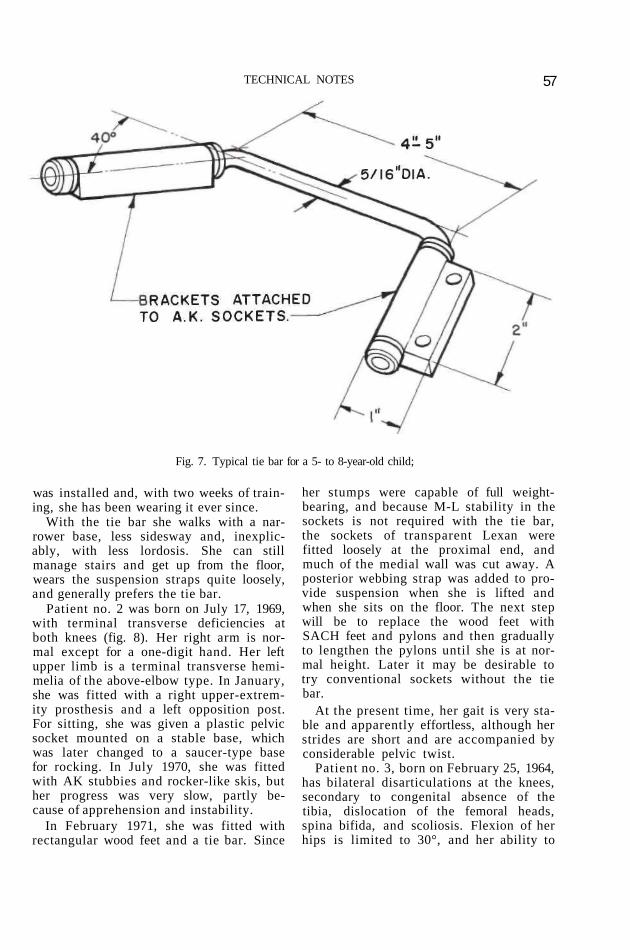
TECHNICAL NOTES 57
Fig. 7. Typical tie bar for a 5- to 8-year-old child;
was installed and, with two weeks of training, she has been wearing it ever since.
With the tie bar she walks with a narrower base, less sidesway and, inexplicably, with less lordosis. She can still manage stairs and get up from the floor, wears the suspension straps quite loosely, and generally prefers the tie bar.
Patient no. 2 was born on July 17, 1969, with terminal transverse deficiencies at both knees (fig. 8). Her right arm is normal except for a one-digit hand. Her left upper limb is a terminal transverse hemi-melia of the above-elbow type. In January, she was fitted with a right upper-extremity prosthesis and a left opposition post. For sitting, she was given a plastic pelvic socket mounted on a stable base, which was later changed to a saucer-type base for rocking. In July 1970, she was fitted with AK stubbies and rocker-like skis, but her progress was very slow, partly because of apprehension and instability.
In February 1971, she was fitted with rectangular wood feet and a tie bar. Since
her stumps were capable of full weight-bearing, and because M-L stability in the sockets is not required with the tie bar, the sockets of transparent Lexan were fitted loosely at the proximal end, and much of the medial wall was cut away. A posterior webbing strap was added to provide suspension when she is lifted and when she sits on the floor. The next step will be to replace the wood feet with SACH feet and pylons and then gradually to lengthen the pylons until she is at normal height. Later it may be desirable to try conventional sockets without the tie bar.
At the present time, her gait is very stable and apparently effortless, although her strides are short and are accompanied by considerable pelvic twist.
Patient no. 3, born on February 25, 1964, has bilateral disarticulations at the knees, secondary to congenital absence of the tibia, dislocation of the femoral heads, spina bifida, and scoliosis. Flexion of her hips is limited to 30°, and her ability to
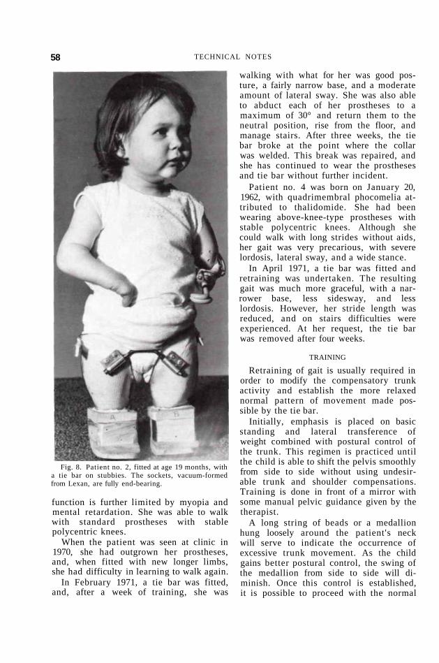
58 TECHNICAL NOTES
Fig. 8. Patient no. 2, fitted at age 19 months, with a tie bar on stubbies. The sockets, vacuum-formed from Lexan, are fully end-bearing.
function is further limited by myopia and mental retardation. She was able to walk with standard prostheses with stable polycentric knees.
When the patient was seen at clinic in 1970, she had outgrown her prostheses, and, when fitted with new longer limbs, she had difficulty in learning to walk again.
In February 1971, a tie bar was fitted, and, after a week of training, she was
walking with what for her was good posture, a fairly narrow base, and a moderate amount of lateral sway. She was also able to abduct each of her prostheses to a maximum of 30° and return them to the neutral position, rise from the floor, and manage stairs. After three weeks, the tie bar broke at the point where the collar was welded. This break was repaired, and she has continued to wear the prostheses and tie bar without further incident.
Patient no. 4 was born on January 20, 1962, with quadrimembral phocomelia attributed to thalidomide. She had been wearing above-knee-type prostheses with stable polycentric knees. Although she could walk with long strides without aids, her gait was very precarious, with severe lordosis, lateral sway, and a wide stance.
In April 1971, a tie bar was fitted and retraining was undertaken. The resulting gait was much more graceful, with a narrower base, less sidesway, and less lordosis. However, her stride length was reduced, and on stairs difficulties were experienced. At her request, the tie bar was removed after four weeks.
TRAINING
Retraining of gait is usually required in order to modify the compensatory trunk activity and establish the more relaxed normal pattern of movement made possible by the tie bar.
Initially, emphasis is placed on basic standing and lateral transference of weight combined with postural control of the trunk. This regimen is practiced until the child is able to shift the pelvis smoothly from side to side without using undesirable trunk and shoulder compensations. Training is done in front of a mirror with some manual pelvic guidance given by the therapist.
A long string of beads or a medallion hung loosely around the patient's neck will serve to indicate the occurrence of excessive trunk movement. As the child gains better postural control, the swing of the medallion from side to side will diminish. Once this control is established, it is possible to proceed with the normal

TECHNICAL NOTES 59
phases of gait training. Short steps should be taken at first, in order to maintain trunk control through each weight transference. Without concentration, the child may revert to a trunk-swivel-type gait pattern, and his walking should therefore be supervised. As confidence and skill improve, the child will gradually increase both step length and speed.
An A.D.L. assessment is essential, and some functional retraining may be required as well. -Sheila Hubbard, P.&O.T.; Colin A. McLaurin, Sc.D.; Ontario Crippled Children's Centre, Toronto 350, Canada.
Cosmetic Covers for Modular Prostheses
With the revival of the modular concept in the prosthetic field, prosthetists are once again faced with the problem of finding suitable materials and techniques for the fabrication of cosmetic covers. The general acceptance of the modular principle depends largely on whether or not it is possible to fabricate these customized covers in the limb shop economically.
Ingold in Basel, Switzerland, who at the turn of the century first used modular systems on a large scale, failed in the end because the covers he used were, although almost indestructible, difficult to make. Moreover, being made from fiber, the cover acted as resonators, amplifying the sounds made by loose rivets, etc. Since then, numerous new materials have been developed, particularly the various families of plastics, many of which at first glance seem to lend themselves naturally to use for cosmetic covers. However, the criteria that have been established for cosmetic covers limit the number of these materials that can be used to a very few.
Essentially, cosmetic covers should be light, hygienic, possibly washable, and should stand up for a reasonable period of time. But probably the most important criterion is that the techniques required should not involve fancy equipment or highly specialized skills. Only if these requirements are fulfilled will the modular
technique become part of the daily routine in the limb shops.
In recent years, several prosthetic groups experimenting with cosmetic materials have produced tangible results. From the Miinster group in Germany came the use of open-cell polyurethane-foam covers covered with Helanca stockinette. This method is especially suited for use with articulated prostheses because of the low durometer of the poly-urethane foam. The shaping of this foam, however, is very time-consuming. The stockinette used to cover the finished leg is subject to "runs," absorbs water and perspiration, and generally soils very easily, especially with younger patients.
Foort, Hobson, et al., in Winnipeg, concentrated their efforts on producing a range of pressure-molded polyurethane shin covers to complement their own modular systems. These covers are very durable, but their wall thickness limits their use on patients with fleshy stumps. Moreover, the fabrication of these covers requires a whole range of expensive molds that only a large rehabilitation center could afford.
Another approach was taken in the United States, where the Veterans Administration Prosthetics Center reported the
Fig. 1. The foam blocks are squared off—the centres are marked on both ends.

60 TECHNICAL NOTES
Fig. 2. The hole is cut using a piece of metal tubing.
Fig. 3. Socket outlines are marked.

TECHNICAL NOTES 61
Fig 4 The hole is enlarged on the router. Maximum depth of hole is indicated by masking tape on router shaft.
Fig. 5. Socket pylon assembly is sprayed with silicone. Masking tape is omitted.
use of polyethylene foam, which after shaping either can be covered with garter hose or painted with cordo-bond skin. This method produces a nice cover; its draw
back, however, lies in the fact that it is impossible to thin or "feather" out the foam sufficiently to bring it all the way to the proximal edge of the socket. Thus the por-

Fig. 6. The hole is coated with vinyl paint.
Fig. 7. Excessive foam is cut away on band saw.
62

TECHNICAL NOTES 63
Fig. 8. The leg is shaped in conventional manner on the router.
tion of the socket above the patellar-ten-don shelf is left exposed. This foam is also rigid and therefore is unsuitable for use with articulated prostheses.
A NEW TECHNIQUE
Because of the problems cited, we have sought an alternative to these methods, and in the past 18 months have successfully used a vinyl foam produced and marketed by B. F. Goodrich under the designation "VS-400."1
This paper will attempt to describe in detail the technique used in fabricating a cosmetic cover from VS-400 for an endo-skeletal below-knee prosthesis.
At a later time the author hopes to be able to produce a complete manual that will discuss the fabrication of covers for the full range of endoskeletal limbs.
The process of making a cosmetic cover is simple. This foam has a density of approximately 4 Ib/cu ft and is firm enough to be sanded, yet is flexible enough to be used with articulated limbs. Furthermore, the cellular structure of the foam is so fine and uniform that, after sanding has been completed, the covers are simply painted, thus giving the foam not only a tough but also a washable skin. After the size of the cover has been determined from drawings, etc., a block of foam is cut from the sheet.2
1 Closed-cell vinyl sponge, 4 lb/cu ft; price (un-trimmed) 48" x 64" x 3" = $44.00; manufactured by B. F. Goodrich, and supplied by Jacobs & Thompson, 89 Kenhar Drive, Weston, Ont., Canada.
2 When a thickness of more than 3 in. is required, two pieces of vinyl foam are bonded together using phenolic neoprene cement #9015, obtainable from Jacobs & Thompson at $9.70 per gal.

TECHNICAL NOTES
This block, after the ends are squared off, should still be at least one full inch longer than the distance from the proximal brim of the socket to the top of the foot. The foam is compressible, and this extra length will ensure good contact with the top of the foot.
The centers of both ends of the block are then marked with a soft pencil (fig. 1). (It is not advisable to use felt pens as the ink from these pens tends to soak into the foam and later to show on the finished product.) A piece of metal tubing, sharpened to a keen edge at one end, is then used to cut a hole longitudinally through the foam block. This is easily accomplished by spinning the tubing in the chuck of a lathe. To achieve a smooth cut, however, it is necessary to wet the tubing inside and out. Needless to say, the tubing should be of the same outside diameter as the pylon
Fig. 9. The cosmetic cover is painted by hand.
Fig. 10. The finished leg.
that is to be used (fig. 2). Next, the pylon socket assembly is placed upon the foam block and its lateral silhouette transferred onto the foam with a soft pencil (fig. 3).
Also, the brim outlines of the socket are drawn on the top of the foam block. If necessary, a simple paper pattern of the brim contours can be made for this purpose. Front and back should be clearly marked on the foam to avoid any confusion during the "fitting in" of the socket.
With a sharp knife, the drill hole in the top of the foam block is increased and then further enlarged, using a coarse sanding cone. The hole to be made should ideally be of the same size and shape as the socket. In practice, however, it is better to keep the hole slightly smaller in its circumferential dimensions to ensure inti-
64

TECHNICAL NOTES 65
mate fit between the socket and the foam. The hole should be made deep enough not only to accommodate the full length of the socket but also to allow it to be recessed at least one-quarter inch. The maximum depth of the hole should be marked on the shaft of the router with a strip of masking tape (fig. 4).
After the outside proximal brim of the socket has been covered with 3/4-in. masking tape, a coating of silicone is applied. This coating will prevent bonding of the foam to the socket, except for the 3/4-in. strip along the proximal edge where it is desirable (fig. 5). Using a brush, the hole in the foam block is then given a soaking of vinyl paint3 (fig. 6).
After the masking tape has been removed from the socket, the whole socket-pylon assembly is pushed into the foam block. If there is any gapping along the brim, the foam must be brought into contact with the socket by applying several turns of friction tape. The foot should be installed to help keep the foam in place.
The vinyl paint applied inside the hole requires about half a day's drying time to form an "inner skin" and only then can shaping of the foam commence. If un-trimmed sheets of foam have been purchased, they will have a self-formed skin from the manufacturing process. This skin should be sliced away before painting to expedite the drying process.
SHAPING AND PAINTING With the foot in place, and properly
toed out, the silhouettes of the leg are marked on the foam. Excess material is then cut away on the band saw and the corners trimmed. Band-saw blades with 24 teeth per inch running at high speeds will cut the foam without tearing it (fig. 7).
The final shaping can be done on a Trautmann Carver, starting out with a coarse sanding cone and progressing to a 3-in. sanding drum and #120 garnet paper (fig. 8). The sanding should never be di-
3 Liquid vinyl, also called P.V.C. paint; price: 5 gal. at $7.50 per gal.; supplied by Jacobs & Thompson.
rected against the edges of the foam but rather toward the edge or parallel with it. If the paint applied inside the foam block has been allowed to form a reinforcing skin, the foam can, if necessary, be sanded right down to this inner skin. The thickness of the foam can always be checked with a sharp awl. Any uneven spots can be sanded smooth by hand.
To finish the prosthesis, it is simply hand-painted with a vinyl paint made of clear vinyl and Hosmer pigment4 (fig. 9). The first coat should be allowed to dry for 30 min. before the second coat is applied. Between coats, the foot can be taken off to allow painting of the distal end of the cover. When the foot is reinstalled, the cover will bond itself to the top of the foot (fig. 10). Two coats of paint will normally suffice and give the leg a beautiful long-lasting finish. William F. Sauter, C.P.O.(C) Ontario Crippled Children's Centre Toronto 350, Canada
A Device for Applying Above-Knee, Total-Contact Sockets
At the Calgary General Hospital, we have found that a fair number of the older above-knee amputees have difficulty putting on their quadrilateral, total-contact sockets without assistance. It appeared that the main problem was their decreased strength and the inability to "pump" the stump into the socket while bending forward to pull off the stump stocking.
The aid we devised (fig. 1) permits the patient to stand upright while applying the prosthesis and to exert a greater pull on the stump stocking. The device also works well with patients who have the use of only one hand. The device is made from 1 in. x Va in. aluminum bent in an L shape, the longer part of the L being 15 in. long and the shorter part, or handle, being 5V2 in. The end of the 15-in. section has a 2-in. split in the center, which is bent
'A . J. Hosmer Corp., P. O. Box 37, Campbell, Calif. 95008.

Fig. 1.
Fig. 2.
Fig. 3.
Fig. 4. 66

TECHNICAL NOTES 67
out to form a V. (This V should be thoroughly smoothed, or the stump stocking will catch on it and rip.)
The patient should be provided with a stockinette stump stocking 3 to 4 ft. long. The stocking is put on the stump in the normal manner, the distal end is threaded through the valve opening, and the stump is inserted in the socket (fig. 2). The stocking is placed through the V in the device, as close to the valve opening as possible,
and the end of the stump stocking is wound around the handle (figs. 3 and 4). The patient stands upright and pushes down on the handle as he "pumps." When the handle becomes difficult to reach, the patient replaces the V next to the valve opening, winds more of the stump stocking around the handle, and pushed again. Gillian E. Wong, O.T.Reg., Calgary General Hospital, 841 Centre Ave. East(61), Calgary, Alberta, Canada.